Lower Limb PAIN - OSU Center for Continuing Medical … proximal muscle One distal muscle One muscle...
55
Lower Limb PAIN „the skinny‟ Is it radiculopathy, vulnerability, or Parsonage-Turner?
-
Upload
nguyennhan -
Category
Documents
-
view
215 -
download
1
Transcript of Lower Limb PAIN - OSU Center for Continuing Medical … proximal muscle One distal muscle One muscle...

Lower Limb PAIN
„the skinny‟
Is it radiculopathy, vulnerability, or Parsonage-Turner?


Check back for symmetry
Stand quietly
Both feel weight bearing
One foot weight bearing
Forward flexed


Exam prior to EDX
Straight Limb Raising
Recumbent
Recumbent with dorsiflexion after lowering to no sx
Sitting
Sitting with neck flexion after SLR lowered

L-5 PX
Walk on heels (only gross test)
Check strength of Ext H L
NB. Must do with ankle plantar flexed and push on proximal phalanx
Measure atrophy of leg (greatest circumference)
MSR – lateral HS

NOT DISTAL PHALANX

PROXIMAL PHALANX

S-1 Radiculopathy
Walking on toes is only a gross test
Must do heel raises unilaterally and compare (10)
MSR - Ankle jerk
Numbness lateral foot and sole

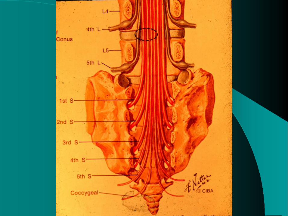
EDX of Lumbar radiculopathy
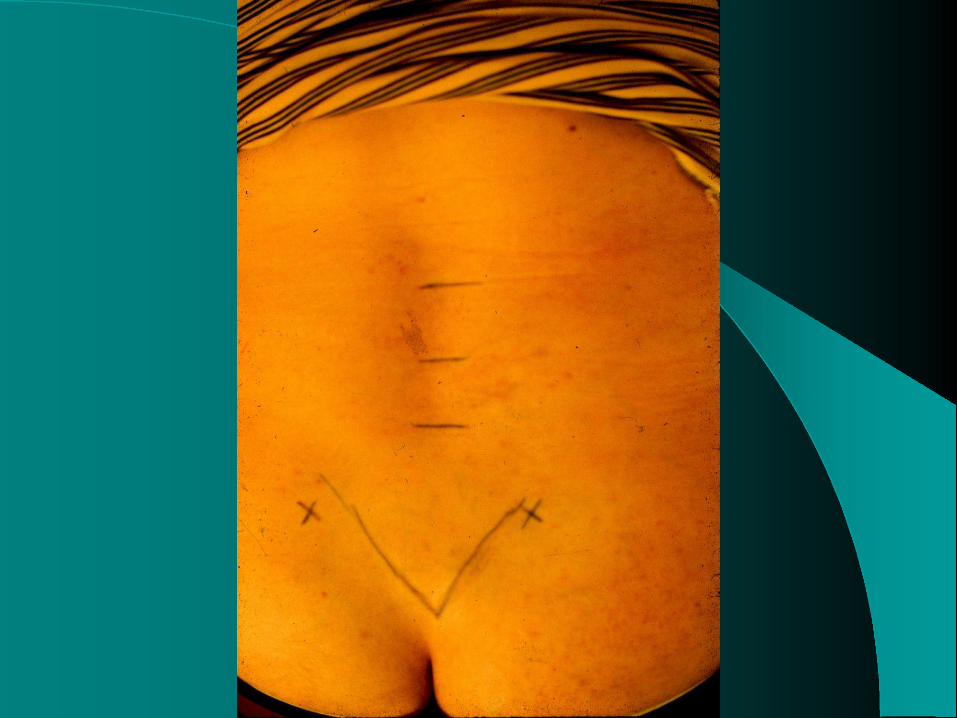
Prone position is best
Land marks
Mark L-4 spinous process at level of ilium crest
Mark L-5 – next caudal spinous process
Mark S-1 – next caudal spinous process
Draw diagonal line from post. sup. iliac spine to midline


Maximize relaxation
Pillow under abdomen
Pillow under ankles
If still cannot get relaxation – use other hand to poke fingers in abdomen


Muscles to explore
Paraspinals
Same root but 2 different nerves
One proximal muscle
One distal muscle
One muscle ABOVE suspected root
One muscle below suspected root


Example – L-5 Radiculopathy
Explore
Ant tib
Flex dig long
Soleus (distal to suspected root)
Vastus medialis (proximal to suspected root}
Tensor fascia lata (a proximal muscle)
Paraspinals

Chronology of L/S radiculopathy
When radicular pain begins:
Recruitment will be reduced (if significant weakness)
H reflex latency will be prolonged
Early “polyphasic MUP‟s” will appear

Needle EMG Abnormalities - chronology
1st week – recruitment frequency will be increased
By 7-8 days – positive waves in paraspinals
(Caution – a train will result if in end plate
area!)
3rd week – abnormal irritability in paraspinals and proximal limb muscles
4th week all findings

Recruitment frequency
In normal muscles the 2d MU will appear when the 1st MU is firing 10-12 hz
L-5 radiculopathy – ext dig long 16-18 hz
Compare with contralateral muscle
Easiest – a single joint muscle
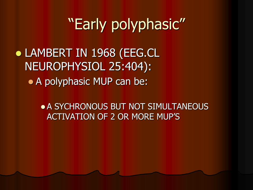
“Early polyphasic”
LAMBERT IN 1968 (EEG.CL NEUROPHYSIOL 25:404):
A polyphasic MUP can be:
A SYCHRONOUS BUT NOT SIMULTANEOUS ACTIVATION OF 2 OR MORE MUP‟S


“Early polyphasic”
2 axons conduct at different rates thus impulses arrive slightly separated
Looks like a polyphasic MUP
Normal amplitude
Increased duration
Several MUP‟s stucked together


RASMINSKY, M
Ephaptic transmission between single nerve fibers in the spinal nerve roots of dystrophic mice.
J.Physiol. 1980. 305:151

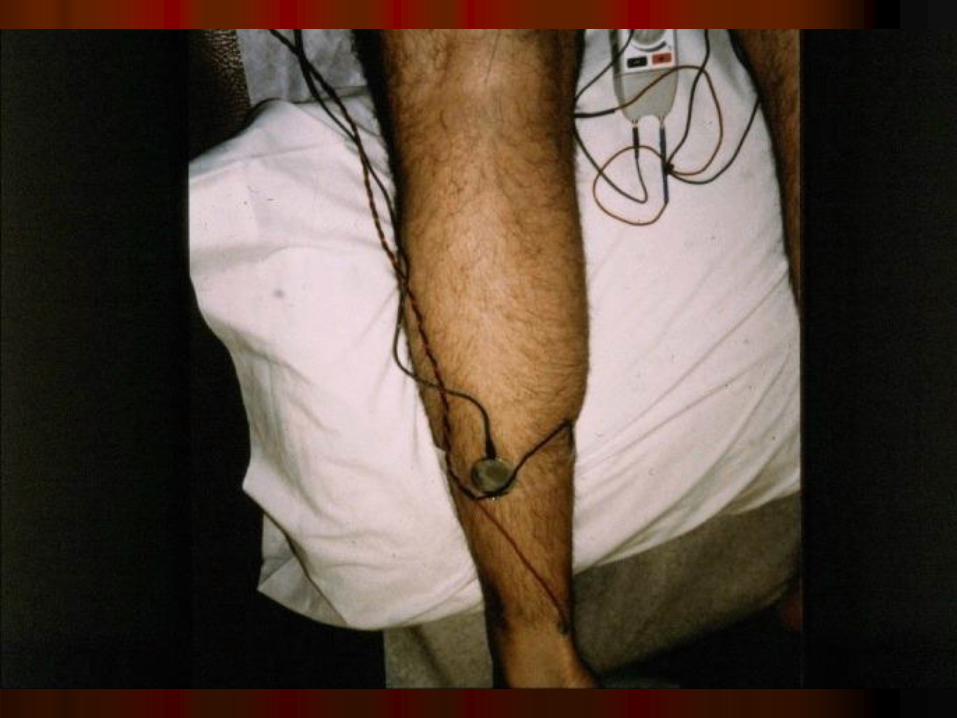
„H‟ REFLEX LATENCY IN LUMBAR RADICULOPATHY
Will be prolonged in S-1 radiculopathy from the onset of radiculopathic pain
Difference in latency, side-to-side, =or< 1 millisec or even .5 millisec is a red flag.
Original study (1974) mean .8 8 =/- S.D. .4 ms
More recent series difference side-to-side .3 ms


Formula to calculate H latency
.46 X distance from stimulation to medial malleolus
+ .1 age in years
+ constant – 9.14
Difference side to side > 1.0 ms (conservative)
My opinion is > .5 ms is “red flag”


Use of H reflex latency
Early in course of L/S radiculopathy
When abnormal irritability is only in paraspinals
Underlying peripheral neuropathy (diabetic)
If muscle exploration is confusing
Post laminectomy with recurrent symptoms

Use of H reflex latency when positive waves are only in
paraspinals 90 – 95% of all first appearing
radiculopathies are L-5 or S-1
Ratio of frequency – L-5:S-1 = 2:1
H latency is prolonged – S-1; if normal – L-5

Prognosis
After 7-10 days an axon undergoing wallerian
degeneration will become inexcitable
Stimulation of nerve to weak muscle will identify the dead axons (NB. Amplitude, compare with contralateral)
L-4– ant tibial or vastus lateralis
L-5 – extensor dig long
S-1 – medial head gastroc


Muscles to explore
One proximal muscle (L-5 eg. tensor fascia lata)
One distal muscle (S-1 eg. Abd hall)
Muscle from 2 different nerves (L-5 eg. Peron. long; flex dig long) BUT same root
Paraspinous – level above and below
Contralateral muscle of most abnormality

EMG of PARASPINALS S/P surgery
Not significant if abnormalities are all along scar
Can be significant if localized and:
> 3 cm lateral to scar
> 3 cm deep
Correlate with sx

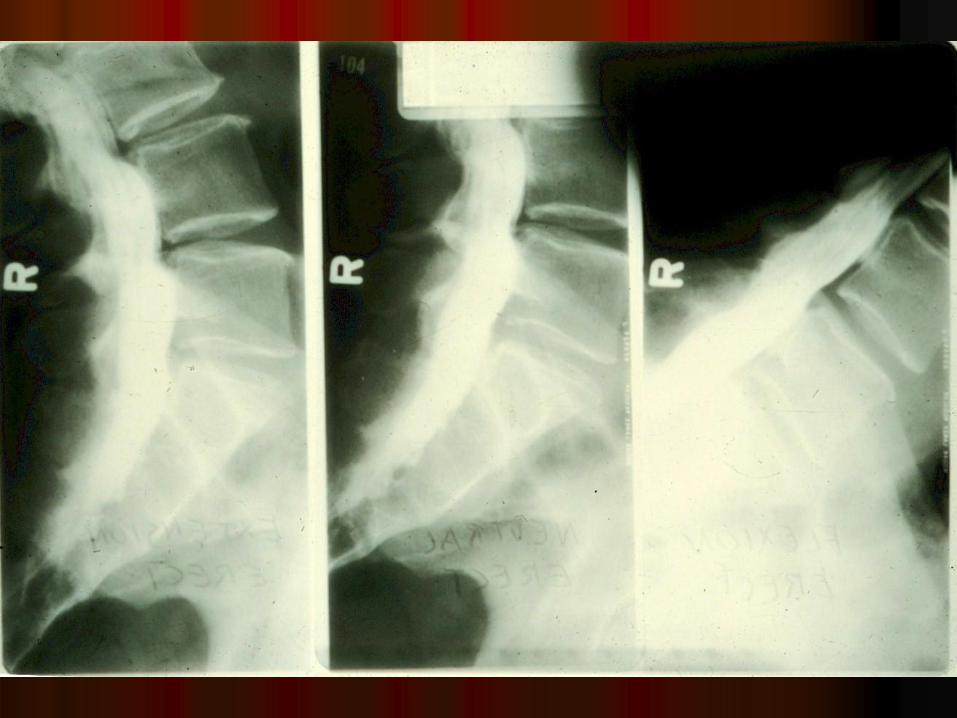
Dynamic myelogram
Note the protrusion when lumbar spine is extended
This demonstrates the absurdity of McKenzie exercises
William flexion exercise program is best

Back surgery

EDX L/S Summary
Early – Recruitment frequency; H
>10days – positive waves in paraspinal; CMAP amplitude = prognosis
>18 days all


Neuralgic Amyotrophy
Ernest W Johnson MD
Emeritus Professor, PM&R
The Ohio State University

What is it?
Syndrome of pain and weakness occurring in a limb with the pain preceding the weakness by several days
More common in upper limb
Lower limb – proximal muscles > distal


Parsonage-Turner syndrome
Original 136 cases
12 occurred after operation
10 after trauma
Most after infections
NB. Often occurred after serum injection

Isolated nerves in N-A
Phrenic N
Long thoracic N of Bell
Anterior Interosseus N
Axillary N
Suprascapular N
Sensory N – lateral antebrachial cutaneous LL – Femoral; sciatic medial>lateral div.

Parsonage-Turner syndrome (consensus)
Brachial plexopathy
Within 1 week or co-incident with -- surgery; or ?viral infection
Severe pain in shoulder
When pain abates, weakness and atrophy are apparent
Prognosis is generally good

summary
Parsonage-Turner syndrome presents:
Acute shoulder /upper limb pain following an operation, viral infection, serum injection
Weakness occurs in 1-3 weeks and acute pain reduces
Most symptoms gone by 12-18 months

references
Amato,A et al: Chronic relapsing brachial plexus neuropathy with persisting conduction block. Muscle & Nerve. 1997.20:1303
Magee, KR & DeJong, RN: Paralytic brachial neuritis: discussion of clinical features with review of 23 cases. JAMA.1960.174:1258.
Martin, W & Kraft, G: Shoulder girdle neuritis: a clinical and EDX evaluation. Military Medicine.1974. 139:21.
Tsairis,P et al: Natural history of brachial plexus neuropathy. Arch Neurol. 1972. 27:109