Lower Jaw Movements Measured by Optoelectronic …1173036/FULLTEXT01.pdf · range of motion while...
27
Student Tandläkarprogrammet, 300 högskolepoäng Examensarbete, 30 högskolepoäng Ht 2017 Lower Jaw Movements Measured by Optoelectronic Movement Recording A pilot study Christopher Staversjö Magnus Wänman
Transcript of Lower Jaw Movements Measured by Optoelectronic …1173036/FULLTEXT01.pdf · range of motion while...

Student
Tandläkarprogrammet, 300 högskolepoäng
Examensarbete, 30 högskolepoäng
Ht 2017
Lower Jaw Movements Measured
by Optoelectronic Movement
Recording
A pilot study
Christopher Staversjö
Magnus Wänman

Lower Jaw Movements Measured by Optoelectronic
Movement Recording
A pilot study
2017
Authors: Christopher Staversjö and Magnus Wänman
Tutor: Catharina Österlund

ABSTRACT
Due to the complex nature of jaw movements, three-dimensional (3D) movement
recording provide information about the jaw movement capacity. The aim of the present
report was to test the reliability of measuring lower jaw movements using a 3D
movement recording system and to calculate the lower jaw movement volume.
Lower jaw movements, recorded by 3D optoelectronic movement analysis system
(MacReflex®) was compared with reference values from a digital caliper. Pre-tests
were performed to develop a software to calculate the lower jaw movements in separate
dimensions and its volume. Pilot tests with two test persons followed to register the
lower jaw movements and calculate lower jaw movement volume.
The results indicate low reliability of lower jaw movements measured by movement
recording system compared with reference values from digital caliper, reflected by delta
values ( = max-min). The values from the movement recording system indicate high
variability reflected by higher levels of standard deviation for movement recorded
values compared with digital caliper and by percentage values calculated from the
differences between mean values of movement recording and digital caliper. The
calculated lower jaw movement volume was 10.3 cm3 and 17.2 cm3 for the test persons,
respectively.
Conclusively, the results imply that further testing of the method is needed with larger
series and test-retest reliability analysis to evaluate the possibility to improve accuracy
of tracing jaw movements with recording device. The 3D-movement recording system
together with the software could be used for calculation of lower jaw movement volume
but its accuracy could not be validated.
3
INTRODUCTION
Temporomandibular disorders
The ability to perform normal jaw functions such as biting, chewing, swallowing,
yawning and speech, without being restricted by pain or dysfunction is significant from
a health perspective. Temporomandibular disorders (TMD) is a generic term for pain
and dysfunction affecting the jaw muscles and the temporomandibular joint (TMJ) or
surrounding tissues (Okeson, 2013). TMD is characterized by pain and dysfunction in
the regions of the temples, TMJ and jaw muscles, impaired movement capacity of the
lower jaw and TMJ noises (Dworkin & LeResche, 1992). TMD and orofacial pain can
pose a negative effect on quality of life and may also affect the ability to perform daily
activities (Dahlstrom & Carlsson, 2010; Shueb et al., 2015). TMD pain is a common
longstanding pain condition in the jaw-face region affecting approximately 10 percent
of the adult population (LeResche, 1997). The need for treatment owing to TMD has
been estimated to be in the range from 1-30 percent of the population with a mean value
of 16 percent (Al-Jundi et al., 2008). Diagnostic Criteria for Temporomandibular
disorders (DC/TMD) is a recently launched method for clinical use as well as for
research (Schiffman et al., 2014). DC/TMD contains diagnostic criteria for the most
common TMD pain–related disorders, TMJ intra articular disorders as well as
degenerative joint disease. In DC/TMD the clinical evaluation of the lower jaw
movement range is an important parameter.
Jaw- and neck sensori-motor function
Jaw function is regulated by the cortical motor centers, the midbrain and the brain stem.
Information necessary for muscle control is provided in complex neural networks. The
brain stem receives input through afferent fibers from peripheral sensory receptors, such
as periodontal mechanoreceptors and the muscle spindles of jaw-closing muscles. This
information is organized in the midbrain, brain stem and cortex to send out suitable
signals through efferent nerve fibers which results in contraction or inhibition of jaw
muscles (Morquette et al., 2012; Okeson, 2013). Normal jaw function involves
coordinated activation of lower jaw, the TMJ, the atlanto-occipital joints, jaw- and neck
muscles and innervation from trigeminal as well as cervical nerves (see for example
4
Eriksson et al., 1998). Normal jaw function is the result of coordinated jaw- and neck
muscle activity with head extension movements during jaw-opening and head flexion
during jaw-closing (Eriksson et al., 2000).
Lower jaw movements
The TMJ movement during jaw opening and closing involve both rotation and
translation of the condyles. The movement of the lower jaw can be divided into border
movements and free functional movements. Border movements are those at the outer
range of motion while functional movements occur during functional activity, such as
chewing, and within the border movements (Okeson, 2013). The maximum range of the
lower jaw movement, in three dimensions, was first described by Ulf Posselt 1952 by
combining the lower jaw border movements in the sagittal, horizontal and frontal planes
(Posselt, 1952) (Figure 2a). Normal maximal lower jaw movement capacity for adults
ranges between 40 - 75 mm for opening and between 6 - 15 mm for protrusion and
laterotrusion (Agerberg, 1974).
Restricted lower jaw movements
Impairment of lower jaw movements can be caused by different conditions. Common
factors include pain conditions affecting the jaw muscles, TMJ or neck (Dworkin et al.,
1990). Other conditions are mechanical obstacles such as TMJ disc displacements, disc
adherences and ankyloses (Okeson, 2013). Furthermore, radiotherapy as a treatment for
cancer/tumours in the head- neck and oro-facial regions can also severely affect jaw
movement capacity (Bensadoun et al., 2010). Motor disorders such as Parkinson’s
disease also affect jaw opening capacity (Bakke et al., 2011). Fear avoidance can cause
limited jaw opening capacity. Acute experimental pain may change jaw motor
coordination with slower and more variable movements in pain catastrophizing
individuals (Akhter et al., 2014).
Measuring lower jaw movements
To measure the range of lower jaw movements a ruler or a digital caliper is normally
used in the dental clinic. The examination according to DC/TMD involves
measurements of pain free jaw opening, maximal unassisted and maximum assisted jaw

5
opening even if it is painful, and laterotrusive right and left and protrusive movements,
even if it is painful (Schiffman et al., 2014). Besides measures with a ruler, different
methods have been tested experimentally to capture the lower jaw movements. One
method is to use optoelectronic devices with light emitting diodes (LED) and a motion
detector (Fang and Kuo, 2008; Travers et al., 2000). Another method is to use an
ultrasonic motion detector. The ultrasonic device capture the distance of movements by
sending out pulses and measuring the time it takes or the pulse to return (Al-Jundi et al.,
2008; Frisoli et al., 2017; Mazzetto et al., 2017). A four-dimensional analysis method
has been tested using a combination of three dimensional (3D) CT of the cranium and
lower jaw, laser scanner and LED-technique (Terajima et al., 2008). The methods
mentioned are expensive and difficult to use in the dental clinics. Studies on simpler
methods has been made using a hand camera and black and white markers (Adly et al.,
2013) or tracking system using a RGB (Red, green, blue) camera and a standard laptop
(Tanaka et al., 2016). Other methods that have been tested include accelerometer, video
fluoroscopy and electromagnetic fields (Adly et al., 2013). In a comparative study using
two-dimensional videography and ultrasonic measurements system to quantify lower
jaw movement showed no significant difference between maximum opening and the
reference system. Laterotrusion showed to be overestimated by the videography system
and to show greater variability (Frisoli et al., 2017).
A change in maximal lower jaw movements is commonly used as a parameter for
treatment outcome. When measuring lower jaw movements with a ruler or digital
caliper, you may miss details in the movement pattern and the total lower jaw volume.
Changes in the lower jaw volume may be a better indicator for the impairment and
improvement of jaw functions. It may be possible to evaluate the envelope of
movement, i.e. the volume of the lower jaw movement patterns, both border and
functional movements. Change in these volumes may turn out to be an indicator of
treatment outcome for jaw function capacity if it can be measured with high reliability.
The lower jaw is capable to move in a six degrees of freedom (Knap et al., 1970). Most
often, in natural jaw function the lower jaw move in more than one dimension. Due to
the complex nature of lower jaw movements, it is possible that three-dimensional (3D)
movement recording analysis of the movements could give more information about the

6
jaw movement capacity and jaw function compared to measures in only one dimension.
Therefore, it is interesting to develop reliable and valid test methods to measure
movements. In this study, we wanted to analyze the reliability of the measurements of
separate lower jaw movements with a wireless 3D optoelectronic movement recording
system and evaluate if it is possible to calculate and visualize the maximal lower jaw
movement volume.
Aims
The aims of the study were:
To evaluate the reliability of the measurements of separate lower jaw
movements (mm) with a wireless 3D optoelectronic movement recording
system.
To compare maximal vertical and horizontal lower jaw movements
registered with a 3D optoelectronic movement recording system to
registrations with a digital caliper.
To calculate and visualize the total lower jaw movement volume (cm3).
Hypotheses
The hypotheses were:
Measurements of the maximal lower jaw movements with wireless 3D
optoelectronic movement recording system can be done with high reliability.
Measurement of maximal lower jaw movements with wireless 3D
optoelectronic movement recording system will not differ significantly from
measurements with a digital calliper.
The maximal lower jaw movement volume can be calculated and visualized
based on registrations of the boarder movements of the lower jaw.
7
MATERIALS AND METHOD
Ethical reflection
The study was approved by the Local Ethical Board, Umeå University. Ethical
considerations that was considered for the study were the risk for short-termed, transient
pain/tiredness in the jaw muscles, TMJ and head-neck region in the two test persons.
The risk for harm was considered very low.
Literature search
Articles were searched on PubMed using the MeSH terms; jaw and movement. As a
complement the MeSH terms, the terms movement recording, movement analysis,
optoelectronic, 3D and lower jaw volume was used. In addition, hand search was done
on google scholar and Libris. Articles was also provided by the supervisor. In total, 20
articles were read in full text.
Test persons
To test the hypotheses and for practical reasons two test persons, the authors, were
included (test person one and test person two).
Movement recording
For the experimental tests, movements of the lower jaw and head were recorded
simultaneously in 3D with wireless optoelectronic system at sampling rate of 50 Hz (Mac
Reflex®; Qualisys, Gothenburg, Sweden) (Josefsson et al). Two cameras recorded the
movements of a tripod of retro-reflective markers attached to the bridge of the nose (to track
head movements) and a single marker on the chin (to track lower jaw movements). Details
of the set-up have been described previously (Eriksson et al., 2000).
Software
There was no known available software compatible with the MacReflex system to
calculate and visualize the lower jaw movements in separate dimensions and the lower
jaw volume. Therefore, we co-worked with the Department of Community Medicine
and Rehabilitation, Umeå University. We supported them with recorded measuring data

8
for developing a custom-made software. The software mathematically compensated for
the head-neck movements, calculated the lower jaw movements relative to the head and
illustrated the lower jaw volume from the recorded movements.
Outcome variables
The outcome variables were;
Jaw opening movement amplitude (mm): the distance from starting position
(slight teeth contact in centric occlusion (CO)) to maximal jaw opening,
including vertical overbite. In the 3D coordinate system referred to as Y-
dimension.
Jaw laterotrusive movement right or left (mm): the distance from starting
position (slight teeth contact in CO) to maximal right or left laterotrusive
movement. In the 3D coordinate system referred to as X-right or left dimension.
Jaw protrusive or retrusive movement (mm): the distance from starting position
(slight teeth contact in CO) to maximal forward protrusive movement. In the 3D
coordinate system referred to as Z-forward or backward dimension.
Calculated total lower jaw movement volume (cm3).
Pre-test
A series of pre-tests was performed, prior to the pilot tests, to sample necessary data to
develop the software for calculation and visualization of the lower jaw movements and
the movement volume. The pre-tests were done with the movement recording system,
reflex markers, jaw movements, a ruler and a cup and a glass with known volume. The
pre-tests compared a known distance or volume with the recorded one.
Pilot test
A series of tests were done to test the test re-test reliability of the measurements of
maximal lower jaw movements using the movement recording system and to compare
these measurements to the reference values of a digital caliper. The pilot test was also
designed to test the possibility to calculate the total lower jaw volume.

9
Two separate series, A and B, of measurements were conducted during five consecutive
days. Pilot test A compared maximal lower jaw movements in opening (Y-dimension),
right and left laterotrusion (X-dimension) and forward protrusion and backward
retrusion (Z-dimension). The X-, Y-, Z- dimensions were recorded with the movement
recording system compared to measurements done with a digital calliper (Table 1a).
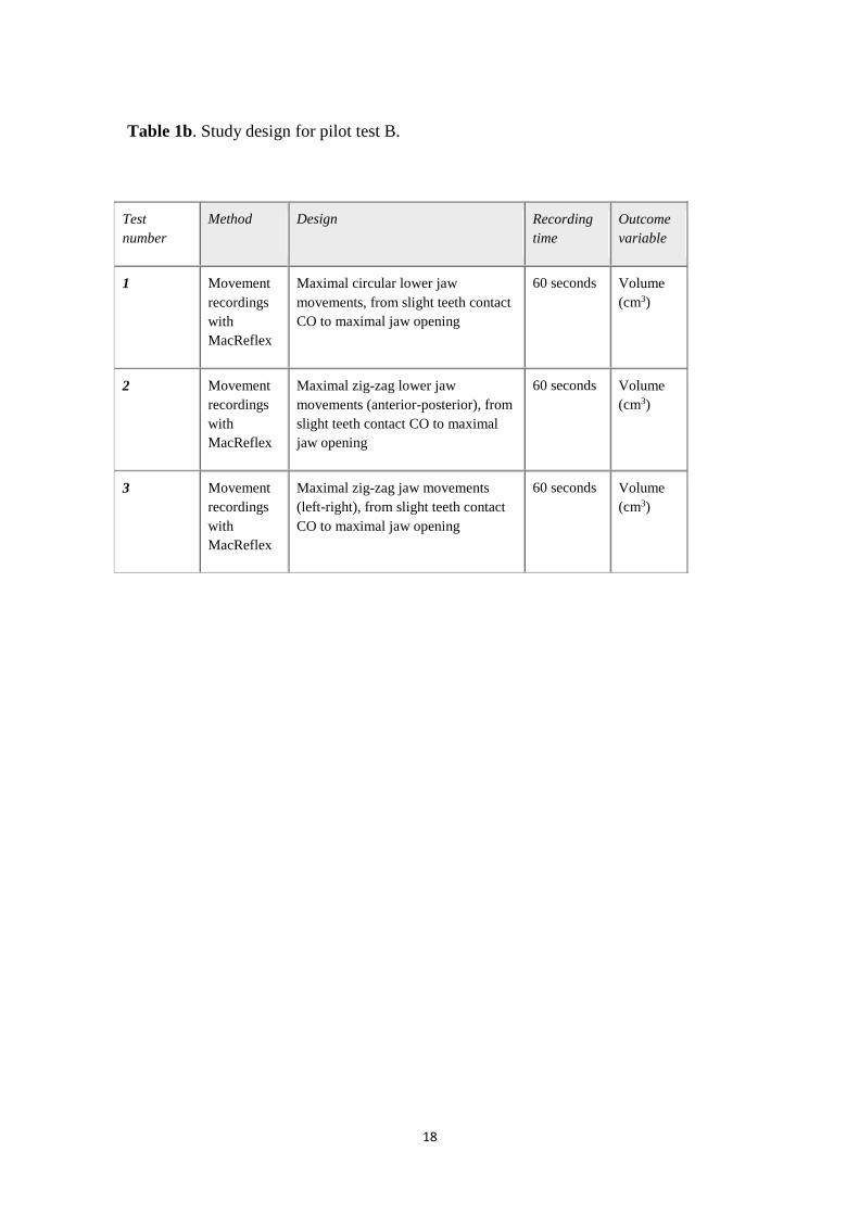
Pilot test B samples lower jaw movement coordinates for calculating the total volume
with the movement recording system (Table 1b).
Statistical methods
The data were analysed by descriptive; mean (mm), min-max (mm) and standard
deviation (SD). As a measure of test-retest reliability for the movements recorded
values by the movement recording system, a mean delta-value (= max-min) was
calculated. As a comparison between values from movement recording and from digital
caliper the formula (m-d)/m X 100 was used (m = mean value of the movement
recording system, d = mean value from the digital caliper).
RESULTS
The reliability of the measurements of separate lower jaw movements with wireless
3D optoelectronic movement recording system
Jaw opening amplitude /y-dimension
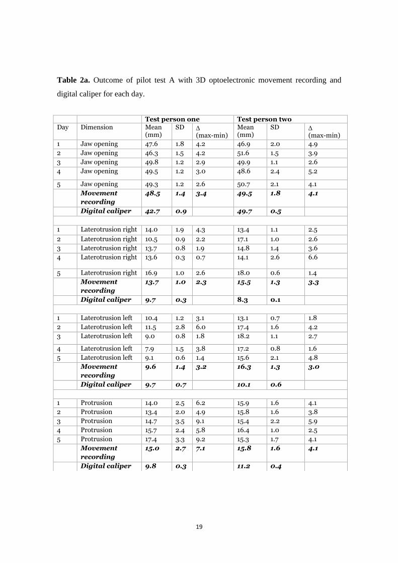
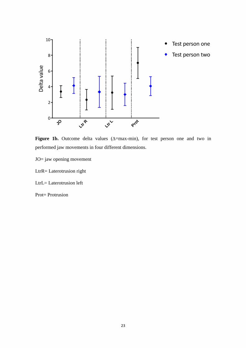
In maximal jaw opening, the mean delta value was 3.4 mm for test person one and
4.1mm for test person two (Table 2a and Figure 1b).
Jaw laterotrusive movement /x-dimension
In maximal jaw laterotrusive movement right, the mean delta value was 2.3 mm for test
person one and 3.3 mm for test person two. In maximal jaw laterotrusive movement left,
the mean delta value for the movement recording was 3.2 mm for test person one and
3.0 mm for test person two (Table 2a, Figure 1b).
10
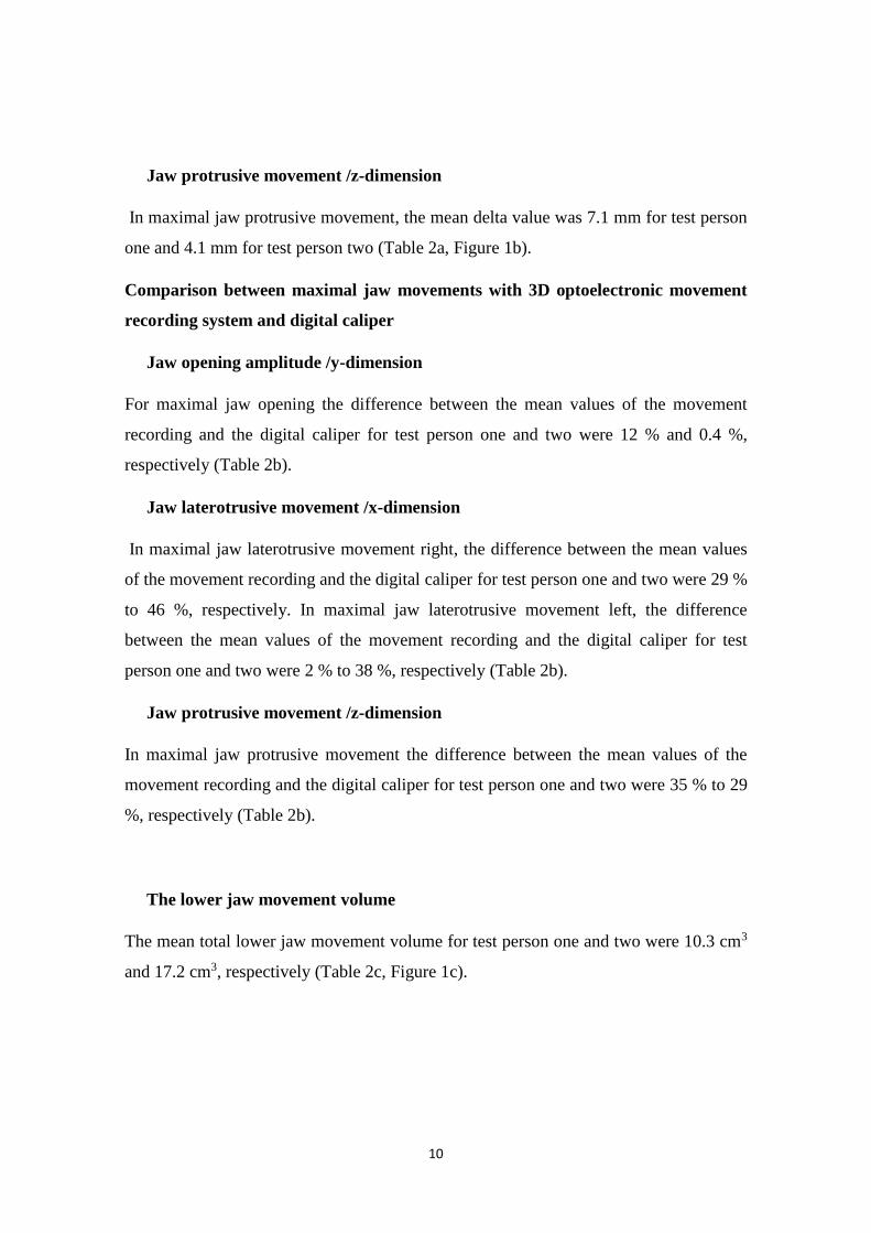
Jaw protrusive movement /z-dimension
In maximal jaw protrusive movement, the mean delta value was 7.1 mm for test person
one and 4.1 mm for test person two (Table 2a, Figure 1b).
Comparison between maximal jaw movements with 3D optoelectronic movement
recording system and digital caliper
Jaw opening amplitude /y-dimension
For maximal jaw opening the difference between the mean values of the movement
recording and the digital caliper for test person one and two were 12 % and 0.4 %,
respectively (Table 2b).
Jaw laterotrusive movement /x-dimension
In maximal jaw laterotrusive movement right, the difference between the mean values
of the movement recording and the digital caliper for test person one and two were 29 %
to 46 %, respectively. In maximal jaw laterotrusive movement left, the difference
between the mean values of the movement recording and the digital caliper for test
person one and two were 2 % to 38 %, respectively (Table 2b).
Jaw protrusive movement /z-dimension
In maximal jaw protrusive movement the difference between the mean values of the
movement recording and the digital caliper for test person one and two were 35 % to 29
%, respectively (Table 2b).
The lower jaw movement volume
The mean total lower jaw movement volume for test person one and two were 10.3 cm3
and 17.2 cm3, respectively (Table 2c, Figure 1c).

11
DISCUSSION
The main findings of this pilot study were that repeated measurements of the lower jaw
movements with 3D optoelectronic movement recording (MacReflex) did not show
reliable values in comparison with the reference values from the digital caliper. The
outcome values from the movement recording were in general larger than values from
the digital caliper in all dimensions, especially in X- dimension (laterotrusive
movement) and in Z- dimension (protrusive movement). Therefore, we reject the
hypotheses one and two, assuming that measurements of the maximal lower jaw
movements with wireless 3D optoelectronic movement recording system can be done
with high reliability and will not differ significantly from reference values from the
digital caliper. The third hypothesis that the maximal lower jaw movement volume can
be calculated and visualized based on registrations of the boarder movements of the
lower jaw was accepted, but its accuracy could not be validated.
The indicated low reliability, are reflected by the delta () values (Table 2a, Figure 1b).
The low reliability of maximal jaw opening with the optoelectronic system was
disappointing in relation to previous studies showing high reliability of measurements
with calipers (Wahlund et al., 1998). We have no explanation for the large differences
between the reference values from the digital caliper for lateral and protrusive
movements compared to those registered with the movement recording values. One
possibility, may be that the registrations by hand with digital caliper measure one
dimension (the lateral movement, X-dimension), while the movement also involves a
forward component (Z-dimension) and sometimes even a downward movement (Y-
dimension), which is caught by the optoelectronic device. With the movement recording
system, the outcome distance is the combined movement in three- dimensions, the
vector of the distance, which is longer than the movement in only one- dimension.
Therefore, movement analysis certainly provides an added value of the movement
patterns in the jaw system. Another explanation, may be that the software program
overestimates the registered values and needs to be adjusted in the mathematical
formulas used in the software. To explore that, further analysis will be needed to assure
or improve the software program.

12
To be sure that the reflex marker set up was appropriate for the tests, we used the same
set up that has been described previously. The reflex markers settings on the bridge of
the noose and chin versus teeth attached markers can be reliably used for jaw movement
analysis (Häggman-Henrikson et al., 1998). Moreover, the accuracy of the MacReflex
system for precision in measurements has been shown to be high (Eriksson et al., 2000).
To be sure that the movement recording system (MacReflex) was calibrated in X-, Y-,
Z- dimensions, calibration measurements were done with the aid of a calibration frame,
showing exactly correspondence between values from the recording system and the
values from a digital caliper.
The values from movement recording system indicate high variability as reflected by
the higher levels of standard deviation for movement recorded values compared with
digital caliper and by the percentage values calculated from the differences between the
mean values of the movement recording and the digital caliper (Table 2b, Figure 1a).
One possible explanation for the variability in the movement recording values may be
that measurement of the maximal interincisal distance during opening with a calliper
clearly define the end- point while free movements can involve a higher level of
variability. The variability in the free movements registered by the optoelectronic device
can be interpreted as a variability in the sensory-motor system. This variability in
movement outcome can be an advantage when the jaw sensori-motor system is affected
by pain, injury or disease.
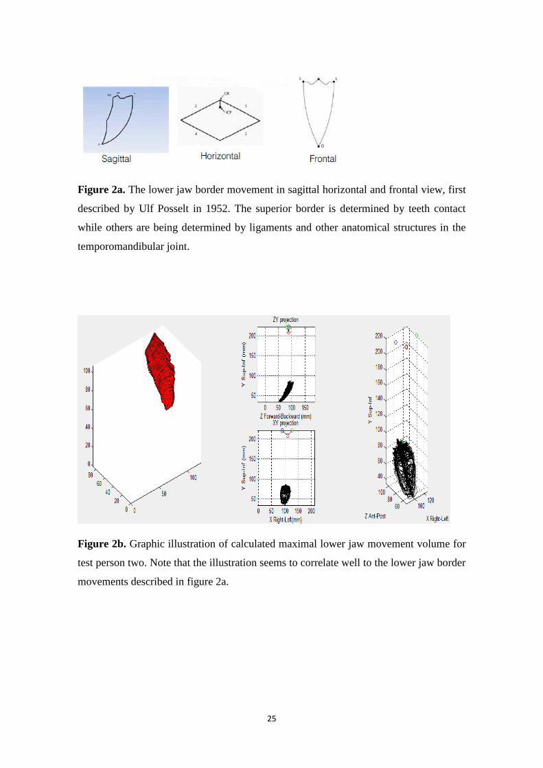
The software could calculate and visualize the total lower jaw movement volume from
zig-zag movements of the lower jaw. The graphic illustrations of the envelope of jaw
mobility visualized seem to correspond to the lower jaw border movements described
by Posselt (Posselt, 1952) (Figure 2a and 2b). When calculating lower jaw movement
volume, the software stratifies the measured coordinates according to a factor called
“stepsize”. In this pilot study stepsize was set at 1.0. In future studies, optimizing of step
size levels may prove a smoother outline and illustrated movement volume.

13
A previous study compared measurements obtained by digital caliper and a 3D
ultrasonic system. The study also found differences in protrusion movements between
the methods (Mazzetto et al., 2017). It is hard to compare the outcome values from two
different methods for movement measurements. The digital caliper is a reliable
measurement method for movements especially when the movement has one direction.
The caliper is cheap, easy to handle and useful in the clinic, but it may not give the full
picture of the movement patterns. A movement recording system if it is reliable can
allow for more detailed quantification and visualization of complex movement patterns,
and that are of value for the specialist and researcher.
Conclusively, the results of the pilot study imply that further testing of the method is
needed with larger series and test-retest reliability analysis to evaluate the possibility to
improve accuracy of tracing jaw movements with recording device. The pilot study has
thus produced some insight and more questions that need to be addressed before the 3D-
movement recording system (MacReflex) together with a software program can be used
for lower jaw volume calculations and included in treatment outcome analyses.
ACKNOWLEDGEMENTS
We want to thank Helena Grip, Department of Community Medicine and
Rehabilitation, Umeå University who developed the software used for calculating
values for separate lower jaw movements and illustrating the lower jaw movement
volume. We also want to thank our tutor Catharina Österlund, Umeå University.
14
REFERENCES
Adly MS, Youssif AAA, Eldin AS (2013). Recording and measuring of jaw movements
using a computer vision system. SYSTEM 81.
Agerberg G (1974). On mandibular dysfunction and mobility (dissertation) Umeå:
University of Umeå.
Akhter R, Benson J, Svensson P, Nicholas MK, Peck CC, Murray GM (2014).
Experimental jaw muscle pain increases pain scores and jaw movement variability in
higher pain catastrophizers. J Orofac Pain 28: 191-204.
Al-Jundi MA, John MT, Setz JM, Szentpetery A, Kuss O (2008). Meta-analysis of
treatment need for temporomandibular disorders in adult nonpatients. J Orofac Pain 22:
97-107.
Bakke M, Larsen SL, Lautrup C, Karlsborg M (2011). Orofacial function and oral
health in patients with Parkinson's disease. Eur J Oral Sci 119: 27-32.
Bensadoun RJ, Riesenbeck D, Lockhart PB, Elting LS, Spijkervet FK, Brennan MT
(2010). A systematic review of trismus induced by cancer therapies in head and neck
cancer patients. Support Care Cancer 18: 1033-1038.
Best N, Best S, Loudovici-Krug D, Smolenski UC (2013). Measurement of mandible
movements using a vernier caliper--an evaluation of the intrasession-, intersession- and
interobserver reliability. Cranio 31: 176-180.
Dahlstrom L, Carlsson GE (2010). Temporomandibular disorders and oral health-
related quality of life. A systematic review. Acta Odontol Scand 68: 80-85.
Dworkin SF, Huggins KH, LeResche L, Von Korff M, Howard J, Truelove E et al.
(1990). Epidemiology of signs and symptoms in temporomandibular disorders: clinical
signs in cases and controls. J Am Dent Assoc 120: 273-281.
Dworkin SF, LeResche L (1992). Research diagnostic criteria for temporomandibular
disorders: review, criteria, examinations and specifications, critique. J Craniomandib
Disord 6: 301-355.
15
Eriksson PO, Haggman-Henrikson B, Nordh E, Zafar H (2000). Co-ordinated
mandibular and head-neck movements during rhythmic jaw activities in man. J Dent
Res 79: 1378-1384.
Fang, J J, Kuo, T H. (2008). Modelling of mandibular movement. Comput Biol Med 38:
1152-1162.
Frisoli M, Edelhoff JM, Gersdorff N, Nicolet J, Braidot A, Engelke W (2017).
Comparative study using video analysis and an ultrasonic measurement system to
quantify mandibular movement. Cranio 35: 19-29.
Josefsson T, Nordh E, Eriksson P-O (1996). A flexible high-precision video system for
digital recording of motor acts through lightweight reflex markers. Computer Methods
and Programs in Biomedicine 49: 119-129.
Knap FJ, Richardson BL, Bogstad J (1970). Study of Mandibular Motion in Six
Degrees of Freedom. J Dent Res 49: 289-292.
LeResche L (1997). Epidemiology of temporomandibular disorders: implications for the
investigation of etiologic factors. Crit Rev Oral Biol Med: 291-305.
Mazzetto MOD MP, Anacleto MADM, Rodrigues CADM, Braganca RMD, Paiva GD,
Valencise Magri LDM (2017). Comparison of mandibular movements in TMD by
means of a 3D ultrasonic system and digital caliper rule. Cranio: 46-51.
Morquette P, Lavoie R, Fhima MD, Lamoureux X, Verdier D, Kolta A (2012).
Generation of the masticatory central pattern and its modulation by sensory feedback.
Prog Neurobiol 96: 340-55.
Okeson JP (2013). Management of temporomandibular disorders and occlusion. 7nd
rev. ed. St. Louis: Mosby.
Posselt U (1952). Studies in the mobility of the human mandible. Copenhagen: U.
Posselt.

16
Schiffman E, Ohrbach R, Truelove E, Look J, Anderson G, Goulet J P, et al (2014).
Diagnostic Criteria for Temporomandibular Disorders (DC/TMD) for Clinical and
Research Applications: recommendations of the International RDC/TMD Consortium
Network* and Orofacial Pain Special Interest Groupdagger. J Orofac Pain 28: 6-27.
Shueb SS, Nixdorf DR, John MT, Alonso BF, Durham J (2015). What is the impact of
acute and chronic orofacial pain on quality of life? J Dent 43: 1203-1210.
Tanaka Y, Yamada T, Maeda Y, Ikebe K (2016). Markerless three-dimensional tracking
of masticatory movement. J Biomech 49: 442-449.
Terajima M, Endo M, Aok, Y, Yuuda K, Hayasaki H, Goto TK, et al (2008). Four-
dimensional analysis of stomatognathic function. Am J Orthod Dentofacial Orthop 134:
276-287.
Travers KH, Buschang PH, Hayasaki H, Throckmorton GS (2000). Associations
between incisor and mandibular condylar movements during maximum mouth opening
in humans. Arch Oral Biol 45: 267-275.
Wahlund K, List T, Dworkin S. F (1998). Temporomandibular disorders in children and
adolescents: reliability of a questionnaire, clinical examination, and diagnosis. J orofac
Pain, 12.

17
Table 1a. Study design for pilot test A.
Test
number
Method Design Number
of tests
Number of
days
Outcome
variable
1 Movement
measurement,
with the aid of
Mac Reflex
system and a
digital caliper
Maximal jaw
opening (Y-
dimension). Start
position light
teeth contact in
CO
5 5 Jaw
movement
amplitude
(mm)
2 Movement
measurement,
with the aid of
Mac Reflex
system and a
digital caliper
Maximal
laterotrusive
movement (X-
dimensions). Start
position light
teeth contact in
CO
5 5 Jaw
laterotrusive
movement
(mm)
3 Movement
measurement,
with the aid of
Mac Reflex
system and a
digital caliper
Maximal
protrusive
movement (Z-
dimensions). Start
position light
teeth contact in
CO
5 5 Jaw
protrusive
movement
(mm)
18
Table 1b. Study design for pilot test B.
Test
number
Method Design Recording
time
Outcome
variable
1 Movement
recordings
with
MacReflex
Maximal circular lower jaw
movements, from slight teeth contact
CO to maximal jaw opening
60 seconds Volume
(cm3)
2 Movement
recordings
with
MacReflex
Maximal zig-zag lower jaw
movements (anterior-posterior), from
slight teeth contact CO to maximal
jaw opening
60 seconds Volume
(cm3)
3 Movement
recordings
with
MacReflex
Maximal zig-zag jaw movements
(left-right), from slight teeth contact
CO to maximal jaw opening
60 seconds Volume
(cm3)
19
Table 2a. Outcome of pilot test A with 3D optoelectronic movement recording and
digital caliper for each day.
Test person one Test person two
Day Dimension Mean (mm)
SD (max-min)
Mean (mm)
SD (max-min)
1 Jaw opening 47.6 1.8 4.2 46.9 2.0 4.9
2 Jaw opening 46.3 1.5 4.2 51.6 1.5 3.9
3 Jaw opening 49.8 1.2 2.9 49.9 1.1 2.6
4 Jaw opening 49.5 1.2 3.0 48.6 2.4 5.2
5 Jaw opening 49.3 1.2 2.6 50.7 2.1 4.1
Movement
recording
48.5 1.4 3.4 49.5 1.8 4.1
Digital caliper 42.7 0.9 49.7 0.5
1 Laterotrusion right 14.0 1.9 4.3 13.4 1.1 2.5
2 Laterotrusion right 10.5 0.9 2.2 17.1 1.0 2.6
3 Laterotrusion right 13.7 0.8 1.9 14.8 1.4 3.6
4
Laterotrusion right 13.6 0.3 0.7 14.1 2.6 6.6
5 Laterotrusion right 16.9 1.0 2.6 18.0 0.6 1.4
Movement
recording
13.7 1.0 2.3 15.5 1.3 3.3
Digital caliper 9.7 0.3 8.3 0.1
1 Laterotrusion left 10.4 1.2 3.1 13.1 0.7 1.8
2 Laterotrusion left 11.5 2.8 6.0 17.4 1.6 4.2
3 Laterotrusion left 9.0 0.8 1.8 18.2 1.1 2.7
4 Laterotrusion left 7.9 1.5 3.8 17.2 0.8 1.6
5 Laterotrusion left 9.1 0.6 1.4 15.6 2.1 4.8
Movement
recording
9.6 1.4 3.2 16.3 1.3 3.0
Digital caliper 9.7 0.7 10.1 0.6
1 Protrusion 14.0 2.5 6.2 15.9 1.6 4.1
2 Protrusion 13.4 2.0 4.9 15.8 1.6 3.8
3 Protrusion 14.7 3.5 9.1 15.4 2.2 5.9
4 Protrusion 15.7 2.4 5.8 16.4 1.0 2.5
5 Protrusion 17.4 3.3 9.2 15.3 1.7 4.1
Movement
recording
15.0 2.7 7.1
15.8 1.6 4.1
Digital caliper 9.8 0.3 11.2 0.4

20
Table 2b. Outcome of pilot test A with 3D optoelectronic movement recording and
digital caliper on test person one and two.
Test person one Test person two Movement
recording Digital caliper Difference
between (m) and (d) in percent
Movement recording
Digital caliper Difference between
(m) and (d) in percent
Movement
Dimension
Mean (mm)
Min-Max
(mm)
Mean (mm)
Min-Max
(mm)
(m-d)/m X 100
Mean (mm)
Min-Max
(mm)
Mean (mm)
Min-Max
(mm)
(m-d)/m X 100
Y-opening
48.5 44.3-
51.8
42.7 43.5-
47.5
12 % 49.5 44.3-
54.0
49.7 52.2-
54.2
0.4 %
X-right 13.7 9.8-
18.3
9.7 9.3-10 29 % 15.5 10.1-
18.8
8.3 8.0-
8.3
46 %
X-left 9.6 5.9-
15.7
9.7 9.2-
10.9
2 % 16.3 12.1-
20.1
10.1 8.3-
10.4
38 %
Z-forward
15.0 11.0-
21.9
9.8 9.6-
10.2
35 % 15.8 13.2-
19.1
11.2 9.0-
10.4
29 %

21
Table 2c. Outcome of pilot test B with 3D optoelectronic movement recording and
calculated movement volumes. The table also shows recorded min and max values for
maximal recorded movements in Y, X and Z – dimensions.
Pilot test B for test persons one and two
Lower jaw
movement volume
Total Y movement Total X movement Total Z movement
Test
person
Mean
(cm3)
Min-
Max
(cm3)
Mean
(mm)
Min-
Max
(mm)
Mean
(mm)
Min-
Max
(mm)
Mean
(mm)
Min-
max
(mm)
1 10.3 9.3-
11.4
47.6 44.2-
52,4
28.2 24.5-30.7 51.4 46.0-
59.5
2 17.2 13.9-
22.2
53.0 48.2 –
57.0
40.9 36.6 –
43.0
55.5 47.5

22
JOmovrec
JOdigcal
LtrRmovrec
LtrRdigcal
LtrLmovrec
LtrLdigcal
Protmovrec
Protdigcal
0
10
20
30
40
50
60Movementvalue(m
m)
Testpersonone
Testpersontwo
Figure 1a. Outcome of jaw movement values for test person one and two with
comparison between values from movement recording (MacReflex) compared with
digital caliper.
JO= jaw opening movement
LtrR= Laterotrusion right
LtrL= Laterotrusion left
Prot= Protrusion
Movrec= movement recording
Digcap= Digital caliper
23
JOLtr
RLtr
LPro
t0
2
4
6
8
10Deltavalue
Testpersonone
Testpersontwo
Figure 1b. Outcome delta values (=max-min), for test person one and two in
performed jaw movements in four different dimensions.
JO= jaw opening movement
LtrR= Laterotrusion right
LtrL= Laterotrusion left
Prot= Protrusion

24
0
5
10
15
20
25Jawmovementvolume(cm
³ )Testpersonone
Testpersontwo
Figure 1c. Outcome of lower jaw movement volume for test person one and two.
25
Figure 2a. The lower jaw border movement in sagittal horizontal and frontal view, first
described by Ulf Posselt in 1952. The superior border is determined by teeth contact
while others are being determined by ligaments and other anatomical structures in the
temporomandibular joint.
Figure 2b. Graphic illustration of calculated maximal lower jaw movement volume for
test person two. Note that the illustration seems to correlate well to the lower jaw border
movements described in figure 2a.

Umeå University
Department of Odontology
SE-901 87 Umeå, Sweden
www.umu.se