Lower Extremity-WCA Student Slides
36
Lower Extremity The Fun Begins
Transcript of Lower Extremity-WCA Student Slides

Lower Extremity The Fun Begins
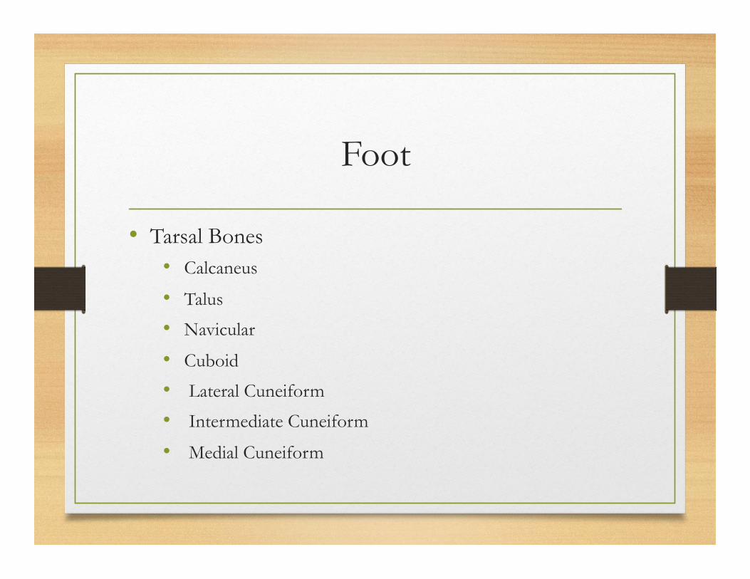
Foot
• Tarsal Bones • Calcaneus
• Talus
• Navicular
• Cuboid
• Lateral Cuneiform
• Intermediate Cuneiform
• Medial Cuneiform
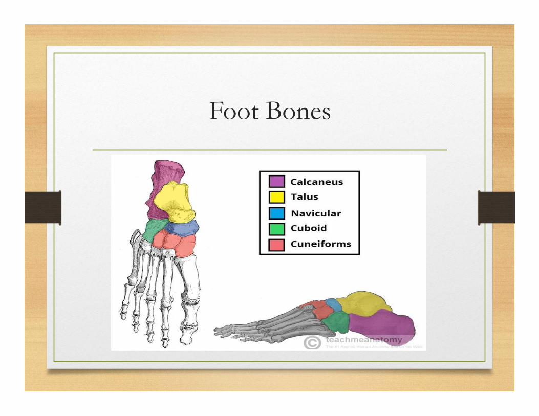
Foot Bones
Foot
• Metatarsals • Labeled 1-5 from great toe to pinky toe
• Metatarsal Heads
Phalanges
• 2 in great toe
• 3 in each of the four lesser toes
Foot
• Arches • Longitudinal
• Transverse
• The foot also has 33 joints, over 100 tendons and countless ligaments
• Transverse Tarsal Joints
• Inversion and Eversion
Injuries to the Foot
• Too many to list them all
• Common Injuries: • Turf Toe - Mid-Foot Sprain
• Spring Ligament - Plantar Fasciitis
• Heel Bruise - Fractures • Other Foot Issues
• Calluses – Keep them in check
• Blisters – following slide
• Athletes Foot Fungus – Damp, warm, dark environment

Ankle Joint
• True Joint - articulation between the talus and the tibia
• Tibia - medial malleolus
• Fibula - lateral malleolus
• Fibula non-weight bearing bone that serves as a point for muscle attachment of the muscles that control the ankle, foot and toes.

Ankle
• Stability • Medial Ligament Complex – Deltoid Ligament-Large complex
ligament • MOI-Eversion
• Testing – Eversion stress test in DF, Neutral, PF
• Tibiofibular Ligaments
• Rotation of body over ankle (cleats stick in turf)
• Can also be sprained along with Medial or Lateral sprain
• Testing – Eversion stress in neutral and DF
• Testing – Squeeze Test

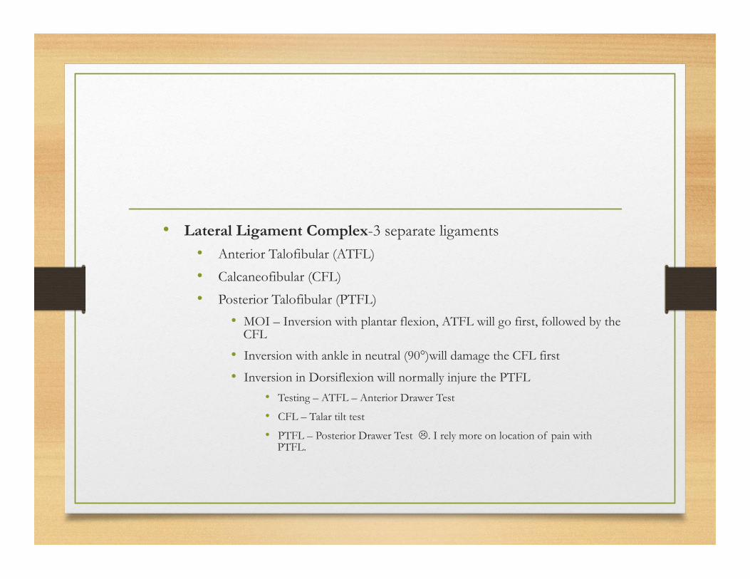
• Lateral Ligament Complex-3 separate ligaments • Anterior Talofibular (ATFL)
• Calcaneofibular (CFL)
• Posterior Talofibular (PTFL)
• MOI – Inversion with plantar flexion, ATFL will go first, followed by the CFL
• Inversion with ankle in neutral (90°)will damage the CFL first
• Inversion in Dorsiflexion will normally injure the PTFL
• Testing – ATFL – Anterior Drawer Test
• CFL – Talar tilt test
• PTFL – Posterior Drawer Test L. I rely more on location of pain with PTFL.
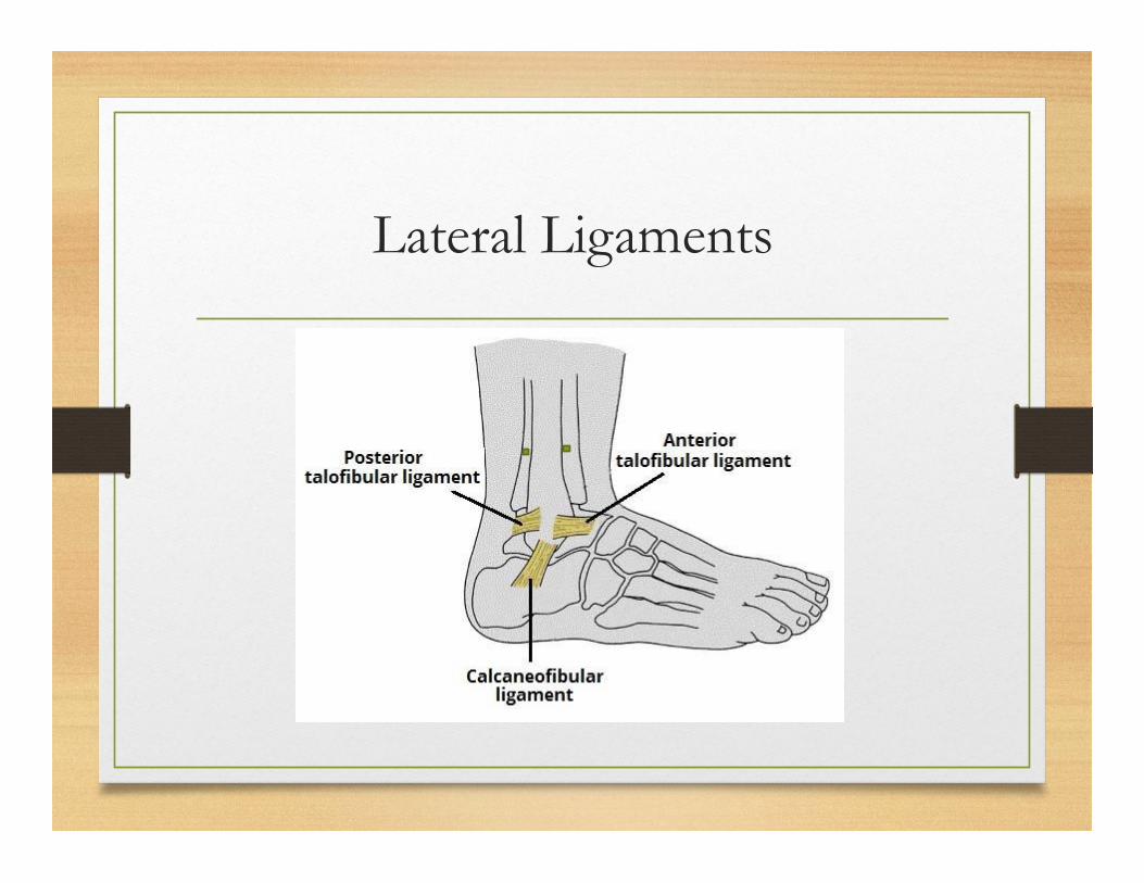
Lateral Ligaments

Back to HOPS
• Ask them how it happened – do they remember what position their ankle was in when force was applied? • “Landed on someone’s foot coming down from a jump”
• “I was standing there watching the play and somebody rolled into my ankle”
• “My heel fell into a hole; my toes were pointing upwards”
• Where is the swelling?
• Where is the pain – Anterior? Distal Fibula? Posterior?
• Then palpate and your stress tests

Other Ankle Issues
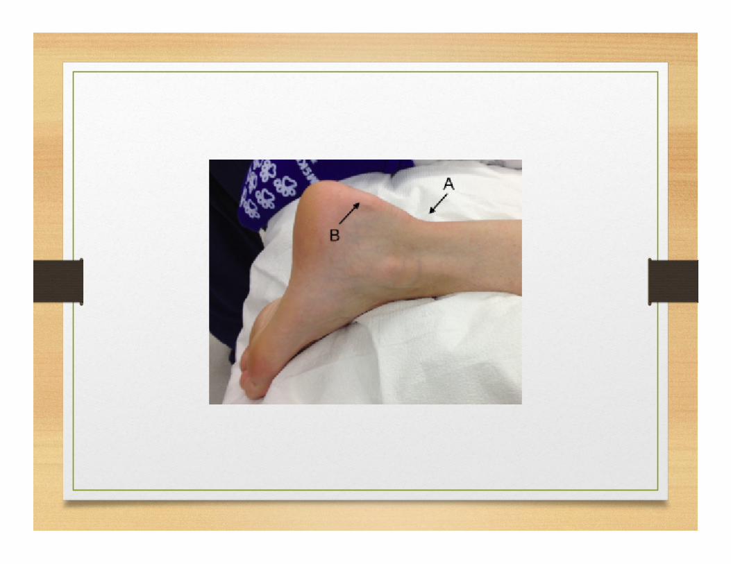
• Fracture • Avulsion Fractures – palpation bilaterally
• Complete Fractures (picture)
• Dislocation • Uncommon by itself– normally associated with a fracture

Lower Leg
• Tibia • Fibula • Interosseous Membrane
• Lower Leg Injuries
• Contusions – Can be very serious • Compartment Syndrome
• Acute versus Chronic


Lower Leg injuries
• Stress Syndrome/Stress Fracture • Either tibia or fibula
• History, palpation Cues
• Muscle Strain (medial tibial stress syndrome) • History, palpation of associated structures, testing
• Must palpate the arch – tibialis posterior, FHL, FDL, spring ligament (following slide)
• Achilles Tendon Strain


Knee
• Articulation of tibia and femur
• Largest joint in the body
• Capsule surrounds the condyles of the femur and extends three fingers above the superior pole of the patella. (joint effusion picture)

Knee
• Stability provided my numerous muscles and ligaments
• Muscles Include • Gastrocnemius
• Hamstrings • Biceps Femoris – lateral insertion
• Semimembranosus – posterior medial insertion
• Semitendinosus – anterior medial insertion
• Quadriceps • Vastus lateralis, medialis, intermedius
• Rectus femoris
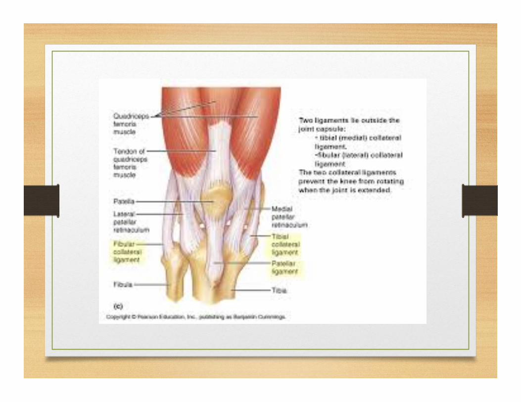

Collateral Ligaments
• Ligament Structures • Lateral Collateral Ligament – Thin, cord ligament
• Becomes looser in flexion
• Your MOI, palpation and testing should guide you
• MOI-Medial Blow
• Palpation – from the head of the fibula superiorly across the joint line to the lateral femur
• Testing – ADDUCTION stress test at 0°(or as close as possible), 15° and 30°. Expect the knee to feel loose(r) after you take it out of full extension.

Collateral Ligaments
• Medial Collateral Ligament – Broad flat band ligament • Most commonly sprained ligament in the knee
• Some fibers stay taut through entire range of motion
• Deep and superficial layers
• MOI-Blow from lateral side
• Palpation – two fingers wide from 90° medial of the tibial tuberosity superiorly across the joint to the medial femoral epicondyle.
• Testing – ABDUCTION stress test at 0°, 15°, 30°. Knee should remain stable in all three degrees as opposed to the LCL
• With either of these two the swelling will be located on the injured side but will be more spread out than it would be with a cruciate ligament injury


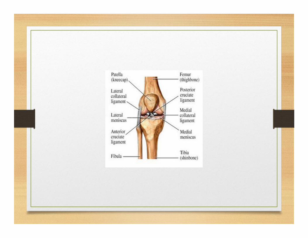
Cruciate Ligaments
• Named according to the tibial attachment • Posterior Cruciate Ligament
• Tibia forced posterior or femur forced anterior (direct blow)
• Forced Deep flexion
• Stronger than ACL, located nearer to center of the joint
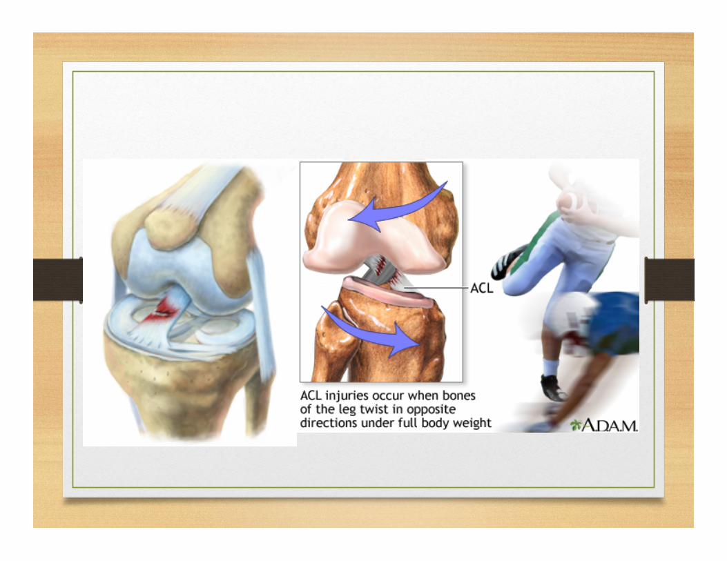
• Anterior Cruciate Ligament • Get’s all the publicity, injured much more frequently than PCL
• Main stabilizing ligament in the knee
• Tibia forced anterior or femur posterior (direct blow)
• Deceleration, hamstrings don’t react quickly enough
• Deep flexion
• Rotation over knee, foot normally fixed (cleats stick)
Testing of Cruciate Ligaments
• PCL – Godfrey’s 90/90 test, Posterior Sag Test • ACL Testing
• Does joint effusion occur rapidly and completely surround the knee joint anteriorly? • Lachman’s test before swelling sets in. If swelling is already
present, WHERE the swelling is located and the AMOUNT OF TIME that has past since the injury occurred can guide you.
• ACL or PCL? Both will look the same objectively and it is quite possible that the MOI could be the same. In either case, a doctors referral will be necessary


Meniscus Injuries
• Lateral Meniscus – Greater freedom of movement • Medial Meniscus
• Each is firmly attached at the periphery to the tibia
• It is said that menisci tear when they are trapped, pinched or crushed between the femur and the tibial plateau.
• Tears • Flap, longitudinal, bucket handle
• The actual tear is not of importance – most will need surgical intervention
• Testing – Sweep Test

Meniscus

Meniscus Tears
Flap Tear Radial Tear Longitudinal Tear Parrot Beak Tear Bucket Handle tear

This is What Makes You Smarter
• Cruciate ligament injuries will cause major effusion in a short period of time (normally less than 24 hours). This is due to the great blood supply to these ligaments. Bloody Effusion
• Meniscal injuries will swell much slower (week(s) because they have a poor blood supply and the increase in joint swelling is caused by synovial fluid. This would yield a clear or straw colored effusion. This is where the sweep test is very effective in establishing an effusion.
Other Issues Around the Knee
• Pre patellar bursitis (next slide)
• Patellar Subluxation
• Chondromalacia Patella
• Patellar Tendinitis – Jumpers Knee
• Osgood Schlatter Disease
• ITB Friction Syndrome