Low-Intensity Pulsed Ultrasound Accelerates Rat …curreyj/BNG-338_files/Azume et al J Bone... ·...
10
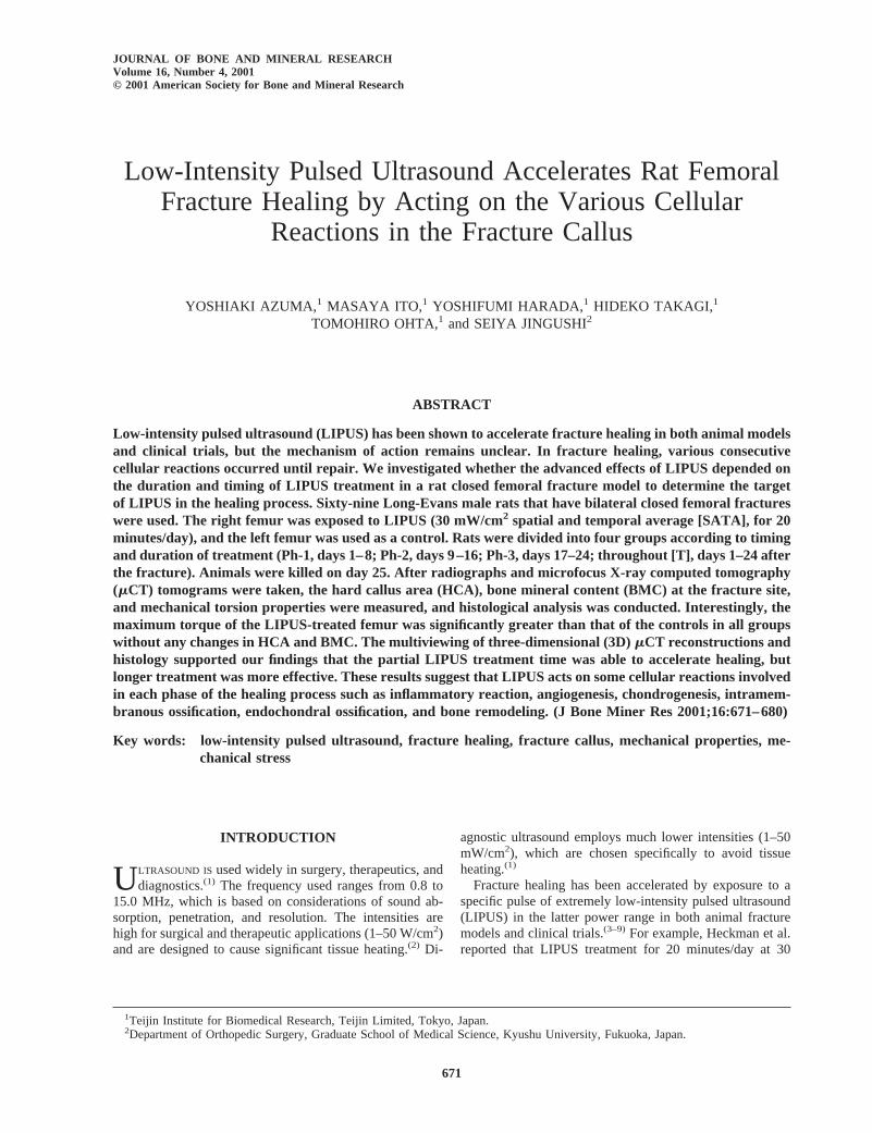
Low-Intensity Pulsed Ultrasound Accelerates Rat Femoral Fracture Healing by Acting on the Various Cellular Reactions in the Fracture Callus YOSHIAKI AZUMA, 1 MASAYA ITO, 1 YOSHIFUMI HARADA, 1 HIDEKO TAKAGI, 1 TOMOHIRO OHTA, 1 and SEIYA JINGUSHI 2 ABSTRACT Low-intensity pulsed ultrasound (LIPUS) has been shown to accelerate fracture healing in both animal models and clinical trials, but the mechanism of action remains unclear. In fracture healing, various consecutive cellular reactions occurred until repair. We investigated whether the advanced effects of LIPUS depended on the duration and timing of LIPUS treatment in a rat closed femoral fracture model to determine the target of LIPUS in the healing process. Sixty-nine Long-Evans male rats that have bilateral closed femoral fractures were used. The right femur was exposed to LIPUS (30 mW/cm 2 spatial and temporal average [SATA], for 20 minutes/day), and the left femur was used as a control. Rats were divided into four groups according to timing and duration of treatment (Ph-1, days 1– 8; Ph-2, days 9 –16; Ph-3, days 17–24; throughout [T], days 1–24 after the fracture). Animals were killed on day 25. After radiographs and microfocus X-ray computed tomography (mCT) tomograms were taken, the hard callus area (HCA), bone mineral content (BMC) at the fracture site, and mechanical torsion properties were measured, and histological analysis was conducted. Interestingly, the maximum torque of the LIPUS-treated femur was significantly greater than that of the controls in all groups without any changes in HCA and BMC. The multiviewing of three-dimensional (3D) mCT reconstructions and histology supported our findings that the partial LIPUS treatment time was able to accelerate healing, but longer treatment was more effective. These results suggest that LIPUS acts on some cellular reactions involved in each phase of the healing process such as inflammatory reaction, angiogenesis, chondrogenesis, intramem- branous ossification, endochondral ossification, and bone remodeling. (J Bone Miner Res 2001;16:671– 680) Key words: low-intensity pulsed ultrasound, fracture healing, fracture callus, mechanical properties, me- chanical stress INTRODUCTION U LTRASOUND IS used widely in surgery, therapeutics, and diagnostics. (1) The frequency used ranges from 0.8 to 15.0 MHz, which is based on considerations of sound ab- sorption, penetration, and resolution. The intensities are high for surgical and therapeutic applications (1–50 W/cm 2 ) and are designed to cause significant tissue heating. (2) Di- agnostic ultrasound employs much lower intensities (1–50 mW/cm 2 ), which are chosen specifically to avoid tissue heating. (1) Fracture healing has been accelerated by exposure to a specific pulse of extremely low-intensity pulsed ultrasound (LIPUS) in the latter power range in both animal fracture models and clinical trials. (3–9) For example, Heckman et al. reported that LIPUS treatment for 20 minutes/day at 30 1 Teijin Institute for Biomedical Research, Teijin Limited, Tokyo, Japan. 2 Department of Orthopedic Surgery, Graduate School of Medical Science, Kyushu University, Fukuoka, Japan. JOURNAL OF BONE AND MINERAL RESEARCH Volume 16, Number 4, 2001 © 2001 American Society for Bone and Mineral Research 671
-
Upload
vuongquynh -
Category
Documents
-
view
217 -
download
0
Transcript of Low-Intensity Pulsed Ultrasound Accelerates Rat …curreyj/BNG-338_files/Azume et al J Bone... ·...
Low-Intensity Pulsed Ultrasound Accelerates Rat FemoralFracture Healing by Acting on the Various Cellular
Reactions in the Fracture Callus
YOSHIAKI AZUMA, 1 MASAYA ITO, 1 YOSHIFUMI HARADA,1 HIDEKO TAKAGI, 1
TOMOHIRO OHTA,1 and SEIYA JINGUSHI2
ABSTRACT
Low-intensity pulsed ultrasound (LIPUS) has been shown to accelerate fracture healing in both animal modelsand clinical trials, but the mechanism of action remains unclear. In fracture healing, various consecutivecellular reactions occurred until repair. We investigated whether the advanced effects of LIPUS depended onthe duration and timing of LIPUS treatment in a rat closed femoral fracture model to determine the targetof LIPUS in the healing process. Sixty-nine Long-Evans male rats that have bilateral closed femoral fractureswere used. The right femur was exposed to LIPUS (30 mW/cm2 spatial and temporal average [SATA], for 20minutes/day), and the left femur was used as a control. Rats were divided into four groups according to timingand duration of treatment (Ph-1, days 1–8; Ph-2, days 9–16; Ph-3, days 17–24; throughout [T], days 1–24 afterthe fracture). Animals were killed on day 25. After radiographs and microfocus X-ray computed tomography(mCT) tomograms were taken, the hard callus area (HCA), bone mineral content (BMC) at the fracture site,and mechanical torsion properties were measured, and histological analysis was conducted. Interestingly, themaximum torque of the LIPUS-treated femur was significantly greater than that of the controls in all groupswithout any changes in HCA and BMC. The multiviewing of three-dimensional (3D)mCT reconstructions andhistology supported our findings that the partial LIPUS treatment time was able to accelerate healing, butlonger treatment was more effective. These results suggest that LIPUS acts on some cellular reactions involvedin each phase of the healing process such as inflammatory reaction, angiogenesis, chondrogenesis, intramem-branous ossification, endochondral ossification, and bone remodeling. (J Bone Miner Res 2001;16:671–680)
Key words: low-intensity pulsed ultrasound, fracture healing, fracture callus, mechanical properties, me-chanical stress
INTRODUCTION
ULTRASOUND IS used widely in surgery, therapeutics, anddiagnostics.(1) The frequency used ranges from 0.8 to
15.0 MHz, which is based on considerations of sound ab-sorption, penetration, and resolution. The intensities arehigh for surgical and therapeutic applications (1–50 W/cm2)and are designed to cause significant tissue heating.(2) Di-
agnostic ultrasound employs much lower intensities (1–50mW/cm2), which are chosen specifically to avoid tissueheating.(1)
Fracture healing has been accelerated by exposure to aspecific pulse of extremely low-intensity pulsed ultrasound(LIPUS) in the latter power range in both animal fracturemodels and clinical trials.(3–9) For example, Heckman et al.reported that LIPUS treatment for 20 minutes/day at 30
1Teijin Institute for Biomedical Research, Teijin Limited, Tokyo, Japan.2Department of Orthopedic Surgery, Graduate School of Medical Science, Kyushu University, Fukuoka, Japan.
JOURNAL OF BONE AND MINERAL RESEARCHVolume 16, Number 4, 2001© 2001 American Society for Bone and Mineral Research
671
mW/cm2 led to a significant 24% reduction in the timerequired for clinical healing in a prospective randomizedplacebo-controlled study of closed or grade I open tibialfractures.(8) Additionally, Kristiansen et al. showed that thetime until union was 38% shorter for patients treated withLIPUS than for those treated with placebo in a multicenterprospective randomized placebo-controlled clinical trial ofdorsally angulated fractures of the distal radius.(9) However,the physical process through which LIPUS stimulates livingtissue remains unclear. LIPUS is a form of mechanicalenergy that can be transmitted into living tissue as high-frequency acoustical pressure waves. The micromechanicalstrains produced by these pressure waves in living tissue canresult in biochemical events at the cellular level.(10–12)Du-arte indicated that a pulsed ultrasound intensity of 49.6mW/cm2 and 57 mW/cm2 spatial average and temporalaverage (SATA) was nonthermal.(4) In this experiment, weused LIPUS (30 mW/cm2), which is considered to havelittle thermal effect and to produce stable cavitation andstreaming. Wang et al. reported a technique for exposingstandard closed femoral fractures to LIPUS and confirmedthat LIPUS stimulation enhanced the mechanical propertiesof the healing callus.(7) Their observations imply that cellsin the fracture callus sense and respond to the mechanicalenergy transferred by the LIPUS.
Fracture healing occurs in a stepwise process of variouscellular reactions until the fracture has been repaired.(13)
Cell types, such as endothelial cells, fibroblasts, osteoblasts,chondrocytes, and osteocytes, all of which respond to me-chanical stress or mechanical strain,(14–18) play importantroles in the fracture healing process.(19) The mechanicalenergy transferred by LIPUS therefore might act on variousprocesses such as inflammatory reaction, angiogenesis,chondrogenesis, intramembranous ossification, endochon-dral ossification, or bone remodeling in the fracture callus.
In previous experiments using a rat closed femoral frac-ture model, we found accelerating effects of daily LIPUStreatment until 21 days after the fracture (in preparation).Because in this model a series of stage-specific cellularreactions were observed until repair,(20) we investigatedwhether the advanced effects of LIPUS depended on dura-tion and timing of LIPUS treatment in this model to deter-mine the target reaction of LIPUS in the fracture healingprocess in vivo. In the present study, we divided the intervalup to 25 days after the fracture into three periods (Ph-1,Ph-2, and Ph-3), each of which involved different essentialreactions for fracture healing,(20) and investigated the ef-fects of partial treatment with LIPUS on the fracture heal-ing. If the advanced effects of LIPUS on fracture healing arebased on an effect on a specific reaction, the efficiency ofacceleration should depend on the timing of the partialLIPUS treatment. In this study, we investigated the rela-tionship between the timing of the partial LIPUS treatmentand the efficiency of accelerating action by estimating me-chanical properties, bone mass, histology, and the micro-structure of the callus, the latter by using the three-dimensional (3D) microfocus X-ray computed tomography(mCT) technique.
MATERIALS AND METHODS
Closed femoral fracture model
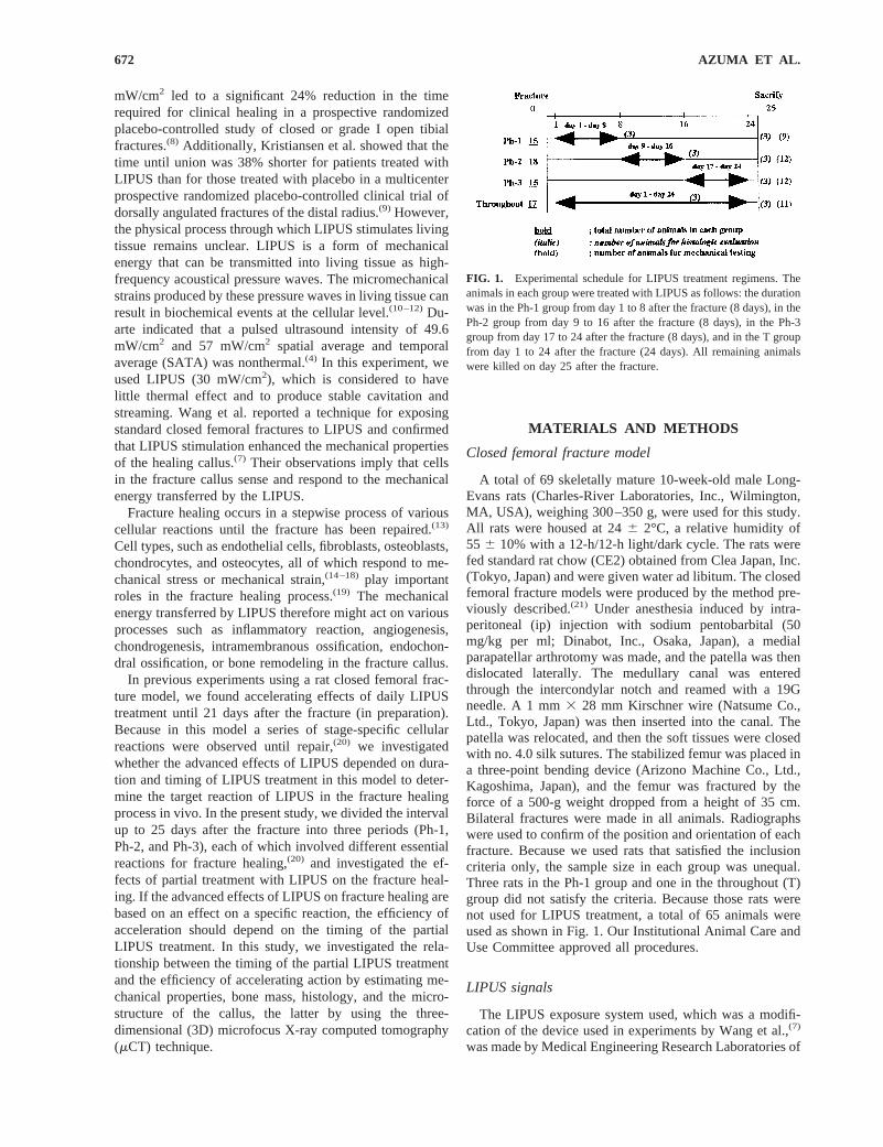
A total of 69 skeletally mature 10-week-old male Long-Evans rats (Charles-River Laboratories, Inc., Wilmington,MA, USA), weighing 300–350 g, were used for this study.All rats were housed at 246 2°C, a relative humidity of55 6 10% with a 12-h/12-h light/dark cycle. The rats werefed standard rat chow (CE2) obtained from Clea Japan, Inc.(Tokyo, Japan) and were given water ad libitum. The closedfemoral fracture models were produced by the method pre-viously described.(21) Under anesthesia induced by intra-peritoneal (ip) injection with sodium pentobarbital (50mg/kg per ml; Dinabot, Inc., Osaka, Japan), a medialparapatellar arthrotomy was made, and the patella was thendislocated laterally. The medullary canal was enteredthrough the intercondylar notch and reamed with a 19Gneedle. A 1 mm 3 28 mm Kirschner wire (Natsume Co.,Ltd., Tokyo, Japan) was then inserted into the canal. Thepatella was relocated, and then the soft tissues were closedwith no. 4.0 silk sutures. The stabilized femur was placed ina three-point bending device (Arizono Machine Co., Ltd.,Kagoshima, Japan), and the femur was fractured by theforce of a 500-g weight dropped from a height of 35 cm.Bilateral fractures were made in all animals. Radiographswere used to confirm of the position and orientation of eachfracture. Because we used rats that satisfied the inclusioncriteria only, the sample size in each group was unequal.Three rats in the Ph-1 group and one in the throughout (T)group did not satisfy the criteria. Because those rats werenot used for LIPUS treatment, a total of 65 animals wereused as shown in Fig. 1. Our Institutional Animal Care andUse Committee approved all procedures.
LIPUS signals
The LIPUS exposure system used, which was a modifi-cation of the device used in experiments by Wang et al.,(7)
was made by Medical Engineering Research Laboratories of
FIG. 1. Experimental schedule for LIPUS treatment regimens. Theanimals in each group were treated with LIPUS as follows: the durationwas in the Ph-1 group from day 1 to 8 after the fracture (8 days), in thePh-2 group from day 9 to 16 after the fracture (8 days), in the Ph-3group from day 17 to 24 after the fracture (8 days), and in the T groupfrom day 1 to 24 after the fracture (24 days). All remaining animalswere killed on day 25 after the fracture.
672 AZUMA ET AL.

Teijin, Ltd. (Tokyo, Japan). It consisted of a function gen-erator (model HP33120A; Hewlett-Packard Japan, Ltd., To-kyo, Japan), a power supply, and six lead-zirconate titanatetransducers (PZT-4, effective area of 3.88 cm2). With it, sixanimals could be exposed to LIPUS at the same time. TheLIPUS signal was generated with a transducer and wascomposed of a 200-ms burst sine wave of 1.5 MHz repeatingat a frequency of 1.0 kHz. The 200-ms burst of pressurepulses was followed by an off time of 800ms and wasrepeated every millisecond. The LIPUS intensity was 30mW/cm2 as an SATA. The LIPUS signal and times per dayused in this study were the same as those recommended fora clinical device (Sonic Accelerated Fracture Healing Sys-tem [SAFHS; Exogen, Inc., Piscataway, NJ, USA]). TheLIPUS signal conditions were calibrated with a hydrophonesystem that consisted of an oscilloscope (model OS-111;Hewlett-Packard Japan, Ltd.) and a PZT-4 transducer fordetection. The LIPUS intensity was calibrated with an ul-trasound power meter (model 101; OR Instruments, Inc.,Seattle, WA, USA) before LIPUS exposure to the animals.The interexperiment CVs of each transducer was within10% for the LIPUS intensity.
Duration and timing of LIPUS treatment
The animals were treated with LIPUS while they wereunder ip anesthesia with ketamine (20–60 mg/kg) and xy-lazine (2.5 mg/kg). The anesthetized rats were placed ontheir back on the holder. Ultrasound gel (Nippon KohdenCo., Ltd., Tokyo, Japan) was applied to the shaved fracturearea in the medial aspects of both thighs of all animals. Thetransducer was placed close to the skin covered with ultra-sound gel at the femoral fracture site. The animals wereexposed to LIPUS once daily for 20 minutes. The rightfemur of the rats was exposed to the LIPUS transducer, andthe left femur was used as the nontreated contralateralcontrol. The duration and timings of the LIPUS treatmentare shown in Fig. 1. The animals in each group were treatedwith LIPUS as follows: LIPUS treatments were performedin the Ph-1 group for 8 days, from day 1 to 8 after fracture;in the Ph-2 group for 8 days, from day 9 to 16 after fracture;in the Ph-3 group for 8 days, from day 17 to 24 afterfracture; and in the T group for 24 days, from day 1 to 24after fracture. Animals were killed on day 25 after thefracture.
Radiography and bone mineral content at thefracture site
The femora were collected on day 25 after the fracture.All femora were radiographed with a soft X-ray system(model CSM; Softex Co., Ltd., Tokyo, Japan). After the softtissues had been dissected from the femora, the intramed-ullary pins were removed. Bone mineral content (BMC) inthe area near the fracture site was measured with a dual-energy X-ray absorptiometer (DXA; model QDR-2000; Ho-logic, Inc., Waltham, MA, USA). The scan field size was3.48 cm2 3 1.41 cm2, and the resolution was 0.025430.0127 cm2. The area near the fracture site of each femurwas determined as the area extending 3.6 mm on either side
of the fracture line, which was measured with software forhigh-resolution analysis. The CV for the measurement ofthe BMC of standard samples by this technique was 0.8%.Specimens were stored in saline-soaked gauze at220°C.
Hard callus area
Radiographic signals of each femur and the standard scalewere captured as digitized images with a scanner and storedon a Macintosh computer using Adobe PhotoShop version 4software. The area of the outer hard callus was measured bytracing the callus portion with “National Institutes of Health(NIH) image version 1.6” image analysis software (NIH,Bethesda, MD, USA). The area represents the number ofpixels within each tracing. The standard scale also wasmeasured, and the pixel value of each hard callus area(HCA) was transferred to the value of the real scale.
Mechanical testing
Just before mechanical testing, the specimens werethawed at room temperature for 2 h. Both bone ends wereembedded in polyacrylic resins (GC-OSTORON; GC Den-tal Products Co., Ltd., Aichi, Japan). A custom-made jigensured consistent alignment of the bone axis with the axisof the testing machine. All specimens were tested to failurein torsion at room temperature on an electromechanicaltesting machine (model MZ-500D; Maruto Machine, Inc.,Tokyo, Japan) at the rate of 1.5°/s with 111.5 g of weight asaxial load. Maximal torque until failure and torsional stiff-ness (the tangent at the point of maximal slope) werecalculated from the load-deformation curve. The biome-chanical stages of fracture union were determined based onpattern of failure according to White et al.(22)
Histological evaluation
Three rats per each group were killed, and the femorawere collected on day 25 after the fracture for histologicalevaluation. To observe the effect of LIPUS in progress onday 9 and day 17 after fracture, we killed three rats in thePh-1 and Ph-2 groups, respectively. The femora were fixedfor several days in 10% neutral buffered formalin. After themCT images had been obtained, the femora were treatedwith 10% formic acid for 1 h, transiently. Then, the femorawere decalcified with 10% EDTA solution and embedded inparaffin. Three to five sections (6mm thick) were cutthrough the long axis of each femur in the sagittal plane andstained with Masson’s trichrome stain and hematoxylin andeosin stain. The section that had the maximum diameter waschosen as the representative section for each fracture.
3D mCT analysis
The mCT (model NX-CP-C80H-IL) equipment that pro-duced the tomograms had been especially developed byNittetsu ELEX Co., Ltd. (Osaka, Japan). The device has amaximum experimental spatial resolution of 2.5mm and aminimum slice thickness of 5mm for measurements of aceramic standard sample. The fan beam ofmCT has a small
673ULTRASOUND ACCELERATES RAT FEMORAL FRACTURE REPAIR

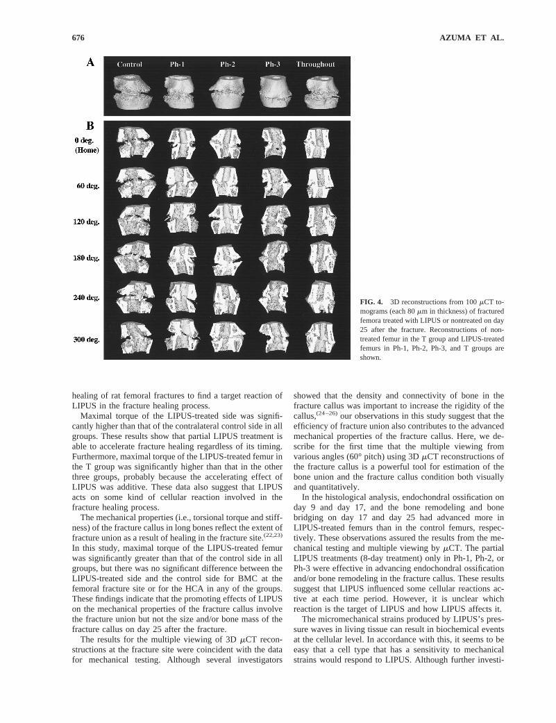
focal spot size, 3mm in diameter, a conventional microfo-cus open vacuum-type X-ray source, rotating sample stage,1024 linear response image sensors, and image intensifier(image sensors). A total of 100 cross-sectional tomogramsper femur were obtained with a slice thickness of 80mm andreconstructed at 5123 512 pixels, by use of an accelerationvoltage of 30 kV and current of 0.1 mA. To evaluate thebone bridging at the fracture site by use of a 3D reconstruc-tion technique, we took 50 slices extending (4000mm) oneither side of the fracture line. The 3D reconstruction of 100tomograms was performed by the volume-rendering method(software, VIP-Station; Teijin System Technology, Inc.,Kanagawa, Japan) using a computer (model SUNSPARK-5; Sun Microsystems, Inc., Palo Alto, CA, USA).By use of the computer software, the threshold of eachimage was kept at the same level to observe trabecular boneand cortex of the healing callus. At this threshold, we neverdetected the soft callus of the femur. Electronic sectionswere cut through the long axis of each femur in the sagittalplane on 3D reconstructed images. The home sagittal plane(0°) was identified as that which was used to cut through thelong axis of the original 3D reconstructed image of eachfemur. To estimate the bone bridging in detail, we rotatedthe sagittal plane clockwise about the long axis at 60°increments of pitch (0°, 60°, 120°, 180°, 240°, and 300°)from the home position in a circuit and cut electronicsections through the long axis of each femur in each sagittalplane.
Statistical analysis
The primary analyses conducted in this study were in-traexperimental group comparisons of the treated fractureswith the untreated contralateral fractures. Pairedt-testingwas used for analyses of HCA, BMC, and mechanicalmeasurement data. The significance of interexperimentalgroup comparison of mechanical measurement data wasdetermined by a one-way analysis of variance (ANOVA)test. A confidence level of 95% (p , 0.05) was chosen forsignificance.
RESULTS
Mechanical properties of fracture callus
The maximal torque and stiffness in torsion of the frac-tured femur on the LIPUS-treated side was significantlyhigher than that of the contralateral control side in all groups(Fig. 2). This indicates that the partial treatment with LIPUSduring Ph-1, Ph-2, or Ph-3 also is able to improve themechanical properties of the fracture callus, as it does withtreatment throughout the 24 days. Furthermore, the maximaltorque for the LIPUS-treated side in the T group (1936 39N-mm) was significantly higher than that for the other threegroups: Ph-1, 1706 39 N-mm; Ph-2, 1596 31 N-mm; andPh-3, 1616 34 N-mm (Fig. 2A). The data for stiffness wasalmost the same as that for maximal torque (Fig. 2B). Theseresults show that the continuous treatment with LIPUS wasmore effective toward the improvement of the mechanicalproperties of the fracture callus than the partial treatment.
Radiographs and 3DmCT analysis
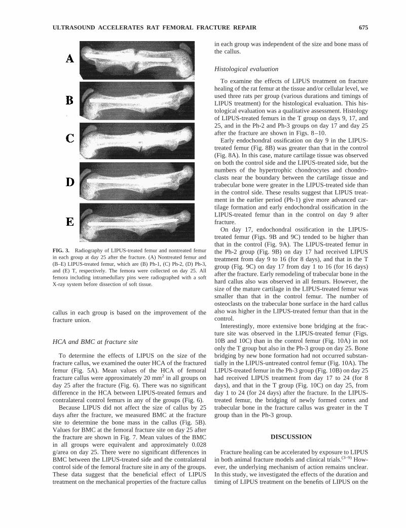
As shown in Fig. 3, in the LIPUS-treated femur, theobliteration of the fracture line at the fracture gap in allgroups (Figs. 3B–3E) was more advanced than that in thecontrol femur (Fig. 3A).
The fracture line of the LIPUS-treated femur was bridgedby new bone formation 3D in all groups (Fig. 4). In contrast,bridging of the fracture site by new bone formation did notoccur substantially in the contralateral LIPUS-untreatedcontrol femur. In the LIPUS-treated femur, the bridging ofnewly formed cortex and trabecular bone in the fracturecallus in the T group was greater than that in the Ph-1, Ph-2,and Ph-3 groups. These data show that the beneficial effectof LIPUS treatment on the mechanical properties of fracture
FIG. 2. Effects of LIPUS treatment of various durations and timingson (A) torsional torque and (B) torsional stiffness of fracture callus onday 25 in rat femoral fractures. The torsional torque of LIPUS-treatedfemurs (closed bars) was significantly higher than that of the nontreatedfemurs (open bars) in the T group (n 5 11), Ph-1 group (n 5 9), Ph-2group (n 5 12), and Ph-3 group (n 5 12). The horizontal bars represent6 1 SD. The value ofp , 0.01 for each treatment group compared withthe control side. Pairedt-testing was used for statistical analysis tocompare between the LIPUS-treated side and -nontreated side in eachgroup. The T group showed a significant difference as compared withthe partially treated groups. One-way ANOVA was used for statisticalanalysis to test for significance versus the T group.
674 AZUMA ET AL.
callus in each group is based on the improvement of thefracture union.
HCA and BMC at fracture site
To determine the effects of LIPUS on the size of thefracture callus, we examined the outer HCA of the fracturedfemur (Fig. 5A). Mean values of the HCA of femoralfracture callus were approximately 20 mm2 in all groups onday 25 after the fracture (Fig. 6). There was no significantdifference in the HCA between LIPUS-treated femurs andcontralateral control femurs in any of the groups (Fig. 6).
Because LIPUS did not affect the size of callus by 25days after the fracture, we measured BMC at the fracturesite to determine the bone mass in the callus (Fig. 5B).Values for BMC at the femoral fracture site on day 25 afterthe fracture are shown in Fig. 7. Mean values of the BMCin all groups were equivalent and approximately 0.028g/area on day 25. There were no significant differences inBMC between the LIPUS-treated side and the contralateralcontrol side of the femoral fracture site in any of the groups.These data suggest that the beneficial effect of LIPUStreatment on the mechanical properties of the fracture callus
in each group was independent of the size and bone mass ofthe callus.
Histological evaluation
To examine the effects of LIPUS treatment on fracturehealing of the rat femur at the tissue and/or cellular level, weused three rats per group (various durations and timings ofLIPUS treatment) for the histological evaluation. This his-tological evaluation was a qualitative assessment. Histologyof LIPUS-treated femurs in the T group on days 9, 17, and25, and in the Ph-2 and Ph-3 groups on day 17 and day 25after the fracture are shown in Figs. 8–10.
Early endochondral ossification on day 9 in the LIPUS-treated femur (Fig. 8B) was greater than that in the control(Fig. 8A). In this case, mature cartilage tissue was observedon both the control side and the LIPUS-treated side, but thenumbers of the hypertrophic chondrocytes and chondro-clasts near the boundary between the cartilage tissue andtrabecular bone were greater in the LIPUS-treated side thanin the control side. These results suggest that LIPUS treat-ment in the earlier period (Ph-1) give more advanced car-tilage formation and early endochondral ossification in theLIPUS-treated femur than in the control on day 9 afterfracture.
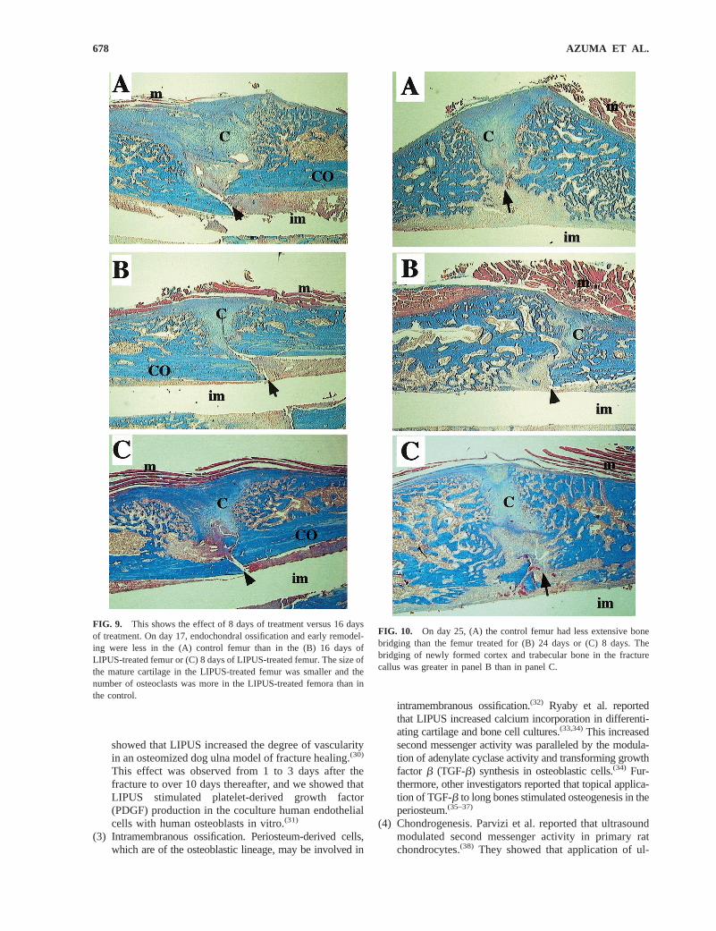
On day 17, endochondral ossification in the LIPUS-treated femur (Figs. 9B and 9C) tended to be higher thanthat in the control (Fig. 9A). The LIPUS-treated femur inthe Ph-2 group (Fig. 9B) on day 17 had received LIPUStreatment from day 9 to 16 (for 8 days), and that in the Tgroup (Fig. 9C) on day 17 from day 1 to 16 (for 16 days)after the fracture. Early remodeling of trabecular bone in thehard callus also was observed in all femurs. However, thesize of the mature cartilage in the LIPUS-treated femur wassmaller than that in the control femur. The number ofosteoclasts on the trabecular bone surface in the hard callusalso was higher in the LIPUS-treated femur than that in thecontrol.
Interestingly, more extensive bone bridging at the frac-ture site was observed in the LIPUS-treated femur (Figs.10B and 10C) than in the control femur (Fig. 10A) in notonly the T group but also in the Ph-3 group on day 25. Bonebridging by new bone formation had not occurred substan-tially in the LIPUS-untreated control femur (Fig. 10A). TheLIPUS-treated femur in the Ph-3 group (Fig. 10B) on day 25had received LIPUS treatment from day 17 to 24 (for 8days), and that in the T group (Fig. 10C) on day 25, fromday 1 to 24 (for 24 days) after the fracture. In the LIPUS-treated femur, the bridging of newly formed cortex andtrabecular bone in the fracture callus was greater in the Tgroup than in the Ph-3 group.
DISCUSSION
Fracture healing can be accelerated by exposure to LIPUSin both animal fracture models and clinical trials.(3–9) How-ever, the underlying mechanism of action remains unclear.In this study, we investigated the effects of the duration andtiming of LIPUS treatment on the benefits of LIPUS on the
FIG. 3. Radiography of LIPUS-treated femur and nontreated femurin each group at day 25 after the fracture. (A) Nontreated femur and(B–E) LIPUS-treated femur, which are (B) Ph-1, (C) Ph-2, (D) Ph-3,and (E) T, respectively. The femora were collected on day 25. Allfemora including intramedullary pins were radiographed with a softX-ray system before dissection of soft tissue.
675ULTRASOUND ACCELERATES RAT FEMORAL FRACTURE REPAIR
healing of rat femoral fractures to find a target reaction ofLIPUS in the fracture healing process.
Maximal torque of the LIPUS-treated side was signifi-cantly higher than that of the contralateral control side in allgroups. These results show that partial LIPUS treatment isable to accelerate fracture healing regardless of its timing.Furthermore, maximal torque of the LIPUS-treated femur inthe T group was significantly higher than that in the otherthree groups, probably because the accelerating effect ofLIPUS was additive. These data also suggest that LIPUSacts on some kind of cellular reaction involved in thefracture healing process.
The mechanical properties (i.e., torsional torque and stiff-ness) of the fracture callus in long bones reflect the extent offracture union as a result of healing in the fracture site.(22,23)
In this study, maximal torque of the LIPUS-treated femurwas significantly greater than that of the control side in allgroups, but there was no significant difference between theLIPUS-treated side and the control side for BMC at thefemoral fracture site or for the HCA in any of the groups.These findings indicate that the promoting effects of LIPUSon the mechanical properties of the fracture callus involvethe fracture union but not the size and/or bone mass of thefracture callus on day 25 after the fracture.
The results for the multiple viewing of 3DmCT recon-structions at the fracture site were coincident with the datafor mechanical testing. Although several investigators
showed that the density and connectivity of bone in thefracture callus was important to increase the rigidity of thecallus,(24–26)our observations in this study suggest that theefficiency of fracture union also contributes to the advancedmechanical properties of the fracture callus. Here, we de-scribe for the first time that the multiple viewing fromvarious angles (60° pitch) using 3DmCT reconstructions ofthe fracture callus is a powerful tool for estimation of thebone union and the fracture callus condition both visuallyand quantitatively.
In the histological analysis, endochondral ossification onday 9 and day 17, and the bone remodeling and bonebridging on day 17 and day 25 had advanced more inLIPUS-treated femurs than in the control femurs, respec-tively. These observations assured the results from the me-chanical testing and multiple viewing bymCT. The partialLIPUS treatments (8-day treatment) only in Ph-1, Ph-2, orPh-3 were effective in advancing endochondral ossificationand/or bone remodeling in the fracture callus. These resultssuggest that LIPUS influenced some cellular reactions ac-tive at each time period. However, it is unclear whichreaction is the target of LIPUS and how LIPUS affects it.
The micromechanical strains produced by LIPUS’s pres-sure waves in living tissue can result in biochemical eventsat the cellular level. In accordance with this, it seems to beeasy that a cell type that has a sensitivity to mechanicalstrains would respond to LIPUS. Although further investi-
FIG. 4. 3D reconstructions from 100mCT to-mograms (each 80mm in thickness) of fracturedfemora treated with LIPUS or nontreated on day25 after the fracture. Reconstructions of non-treated femur in the T group and LIPUS-treatedfemurs in Ph-1, Ph-2, Ph-3, and T groups areshown.
676 AZUMA ET AL.

gations are needed to define the target cells in each timeperiod, the results obtained in the present study allow us tomake the following speculation about the candidate:
(1) Inflammatory reaction. In the inflammatory phase, ahematoma is formed from blood vessels ruptured by theinjury; and soon thereafter, inflammatory cells such asmacrophages and/or lymphocytes invade the clot. It wasreported that macrophages contain a stretch-sensitive
potassium channel, the activity of which was modifiedby mechanical stress.(27,28)Furthermore, it was reportedthat high-energy ultrasound modulated the activity ofthymocytes.(29)
(2) Blood flow and neovascularization. After or overlap-ping the end stage of inflammatory reactions, the for-mation of granulation tissue and the invasion of multi-potential mesenchymal stem cells occur. In this stage,the concurrent proliferation of blood vessels within theperiosteal tissues and marrow space helps to route theappropriate cells to the fracture site. Rawool et al.
FIG. 5. (A) The BMC of femoral diaphysis at fracture site wasmeasured, and (B) anterior and posterior HCAs were traced and mea-sured with image analysis software.
FIG. 6. Effects of LIPUS treatment of various durations and timingson the HCA of the fracture callus on day 25 after the femoral fracture.The HCA of nontreated femur or LIPUS-treated femur is represented asopen bars or closed bars, respectively. There was no significant differ-ence in the HCA between the nontreated femur and LIPUS-treatedfemur in the T group (n 5 11), Ph-1 group (n 5 9), Ph-2 group (n 512), or Ph-3 group (n 5 12). The horizontal bars represent61 SD.Pairedt-testing was used for statistical analysis.
FIG. 7. Effects of LIPUS treatment of various durations and timingson BMC of the fracture callus near the fracture site on day 25 after thefracture. The HCA of the nontreated femur and LIPUS-treated femur isrepresented as open bars and closed bars, respectively. There was nosignificant difference in BMC between the nontreated femur and theLIPUS-treated femur in the T group (n 5 11), Ph-1 group (n 5 9), Ph-2group (n 5 12), or Ph-3 group (n 5 12). The horizontal bars repre-sent6 1SD. Pairedt-testing was used for statistical analysis.
FIG. 8. Effects of LIPUS treatment on the histology of fracturehealing at day 9. Photomicrographs show the appearance of the fracturehealing in (A) the control limb and (B) the treated limb. Early endo-chondral ossification on day 9 in the LIPUS-treated femur was greaterthan in the control. Sections were stained with Masson’s trichromestain. Arrowheads show the fracture sites. Specific regions are labeledas follows: cortical bone, co; muscle, m; intramedullary canal, im; andcartilage, c.
677ULTRASOUND ACCELERATES RAT FEMORAL FRACTURE REPAIR
showed that LIPUS increased the degree of vascularityin an osteomized dog ulna model of fracture healing.(30)
This effect was observed from 1 to 3 days after thefracture to over 10 days thereafter, and we showed thatLIPUS stimulated platelet-derived growth factor(PDGF) production in the coculture human endothelialcells with human osteoblasts in vitro.(31)
(3) Intramembranous ossification. Periosteum-derived cells,which are of the osteoblastic lineage, may be involved in
intramembranous ossification.(32) Ryaby et al. reportedthat LIPUS increased calcium incorporation in differenti-ating cartilage and bone cell cultures.(33,34)This increasedsecond messenger activity was paralleled by the modula-tion of adenylate cyclase activity and transforming growthfactor b (TGF-b) synthesis in osteoblastic cells.(34) Fur-thermore, other investigators reported that topical applica-tion of TGF-b to long bones stimulated osteogenesis in theperiosteum.(35–37)
(4) Chondrogenesis. Parvizi et al. reported that ultrasoundmodulated second messenger activity in primary ratchondrocytes.(38) They showed that application of ul-
FIG. 9. This shows the effect of 8 days of treatment versus 16 daysof treatment. On day 17, endochondral ossification and early remodel-ing were less in the (A) control femur than in the (B) 16 days ofLIPUS-treated femur or (C) 8 days of LIPUS-treated femur. The size ofthe mature cartilage in the LIPUS-treated femur was smaller and thenumber of osteoclasts was more in the LIPUS-treated femora than inthe control.
FIG. 10. On day 25, (A) the control femur had less extensive bonebridging than the femur treated for (B) 24 days or (C) 8 days. Thebridging of newly formed cortex and trabecular bone in the fracturecallus was greater in panel B than in panel C.
678 AZUMA ET AL.

trasound at 50 mW/cm2 and at higher intensities in-duced a real-time increase in the intracellular level ofcalcium. And other investigators showed that exposureof cultured chondrocytes to LIPUS stimulated an up-regulation of aggrecan gene expression.(39,40)
(5) Endochondral ossification. Invasion of newly formedblood vessels into the cartilage tissue, degradation ofcartilage tissue, and bone formation are involved inendochondral ossification. The growth, differentiationand activity of the cells such as chondrocytes, osteo-blasts, endothelial cells, and chondroclasts/osteoclastsrelated to those reactions may be important in thisprocess. In a previous study, we showed that the num-ber of multinuclear cells (MNCs) near the boundarybetween cartilage tissue and trabecular bone in theLIPUS-treated femur (156 3 MNCs/mm) was greaterin comparison with that in the control femur (86 2MNCs/mm) on day 21 after the fracture (in prepara-tion). However, it is unclear whether this effect wasbased on the direct action of LIPUS on osteoclastogen-esis. Soma et al. showed that the application of bilateralstretch stress to osteoblastic cells stimulated the produc-tion of some factors that could up-regulate osteoclasto-genesis.(41)
(6) Bone remodeling. Tanzer et al. reported that LIPUSstimulated bone ingrowth into porous coated dog fem-oral implants.(42) In contrast, Spadaro et al. reported thatphyseal bone growth was far less sensitive to LIPUSthan was fracture repair.(43) It remains unclear whetherand/or how LIPUS directly modulates bone metabolisminvolved in bone formation and bone resorption.
In conclusion, we investigated whether the acceleratingeffects of LIPUS on the healing of rat femoral fracturesdepended on the duration and timing of LIPUS treatment todetermine the target reaction of LIPUS in the fracture heal-ing process. Although we could not define the main target,we found out that LIPUS accelerated the rat femoral frac-ture healing regardless of the timing of LIPUS treatment.Thus, LIPUS appears to act on various cellular reactionsinvolved in the fracture healing process.
REFERENCES
1. Ziskin MC 1987 Application of ultrasound in medicine—comparison with other modalities. In: Rapacholi MH, Gran-dolfo M, Rindi A (eds.) Ultrasound: Medical Applications,Biological Effects, and Hazard Potential. Plenum Press, NewYork, NY, USA, pp. 49–59.
2. Wells PNT 1985 Surgical applications of ultrasound. In: Ny-borg WL, Ziskin MC (eds.) Biological Effects of Ultrasound.Churchill Livingstone, New York, NY, USA, pp. 157–167.
3. Xavier CAM, Duarte LR 1983 Estimulaci ultra-sonica de calloosseo: Applicaca clinica. Rev Bras Orthop18:73–80.
4. Duarte LR 1983 The stimulation of bone growth by ultra-sound. Arch Orthop Trauma Surg101:153–159.
5. Klug W, Franke WG, Knoch HG 1986 Scintigraphic control ofbone-fracture healing under ultrasonic stimulation: An animalexperimental study. Eur J Nucl Med11:494–497.
6. Pilla AA, Mont MA, Nasser PR, Khan SA, Figueiredo M,Kaufman JJ, Siffert RS 1990 Non-invasive low-intensitypulsed ultrasound accelerates bone healing in the rabbit. J Or-thop Trauma4:246–253.
7. Wang S-J, Lewallen DG, Bolander ME, Chao EYS, IlstrupDM, Greenleaf JF 1994 Low intensity ultrasound treatmentincreases strength in a rat femoral fracture model. J Orthop Res12:40–47.
8. Heckman JD, Ryaby JP, McCabe J, Frey JJ, Kilcoyne RF 1994Acceleration of tibial fracture-healing by non-invasive, low-intensity pulsed ultrasound. J Bone Joint Surg Am76:26–34.
9. Kristiansen TK, Ryaby JP, McCabe J, Frey JJ, Roe LR 1997Accelerated healing of distal radial fractures with the use ofspecific, low-intensity ultrasound. A multicenter, prospective,randomized, double-blind, placebo-controlled study. J BoneJoint Surg Am79:961–973.
10. Binderman I, Zor U, Kaye AM, Shimshoni Z, Harell A,Somjen D 1988 The transduction of mechanical force intobiochemical events in bone cells may involve activation ofphospholipase A2. Calcif Tissue Int42:261–266.
11. Buckley MJ, Banes AJ, Levin LG, Sumpio BE, Sato M, JordanR, Gilbert J, Link GW, Tran Son Tay R 1988 Osteoblastsincrease their rate of division and align in response to cyclic,mechanical tension in vitro. Bone Miner4:225–236.
12. Rubin J, Biskobing D, Fan X, Rubin C, McLeod K, Taylor WR1997 Pressure regulates osteoclast formation and MCSF ex-pression in marrow culture. J Cell Physiol170:81–87.
13. Bolander ME 1992 Regulation of fracture repair by growthfactors. Proc Soc Exp Biol Med200:165–170.
14. Schwachtgen JL, Houston P, Campbell C, Sukhatme V, Brad-dock M 1998 Fluid shear stress activation of egr-1 transcrip-tion in cultured human endothelial and epithelial cells is me-diated via the extracellular signal-related kinase 1/2 mitogen-activated protein kinase pathway. J Clin Invest101:2540–2549.
15. MacKenna DA, Dolfi F, Vuori K, Ruoslahti E 1998 Extracel-lular signal-regulated kinase and c-Jun NH2-terminal kinaseactivation by mechanical stretch is integrin-dependent andmatrix-specific in rat cardiac fibroblasts. J Clin Invest101:301–310.
16. Pavalko FM, Chen NX, Turner CH, Burr DB, Atkinson S,Hsieh YF, Qiu J, Duncan RL 1998 Fluid shear-induced me-chanical signaling in MC3T3–E1 osteoblasts requirescytoskeleton-integrin interactions. Am J Physiol275:C1591–C1601.
17. Quinn TM, Grodzinsky AJ, Buschmann MD, Kim YJ, Hun-ziker EB 1998 Mechanical compression alters proteoglycandeposition and matrix deformation around individual cells incartilage explants. J Cell Sci111:573–583.
18. Ajubi NE, Klein-Nulend J, Alblas MJ, Burger EH, NijweidePJ 1999 Signal transduction pathways involved in fluid flow-induced PGE2 production by cultured osteocytes. Am JPhysiol276:E171–E178.
19. Einhorn TA 1998 The cell and molecular biology of fracturehealing. Clin Orthop55(Suppl 3):S7–S21.
20. Jingushi S, Joyce ME, Bolander ME 1992 Genetic expressionof extracellular matrix proteins correlates with histologicchanges during fracture repair. J Bone Miner Res7:1045–1055.
21. Bonnarens F, Einhorn TA 1984 Production of a standardclosed fracture in laboratory animal bone. J Orthop Res2:97–101.
22. White AA III, Panjabi MM, Southwick WO 1977 The fourbiomechanical stages of fracture repair. J Bone Joint Surg Am59:188–192.
23. Black J, Perdigon P, Brown N, Pollack SR 1984 Stiffness andstrength of fracture callus. Relative rates of mechanical mat-uration as evaluated by a uniaxial tensile test. Clin Orthop182:278–288.
24. Markel MD, Wikenheiser MA, Chao EY 1990 A study offracture callus material properties: Relationship to the tor-sional strength of bone. J Orthop Res8:843–850.
679ULTRASOUND ACCELERATES RAT FEMORAL FRACTURE REPAIR

25. Chakkalakal DA, Lippiello L, Wilson RF, Shindell R, Con-nolly JF 1990 Mineral and matrix contributions to rigidity infracture healing. J Biomech23:425–434.
26. Augat P, Merk J, Genant HK, Claes L 1997 Quantitativeassessment of experimental fracture repair by peripheral com-puted tomography. Calcif Tissue Int60:194–199.
27. Martin DK, Bootcov MR, Campbell TJ, French PW, Breit SN1995 Human macrophages contain a stretch-sensitive potas-sium channel that is activated by adherence and cytokines. JMembr Biol 147:305–315.
28. Mattana J, Sankaran RT, Singhal PC 1995 Repetitive mechan-ical strain suppresses macrophage uptake of immunoglobulinG complexes and enhances cyclic adenosine monophosphatesynthesis. Am J Pathol147:529–540.
29. Dooley DA, Child SZ, Carstensen EL, Miller MW 1983 Theeffects of continuous wave and pulsed ultrasound on rat thy-mocytes in vitro. Ultrasound Med Biol9:379–384.
30. Rawool NM, Goldberg BB, Forsberg F, Winder AA, TallahRJ, Hume E 1997 Power doppler assessment of vascularchanges during fracture treatment with low intensity ultra-sound. Trans 83rd Radiol Soc North Am83:421.
31. Ito M, Azuma Y, Ohta T, Komoriya K 2000 Effects of ultra-sound and 1,25-dihydroxyvitamin D3 on growth factor secre-tion in co-cultures of osteoblasts and endothelial cells. Ultra-sound Med Biol26:161–166.
32. Utvag SE, Grundnes O, Reikeras O 1999 Early muscle-periosteal lesion inhibits fracture healing in rats. Acta OrthopScand70:62–66.
33. Ryaby JT, Bachner EJ, Bendo JA, Dalton PF, Tannenbaum S,Pilla AA 1989 Low intensity pulsed ultrasound increases cal-cium incorporation in both differentiating cartilage and bonecell cultures. Trans Orthop Res Soc14:15.
34. Ryaby JT, Mathew J, Duarte-Alves P 1992 Low intensitypulsed ultrasound affects adenylate cyclase and TGF-b syn-thesis in osteoblastic cells. Trans Orthop Res Soc17:590.
35. Joyce ME, Roberts AB, Sporn MB, Bolander ME 1990 Trans-forming growth factor-beta and the initiation of chondrogen-esis and osteogenesis in the rat femur. J Cell Biol110:2195–2207.
36. Joyce ME, Jingushi S, and Bolander ME 1990 Transforminggrowth factor-beta in the regulation of fracture repair. OrthopClin North Am 21:199–209.
37. Taniguchi Y, Tanaka T, Gotoh K, Satoh R, Inazu M 1993Transforming growth factor beta-1 induced cellular heteroge-neity in the periosteum of rat parietal bones. Calcif Tissue Int53:122–126.
38. Parvizi J, Parpura V, Kinnick RR, Greenleaf JF, Bolander ME1997 Low intensity ultrasound increases intracellular concen-tration of calcium in chondrocytes. Trans Orthop Res Soc22:465.
39. Yang K-H, Parvizi J, Wang S-J, Lewallen DG, Kinnick RR,Greenleaf JF, Bolander ME 1996 Exposure to low-intensityultrasound increases aggrecan gene expression in a rat femurfracture model. J Orthop Res14:802–809.
40. Wu C-C, Lewallen DG, Bolander ME, Bronk J, Kinnick R,Greenleaf JF 1996 Exposure to low intensity ultrasound stim-ulates aggrecan gene expression by cultured chondrocytes.Trans Orthop Res Soc21:622.
41. Soma S, Matsumoto S, Takano-Yamamoto T 1997 Enhance-ment by conditioned medium of stretched calvarial bone cellsof the osteoclast-like cell formation induced by parathyroidhormone in mouse bone marrow cultures. Arch Oral Biol42:205–211.
42. Tanzer M, Harvey E, Kay A, Morton P, Bobyn JD 1996 Effectof noninvasive low intensity ultrasound on bone growth intoporous-coated implants. J Orthop Res14:901–906.
43. Spadaro JA, Albanese SA 1998 Application of low-intensityultrasound to growing bone in rats. Ultrasound Med Biol24:567–573.
Address reprint requests to:Yoshiaki Azuma, Ph.D.
Pharmacological Research DepartmentTeijin Institute for Biomedical Research, Teijin Limited
4–3-2 Asahigaoka, Hino CityTokyo 191-8512, Japan
Received in original form July 30, 1999; in revised form October31, 2000; accepted October 31, 2000.
680 AZUMA ET AL.