Loss and gain of Drosophila TDP-43 impair synaptic efficacy and
19
Loss and gain of Drosophila TDP-43 impair synaptic efficacy and motor control leading to age-related neurodegeneration by loss-of-function phenotypes Danielle C. Diaper 1, { , Yoshitsugu Adachi 1, { , Ben Sutcliffe 3 , Dickon M. Humphrey 1 , Christopher J.H. Elliott 4 , Alan Stepto 1 , Zoe N. Ludlow 1 , Lies Vanden Broeck 5 , Patrick Callaerts 5 , Bart Dermaut 5,6 , Ammar Al-Chalabi 2 , Christopher E. Shaw 2 , Iain M. Robinson 3 and Frank Hirth 1,∗ 1 Department of Neuroscience and 2 Department of Clinical Neuroscience, Institute of Psychiatry, MRC Centre for Neurodegeneration Research, King’s College London, London SE5 8AF, UK, 3 Peninsula College of Medicine and Dentistry, Universities of Exeter and Plymouth, Plymouth PL6 8BU, UK, 4 Department of Biology, University of York, York YO10 5DD, UK, 5 Laboratory of Behavioural & Developmental Genetics, Centre for Human Genetics, University of Leuven & VIB Centre for the Biology of Disease, Leuven 3000, Belgium and 6 INSERM U744, Pasteur Institute of Lille, University of Lille North of France, Lille, France Received January 2, 2013; Revised and Accepted January 3, 2013 Cytoplasmic accumulation and nuclear clearance of TDP-43 characterize familial and sporadic forms of amyotrophic lateral sclerosis and frontotemporal lobar degeneration, suggesting that either loss or gain of TDP-43 function, or both, cause disease formation. Here we have systematically compared loss- and gain- of-function of Drosophila TDP-43, TAR DNA Binding Protein Homolog (TBPH), in synaptic function and morphology, motor control, and age-related neuronal survival. Both loss and gain of TBPH severely affect development and result in premature lethality. TBPH dysfunction caused impaired synaptic transmission at the larval neuromuscular junction (NMJ) and in the adult. Tissue-specific knockdown together with electro- physiological recordings at the larval NMJ also revealed that alterations of TBPH function predominantly affect pre-synaptic efficacy, suggesting that impaired pre-synaptic transmission is one of the earliest events in TDP-43-related pathogenesis. Prolonged loss and gain of TBPH in adults resulted in synaptic defects and age-related, progressive degeneration of neurons involved in motor control. Toxic gain of TBPH did not downregulate or mislocalize its own expression, indicating that a dominant-negative effect leads to progressive neurodegeneration also seen with mutational inactivation of TBPH. Together these data suggest that dysfunction of Drosophila TDP-43 triggers a cascade of events leading to loss-of-function phenotypes whereby impaired synaptic transmission results in defective motor behavior and progressive deconstruction of neuronal connections, ultimately causing age-related neurodegeneration. INTRODUCTION Tar DNA binding protein of 43 kDa (TDP-43) has been iden- tified as the major disease protein present in cytoplasmic inclusions in amyotrophic lateral sclerosis (ALS) and fronto- temporal lobar degeneration (FTLD). Full length TDP-43 protein as well as 25 kDa, C-terminal fragments were found to be hyper-phosphorylated and ubiquitinated in aggregates present in affected brain and spinal cord of ALS and FTLD cases (1,2). TDP-43 inclusions are found as secondary histo- pathological feature in other neurodegenerative disorders in- cluding Alzheimer’s, Parkinson’s and Huntington’s disease, thereby defining a novel proteinopathy (3,4). Dominant † The authors wish it to be known that, in their opinion, the first two authors should be regarded as joint First Authors. ∗ To whom correspondence should be addressed at: Department of Neuroscience, Institute of Psychiatry, King’s College London, PO Box 37, 16 De Crespigny Park, SE5 8AF London, UK. Tel: +44 2078480786; Fax: +44 2077080017; Email: [email protected] # The Author 2013. Published by Oxford University Press. This is an Open Access article distributed under the terms of the Creative Commons Attribution License (http://creativecommons.org/licenses/by-nc/ 3.0/), which permits non-commercial use, distribution, and reproduction in any medium, provided the original work is properly cited. For commercial re-use, please contact [email protected] Human Molecular Genetics, 2013, Vol. 22, No. 8 1539–1557 doi:10.1093/hmg/ddt005 Advance Access published on January 10, 2013 Downloaded from https://academic.oup.com/hmg/article/22/8/1539/625893 by guest on 02 December 2021
Transcript of Loss and gain of Drosophila TDP-43 impair synaptic efficacy and

Loss and gain of Drosophila TDP-43 impair synapticefficacy and motor control leading to age-relatedneurodegeneration by loss-of-function phenotypes
Danielle C. Diaper1,{, Yoshitsugu Adachi1,{, Ben Sutcliffe3, Dickon M. Humphrey1,
Christopher J.H. Elliott4, Alan Stepto1, Zoe N. Ludlow1, Lies Vanden Broeck5, Patrick Callaerts5,
Bart Dermaut5,6, Ammar Al-Chalabi2, Christopher E. Shaw2, Iain M. Robinson3 and Frank Hirth1,∗
1Department of Neuroscience and 2Department of Clinical Neuroscience, Institute of Psychiatry, MRC Centre for
Neurodegeneration Research, King’s College London, London SE5 8AF, UK, 3Peninsula College of Medicine and
Dentistry, Universities of Exeter and Plymouth, Plymouth PL6 8BU, UK, 4Department of Biology, University of York,
York YO10 5DD, UK, 5Laboratory of Behavioural & Developmental Genetics, Centre for Human Genetics, University
of Leuven & VIB Centre for the Biology of Disease, Leuven 3000, Belgium and 6INSERM U744, Pasteur Institute of
Lille, University of Lille North of France, Lille, France
Received January 2, 2013; Revised and Accepted January 3, 2013
Cytoplasmic accumulation and nuclear clearance of TDP-43 characterize familial and sporadic forms ofamyotrophic lateral sclerosis and frontotemporal lobar degeneration, suggesting that either loss or gain ofTDP-43 function, or both, cause disease formation. Here we have systematically compared loss- and gain-of-function of Drosophila TDP-43, TAR DNA Binding Protein Homolog (TBPH), in synaptic function andmorphology, motor control, and age-related neuronal survival. Both loss and gain of TBPH severely affectdevelopment and result in premature lethality. TBPH dysfunction caused impaired synaptic transmissionat the larval neuromuscular junction (NMJ) and in the adult. Tissue-specific knockdown together with electro-physiological recordings at the larval NMJ also revealed that alterations of TBPH function predominantlyaffect pre-synaptic efficacy, suggesting that impaired pre-synaptic transmission is one of the earliestevents in TDP-43-related pathogenesis. Prolonged loss and gain of TBPH in adults resulted in synapticdefects and age-related, progressive degeneration of neurons involved in motor control. Toxic gain ofTBPH did not downregulate or mislocalize its own expression, indicating that a dominant-negative effectleads to progressive neurodegeneration also seen with mutational inactivation of TBPH. Together thesedata suggest that dysfunction of Drosophila TDP-43 triggers a cascade of events leading to loss-of-functionphenotypes whereby impaired synaptic transmission results in defective motor behavior and progressivedeconstruction of neuronal connections, ultimately causing age-related neurodegeneration.
INTRODUCTION
Tar DNA binding protein of 43 kDa (TDP-43) has been iden-tified as the major disease protein present in cytoplasmicinclusions in amyotrophic lateral sclerosis (ALS) and fronto-temporal lobar degeneration (FTLD). Full length TDP-43protein as well as 25 kDa, C-terminal fragments were found
to be hyper-phosphorylated and ubiquitinated in aggregatespresent in affected brain and spinal cord of ALS and FTLDcases (1,2). TDP-43 inclusions are found as secondary histo-pathological feature in other neurodegenerative disorders in-cluding Alzheimer’s, Parkinson’s and Huntington’s disease,thereby defining a novel proteinopathy (3,4). Dominant
†The authors wish it to be known that, in their opinion, the first two authors should be regarded as joint First Authors.
∗To whom correspondence should be addressed at: Department of Neuroscience, Institute of Psychiatry, King’s College London, PO Box 37, 16 DeCrespigny Park, SE5 8AF London, UK. Tel: +44 2078480786; Fax: +44 2077080017; Email: [email protected]
# The Author 2013. Published by Oxford University Press.This is an Open Access article distributed under the terms of the Creative Commons Attribution License (http://creativecommons.org/licenses/by-nc/3.0/), which permits non-commercial use, distribution, and reproduction in any medium, provided the original work is properly cited. For commercialre-use, please contact [email protected]
Human Molecular Genetics, 2013, Vol. 22, No. 8 1539–1557doi:10.1093/hmg/ddt005Advance Access published on January 10, 2013
Dow
nloaded from https://academ
ic.oup.com/hm
g/article/22/8/1539/625893 by guest on 02 Decem
ber 2021

missense mutations in the TARDBP gene have been identifiedin familial and sporadic cases of ALS and FTLD (5–8), indi-cating that TDP-43 dysfunction may cause disease formation.
TDP-43 encodes an evolutionarily conserved protein com-prising a bipartite nuclear localization signal, two RNA recog-nition motifs (RRMs), a nuclear export signal, a glycine andglycine/glutamine/asparagine-rich C-terminal region. TheC-terminus resembles a prion-like domain (9,10) predominantlymutated in TDP-43-related ALS and FTLD cases (11,12). Invitro studies showed that TDP-43 is involved in transcriptionalregulation, RNA biogenesis and splicing (13). The C-terminaldomain of TDP-43 regulates tissue-specific gene expression,transcriptional repression, and alternative splicing, and is essen-tial for binding to heterogeneous nuclear ribonucleoproteinsinvolved in microRNA and mRNA biogenesis, as well asRNA turnover (12,13).
The pathophysiological role of TDP-43 has been addressedin cell culture and animal models using mutation, alterationor expression of disease-specific variants of TDP-43 andits homologs in worms, flies, zebrafish, mice and rats(11,12,14). These studies revealed that post-translationalmodification (phosphorylation, ubiquitylation), truncation,mislocalization, nuclear clearance, cytoplasmic accumulationand defective autoregulation can all impact either directly orindirectly onto TDP-43 toxicity (12). The resulting phenotypesinclude lethality or reduced lifespan, defects in axogenesis andneurite branching, altered synaptic morphology, axon andneuron degeneration, astrogliosis, defective fat metabolism,impaired motor behavior and cognitive deficits (11,14). Theavailable data suggest that TDP-43 toxicity is either directlyor indirectly related to disease formation. So far, however,no consensus has emerged as to whether loss (i.e. nuclearclearance) or toxic gain (i.e. cytoplasmic accumulation) ofTDP-43, or both, are causally related to disease onset andprogression. Moreover, despite the fact that a large numberof TDP-43 RNA targets have been identified (15–18), theinitiating events and pathogenic pathways underlying TDP-43-mediated neurodegeneration remain to be identified.
Here we show that similar to its human homolog, DrosophilaTDP-43, TAR DNA binding protein homolog (TBPH), isexpressed in neurons, glia and muscle cells. Both loss of func-tion and overexpression of TBPH result in decreased viability,impaired synaptic transmission, defective locomotion andage-related progressive neurodegeneration. Together these datasuggest that both increased and decreased levels of TDP-43 ini-tiate a loss-of-function phenotype, resulting in decreased neur-onal viability and subsequent degenerative cell loss thatcharacterize motor neuron diseases like ALS and FTLD.
RESULTS
Drosophila TDP-43, TBPH, is expressed in neurons,glia and muscle cells
BLAST search of the annotated Drosophila melanogastergenome identified two genes with sequence similarities toTARDBP/TDP-43, namely TBPH (19) and CG7804.Genomic sequence analysis revealed that the predicted six iso-forms of TBPH are encoded by 5–6 exons, whereas the codingregion of CG7804 is intronless with sequence similarity to
exon 1–4 of TBPH. The differences in exon–intron structureand coding sequence similarities between CG7804 and TBPHsuggest that CG7804 is paralogous to TBPH and likely to be anewly evolved retrogene that derived from TBPH rather thanTARDBP (20). Moreover, in contrast to TBPH, CG7804 isnot expressed in CNS and lacks a prion-like C-terminus thatis predominantly mutated in ALS and FTLD (SupplementaryMaterial, Fig. S1). We therefore focused on TBPH.
We generated polyclonal antibodies against TBPH-specificpeptide sequences. Western blot analysis of control tissueidentified a 58 kDa protein encoded by TBPH which wasabsent in deletions uncovering the genomic locus (Supplemen-tary Material, Fig. S2); we did not detect any cleavage pro-ducts, TBPH fragments, dimers nor band that indicatespost-translational modifications (Supplementary Material,Fig. S2). Whole-mount immunocytochemistry revealed that,similar to human TDP-43, TBPH is expressed throughout de-velopment and adulthood in the nucleus of neurons, glia andmuscle cells (Fig. 1). Perinuclear, cytoplasmic TBPH expressionwas detectable in post-mitotic neurons (Fig. 1L–O), althoughimmunoreactivity was less intense than nuclear anti-TBPH la-beling. These data suggest that similar to TDP-43, and predictedby nuclear localization and export signals (21,22), TBPH ispredominantly localized to the nucleus but also shuttles intothe cytoplasm.
Both loss and gain of TBPH affect survivaland motor behavior
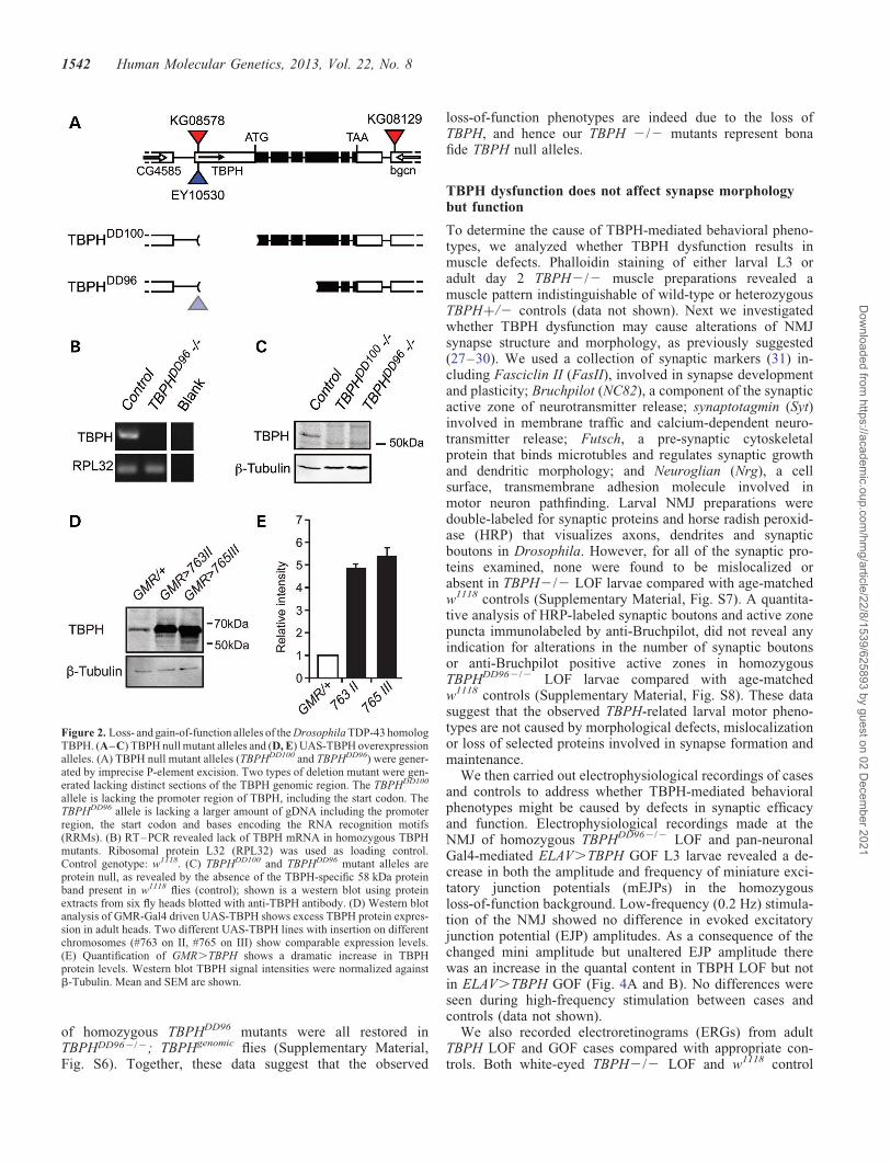
To study TDP-43 dysfunction in Drosophila, we generatedspecific loss- and gain-of-function alleles of TBPH. ImpreciseP-element excision resulted in two independent deletions thatremoved the promoter region and start codon, or the promoterregion, start codon, exons 1–3 and parts of exon 4 coding forthe TBPH RRMs (Fig. 2A). RT–PCR and western blot ana-lyses determined the absence of TBPH RNA and protein(Fig. 2B and C). For tissue-specific TBPH LOF, we generatedtwo independent hairpin-loop UAS-RNAi lines that targetexon 5 of all predicted TBPH isoforms (Supplementary Mater-ial, Fig. S3). BLAST search confirmed that our chosen targetsequences do not detect off-targets, in contrast to other avail-able TBPH RNAi lines, and western blot analysis revealed thatTub-Gal4 driven UAS-TBPH-RNAi is able to knock down en-dogenous TBPH protein expression below detection level(Supplementary Material, Fig. S3).
For gain of TBPH function (GOF), several independentUAS lines were generated with single inserts on differentchromosomal locations. Expression efficacy was tested usingeye-specific GMR-Gal4 activation. Western blot analysisrevealed that GMR.TBPH showed increased amounts ofTBPH protein (Fig. 2D) comparable among different insertionlines (Fig. 2E). Targeted misexpression resulted in excessiveaccumulation of TBPH both in the nucleus and cytoplasm(Supplementary Material, Fig. S4).
Analysis of zygotic TBPH LOF (TBPH2/2) and Gal4-mediated GOF mutants revealed that both affect developmentand lifespan: only 10–20% of progeny eclosed as adult whilethe majority of mutant cases died during late larval/pupalstages (Fig. 3A). Adult TBPH2/2 mutant and pan-neuronalGal4-specific ELAV.TBPH GOF flies were characterized by
1540 Human Molecular Genetics, 2013, Vol. 22, No. 8
Dow
nloaded from https://academ
ic.oup.com/hm
g/article/22/8/1539/625893 by guest on 02 Decem
ber 2021

shortened lifespan, with TBPH2/2 adults dying around day 7and pan-neuronal Gal4-specific ELAV.TBPH flies dyingaround day 35, when compared with wild-type controls withan average lifespan of 80 days (23,24) (data not shown).Affected larvae exhibited impaired peristalsis and defectivelocomotion, which was more severe in TBPH2/2 LOFmutants compared with pan-neuronal Gal4-specific ELAV.TBPH GOF (Fig. 3B). Adult TBPH2/2 LOF mutant escapersand pan-neuronal Gal4-specific ELAV.TBPH GOF flies werecharacterized by inexistent or severely impaired innate escapeand climbing behaviors (Fig. 3C).
To further investigate motor behavior, we used an open-field paradigm together with video-assisted motion tracking(23,24) (Fig. 3D) and compared adult TBPH2/2 mutantLOF escapers with EB1-Gal4-mediated UAS-TBPH GOFflies targeting TBPH to ellipsoid body neurons of the centralcomplex that are upper motor neurons due to their prominentrole in the higher control of locomotion (25). Analysis of adultTBPH2/2 mutant LOF escapers and EB1.TBPH GOF fliesrevealed severely reduced walking activity and distance trav-eled; motor activity remained low over time with onlyTBPH2/2 flies walking slower (Fig. 3D–H). In addition,
both LOF and GOF flies were characterized by severe gait ab-normalities (Fig. 3I). These data suggest that TBPH dysfunc-tion affects lifespan and motor behavior.
To confirm that the phenotypes observed in homozygousTBPH2/2 LOF flies were indeed due to the absence ofTBPH, we carried out a complementation test using a defi-ciency line uncovering the genomic region of chromosome2R (Df(2R)106) including the TBPH locus, which revealedearly larval lethality suggesting non-complementation. In add-ition and to rule out hidden second-site mutations, we first ana-lyzed heteroallelic combinations using our TBPHDD96 andTBPHDD100 alleles together with a recently reported deletion(26), which also revealed non-complementation. Secondly,we generated a genomic TBPH construct covering the entirecoding region of TBPH as well as 3′- and 5′- regions upstreamof the TBPH locus (Supplementary Material, Fig. S5). Toanalyze its rescue potential, we integrated TBPHgenomic on3R using the attP86Fb landing site and crossed it into theTBPHDD96 mutant background. Analysis of homozygousTBPHDD962/2 flies carrying one or two copies of TBPHgenomic
revealed full rescue. Thus, development, larval locomotion,eclosion, adult climbing, walking and tripod gait phenotypes
Figure 1. TBPH is expressed in neurons, glia and muscle cells. (A–E) Endogenous TBPH expression was examined at the neuromuscular junction of musclegroup 6/7 of abdominal segment II in w1118 wandering third instar larvae. TBPH is expressed in the nucleus of muscle cells (C–E). Images are z-projections of a10 mm area scanned in 2 mm steps. (F–J) Endogenous TBPH expression was examined in the adult brain of flies expressing membrane-bound GFP in ellipsoidbody neurons of the central brain that are considered to be upper motor neurons [EB1.mCD8::GFP; CNS hemisphere shown in (J), the dotted white box corre-sponds to (F–I)]. TBPH is expressed in neurons (F and G, arrow) and glia (H, arrowhead) throughout the adult brain, including ellipsoid body neurons. Imagesare single z-slices taken in 1 mm steps. (K–O) TBPH expression is also seen in perinuclear regions of ellipsoid body neurons as shown by its co-localization withmembrane bound mCD8::GFP (white arrowheads). Images are single z-sections taken in 1.5 mm steps. Scale bars; 50 mm (A–E); 10 mm (F–I, K–O).
Human Molecular Genetics, 2013, Vol. 22, No. 8 1541
Dow
nloaded from https://academ
ic.oup.com/hm
g/article/22/8/1539/625893 by guest on 02 Decem
ber 2021
of homozygous TBPHDD96 mutants were all restored inTBPHDD962/2; TBPHgenomic flies (Supplementary Material,Fig. S6). Together, these data suggest that the observed
loss-of-function phenotypes are indeed due to the loss ofTBPH, and hence our TBPH 2/2 mutants represent bonafide TBPH null alleles.
TBPH dysfunction does not affect synapse morphologybut function
To determine the cause of TBPH-mediated behavioral pheno-types, we analyzed whether TBPH dysfunction results inmuscle defects. Phalloidin staining of either larval L3 oradult day 2 TBPH2/2 muscle preparations revealed amuscle pattern indistinguishable of wild-type or heterozygousTBPH+/2 controls (data not shown). Next we investigatedwhether TBPH dysfunction may cause alterations of NMJsynapse structure and morphology, as previously suggested(27–30). We used a collection of synaptic markers (31) in-cluding Fasciclin II (FasII), involved in synapse developmentand plasticity; Bruchpilot (NC82), a component of the synapticactive zone of neurotransmitter release; synaptotagmin (Syt)involved in membrane traffic and calcium-dependent neuro-transmitter release; Futsch, a pre-synaptic cytoskeletalprotein that binds microtubles and regulates synaptic growthand dendritic morphology; and Neuroglian (Nrg), a cellsurface, transmembrane adhesion molecule involved inmotor neuron pathfinding. Larval NMJ preparations weredouble-labeled for synaptic proteins and horse radish peroxid-ase (HRP) that visualizes axons, dendrites and synapticboutons in Drosophila. However, for all of the synaptic pro-teins examined, none were found to be mislocalized orabsent in TBPH2/2 LOF larvae compared with age-matchedw1118 controls (Supplementary Material, Fig. S7). A quantita-tive analysis of HRP-labeled synaptic boutons and active zonepuncta immunolabeled by anti-Bruchpilot, did not reveal anyindication for alterations in the number of synaptic boutonsor anti-Bruchpilot positive active zones in homozygousTBPHDD962/2 LOF larvae compared with age-matchedw1118 controls (Supplementary Material, Fig. S8). These datasuggest that the observed TBPH-related larval motor pheno-types are not caused by morphological defects, mislocalizationor loss of selected proteins involved in synapse formation andmaintenance.
We then carried out electrophysiological recordings of casesand controls to address whether TBPH-mediated behavioralphenotypes might be caused by defects in synaptic efficacyand function. Electrophysiological recordings made at theNMJ of homozygous TBPHDD962/2 LOF and pan-neuronalGal4-mediated ELAV.TBPH GOF L3 larvae revealed a de-crease in both the amplitude and frequency of miniature exci-tatory junction potentials (mEJPs) in the homozygousloss-of-function background. Low-frequency (0.2 Hz) stimula-tion of the NMJ showed no difference in evoked excitatoryjunction potential (EJP) amplitudes. As a consequence of thechanged mini amplitude but unaltered EJP amplitude therewas an increase in the quantal content in TBPH LOF but notin ELAV.TBPH GOF (Fig. 4A and B). No differences wereseen during high-frequency stimulation between cases andcontrols (data not shown).
We also recorded electroretinograms (ERGs) from adultTBPH LOF and GOF cases compared with appropriate con-trols. Both white-eyed TBPH2/2 LOF and w1118 control
Figure 2. Loss- and gain-of-function alleles of the Drosophila TDP-43 homologTBPH. (A–C) TBPH null mutant alleles and (D, E) UAS-TBPH overexpressionalleles. (A) TBPH null mutant alleles (TBPHDD100 and TBPHDD96) were gener-ated by imprecise P-element excision. Two types of deletion mutant were gen-erated lacking distinct sections of the TBPH genomic region. The TBPHDD100
allele is lacking the promoter region of TBPH, including the start codon. TheTBPHDD96 allele is lacking a larger amount of gDNA including the promoterregion, the start codon and bases encoding the RNA recognition motifs(RRMs). (B) RT–PCR revealed lack of TBPH mRNA in homozygous TBPHmutants. Ribosomal protein L32 (RPL32) was used as loading control.Control genotype: w1118. (C) TBPHDD100 and TBPHDD96 mutant alleles areprotein null, as revealed by the absence of the TBPH-specific 58 kDa proteinband present in w1118 flies (control); shown is a western blot using proteinextracts from six fly heads blotted with anti-TBPH antibody. (D) Western blotanalysis of GMR-Gal4 driven UAS-TBPH shows excess TBPH protein expres-sion in adult heads. Two different UAS-TBPH lines with insertion on differentchromosomes (#763 on II, #765 on III) show comparable expression levels.(E) Quantification of GMR.TBPH shows a dramatic increase in TBPHprotein levels. Western blot TBPH signal intensities were normalized againstb-Tubulin. Mean and SEM are shown.
1542 Human Molecular Genetics, 2013, Vol. 22, No. 8
Dow
nloaded from https://academ
ic.oup.com/hm
g/article/22/8/1539/625893 by guest on 02 Decem
ber 2021
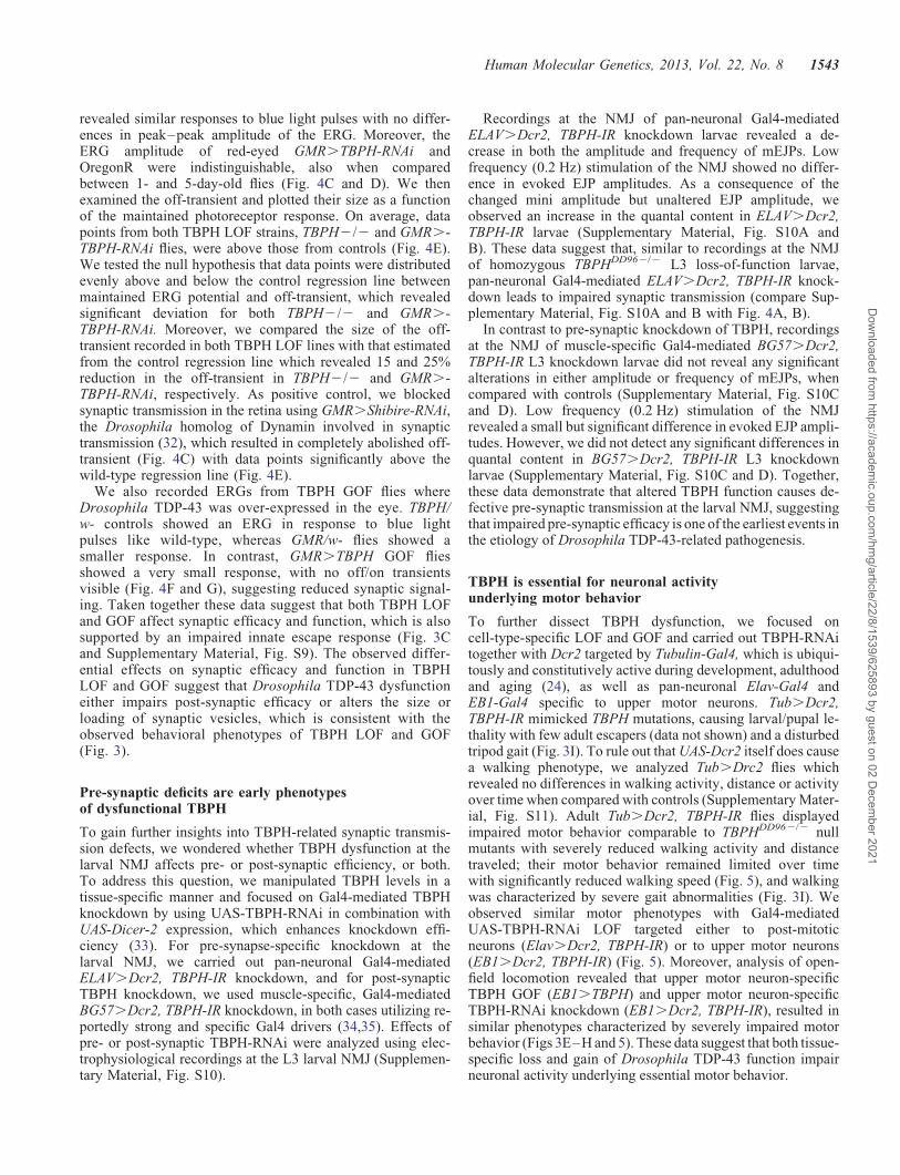
revealed similar responses to blue light pulses with no differ-ences in peak–peak amplitude of the ERG. Moreover, theERG amplitude of red-eyed GMR.TBPH-RNAi andOregonR were indistinguishable, also when comparedbetween 1- and 5-day-old flies (Fig. 4C and D). We thenexamined the off-transient and plotted their size as a functionof the maintained photoreceptor response. On average, datapoints from both TBPH LOF strains, TBPH2/2 and GMR.-TBPH-RNAi flies, were above those from controls (Fig. 4E).We tested the null hypothesis that data points were distributedevenly above and below the control regression line betweenmaintained ERG potential and off-transient, which revealedsignificant deviation for both TBPH2/2 and GMR.-TBPH-RNAi. Moreover, we compared the size of the off-transient recorded in both TBPH LOF lines with that estimatedfrom the control regression line which revealed 15 and 25%reduction in the off-transient in TBPH2/2 and GMR.-TBPH-RNAi, respectively. As positive control, we blockedsynaptic transmission in the retina using GMR.Shibire-RNAi,the Drosophila homolog of Dynamin involved in synaptictransmission (32), which resulted in completely abolished off-transient (Fig. 4C) with data points significantly above thewild-type regression line (Fig. 4E).
We also recorded ERGs from TBPH GOF flies whereDrosophila TDP-43 was over-expressed in the eye. TBPH/w- controls showed an ERG in response to blue lightpulses like wild-type, whereas GMR/w- flies showed asmaller response. In contrast, GMR.TBPH GOF fliesshowed a very small response, with no off/on transientsvisible (Fig. 4F and G), suggesting reduced synaptic signal-ing. Taken together these data suggest that both TBPH LOFand GOF affect synaptic efficacy and function, which is alsosupported by an impaired innate escape response (Fig. 3Cand Supplementary Material, Fig. S9). The observed differ-ential effects on synaptic efficacy and function in TBPHLOF and GOF suggest that Drosophila TDP-43 dysfunctioneither impairs post-synaptic efficacy or alters the size orloading of synaptic vesicles, which is consistent with theobserved behavioral phenotypes of TBPH LOF and GOF(Fig. 3).
Pre-synaptic deficits are early phenotypesof dysfunctional TBPH
To gain further insights into TBPH-related synaptic transmis-sion defects, we wondered whether TBPH dysfunction at thelarval NMJ affects pre- or post-synaptic efficiency, or both.To address this question, we manipulated TBPH levels in atissue-specific manner and focused on Gal4-mediated TBPHknockdown by using UAS-TBPH-RNAi in combination withUAS-Dicer-2 expression, which enhances knockdown effi-ciency (33). For pre-synapse-specific knockdown at thelarval NMJ, we carried out pan-neuronal Gal4-mediatedELAV.Dcr2, TBPH-IR knockdown, and for post-synapticTBPH knockdown, we used muscle-specific, Gal4-mediatedBG57.Dcr2, TBPH-IR knockdown, in both cases utilizing re-portedly strong and specific Gal4 drivers (34,35). Effects ofpre- or post-synaptic TBPH-RNAi were analyzed using elec-trophysiological recordings at the L3 larval NMJ (Supplemen-tary Material, Fig. S10).
Recordings at the NMJ of pan-neuronal Gal4-mediatedELAV.Dcr2, TBPH-IR knockdown larvae revealed a de-crease in both the amplitude and frequency of mEJPs. Lowfrequency (0.2 Hz) stimulation of the NMJ showed no differ-ence in evoked EJP amplitudes. As a consequence of thechanged mini amplitude but unaltered EJP amplitude, weobserved an increase in the quantal content in ELAV.Dcr2,TBPH-IR larvae (Supplementary Material, Fig. S10A andB). These data suggest that, similar to recordings at the NMJof homozygous TBPHDD962/2 L3 loss-of-function larvae,pan-neuronal Gal4-mediated ELAV.Dcr2, TBPH-IR knock-down leads to impaired synaptic transmission (compare Sup-plementary Material, Fig. S10A and B with Fig. 4A, B).
In contrast to pre-synaptic knockdown of TBPH, recordingsat the NMJ of muscle-specific Gal4-mediated BG57.Dcr2,TBPH-IR L3 knockdown larvae did not reveal any significantalterations in either amplitude or frequency of mEJPs, whencompared with controls (Supplementary Material, Fig. S10Cand D). Low frequency (0.2 Hz) stimulation of the NMJrevealed a small but significant difference in evoked EJP ampli-tudes. However, we did not detect any significant differences inquantal content in BG57.Dcr2, TBPH-IR L3 knockdownlarvae (Supplementary Material, Fig. S10C and D). Together,these data demonstrate that altered TBPH function causes de-fective pre-synaptic transmission at the larval NMJ, suggestingthat impaired pre-synaptic efficacy is one of the earliest events inthe etiology of Drosophila TDP-43-related pathogenesis.
TBPH is essential for neuronal activityunderlying motor behavior
To further dissect TBPH dysfunction, we focused oncell-type-specific LOF and GOF and carried out TBPH-RNAitogether with Dcr2 targeted by Tubulin-Gal4, which is ubiqui-tously and constitutively active during development, adulthoodand aging (24), as well as pan-neuronal Elav-Gal4 andEB1-Gal4 specific to upper motor neurons. Tub.Dcr2,TBPH-IR mimicked TBPH mutations, causing larval/pupal le-thality with few adult escapers (data not shown) and a disturbedtripod gait (Fig. 3I). To rule out that UAS-Dcr2 itself does causea walking phenotype, we analyzed Tub.Drc2 flies whichrevealed no differences in walking activity, distance or activityover time when compared with controls (Supplementary Mater-ial, Fig. S11). Adult Tub.Dcr2, TBPH-IR flies displayedimpaired motor behavior comparable to TBPHDD962/2 nullmutants with severely reduced walking activity and distancetraveled; their motor behavior remained limited over timewith significantly reduced walking speed (Fig. 5), and walkingwas characterized by severe gait abnormalities (Fig. 3I). Weobserved similar motor phenotypes with Gal4-mediatedUAS-TBPH-RNAi LOF targeted either to post-mitoticneurons (Elav.Dcr2, TBPH-IR) or to upper motor neurons(EB1.Dcr2, TBPH-IR) (Fig. 5). Moreover, analysis of open-field locomotion revealed that upper motor neuron-specificTBPH GOF (EB1.TBPH) and upper motor neuron-specificTBPH-RNAi knockdown (EB1.Dcr2, TBPH-IR), resulted insimilar phenotypes characterized by severely impaired motorbehavior (Figs 3E–H and 5). These data suggest that both tissue-specific loss and gain of Drosophila TDP-43 function impairneuronal activity underlying essential motor behavior.
Human Molecular Genetics, 2013, Vol. 22, No. 8 1543
Dow
nloaded from https://academ
ic.oup.com/hm
g/article/22/8/1539/625893 by guest on 02 Decem
ber 2021

Figure 3. Both loss and gain of TBPH affect survival and motor behavior. (A) Survival analysis quantified the number of larvae that survived to adulthood. Thenumber of fully eclosed adults was significantly reduced in loss-of-function (LOF) TBPH null mutants and gain-of-function (GOF) ELAV.TBPH flies, com-pared with respective control (w1118/+, control A; Elav/+, control B). (B) Both TBPH LOF and GOF show impaired larval locomotion with a reduction in the
1544 Human Molecular Genetics, 2013, Vol. 22, No. 8
Dow
nloaded from https://academ
ic.oup.com/hm
g/article/22/8/1539/625893 by guest on 02 Decem
ber 2021

TBPH dysfunction differentially affects synaptic integrityin an age-related manner
Next we investigated whether impaired synaptic activitycaused by TBPH dysfunction may lead to synaptic alterationsin aging flies. We therefore analyzed brains of aged flies withor without either UAS-TBPH-IR LOF or UAS-TBPH GOF tar-geted to EB1-Gal4-specific upper motor neurons. TheEB1-Gal4 driver becomes active during late pupal stagesand remains active during adulthood and aging in a populationof roughly 80 upper motor neurons (ellipsoid body ringneurons). In contrast to severe motor phenotypes (seeFigs 3E–H and 5G–J), dysfunction or loss of this small popu-lation of upper motor neurons does not significantly alter thelifespan of flies and hence is an ideal assay system to studyDrosophila TDP-43 dysfunction at the cellular and synapticlevel (see Figs 6 and 7). We used EB1-Gal4 drivenUAS-TBPH-IR LOF or UAS-TBPH GOF and co-expressed amembrane-bound form of GFP (UAS-mCD8::GFP) that alsovisualizes axons and synaptic terminals, and co-labeledwhole mount brains with anti-Fasciclin 2 (Fas2) that is alsoexpressed at pre- and post-synaptic sites of the ellipsoidbody ring neuropil (Supplementary Material, Fig. S12).
Analysis of brains of Gal4-mediated upper motor neuron-specific EB1.Dcr2, TBPH-IR at day 5, 20 and 40 did notreveal any significant differences at synaptic termini of theEB ring neuropil based on distribution, intensity, and shapeof mCD8::GFP or Fas2 expression (Supplementary Material,Fig. S12A–J); however, by day 40, but not at day 5 or day20, we consistently observed misshapen, fragmented and scat-tered cell bodies of EB ring neurons, together with a severe re-duction in the number and fasciculation of axonal projections,when compared with age-matched controls (SupplementaryMaterial, Fig. S12E–H, compare with Supplementary Mater-ial, Fig. S12A–D). In contrast, we observed strong reductionalready by day 5 of mCD8::GFP labeling in cell body mem-branes, axons and synaptic termini of TBPH GOF brains(EB1.mCD8::GFP, TBPH), which was more pronouncedby day 40; Fas2 immunoreactivity, however, appeared un-affected (Supplementary Material, Fig. S12K–T).
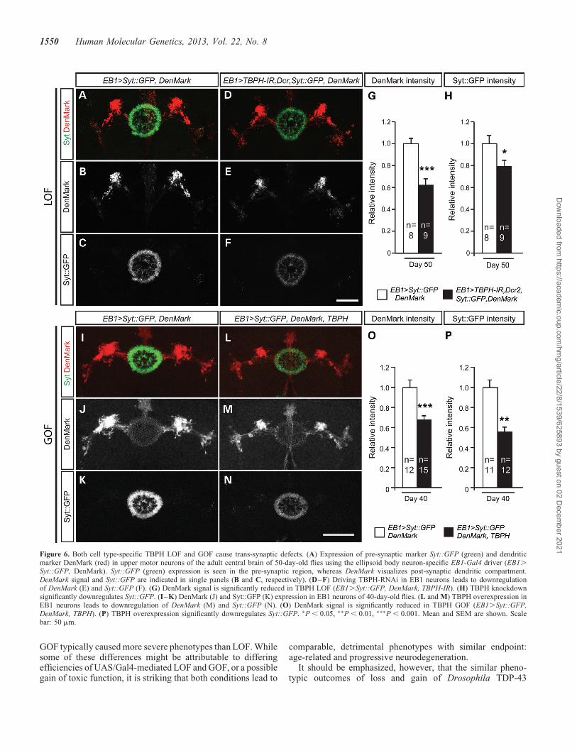
To determine in more detail whether the age-related synap-tic alterations were either pre- or post-synaptic, we made useof the pre-synaptic marker synaptotagmin fused to GFP (UAS-Syt::GFP) in combination with the post-synaptic markerDenMark, a fusion protein of telencephalin and mCherry,which labels the somatodendritic compartment in Drosophila(36) (Fig. 6). Analysis of day 5, day 40 and day 50 adultEB1.Syt::GFP, DenMark controls revealed that expression
of these markers alone did not cause obvious phenotypes(Fig. 6A–C, I–K, and data not shown). However, whenco-expressed together with UAS-TBPH-IR and UAS-Dcr2 inupper motor neurons, Gal4-mediated upper motor neuron-specific EB1.Dcr2, TBPH-IR RNAi-mediated LOF specific-ally caused down-regulation of both pre-synaptic Syt::GFPand dendritic DenMark at day 50 (Fig. 6D–F). To rule out prep-aration or immunolabeling artifacts, independent replicates offly brains were similarly processed and analyzed with identicalconfocal microscopy settings, which established significant re-duction of reporter gene expression in pre-synaptic (Fig. 6H)and post-synaptic compartments when compared with age-matched controls (Fig. 6G). Similar analyses were carried outon day 5 and day 40 Gal4-mediated upper motor neuron-specificGOF flies, with adult EB1.Syt::GFP, DenMark, TBPH fliesshowing a down-regulation of both pre-synaptic Syt::GFP anddendritic DenMark at day 5 and day 40 (Fig. 6L–N) comparedwith EB1.Syt::GFP, DenMark controls (Fig. 6I–K). Inde-pendent replicates of brains were similarly analyzed andshowed a significant reduction of reporter gene expression inpre-synaptic (Fig. 6P) and post-synaptic compartments whencompared with age-matched controls (Fig. 6O and data notshown). Together these findings suggest that upper motorneuron-specific TBPH GOF and RNAi LOF affect synaptic in-tegrity; in both cases, phenotypes occur in a progressive andage-related manner, and are associated with severely impairedmotor behavior (Figs 3D–H and 5).
TBPH dysfunction results in age-related and progressiveneurodegeneration
Synaptic deficits and subsequent neurodegeneration are con-sidered to be hallmarks of ALS and other neurodegenerativediseases (37,38). We therefore investigated whether TBPHLOF and GOF can lead to degenerative cell loss. To visualizeand number targeted neurons, we analyzed brains of aged fliesthat co-express UAS-mCD8::GFP with or without eitherUAS-TBPH-IR LOF or UAS-TBPH GOF again targeted toEB1-Gal4 specific upper motor neurons (Fig. 7).
Analysis of EB1.mCD8::GFP, Dcr2, TBPH-IR brains atday 5 and 20 did not reveal any significant differences inneuron numbers; however, by day 40 and 50, we observed asignificant reduction of targeted EB neurons (Fig. 7G),showing a worsening of the phenotype over time. For theremaining EB neurons, mCD8::GFP labeling revealed missha-pen, fragmented and scattered cell bodies when compared withage-matched controls (Figs 7D–F, compare with 7A–C).
number of peristaltic waves per minute compared with controls (w1118/+, control A; Elav/+, control B). (C) TBPH LOF and GOF adults also show poor climb-ing performance in a startle-induced climbing assay (n ¼ 15) compared with controls (w1118/+, control A; Elav/+, control B). (D–H) Video-assisted motiontracking of TBPHDD962/2 LOF and cell type-specific EB1.TBPH GOF targeted to upper motor neurons. (D) Representative walking tracks recorded over 3 minfor TBPHDD962/2 and EB1.TBPH flies, together with their respective controls (w1118/+, control A; EB1/+, control C). (E–G) TBPHDD962/2 LOF andEB1.TBPH GOF flies show a reduction in walking activity over time, activity and total distance traveled compared with the respective controls (w1118/+,control A; EB1/+, control C). (H) TBPHDD962/2 null mutants show reduced walking velocity compared with controls, whereas EB1-specific overexpressionof TBPH does not affect the mean velocity (w1118/+, control A; EB1/+, control C). (I) TBPHDD962/2 LOF, Tub.TBPH-RNAi downregulation and ELAV.
TBPH GOF flies display disturbed tripod gait. Left cartoon shows tripod gait with left foreleg (1), right middle leg (2) and left hind leg (3). Control flies (w1118)walk in a stereotyped alternating tripod gait pattern. This pattern is disrupted in TBPHDD962/2 null mutants (TBPH2/2), ubiquitous RNAi-mediated TBPHknockdown flies (Tub.TBPH-RNAi) and pan-neuronal Elav-Gal4-mediated TBPH overexpression flies (ELAV.TBPH). Box-plots show median, upper andlower quartiles (box); whiskers contain data 1.5× the interquartile range; + indicates a data point within 3× the interquartile range (outliers). ∗∗P , 0.01;∗∗∗P , 0.001. Mean and SEM are shown (A, E).
Human Molecular Genetics, 2013, Vol. 22, No. 8 1545
Dow
nloaded from https://academ
ic.oup.com/hm
g/article/22/8/1539/625893 by guest on 02 Decem
ber 2021
Figure 4. TBPH dysfunction affects synaptic efficacy. (A) Representative excitatory junction potential (EJP) traces are shown for LOF. EJP amplitudes forTBPHDD962/2 LOF and pan-neuronal ELAV.TBPH GOF flies are not significantly different from the respective controls (control A, w1118/+ and controlB, Elav/+), however, quantal content is significantly increased in TBPH mutant larvae. (B) Representative traces of spontaneous neurotransmitter release
1546 Human Molecular Genetics, 2013, Vol. 22, No. 8
Dow
nloaded from https://academ
ic.oup.com/hm
g/article/22/8/1539/625893 by guest on 02 Decem
ber 2021

A comparable but more severe degenerative phenotype wasobserved in TBPH GOF. Brains of 5-day-old EB1.mCD8::GFP, TBPH flies showed reduced mCD8::GFP ex-pression in perikarya as well as axonal extensions and synapticarborizations of ellipsoid body ring neurons. GFP expressionwas further reduced by day 40 (Fig. 7K–M, compare with7H–J) and almost undetectable in day 60 old EB1.mCD8::GFP, TBPH flies when compared with age-matchedcontrols. Analysis of independent replicates of brains of age-matched EB1.mCD8::GFP, TBPH and EB1. mCD8::GFPflies that were similarly processed and analyzed with identicalconfocal microscopy settings, established significant and pro-gressive, age-related loss of upper motor neurons (Fig. 7N).
To independently confirm age-related and progressive lossof neurons, we studied pox neuro (poxn) expression, whichis specific to ellipsoid body ring neurons (39). Notably, lossof poxn-expressing cells progressed between day 5 and day40 in EB1.mCD8::GFP, TBPH flies (Supplementary Mater-ial, Fig. S13). In addition to the decrease and subsequent lossof mCD8::GFP and poxn expression, degenerative cell losswas confirmed by the specific and age-related decrease inthe number of ellipsoid body ring neurons that expressed ex-cessive amounts of TBPH in EB1.mCD8::GFP, TBPHflies (Supplementary Material, Fig. S4 and data not shown).TUNEL labeling of brains of aged EB1.mCD8::GFP,TBPH flies did not indicate increased apoptotic activity (Sup-plementary Material, Fig. S14), suggesting that progressiveloss of upper motor neurons was not caused by programmedcell death. Together these data suggest that prolonged dys-function of Drosophila TDP-43 causes age-related and pro-gressive neurodegeneration.
Gain of TBPH does not downregulate or mislocalizeendogenous TBPH
The phenotypic similarities between tissue-specific TBPHLOF and GOF may suggest that the observed synaptic deficits,impaired motor behavior and age-related neurodegenerationmight be due to a common pathogenic mechanism. Previousin vitro and in vivo studies indicated that TDP-43 regulatesits own expression by a negative feedback loop (17,40,41)that may participate in a feed-forward mechanism wherebycytoplasmic aggregation depletes nuclear TDP-43 function,essentially leading to a loss-of-function phenotype. Wetested this hypothesis and over-expressed UAS constructscoding for tagged or untagged full length TBPH (Fig. 8).We utilized GMR-Gal4 and specifically targeted TBPH GOFto differentiating photoreceptor cells which resulted in a
rough eye phenotype due to photoreceptor cell degeneration(42) (data not shown). Western blot analysis of heads of1–3-day-old cases and controls identified a 58 kDa band ineach condition similar to the control (Fig. 8A). Quantificationof independent replicates confirmed that GMR-Gal4 specificTBPH GOF with C- and/or N-terminal tags did not alter en-dogenous levels of TBPH expression (Fig. 8B).
We then tested whether GMR-Gal4 specific TBPH GOFmay lead to aggregate formation and/or deplete nuclear ex-pression, hence affecting the function of endogenous TBPHwithout changing overall TBPH protein levels. Third instareye imaginal discs were immunolabeled with anti-TBPH andanti-FLAG or anti-HA and counterstained with DAPI to deter-mine potential aggregates and nuclear localization. In none ofthe cases examined did we detect aggregate formation, nor didwe observe loss or alteration of nuclear TBPH expression(Fig. 8C–N and data not shown). We also tested whetherGMR-Gal4-specific TBPH GOF may result in ubiquitinatedTBPH and thus render the endogenous protein non-functionaleven though expression levels and nuclear localizationappeared unaltered. However, immunolabeling with anti-FK2, which recognizes poly-ubiquitination, did not revealany differences to controls (data not shown). Together thesedata suggest that gain of Drosophila TDP-43 function doesnot alter its own expression level or localization, although itresults in severe synaptic, behavioral and neurodegenerativephenotypes.
DISCUSSION
Here we have systematically compared the phenotypicconsequences of loss and gain of TDP-43 function in Dro-sophila. Our results demonstrate that deregulation of TBPHaffects neuronal function and viability. In either case, deregu-lated TBPH initiates synaptic deficits and impaired motor be-havior, ultimately causing age-related and progressiveneurodegeneration. Our findings indicate that commonloss-of-function phenotypes underlie TDP-43 dysfunction inDrosophila, which has implications for understandingTDP-43-mediated pathogenesis in ALS and FTLD.
Both loss and gain of TDP-43 trigger disease formation
TDP-43 related ALS and FTLD cases are characterized byboth nuclear clearance and the cytoplasmic accumulation offull length, truncated and modified forms of TDP-43 (1).TDP-43 has been shown to autoregulate its own expression(17,41), and its ubiquitylation, phosphorylation, aggregation,
(mEJP) shown for TBPHDD962/2 LOF. Both the mEJP amplitude and frequency are significantly reduced in TBPH mutant larvae. The mEJP is unaffected inELAV.TBPH gain-of-function flies (w1118/+, control A; Elav/+, control B). (C) Representative ERG traces of the indicated TBPH LOF conditions. The white-eyed TBPHDD962/2 mutants and w1118 flies show a strong, habituating response, steady maintained response and off-transient. The red-eyed OregonR, GMR.-Shibire-IR and GMR.TBPH-IR show a smaller amplitude response; GMR.Shibire-IR flies lack on- and off transients. Chequered bar indicates duration of lightpulse. (D) Mean peak–peak amplitude of the ERG from the indicated conditions at day 1 and day 5 timepoints. (E) TBPHDD962/2 mutant and GMR.TBPH-IRhave significantly smaller off-transients than the respective eye color-matched controls. The negative GMR.Shibire-IR controls, in which synaptic transmissionis blocked, show a severely reduced off-transient. The solid line shows the regression line between off-transient and maintained response for wild-type flies. (F)Representative ERG traces of the indicated TBPH GOF conditions. The UAS-TBPH/+ and GMR/+ flies show a normal amplitude response, the smaller responseseen in the GMR/+ flies is due to the darker red pigment. GMR.UAS-TBPH flies have a very small response, with no off/on transients visible. (G) Mean peak–peak amplitude of the ERG from the indicated GOF conditions at day 1 is significantly decreased in GMR.TBPH flies compared with the controls (D, E, n ¼ 9;∗P , 0.05, ∗∗P , 0.01, ∗∗∗P , 0.001). Mean and SEM are shown (A, B, D, G).
Human Molecular Genetics, 2013, Vol. 22, No. 8 1547
Dow
nloaded from https://academ
ic.oup.com/hm
g/article/22/8/1539/625893 by guest on 02 Decem
ber 2021

1548 Human Molecular Genetics, 2013, Vol. 22, No. 8
Dow
nloaded from https://academ
ic.oup.com/hm
g/article/22/8/1539/625893 by guest on 02 Decem
ber 2021

truncation, mislocalization, nuclear clearance and defectiveautoregulation can contribute either directly or indirectly todisease formation (12). However, no consensus has emergedas to whether loss or toxic gain of TDP-43 function, or both,are causally related to disease onset and progression.
In our study, we systematically compared loss- and gain ofTBPH, the Drosophila homolog of TDP-43, and analyzed theireffect on synaptic function and morphology, motor control andage-related neuronal survival. Our results demonstrate thatloss of function, RNAi-mediated downregualtion and overex-pression of wild-type Drosophila TDP-43 are sufficient tocause age-related neurodegeneration. In the case of TDP-43overexpression, we did not detect alterations in expressionlevels or mislocalization of the endogenous protein; we alsofound no evidence for ubiquitylation, aggregation, truncationor autoregulation, and based on western blot data, excessivephosphorylation appears unlikely. Our data thus argue thatTDP-43 truncation, modification, altered autoregulation oraggregates are not a prerequisite for toxicity and age-relatedneurodegeneration, which is also supported by data in othermodel systems (28,41,43–46).
Previous studies showed that TDP-43 can form homodimers(47) and functions in multiprotein/RNA complexes (15,48,49)where multiple TDP-43 molecules are incorporated into eachribonucleoprotein complex (50). These data suggest that equi-librated, physiological levels of TDP-43 and a stoichiometricrelationship with ribonucleoprotein complex components areessential for proper biogenesis, spatio-temporal expressionand hence, regulation of TDP-43 target genes. Our results es-tablish that deregulation of TDP-43 levels is a core eventunderlying disease formation, which can be triggered byeither decreasing or increasing levels of TDP-43. This sug-gests that de-regulation of TDP-43 levels, by loss of function,downregulation or overexpression, alters the stoichiometry ofparticipating components and, hence, ribonucleoproteincomplex formation required for proper physiological function.In either condition, the net result of de-regulated TDP-43 isimpaired physiological function which, when continuing,affects neuronal survival, ultimately causing progressive cellloss. In support of this notion, we determined synaptic deficitsas one of the earliest phenotypes of TDP-43 dysfunction.
Pre-synaptic deficits are early, initiating events ofTDP-43-related pathogenesis
In our study, we found that both loss and gain of DrosophilaTDP-43 result in synaptic deficits and impaired motor behav-ior, followed by progressive, age-related neurodegeneration.
In contrast to previous reports in Drosophila (27,28,29), wedid not detect any obvious structural defects at the larvalneuromuscular junction or mislocalization of synaptic proteinsthat might explain the observed behavioral phenotypes. Thisdiscrepancy might be attributable to differences in geneticbackground and/or methods used. Motor terminals aredynamic structures that show developmental and circadianfluctuations in shape and size (51). In our study, we usedbona fide TBPH null alleles together with genomic rescue,as well as overexpression alleles. Instead of structuraldefects at the larval NMJ, our experiments reveal that affectedflies are characterized by defective synaptic transmission, aphenotype detectable both at the larval NMJ and in theadult. In addition, our tissue-specific RNAi-mediated knock-down experiments identified pre-synaptic, rather than post-synaptic deficits as the earliest detectable phenotypes at thelarval NMJ, suggesting that pre-synaptic deficits are initiatingevents in disease formation.
Our observations therefore allow for the first time the de-scription of a sequence of events whereby TDP-43 dysfunctioncauses impaired synaptic transmission and motor abnormal-ities, followed by the subsequent loss of neuronal connectionsthat precede degenerative cell death in an age-related and pro-gressive manner. Such a sequence of events suggests thatTDP-43 related pathogenesis progresses from the synapseand axon to the neuronal cell body, thereby resembling affin-ities to the ‘dying back’ phenomena, a hallmark of severalneurodegenerative diseases (37). Comparable events havebeen, at least to some extent, reported for Wallerian degener-ation (52), and recent genetic evidence indicates that neuronal‘dying back’ underlies an active process of self-destructiondistinct from apoptotic cell death (53). A ‘dying back’process also characterizes human ALS patients and mousesuperoxide dismutase 1 (SOD1) models of ALS (38,54).Like TDP-43 inclusions, SOD1 mutations are commonly iden-tified in familial ALS (55). Although further, conclusiveexperiments are required, it is tempting to speculate thatTDP-43 might not only regulate RNAs related to synapticfunction (15,17), but potentially also negatively regulates thetranscription or translation of target genes involved in theactive self-destruction of neuronal connections.
TDP-43 dysfunction results in loss-of-function phenotypes
In addition to similarities between loss and gain of functionphenotypes, we also observed phenotypic differences, includ-ing electrophysiology at the larval NMJ but also at the level ofsynaptic integrity and neurodegeneration in adult brain, where
Figure 5. Similar to TBPH mutants, cell-type specific knockdown of TBPH leads to impaired motor behavior. Gal4-mediated UAS-TBPH-RNAi knockdowntargeted either ubiquitously (Tub-Gal4) or to all neurons (Elav-Gal4), or targeted to upper motor neurons (EB1-Gal4). RNAi efficiency was enhanced byco-expressing UAS-Dcr2. (A–J) Motor behavior analysis using video assisted open-field motion tracking. (A) Representative walking tracks recorded over30 min for 5-day-old Tub.Dcr2, TBPH-RNAi flies and heterozygous Dcr2,TBPH-RNAi controls. (C–F) Tracking analyses reveal that, comparable toTBPHDD962/2 mutants, Tub.Dcr2,TBPH-RNAi flies have a significantly reduced average activity, total distance travelled, mean speed and activity overtime when compared with heterozygous Dcr2,TBPH-RNAi controls. (G–J) Dcr2, TBPH-RNAi expression was activated by either the pan-neuronalELAV-Gal4 driver, or the EB1-Gal4 driver (ELAV.Dcr2,TBPH-RNAi or EB1.Dcr2,TBPH-RNAi, respectively). Tracking analyses reveal that, comparableto TBPHDD962/2 mutants, the walking activity, total distance travelled, mean speed and activity over time of 5-day-old ELAV.Dcr2,TBPH-RNAi and EB1.-Dcr2,TBPH-RNAi flies is significantly reduced when compared with that of the control (heterozygous Dcr2,TBPH-RNAi). Box-plots show median, upper andlower quartiles (box); whiskers contain data 1.5× the interquartile range; +indicates a data point within 3× the interquartile range (outliers). (n ¼ 24; ∗P ,
0.05, ∗∗P , 0.01, ∗∗∗P , 0.001). Mean and SEM are shown (F, J).
Human Molecular Genetics, 2013, Vol. 22, No. 8 1549
Dow
nloaded from https://academ
ic.oup.com/hm
g/article/22/8/1539/625893 by guest on 02 Decem
ber 2021
GOF typically caused more severe phenotypes than LOF. Whilesome of these differences might be attributable to differingefficiencies of UAS/Gal4-mediated LOF and GOF, or a possiblegain of toxic function, it is striking that both conditions lead to
comparable, detrimental phenotypes with similar endpoint:age-related and progressive neurodegeneration.
It should be emphasized, however, that the similar pheno-typic outcomes of loss and gain of Drosophila TDP-43
Figure 6. Both cell type-specific TBPH LOF and GOF cause trans-synaptic defects. (A) Expression of pre-synaptic marker Syt::GFP (green) and dendriticmarker DenMark (red) in upper motor neurons of the adult central brain of 50-day-old flies using the ellipsoid body neuron-specific EB1-Gal4 driver (EB1.
Syt::GFP, DenMark). Syt::GFP (green) expression is seen in the pre-synaptic region, whereas DenMark visualizes post-synaptic dendritic compartment.DenMark signal and Syt::GFP are indicated in single panels (B and C, respectively). (D–F) Driving TBPH-RNAi in EB1 neurons leads to downregulationof DenMark (E) and Syt::GFP (F). (G) DenMark signal is significantly reduced in TBPH LOF (EB1.Syt::GFP, DenMark, TBPH-IR). (H) TBPH knockdownsignificantly downregulates Syt::GFP. (I–K) DenMark (J) and Syt::GFP (K) expression in EB1 neurons of 40-day-old flies. (L and M) TBPH overexpression inEB1 neurons leads to downregulation of DenMark (M) and Syt::GFP (N). (O) DenMark signal is significantly reduced in TBPH GOF (EB1.Syt::GFP,DenMark, TBPH). (P) TBPH overexpression significantly downregulates Syt::GFP. ∗P , 0.05, ∗∗P , 0.01, ∗∗∗P , 0.001. Mean and SEM are shown. Scalebar: 50 mm.
1550 Human Molecular Genetics, 2013, Vol. 22, No. 8
Dow
nloaded from https://academ
ic.oup.com/hm
g/article/22/8/1539/625893 by guest on 02 Decem
ber 2021

function, including age-related neurodegeneration, do not ne-cessarily implicate similar mechanisms related to TDP-43mediated pathogenesis. It is conceivable but remains to beshown that loss, downregulation or gain of TDP-43 functioncould trigger distinct pathogenic mechanisms that all convergeon a functional node which when defective initiates diseaseformation. Previous studies suggest that such a functionalnode is likely the stochiometric activity of TDP-43 in ribonu-cleoprotein complexes that are essential for the regulation andcorrect processing of multiple target genes, including thoserelated to synaptic function (15,17). This model is consistentwith familial cases of ALS and FTLD, were TDP-43 is pre-dominantly mutated in the C-terminal, prion-like domain(5–8) that regulates tissue-specific gene expression, transcrip-tional repression and alternative splicing, and is essential forbinding to ribonucleoproteins involved in microRNA andmRNA biogenesis, as well as RNA turnover (9,10,12,13).According to this model, C-terminal, but also RRM mutations
may hinder the participation of TDP-43 in ribonucleoproteincomplex formation and hence, their function, leading tophysiological deficits and eventually neurodegeneration.
In summary, our findings demonstrate that both loss and gainof TDP-43 function result in functional deficits wherebyimpaired synaptic transmission and defective motor behaviorprecede progressive deconstruction of neuronal connections, ul-timately causing age-related neurodegeneration. Our data there-fore provide in vivo evidence for common loss-of-functionphenotypes underlying TDP-43 dysfunction that likely alsocharacterize TDP-43-mediated ALS and FTLD.
MATERIALS AND METHODS
Fly stocks
Fly stocks were maintained at 258C on standard cornmealfood, unless for aging experiments where aged flies
Figure 7. Upper motor neuron-specific RNAi-mediated downregulation and gain of TBPH causes loss of neuronal connections and age-related neurodegenera-tion. (A–C) Control flies with mCD8::GFP overexpression in upper motor neurons at day 40 (B, enlargement of dashed box in A; C, additional example). (D–F)TBPH LOF flies (EB1.mCD8::GFP, Dcr2, TBPH-IR) with TBPH-RNAi targeted to upper motor neurons at day 40. The GFP expression in the ellipsoid bodystructure does not diminish in the aged brain, however, dispersed cells with fragmented and granular GFP expression are detectable (E, enlargement of dashedbox in D; F, additional example). (G) Quantification of EB1 neuron cell number (GFP positive) in control and TBPH LOF flies at day 5, 40 and 50. A significantdecrease in neuron number is observed in aged TBPH LOF flies. (H–J) Control flies with mCD8::GFP overexpression in EB1 ring neurons at day 40 (I–J,enlarged example of cell bodies). (K–M) TBPH and mCD8::GFP overexpression in upper motor neurons leads to an age-related reduction in mCD8::GFP ex-pression (L–M, enlarged example of cell bodies). (N) Quantification of EB1 cell numbers (GFP positive) in control and TBPH GOF flies at day 5, 40 and 60. Asignificant decrease in neuron number is observed over time in TBPH GOF flies. ∗∗P , 0.01, ∗∗∗P , 0.001. Mean and SEM are indicated. Scale bar: 50 mm.
Human Molecular Genetics, 2013, Vol. 22, No. 8 1551
Dow
nloaded from https://academ
ic.oup.com/hm
g/article/22/8/1539/625893 by guest on 02 Decem
ber 2021

were maintained on 15% sugar/yeast medium (23). Thefollowing strains were used: Oregon R (wild-type); w1118;w; TBPHnull/CyO,GFP; + (26) Df(2R)106/SM5; w; +;Elav-Gal4; GMR-Gal4; and Tub-Gal4; UAS-mCD8::GFP(56); UAS-Dcr2; UAS-Shibire-RNAi (Bloomington StockCentre); p{Gawb}C57 (BG57, ref. 34); EB1-Gal4 (57); UAS-DenMark, UAS-Syt::GFP (36); UAS-RFP-TBPH (58).
Transgenic constructs
The TBPH gene was amplified by PCR from cDNA cloneGH09868 (DGRC). PCR primers to amplify TBPH inpUAST vector are forward primer: 5′-GGAGATCTATGGA
TTTCGTTCAAGTGTCGG-3′; reverse primer: 5′-GGCTCGAGTTAAAGAAAGTTTGACTTCTCCGC-3′.Primers used to generate Flag-TBPH-HA are: forward primer:5′-GGAGATCTATGGACTACAAGGACGACGATGACAAGGATTTCGTTCAAGTGTCGG-3′; reverse primer: 5′-GGCTCGAGTTAGGCATAGTCTGGGACGTCATATGGATAAAGAAAGTTTGACTTCTCCGC-3′.
BglII and XhoI sites were used to insert TBPH into pUASTvector. DNA sequence was confirmed by sequencing. TheUAS-TBPHYA765 line was used for all behavioral, electrophysi-ology and immunohistochemistry experiments. To specificallyknockdown TBPH, the target region was chosen by E-RNAiweb service (59). TBPH RNAi construction was carried out
Figure 8. GMR-Gal4-mediated gain of TBPH does not downregulate or mislocalize endogenous TBPH. (A) Western blot analysis of GMR-Gal4-drivenuntagged (58 kDa) or tagged UAS-TBPH (Flag-TBPH-HA, 60 kDa; RFP-TBPH, 85 kDa). Levels of endogenous TBPH (arrow at 58 kDa) are unaffected inday 1–3 flies overexpressing either form of TBPH. (B) Quantification of endogenous TBPH shows no alteration in TBPH protein levels. Endogenous TBPHsignal intensities were normalized against b-Tubulin. Mean and SEM are indicated. (C, G, K) Third instar eye imaginal discs immunolabeled with anti-TBPHdoes not reveal TBPH-positive aggregates in cell bodies nor in the optic stalk (dotted area). (D–F, H–J, L–N) Third instar eye imaginal discs immunolabeledwith anti-TBPH and DAPI identifies that nuclear expression of endogenous TBPH is maintained in gain of TBPH. Note that signal intensity in I, J, M and N isadjusted for overexpressed TBPH to avoid saturation of the signal. Scale bar: 10 um.
1552 Human Molecular Genetics, 2013, Vol. 22, No. 8
Dow
nloaded from https://academ
ic.oup.com/hm
g/article/22/8/1539/625893 by guest on 02 Decem
ber 2021

as previously described (33). PCR primers with EcoRI(forward) and XbaI (reverse) sites were designed to subcloneTBPH RNAi construct into pMF3 vector (33).
TBPH-RNAi: forward primer: 5′-GGGAATTCCACTCATACCACCCACAGGG-3′; reverse primer: 5′-GGTCTAGAGTTATGCGGCTGGTTCATTC-3′.
Insertion of RNAi construct was confirmed by restrictionenzyme digestion and sequencing. Plasmid injection and gen-eration of transgenic flies were performed by BestGene Inc(CA, USA).
To generate a TBPH genomic rescue construct, we usedTBPH genomic DNA from pacman bac library (CH322-116J04, bacpac.chori.org) covering position 19751906–19744713 on the second chromosome, which encodes theentire TBPH coding sequence. TBPHgenomic was integratedinto the pUC 3GLA plasmid (gift from Matthias Soller) byrecombination. TBPHgenomic in pUC 3GLA was used to generatetransgenic flies by site-specific integration into the attp86Fbfly strain (60) and flies harboring TBPHgenomic pUC 3GLAwere selected by compound eye-specific GFP expression.
P-element-mediated mutagenesis
The TBPHDD96 and TBPHDD100 null alleles were generated byimprecise excision of the EY10530 P-element, which isinserted in the promoter region of the TBPH locus. It wasmobilized using HoP1 D2-3 transposase (gift from IrisSalecker). Four hundred and fifty excision lines were estab-lished from individual events and genomic deletions withinthe TBPH locus were identified by PCR and confirmed asprotein null by western blot. TBPHDD96 and TBPHDD100
breakpoints are 19750093 . . . 19747817 and 19750093 . . .19747925, respectively. The primers used to confirmthe presence of neighboring gene CG4585 were: forwardprimer: 5′-CTGAGCATCTTCTGCGAC-3′; reverse primer:5′-GAAATTAGCTCGCCATGG-3′.
Breakpoints and P-element insertion sites were sequencedusing the following primers:TBPHDD100, forward primer:5′-TTCCATGGCGAGCTAATT-3′; reverse primer: 5′- GTGTCCAGGTTGCGGTACTT-3′ TBPHDD96 forward primer:5′-CGAGTTGCCGCCGTAAT-3′; reverse primer: 5′-CCTTTCACTCGCACTTAT-3′. Sequencing was carried out atMWG Eurofins.
Generation of TBPH antibody
The anti-TBPH antibody was generated by injecting two rabbitswith peptide sequences PQGNHMNPGRNGHHR, correspond-ing to residues 291–305 and QSSGSQNAAEKSNFL, residues517–531. Immunization was carried out by Eurogentec.
Immunohistochemistry
Adult and larval CNS dissection was carried out as previouslydescribed (23). Larval NMJ dissections were carried outaccording to established protocol (61). Primary antibodiesused were: mouse anti-Repo (1:20); rat anti-Elav (1:30);
mouse anti-Futsch (1:50); mouse 3C11 (1:100); mouse anti-synaptotagmin (1:25); mouse anti-FasII (1:10); mouse NC82(anti-bruchpilot) (1:100) and mouse anti-Neuroglian (1:4) allobtained from the Developmental Studies Hybridoma Bank(DSHB) under the auspices of the NICHD and maintainedby The University of Iowa; rabbit anti-TBPH antibody(1:3000); rabbit anti-Poxn (1:100; Adachi, Ludlow & Hirth,unpublished); mouse anti-polyubiquitin (FK2, 1:200;ENZO); rabbit anti-FLAG (1:500; Cell Signaling Tech);mouse anti-HA (1:40; Roche); goat anti-HRPCy3 (1:100; Stra-tech), 488-Phalloidin (1:1000; Invitrogen). Secondary anti-bodies were Alexa fluor 488, 568 and 647 (each 1:150;Invitrogen).
Image acquisition and analysis
Images were obtained either with Motic BA400 or Leica TCSSP5 confocal microscope with Leica Application SuiteAdvanced Fluorescence (LAS AF) version 2.0.2 software.For confocal images, channels were scanned sequentially.For comparative images, the same microscope settings wereused for the control and experimental genotypes. For Figs 6and 7, confocal z-stacks were rendered as 3-D projections.Signal quantification of digital images was carried out usingFiji (FIJI). Significance of GFP and FasII signal intensitywas calculated using unpaired Student’s t-test. Equal varianceassumptions were based on Levene’s test for equality of vari-ance.
NMJ bouton count
Early L1 larvae were picked from 4 h egg collection plates(fruit agar) and placed on cornmeal food. At wandering L3,larvae were dissected according to established protocol (61).All preparations were dissected within a 1.5 h window toavoid differences in circadian rhythm effect on boutonnumber. Z-stacks were taken of the MN6/7-Ib motor neuronsinnervating muscle group 6/7 in segment A3. Boutons werecounted by hand and the number of NC82 puncta were ana-lyzed using the ITCN plug-in for ImageJ.
Terminal deoxynucleotidyl transferase dUTP nickend labeling (TUNEL) assay
Tissues of interest were fixed in 4% formaldehyde and subse-quently permeabilized in 100 mM Citrate/0.1% Triton X-100for 30 min at 658C. Tissues were briefly washed with 0.5%Triton-X 100 in PBS and rinsed twice with TUNEL assaybuffer (In Situ Cell Death Detection Kit; Roche). After incu-bating for 90 min at 378C, TdT enzyme was added andfurther incubated for 3 h at 378C. For positive control,freshly dissected tissue was soaked in 2N HCl for 30 min toinduce apoptosis.
Western blots
Fly tissue was lysed in RIPA buffer (150 mM NaCl, 1% NP-40,5 mM EDTA, 0.5% sodium, deoxycholate and 0.1% SDS,50 mM Tris, pH8.0) containing complete proteinase inhibitor(Roche) and phosstop phosphatase inhibitor (Roche).
Human Molecular Genetics, 2013, Vol. 22, No. 8 1553
Dow
nloaded from https://academ
ic.oup.com/hm
g/article/22/8/1539/625893 by guest on 02 Decem
ber 2021

Samples were centrifuged to take the RIPA-soluble fraction.SDS–PAGE was run in 8% SDS gel. Rabbit anti-TBPH anti-body was used at 1:2000–3000 and anti-b-Tub (DSHB) wasused at 1:300. Secondary antibodies were IRDye 800 conju-gated goat anti-rabbit (1:10000, Rockland Immunochemicals)and Alexa Fluor 680 goat anti-mouse (1:10000, Invitrogen).Membrane images were acquired using Odyssey.
Reverse transcriptase PCR
RNA was extracted by homogenizing whole flies in Trizol,adding chloroform, incubating on ice for 15 min, followedby centrifugation at 16 000g at 48C. The upper phase wasadded to isopropanol and incubated and spun as before. Thepellet was washed in 70% ethanol and dissolved in nuclease-free H2O. RNA purity was measured using a Nanodrop.DNase treatment was carried out according to manufacturer’sinstructions (Ambion). cDNA was generated by mixing 1 mgRNA and random hexamers, heating to 708C and adding5 × M-MLV reaction buffer, M-MLV-RT, RNAsin anddNTPs according to manufacturer’s instructions (Ambion).Samples were incubated at 378C for 1 h and 708C for15 min. PCR products were run on 1% agarose gel. Theprimers used were as follows; TBPH: forward primer: 5′-ATCTTGGATGGCTCAGAACG-3′, reverse primer: 5′-GTCGGTCTTTATTCCGTTGG-3′ RPL32: forward primer:5′-CGCCGCTTCAAGGGACAGTATC-3′, reverse primer:5′-CGACAATCTCCTTGCGCTTCTT-3′.
Footprint/gait analysis
Flies were briefly anaesthetized with CO2 and one-third of thewings were removed to prevent escape. Flies were left torecover in a petri dish for 10–15 min before being tappedonto a soot-covered glass microscope slide (slides were heldover a candle flame until covered in an even coating ofsoot). Footprints were visualized by lighting the slide frombelow and images were captured with a Q-capture camera.
Startle-induced negative geotaxis
Flies’ climbing ability was assayed as described earlier (23).Equal variance assumptions were based on Levene’s test forequality of variance.
Eclosion analysis
Embryos were collected on fruit agar plates in 5 h batches andleft in a 258C incubator until larvae had hatched. Fifty firstinstar larvae were placed in a vial and a tally made of allfully eclosed adult flies, mean values were calculated andplotted as a percentage of total number of larvae picked. Sig-nificance was calculated using unpaired t-test. This wascarried out in triplicate for each genotype.
Larval motility
Thirty wandering third instar larvae were individually placedon a fruit agar plate and allowed to recover for 30 s. Thenumber of peristaltic waves, travelling in either direction,
was scored over 1 min. Significance was calculated using un-paired t-test (two-tailed). Equal variance assumptions werebased on Levene’s test for equality of variance.
Video-assisted motion tracking
Tracking arenas were modified six-well tissue culture plates(35 mm diameter wells) filled with silicon rubber (Sylguard)to leave a 3 mm space so that flies could walk freely but nothop or fly. 18–24 flies were briefly anaesthetized with CO2,placed in separate arenas and left to recover at 258C for45 min before being placed above an array of white LEDswithin a temperature-controlled incubator. Tracking wascarried out at 258C. A black and white CCD camera(Hitachi, MP-M1A) positioned above the arenas was con-nected to a PC via an analog capture card (Integral Technolo-gies, Flashbus MV Lite). Recordings were carried out duringthe same time slot. Recorded videos were converted to flymovie format using the motmot package (62) and loadedinto Ctrax software (63) to analyze the positions of the fliesthroughout the video. Position data for the 30 min file wasexported as a Matlab-compatable (Mathworks) matrix file.Errors in the tracking were fixed using Matlab (Mathworks)as well as FixErrors GUI (63), which is described in furtherdetail at http://ctrax.sourceforge.net/fixerrors.html. Fixed tra-jectories were analyzed in Matlab using custom scripts(written by D.M.H). This analysis determined the mean vel-ocity, mean activity, activity over time and mean cumulativedistance traveled by the population of flies in the arena. Activitywas defined as movement per frame above a velocity of 2 mm/s. Average activity was the percentage of frames where the flywas active (.2 mm/s velocity). Mean velocity was the averageof velocities in each frame of the recording only when the flywas active. Box-plots were generated in Matlab where theboxes show median, upper and lower quartiles; whiskerscontain data 1.5× the interquartile range; + indicates a datapoint within 3× the interquartile range. Significance was calcu-lated using the Mann–Whitney U-test with a Bonferroni correc-tion to account for multiple comparisons.
Escape response
The amplitude of the escape response was carried out as pre-viously described (64). In summary, anaesthetized flies werefixed to a tungsten pin on the end of a cocktail stick usingglue gum. The pin was placed so that it covered the thorax.Flies were left to recover for 20 min before being mountedover the flexible ergometer and lowered gently until the fliesreached out and place their legs on the jump platform, bringingit up to a resting position. A stimulating electrode was placedin the eye and one in the cervical connective to stimulate theGiant Fiber System. A supra-threshold 0.8 mV was appliedand as the flies jumped, the amount of beam displacementwas measured.
Electrophysiology
Wandering third instar larvae were dissected in HL3 buffer(65) containing 1.8 mM CaCl2. Nerves innervating the bodymuscle walls were cut near the ventral ganglion and stimulated
1554 Human Molecular Genetics, 2013, Vol. 22, No. 8
Dow
nloaded from https://academ
ic.oup.com/hm
g/article/22/8/1539/625893 by guest on 02 Decem
ber 2021

using a suction electrode and isolated pulse stimulator Digiti-mer DS2A (constant current modification), with a currentdouble that needed to initiate a compound response. Allrecordings for TBPH LOF/GOF and appropriate controlswere made intracellularly in muscle 6, abdominal segment 3,at ambient room temperature using microelectrodes filledwith 3 M KCl that had tip resistances of 5–10 MV. In 1.8 mM
extracellular Ca2+ resting membrane potentials were261.1+ 1.0 mV (n ¼ 10) and 255.8+ 2.5 mV (n ¼ 9, P ,0.05) and input resistances were 10.1+ 0.7 MV (n ¼ 10) and9.6+ 0.4 MV (n ¼ 9, P . 0.05) for w1118 and TBPH2/2larvae, respectively. Resting membrane potentials were254.1+ 1.4 mV (n ¼ 7) and 252.0+ 1.5 mV (n ¼ 8, P ¼0.0018) and input resistances were 5.9+ 0.2 MV (n ¼ 7) and6.4+ 0.3 MV (n ¼ 8, P , 0.05) for ELAV/+ and ELAV.TBPH, respectively. Data, filtered at 1 kHz and digitized at10 kHz, were acquired using an Axopatch 200B amplifier anda Digidata 1320A data acquisition board (Molecular DevicesData) were acquired using pClamp8.02 (Molecular Devices)and analyzed with Clampfit8.02 (Molecular Devices) or MiniA-nalysis (Synaptosoft). EJP amplitude histograms were con-structed by averaging 50 separate events stimulated at 0.2 Hzfrom an individual muscle cell and then calculating the meanresponse from at least six larvae per line. Spontaneous releaseevents were recorded for 120 s without any electrical stimula-tion. All events were analyzed and mean mEJP amplitudesfrom individual larvae were averaged to generate the histo-grams. To obtain the frequency of spontaneous release eventsthe number of events for each recording was divided by 120and then averaged to generate histograms. Significance wascalculated using unpaired Students t-test (two-tailed).
Electroretinograms
Female flies were aspirated into a pooter, and then gentlyblown into a truncated pipette tip. They were restrained withnail varnish (Creative Nail Design). No anaesthesia wasused. Blunt glass pipettes, filled with simple Drosophilasaline (130 mM NaCl, 4.7 mM KCl, 1.9 mM CaCl2 (66) wereused as recording electrodes, placing one tip centrally on thesurface of the eye and the other in the mouthparts. Flieswere adapted to the dark for 2 min and then presented witha standard blue light stimulus from three Kingbright,KAF-5060PBESEEVGC light-emitting diodes (maximumemission wavelength 465 nm) situated 6 cm away from theeye. Light pulses were monitored with a BPX65 photodiode(Centronics) next to the head of the fly. In the dark, the photo-diode current was 0.5 nA; during the stimuli 400 nA. Each flywas tested with at least three blue light stimuli, .10 s apart.Their responses were captured using Dasylab software(measX, Monchengladbach, Stuttgart) and the average wave-form calculated. At least nine flies of each genotype weretested at each timepoint. Significance was calculated usingBonferroni post hoc analysis.
Statistical analysis
Statistical analysis was carried out as previously described(24); for details of the statistical tests used, see SupplementaryMaterial, Table S1.
SUPPLEMENTARY MATERIAL
Supplementary Material is available at HMG online.
ACKNOWLEDGEMENTS
We thank B. Hassan, T. Lee, I. Salecker, A. Voigt, D.C. Zar-nescu, the Bloomington Stock Centre, the Vienna DrosophilaRNAi Centre and the Developmental Studies HybridomaBank at the University of Iowa for stocks and reagents.
Conflict of Interest statement. None declared.
FUNDING
This work was supported by the UK Medical Research Council(G-0802208 to I.M.R. and G-070149 to F.H.), the Royal Society(Hirth/2007/R2 to F.H.), the Motor Neurone Disease Associ-ation (Hirth/Oct07/6233 to F.H., A.A.-C., C.E.S. and Hirth/Mar12/6085 to F.H. and C.E.S.), Parkinson’s UK (G-0714to F.H.), Alzheimer’s Research UK (ARUK-PhD2012-18 toF.H.) and the Fondation Thierry Latran (2/2011/DrosAlsto F.H.). Funding to pay the Open Access publication chargesfor this article was provided by the UK Medical ResearchCouncil and the Fondation Thierry Latran.
REFERENCES
1. Neumann, M., Sampathu, D.M., Kwong, L.K., Truax, A.C., Micsenyi,M.C., Chou, T.T., Bruce, J., Schuck, T., Grossman, M., Clark, C.M. et al.(2006) Ubiquitinated TDP-43 in frontotemporal lobar degeneration andamyotrophic lateral sclerosis. Science, 314, 130–133.
2. Arai, T., Hasegawa, M., Akiyama, H., Ikeda, K., Nonaka, T., Mori, H.,Mann, D., Tsuchiya, K., Yoshida, M., Hashizume, Y. et al. (2006)TDP-43 is a component of ubiquitin-positive tau-negative inclusions infrontotemporal lobar degeneration and amyotrophic lateral sclerosis.Biochem. Biophys. Res. Commun., 351, 602–611.
3. Forman, M.S., Trojanowski, J.Q. and Lee, V.M. (2007) TDP-43: a novelneurodegenerative proteinopathy. Curr. Opin. Neurobiol., 17, 548–555.
4. Chen-Plotkin, A.S., Trojanowski, J.Q. and Lee, V.M. (2010) TARDNA-binding protein 43 in neurodegenerative disease. Nat. Rev. Neurol.,6, 211–220.
5. Sreedharan, J., Blair, I.P., Tripathi, V.B., Hu, X., Vance, C., Rogelj, B.,Ackerley, S., Durnall, J.C., Williams, K.L., Buratti, E. et al. (2008)TDP-43 mutations in familial and sporadic amyotrophic lateral sclerosis.Science, 319, 1668–1672.
6. Gitcho, M.A., Baloh, R.H., Chakraverty, S., Mayo, K., Norton, J.B.,Levitch, D., Hatanpaa, K.J., White, C.L. 3rd, Bigio, E.H., Caselli, R. et al.(2008) TDP-43 A315T mutation in familial motor neuron disease. Ann.Neurol., 63, 535–538.
7. Kabashi, E., Valdmanis, P.N., Dion, P., Spiegelman, D., McConkey, B.J.,Vande Velde, C., Bouchard, J.P., Lacomblez, L., Pochigaeva, K.,Salachas, F. et al. (2008) TARDBP mutations in individuals with sporadicand familial amyotrophic lateral sclerosis. Nat. Genet., 40, 572–574.
8. Yokoseki, A., Shiga, A., Tan, C.F., Tagawa, A., Kaneko, H., Koyama, A.,Eguchi, H., Tsujino, A., Ikeuchi, T., Kakita, A. et al. (2008) TDP-43mutation in familial amyotrophic lateral sclerosis. Ann. Neurol., 63, 538–542.
9. Ayala, Y.M., Pantano, S., D’Ambrogio, A., Buratti, E., Brindisi, A.,Marchetti, C., Romano, M. and Baralle, F.E. (2005) Human, Drosophila,and C. elegans TDP43: nucleic acid binding properties and splicingregulatory function. J. Mol. Biol., 348, 575–588.
10. Gitler, A.D. and Shorter, J. (2011) RNA-binding proteins with prion-likedomains in ALS and FTLD-U. Prion, 5, 179–187.
11. Da Cruz, S. and Cleveland, D.W. (2011) Understanding the role ofTDP-43 and FUS/TLS in ALS and beyond. Curr. Opin. Neurobiol, 21,904–919.
Human Molecular Genetics, 2013, Vol. 22, No. 8 1555
Dow
nloaded from https://academ
ic.oup.com/hm
g/article/22/8/1539/625893 by guest on 02 Decem
ber 2021

12. Lee, E.B., Lee, V.M.Y. and Trojanowski, J.Q. (2012) Gains and losses:molecular mechanisms of TDP43-mediated neurodegeneration. Nat. Rev.
Neurosci, 13, 38–50.13. Buratti, E. and Baralle, F.E. (2010) The multiple roles of TDP-43 in
pre-mRNA processing and gene expression regulation. RNA Biol., 7,420–429.
14. Wegorzewska, I. and Baloh, R.H. (2011) TDP-43 based animal models ofneurodegeneration: new insights into ALS pathology andpathophysiology. Neurodegenerat. Dis., 8, 262–274.
15. Sephton, C.F., Cenik, C., Kucukural, A., Dammer, E.B., Cenik, B., Han,Y., Dewey, C.M., Roth, F.P., Herz, J., Peng, J. et al. (2011) Identificationof neuronal RNA targets of TDP-43-containing ribonucleoproteincomplexes. J. Biol. Chem., 286, 1204–1215.
16. Tollervey, J.R., Curk, T., Rogelj, B., Briese, M., Cereda, M., Kayikci, M.,Konig, J., Hortobagyi, T., Nishimura, A.L., Zupunski, V. et al. (2011)Characterizing the RNA targets and position-dependent splicingregulation by TDP-43. Nat. Neurosci., 14, 452–458.
17. Polymenidou, M., Lagier-Tourenne, C., Hutt, K.R., Huelga, S.C., Moran,J., Liang, T.Y., Ling, S.C., Sun, E., Wancewicz, E., Mazur, C. et al.
(2011) Long pre-mRNA depletion and RNA missplicing contribute toneuronal vulnerability from loss of TDP-43. Nat. Neurosci., 14, 459–468.
18. Lagier-Tourenne, C., Polymenidou, M., Hutt, K.R., Vu, A.Q., Baughn,M., Huelga, S.C., Clutario, K.M., Ling, S.C., Liang, T.Y., Mazur, C. et al.
(2012) Divergent roles of ALS-linked proteins FUS/TLS and TDP-43intersect in processing long pre-mRNAs. Nat. Neurosci., 15, 1488–1497.
19. Lukacsovich, T., Asztalos, Z., Juni, N., Awano, W. and Yamamoto, D.(1999) The Drosophila melanogaster 60A chromosomal division isextremely dense with functional genes: their sequences, genomicorganization, and expression. Genomics, 57, 43–56.
20. Zhou, Q., Zhang, G., Zhang, Y., Xu, S., Zhao, R., Zhan, Z., Li, X., Ding,Y., Yang, S. and Wang, W. (2008) On the origin and evolution of newgenes in Drosophila. Genome Res., 18, 1446–1455.
21. Strong, M.J., Volkening, K., Hammond, R., Yang, W., Strong, W.,Leystra-Lantz, C. and Shoesmith, C. (2007) TDP43 is a human lowmolecular weight neurofilament (hNFL) mRNA-binding protein. Mol.
Cell Neurosci., 35, 320–327.
22. Ayala, Y.M., Zago, P., D’Ambrogio, A., Xu, Y.F., Petrucelli, L., Buratti,E. and Baralle, F.E. (2008) Structural determinants of the cellularlocalization and shuttling of TDP-43. J. Cell Sci., 121, 3778–3785.
23. White, K.E., Humphrey, D.M. and Hirth, F. (2010) The dopaminergicsystem in the aging brain of Drosophila. Front. Neurosci., 4, 205–333.
24. Humphrey, D.M., Parsons, R.B., Ludlow, Z.N., Riemensperger, T.,Esposito, G., Verstreken, P., Jacobs, H.T., Birman, S. and Hirth, F. (2012)Alternative oxidase rescues mitochondria-mediated dopaminergic cell lossin Drosophila. Hum. Mol. Genet., 21, 2698–2712.
25. Strauss, R. (2002) The central complex and the genetic dissection oflocomotor behaviour. Curr. Opin. Neurobiol., 12, 633–638.
26. Fiesel, F.C., Voigt, A., Weber, S.S., Van den Haute, C., Waldenmaier, A.,Gorner, K., Walter, M., Anderson, M.L., Kern, J.V., Rasse, T.M. et al.
(2010) Knockdown of transactive response DNA-binding protein(TDP-43) downregulates histone deacetylase 6. EMBO J., 29, 209–221.
27. Feiguin, F., Godena, V.K., Romano, G., D’Ambrogio, A., Klima, R. andBaralle, F.E. (2009) Depletion of TDP-43 affects Drosophila motoneuronsterminal synapsis and locomotive behavior. FEBS Lett., 583, 1586–1592.
28. Lin, M.J., Cheng, C.W. and Shen, C.K. (2011) Neuronal function anddysfunction of Drosophila dTDP. PLoS One, 6, e20371.
29. Godena, V.K., Romano, G., Romano, M., Appocher, C., Klima, R.,Buratti, E., Baralle, F.E. and Feiguin, F. (2011) TDP-43 regulatesDrosophila neuromuscular junctions growth by modulating Futsch/MAP1B levels and synaptic microtubule organization. PLoS One, 6,e17808.
30. Wang, J.W., Brent, J.R., Tomlinson, A., Shneider, N.A. and McCabe,B.D. (2011) The ALS-associated proteins FUS and TDP-43 functiontogether to affect Drosophila locomotion and life span. J. Clin. Invest.,121, 4118–4126.
31. Collins, C.A. and DiAntonio, A. (2007) Synaptic development: insightsfrom Drosophila. Curr. Opin. Neurobiol., 17, 35–42.
32. Kitamoto, T. (2001) Conditional modification of behaviour in Drosophila
by targeted expression of a temperature-sensitive shibire allele in definedneurons. J. Neurobiol., 47, 81–92.
33. Dietzl, G., Chen, D., Schnorrer, F., Su, K.C., Barinova, Y., Fellner, M.,Gasser, B., Kinsey, K., Oppel, S., Scheiblauer, S. et al. (2007) A
genome-wide transgenic RNAi library for conditional gene inactivation inDrosophila. Nature, 448, 151–156.
34. Budnik, V., Koh, Y.H., Guan, B., Hartmann, B., Hough, C., Woods, D.and Gorczyca, M. (1996) Regulation of synapse structure and function bythe Drosophila tumor suppressor gene dlg. Neuron, 17, 627–640.
35. Hirth, F. (2010) Drosophila melanogaster in the study of humanneurodegeneration. CNS Neurol. Disord. Drug Targets, 9, 504–523.
36. Nicolaı, L.J., Ramaekers, A., Raemaekers, T., Drozdzecki, A., Mauss,A.S., Yan, J., Landgraf, M., Annaert, W. and Hassan, B.A. (2010)Genetically encoded dendritic marker sheds light on neuronal connectivityin Drosophila. Proc. Natl Acad. Sci. USA, 107, 20553–20558.
37. Raff, M.C., Whitmore, A.V. and Finn, J.T. (2002) Axonal self-destructionand neurodegeneration. Science, 296, 868–871.
38. Fischer, L.R., Culver, D.G., Tennant, P., Davis, A.A., Wang, M.,Castellano-Sanchez, A., Khan, J., Polak, M.A. and Glass, J.D. (2004)Amyotrophic lateral sclerosis is a distal axonopathy: evidence in mice andman. Exp. Neurol., 185, 232–240.
39. Boll, W. and Noll, M. (2002) The Drosophila Pox neuro gene: control ofmale courtship behaviour and fertility by a complete dissection of allenhancers. Development, 129, 5667–5681.
40. Ayala, Y.M., De Conti, L., Avendano-Vazquez, S.E., Dhir, A., Romano,M., D’Ambrogio, A., Tollervey, J., Ule, J., Baralle, M., Buratti, E. et al.
(2011) TDP-43 regulates its mRNA levels through a negative feedbackloop. EMBO J., 30, 277–288.
41. Igaz, L.M., Kwong, L.K., Lee, E.B., Chen-Plotkin, A., Swanson, E.,Unger, T., Malunda, J., Xu, Y., Winton, M.J., Trojanowski, J.Q. et al.
(2011) Dysregulation of the ALS-associated gene TDP-43 leads toneuronal death and degeneration in mice. J. Clin. Invest., 121, 726–738.
42. Ritson, G.P., Custer, S.K., Freibaum, B.D., Guinto, J.B., Geffel, D.,Moore, J., Tang, W., Winton, M.J., Neumann, M., Trojanowski, J.Q. et al.
(2010) TDP-43 mediates degeneration in a novel Drosophila model ofdisease caused by mutations in VCP/p97. J. Neurosci., 30, 7729–7739.
43. Miguel, L., Frebourg, T., Campion, D. and Lecourtois, M. (2010) Bothcytoplasmic and nuclear accumulations of the protein are neurotoxicin Drosophila models of TDP-43 proteinopathies. Neurobiol. Dis., 41,398–406.
44. Guo, W., Chen, Y., Zhou, X., Kar, A., Ray, P., Chen, X., Rao, E.J., Yang,M., Ye, H., Zhu, L. et al. (2011) An ALS-associated mutation affectingTDP-43 enhances protein aggregation, fibril formation and neurotoxicity.Nat. Struct. Mol. Biol., 18, 822–830.
45. Xu, Y.F., Gendron, T.F., Zhang, Y.J., Lin, W.L., D’Alton, S., Sheng, H.,Casey, M.C., Tong, J., Knight, J., Yu, X. et al. (2010) Wild-type humanTDP-43 expression causes TDP-43 phosphorylation, mitochondrialaggregation, motor deficits, and early mortality in transgenic mice.J. Neurosci., 30, 10851–10859.
46. Shan, X., Chiang, P.M., Price, D.L. and Wong, P.C. (2010) Altereddistributions of Gemini of coiled bodies and mitochondria in motorneurons of TDP-43 transgenic mice. Proc. Natl Acad. Sci. USA, 107,16325–16330.
47. Kuo, P.H., Doudeva, L.G., Wang, Y.T., Shen, C.K. and Yuan, H.S. (2009)Structural insights into TDP-43 in nucleic-acid binding and domaininteractions. Nucleic Acids Res., 37, 1799–1808.
48. Freibaum, B.D., Chitta, R.K., High, A.A. and Taylor, J.P. (2010) Globalanalysis of TDP-43 interacting proteins reveals strong association withRNA splicing and translation machinery. J. Proteome Res., 9, 1104–1120.
49. Kim, S.H., Shanware, N.P., Bowler, M.J. and Tibbetts, R.S. (2010)Amyotrophic lateral sclerosis-associated proteins TDP-43 and FUS/TLSfunction in a common biochemical complex to co-regulate HDAC6mRNA. J. Biol. Chem., 285, 34097–34105.
50. Ling, S.C., Albuquerque, C.P., Han, J.S., Lagier-Tourenne, C., Tokunaga,S., Zhou, H. and Cleveland, D.W. (2010) ALS-associated mutations inTDP-43 increase its stability and promote TDP-43 complexes with FUS/TLS. Proc. Natl Acad. Sci. USA, 107, 13318–13323.
51. Mehnert, K.I., Beramendi, A., Elghazali, F., Negro, P., Kyriacou, C.P. andCantera, R. (2007) Circadian changes in Drosophila motor terminals. Dev.
Neurobiol., 67, 415–421.52. Conforti, L., Adalbert, R. and Coleman, M.P. (2007) Neuronal death:
where does the end begin? Trends Neurosci., 30, 159–166.53. Osterloh, J.M., Yang, J., Rooney, T.M., Fox, A.N., Adalbert, R., Powell,
E.H., Sheehan, A.E., Avery, M.A., Hackett, R., Logan, M.A. et al. (2012)dSarm/Sarm1 is required for activation of an injury-induced axon deathpathway. Science, 337, 481–484.
1556 Human Molecular Genetics, 2013, Vol. 22, No. 8
Dow
nloaded from https://academ
ic.oup.com/hm
g/article/22/8/1539/625893 by guest on 02 Decem
ber 2021

54. Azzouz, M., Leclerc, N., Gurney, M., Warter, J.M., Poindron, P. andBorg, J. (1997) Progressive motor neuron impairment in an animalmodel of familial amyotrophic lateral sclerosis. Muscle Nerve, 20,45–51.
55. Pasinelli, P. and Brown, R.H. (2006) Molecular biology of amyotrophiclateral sclerosis: insights from genetics. Nat. Rev. Neurosci., 7, 710–723.
56. Zhang, Y.Q., Rodesch, C.K. and Broadie, K. (2002) Living synapticvesicle marker: synaptotagmin-GFP. Genesis, 34, 142–145.
57. Wang, J., Zugates, C.T., Liang, I.H., Lee, C.H. and Lee, T. (2002)
Drosophila Dscam is required for divergent segregation of sister branchesand suppresses ectopic bifurcation of axons. Neuron, 33, 559–571.
58. Estes, P.S., Boehringer, A., Zwick, R., Tang, J.E., Grigsby, B. andZarnescu, D.C. (2011) Wild-type and A315T mutant TDP-43 exert
differential neurotoxicity in a Drosophila model of ALS. Hum. Mol.
Genet, 20, 2308–2321.
59. Horn, T. and Boutros, M. (2010) E-RNAi: a web application for the
multi-species design of RNAi reagents—2010 update. Nucl. Acids Res.,38, W332–W339.
60. Bischof, J., Maeda, R.K., Hediger, M., Karch, F. and Basler, K. (2007) Anoptimized transgenesis system for Drosophila using germ-line-specificphiC31 integrases. Proc. Natl. Acad. Sci. USA, 104, 3312–3317.
61. Brent, J.R., Werner, K.M. and McCabe, B.D. (2009) Drosophila LarvalNMJ Dissection. J. Vis. Exp., 24, e1107.
62. Straw, A.D. and Dickinson, M.H. (2009) Motmot, an open-source toolkitfor realtime video acquisition and analysis. Source Code Biol. Med., 4, 5.
63. Branson, K., Robie, A.A., Bender, J.A., Perona, P. and Dickinson, M.H.(2009) High-throughput ethomics in large groups of Drosophila. Nat.Methods, 6, 451–457.
64. Elliott, C.J.H. and Sparrow, J.C. (2012) In vivo measurement of muscleoutput in intact Drosophila. Methods, 56, 78–86.
65. Stewart, B.A., Atwood, H.L., Renger, J.J., Wang, J. and Wu, C.F. (1994)Improved stability of Drosophila larval neuromuscular preparations inhaemolymph-like physiological solutions. J. Comp. Physiol. A, 175,179–191.
66. Heisenberg, M. (1971) Separation of receptor and lamina potentials inthe electroretinogram of normal and mutant Drosophila. J. Exp. Biol., 55,85–100.
Human Molecular Genetics, 2013, Vol. 22, No. 8 1557
Dow
nloaded from https://academ
ic.oup.com/hm
g/article/22/8/1539/625893 by guest on 02 Decem
ber 2021