Loscalzo Cardiovascular Ch11 p086-098 - McGraw … negative deflection if the wave spreads toward...
13
86 The electrocardiogram (ECG) is a graphic recording of electric potentials generated by the heart.The signals are detected by means of metal electrodes attached to the extremities and chest wall and are then amplified and recorded by the electrocardiograph. ECG leads actually display the instantaneous differences in potential between these electrodes. The clinical utility of the ECG derives from its immediate availability as a noninvasive, inexpensive, and highly versatile test. In addition to its use in detecting arrhythmias, conduction disturbances, and myocardial ischemia, electrocardiography may reveal other findings related to life-threatening metabolic disturbances (e.g., hyperkalemia) or increased susceptibility to sudden cardiac death (e.g., QT prolongation syndromes). The widespread use of coronary fibrinolysis and acute percu- taneous coronary interventions in the early therapy of acute myocardial infarction (Chap. 35) has refocused attention on the sensitivity and specificity of ECG signs of myocardial ischemia. ELECTROPHYSIOLOGY (See also Chaps. 15 and 16) Depolarization of the heart is the initiating event for cardiac contraction.The elec- tric currents that spread through the heart are produced by three components: cardiac pacemaker cells, specialized conduction tissue, and the heart muscle itself.The ECG, however, records only the depolarization (stimulation) and repolarization (recovery) potentials generated by the atrial and ventricular myocardium. The depolarization stimulus for the normal heartbeat originates in the sinoatrial (SA) node (Fig. 11-1), or sinus node, a collection of pacemaker cells.These cells fire spon- taneously; that is, they exhibit automaticity. The first phase of cardiac electrical activation is the spread of the depolarization wave through the right and left atria, fol- lowed by atrial contraction. Next, the impulse stimulates pacemaker and specialized conduction tissues in the atri- oventricular (AV) nodal and His-bundle areas; together, these two regions constitute the AV junction.The bun- dle of His bifurcates into two main branches, the right and left bundles, which rapidly transmit depolarization wavefronts to the right and left ventricular myocardium by way of Purkinje fibers. The main left bundle bifur- cates into two primary subdivisions, a left anterior fascicle and a left posterior fascicle. The depolarization wavefronts then spread through the ventricular wall, from endocardium to epicardium, triggering ventricular contraction. Since the cardiac depolarization and repolarization waves have direction and magnitude, they can be represented by vectors. Vectorcardiograms that measure and display these instantaneous potentials are no longer used much in clinical practice. However, the general principles of vector analysis remain fundamental to understanding the genesis Ary L. Goldberger ELECTROCARDIOGRAPHY CHAPTER 11 CHAPTER 11 Electrophysiology . . . . . . . . . . . . . . . . . . . . . . . . . . . . . . . . . . 86 ECG Waveforms and Intervals . . . . . . . . . . . . . . . . . . . . . . . . 87 ECG Leads . . . . . . . . . . . . . . . . . . . . . . . . . . . . . . . . . . . . . . . 88 ■ Genesis of the Normal ECG . . . . . . . . . . . . . . . . . . . . . . . . . . 89 P Wave . . . . . . . . . . . . . . . . . . . . . . . . . . . . . . . . . . . . . . . . . . 89 QRS Complex . . . . . . . . . . . . . . . . . . . . . . . . . . . . . . . . . . . . . 89 T Wave and U Wave . . . . . . . . . . . . . . . . . . . . . . . . . . . . . . . . 90 ■ Major ECG Abnormalities . . . . . . . . . . . . . . . . . . . . . . . . . . . . 90 Cardiac Enlargement and Hypertrophy . . . . . . . . . . . . . . . . . . 90 Bundle Branch Blocks . . . . . . . . . . . . . . . . . . . . . . . . . . . . . . 91 Myocardial Ischemia and Infarction . . . . . . . . . . . . . . . . . . . . 93 Metabolic Factors and Drug Effects . . . . . . . . . . . . . . . . . . . . 95 Electrical Alternans . . . . . . . . . . . . . . . . . . . . . . . . . . . . . . . . . 97 Clinical Interpretation of the ECG . . . . . . . . . . . . . . . . . . . . . . 97 Computerized Electrocardiography . . . . . . . . . . . . . . . . . . . . . 98 ■ Further Readings . . . . . . . . . . . . . . . . . . . . . . . . . . . . . . . . . . 98
Transcript of Loscalzo Cardiovascular Ch11 p086-098 - McGraw … negative deflection if the wave spreads toward...
86
The electrocardiogram (ECG) is a graphic recording ofelectric potentials generated by the heart.The signals aredetected by means of metal electrodes attached to theextremities and chest wall and are then amplified andrecorded by the electrocardiograph. ECG leads actuallydisplay the instantaneous differences in potential betweenthese electrodes.
The clinical utility of the ECG derives from itsimmediate availability as a noninvasive, inexpensive, andhighly versatile test. In addition to its use in detectingarrhythmias, conduction disturbances, and myocardialischemia, electrocardiography may reveal other findingsrelated to life-threatening metabolic disturbances (e.g.,hyperkalemia) or increased susceptibility to suddencardiac death (e.g., QT prolongation syndromes). Thewidespread use of coronary fibrinolysis and acute percu-taneous coronary interventions in the early therapy ofacute myocardial infarction (Chap. 35) has refocusedattention on the sensitivity and specificity of ECG signsof myocardial ischemia.
ELECTROPHYSIOLOGY
(See also Chaps. 15 and 16) Depolarization of the heartis the initiating event for cardiac contraction. The elec-tric currents that spread through the heart are producedby three components: cardiac pacemaker cells, specializedconduction tissue, and the heart muscle itself.The ECG,
however, records only the depolarization (stimulation)and repolarization (recovery) potentials generated by theatrial and ventricular myocardium.
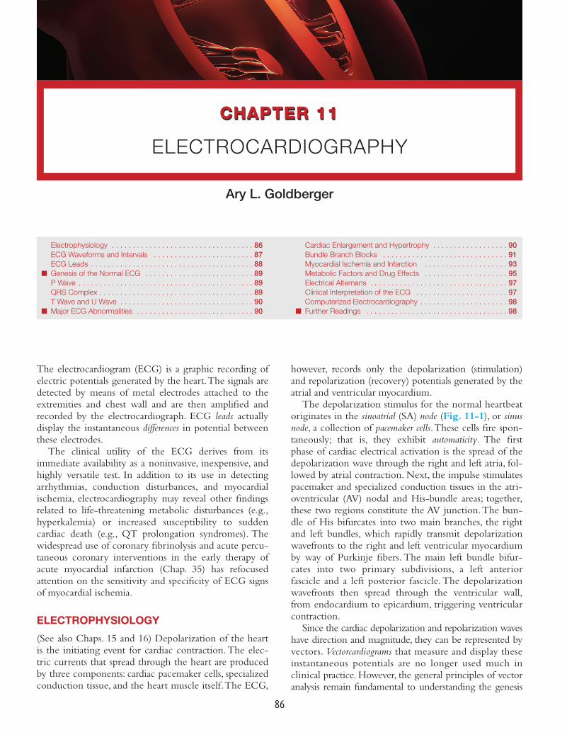
The depolarization stimulus for the normal heartbeatoriginates in the sinoatrial (SA) node (Fig. 11-1), or sinusnode, a collection of pacemaker cells.These cells fire spon-taneously; that is, they exhibit automaticity. The firstphase of cardiac electrical activation is the spread of thedepolarization wave through the right and left atria, fol-lowed by atrial contraction. Next, the impulse stimulatespacemaker and specialized conduction tissues in the atri-oventricular (AV) nodal and His-bundle areas; together,these two regions constitute the AV junction.The bun-dle of His bifurcates into two main branches, the rightand left bundles, which rapidly transmit depolarizationwavefronts to the right and left ventricular myocardiumby way of Purkinje fibers. The main left bundle bifur-cates into two primary subdivisions, a left anteriorfascicle and a left posterior fascicle. The depolarizationwavefronts then spread through the ventricular wall,from endocardium to epicardium, triggering ventricularcontraction.
Since the cardiac depolarization and repolarization waveshave direction and magnitude, they can be represented byvectors. Vectorcardiograms that measure and display theseinstantaneous potentials are no longer used much inclinical practice. However, the general principles of vectoranalysis remain fundamental to understanding the genesis
Ary L. Goldberger
ELECTROCARDIOGRAPHY
CHAPTER 11CHAPTER 11
Electrophysiology . . . . . . . . . . . . . . . . . . . . . . . . . . . . . . . . . . 86ECG Waveforms and Intervals . . . . . . . . . . . . . . . . . . . . . . . . 87ECG Leads . . . . . . . . . . . . . . . . . . . . . . . . . . . . . . . . . . . . . . . 88
■ Genesis of the Normal ECG . . . . . . . . . . . . . . . . . . . . . . . . . . 89P Wave . . . . . . . . . . . . . . . . . . . . . . . . . . . . . . . . . . . . . . . . . . 89QRS Complex . . . . . . . . . . . . . . . . . . . . . . . . . . . . . . . . . . . . . 89T Wave and U Wave . . . . . . . . . . . . . . . . . . . . . . . . . . . . . . . . 90
■ Major ECG Abnormalities . . . . . . . . . . . . . . . . . . . . . . . . . . . . 90
Cardiac Enlargement and Hypertrophy . . . . . . . . . . . . . . . . . . 90Bundle Branch Blocks . . . . . . . . . . . . . . . . . . . . . . . . . . . . . . 91Myocardial Ischemia and Infarction . . . . . . . . . . . . . . . . . . . . 93Metabolic Factors and Drug Effects . . . . . . . . . . . . . . . . . . . . 95Electrical Alternans . . . . . . . . . . . . . . . . . . . . . . . . . . . . . . . . . 97Clinical Interpretation of the ECG . . . . . . . . . . . . . . . . . . . . . . 97Computerized Electrocardiography . . . . . . . . . . . . . . . . . . . . . 98
■ Further Readings . . . . . . . . . . . . . . . . . . . . . . . . . . . . . . . . . . 98
Loscalzo_Cardiovascular_Ch11_p086-098.qxd 2/4/10 5:19 PM Page 86

of normal and pathologic ECG waveforms.Vector analysisillustrates a central concept of electrocardiography—thatthe ECG records the complex spatial and temporal sum-mation of electrical potentials from multiple myocardialfibers conducted to the surface of the body.This principleaccounts for inherent limitations in both ECG sensitivity(activity from certain cardiac regions may be canceled outor may be too weak to be recorded) and specificity (thesame vectorial sum can result from either a selective gainor a loss of forces in opposite directions).
ECG WAVEFORMS AND INTERVALS
The ECG waveforms are labeled alphabetically, begin-ning with the P wave, which represents atrial depolar-ization (Fig. 11-2). The QRS complex represents
ventricular depolarization, and the ST-T-U complex(ST segment,T wave, and U wave) represents ventricularrepolarization. The J point is the junction between theend of the QRS complex and the beginning of the STsegment.Atrial repolarization is usually too low in ampli-tude to be detected, but it may become apparent in suchconditions as acute pericarditis or atrial infarction.
The QRS-T waveforms of the surface ECG corre-spond in a general way with the different phases ofsimultaneously obtained ventricular action potentials, theintracellular recordings from single myocardial fibers(Chap. 15). The rapid upstroke (phase 0) of the actionpotential corresponds to the onset of QRS.The plateau(phase 2) corresponds to the isoelectric ST segment, andactive repolarization (phase 3) to the inscription of theT wave. Factors that decrease the slope of phase 0 byimpairing the influx of Na+ (e.g., hyperkalemia, or drugssuch as flecainide) tend to increase QRS duration.Conditions that prolong phase 2 (use of amiodarone,hypocalcemia) increase the QT interval. In contrast,shortening of ventricular repolarization (phase 2), as bydigitalis administration or hypercalcemia, abbreviates theST segment.
The electrocardiogram is ordinarily recorded on spe-cial graph paper which is divided into 1 mm2 gridlikeboxes. Since the ECG paper speed is generally 25 mm/s,the smallest (1 mm) horizontal divisions correspond to0.04 (40 ms), with heavier lines at intervals of 0.20 s(200 ms).Vertically, the ECG graph measures the ampli-tude of a given wave or deflection (1 mV = 10 mm withstandard calibration; the voltage criteria for hypertrophymentioned below are given in millimeters). There arefour major ECG intervals: R-R, PR, QRS, and QT(Fig. 11-2). The heart rate (beats per minute) can bereadily computed from the interbeat (R-R) intervalby dividing the number of large (0.20 s) time unitsbetween consecutive R waves into 300 or the numberof small (0.04 s) units into 1500.The PR interval mea-sures the time (normally 120–200 ms) between atrialand ventricular depolarization, which includes the phys-iologic delay imposed by stimulation of cells in the AVjunction area.The QRS interval (normally 100–110 msor less) reflects the duration of ventricular depolariza-tion.The QT interval includes both ventricular depolar-ization and repolarization times and varies inverselywith the heart rate. A rate-related (“corrected”) QTinterval, QTc, can be calculated as QT/√RR and normallyis ≤0.44 s. (Some references give QTc upper normal limitsas 0.43 s in men and 0.45 s in women.)
The QRS complex is subdivided into specific deflec-tions or waves. If the initial QRS deflection in a givenlead is negative, it is termed a Q wave; the first positivedeflection is termed an R wave. A negative deflectionafter an R wave is an S wave. Subsequent positive ornegative waves are labeled R′ and S′, respectively.
CHAPTER 11Electrocardiography
87
Ventricularmyocardium
Purkinjefibers
Left bundlebranch
Ventricular septum
Right bundle branch
His bundle
AV junction
Sinoatrial (SA)node
AV nodeRA
LA
LV
RV
FIGURE 11-1Schematic of the cardiac conduction system.
QRS
PST
T
U
PR interval
J
QRS interval
QT interval
FIGURE 11-2Basic ECG waveforms and intervals. Not shown is the R-Rinterval, the time between consecutive QRS complexes.
Loscalzo_Cardiovascular_Ch11_p086-098.qxd 2/4/10 5:19 PM Page 87

Lowercase letters (qrs) are used for waves of relativelysmall amplitude. An entirely negative QRS complex istermed a QS wave.
ECG LEADS
The 12 conventional ECG leads record the difference inpotential between electrodes placed on the surface ofthe body. These leads are divided into two groups: sixlimb (extremity) leads and six chest (precordial) leads.Thelimb leads record potentials transmitted onto the frontalplane (Fig. 11-3A), and the chest leads record potentialstransmitted onto the horizontal plane (Fig. 11-3B). Thesix limb leads are further subdivided into three standard“bipolar” leads (I, II, and III) and three augmented“unipolar” leads (aVR, aVL, and aVF). Each standard leadmeasures the difference in potential between electrodesat two extremities: lead I = left arm – right arm voltages,lead II = left leg – right arm, and lead III = left leg –left arm.The unipolar leads measure the voltage (V) at onelocus relative to an electrode (called the central terminalor indifferent electrode) that has approximately zero potential.Thus, aVR = right arm, aVL = left arm, and aVF = leftleg (foot). The lowercase a indicates that these unipolarpotentials are electrically augmented by 50%. The rightleg electrode functions as a ground.The spatial orienta-tion and polarity of the six frontal plane leads is repre-sented on the hexaxial diagram (Fig. 11-4).
The six chest leads (Fig. 11-5) are unipolar recordingsobtained by electrodes in the following positions: lead V1,fourth intercostal space, just to the right of the sternum;lead V2, fourth intercostal space, just to the left of thesternum; lead V3, midway between V2 and V4; lead V4,midclavicular line, fifth intercostal space; lead V5, anterioraxillary line, same level as V4; and lead V6, midaxillary line,same level as V4 and V5.
Together, the frontal and horizontal plane electrodesprovide a three-dimensional representation of cardiacelectrical activity. Each lead can be likened to a differentcamera angle “looking” at the same events—atrial and ven-tricular depolarization and repolarization—from different
spatial orientations. The conventional 12-lead ECG canbe supplemented with additional leads under special cir-cumstances. For example, right precordial leads V3R,V4R, etc., are useful in detecting evidence of acute rightventricular ischemia. Bedside monitors and ambulatoryECG (Holter) recordings usually employ only one ortwo modified leads. Intracardiac electrocardiogra-phy and electrophysiologic testing are discussedin Chaps. 15 and 16.
The ECG leads are configured so that a positive(upright) deflection is recorded in a lead if a wave of depo-larization spreads toward the positive pole of that lead, and
SECTION IIDiagnosis of Cardiovascular Disorders
88
A
Superior
Inferior
Right Left
+
–––
–
––
+
+++
+aVR aVL
IIIII aVF
B
Posterior
Right Left
+
+
++++
V6
V5
V4V3V2V1
Anterior
––––
– –
I
FIGURE 11-3The six frontal plane (A) and six horizontalplane (B) leads provide a three-dimensional rep-resentation of cardiac electrical activity.
Ext
rem
eax
isde
viation
Normal
axis
Right axis
deviation
–90°–aVF –60°
–III
–30°+aVL
0°+I
+30°–aVR
+60°+II+90°
+aVF
+120°+III
+150°– aVL
180°–I
–150°+aVR
–120°–II
Left axis deviation
FIGURE 11-4The frontal plane (limb or extremity) leads are representedon a hexaxial diagram. Each ECG lead has a specific spatialorientation and polarity. The positive pole of each lead axis(solid line) and negative pole (hatched line) are designated bytheir angular position relative to the positive pole of lead I(0°). The mean electrical axis of the QRS complex is measuredwith respect to this display.
Loscalzo_Cardiovascular_Ch11_p086-098.qxd 2/4/10 5:19 PM Page 88

a negative deflection if the wave spreads toward the nega-tive pole. If the mean orientation of the depolarizationvector is at right angles to a given lead axis, a biphasic (equallypositive and negative) deflection will be recorded.
GENESIS OF THE NORMAL ECG
P WAVE
The normal atrial depolarization vector is orienteddownward and toward the subject’s left, reflecting thespread of depolarization from the sinus node to the rightand then the left atrial myocardium. Since this vectorpoints toward the positive pole of lead II and toward thenegative pole of lead aVR, the normal P wave will bepositive in lead II and negative in lead aVR. By contrast,activation of the atria from an ectopic pacemaker in thelower part of either atrium or in the AV junction regionmay produce retrograde P waves (negative in lead II,positive in lead aVR). The normal P wave in lead V1
may be biphasic with a positive component reflectingright atrial depolarization, followed by a small (<1 mm2)negative component reflecting left atrial depolarization.
QRS COMPLEX
Normal ventricular depolarization proceeds as a rapid,continuous spread of activation wavefronts. This com-plex process can be divided into two major, sequentialphases, and each phase can be represented by a meanvector (Fig. 11-6). The first phase is depolarization ofthe interventricular septum from the left to the rightand anteriorly (vector 1). The second results from thesimultaneous depolarization of the right and left ventri-cles; it is normally dominated by the more massive leftventricle, so that vector 2 points leftward and posteriorly.
Therefore, a right precordial lead (V1) will record thisbiphasic depolarization process with a small positivedeflection (septal r wave) followed by a larger negativedeflection (S wave). A left precordial lead, e.g. V6, willrecord the same sequence with a small negative deflec-tion (septal q wave) followed by a relatively tall positivedeflection (R wave). Intermediate leads show a relativeincrease in R-wave amplitude (normal R-wave progres-sion) and a decrease in S-wave amplitude progressingacross the chest from the right to left.The precordial leadwhere the R and S waves are of approximately equalamplitude is referred to as the transition zone (usuallyV3 or V4) (Fig. 11-7).
The QRS pattern in the extremity leads may varyconsiderably from one normal subject to another
CHAPTER 11Electrocardiography
89
V1
V3R
V4R
V2
V3
V4 V5V6
FIGURE 11-5The horizontal plane (chest or precordial) leads areobtained with electrodes in the locations shown.
– –––––
2
1
+ + ++
+
+
V1 V2V3
V4
V5
V6
C
V1
r
S
V1
RV LV
2q
V6
R
B
V1
rRV
LV
1q
V6
A
FIGURE 11-6Ventricular depolarization can be divided into two majorphases, each represented by a vector. A. The first phase(arrow 1) denotes depolarization of the ventricular septum,beginning on the left side and spreading to the right. Thisprocess is represented by a small “septal” r wave in lead V1
and a small septal q wave in lead V6. B. Simultaneous depo-larization of the left and right ventricles (LV and RV) consti-tutes the second phase. Vector 2 is oriented to the left andposteriorly, reflecting the electrical predominance of theLV. C. Vectors (arrows) representing these two phases areshown in reference to the horizontal plane leads. (AfterGoldberger, 2006.)
Loscalzo_Cardiovascular_Ch11_p086-098.qxd 2/4/10 5:19 PM Page 89

depending on the electrical axis of the QRS, whichdescribes the mean orientation of the QRS vector withreference to the six frontal plane leads. Normally, theQRS axis ranges from −30° to +100° (Fig. 11-4). Anaxis more negative than −30° is referred to as left axisdeviation, while an axis more positive than +100° isreferred to as right axis deviation. Left axis deviation mayoccur as a normal variant but is more commonly associ-ated with left ventricular hypertrophy, a block in theanterior fascicle of the left bundle system (left anteriorfascicular block or hemiblock), or inferior myocardialinfarction. Right axis deviation may also occur as a nor-mal variant (particularly in children and young adults); asa spurious finding due to reversal of the left and rightarm electrodes; or in conditions such as right ventricularoverload (acute or chronic), infarction of the lateral wallof the left ventricle, dextrocardia, left pneumothorax, orleft posterior fascicular block.
T WAVE AND U WAVE
Normally, the mean T-wave vector is oriented roughlyconcordant with the mean QRS vector (within about45° in the frontal plane). Since depolarization and repo-larization are electrically opposite processes, this normalQRS–T-wave vector concordance indicates that repolar-ization must normally proceed in the reverse directionfrom depolarization (i.e., from ventricular epicardium toendocardium). The normal U wave is a small, roundeddeflection (≤1 mm) that follows the T wave and usuallyhas the same polarity as the T wave. An abnormalincrease in U-wave amplitude is most commonly due todrugs (e.g., dofetilide, amiodarone, sotalol, quinidine, pro-cainamide, disopyramide) or to hypokalemia.Very promi-nent U waves are a marker of increased susceptibility to
the torsades de pointe type of ventricular tachycardia(Chap. 16). Inversion of the U wave in the precordial leadsis abnormal and may be a subtle sign of ischemia.
MAJOR ECG ABNORMALITIES
CARDIAC ENLARGEMENT ANDHYPERTROPHY
Right atrial overload (acute or chronic) may lead to anincrease in P-wave amplitude (≥2.5 mm) (Fig. 11-8).Left atrial overload typically produces a biphasic P wavein V1 with a broad negative component or a broad(≥120 ms), often notched P wave in one or more limbleads (Fig. 11-8). This pattern may also occur with leftatrial conduction delays in the absence of actual atrialenlargement, leading to the more general designation ofleft atrial abnormality.
Right ventricular hypertrophy due to a pressure load(as from pulmonic valve stenosis or pulmonary arteryhypertension) is characterized by a relatively tall R wavein lead V1 (R ≥ S wave), usually with right axis deviation(Fig. 11-9); alternatively, there may be a qR pattern inV1 or V3R. ST depression and T-wave inversion in theright to midprecordial leads are also often present.This pattern, formerly called right ventricular “strain,” isattributed to repolarization abnormalities in acutely orchronically overloaded muscle. Prominent S waves mayoccur in the left lateral precordial leads. Right ventricu-lar hypertrophy due to ostium secundum–type atrialseptal defects, with the accompanying right ventricularvolume overload, is commonly associated with an incom-plete or complete right bundle branch block patternwith a rightward QRS axis.
SECTION IIDiagnosis of Cardiovascular Disorders
90
aVRI
II
III
aVL
aVF
V1
V2
V3
V4
V5
V6
FIGURE 11-7Normal electrocardiogram from a healthy subject. Sinusrhythm is present with a heart rate of 75 beats per minute.PR interval is 0.16 s; QRS interval (duration) is 0.08 s;
QT interval is 0.36 s; QTc is 0.40 s; the mean QRS axis is about+70°. The precordial leads show normal R-wave progressionwith the transition zone (R wave = S wave) in lead V3.
Loscalzo_Cardiovascular_Ch11_p086-098.qxd 2/4/10 5:19 PM Page 90

Acute cor pulmonale due to pulmonary embolism, forexample, may be associated with a normal ECG or avariety of abnormalities. Sinus tachycardia is the mostcommon arrhythmia, although other tachyarrhythmias,such as atrial fibrillation or flutter, may occur.The QRSaxis may shift to the right, sometimes in concert withthe so-called S1Q3T3 pattern (prominence of the S wavein lead I, Q wave in lead III, with T-wave inversion inlead III). Acute right ventricular dilation may also beassociated with slow R-wave progression and T-waveinversions in V1–V4 simulating acute anterior infarc-tion. A right ventricular conduction disturbance mayappear.
Chronic cor pulmonale due to obstructive lung disease(Chap. 17) usually does not produce the classic ECGpatterns of right ventricular hypertrophy noted above.Instead of tall right precordial R waves, chronic lungdisease more typically is associated with small R wavesin right-to-midprecordial leads (slow R-wave progres-sion) due in part to downward displacement of thediaphragm and the heart. Low-voltage complexes arecommonly present, owing to hyperaeration of the lungs.
A number of different voltage criteria for left ventricu-lar hypertrophy (Fig. 11-9) have been proposed on thebasis of the presence of tall left precordial R waves anddeep right precordial S waves [e.g., SV1 + (RV5 or RV6)>35 mm]. Repolarization abnormalities (ST depressionwith T-wave inversions, formerly called the left ventric-ular “strain” pattern) may also appear in leads withprominent R waves. However, prominent precordialvoltages may occur as a normal variant, especially in ath-letic or young individuals. Left ventricular hypertrophymay increase limb lead voltage with or withoutincreased precordial voltage (e.g., RaVL + SV3 > 20 mmin women and >28 mm in men). The presence of leftatrial abnormality increases the likelihood of underlyingleft ventricular hypertrophy in cases with borderlinevoltage criteria. Left ventricular hypertrophy often pro-gresses to incomplete or complete left bundle branchblock.The sensitivity of conventional voltage criteria forleft ventricular hypertrophy is decreased in obese per-sons and in smokers. ECG evidence for left ventricularhypertrophy is a major noninvasive marker of increasedrisk of cardiovascular morbidity and mortality, includingsudden cardiac death. However, because of false-positiveand false-negative diagnoses, the ECG is of limited utilityin diagnosing atrial or ventricular enlargement. Moredefinitive information is provided by echocardiography(Chap. 12).
BUNDLE BRANCH BLOCKS
Intrinsic impairment of conduction in either the rightor left bundle system (intraventricular conduction dis-turbances) leads to prolongation of the QRS interval.Withcomplete bundle branch blocks, the QRS interval
CHAPTER 11Electrocardiography
91
RA
LA
Normal Right Left
II
V1
RA
RARA
RA
RA
RA
LA
LALALA
LA LA
V1
FIGURE 11-8Right atrial (RA) overload may cause tall, peaked Pwaves in the limb or precordial leads. Left atrial (LA)abnormality may cause broad, often notched P waves in thelimb leads and a biphasic P wave in lead V1 with a prominentnegative component representing delayed depolarization ofthe LA. (After MK Park, WG Guntheroth: How to ReadPediatric ECGs, 4th ed. St. Louis, Mosby/Elsevier, 2006.)
QRS in hypertrophy
V1
Main QRS vector
Normal
RVHor or
V1
V6
V6
LVH
FIGURE 11-9Left ventricular hypertrophy (LVH) increases the ampli-tude of electrical forces directed to the left and posteri-orly. In addition, repolarization abnormalities may causeST-segment depression and T-wave inversion in leads with aprominent R wave. Right ventricular hypertrophy (RVH) mayshift the QRS vector to the right; this effect usually is associ-ated with an R, RS, or qR complex in lead V1. T-wave inver-sions may be present in right precordial leads.
Loscalzo_Cardiovascular_Ch11_p086-098.qxd 2/4/10 5:19 PM Page 91

is ≥120 ms in duration; with incomplete blocks the QRSinterval is between 100 and 120 ms.The QRS vector isusually oriented in the direction of the myocardialregion where depolarization is delayed (Fig. 11-10).Thus, with right bundle branch block, the terminalQRS vector is oriented to the right and anteriorly(rSR′ in V1 and qRS in V6, typically). Left bundle branchblock alters both early and later phases of ventriculardepolarization.The major QRS vector is directed to theleft and posteriorly. In addition, the normal early left-to-right pattern of septal activation is disrupted such thatseptal depolarization proceeds from right to left as well.As a result, left bundle branch block generates wide,predominantly negative (QS) complexes in lead V1 andentirely positive (R) complexes in lead V6. A patternidentical to that of left bundle branch block, precededby a sharp spike, is seen in most cases of electronic rightventricular pacing because of the relative delay in leftventricular activation.
Bundle branch block may occur in a variety of con-ditions. In subjects without structural heart disease, rightbundle branch block is seen more commonly than leftbundle branch block. Right bundle branch block alsooccurs with heart disease, both congenital (e.g., atrialseptal defect) and acquired (e.g., valvular, ischemic). Leftbundle branch block is often a marker of one of fourunderlying conditions associated with increased risk of
cardiovascular morbidity and mortality: coronary heartdisease (frequently with impaired left ventricular func-tion), hypertensive heart disease, aortic valve disease, andcardiomyopathy. Bundle branch blocks may be chronicor intermittent. A bundle branch block may be rate-related; for example, it often occurs when the heart rateexceeds some critical value.
Bundle branch blocks and depolarization abnormalitiessecondary to artificial pacemakers not only affect ventric-ular depolarization (QRS) but are also characteristicallyassociated with secondary repolarization (ST-T) abnormali-ties. With bundle branch blocks, the T wave is typicallyopposite in polarity to the last deflection of the QRS(Fig. 11-10).This discordance of the QRS–T-wave vectorsis caused by the altered sequence of repolarization thatoccurs secondary to altered depolarization. In contrast,primary repolarization abnormalities are independent of QRSchanges and are related instead to actual alterations in theelectrical properties of the myocardial fibers themselves(e.g., in the resting membrane potential or action potentialduration), not just to changes in the sequence of repolar-ization. Ischemia, electrolyte imbalance, and drugs suchas digitalis all cause such primary ST–T-wave changes.Primary and secondary T-wave changes may coexist. Forexample, T-wave inversions in the right precordial leadswith left bundle branch block or in the left precordialleads with right bundle branch block may be importantmarkers of underlying ischemia or other abnormalities.A distinctive abnormality simulating right bundle branchblock with ST-segment elevations in the right chest leadsis seen with the Brugada pattern (Chap. 16).
Partial blocks (fascicular or “hemiblocks”) in the leftbundle system (left anterior or posterior fascicular blocks)generally do not prolong the QRS duration substantiallybut instead are associated with shifts in the frontal planeQRS axis (leftward or rightward, respectively). Morecomplex combinations of fascicular and bundle branchblocks may occur involving the left and right bundlesystem. Examples of bifascicular block include right bundlebranch block and left posterior fascicular block, rightbundle branch block with left anterior fascicular block,and complete left bundle branch block. Chronic bifascic-ular block in an asymptomatic individual is associatedwith a relatively low risk of progression to high-degreeAV heart block. In contrast, new bifascicular blockwith acute anterior myocardial infarction carries a muchgreater risk of complete heart block. Alternation of rightand left bundle branch block is a sign of trifascicular disease.However, the presence of a prolonged PR interval andbifascicular block does not necessarily indicate trifasci-cular involvement, since this combination may arisewith AV node disease and bifascicular block. Intraven-tricular conduction delays can also be caused by extrinsic(toxic) factors that slow ventricular conduction, particu-larly hyperkalemia or drugs (e.g., class 1 antiarrhythmicagents, tricyclic antidepressants, phenothiazines).
SECTION IIDiagnosis of Cardiovascular Disorders
92
T
R′
R
S
V1Normal
RBBB
LBBB
q
S
R
V6
T
FIGURE 11-10Comparison of typical QRS-T patterns in right bundlebranch block (RBBB) and left bundle branch block (LBBB)with the normal pattern in leads V1 and V6. Note the sec-ondary T-wave inversions (arrows) in leads with an rSR′ com-plex with RBBB and in leads with a wide R wave with LBBB.
Loscalzo_Cardiovascular_Ch11_p086-098.qxd 2/4/10 5:19 PM Page 92

Prolongation of QRS duration does not necessarilyindicate a conduction delay but may be due to preexcitationof the ventricles via a bypass tract, as in Wolff-Parkinson-White (WPW) patterns (Chap. 16) and related variants.The diagnostic triad of WPW consists of a wide QRScomplex associated with a relatively short PR interval andslurring of the initial part of the QRS (delta wave), thelatter effect due to aberrant activation of ventricularmyocardium.The presence of a bypass tract predisposes toreentrant supraventricular tachyarrhythmias.
MYOCARDIAL ISCHEMIA AND INFARCTION
(See also Chap. 35) The ECG is a cornerstone in thediagnosis of acute and chronic ischemic heart disease.The findings depend on several key factors: the natureof the process [reversible (i.e., ischemia) versus irreversible(i.e., infarction)], the duration (acute versus chronic),extent (transmural versus subendocardial), and localization(anterior versus inferoposterior), as well as the presenceof other underlying abnormalities (ventricular hypertro-phy, conduction defects).
Ischemia exerts complex time-dependent effects onthe electrical properties of myocardial cells. Severe, acuteischemia lowers the resting membrane potential andshortens the duration of the action potential. Such changescause a voltage gradient between normal and ischemiczones. As a consequence, current flows between theseregions. These currents of injury are represented on thesurface ECG by deviation of the ST segment (Fig. 11-11).When the acute ischemia is transmural, the ST vector isusually shifted in the direction of the outer (epicardial) lay-ers, producing ST elevations and sometimes, in the earlieststages of ischemia, tall, positive so-called hyperacuteT waves over the ischemic zone.With ischemia confinedprimarily to the subendocardium, the ST vector typically shiftstoward the subendocardium and ventricular cavity, so thatoverlying (e.g., anterior precordial) leads show ST-segmentdepression (with ST elevation in lead aVR). Multiple fac-tors affect the amplitude of acute ischemic ST deviations.Profound ST elevation or depression in multiple leads
usually indicates very severe ischemia. From a clinicalviewpoint, the division of acute myocardial infarctioninto ST segment elevation and non-ST elevation typesis useful since the efficacy of acute reperfusion therapy islimited to the former group.
The ECG leads are more helpful in localizing regionsof ST elevation than non-ST elevation ischemia. Forexample, acute transmural anterior (including apical andlateral) wall ischemia is reflected by ST elevations orincreased T-wave positivity in one or more of the pre-cordial leads (V1–V6) and leads I and aVL. Inferior wallischemia produces changes in leads II, III, and aVF. Pos-terior wall ischemia may be indirectly recognized byreciprocal ST depressions in leads V1–V3. Prominentreciprocal ST depressions in these leads also occur withcertain inferior wall infarcts, particularly those withposterior or lateral wall extension. Right ventricularischemia usually produces ST elevations in right-sidedchest leads (Fig. 11-5). When ischemic ST elevationsoccur as the earliest sign of acute infarction, they aretypically followed within a period ranging from hoursto days by evolving T-wave inversions and often byQ waves occurring in the same lead distribution. (T-waveinversions due to evolving or chronic ischemia correlatewith prolongation of repolarization and are often associatedwith QT lengthening.) Reversible transmural ischemia, forexample, due to coronary vasospasm (Prinzmetal’s variantangina, and possibly the tako-tsubo cardiomyopathy syn-drome), may cause transient ST-segment elevations with-out development of Q waves. Depending on the severityand duration of such ischemia, the ST elevations mayeither resolve completely in minutes or be followed byT-wave inversions that persist for hours or even days.Patients with ischemic chest pain who present with deepT-wave inversions in multiple precordial leads (e.g.,V1–V4)with or without cardiac enzyme elevations typically havesevere obstruction in the left anterior descending coronaryartery system (Fig. 11-12). In contrast, patients whosebaseline ECG already shows abnormal T-wave inversionsmay develop T-wave normalization (pseudonormaliza-tion) during episodes of acute transmural ischemia.
CHAPTER 11Electrocardiography
93
FIGURE 11-11Acute ischemia causes a current of injury. With predomi-nant subendocardial ischemia (A), the resultant ST vector willbe directed toward the inner layer of the affected ventricleand the ventricular cavity. Overlying leads therefore will
record ST depression. With ischemia involving the outerventricular layer (B) (transmural or epicardial injury), the STvector will be directed outward. Overlying leads will recordST elevation.
V5
ST
ST
A B
ST
ST
V5
Loscalzo_Cardiovascular_Ch11_p086-098.qxd 2/4/10 5:19 PM Page 93

With infarction, depolarization (QRS) changes oftenaccompany repolarization (ST-T) abnormalities. Necrosisof sufficient myocardial tissue may lead to decreasedR-wave amplitude or abnormal Q waves in the anterior orinferior leads (Fig. 11-13). Previously, abnormal Q waveswere considered to be markers of transmural myocardialinfarction, while subendocardial infarcts were thought notto produce Q waves. However, careful ECG-pathologycorrelative studies have indicated that transmural infarctsmay occur without Q waves and that subendocardial(nontransmural) infarcts may sometimes be associatedwith Q waves. Therefore, infarcts are more appropriatelyclassified as “Q-wave” or “non-Q-wave.”The major acuteECG changes in syndromes of ischemic heart diseaseare summarized schematically in Fig. 11-14. Loss of
depolarization forces due to posterior or lateral infarctionmay cause reciprocal increases in R-wave amplitude inleads V1 and V2 without diagnostic Q waves in any of theconventional leads.Atrial infarction may be associated withPR-segment deviations due to an atrial current of injury,changes in P-wave morphology, or atrial arrhythmias.
In the weeks and months following infarction, theseECG changes may persist or begin to resolve. Completenormalization of the ECG following Q-wave infarctionis uncommon but may occur, particularly with smallerinfarcts. In contrast, ST-segment elevations that persistfor several weeks or more after a Q-wave infarct usuallycorrelate with a severe underlying wall motion disorder(akinetic or dyskinetic zone), although not necessarily afrank ventricular aneurysm.
SECTION IIDiagnosis of Cardiovascular Disorders
94 V1 V2 V4 V5 V6V3
FIGURE 11-12Severe anterior wall ischemia (with or without infarction)may cause prominent T-wave inversions in the precordial
leads. This pattern is usually associated with a high-gradestenosis of the left anterior descending coronary artery.
A
B
ECG sequence with anterior Q wave infarction
Early
Evolving
Early
Evolving
ECG sequence with inferior Q wave infarction
I II III
I II III
aVR aVL aVF V2 V4 V6
aVR aVL aVF V2 V4 V6
FIGURE 11-13Sequence of depolarization and repolarization changeswith (A) acute anterior and (B) acute inferior wall Q-waveinfarctions. With anterior infarcts, ST elevation in leads I, aVL,and the precordial leads may be accompanied by reciprocal
ST depressions in leads II, III, and aVF. Conversely, acuteinferior (or posterior) infarcts may be associated with recip-rocal ST depressions in leads V1 to V3. (After Goldberger,2006.)
Loscalzo_Cardiovascular_Ch11_p086-098.qxd 2/4/10 5:19 PM Page 94

CHAPTER 11Electrocardiography
95
ECG changes due to ischemia may occur sponta-neously or may be provoked by various exercise protocols(stress electrocardiography) (Chap. 33). In patients withsevere ischemic heart disease, exercise testing is mostlikely to elicit signs of subendocardial ischemia (horizon-tal or downsloping ST depression in multiple leads).ST-segment elevation during exercise is most oftenobserved after a Q-wave infarct.This repolarization changedoes not necessarily indicate active ischemia but correlatesstrongly with the presence of an underlying ventricularwall motion abnormality. However, in patients withoutprior infarction, transient ST-segment elevation with exer-cise is a reliable sign of transmural ischemia.
The ECG has important limitations in both sensitivityand specificity in the diagnosis of ischemic heart disease.Although a single normal ECG does not excludeischemia or even acute infarction, a normal ECG through-out the course of an acute infarct is distinctly uncommon.Prolonged chest pain without diagnostic ECG changes,therefore, should always prompt a careful search for othernoncoronary causes of chest pain (Chap. 4). Furthermore,the diagnostic changes of acute or evolving ischemia areoften masked by the presence of left bundle branch block,electronic ventricular pacemaker patterns, and WPW pre-excitation. On the other hand, clinicians may overdiag-nose ischemia or infarction based on the presence of ST-segment elevations or depressions,T-wave inversions, tallpositive T waves, or Q waves not related to ischemic heartdisease (pseudoinfarct patterns). For example, ST-segment
elevations simulating ischemia may occur with acutepericarditis or myocarditis, as a normal variant (“earlyrepolarization” pattern), or in a variety of other condi-tions (Table 11-1). Similarly, tall, positive T waves do notinvariably represent hyperacute ischemic changes but mayalso be caused by normal variants, hyperkalemia, cere-brovascular injury, and left ventricular volume overloaddue to mitral or aortic regurgitation, among other causes.
ST-segment elevations and tall, positive T waves arecommon findings in leads V1 and V2 in left bundlebranch block or left ventricular hypertrophy in theabsence of ischemia.The differential diagnosis of Q waves(Table 11-2) includes physiologic or positional variants,ventricular hypertrophy, acute or chronic noncoronarymyocardial injury, hypertrophic cardiomyopathy, and ven-tricular conduction disorders. Digoxin, ventricular hyper-trophy, hypokalemia, and a variety of other factors maycause ST-segment depression mimicking subendocardialischemia. Prominent T-wave inversion may occur withventricular hypertrophy, cardiomyopathies, myocarditis,and cerebrovascular injury (particularly intracranial bleeds),among many other conditions.
METABOLIC FACTORS AND DRUG EFFECTS
A variety of metabolic and pharmacologic agentsalter the ECG and, in particular, cause changes inrepolarization (ST-T-U) and sometimes QRS prolon-gation. Certain life-threatening electrolyte disturbances
FIGURE 11-14Variability of ECG patterns with acute myocardial ischemia.The ECG also may be normal or nonspecifically abnormal.Furthermore, these categorizations are not mutually exclusive.For example, a non-ST elevation infarct may evolve into aQ-wave infarct; ST elevations may be followed by a non-Q-wave infarct; or ST depressions and T-wave inversions may befollowed by a Q-wave infarct. (After Goldberger, 2006.)
Noninfarctionsubendocardial
ischemia(classic angina)
Transient STdepressions
Noninfarctiontransmural ischemia
Transient ST elevationsor paradoxical T-wave
normalization,sometimes followed
by T-waveinversions
Non-Q wave(Non-ST elevation)
infarctionST depressions or
T-waveinversions
without Q waves
MYOCARDIALISCHEMIA
ST elevation/Q wave infarction
New Q waves precededby hyperacute
T-waves/ST elevationsand followed by
T-wave inversions
TABLE 11-1
DIFFERENTIAL DIAGNOSIS OF ST SEGMENTELEVATIONS
Ischemia/myocardial infarctionNoninfarction, transmural ischemia (Prinzmetal’s angina, and possibly tako-tsubo syndrome)
Acute myocardial infarctionPostmyocardial infarction (ventricular aneurysm pattern)
Acute pericarditisNormal variant (“early repolarization” pattern)Left ventricular hypertrophy/left bundle branch blocka
Other (rarer)Brugada pattern (right bundle branch block-like pattern with ST elevations in right precordial leads)a
Class 1C antiarrhythmic drugsa
DC cardioversionHypercalcemiaa
Hyperkalemiaa
Hypothermia (J wave/Osborn waves)Myocardial injury
MyocarditisTumor invading left ventricleTrauma to ventricles
aUsually localized to V1-V2 or V3.Source: Modified from Goldberger, 2006.
Loscalzo_Cardiovascular_Ch11_p086-098.qxd 2/4/10 5:19 PM Page 95

may be diagnosed initially and monitored from theECG. Hyperkalemia produces a sequence of changes(Fig. 11-15), usually beginning with narrowing andpeaking (tenting) of the T waves. Further elevation ofextracellular K+ leads to AV conduction disturbances,
diminution in P-wave amplitude, and widening of theQRS interval. Severe hyperkalemia eventually causescardiac arrest with a slow sinusoidal type of mechanism(“sine-wave” pattern) followed by asystole. Hypokalemia(Fig. 11-16) prolongs ventricular repolarization, oftenwith prominent U waves. Prolongation of the QT inter-val is also seen with drugs that increase the duration ofthe ventricular action potential—class 1A antiarrhythmicagents and related drugs (e.g., quinidine, disopyramide,procainamide, tricyclic antidepressants, phenothiazines)and class III agents [amiodarone (Fig. 11-16), sotalol,ibutilide]. Marked QT prolongation, sometimes withdeep, wide T-wave inversions, may occur with intracranialbleeds, particularly subarachnoid hemorrhage (“CVAT-wave” pattern) (Fig. 11-16). Systemic hypothermia alsoprolongs repolarization, usually with a distinctive convexelevation of the J point (Osborn wave). Hypocalcemia typi-cally prolongs the QT interval (ST portion), while hyper-calcemia shortens it (Fig. 11-17). Digitalis glycosides alsoshorten the QT interval, often with a characteristic“scooping” of the ST–T-wave complex (digitalis effect).
Many other factors are associated with ECG changes,particularly alterations in ventricular repolarization.T-waveflattening, minimal T-wave inversions, or slight ST-segmentdepression (“nonspecific ST–T-wave changes”) may occurwith a variety of electrolyte and acid-base disturbances, avariety of infectious processes, central nervous systemdisorders, endocrine abnormalities, many drugs, ischemia,hypoxia, and virtually any type of cardiopulmonary abnor-mality.While subtle ST–T-wave changes may be markers ofischemia, transient nonspecific repolarization changes mayalso occur following a meal or with postural (orthostatic)change, hyperventilation, or exercise in healthy individuals.
SECTION IIDiagnosis of Cardiovascular Disorders
96 TABLE 11-2
DIFFERENTIAL DIAGNOSIS OF Q WAVES (WITH SELECTED EXAMPLES)
Physiologic or positional factorsNormal variant “septal” q wavesNormal variant Q waves in V1 to V2, aVL, III, and aVFLeft pneumothorax or dextrocardia: loss of lateral R-wave progression
Myocardial injury or infiltrationAcute processes: myocardial ischemia or infarction, myocarditis, hyperkalemia
Chronic processes: myocardial infarction, idiopathic cardiomyopathy, myocarditis, amyloid, tumor, sarcoid,scleroderma, Chagas’ disease, echinococcus cyst
Ventricular hypertrophy/enlargementLeft ventricular (slow R-wave progressiona)Right ventricular (reversed R-wave progressionb or slow R-wave progressiona, particularly with chronicobstructive lung disease)
Hypertrophic cardiomyopathy (may simulate anterior, inferior, posterior, or lateral infarcts)
Conduction abnormalitiesLeft bundle branch block (slow R-wave progressiona)Wolff-Parkinson-White patterns
aSmall or absent R waves in the right to midprecordial leads.bProgressive decrease in R-wave amplitude from V1 to the mid- orlateral precordial leads.Source: After Goldberger, 2006.
Hyperkalemia
Mild-Moderate Moderate-Severe Very Severe
Lead I
Lead II
V1
V2
V1P
T
P
T
V2
1s
1mV
FIGURE 11-15The earliest ECG change with hyperkalemia is usuallypeaking (“tenting”) of the T waves. With further increases inthe serum potassium concentration, the QRS complexes
widen, the P waves decrease in amplitude and may disap-pear, and finally a sine-wave pattern leads to asystole unlessemergency therapy is given. (After Goldberger, 2006.)
Loscalzo_Cardiovascular_Ch11_p086-098.qxd 2/4/10 5:19 PM Page 96

ELECTRICAL ALTERNANS
Electrical alternans—a beat-to-beat alternation in oneor more components of the ECG signal—is a commontype of nonlinear cardiovascular response to a variety ofhemodynamic and electrophysiologic perturbations.Total electrical alternans (P-QRS-T) with sinus tachy-cardia is a relatively specific sign of pericardial effusion,usually with cardiac tamponade. The mechanism relatesto a periodic swinging motion of the heart in the effu-sion at a frequency exactly one-half the heart rate.Repolarization (ST-T or U wave) alternans is a sign ofelectrical instability and may precede ventricular tach-yarrhythmias.
CLINICAL INTERPRETATION OF THE ECG
Accurate analysis of ECGs requires thoroughness andcare.The patient’s age, gender, and clinical status shouldalways be taken into account. For example, T-waveinversions in leads V1–V3 are more likely to represent anormal variant in a healthy young adult woman (“per-sistent juvenile T-wave pattern”) than in an elderly manwith chest discomfort. Similarly, the likelihood thatST-segment depression during exercise testing representsischemia depends partly on the prior probability ofcoronary artery disease.
Many mistakes in ECG interpretation are errors ofomission. Therefore, a systematic approach is essential.The following 14 points should be analyzed carefully inevery ECG: (1) standardization (calibration) and techni-cal features (including lead placement and artifacts);(2) rhythm; (3) heart rate; (4) PR interval/AV conduc-tion; (5) QRS interval; (6) QT/QTc interval; (7) meanQRS electrical axis; (8) P waves; (9) QRS voltages; (10)precordial R-wave progression; (11) abnormal Q waves;(12) ST segments; (13) T waves; (14) U waves.
Only after analyzing all these points should the inter-pretation be formulated. Where appropriate, importantclinical correlates or inferences should be mentioned.For example, the combination of left atrial abnormality(enlargement) and signs of right ventricular hypertrophysuggests mitral stenosis. Low voltage with sinus tachy-cardia raises the possibility of pericardial tamponadeor chronic obstructive lung disease. Sinus tachycardiawith QRS and QT-(U) prolongation, especially inthe context of mental status changes, suggests tricyclic
CHAPTER 11Electrocardiography
97Hypokalemia Hypothermia Amiodarone
II
Tricyclic overdose
I
Subarachnoid hemorrhage
III V3
V2
V3
U
V5 V4T
FIGURE 11-16A variety of metabolic derangements, drug effects, andother factors may prolong ventricular repolarization with QTprolongation or prominent U waves. Prominent repolariza-tion prolongation, particularly if due to hypokalemia, inher-ited “channelopathies,” or certain pharmacologic agents,indicates increased susceptibility to torsades de pointes–type
ventricular tachycardia (Chap. 16). Marked systemic hypother-mia is associated with a distinctive convex “hump” at the J point (Osborn wave, arrow) due to altered ventricular actionpotential characteristics. Note QRS and QT prolongationalong with sinus tachycardia in the case of tricyclic antide-pressant overdose.
Hypocalcemia
QT 0.48 sQTC 0.52
I
II
Normal
QT 0.36 sQTC 0.41
I
II
Hypercalcemia
QT 0.26 sQTC 0.36
I
II
FIGURE 11-17 Prolongation of the Q-T interval (ST-segment portion) istypical of hypocalcemia. Hypercalcemia may cause abbrevi-ation of the ST segment and shortening of the QT interval.
Loscalzo_Cardiovascular_Ch11_p086-098.qxd 2/4/10 5:19 PM Page 97

antidepressant overdose (Fig. 11-16). The triad ofpeaked T waves (hyperkalemia), a long QT due to STsegment lengthening (hypocalcemia) and left ventric-ular hypertrophy (systemic hypertension) suggestschronic renal failure. Comparison with previousECGs is essential. The diagnosis and managementof specific cardiac arrhythmias and conductiondisturbances are discussed in Chaps. 15 and 16.
COMPUTERIZED ELECTROCARDIOGRAPHY
Computerized ECG systems are widely used. Digitalsystems provide for convenient storage and immediateretrieval of thousands of ECG records. Despite advances,computer interpretation of ECGs has important limita-tions. Incomplete or inaccurate readings are most likelywith arrhythmias and complex abnormalities.Therefore,computerized interpretation (including measurements ofbasic ECG intervals) should not be accepted withoutcareful clinician review.
FURTHER READINGS
ASHLEY EA et al:An evidence-based review of the resting electrocar-diogram as a screening technique for heart disease. Prog CardiovascDis 44:55, 2001
GOLDBERGER AL: Clinical Electrocardiography: A Simplified Approach,7th ed. St. Louis, Mosby/Elsevier, 2006
GUGLIN MF,THATAI D: Common errors in computer electrocardio-gram interpretation. Int J Cardiol 106:232, 2006
KRIGFIELD P, et al: Recommendations for the standardization andinterpretation of the electrocardiogram. Part I: The electrocar-diogram and its standardization. J Am Coll Cardiol 49:1109,2007
MIRVIS DM, GOLDBERGER AL: Electrocardiography, in Braunwald’sHeart Disease:A Textbook of Cardiovascular Medicine, 8th ed, P Libbyet al (eds). Philadelphia, Saunders, 2008
SURAWICZ B, KNILANS TK: Chou’s Electrocardiography in ClinicalPractice, 5th ed. Philadelphia, Saunders, 2001
WAGNER G, et al: Recommendations for the standardization andinterpretation of the electrocardiogram. Part VI: Acute myocar-dial ischemia. J Am Coll Cardiol 53:1003, 2009
SECTION IIDiagnosis of Cardiovascular Disorders
98
Loscalzo_Cardiovascular_Ch11_p086-098.qxd 2/4/10 5:19 PM Page 98