Look Inside Fundamentals
30
description
"Look Inside" pages from Fundamentals of Musculoskeletal Pain
Transcript of Look Inside Fundamentals

Mission Statement of IASP Press®
IASP brings together scientists, clinicians, health care providers, and policy makers to stimulate and support the study of pain and to translate that knowledge into improved pain relief worldwide. IASP Press publishes timely, high-quality, and reasonably priced books relating to pain research and treatment.
Fundamentals of Musculoskeletal Pain
Editors
Thomas Graven-Nielsen, Dr Med Sci, PhDDepartment of Health Sciences and Technology, Center for Sensory-Motor
Interaction, Aalborg University, Aalborg, Denmark
Lars Arendt-Nielsen, Dr Med Sci, PhD Department of Health Sciences and Technology, Center for Sensory-Motor
Interaction, Aalborg University, Aalborg, Denmark
Siegfried Mense, Dr MedInstitute of Anatomy and Cell Biology, University of Heidelberg,
Heidelberg, Germany
IASP PRESS® • SEATTLE
© 2008 IASP Press®International Association for the Study of Pain®
All rights reserved. No part of this publication may be reproduced, stored in a retrieval system, or transmitted, in any form or by any means, electronic, mechanical, photocopy-ing, recording, or otherwise, without the prior written permission of the publisher.
Timely topics in pain research and treatment have been selected for publication, but the information provided and opinions expressed have not involved any verification of the findings, conclusions, and opinions by IASP®. Thus, opinions expressed in Fundamentals of Musculoskeletal Pain do not necessarily reflect those of IASP or of the Officers and Councilors.
No responsibility is assumed by IASP for any injury and/or damage to persons or prop-erty as a matter of product liability, negligence, or from any use of any methods, products,instruction, or ideas contained in the material herein. Because of the rapid advances in the medical sciences, the publisher recommends that there should be independent verifi-cation of diagnoses and drug dosages.
Library of Congress Cataloging-in-Publication Data
Fundamentals of musculoskeletal pain / editors, Thomas Graven-Nielsen, Lars Arendt-Nielsen, Siegfried Mense. p. ; cm. Includes bibliographical references. Summary: “Basic scientists and clinicians explain the fundamentals of musculoskeletal pain and describe current research that can benefit clinical strategies. Part I reviews the peripheral and central mechanisms of muscle pain. Part II describes key factors in pain sensitivity, including sex-related and genetic factors. Part III describes the effects of mus-cle pain on motor function.”--Provided by publisher. ISBN 978-0-931092-72-5 (hardcover : alk. paper) 1. Myalgia. I. Graven-Nielsen, Thomas, 1970- II. Arendt-Nielsen, Lars, 1958- III. Mense, Siegfried. IV. International Association for the Study of Pain. [DNLM: 1. Musculoskeletal Diseases--physiopathology. 2. Pain--physiopathology. WE 140 F981 2008] RC935.M77.F86 2008 616.7’42--dc22 2008014881
Published by:IASP PressInternational Association for the Study of Pain111 Queen Anne Ave N, Suite 501Seattle, WA 98109-4955, USAFax: 206-283-9403www.iasp-pain.org
Printed in the United States of America
Contents
List of Contributing Authors ixPreface xiii Acknowledgments xvi
Part I Basic Mechanisms of Muscle Pain1. Morphology and Functional Types of Muscle Nociceptors Siegfried Mense and Ulrich Hoheisel 3
2. Physiological Properties of Thin-Fiber Muscle Afferents: Excitation and Modulatory Effects Brian E. Cairns 19
3. Functional Role of Peripheral Glutamate Receptors in Craniofacial Muscle Pain and Hyperalgesia Jin Y. Ro 33
4. Facilitated Response of Muscle Thin-Fiber Receptors in Mechanical Hyperalgesia after Exercise Kazue Mizumura and Toru Taguchi 47
5. Mechanisms of Central Nervous Hyperexcitability Due to Activation of Muscle Nociceptors Siegfried Mense and Ulrich Hoheisel 61
6. Bilateral, Long-Lasting Hyperalgesia Due to Repeated Excitation of Muscle Nociceptors: A Role for ASIC3 Roxanne Y. Walder and Kathleen A. Sluka 75
7. Central Mechanisms of Craniofacial Musculoskeletal Pain: A Review Barry J. Sessle 87
8. New Perspectives on Descending Pain-Modulating Systems in Musculoskeletal Pain Wahida Rahman and Anthony H. Dickenson 105
9. Peripheral Aspects of Cytokines in Musculoskeletal Pain Sigvard Kopp and Per Alstergren 119
10. Serotonergic Receptor Involvement in Muscle Pain and Hyperalgesia Malin Ernberg 139
v

vi Contents
11. Human Models and Clinical Manifestations of Musculoskeletal Pain and Pain-Motor Interactions Thomas Graven-Nielsen and Lars Arendt-Nielsen 155
12. Central Representation of Muscle Pain and Hyperalgesia Peter Svensson and Randi Abrahamsen 189
Part II Key Factors Determining Muscle Pain Sensitivity 13. Referred Muscular Hyperalgesia from Visceral Structures Maria Adele Giamberardino, Raffaele Costantini, and Giannapia Affaitati 207
14. Sex-Related Differences in Muscle Afferent Discharge Brian E. Cairns and Xudong Dong 223
15. Sex-Related Differences in Clinical and Experimental Muscle Pain Stefan Lautenbacher 235
16. Sex-Related Differences in Delayed-Onset Muscle Pain Erin A. Dannecker 249
17. Biopsychological and Genetic Risk Factors for Temporomandibular Joint Disorders and Related Conditions William Maixner 263
18. Genetic Aspects of Clinical Conditions of Deep Tissue Pain Laurence A. Bradley 281
19. Peripheral Opioid Analgesia in Experimental Muscle Pain Irmgard Tegeder 297
20. Augmented Central Pain Processing in Fibromyalgia Patients Richard H. Gracely 311
21. Clinical Manifestations of Muscle and Joint Pain Henning Bliddal and Michele Curatolo 327
22. Translational Aspects of Musculoskeletal Pain: From Animals to Patients Lars Arendt-Nielsen and Thomas Graven-Nielsen 347
Part III Effects of Muscle Pain on Motor Function23. Effects of Experimental Muscle Pain on Muscle Spindle Sensitivity Jin Y. Ro, Norman F. Capra, and Radi Masri 369
24. Proprioception and Neck/Shoulder Pain Mats Djupsjöbacka 385
25. Functional Adaptations in Work-Related Pain Conditions Pascal Madeleine 401
26. Neuromuscular Control of the Cervical Spine in Neck Pain Disorders Deborah Falla 417
27. Pain and Jaw Motor Function Peter Svensson 431
28. Changes in Sensorimotor Control in Low Back Pain Paul W. Hodges 445
29. Effect of Experimental Muscle Pain on Motor Unit Properties Dario Farina 461
Index 477
Contents vii
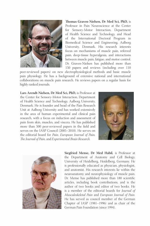
Thomas Graven-Nielsen, Dr Med Sci, PhD, is Professor in Pain Neuroscience at the Center for Sensory-Motor Interaction, Department of Health Science and Technology, and Head of the International Doctoral Program in Biomedical Science and Engineering, Aalborg University, Denmark. His research interests focus on mechanisms of muscle pain, referred pain, deep-tissue hyperalgesia, and interactions between muscle pain, fatigue, and motor control. Dr. Graven-Nielsen has published more than 150 papers and reviews (including over 110
peer-reviewed papers) on new electrophysiological methods and basic muscle pain physiology. He has a background of extensive national and international collaborations on muscle pain research. He reviews papers on a regular basis for highly ranked journals.
Lars Arendt-Nielsen, Dr Med Sci, PhD, is Professor at the Center for Sensory-Motor Interaction, Department of Health Science and Technology, Aalborg University, Denmark. He is founder and head of the Pain Research Unit at Aalborg University and has worked extensively in the area of human experimental and clinical pain research, with a focus on induction and assessment of pain from skin, muscles, and viscera. He has published more than 500 peer-reviewed papers in the field and serves on the IASP Council (2005–2010). He serves on the editorial board for Pain, European Journal of Pain, The Journal of Pain, and Experimental Brain Research.
Siegfried Mense, Dr Med Habil, is Professor at the Department of Anatomy and Cell Biology, University of Heidelberg, Heidelberg, Germany. He is professionally educated as physician, physiologist, and anatomist. His research interests lie within the neuroanatomy and neurophysiology of muscle pain. Dr. Mense has published more than 180 scientific articles, including book contributions, and is the author of two books and editor of two books. He is a member of the editorial boards for Journal of Musculoskeletal Pain and European Journal of Pain. He has served as council member of the German Chapter of IASP (1981–1996) and as chair of the German Pain Foundation (since 1994).
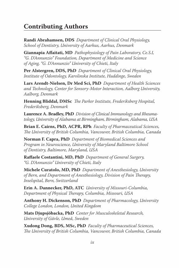
Randi Abrahamsen, DDS Department of Clinical Oral Physiology, School of Dentistry, University of Aarhus, Aarhus, DenmarkGiannapia Affaitati, MD Pathophysiology of Pain Laboratory, Ce.S.I, “G. D’Annunzio” Foundation, Department of Medicine and Science of Aging, “G. D’Annunzio” University of Chieti, ItalyPer Alstergren, DDS, PhD Department of Clinical Oral Physiology, Institute of Odontology, Karolinska Institute, Huddinge, SwedenLars Arendt-Nielsen, Dr Med Sci, PhD Department of Health Sciences and Technology, Center for Sensory-Motor Interaction, Aalborg University, Aalborg, DenmarkHenning Bliddal, DMSc The Parker Institute, Frederiksberg Hospital, Frederiksberg, DenmarkLaurence A. Bradley, PhD Division of Clinical Immunology and Rheuma-tology, University of Alabama at Birmingham, Birmingham, Alabama, USABrian E. Cairns, PhD, ACPR, RPh Faculty of Pharmaceutical Sciences, The University of British Columbia, Vancouver, British Columbia, CanadaNorman F. Capra, PhD Department of Biomedical Sciences and Program in Neuroscience, University of Maryland Baltimore School of Dentistry, Baltimore, Maryland, USARaffaele Costantini, MD, PhD Department of General Surgery, “G. D’Annunzio” University of Chieti, ItalyMichele Curatolo, MD, PhD Department of Anesthesiology, University of Bern, and Department of Anesthesiology, Division of Pain Therapy, Inselspital, Bern, SwitzerlandErin A. Dannecker, PhD, ATC University of Missouri-Columbia, Department of Physical Therapy, Columbia, Missouri, USAAnthony H. Dickenson, PhD Department of Pharmacology, University College London, London, United KingdomMats Djupsjöbacka, PhD Center for Musculoskeletal Research, University of Gävle, Umeå, SwedenXudong Dong, BDS, MSc, PhD Faculty of Pharmaceutical Sciences, The University of British Columbia, Vancouver, British Columbia, Canada
Contributing Authors
ix

x Contributing Authors
Malin Ernberg, DDS, PhD Department of Clinical Oral Physiology, Institute of Odontology, Karolinska Institute, Huddinge, SwedenDeborah Falla, PhD Center for Sensory-Motor Interaction, Department of Health Science and Technology, Aalborg University, Aalborg, DenmarkDario Farina, PhD Center for Sensory-Motor Interaction, Department of Health Science and Technology, Aalborg University, Aalborg, DenmarkMaria Adele Giamberardino, MD Pathophysiology of Pain Laboratory, Ce.S.I, “G. D’Annunzio” Foundation, Department of Medicine and Science of Aging, “G. D’Annunzio” University of Chieti, ItalyRichard H. Gracely, PhD Departments of Medicine-Rheumatology and Neurology, University of Michigan Health System, Ann Arbor, Michigan, USAThomas Graven-Nielsen, Dr Med Sci, PhD Department of Health Sciences and Technology, Center for Sensory-Motor Interaction, Aalborg University, Aalborg, DenmarkPaul W. Hodges, MedDr, PhD, BPhty(Hons) NHMRC Centre of Clinical Research Excellence in Spinal Pain, Injury and Health, School of Health and Rehabilitation Sciences, The University of Queensland, Brisbane, Queensland, AustraliaUlrich Hoheisel, Dr Rer Nat, Institute of Pharmacology and Toxicology, Charité, Humboldt University, Berlin, GermanySigvard Kopp, DDS, PhD Department of Clinical Oral Physiology, Institute of Odontology, Karolinska Institute, Huddinge, SwedenStefan Lautenbacher, PhD Department of Physiological Psychology, University of Bamberg, Bamberg, GermanyPascal Madeleine, PhD Laboratory for Work-Related Pain and Biomechanics, Center for Sensory-Motor Interaction, Department of Health Science and Technology, Aalborg University, Aalborg, DenmarkWilliam Maixner, DDS, PhD Center for Neurosensory Disorders, School of Dentistry, University of North Carolina, Chapel Hill, North Carolina, USARadi Masri, BDS, PhD Department of Endodontics, Prosthodontics and Operative Dentistry, and Program in Neuroscience, University of Maryland Baltimore School of Dentistry, Baltimore, Maryland, USA

Contributing Authors xi
Siegfried Mense, Dr Med Institute of Anatomy and Cell Biology, University of Heidelberg, Heidelberg, GermanyKazue Mizumura, MD, PhD Department of Neuroscience II, Research Institute of Environmental Medicine, Nagoya University, Nagoya, JapanWahida Rahman, PhD Department of Pharmacology, University College London, London, United KingdomJin Y. Ro, PhD Department of Biomedical Sciences and Program in Neu-roscience, University of Maryland Baltimore School of Dentistry, Balti-more, Maryland, USABarry J. Sessle, MDS, PhD, DSc(hc), FRSC Faculty of Dentistry and Centre for the Study of Pain, Toronto, Ontario, CanadaKathleen A. Sluka, PT, PhD Physical Therapy and Rehabilitation Science Graduate Program, Pain Research Program, University of Iowa, Iowa City, Iowa, USAPeter Svensson, DDS, PhD, Dr Odont Department of Clinical Oral Physiology, School of Dentistry, University of Aarhus, Aarhus, Denmark; Department of Oral and Maxillofacial Surgery, Aarhus University Hospital, Aarhus, Denmark; Orofacial Pain Laboratory, Center for Sensory-Motor Interaction, Aalborg University, Aalborg, DenmarkToru Taguchi, PhD Department of Neuroscience II, Research Institute of Environmental Medicine, Nagoya University, Nagoya, JapanIrmgard Tegeder, Dr Med Pharmazentrum Frankfurt, Institute of Clinical Pharmacology/ZAFES, Clinic of the Johann Wolfgang Goethe University, Frankfurt am Main, GermanyRoxanne Y. Walder, PhD Physical Therapy and Rehabilitation Science Graduate Program, Pain Research Program, University of Iowa, Iowa City, Iowa, USA

Preface
xiii
The clinical importance of pain from musculoskeletal structures is obvi-ous. Musculoskeletal pain is a diagnostic and therapeutic problem, and further insights into the peripheral and central neurobiological mecha-nisms are needed to improve diagnosis, therapy, and the implementation of mechanism-based treatment regimes. It has become increasingly evi-dent that muscle hyperalgesia, referred pain, referred hyperalgesia, and widespread hyperalgesia play an important role in chronic musculoskel-etal pain. Besides the sensory consequences of musculoskeletal pain, the motor control systems are also affected, changing the drive to the muscles and the related biomechanics.
This book integrates the research findings within the field of mus-culoskeletal pain into a comprehensive publication that will update the reader on novel mechanisms involved in the sensory and motor charac-teristics of such pain. The authors attempt to translate findings from basic animal studies, and from human experimental pain studies, into potential clinical mechanisms.
The historical perspective of muscle pain investigations is exten-sive. Articular and muscular forms of rheumatism had been differentiated by the 18th century. Muscular rheumatism was defined as pain and stiff-ness in muscle and soft tissue. Other authors note the use of alternative terms: Muskelschwiele (muscle callus; defined in 1843), “muscular rheu-matism” (1900), fibrositis (1915), Myogelose (muscle gelling; 1919), Mus-kelhärten (muscle hardenings; 1925), myalgia (1942), myogelosis (1942), nonarticular rheumatism (1951), and myofascial pain (1952) (Reyn-olds MD. The development of the concept of fibrositis. J Hist Med Al-lied Sci 1983;38:5–35; Simons DG. Muscular pain syndromes. In: Fricton JR, Awad E, editors. Myofascial pain and fibromyalgia. New York: Raven Press; 1990, pp 1–41). In the 1930s, Lewis and Kellgren pioneered the experimental approach to the study of muscle hyperalgesia and referred pain in humans and introduced the concept of experimentally induced muscle pain. Some of the first systematic recordings from thin-caliber muscle afferent fibers in animals were made in the early 1960s by Pain-tal (J Physiol 1960;152:250–270) and Iggo (J Physiol 1961;155:52–53),

xiv Preface
who reported responsiveness to noxious and innocuous stimuli. The next major breakthrough in muscle pain physiology was in the mid-1970s, when Mense, Kniffki, and Schmidt thoroughly characterized a subgroup of thin-afferent nerve fibers as muscle nociceptors. This work was later followed by numerous animal investigations addressing the central con-sequences of peripheral muscle nociception. For over a decade there has been extensive work on translating the basic animal findings to clinical manifestations, especially in experimental muscle pain studies in hu-mans. Over the last 10 years the relative publication rate per year within the field of musculoskeletal pain has been higher than for pain in gen-eral. This trend reflects the need to develop new pharmacological targets for chronic musculoskeletal pain (including fibromyalgia), which is now a major focus of many pharmaceutical companies.
This volume includes contributions mainly based on presenta-tions from the 7th IASP Research Symposium, “Fundamentals of Mus-culoskeletal Pain,” which took place in May 2007 at Aalborg University’s Center for Sensory-Motor Interaction and was organized by Profs. Thom-as Graven-Nielsen and Lars Arendt-Nielsen. More than 180 clinicians and basic scientists participated in the 3-day symposium. The symposium attracted participants from Europe and from 18 countries outside Europe, including Australia, Brazil, Canada, Egypt, Israel, Japan, New Zealand, Russia, and Switzerland. Twenty-seven plenary lectures were given by in-vited international speakers, and there were also poster presentations and oral presentations based on peer-reviewed accepted abstracts.
The present book is organized in three main sections: (I) Basic Mechanisms of Muscle Pain, (II) Key Factors Determining Muscle Pain Sensitivity, and (III) Effects of Muscle Pain on Motor Function. The first section focuses on morphology and functional types of peripheral mus-cle nociceptors; on central neurophysiological mechanisms involved in nociception, such as central sensitization and descending modulation of spinal mechanisms; and on cortical representation of muscle nocicep-tion. The potential human correlates are outlined. The next section pres-ents factors that can influence muscle nociceptive mechanisms, including genetics, gender, chronic pain, analgesics, and pain from other tissues. Part III outlines the effects of musculoskeletal pain on muscle function in

Preface xv
contributions covering lower back, neck, and jaw muscle systems to-gether with advanced neurophysiological assessments of muscle function, from proprioceptive afferents to motor units. Each section emphasizes the translational aspects and includes contributions from animal stud-ies, human experimental studies, and clinical findings. All authors are sincerely acknowledged for meeting our publication deadlines with their enthusiastic contributions of high-quality manuscripts and for their help with peer reviews.
Prof. Thomas Graven-Nielsen, Dr Med Sci, PhD Aalborg University, Denmark
Prof. Lars Arendt-Nielsen, Dr Med Sci, PhD Aalborg University, Denmark
Prof. Siegfried Mense, Dr Med University of Heidelberg, Germany


64 S. Mense and U. Hoheisel
Neurotransmitters and Neuropeptides Involved in Myositis-Induced Central Sensitization
The activation of NMDA and NK1 receptors contributes to myositis-in-duced central sensitization. In experiments on rats, intrathecal admin-istration of antagonists to NK1 and NMDA receptors prevented the
Fig. 1. Some of the mechanisms involved in central sensitization. To the right, a presyn-aptic terminal of a nociceptive muscle afferent fiber is shown that contacts a postsynaptic neuron. The upper part depicts a microglial cell as it releases cytokines and brain-derived neurotrophic factor (BDNF). AMPA/KA = α-amino-3-hydroxy-5 methyl-4-isoxazole pro-pionic acid/kainate, CaMK II = calcium/calmodulin protein kinase II, cAMP = cyclic ade-nosine monophosphate, IL-6 = interleukin-6, NO = nitric oxide, NK1 = neurokinin 1, NMDA = N-methyl-d-aspartate, PGs = prostaglandins, PKA = protein kinase A, PKC = protein ki-nase C, TNF-α = tumor necrosis factor alpha, TrkB = tyrosine kinase B (BDNF receptor).

80 R.Y. Walder and K.A. Sluka
C-fiber stimulation of a cutaneous nerve (Wall and Woolf, 1984). Thus, the biochemical, anatomical, and physiological differences support the hypothesis that injury to muscle results in distinctly different behavioral responses when compared to injury to the skin. These differences are ob-served in both the peripheral and central nervous systems.
Acid-Sensing Ion Channels and Pain
Effects of tissue acidosis on nociception. A decrease in pH is observed following inflammation, hematomas, and isometric exercise (Revici et al., 1949; Hood et al., 1988; Pan et al., 1988; Issberner et al., 1996). Tis-sue acidosis occurs in several physiological and disease states, including arthritis, inflammation, ischemia, myofascial pain, and cancer (Pan et al., 1988; Issberner et al., 1996; Reeh and Steen, 1996; Shah et al., 2005). In fact, there is a positive correlation between the pain experienced and lo-cal acidity (Issberner et al., 1996). Acidosis can be mimicked by infus-ing acidic solutions into muscle and joints. Constant infusion of pH 5.2 phosphate buffer into the flexor carpi radialis muscle in human subjects produces pain with a rating of 20% on the visual analogue scale that shows no adaptation during infusion (Issberner et al., 1996). Thus, acid infusion into muscle results in pain and can play a role in the develop-ment of referred pain and mechanical hyperalgesia.
ASIC3 and pain. Low pH activates acid-sensing ion chan-nels (ASICs), which are encoded by four different genes. Three of the ASICs—ASIC1, ASIC2, and ASIC3—are found on primary afferent neurons (Waldmann and Lazdunski, 1998; Waldmann, 2001). Of these, ASIC3 plays a key role in mechanical hyperalgesia induced by muscle in-sult. ASIC3 is found in large- and small-diameter primary afferents and in free nerve endings of the skin and colocalizes with substance P recep-tors in small DRG neurons (Price et al., 2001). Further ASIC3 is found in DRG innervating muscle (Sluka et al., 2003; Molliver et al., 2005). In knockout mice lacking ASIC3 (ASIC3–/–) and without tissue injury, the behavioral response to mechanical and heat stimuli is similar to that of wild-type littermates (Price et al., 2001), indicating that ASIC3 does not play a role in normal sensory processing.

112 W. Rahman and A. Dickenson
Descending Excitatory Serotonergic Pathways and Chronic Pain
Serotonergic modulation of spinal nociceptive transmission is bidirec-tional; thus, in addition to well-documented inhibitory effects, the brain can amplify spinal pain processes through a serotonergic circuit (Oyama et al., 1996; Dubner and Ren, 1999; Ossipov et al., 2001; Millan, 2002). Evidence from studies in neuropathic rats suggests an overdrive or inap-propriate activation of descending facilitation onto mechanically evoked spinal neuronal responses mediated by serotonin acting on spinal 5-HT3 receptors (Suzuki et al., 2004). Depletion of endogenous spinal 5-HT re-duced mechanical allodynia in neuropathic rats and also in a model of spinal cord injury (Oatway et al., 2004; Rahman et al., 2004).
Further evidence for the involvement of 5-HT3 receptor systems in pain behavior has emerged from a recently developed model of can-cer-induced bone pain in rats (Donovan-Rodriguez et al., 2006). Antag-onism of spinal 5-HT3 receptors produced much greater reductions in mechanical- and thermal-evoked neuronal responses compared with the effects of the drug in sham controls, again suggesting an enhanced de-scending excitatory drive onto spinal neurons in this model of chronic pain. In contrast, in a model of acute inflammation, although descending facilitation was demonstrated in both sham and inflamed rats, no differ-ence was seen between the animal groups. These findings suggest that the pattern of descending facilitatory influences may vary with time and may depend on particular pathophysiological states. A recent clinical study demonstrated a significant, albeit small, reduction in pain scores in neuropathic pain patients given an i.v. injection of the selective 5-HT3-receptor antagonist ondansetron, compared with a placebo control (Mc-Cleane et al., 2003). Similar evidence exists for patients with fibromyalgia that exhibit diffuse widespread pain, where oral and i.v. administration of the 5-HT3 antagonist tropisetron significantly reduced pain scores (Papa-dopoulos et al., 2000; Stratz et al., 2001). Whether models of longer-term joint inflammation such as osteoarthritis and other deep tissue pains also exhibit enhanced descending facilitation remains to be determined, although preliminary data from our laboratory suggests that this may indeed
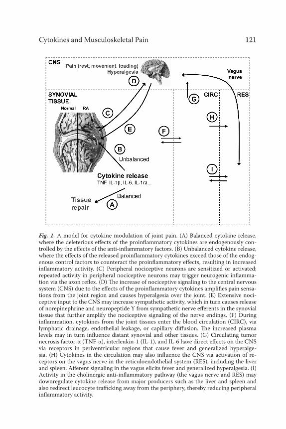
Cytokines and Musculoskeletal Pain 121
Fig. 1. A model for cytokine modulation of joint pain. (A) Balanced cytokine release, where the deleterious effects of the proinflammatory cytokines are endogenously con-trolled by the effects of the anti-inflammatory factors. (B) Unbalanced cytokine release, where the effects of the released proinflammatory cytokines exceed those of the endog-enous control factors to counteract the proinflammatory effects, resulting in increased inflammatory activity. (C) Peripheral nociceptive neurons are sensitized or activated; repeated activity in peripheral nociceptive neurons may trigger neurogenic inflamma-tion via the axon reflex. (D) The increase of nociceptive signaling to the central nervous system (CNS) due to the effects of the proinflammatory cytokines amplifies pain sensa-tions from the joint region and causes hyperalgesia over the joint. (E) Extensive noci-ceptive input to the CNS may increase sympathetic activity, which in turn causes release of norepinephrine and neuropeptide Y from sympathetic nerve efferents in the synovial tissue that further amplify the nociceptive signaling of the nerve endings. (F) During inflammation, cytokines from the joint tissues enter the blood circulation (CIRC), via lymphatic drainage, endothelial leakage, or capillary diffusion. The increased plasma levels may in turn influence distant synovial and other tissues. (G) Circulating tumor necrosis factor-α (TNF-α), interleukin-1 (IL-1), and IL-6 have direct effects on the CNS via receptors in periventricular regions that cause fever and generalized hyperalge-sia. (H) Cytokines in the circulation may also influence the CNS via activation of re-ceptors on the vagus nerve in the reticuloendothelial system (RES), including the liver and spleen. Afferent signaling in the vagus elicits fever and generalized hyperalgesia. (I) Activity in the cholinergic anti-inflammatory pathway (the vagus nerve and RES) may downregulate cytokine release from major producers such as the liver and spleen and also redirect leucocyte trafficking away from the periphery, thereby reducing peripheral inflammatory activity.
170 T. Graven-Nielsen and L. Arendt-Nielsen
referred pain areas in pain patients suggest that the efficacy of central processing is increased (central sensitization). Moreover, the expansion of areas of referred pain in fibromyalgia patients was partly inhibited by an NMDA-receptor antagonist (ketamine), which was thus shown to inhibit central sensitization (Graven-Nielsen et al., 2000). Extended areas of re-ferred pain from the tibialis anterior muscle, indicating central sensitiza-tion, have also been shown in patients suffering from other chronic mus-culoskeletal pain conditions (Arendt-Nielsen and Graven-Nielsen, 2003).
Muscle Hyperalgesia
Deep tissue hyperalgesia characterizes “an increased response to a stim-ulus which is normally painful,” as opposed to allodynia, which describes “pain due to a stimulus which does not normally provoke pain.” Accord-ing to these definitions, a decreased pain threshold should be defined as allodynia. Nonetheless, the nomenclature used in numerous psycho-physical studies is hyperalgesia in the case of decreased pain thresholds and also often in the case of pain due to a nonpainful stimulus, such as in secondary hyperalgesia, where pain can be evoked by weak mechanical stimulation.
Assessment of Muscle Hyperalgesia
Psychophysical determinations can be divided into response-dependent and stimulus-dependent methods (Gracely, 2006). Response-dependent methods are constructed by a series of fixed stimulus intensities with a score for each stimulus. The score can be determined using a visual an-alogue scale (VAS), a verbal descriptor scale, magnitude estimation, or cross-modality matching. The stimulus-dependent methods are based on adjustment of the stimulus intensity until a predefined response, typically a threshold (e.g., detection, pain, or tolerance), is reached.
Stimulus-response functions are more informative than a thresh-old determination because suprathreshold response characteristics can be derived from the data. For example, stimulus-response functions clear-ly differentiate between low- and high-intensity stimuli. Nevertheless, the

208 M.A. Giamberardino et al.
Referred Muscle Pain/Hyperalgesia from Visceral Structures in the Clinical Setting
Referred pain occurs constantly in visceral nociception. In fact, after a transitory phase (lasting a few minutes or hours) in which visceral pain is perceived as a midline, poorly discriminated sensation accompanied by marked neurovegetative signs and emotional reactions (“true viscer-al pain”), the symptom is “transferred” to somatic areas of the body wall neuromerically connected to the involved organ (Procacci et al., 1986). The referral area most often becomes the site of secondary hyperalge-sia (increased sensitivity to painful stimuli and decreased pain thresh-old), especially if the algogenic condition of the internal organ recurs frequently or is prolonged. This condition may involve all three somatic tissues of the body wall—skin, subcutis, and muscle—but it is most of-ten localized at the muscle level (referred muscle pain without and with hyperalgesia) (Vecchiet et al., 1989, 1992). In myocardial infarction, for instance, referred pain typically occurs in the precordial region and up-per limbs, mostly the left arm; hyperalgesia affects the pectoralis major muscle and forearm muscles. The trapezius and deltoid muscles are less frequently involved. In a low percentage of cases, pain is also referred to the subcutis and skin, within dermatomes C8–T1 on the ulnar side of the arm and forearm, and hyperalgesia is found at the same level (Procacci et al., 1986; Loeser, 2001). Fig. 1 shows patterns of referred muscle hyperal-gesia from the heart.
In urinary colic from calculosis, pain is referred to the lumbar region of the affected side, radiating toward the ipsilateral flank and an-teriorly toward the groin, with hyperalgesia characteristically affecting the quadratus lumborum muscle and external oblique muscle (Vecchi-et et al., 1989). In biliary calculosis, pain is perceived in the upper right quadrant of the abdomen, radiating toward the back; hyperalgesia typi-cally develops in the rectus abdominis muscle, around the cystic point area (at the level of junction of the 10th rib and the outer margin of the same muscle) (Giamberardino et al., 2005). Fig. 2 shows patterns of re-ferred muscle hyperalgesia from the biliary tract. In dysmenorrhea, pain

Muscle Hyperalgesia in Visceral Pain 209
is referred to the lowest abdominal quadrants, radiating toward the groin and the upper part of the thighs; hyperalgesia develops in the rectus ab-dominis muscle and in muscles of the pelvic region (Giamberardino et al., 1997). In irritable bowel syndrome (IBS), pain is perceived in the ab-domen, around the umbilical area; hyperalgesia of the rectus abdominis muscle at the same level is a typical finding (Caldarella et al., 2006). In all the previous examples, hyperalgesia may also eventually involve the sub-cutis and skin overlying the tender muscles in cases of repeated and/or prolonged painful episodes (Vecchiet et al., 1999).
Muscle hypersensitivity in the referred pain zones can easily be detected clinically by manual compression, the maneuver provoking an evident pain reaction by the patient. However, precise quantification of the extent of the sensory changes can only be provided by instrumen-tal measurements. Evaluation of pain thresholds to different stimuli has been usefully employed to this purpose (Vecchiet et al., 1989; Giambe-rardino et al., 1997, 2005; Arendt-Nielsen and Svensson, 2001). The ap-plication of pressure, chemical, and electrical stimuli has allowed study of the development and progression of muscle hyperalgesia in referred pain areas from different internal organs. As a general rule, irrespective of the specific organ involved, researchers have found that in recurrent visceral pain conditions, such as urinary or biliary colics or dysmenorrhea, mus-cle hyperalgesia (evidenced by a significant decrease in pain threshold)
Fig. 1. Patterns of referred muscle hyperalgesia from the heart.

15
Fundamentals of Musculoskeletal Painedited by Thomas Graven-Nielsen, Lars Arendt-Nielsen, and Siegfried MenseIASP Press, Seattle, © 2008
235
Sex-Related Differences in Clinical and Experimental Muscle Pain
Stefan LautenbacherDepartment of Physiological Psychology, University of Bamberg, Bamberg, Germany
This chapter deals with two issues: first, is there a sex difference in the presentation of muscle pain, and second, if so, how might we account for it? The first part of the chapter presents epidemiological evidence and discusses associated factors that might guide attempts to explain the evi-dence. However, satisfactory explanations also require evidence based on experimentally induced muscle pain, which will be reviewed in the sec-ond part of the chapter.
Epidemiological and Clinical Evidence
Epidemiological reports indicate that musculoskeletal pain is a major medical and economic problem. The pain and associated disability are linked with a significant loss of productivity and substantial health care expenditures for women. Leijon et al. (1998) observed that about 30% of all sick-leave days in Sweden are due to neck/shoulder or low back pain, which were defined as being of musculoskeletal origin. The authors

302 I. Tedeger
Peripheral Opioid Analgesia in Humans
Peripheral Opioids in Experimental Muscle Pain and Inflammation
In the delayed-onset muscle soreness (DOMS) model, muscle pain is produced by strenuous muscle exercise including eccentric muscle con-tractions (Cleak and Eston, 1992; see Chapter 4 by Mizumura and Tagu-chi and Chapter 16 by Dannecker). The pain, which reaches its maximum between 24 and 48 hours after exercise, is thought to be due to an in-flammatory response (Smith, 1991; MacIntyre et al., 1995) elicited by mi-cro-injuries of muscle fibers (Cleak and Eston, 1992). Levels of cytokines, prostaglandins, and glutamate are increased in painful muscles (Tegeder et al., 2002). Further, plasma levels of pro-inflammatory cytokines such as IL-6 increase during DOMS (Croisier et al., 1999). Intravenously ad-ministered morphine-6-β-glucuronide (M6G), a metabolite of morphine with potent opioid agonist activity, which penetrates the central nervous system only very slowly, reduced muscle pain in the DOMS model and abolished mechanical hyperalgesia in the freeze lesion model of mild lo-cal skin inflammation (Tegeder et al., 2003) (Fig. 1). The peripheral lo-calization of this effect was confirmed by the absence of any effects on pupil size, indicating that M6G had not penetrated the brain in relevant amounts when peripheral analgesia was observed (Fig. 2). In contrast to its effects in these inflammatory models, M6G did not increase pain threshold and tolerance to electrically evoked pain caused predominantly by activation of C fibers without hyperalgesia or inflammatory reactions. The positive control morphine, however, reduced pain in all models and decreased pupil diameter, indicating that it had acted centrally (Tegeder et al., 2003). Target tissue and plasma concentrations of morphine and M6G were confirmed by microdialysis. M6G target concentrations were achieved that are known to be devoid of central effects (Fig. 3).
Regional intravenous 0.01% morphine infusion reduced thermal, but not mechanical, hyperalgesia exclusively in inflamed skin areas (Kop-pert et al., 1999). Central analgesic effects were excluded by the absence of measurable plasma concentrations of morphine and its metabolites.

Augmented Central Processing in Fibromyalgia 313
Increased Pain Sensitivity in Fibromyalgia Is Not Confined to Muscles
Given the role of distress, are patients still significantly tender when as-sessed by more controlled methods? Petzke et al. (2003) addressed this important question by assessing tenderness using both controlled and uncontrolled methods in 43 patients with FM and 28 control subjects. FM patients were significantly more sensitive according to both ascend-ing and random direct scaling methods, supporting previous findings. This study also found increased sensitivity to contact heat using a ran-dom staircase technique, consistent with similar results using direct scal-ing (Geisser et al. 2003) and threshold assessment (Lautenbacher et al., 1994; Gibson et al., 1995) of reactions to painful heat. These studies indi-cate that increased pain sensitivity is not confined to muscles, as was also demonstrated by increased pressure sensitivity at a region of the thumb that is devoid of muscle (Petzke et al., 2003). All of the studies using heat applied the painful stimulus to the skin, which probably activated Aδ and C-fiber cutaneous nociceptors. The mechanism of pain augmentation in FM is not limited to muscles and may involve nociceptors in deep soft tissue, periosteum, and bone. The “myalgia” component of fibromyalgia reflects the historical origins of the taxonomy (e.g., fibromyositis) used to describe this disorder, but fibromyalgia may be a misnomer in describing the underlying mechanism.
Petzke et al. (2003) administered both heat and pressure to the same 43 FM patients and observed correlations of 0.41 for ascending and 0.36 for random methods, indicating that only 0.13–0.17 of the variance in sensitivity to heat can be explained by sensitivity to pressure. This modest association provides supporting evidence for the validity of the subjective responses in the sense that the augmentation does not repre-sent a reporting style that would be assumed to influence responses to any painful stimulus modality. The modest correlations also suggest that the augmented pain sensitivity in FM may not result from a simple uni-tary mechanism but rather may represent the combined effect of multi-ple mechanisms. One possible source of variability may relate to primary
338 H. Bliddal and M. Curatolo
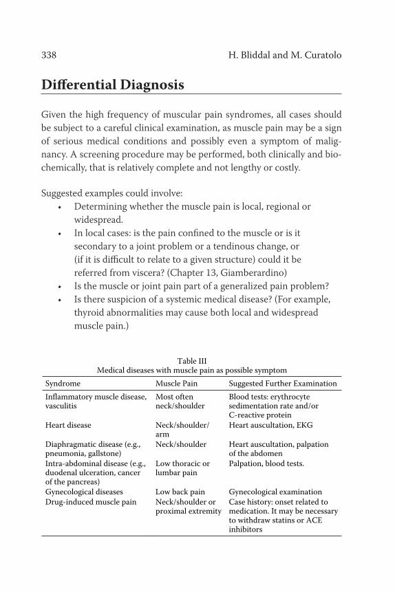
Differential Diagnosis
Given the high frequency of muscular pain syndromes, all cases should be subject to a careful clinical examination, as muscle pain may be a sign of serious medical conditions and possibly even a symptom of malig-nancy. A screening procedure may be performed, both clinically and bio-chemically, that is relatively complete and not lengthy or costly.
Suggested examples could involve: • Determining whether the muscle pain is local, regional or widespread. • In local cases: is the pain confined to the muscle or is it secondary to a joint problem or a tendinous change, or (if it is difficult to relate to a given structure) could it be referred from viscera? (Chapter 13, Giamberardino) • Is the muscle or joint pain part of a generalized pain problem? • Is there suspicion of a systemic medical disease? (For example, thyroid abnormalities may cause both local and widespread muscle pain.)
Table III
Medical diseases with muscle pain as possible symptom Syndrome Muscle Pain Suggested Further Examination Inflammatory muscle disease, vasculitis
Most often neck/shoulder
Blood tests: erythrocyte sedimentation rate and/or C-reactive protein
Heart disease Neck/shoulder/ arm
Heart auscultation, EKG
Diaphragmatic disease (e.g., pneumonia, gallstone)
Neck/shoulder Heart auscultation, palpation of the abdomen
Intra-abdominal disease (e.g., duodenal ulceration, cancer of the pancreas)
Low thoracic or lumbar pain
Palpation, blood tests.
Gynecological diseases Low back pain Gynecological examination Drug-induced muscle pain Neck/shoulder or
proximal extremity Case history: onset related to medication. It may be necessary to withdraw statins or ACE inhibitors

Clinical Manifestations of Muscle and Joint Pain 339
Important red flags: • Central distribution of muscle and joint pain close to the body in neck/shoulder/pelvic areas • Diffuse spread of the muscle pain • Lack of a precipitating factor
Table III gives examples of medical diseases that may be accompanied by muscle pain, sometimes as the primary symptom. Generalized disease or truncal muscle pain without reason should lead to a full clinical exami-nation along with supplementary blood tests, which will uncover most relevant diseases (Tables IV–V).
Table IV Blood tests of relevance for muscle pain
Blood Test
Possible Diseases with Muscle Pain as a Symptom
Hemoglobin, leucocyte count Anemia, leucemia Erythrocyte sedimentation rate or C-reactive protein
Systemic inflammation
Thyroid-stimulating hormone Hypo- or hyperthyroidism Alkaline phosphatase Malignancy, bone marrow disease Calcium Hypocalcemia Note: More seldom seen: amylases (pancreatic diseases), creatine kinase (myositis), other transaminases (liver disease), and antibodies against various viruses.
Table V
Supplementary blood tests of relevance for joint pain Blood Test Possible Disease with Joint Pain
as a Symptom Erythrocyte sedimentation rate or C-reactive protein
Inflammatory joint disease
Uric acid Gout Rheumatoid factor Rheumatoid arthritis Anticyclic citrullinated peptide (anti-CCP)
Rheumatoid arthritis
Antinuclear antibodies (ANA) Collagenoses, e.g., lupus erythematosus
Sensory-Motor Changes in Low Back Pain 449
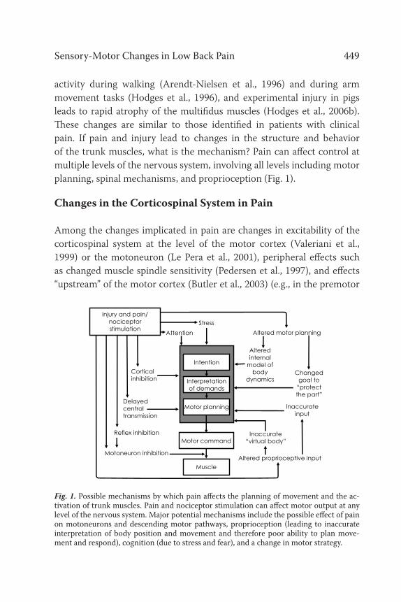
activity during walking (Arendt-Nielsen et al., 1996) and during arm movement tasks (Hodges et al., 1996), and experimental injury in pigs leads to rapid atrophy of the multifidus muscles (Hodges et al., 2006b). These changes are similar to those identified in patients with clinical pain. If pain and injury lead to changes in the structure and behavior of the trunk muscles, what is the mechanism? Pain can affect control at multiple levels of the nervous system, involving all levels including motor planning, spinal mechanisms, and proprioception (Fig. 1).
Changes in the Corticospinal System in Pain
Among the changes implicated in pain are changes in excitability of the corticospinal system at the level of the motor cortex (Valeriani et al., 1999) or the motoneuron (Le Pera et al., 2001), peripheral effects such as changed muscle spindle sensitivity (Pedersen et al., 1997), and effects “upstream” of the motor cortex (Butler et al., 2003) (e.g., in the premotor
Fig. 1. Possible mechanisms by which pain affects the planning of movement and the ac-tivation of trunk muscles. Pain and nociceptor stimulation can affect motor output at any level of the nervous system. Major potential mechanisms include the possible effect of pain on motoneurons and descending motor pathways, proprioception (leading to inaccurate interpretation of body position and movement and therefore poor ability to plan move-ment and respond), cognition (due to stress and fear), and a change in motor strategy.
Corticalinhibition
Delayedcentraltransmission
Motoneuron inhibition
Reflex inhibition
Interpretationof demands
Motor command
Intention
Muscle
Motor planning
Altered motor planning
Alteredinternal
model of body
dynamicsChanged
goal to“protectthe part”
Inaccurate“virtual body”
Inaccurateinput
Altered proprioceptive input
AttentionStress
Injury and pain/nociceptorstimulation