Long-range conformational effects of proteolytic removal of the last ...
8
Biochem. J. (1995) 307, 527-534 (Printed in Great Britain) Long-range conformational effects of proteolytic removal of the last three residues of actin Hanna STRZELECKA-GOLASZEWSKA,*t Mafgorzata MOSSAKOWSKA,*t Aleksandra WOZNIAK,* Joanna MORACZEWSKA* and Haruto NAKAYAMAt *Department of Muscle Biochemistry, Nencki Institute of Experimental Biology, 3 Pasteur Street, PL-02-093 Warsaw, Poland, and tBiological Function Section, Kansai Advanced Research Center, Communications Research Laboratory, Kobe, Japan Trunca-ted derivatives of actin devoid of either the last two (actin-2d) or three residues (actin-3O) were used to study the role of the C-terminal segment in the polymerization of actin. The monomer critical concentration and polymerization rate in- creased in the order: intact actin < actin2C < actin-3C. Con- versely, the rate of hydrolysis of actin-bound ATP during spontaneous polymerization of Mg-actin decreased in the same order, so that, for actin-3C, the ATP hydrolysis significantly lagged behind the polymer growth. Probing the conformation of the nucleotide site in the monomer form by measuring the rates of the bound nucleotide exchange revealed a similar change upon removal of either the two or three residues from the C-terminus. The C-terminal truncation also resulted in a slight decrease in the rate of subtilisin cleavage of monomeric actin within the DNAse- I binding loop, whereas in F-actin subunits the susceptibility of INTRODUCTION On the basis of the atomic model of F-actin, Holmes et al. [1] have suggested that the C-terminal phenylalanine (Phe375), loca- ted in subdomain 1 of the actin molecule [2], interacts with the N- terminal part of the DNase-I-binding loop (residues 39-51) on the top of subdomain 2 in the neighbouring polymer subunit along the two-start F-actin helix. The exact location of the C- terminal segment in both the monomer and polymer structure is, however, still uncertain. The three C-terminal residues of actin were proteolytically removed prior to crystallization of the actin- DNase I complex that was used by Kabsch et al. [21 to obtain the first atomic model of G-actin. In the two other atomic models of G-actin, based on the crystal structure of gelsolin segment I-actin complex [3] and of the profilin-actin complex [4], the position of the C-terminal residues might be changed by their interaction with segment 1 of gelsolin and with profilin respectively. The role of the C-terminus in actin-actin interactions has been studied using actin modified at the penultimate cysteine residue (Cys374) with certain thiol compounds [5,6], truncated actin lacking either the last two [7,8] or three amino acid residues [9], and mutant fl-actin from yeast in which Cys374 was replaced with a serine residue [10]. All these modifications resulted in destabi- lization of the actin filament. To obtain more information on the involvement of the C- terminal segment of the actin polypeptide chain in polymer formation and stabilization, we have directly compared certain aspects of polymerization of truncated actin derivatives devoid of the last two (actin.2c) and the last three residues (actin.3C). this and of another site within this loop, specifically cleaved by a proteinase from Escherichia coli A2 strain, gradually incre-aed upon sequential removal of the two and of the third residue froih the C-terminus. From these and other observations made in this work it has been concluded that perturbation of the C-termiltal structure in monomeric actin is transmitted to the cleft, where nucleotide and bivalent cation are bound, and to the DNAo- binding loop on the top of subdomain 2. Further changes at these sites, observed on the polymer level, seem to result from elimination of the intersubunit contact between the C-terminal residues and the DNAse-I binding loop. It is suggested that formation of this contact plays an essential role in regulating the hydrolysis of actin-bound ATP associated with the polymeri- zation process. Influence of removal of the C-terminal residues on the con- formation of other regions of the actin molecule in the monomer and polymer form was investigated by the method of limited proteolysis and by measuring the rates of dissociation of the bound nucleotide and of the tightly bound bivalent cation. The results show that the influence of the proteolytic modifications of the C-terminal segment on polymer formation and its structure is more complex than might be expected from elimination of the putative actin-actin contact made by Phe375 only. Evidence is presented showing that structural perturbations in the C-terminal segment are propagated to distant regions of the monomer molecule, and that proteolytic removal of Lys373 in addition to Cys374-Phe375 has additional long-range conformational effects on the polymer subunits. Some of the present results have been presented in a preliminary form [11]. MATERIALS AND METHODS Protein preparations Rabbit skeletal-muscle actin was prepared as described by Spudich and Watt [12]. Both intact actin and its C-terminal truncated derivatives were stored in buffer containing 2 mM Hepes, pH 7.6, 0.2 mM ATP, 0.1 mM CaCl2 and 0.02 % NaN3 (buffer G). In all experiments on actin (intact or modified) containing Mg2" at the single high-affinity site for bivalent cation, Ca-G-actin was transformed into Mg-G-actin directly before starting the measurements, by a 5-10 min incubation with 0.2 mM EGTA/50 ,uM MgCl2 at room temperature. Abbreviations used: 1,5-IAEDANS, N-iodoacetyl-N'-(5-sulpho-1-naphthyl)ethylenediamine; AEDANS, acetyl-N'-(5-sulpho-1-naphthyl)ethylenedi- amine; c-ATP, 1-N6-etheno-ATP; SBTI, soybean trypsin inhibitor; PMSF, phenylmethanesulphonyl fluoride; ECP, Escherichia coliA2 strain proteinase (EC 3.4.21.62). $ To whom correspondence should be sent. 527
Transcript of Long-range conformational effects of proteolytic removal of the last ...

Biochem. J. (1995) 307, 527-534 (Printed in Great Britain)
Long-range conformational effects of proteolytic removal of the last threeresidues of actinHanna STRZELECKA-GOLASZEWSKA,*t Mafgorzata MOSSAKOWSKA,*t Aleksandra WOZNIAK,* Joanna MORACZEWSKA*and Haruto NAKAYAMAt*Department of Muscle Biochemistry, Nencki Institute of Experimental Biology, 3 Pasteur Street, PL-02-093 Warsaw, Poland, and tBiological Function Section,Kansai Advanced Research Center, Communications Research Laboratory, Kobe, Japan
Trunca-ted derivatives of actin devoid of either the last two(actin-2d) or three residues (actin-3O) were used to study the roleof the C-terminal segment in the polymerization of actin. Themonomer critical concentration and polymerization rate in-creased in the order: intact actin < actin2C < actin-3C. Con-versely, the rate of hydrolysis of actin-bound ATP duringspontaneous polymerization of Mg-actin decreased in the sameorder, so that, for actin-3C, the ATP hydrolysis significantlylagged behind the polymer growth. Probing the conformation ofthe nucleotide site in the monomer form by measuring the ratesof the bound nucleotide exchange revealed a similar change uponremoval of either the two or three residues from the C-terminus.The C-terminal truncation also resulted in a slight decrease in therate of subtilisin cleavage of monomeric actin within the DNAse-I binding loop, whereas in F-actin subunits the susceptibility of
INTRODUCTION
On the basis of the atomic model of F-actin, Holmes et al. [1]have suggested that the C-terminal phenylalanine (Phe375), loca-ted in subdomain 1 of the actin molecule [2], interacts with the N-terminal part of the DNase-I-binding loop (residues 39-51) on
the top of subdomain 2 in the neighbouring polymer subunitalong the two-start F-actin helix. The exact location of the C-terminal segment in both the monomer and polymer structure is,however, still uncertain. The three C-terminal residues of actinwere proteolytically removed prior to crystallization of the actin-DNase I complex that was used by Kabsch et al. [21 to obtain thefirst atomic model of G-actin. In the two other atomic models ofG-actin, based on the crystal structure of gelsolin segment I-actincomplex [3] and of the profilin-actin complex [4], the position ofthe C-terminal residues might be changed by their interactionwith segment 1 of gelsolin and with profilin respectively.The role of the C-terminus in actin-actin interactions has been
studied using actin modified at the penultimate cysteine residue(Cys374) with certain thiol compounds [5,6], truncated actinlacking either the last two [7,8] or three amino acid residues [9],and mutant fl-actin from yeast in which Cys374 was replaced witha serine residue [10]. All these modifications resulted in destabi-lization of the actin filament.To obtain more information on the involvement of the C-
terminal segment of the actin polypeptide chain in polymerformation and stabilization, we have directly compared certainaspects of polymerization of truncated actin derivatives devoidof the last two (actin.2c) and the last three residues (actin.3C).
this and of another site within this loop, specifically cleaved bya proteinase from Escherichia coli A2 strain, gradually incre-aedupon sequential removal of the two and of the third residue froihthe C-terminus. From these and other observations made in thiswork it has been concluded that perturbation of the C-termiltalstructure in monomeric actin is transmitted to the cleft, wherenucleotide and bivalent cation are bound, and to the DNAo-binding loop on the top of subdomain 2. Further changes atthese sites, observed on the polymer level, seem to result fromelimination of the intersubunit contact between the C-terminalresidues and the DNAse-I binding loop. It is suggested thatformation of this contact plays an essential role in regulating thehydrolysis of actin-bound ATP associated with the polymeri-zation process.
Influence of removal of the C-terminal residues on the con-formation of other regions of the actin molecule in the monomerand polymer form was investigated by the method of limitedproteolysis and by measuring the rates of dissociation of thebound nucleotide and of the tightly bound bivalent cation. Theresults show that the influence of the proteolytic modifications ofthe C-terminal segment on polymer formation and its structureis more complex than might be expected from elimination of theputative actin-actin contact made by Phe375 only. Evidence ispresented showing that structural perturbations in the C-terminalsegment are propagated to distant regions of the monomermolecule, and that proteolytic removal of Lys373 in addition toCys374-Phe375 has additional long-range conformational effectson the polymer subunits.Some ofthe present results have been presented in a preliminary
form [11].
MATERIALS AND METHODSProtein preparationsRabbit skeletal-muscle actin was prepared as described bySpudich and Watt [12]. Both intact actin and its C-terminaltruncated derivatives were stored in buffer containing 2 mMHepes, pH 7.6, 0.2 mM ATP, 0.1 mM CaCl2 and 0.02% NaN3(buffer G). In all experiments on actin (intact or modified)containing Mg2" at the single high-affinity site for bivalentcation, Ca-G-actin was transformed into Mg-G-actin directlybefore starting the measurements, by a 5-10 min incubation with0.2 mM EGTA/50,uM MgCl2 at room temperature.
Abbreviations used: 1,5-IAEDANS, N-iodoacetyl-N'-(5-sulpho-1-naphthyl)ethylenediamine; AEDANS, acetyl-N'-(5-sulpho-1-naphthyl)ethylenedi-amine; c-ATP, 1-N6-etheno-ATP; SBTI, soybean trypsin inhibitor; PMSF, phenylmethanesulphonyl fluoride; ECP, Escherichia coliA2 strain proteinase(EC 3.4.21.62).
$ To whom correspondence should be sent.
527
528 H. Strzelecka-Golaszewska and others
Labelling of actin at Cys374 with N-iodoacetyl-N'-(5-sulpho-1-naphthyl)ethylenediamine (1,5-IAEDANS) was performed asdescribed in [13].
Actin2C was prepared essentially as described in [7]. Mg-G-actin was polymerized with 0.1 M KCl and digested with trypsinat an enzyme/protein mass ratio of 1:20 at 25 'C. To minimizethe proteolytic removal of the third residue (Lys373) from the C-terminus [9], F-actin labelled with 1,5-IAEDANS was used andthe removal of Cys374 was monitored by measuring the decreasein the fluorescence intensity of the AEDANS label. As soon asthe fluorescence plateau was reached, proteolysis was terminatedby addition of soybean trypsin inhibitor (SBTI) at three times thetrypsin concentration. The modified actin was collected byultracentrifugation and further freed of residual trypsin and low-molecular-mass digestion products by an additional polymeriza-tion-depolyinerization cycle.
Actin,3 was-prepared by digestion of ATP-Mg-G-actin withtrypsin as described previously [9]. Tryptic removal of Lys373from this form of actin was shown to proceed at a rate 2-3-foldlower than the rate of removal of the Cys374-Phe375 dipeptide [9].To ensure that all three residues were cleaved off, the releaseof Cys374 was monitored as described for actin-2C, using actinlabelled with 1,5-IAEDANS, and the digestion was carried outfor a period of time 3-fold longer than that during which thefluorescence of the AEDANS label decreased to the plateauvalue. The digestion was stopped with SBTI, and truncated actinwas purified by polymerization with 0.1 M KCl/2 mM MgCl2and ultracentrifugation, followed by depolymerization by dialysisagainst buffer G.
G-actin containing 1-N6-etheno-ATP (c-ATP) at the nucleotidesite (e-ATP-G-actin) was obtained by homogenization of centri-fuged F-actin pellets in buffer G containing e-ATP in place ofATP, followed by dialysis against the same buffer solution.
All protein preparations, after dialysis, were clarified by a30 min centrifugation at 150000 g.
Protein concentration in G-actin solutions was determinedspectrophotometrically at 290 nm using an absorption coefficient(e) of 0.63 mg ml-- cm-' [14]. Molar concentrations of ATPand e-ATP were calculated using absorption coefficients of15400 M-l cm-' at 259 nm and 5700 M-1 cm-' at 265 nmrespectively.
AssaysPolymerization of actin was monitored by recording the increasein the light-scattering intensity at 90 0 at 360 or 450 nm, in eithera Hitachi 650-lOS or a Perkin-Elmer LS-5b luminescence spec-trometer.The critical concentrations of actin for polymerization were
determined by measuring the concentration of unpolymerizedactin in steady-state F-actin solutions using the DNase-I in-hibition assay [15]. Actin in buffer G was polymerized with 0.1 MKCI for 1.5-3 h at high protein concentration. It was thendiluted with buffer G supplemented with 0.1 M KCl to severaldifferent protein concentrations over the range 5-10 #M. Thesamples were incubated at 22 'C until the new steady-state levelof polymerization (as monitored by measuring the 90 ° light-scattering intensity) was established, and then the DNase-Iinhibitory activity was determined under standard conditions asdescribed in [16] using DNase-I dissolved in buffer G supple-mented with 1 mM phenylmethanesulphonyl fluoride (PMSF)and dialysed overnight against this solution. The reaction wasstarted immediately after mixing DNase I and actin, and theDNase-I activity was obtained from the slope of the linear part
merized actin was calculated from calibration curves of theDNase-I activity versus known amounts of monomeric actin.ATP hydrolysis associated with actin polymerization was
assayed in aliquots removed at time intervals from the cuvette inwhich the light-scattering intensity of polymerizing actin solutionwas being measured. The reaction was quenched by mixing thesolution with an equal volume of ice-cold solution of 0.6 Mperchloric acid, and Pi was determined by the highly sensitiveMalachite Green method described in [17].The dissociation rates of G-actin-bound nucleotide were
determined by displacing the initially bound nucleotide, ATP ore-ATP, with a large molar excess (10-100-fold) of 6-ATP or ATPrespectively, so that the back reaction could be neglected. Unlessstated otherwise, concentrated actin solution (about 70 ,sM) inbuffer G was diluted to the final concentration of 3.5 ,uM (ATP-G-actin) or 0.4 ,uM (e-ATP-G-actin) with a buffer solutioncontaining 4 mM Hepes, pH 7.6, and either CaC12 at differentconcentrations or 0.2 mM EDTA, and the reaction was im-mediately started by addition of 0.1 mM e-ATP or ATP re-spectively. Displacement of the bound nucleotide was monitoredby measuring the changes in the fluorescence intensity of e-ATPon its binding to, or dissociation from, actin [18].The rates of dissociation of Ca2+ from the high-affinity site in
G-actin were measured using the fluorescent calcium chelatorQuin-2 [19]. Actin in buffer G (70 ,uM) was 3-fold diluted with aCa2+-free buffer G, then a mixture of Quin-2 and MgCl2 at finalconcentrations of 0.5 mM and 0.1 mM respectively was addedand the increase in the fluorescence of Quin-2 was recorded at23 'C. The rate of Ca2+ dissociation from actin was calculatedfrom the time course of the slow increase in the fluorescencefollowing the initial fast change corresponding to binding toQuin-2 of free Ca2 .
Proteolytic digestions of G- and F-actins were carried out at25 'C at a protein concentration of 24 1sM. Unless statedotherwise, the digestion mixtures contained 2 mM or 10 mMHepes, pH 7.6 (for G- and F-actin respectively), 0.2 mM ATP,0.1 mM CaCl2, 0.02% NaN3 and, when present, 0.1 M KC1. Inexperiments on F-actins the polymerization was controlled bymeasuring the increase in light-scattering intensity, and thedigestions were started shortly after the steady-state value wasreached. Digestion with subtilisin and trypsin was stopped withPMSF (2 mM) or SBTI (3 x molar concentration of trypsin)respectively. Activity of the proteinase from E. coli A2 strain wasquenched by mixing with electrophoresis sample buffer [1.5 %SDS/3 % 2-mercaptoethanol/15 % glycerol/40 mM Tris, pH 6.8(all final concns.)] and incubation for 3 min at 100 'C. Thedigests were analysed by SDS/PAGE. Absorbance of CoomassieBlue-stained protein bands on the gels was determined with aShimadzu CS-9000 dual-wavelength flying-spot scanner.
Other methodsFluorescence measurements were performed in either a Perkin-Elmer LS-Sb luminescence spectrometer or a Hitachi 650-lOSspectrofluorimeter. Excitation and emission wavelengths were340 and 460 nm respectively for AEDANS-actin, 350 and 410 nmfor e-ATP and 340 nm and 500 nm for Quin-2. SDS/PAGE wascarried out as described by Laemmli [20] on 10 %-(w/v- poly-acrylamide gel slabs.
ReagentsTrypsin (from bovine pancreas, tosylphenylalaninechloro-methane-treated), subtilisin (Carlsberg; from Bacillus licheni-formis), ATP (disodium salt), e-ATP (sodium salt), DNAse-Iof the curve of DNA hydrolysis. The concentration of unpoly-

Conformational effects of removal of C-terminal residues of actin
(from bovine pancreas), Quin-2 and EGTA were purchased fromSigma. E. coli A2 strain proteinase was a gift of Dr. S. Yu.Khaitlina and A. Morozova (Institute of Cytology, St. Peters-burg, Russia). 1,5-IAEDANS was from Molecular Probes. Hepesand SDS were from Serva.
RESULTSPolymerization properties of Intact and C-terminal truncatedactinsWe have previously shown that ATP-Mg-G-actin devoid of thelast three residues has a 5-fold higher critical concentration butpolymerizes faster than intact ATP-Mg-G-actin [9]. Figure 1shows that the rate of polymerization of actin2C is intermediatebetween those of actin and of intact actin. To reveal better thedifferences, for this experiment the conditions of slow poly-merization (low salt concentration) were chosen.The critical concentration for 0.1 M KCl-induced polymeriza-
tion of actin in ATP-Ca-form, determined using the DNAse-Iinhibition assay [15], was 1.3 ,uM for intact actin, 2.7 4M foractin-2C, and 3.5,uM for actin-3C' These measurements wereconfined to actins with Ca2+ as the tightly bound cation, becausetruncated Mg-F-actins were too quickly depolymerized byDNAse I, as indicated by increase in the rate of DNA hydrolysisduring the assay. This was not observed with Ca-F-actins. Tominimize possible errors due to the protein inactivation, themeasurements were performed on freshly obtained preparationsonly. As reported previously for actin-3C [9], the truncated actinsin their monomer form were fairly stable provided they were keptin Ca2+-bound form. It was checked, by measuring the DNAse-I activity as a function of G-actin concentration, that thetruncated actins do not differ from intact actin in their DNAse-I inhibitory activity (results not shown).
Polymerization of ATP-G-actin is accompanied by hydrolysisof the bound nucleotide to ADP. It has been established thatATP is hydrolysed subsequent to polymer formation, so that atransient ATP-F-actin species is formed and, in the presence offree ATP, a cap of ATP-subunits is present at least at one, thefast-growing, end of the filament at steady state (for a review, see[21]). With Mg-actin, the uncoupling of the hydrolysis frompolymer formation can, however, be observed only when the rateof elongation is very high, i.e. at high actin concentrations, underconditions of 'seeded' polymerization additionally accelerated
200
X 160
0'C*1200 80
.2m 40-J
0 10 20Time (min)
30 40
100
80
60
D 40.N
E 2000 0
.D 100(0aB 80acm
0,m 60._
to 40(0s 20co
@ 100
a 80
60
40
20
0E
0
~0-nm
0 5 10 15 20Time (min)
Figure 2 Time courses of spontaneous polymerizaton of unmodMed andC-terminal truncated ATP-Mg-G-actins and of the accompanying hydrolysisof ATP
Unmodified actin (a), actin-2C (b) and actin.3C (c), 24 FM, in 2 mM Hepes (pH 7.6)/0.2 mMATP/50 ,M CaCI2, were preincubated with 0.1 mM MgCI2/0.2 mM EGTA for 10 min at 20 0Cand then polymerized by the addition of KCI at a final concentration of 90 mM (zero time).O,Light-scattering intensity at 360 nm, normalized to 0 at zero time and 100 at steady state (theresults are means for three different preparations; the vertical bars represent the S.D.); E, A,V, hydrolysed ATP, determined as described in Materials and methods section; the differentsymbols denote the results of the three separate experiments.
by sonication [22]. As Figure 2(c) shows, in contrast with intactactin, spontaneous polymerization of the ATP-Mg-form ofactin3, was characterized by a long delay in ATP hydrolysisrelative to the polymer formation. Only about 0.3 mol ofATP/mol of actin was hydrolysed at the time when the light-scattering intensity reached the steady-state value. This stronguncoupling of the two reactions could not be simply due to fasterpolymerization of actin,3 as compared with intact actin, becausethe hydrolysis on actin-3C was significantly delayed also inrelation to ATP hydrolysis on intact actin. The hydrolysis on
polymerizing actin-2C (Figure 2b) lagged behind the polymerformation too, but significantly less than in actin3C. The resultsobtained for actin3C also show a strong co-operativity of ATPhydrolysis, supporting the earlier conclusion that the hydrolysison an ATP-F-actin subunit is accelerated by interaction withADP-F-actin subunits [22].
Figure 1 Comparison of the kineftcs of polymerization of unmodffled and Influence of the C-terminal residues on the conformation of theC-terminal truncated actins nucleotide site in G-actin
Actin [12 ,uM in 2 mM Hepes (pH 7.6)/0.2 mM ATP/50 ,uM CaCI,2 was preincubated with50 uM MgCI2/200 ,M EGTA for 10 min at 25 OC to replace the tightly bound Ca2+ with Mg2+.Polymerization was then started by addition of KCI at a final concentration of 10 mM (arrow)and the increase in the light scattering intensity at 360 nm was recorded. Curves 1-3 refer to
unmodified actin, actin-2C, and actin-3C respectively. Abbreviation: a.u., arbitrary unit.
To obtain information on possible differences in the confor-mation of the nucleotide site in the monomeric form of intact andtruncated actins, we have compared the rates of dissociation ofthe bound nucleotide from these actins making use of a large
-32
1
529
530 H. Strzelecka-Golaszewska and others
0.60
'^8 0.40
C 0.20
0.10
0.050 2 4 6 8
Time (min)
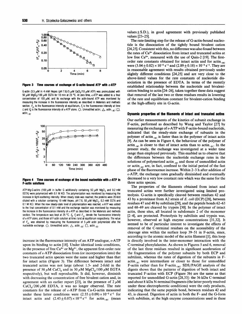
Figure 3 Tlime courses of exchange of G-actln-bound ATP with e-ATP
G-actin (3.5 ,uM in 4 mM Hepes (pH 7.6)/5 ,uM CaCI2/10 ,uM ATP) was preincubated with50 1sM MgCI2/100 ,uM EGTA for 10 min at 23 OC. At zero time, e-ATP was added to a finalconcentration of 100lM, and its exchange with the actin-bound ATP was monitored bymeasuring the increase in the fluorescence intensity as described in Materials and methodssection. F/, is the fluorescence intensity at equilibrium, Ft is the fluorescence intensity at time4 and F0 is the fluorescence intensity of e-ATP alone. 0, Unmodified actin; A, actin-2C; El,
actin3C'
50
200 lo
x
5
1U--
60 120 180 240 300 360 420 480Time (min)
Figure 4 Time courses of exchange of the bound nucleoUde with e-ATP InF-actin solutions
ATP-Mg-G-actins (100 uM in buffer G additionally containing 50 ,uM MgCI2 and 0.2 mMEGTA) were polymerized with 0.1 M KCI. The polymerization was monitored by measuring theincrease in light-scattering intensity. When steady state was reached, the proteins were 20-folddiluted with a solution containing 10 mM Hepes, pH 7.6, 50 uiM MgCI2, 0.2 mM EGTA and0.1 M KCI. When the new steady-state level of polymerization was reached, e-ATP was addedto the final concentration of 0.1 mM and the exchange reaction was monitored by measuringthe increase in the fluorescence intensity of c-ATP as described in the Materials and methodssection. The temperature was kept at 25 °C. Fo, F and Fo denote the fluorescence intensityof c-ATP alone, and those of F-actin solution at time tand at equilibrium respectively. The valueof Fc, was obtained by measuring the fluorescence of 5 1uM actin polymerized after thenucleotide exchange. 0, Unmodified actin; A, actin-2C; a actin 3C'
increase in the fluorescence intensity of an ATP analogue, c-ATPupon its binding to actin [18]. Under identical ionic conditions,in the presence of free Ca2+ or Mg2+, the apparent first-order rateconstants of c-ATP dissociation from (or incorporation into) thetwo truncated actin species were the same and higher than thatfor intact actin (Figure 3). The difference between intact andtruncated actins was not large (about 1.5- and 2-fold in the
presence of 50 ,iM CaCl2 and in 50 ,uM MgCl2/ 100 ,tM EGTArespectively), but well reproducible. It did, however, diminishwith decreasing the concentration of free bivalent cation and, in
agreement with an earlier report [23], in the presence of 2 jtM
CaCl2/200 M EDTA, it was no longer observed. The rate
constants for the release of c-ATP from Ca-G-actin measuredunder these latter conditions were (2.55 + 0.09) x 10-2 s-1 forintact actin and (2.45 + 0.07) x 10-2 S-1 for actin_3C (mean
values + S.D.), in good agreement with previously publishedvalues [23-25].The rate-limiting step for the release of G-actin-bound nucleo-
tide is the dissociation of the tightly bound bivalent cation[24,25]. Consistent with this, no difference was also found betweenthe rates of Ca2+ dissociation from intact and truncated actins atlow free Ca2+, measured with the use of Quin-2 [19]. The first-order rate constants obtained for intact actin and for actin,3Cwere (3.04+0.02) x 10-2 s-I and (2.89+0.18) x 10-2 s-1. They arein reasonable agreement with results obtained previously underslightly different conditions [24,25] and are very close to theabove-listed values for the rate constants of nucleotide dis-sociation in the presence of EDTA. In terms of the recentlyestablished relationship between the nucleotide and bivalent-cation binding to actin [24-26], taken together these data suggestthat removal of the last two or three residues results in loweringof the rate and equilibrium constant for bivalent-cation bindingat the high-affinity site in G-actin.
Dynamic properties of the filaments of Intact and truncated actinsOur earlier measurements of the kinetics of subunit exchange inF-actin, performed as described by Wang and Taylor [27] bymeasuring the exchange of e-ATP with F-actin-bound nucleotide,indicated that the steady-state exchange of subunits in thepolymer of actin,3C is faster than in the polymer of intact actin[9]. As can be seen in Figure 4, the behaviour of the polymer ofactin-2C is closer to that of intact actin than to actin-3C. In thepresent study, the exchange was investigated at a wider timerange than employed previously. This enabled us to observe thatthe differences between the nucleotide exchange rates in thesolutions of polymerized actin-3W and those of unmodified actinor actin,2C are, in fact, confined to the initial period of the fastphase of the fluorescence increase. Within 2-3 h after addition ofe-ATP, the exchange rates gradually diminished and eventuallydecreased to a very low constant rate which was the same for thethree actin species.The properties of the filaments obtained from intact and
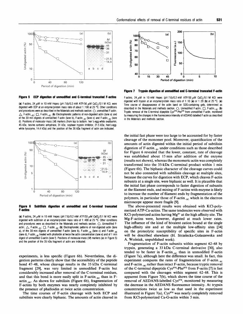
truncated actins were further investigated using limited pro-teolysis. G-actin is specifically cleaved between residues 42 and43 by a proteinase from A2 strain of E. coli (ECP) [28], betweenresidues 47 and 48 by subtilisin [29], and the peptide bonds 62-63and 68-69 are cleaved by trypsin [30]. In the polymer of intactactin, these sites, all located in subdomain 2 of the monomer
[2-4], are protected. Proteolysis by subtilisin and trypsin was,however, observed at high enzyme concentrations [31,32]. Itseemed to be of particular interest to investigate the effects ofremoval of the C-terminal residues on the accessibility of thecleavage sites within the surface loop 39-51 in F-actin, since,according to the atomic model of the actin filament [1], this loopis directly involved in the inter-monomer interaction with theC-terminal phenylalanine. As shown in Figures 5 and 6, removalof the last three residues resulted in significant acceleration ofthe fragmentation of the polymer subunits by both ECP andsubtilisin, whereas the rates of digestion of the subunits in F-actin,2 were intermediate or closer to those for unmodifiedF-actin rather than for F-actin_3C, SDS/PAGE analysis of thedigests shows that the patterns of digestion of both intact andtruncated F-actins with ECP (Figure 5b) are the same as thatreported for unmodified G-actin [28,33]: the 36 kDa C-terminaland about 8 kDa N-terminal fragments (the latter poorly resolvedunder these electrophoretic conditions) were the only products,indicating that the same peptide bond, between residues 42 and43, is cleaved. Digestion of actin in both the F- and the G-formwith subtilisin, at the high enzyme concentrations used in these
Conformational effects of removal of C-terminal residues of actin
, 150
O XV6(D0
\ , 1000
500-.
45 0Period of digestion (min)
Period of digestion (min)
Figure 5 ECP digestion of unmodffied and C-terminal truncated F-actins
(a) F-actins, 24 ,uM in 10 mM Hepes (pH 7.6)/0.2 mM ATP/50 4aM CaCI2/0.1 M KCI, weredigested with ECP at an enzyme/protein mass ratio of about 1:100 at 25 OC. Other conditionsand procedures were as described in the Materials and methods section. 0, unmodified F-actin;AL, F-actin-2c; O, F-actin-3C. (b) Electrophoretic patterns of non-digested actin (lane a) andof the 30 min digests of unmodified F-actin (lane b), F-actin-2c (lane c) and F-actin-3c (laned). Positions of molecular-mass (At markers (from top to bottom: hen's-egg-white ovalbumin,45 kDa; bovine carbonic anhydrase, 31 kDa; soybean trypsin inhibitor, 21.5 kDa; hen's-egg-white lysozyme, 14.4 kDa) and the position of the 36 kDa fragment of actin are indicated.
100E (a) (b)
50 M
lkDal
(3 5-~~~~~~~~~~~~~~31-
~~~~~~~~~~~~~~21.5-14.4-
5
\ a b c d e f
0 4o 80 120Period of digestion (min)
Figure 6 Subtilisin digestion of unmodffmed and C-terminal truncatedF-actins
(a) F-actins, 24 ,M in 10 mM Hepes (pH 7.6)/0.2 mM ATP/50 aM CaCI2/0.1 M KCI weredigested with subtilisin at an enzyme/protein mass ratio of 1:400 at 25 °C. Other conditionsand procedures were as described in the Materials and methods section. 0, Unmodified F-actin; A, F-actin.2c; C1, F-actin-3c. (b) Electrophoretic patterns of non-digested actin (lanea), of the 30 min digests of unmodified F-actin (lane b), F-actin.2c (lane c) and F-actin-3c(lane d), F-actin-3c treated with phalloidin at twice the actin concentration (lane e) and of 1 mindigest of unmodified G-actin (lane f). Positions of molecular-mass (Al markers (as in Figure 5)and the position of the 35 kDa fragment of actin are indicated.
experiments, is less specific (Figure 6b). Nevertheless, the di-gestion patterns clearly show that the accessibility of the peptidebond 47-48, whose cleavage results in the 35 kDa C-terminalfragment [29], was very limited in unmodified F-actin butconsiderably increased after removal of the C-terminal residues,and that this bond is more easily split in F-actin-3c than in F-actin-2C. As shown for subtilisin (Figure 6b), fragmentation ofF-actins by both enzymes was nearly completely inhibited bythe presence of phalloidin at twice actin concentration.The time courses of F-actin cleavage with both ECP and
subtilisin were clearly biphasic. The amounts of actin cleaved in
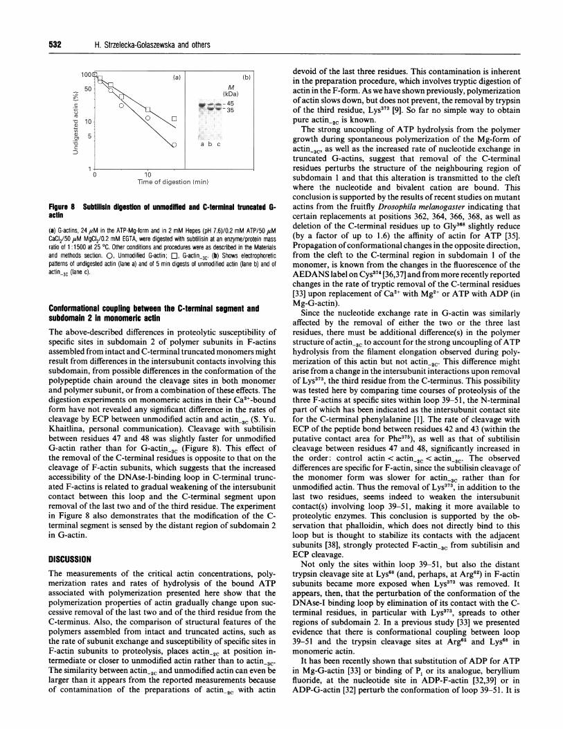
Figure 7 Trypsin digestion of unmodmfied and C-terminal truncated F-actin
F-actins, 24,M in 10 mM Hepes (pH 7.6)/0.2 mM ATP/50,M CaCI2/0.1 M KCI weredigested with trypsin at an enzyme/protein mass ratio of 1 :10 (a) or 1 :20 (b) at 25 'C. (a)Time course of disappearance of the actin band on SDS-containing gels, determined asdescribed in the Materials and methods section. 0, Unmodified F-actin; El, F-actin_3c. (b)Tryptic removal of the C-terminal dipeptide Cys374-Phe375 from unmodified F-actin, monitoredby measuring the changes in the fluorescence intensity of AEDANS-labelled F-actin as describedin the Materials and methods section.
the initial fast phase were too large to be accounted for by fastercleavage of the monomer pool. Moreover, quantification of theamounts of actin digested within the initial period of subtilisindigestion of F-actin-3C under conditions such as those describedfor Figure 6 revealed that the lower, constant, rate of cleavagewas established about 15 min after addition of the enzyme
(results not shown), whereas the monomeric actin was completelytransformed into the 35-kDa C-terminal product within 1 min(Figure 6b). The biphasic character of the cleavage curves couldnot be also connected with subtilisin cleavage at multiple sites,because the curves for digestion with ECP, which cleaves F-actinsubunits at a single site, were biphasic as well. It is plausible thatthe initial fast phase corresponds to faster digestion of subunitsat the filament ends, and mixing of F-actins with enzyme is likelyto increase the number of filament ends by fragmentation of thepolymers, in particular those of F-actin-3C which in the electronmicroscope appear more fragile [9].The above-presented results were obtained with KCl-poly-
merized ATP-Ca-actins. The same tendencies were observed withKCl-polymerized actins having Mg2+ at the high affinity site. TheMg-F-actins were, however, digested at much lower rates.The influence of the kind of bivalent cation bound at the singlehigh-affinity site and at the multiple low-affinity sites [34]on the proteolytic susceptibility of specific sites in F-actinwill be described elsewhere (H. Strzelecka-Golaszewska andA. Wozniak, unpublished work).
Fragmentation of F-actin subunits within segment 62-68 bytrypsin, generating a 33 kDa C-terminal derivative [30], alsotended to be faster in F-actin,3 than in unmodified F-actin(Figure 7a), although here the difference was small. In fact, thisexperiment compares the rates of fragmentation of F-actin.31and F-actin,2c rather than intact F-actin, because tryptic removalof the C-terminal dipeptide Cys374-Phe375 from F-actin [7] is fastcompared with the cleavages within segment 62-68. This isapparent from Figure 7(b), which shows the time course of theremoval of AEDANS-labelled Cys374, monitored by measuringthe decrease in the AEDANS fluorescence intensity. At trypsinconcentrations twice as low as that used in the experimentillustrated in Figure 7(a), Cys374 was nearly completely removedfrom KCl-polymerized Ca-G-actin within 5 min.
1001
, 50
coa)
a,
cD 10
0
531
(b)
0-1-.4
.)c)4-c
'C
5 10
532 H. Strzelecka-Golaszewska and others
50
C-
C,-0 1o
U,
cn
Figure 8 Subtilisin digestion of unmodMed and C-terminal truncated G-actin
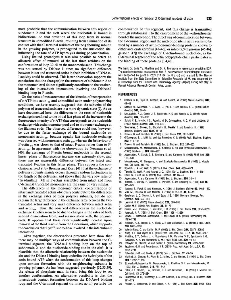
(a) G-actins, 24 4aM in the ATP-Mg-form and in 2 mM Hepes (pH 7.6)/0.2 mM ATP/50 ,tMCaCI2/50 ,tM MgCI2/0.2 mM EGTA, were digested with subtilisin at an enzyme/protein massratio of 1 :1500 at 25 OC. Other conditions and procedures were as described in the Materialsand methods section. 0, Unmodified G-actin; EO, G-actin-3c. (b) Shows electrophoreticpatterns of undigested actin (lane a) and of 5 min digests of unmodified actin (lane b) and ofactin-3C (lane c).
Conformational coupling between the C-terminal segment andsubdomain 2 in monomeric actinThe above-described differences in proteolytic susceptibility ofspecific sites in subdomain 2 of polymer subunits in F-actinsassembled from intact and C-terminal truncated monomers mightresult from differences in the intersubunit contacts involving thissubdomain, from possible differences in the conformation of thepolypeptide chain around the cleavage sites in both monomer
and polymer subunit, or from a combination of these effects. Thedigestion experiments on monomeric actins in their Ca2+-boundform have not revealed any significant difference in the rates ofcleavage by ECP between unmodified actin and actin-3C (S. Yu.Khaitlina, personal communication). Cleavage with subtilisinbetween residues 47 and 48 was slightly faster for unmodifiedG-actin rather than for G-actin-3C (Figure 8). This effect ofthe removal of the C-terminal residues is opposite to that on thecleavage of F-actin subunits, which suggests that the increasedaccessibility of the DNAse-I-binding loop in C-terminal trunc-ated F-actins is related to gradual weakening of the intersubunitcontact between this loop and the C-terminal segment uponremoval of the last two and of the third residue. The experimentin Figure 8 also demonstrates that the modification of the C-terminal segment is sensed by the distant region of subdomain 2in G-actin.
DISCUSSIONThe measurements of the critical actin concentrations, poly-merization rates and rates of hydrolysis of the bound ATPassociated with polymerization presented here show that thepolymerization properties of actin gradually change upon suc-
cessive removal of the last two and of the third residue from theC-terminus. Also, the comparison of structural features of thepolymers assembled from intact and truncated actins, such as
the rate of subunit exchange and susceptibility of specific sites inF-actin subunits to proteolysis, places actin-2, at position in-termediate or closer to unmodified actin rather than to actin-3c.The similarity between actin-2c and unmodified actin can even belarger than it appears from the reported measurements becauseof contamination of the preparations of actin-2. with actin
devoid of the last three residues. This contamination is inherentin the preparation procedure, which involves tryptic digestion ofactin in the F-form. As we have shown previously, polymerizationof actin slows down, but does not prevent, the removal by trypsinof the third residue, Lys373 [9]. So far no simple way to obtainpure actin2C is known.The strong uncoupling of ATP hydrolysis from the polymer
growth during spontaneous polymerization of the Mg-form ofactin-3C, as well as the increased rate of nucleotide exchange intruncated G-actins, suggest that removal of the C-terminalresidues perturbs the structure of the neighbouring region ofsubdomain 1 and that this alteration is transmitted to the cleftwhere the nucleotide and bivalent cation are bound. Thisconclusion is supported by the results of recent studies on mutantactins from the fruitfly Drosophila melanogaster indicating thatcertain replacements at positions 362, 364, 366, 368, as well asdeletion of the C-terminal residues up to Gly368 slightly reduce(by a factor of up to 1.6) the affinity of actin for ATP [35].Propagation of conformational changes in the opposite direction,from the cleft to the C-terminal region in subdomain I of themonomer, is known from the changes in the fluorescence of theAEDANS label on Cys374 [36,37] and from more recently reportedchanges in the rate of tryptic removal of the C-terminal residues[33] upon replacement of Ca2+ with Mg2+ or ATP with ADP (inMg-G-actin).
Since the nucleotide exchange rate in G-actin was similarlyaffected by the removal of either the two or the three lastresidues, there must be additional difference(s) in the polymerstructure of actin3C to account for the strong uncoupling ofATPhydrolysis from the filament elongation observed during poly-merization of this actin but not actin-2C. This difference mightarise from a change in the intersubunit interactions upon removalof Lys373, the third residue from the C-terminus. This possibilitywas tested here by comparing time courses of proteolysis of thethree F-actins at specific sites within loop 39-51, the N-terminalpart of which has been indicated as the intersubunit contact sitefor the C-terminal phenylalanine [1]. The rate of cleavage withECP of the peptide bond between residues 42 and 43 (within theputative contact area for Phe375), as well as that of subtilisincleavage between residues 47 and 48, significantly increased inthe order: control actin < actin2C < actin-3C. The observeddifferences are specific for F-actin, since the subtilisin cleavage ofthe monomer form was slower for actin_3C rather than forunmodified actin. Thus the removal of Lys373, in addition to thelast two residues, seems indeed to weaken the intersubunitcontact(s) involving loop 39-51, making it more available toproteolytic enzymes. This conclusion is supported by the ob-servation that phalloidin, which does not directly bind to thisloop but is thought to stabilize its contacts with the adjacentsubunits [38], strongly protected F-actin-3c from subtilisin andECP cleavage.Not only the sites within loop 39-51, but also the distant
trypsin cleavage site at Lys68 (and, perhaps, at Arg62) in F-actinsubunits became more exposed when Lys373 was removed. Itappears, then, that the perturbation of the conformation of theDNAse-I binding loop by elimination of its contact with the C-terminal residues, in particular with Lys373, spreads to otherregions of subdomain 2. In a previous study [33] we presentedevidence that there is conformational coupling between loop39-51 and the trypsin cleavage sites at Arg62 and Lys68 inmonomeric actin.
It has been recently shown that substitution ofADP for ATPin Mg-G-actin [33] or binding of Pi or its analogue, berylliumfluoride, at the nucleotide site in ADP-F-actin [32,39] or inADP-G-actin [32] perturb the conformation of loop 39-51. It is
Conformational effects of removal of C-terminal residues of actin
most probable that the communication between this region ofsubdomain 2 and the cleft where the nucleotide is bound isbidirectional, so that deviation of this loop from its normalstructure in unmodified F-actin, resulting from elimination of itscontact with the C-terminal residues of the neighbouring subunitin the growing polymer, is propagated to the nucleotide site,influencing the rate of ATP hydrolysis during polymerization.
Using limited proteolysis it was also possible to detect anallosteric effect of removal of the last three residues on theconformation of loop 39-51 in the monomeric actin. This changewas not sensed by DNAse I, since no significant differencebetween intact and truncated actins in their inhibition ofDNAse-I activity could be observed. This latter observation supports theconclusion that the change(s) in the structure of subdomain 2 onthe monomer level do not significantly contribute to the weaken-ing of the intersubunit interactions involving the DNAse-Ibinding loop in F-actin.On the basis of measurements of the kinetics of incorporation
of e-ATP into actin-3W and unmodified actin under polymerizingconditions, we have recently suggested that the subunits of thepolymer of truncated actin are in a more dynamic state [9]. As wehave documented here, the difference in the rates of nucleotideexchange is confined to the initial fast phase of the increase in thefluorescence intensity ofe-ATP that corresponds to the nucleotideexchange with actin monomer pool and to monomer exchange atthe filament ends. The observed difference could not, however,be due to the faster exchange of the bound nucleotide onmonomeric actin-3C, because equally fast nucleotide exchangewas observed with monomeric actin.2C, whereas the behaviour ofF-actin,2 was closer to that of intact F-actin rather than to F-actin_3C' In agreement with the observation by Newman et al.[40], the exchange of F-actin bound nucleotide in the second,linear, phase of fluorescence increase was extremely slow, andthere was no measurable difference between the intact andtruncated F-actins in this latter phase. This supports the con-clusion arrived at by Brenner and Korn [41] that the exchange ofpolymer subunits mainly occurs through random fluctuations inthe length of the polymers, and shows that the very low rates of'treadmilling' [42] in F-actins assembled from intact and fromC-terminal truncated monomers are the same or very similar.The differences in the monomer critical concentrations of
intact and truncated actins obviously contribute to the differencesin the exchange rates of their polymer subunits, but do notexplain the large difference in the exchange rates between the twotruncated actins and very small difference between intact actinand actin-2C. Thus, the observed differences in the nucleotideexchange kinetics seem to be due to changes in the rates of bothsubunit dissociation from, and reassociation with, the polymerends. It appears that these rates significantly increase uponremoval of the third residue from the C-terminus, which supportsthe conclusion that Lys373 is somehow involved in the intersubunitinteraction.Taken together, the observations presented here show that
there may be multiple ways of communication between the C-terminal segment, the DNAse-I binding loop on the top ofsubdomain 2, and the nucleotide-binding site in the cleft. It isplausible that the allosteric relationship between the nucleotidesite and the DNase-I binding loop underlies the hydrolysis of theactin-bound ATP when the conformation of this loop changesupon contact formation with the neighbouring subunits ingrowing polymer. As has been suggested previously [32,33,39],the release of phosphate may, in turn, bring this loop to yetanother conformation. An alternative possibility is that theintersubunit contact formation between the DNAse-I binding
conformation of this segment, and this change is transmittedthrough subdomain 1 to the environment of the y-phosphoesterbond ofthe nucleotide. The direct way ofcommunication betweenthe C-terminal region and the nucleotide site in actin seems to beused by a number of actin-monomer-binding proteins known toeither accelerate (profilin [43-46]) or inhibit (,8-thymosins [45,46],gelsolin [47]) the exchange of G-actin-bound nucleotide, as theC-terminal segment of the actin polypeptide chain participates inthe binding of these proteins [3,4,48].
We thank Dr. Sofia Yu. Khaitlina and Dr. A. Morozova for generously providing ECP.The excellent technical assistance of Mrs. E. Karczewska is acknowledged. This workwas supported by grant 6 P203 011 04 (to H. S.-G.) and a grant to the NenckiInstitute from the State Committee for Scientific Research. M. M. was supported bya fellowship from the Science and Technology Agency (Japan) during her stay inKansai Advance Research Center, Kobe, Japan.
REFERENCES1 Holmes, K. C., Popp, D., Gebhard, W. and Kabsch, W. (1990) Nature (London) 347,
44-492 Kabsch, W., Mannherz, H. G., Suck, D., Pai, E. F. and Holmes, K. C. (1990) Nature
(London) 347, 37-443 McLaughlin, P. J., Gooch, J. T., Mannherz, H.-G. and Weeds, A. G. (1993) Nature
(London) 364, 685-6924 Schutt, C. E., Myslik, J. C., Rozycki, M. D., Goonesekere, N. C. W. and Lindberg, U.
(1993) Nature (London) 365, 810-8165 Stournaras, C., Drewes, G., Blackholm, H., Merkler, I. and Faulstich, H. (1990)
Biochim. Biophys. Acta 1037, 86-916 Drewes, G. and Faulstich, H. (1990) J. Biol. Chem. 265, 3017-30217 O'Donoghue, S. I., Miki, M. and dos Remedios, C. G. (1992) Arch. Biochem. Biophys.
293,110-1168 Drewes, G. and Faulstich, H. (1993) Eur. J. Biochem. 212, 247-2539 Mossakowska, M., Moraczewska, J., Khaitlina, S. Yu. and Strzelecka-Golaszewska, H.
(1993) Biochem. J. 289, 897-90210 Aspenstrom, P., Schutt, C. E., Lindberg, U. and Karlsson, R. (1993) FEBS Left. 329,
163-17011 Mossakowska, M., Nakayama, H. and Strzelecka-Gotaszewska, H. (1933) J. Muscle
Res. Cell Motil. 15, 19912 Spudich, J. A. and Watt, S. (1971) J. Biol. Chem. 246, 4866-487113 Tawada, K., Wahl, P. and Auchet, J.-C. (1978) Eur. J. Biochem. 88, 411-41914 Houk, W. T. and Ule, K. (1974) Anal. Biochem. 62, 66-7415 Aspenstrom, P. and Karlsson, R. (1991) Eur. J. Biochem. 200, 35-4116 Blikstad, I., Markey, F., Carlsson, L., Persson, T. and Lindberg, U. (1978) Cell 15,
935-94317 Kodama, T., Fukui, K. and Kometani, K. (1986) J. Biochem. (Tokyo) 99, 1465-147218 Miki, M., Ohnuma, H. and Mihashi, K. (1974) FEBS Lett. 46,17-1919 Gershman, L. C., Selden, L. A. and Estes, J. E. (1986) Biochem. Biophys. Res.
Commun. 135, 607-61420 Laemmli, K. K. (1970) Nature (London) 227, 680-68521 Carlier M.-F. (1990) Adv. Biophys. 26, 51-7322 Carlier, M.-F., Pantaloni, D. and Korn, E. D. (1987) J. Biol. Chem. 262, 3052-305923 Kasprzak, A. A. (1993) J. Biol. Chem. 268, 13261-1326624 Nowak, E., Strzelecka-Golaszewska, H. and Goody, R. S. (1988) Biochemistry 27,
1785-1 79225 Kinosian, H. J., Selden, L. A., Estes, J. E. and Gershman, L. C. (1993) J. Biol. Chem.
268, 8683-869126 Valentin-Ranc, C. and Carlier, M.-F. (1989) J. Biol. Chem. 264, 20871-2088027 Wang, Y.-L. and Taylor, D. L. (1981) Proc. Natl Acad. Sci. U.S.A. 78, 5503-550728 Khaitlina, S. Y., Collins, J. H., Kuznetsova, I. M., Pershina, V. P., Synakevich, I. G.,
Turoverov, K. K. and Usmanova, A. M. (1991) FEBS Lett. 279, 49-5129 Schwyter, D., Phillips, M. and Reisler, E. (1989) Biochemistry 28, 5889-589530 Jacobson, G. R. and Rosenbusch, J. P. (1976) Proc. Natl Acad. Sci. U.S.A. 73,
2742-274631 Pollender, J.-M. and Gruda, J. (1979) Can. J. Biochem. 57, 49-5532 Muhlrad, A., Cheung, P., Phan, B. C., Miller, C. and Reisler, E. (1994) J. Biol. Chem.
269, 11852-1185833 Strzelecka-Gotaszewska, H., Moraczewska, J., Khaitlina, S. Y. and Mossakowska, M.
(1993) Eur. J. Biochem. 211, 731-74234 Estes, J. E., Selden, L. A., Kinosian, H. J. and Gershman, L. C. (1992) J. Muscle Res.
Cell Motil. 13, 272-28435 Drummond, D. R., Hennessey, E. S. and Sparrow, J. C. (1992) Eur. J. Biochem. 209,
171-179loop and the C-terminal segment (in intact actin) perturbs the
533
36 Frieden, C., Lieberman, D. and Gilbert, H. R. (1980) J. Biol. Chem. 225, 8991-8993

534 H. Strzelecka-Golaszewska and others
37 Frieden, C. and Patane, K. (1985) Biochemistry 24, 4192-419638 Lorenz, M., Popp, D. and Holmes, K. C. (1993) J. Mol. Biol. 234, 826-83639 Orlova, A. and Egelman, E. H. (1992) J. Mol. Biol. 227, 1047-105340 Newman, J., Zaner, K. S., Schick, K. L., Gershman, L. C., Selden, L. A., Kinosian,
K. J., Travis, J. L. and Estes, J. E. (1993) Biophys. J. 64, 1559-156641 Brenner, S. L. and Korn, E. D. (1983) J. Biol. Chem. 258, 5013-502042 Wegner, A. and Neuhaus, J. M. (1981) J. Mol. Biol. 153, 681-69343 Mockrin, S. C. and Korn, E. D. (1980) Biochemistry 19, 5359-5362
44 Nishida, E. (1985) Biochemistry 24, 1160-116445 Goldschmidt-Clermont, P. J., Furman, M. I.,Wachsstock, D., Safer, D., Nachmias, V. T.
and Pollard, T. D. (1992) Mol. Biol. Cell 3,1015-102446 Carlier, M.-F., Jean, C., Rieger, K. J., Lenfant, M. and Pantaloni, D. (1993) Proc. Natl.
Acad. Sci. U.S.A. 90, 5034-503847 Coue, M. and Korn, E. D. (1986) J. Biol. Chem. 261, 1588-159348 Heintz, D., Reichert, A., Mihelic, M., Voelter, W. and Faulstich, H. (1993) FEBS Lett.
329, 9-12
Received 5 August 1994/1 December 1994; accepted 8 December 1994