Long QT Syndrome in Sweden - DiVA...
119
Umeå University Medical Dissertations, New Series No 1515 Long QT Syndrome in Sweden Founder Effects and Associated Cardiac Phenotypes Annika Winbo Department of Clinical Sciences, Pediatrics Umeå 2012
Transcript of Long QT Syndrome in Sweden - DiVA...
Umeå University Medical Dissertations, New Series No 1515
Long QT Syndrome in Sweden Founder Effects and Associated Cardiac Phenotypes
Annika Winbo
Department of Clinical Sciences, Pediatrics
Umeå 2012

Responsible publisher under Swedish law: the Dean of the Medical Faculty
This work is protected by the Swedish Copyright Legislation (Act 1960:729) UMEÅ
New Series No: 1515
ISBN: 978-91-7459-460-7
ISSN: 0346-6612
Cover: Annika Winbo (photo), Erik Winbo (design)
Electronic version available at http://umu.diva-portal.org/
Printed by: Print & Media
Umeå, Sweden 2012

This thesis is dedicated to the participating Long QT Syndrome families,
whose confidence and support enabled the studies
and for whose benefit the studies were incited

i
Table of Contents
Table of Contents i Abstract v Populärvetenskaplig sammanfattning vi
Bakgrund vi Bakomliggande genetisk orsak vi Sjukdomsuttryck (Fenotyp) vi Diagnostik och behandling vii Avhandlingens Syfte vii Metod och Resultat vii Slutsatser ix
Overview of Papers x Thesis at a glance xi Introduction 1
Founder effects and founder populations 1 Northern Swedish founder effects 1 The Long QT Syndrome 2 Autosomal dominant and recessive variants 2 Main clinical aspects of LQTS and JLNS 3
Electrocardiographic phenotype 3 Arrhythmia propensity and cardiac events 4 Auditory phenotype 4
Clinical diagnosis 4 Prophylactic treatment and interventions 4 LQTS- a heterogeneous disease 6 The KCNQ1-encoded potassium channel (Kv7.1) 7 The Kv7.1 channel and cardiac repolarization 8 The Kv7.1 channel and auditory function 9 Correlations between Kv7.1 function and phenotype 9 Remaining heterogeneity within the KCNQ1-subgroup 9 Proposed effects of modifying genes 11 LQTS research in Sweden 12 Resources on the national and regional levels 12
Regional LQTS- a brief history and study setting 12 The LQTS Family Clinic and Centre for Cardiovascular Genetics 12
Overall Aim 15 Specific Aims 15 Overview of aims and methods 16 Overview of study populations papers I-IV 18
Materials and Methods 18 Cases and families 19
ii
A national inventory of JLNS cases 19 Identification of JLNS cases 19 A JLNS prevalence estimate in preadolescent children 20
Case ascertainment 20 Diagnostic strategies for index cases and family members 20 Molecular genetics diagnostic methods and screening strategies used 21
Investigating the possible founder nature of a mutation 22 Genealogical investigations 22 Geographic origin and clustering of ancestors 23 Handling and presenting genealogical and geographic data 23 Haplotype analysis using microsatellite markers 23 Mutation dating in index families sharing a common haplotype 24 Prevalence estimations derived from mutation age 26 Prevalence estimates using Hardy-Weinberg statistics 26 Clinical cardiac phenotype 27 Data collection 27 Definitions of cardiac events 28 Electrocardiographic assessment of QT duration 28 Assessments of mutation-specific phenotypes 29
Investigation of natural history in a pedigree 29 Comparisons to other LQT1 and/or founder populations 30
Statistics: Clinical phenotype 30 Comparisons within or between populations or groups 30 Comparisons between populations or groups using summarized data 30 Repeated electrocardiographic measurements 30
Analysis of genetic modifiers and possible associations to cardiac phenotype
(cardiac events and QTc) 31 Selection of sequence variants and nomenclature 31 Analysis of sequence variants 32 Statistics: Associations between sequence variants and cardiac phenotype in
founder heterozygotes 33 Results 35
p.Y111C, p.R518X and Northern Swedish founder effects 35 Occurrence of p.Y111C and p.R518X in Sweden 35 The p.Y111C mutation- Genealogy and geographical origin 36 The p.R518X mutation- Genealogy and geographical origin 37 A shared haplotype in p.Y111C and p.R518X families, respectively 41 Estimated age of the p.Y111C and p.R518X founder mutations 43 Associated prevalence estimates for p.Y111C and p.R518X 44 Prevalence and mutation spectrum of JLNS in Sweden 45 Geographical distribution of four common JLNS mutations 48 Clinical phenotype 49 Study population characteristics 49

iii
A range of clinical severity evident 50 Triggers and cardiac events 50 Electrocardiographic phenotype 51 Genotype and gender-specific assessment of cardiac phenotypes 52
p.Y111C heterozygotes 52 p.R518X heterozygotes 52 JLNS cases 52
Beta-blocker therapy and other interventions 53 Assessment of the “benign” p.Y111C and p.R518X phenotypes 55
Benign phenotype verified in non-medicated p.Y111C ancestors 55 Phenotypic comparisons between LQT1 populations 56
Using sequence variant analysis to assess phenotypic variability in p.R518X
heterozygotes 57 Association testing of NOS1AP, KCNH2, KCNE1 and SCN5A derived variants
with a frequency >1.0 57 Frequency and allelic phase of 3’UTR derived sequence variants 60
Discussion 63 LQTS in Sweden and the impact of founder effects 63 JLNS in Sweden and the impact of founder effects 64 Findings suggestive of Scandinavian LQTS founder effects 65 LQTS prevalence estimates in Sweden, Norway and Finland 66 LQTS founder populations in Sweden and world wide 68 Main observed clinical phenotypes related to genotype 69 Severe phenotype associated with JLNS 69 A benign phenotype seen in founder heterozygotes 70 Remaining phenotypic variability in LQTS heterozygotes 71
Compound heterozygosity as a potential confounder 71 The contribution of sequence variants to phenotypic expression and variability in
LQTS founder populations 72 Sequence variants with non-allele-specific effects 72 Sequence variants with allele-specific effects 73
Main methodological considerations 74 Overview of main methodological approaches used and discussion regarding
their possible strength of evidence 76 Overview continued 77 Clinical management of LQTS within a temporal context 78 Direct clinical implication of the studies in the thesis 79 In Summary 80
Conclusions 81 Where do we go from here? 82
Contribution statement 83 Acknowledgements 84 References 87

iv
Appendices 1 LQTS Questionnaire, page 1 2 LQTS Questionnaire, page 2 3 LQTS Questionnaire, translated 4

v
Abstract Background We aimed to increase the knowledge regarding the familial
arrhythmogenic disorder Long QT Syndrome (LQTS) and its recessive
variant Jervell and Lange-Nielsen Syndrome (JLNS) in Sweden, including
prevalences and clinical phenotypes. A specific focus was directed towards
two KCNQ1 mutations –p.Y111C and p.R518X- commonly identified in
Swedish LQTS index cases.
Methods Cases and families with LQTS (p.Y111C or p.R518X) and JLNS
were recruited via regional clinical practices, national referrals to the Clinical
Genetics laboratory, Umeå University Hospital, and a national inventory.
Molecular genetics diagnostics was used for case ascertainment. Clinical data
was obtained via medical records, a questionnaire, and/or an interview.
Electrocardiograms were manually assessed. In p.R518X heterozygotes
intra-familial phenotypic variability (QTc and cardiac events) was assessed
by analysis of sequence variants (modifier genes).
The origins of the mutations p.Y111C and p.R518X were investigated using
genealogical and haplotype analysis (microsatellite markers). In families
sharing a common haplotype mutation age and associated prevalence was
analyzed using ESTIAGE and DMLE computer software.
Results We identified p.Y111C (170 mutation-carriers) and p.R518X (101
mutation-carriers) as two major causes of LQTS/JLNS in Sweden. LQTS
phenotype was revealed to be relatively benign in p.Y111C and p.R518X
(annual incidence of life-threatening cardiac events, before therapy 0.05%
and 0.04%, respectively). Gender-specific effects of genetic modifiers on
phenotypic expression were seen. A founder origin, approximately 600-700
years ago in two northern river valleys was established for p.Y111C and
p.R518X, and a high prevalence of LQTS founder descendants suggested.
A minimum JLNS prevalence of 1:200 000 in preadolescent Swedish
children was revealed. JLNS phenotype was mainly severe, with a
cumulative incidence of life-threatening cardiac events of 53% (annual
incidence rate before therapy 5%) and four sudden deaths. Possible founder
effects regarding four KCNQ1 mutations; p.Y111C (8%), p.R518X (50%),
c.572_576del (17%) and p.Q530X (8%) together explained 83% of the JLNS
mutation-spectrum in Sweden, consisting of 8 KCNQ1 mutations.
Conclusion The high prevalence of p.Y111C- and p.R518X-related LQTS
as well as JLNS revealed in Sweden could be explained by the combination of
mild clinical phenotypes in heterozygotes and strong founder effects present
during the population development of northern Sweden.
Increased knowledge regarding the occurrence of LQTS and JLNS as well
as mutation- and/or genotype-specific data constitute prerequisites for
possible improvement of patient management.

vi
Populärvetenskaplig sammanfattning
Bakgrund
Långt QT Syndrom (LQTS) är en ärftlig sjukdom som kännetecknas av en
ökad risk att utveckla livshotande hjärtrytmrubbning (arytmi). Sjukdomen
beskrevs för första gången på slutet av 50-talet, och förekommer i två
varianter med olika ärftlighetsgång; Romano-Wards Syndrom (RWS eller
LQTS) och Jervell och Lange-Nielsens Syndrom (JLNS), namngivna efter de
läkare som först beskrev dem.
LQTS ärvs autosomalt dominant (ett sjukdomsanlag krävs) medan JLNS
ärvs autosomalt recessivt (två anlag krävs, ett från varje förälder). JLNS
kännetecknas, utöver en hög risk för allvarliga arytmier, av medfödd
hörselnedsättning.
Förekomsten av LQTS och JLNS är sedan tidigare okänd i Sverige. Från
internationella studier vet man att LQTS är relativt vanligt förekommande
(prevalens ca 1:2500) medan JLNS är ovanligt (prevalens ca 1 på miljonen).
Bakomliggande genetisk orsak
Den ärftliga komponenten (sjukdomsanlaget) vid LQTS utgörs av mutationer
(felaktigheter i DNA) i gener (proteinrecept) som ansvarar för bildningen av
kanaler (proteiner) som sköter transporten av vissa kroppssalter (joner).
LQTS och JLNS kan orsakas av hundratals olika mutationer i ett flertal
gener. En av de gener som oftast drabbats av mutationer vid både LQTS och
JLNS är KCNQ1-genen. KCNQ1-genen sitter på kromosom 11 och kodar för
en jonkanal som reglerar transporten av kroppssaltet kalium (K+) i många
organ, bland annat i hjärtat och innerörat. Dessa kaliumkanaler är viktiga för
den normala hjärtrytmen samt hörseln.
Sjukdomsuttryck (Fenotyp)
Det vanligaste symptomet vid LQTS utgörs av en plötslig svimning, som
ibland kan likna ett krampanfall. Om arytmin inte går över spontant kan den
i värsta fall leda till plötslig hjärtdöd.
LQTS är, som namnet antyder, generellt sätt förknippat med en förlängd
QT-tid som mäts på vilo-EKG. QT-tiden motsvarar enkelt uttryckt tiden det
tar för hjärtat att bli elektriskt neutralt inför ett nytt hjärtslag. En förlängd
QT-tid innebär en ökad risk att ett nytt slag ska påbörjas innan hjärtat är
redo. Svimningstendensen och QT-förlängningen är ofta, men inte alltid,
kopplade till varandra. Inte alla som ärvt LQTS-orsakande mutationer har
QT-förlängning eller utvecklar symptom under sin livstid.

vii
Diagnostik och behandling
Eftersom QT-tiden inte alltid är lång vid LQTS ställs diagnosen säkrast med
hjälp av mutationsidentifiering (DNA-test, molekylärgenetisk diagnostik).
Behandlingen vid LQTS utgörs av patientinformation (bland annat syftande
till att undvika QT-förlängande läkemedel) och effektiv förebyggande
medicinsk behandling (beta-blockad). Vid JLNS, som är mer svårbehandlat,
kan ytterligare åtgärder behövas som komplement.
Avhandlingens Syfte
Huvudsyftet med den här avhandlingen och dess delstudier var att öka
kunskapen om LQTS i Sverige vad gäller förekomst och sjukdomsuttryck
samt att försöka identifiera möjliga faktorer som kan bidra till klinisk
riskbedömning (riskstratifiering).
Delstudierna fokuserar dels på två mutationer i KCNQ1-genen (p.Y111C och
p.R518X) som visat sig vara ”ovanligt vanligt förekommande” i svenska
LQTS familjer, samt på den ovanliga men allvarliga varianten JLNS.
Metod och Resultat
Via molekylärgenetisk diagnostik identifierades bärarskap av Y111C/KCNQ1
hos 170 individer i 37 svenska LQTS familjer. Bärarskap av R518X/KCNQ1
påvisades hos 101 individer i 19 svenska LQTS familjer. Informerat samtycke
till att ingå i studierna lämnades av alla deltagande fall/familjer.
För att försöka ta reda på varför de två mutationerna p.Y111C och p.R518X
var så vanligt förekommande bland svenska LQTS familjer utforskade vi
familjernas ursprung via släktforskning (genealogi). Ett flertal kopplingar i
form av historiska blodsband samt ett gemensamt ursprung för bärare av
respektive mutation i två olika nordsvenska älvdalar kunde påvisas. Vi
undersökte även om det fanns släktskapsspår i de nu levande bärarnas DNA
(molekylärgenetisk haplotypsanalys). För både p.Y111C och p.R518X sågs
delade haplotyper hos de testade familjerna (DNA-rester som tyder på att
respektive mutation ärvts från en gemensam anmoder/fader- founder).
Mutationernas ålder uppskattades med hjälp av statistisk mjukvara
(ESTIAGE, DMLE) till ~600-700 år, vilket tolkades som att mutationerna
uppstått i älvdalarna i samband med att övre Norrland ”koloniserades”.
Förekomsten av dessa mutationer i den svenska befolkningen idag
uppskattades med hjälp av samma statisiska mjukvara grovt till ~1:4000-
7000 (p.Y111C), respektive 1:2000-4000 (p.R518X).

viii
Vad gäller JLNS i Sverige identifierade vi 19 JLNS fall i 13 familjer, varav fem
nu levande fall under 10 år, vilket innebär en förekomst av JLNS på
>1:200 000 bland barn under 10 år. Detta motsvarar den sedan tidigare
högsta beskrivna prevalensen av JLNS (i Norge).
Mutationspanoramat vid JLNS i Sverige (12 familjer med två mutationer
vardera, identifierade via molekylärgenetisk diagnostik) utgjordes av åtta
olika KCNQ1 mutationer. Hälften av de muterade anlagen utgjordes av
p.R518X, i linje med att förekomsten av denna mutation i populationen
skulle vara hög. Fyra mutationer, inklusive p.R518X och p.Y111C förklarade
83% av JLNS i Sverige. Vissa tecken till svensk-norska foundereffekter
gällande dessa fyra mutationer kunde ses.
Kliniska data (information om eventuell svimning och medicinering)
samlades in via enkät, telefonintervju och/eller via patientjournaler. En
personlig intervju utfördes med alla JLNS fall/familjer. QT-tiden mättes för
hand på insamlade EKG.
Andelen mutationsbärare i respektive grupp som svimmat var 34%, 17%
och 84% för p.Y111C, p.R518X och JLNS. Andelen med normal QT-tid i
samma grupper var 14%, 39% och 0.
Fenotypen (sjukdomsuttrycket) vid bärarskap av enstaka p.Y111C eller
p.R518X mutationer visade sig vara ovanligt mild, vilket också kan ha
bidragit till att mutationerna blivit så vanligt förekommande. Den årliga
förekomsten av livshotande hjärthändelser (hjärtstopp eller plötslig död)
utan behandling beräknades till 0.05% (p.Y111C) respektive 0.04%
(p.R518X). Detta är att jämföra med tidigare beskrivna nivåer motsvarande
0.3% per år för KCNQ1-relaterad LQTS (blandade mutationer) samt 5% per
år hos de svenska JLNS fallen. Vid JLNS sågs en klart allvarligare fenotyp,
där 53% drabbats av livshotande arytmier (hjärtstopp eller plötslig död).
Bland LQTS fall av p.R518X-genotyp framkom, utöver den milda
fenotypen, en atypisk bild där en tredjedel av fallen som svimmat hade
normal QT tid, och ingen korrelation mellan QT-tid och svimningstendens
kunde ses. Vidare sågs QT-tiderna variera starkt inom gruppen.
För att försöka förklara den avvikande bilden bland LQTS fall av p.R518X-
genotyp undersökte vi där förekomst av ytterligare genetiska förändringar
som är vanligt förekommande i befolkningen och som tidigare visats påverka
QT-tiden (sekvensvarianter). Bland de sekvensvarianter vi analyserade fann
vi att två varianter (i en gen vid namn NOS1AP) var associerade till QT-
förlängning specifikt hos män. Vidare fann vi, oväntat, att det i p.R518X
founderfamiljerna tillsammans med mutationen ärvdes sekvensvarianter
(intill KCNQ1 genen) som sannolikt bidrar till att minska mängden felaktiga
jonkanaler som produceras, och därigenom möjligen också skulle kunna
förklara den milda fenotypen.

ix
Slutsatser
De särskilda förutsättningar (foundereffekter) som rådde under
befolkningsutvecklingen i Norrlands älvdalar från 1300-talet och framåt,
samt mutationernas associerade milda fenotyper, har sannolikt bidragit till
att mutationerna p.Y111C och p.R518X berikats i den svenska populationen
och idag återfinns bland världens största LQTS founderpopulationer.
Sammantaget visar våra studier att förekomsten av både LQTS och JLNS i
Sverige är högre än man tidigare trott och att bärarfrekvensen av olika
LQTS-mutationer i den svenska befolkningen sannolikt överstiger 1:1000.
I och med de svenska LQTS founderpopulationerna har vi kunnat utföra
mutationsspecifika beskrivningar av sjukdomsuttrycket vid bärarskap av
p.Y111C och p.R518X (även till viss del p.R518X-relaterad JLNS), samt inom
grupperna identifierat möjliga riskfaktorer associerade med svimning eller
QT-förlängning. Detta möjliggör mutationsspecifik familjeinformation och
riskstratifiering.
Den oväntat höga förekomsten av LQTS samt det variabla och delvis
atypiska sjukdomsuttrycket motiverar att LQTS bör betänkas som möjlig
bakomliggande orsak (differentialdiagnos) vid oklar svimning/kramp
oberoende av markerad QT-förlängning.
Vi (syftande på alla oss som forskar kring LQTS) kan i nuläget säga en hel del
om ökad risk vid LQTS och JLNS, men vi kan inte säkert avgöra vilka som
inte löper risk att drabbas av symptom, särskilt med tanke på att symptom
hos mutationsbärare kan utlösas av exempelvis vissa läkemedel. Vi vet dock
att beta-blockad ger ett effektivt skydd vid LQTS, om den tas regelbundet.
Tills dess att bakgrunden till varför vissa drabbas av svåra symptom och
andra inte bättre klarlagts, kvarstår familjeutredning (där bärarskap i
familjer/släkter fastställs, helst innan symptom uppstått),
familjeinformation och förebyggande medicinering med beta-blockad som
den bästa strategin för att förhindra plötslig död till följd av LQTS.

x
Overview of Papers
I. Low Incidence of Sudden Cardiac Death in a Swedish Y111C Type 1 Long QT Syndrome Population.
Winbo A, Stattin EL, Diamant UB, Jensen SM, Rydberg A.
Circulation: Cardiovascular Genetics 2009;2:558-564.
Reproduced by permission of the American Heart Association
II. Origin of the Swedish Long QT Syndrome Y111C/KCNQ1 Founder Mutation.
Winbo A, Diamant UB, Rydberg A, Persson J, Jensen SM, Stattin EL.
Heart Rhythm 2011;8:541-547.
Reproduced by permission of Elsevier
III. Phenotype and Origin of the Swedish Long QT Syndrome R518X/KCNQ1 Founder Population- Phenotypic Variability partly explained by Gender-Specific Effects of Genetic Modifiers
Winbo A, Stattin EL, Norberg A, Nordin C, Diamant UB, Persson J, Jensen SM, Rydberg A.
Submitted manuscript
IV. Prevalence, Mutation Spectrum and Cardiac Phenotype of the Jervell and Lange-Nielsen Syndrome in Sweden.
Winbo A, Stattin EL, Diamant UB, Persson J, Jensen SM, Rydberg A.
Europace 2012; Epub Date 2012/04/28, doi: 10.1093/europace/eus111
Reproduced by permission of Oxford University Press

xi
Thesis at a glance
Paper Main findings
I An unusually benign clinical phenotype (annual incidence of life-
threatening cardiac events, before therapy 0.05%) was found in 80
Y111C/KCNQ1 mutation-carriers (15 index families), in spite of a
markedly prolonged QTc (481±36 ms) and the dominant-negative
effect of the mutation seen in previous in vitro studies. The benign
phenotype was confirmed in 107 non-medicated ancestors born
between 1968 and 1873, sharing a common founder.
II A common origin in a mid-northern river valley region for the Swedish
p.Y111C population (37 index families, 170 mutation-carriers) was
revealed, including congruent genealogical data, clustering of
ancestors, and a shared haplotype. Mutation age was estimated to
approximately 600 years and the associated prevalence to ~1:4000-
7000 in Sweden (population ~9.5 x 10⁶).
III A common origin in an upper northern river valley region for the
majority of the Swedish p.R518X population (19 index families, 101
mutation-carriers) was revealed, including congruent genealogical
data, clustering of ancestors, and a shared haplotype for 17/19
families. Mutation age was estimated to approximately 700 years, and
the associated prevalence to ~1:2000-4000 in Sweden.
A benign clinical phenotype was seen (annual incidence of life-
threatening cardiac events, before therapy 0.04%) in p.R518X
heterozygotes, possibly related to 3’UTR/KCNQ1 sequence variants
(genetic modifiers) segregating within the haplotype. Gender-specific
effects of NOS1AP sequence variants explained over ¼ of QTc variance
in males.
IV A national inventory identified 19 JLNS cases in 13 index families, and
a JLNS prevalence of ≥1:200 000 in Swedish preadolescent children
(5 living cases below 10 years of age).
The mutation spectrum in genotyped JLNS index families (n=12, 6
homozygous and 6 compound heterozygous) consisted of 8 KCNQ1
mutations, whereof the p.R518X mutation in 12/24 alleles. A severe
phenotype was seen including marked QT prolongation (587±69 ms),
a high frequency of life-threatening cardiac events (63%, including
verified ventricular fibrillation/fast ventricular tachycardia), whereof 4
sudden deaths.

xii

1
Introduction
This thesis could well have been named “Typographical Errors and the
Impact of Spatial and Temporal Timing”. Or, less formally stated, “The
Importance of being at the Right Place at the Right Time”.
The probability that a single typographical error (i.e. point mutation)
occurring in the genome of any one individual would become enriched in a
future subpopulation is grantedly infinitesimal. However, present-day
observations strongly bias against thwarted possibilities, focusing our
attention on that which is present and observable, i.e. that which has already
come to pass.
Founder effects and founder populations The enrichment of alleles on the population level may be caused by what is
known as founder effects. Founder effects relate to the loss of genetic
variation that occurs when a small population subset establishes a new
population. Founder populations consequently constitute descendants of
common founders, sharing alleles identical by descent.
Factors that propagate founder effects include geographical and/or
cultural isolation, population bottlenecks and endogamy.
Northern Swedish founder effects
The population development of the northern Swedish region included the
forming of genetic sub-isolates1 in the separate river valleys spanning the
region from northwest to southeast, a process propagated by:
Few initial settlers
Slow initial population growth (~50% 1571-17501)
Marked population growth (>250% 1750-19501)
Geographic isolation of the separate river valleys
Cultural isolation between ethnic groups (Saami, Finns and Swedes)
Preferences to marry within the same river valley1 and endogamy2
The resultant effects on the population level are reflected in the unique
frequency and distribution of many inherited disorders in the northern
region.3-7 In the clinical setting of my field, a high regional occurrence of
both the dominant and recessive variants of the familial Long QT Syndrome
(LQTS) has been evident. Moreover, as molecular diagnostics of LQTS
became clinically available, two specific mutations in the KCNQ1 gene,
p.Y111C and p.R518X, were revealed to be “uncommonly common” among
LQTS index cases of northern Swedish origin, jointly suggestive of possible
founder effects.

2
The Long QT Syndrome
Autosomal dominant and recessive variants
The Long QT Syndrome is an inherited arrhythmogenic disorder, associated
with familial loss-of-consciousness and electrocardiographic abnormalities.
The disorder occurs in two distinct forms differing in prevalence, inheritance
pattern (Figure 1) and clinical phenotype, named after the doctors that first
described them in 1964-658,9 and 195710, respectively. The distinct forms
include the more common autosomal dominant form -LQTS or Romano-
Ward syndrome8,9 (incidence >1:250011), as well as the rare autosomal
recessive form with concomitant congenital hearing loss –the Jervell and
Lange-Nielsen syndrome10 –JLNS (roughly estimated to affect less than 1 in
a million12)
Figure 1. The figure exemplifies the inheritance patterns (parents, gametes and offspring) for the autosomal
dominant (Long QT Syndrome- LQTS) and autosomal recessive (Jervell and Lange Nielsen Syndrome- JLNS)
forms of the Long QT Syndrome. The black dot signifies the heritable component. N- Normal/ Non-carrier
genotype. The auditory phenotype is inherited as an autosomal recessive trait, while the cardiac phenotype
(electrocardiographic abnormalities and arrhythmia propensity) is inherited as an autosomal dominant trait,
albeit with variable penetrance. Typically JLNS cases are more severely affected also regarding cardiac
phenotype. LQTS and JLNS may occur within the same pedigrees (as above, right).

3
Main clinical aspects of LQTS and JLNS
The main differences in clinical phenotype between LQTS and JLNS entail
the additional feature of congenital hearing loss in JLNS as well as an overall
more severe affectation, both regarding electrocardiographic abnormalities
and arrhythmia propensity, in JLNS as compared to LQTS.
Electrocardiographic phenotype
The main electrocardiographic (ECG) abnormality seen in both forms
constitute a prolongation of the QT interval, corresponding mainly to an
abnormal prolongation of the repolarization phase as measured on a
standard resting ECG (Figure 2).
Figure 2. The QT interval, measured from the first deviation from the baseline after the p wave (Q) to the
end of the T wave (T), here in lead II of a standard 12-lead electrocardiogram recorded in an adult LQTS
female (above) and an adult JLNS female (below). The QT interval, measured in milliseconds (ms) is
corrected for heart rate using the preceding R-R interval (in seconds) by Bazett’s formula (QT/√R-R) in order
to obtain a value for QTc (QT corrected). pQR(S)T, QT- and R-R intervals are annotated (below).
As it is the relative prolongation of the repolarization phase in relation to the
cardiac cycle length that is of main interest, and not the QT duration per se,
QT intervals are typically corrected for heart rate, and the term “QTc”, i.e.
QT corrected, used. Heart rate correction can be performed by different
correction methods,13-15 whereof Bazett’s formula (QTc=QT/√R-R) is the
most frequently used, although it is associated with over- and under-
corrections at high and low heart rates, respectively.
The definition of when QTc is normal or prolonged varies, both
depending on age and gender, and between publications. Commonly used
definitions include QTc <440 (normal), QTc >500 ms (markedly prolonged),
and age and gender adjusted QTc cut-offs (Table 1).

4
Table 1. Age- and gender adjusted QTc cut-offs according to Goldenberg16
Children
<15 years
Adult males
>15 years
Adult females
>15 years
Normal (ms) <440 <430 <450
Borderline (ms) 440-460 430-450 450-470
Prolonged (ms) >460 >450 >470 Adapted from J Cardiovasc Electrophysiol. 2006 Nov;17(11):1161-8.
Arrhythmia propensity and cardiac events
The delayed repolarization in both LQTS forms is associated with an
increased risk of developing ventricular arrhythmias, typically polymorphic
ventricular tachycardia (Torsade de Pointes). Ventricular arrhythmias
typically present either as recurrent syncopal episodes (self-terminating
arrhythmias) or cardiac arrest (sustained arrhythmia), leading to sudden
death if not aborted by resuscitation or defibrillator therapy.
Auditory phenotype
The auditory phenotype in JLNS constitutes a congenital bilateral
sensorineural hearing-loss or deafness. Parents of JLNS cases and cases with
LQTS have normal hearing.
Clinical diagnosis
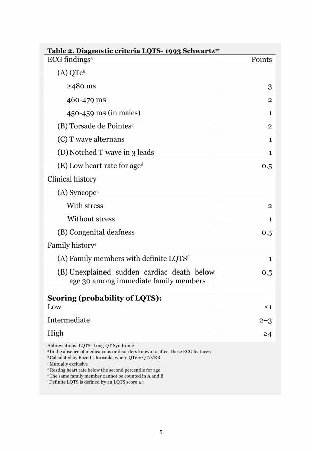
The probability of having LQTS and/or JLNS may be assessed in the clinical
setting using diagnostic criteria including a scoring system that takes ECG
findings, clinical history and family history into account (Table 2).17 The
ECG findings include, in addition to QTc levels, additional associated
abnormalities involving the T wave and bradycardia.
Prophylactic treatment and interventions
The primary prophylactic treatment in LQTS/JLNS, aimed at reducing
arrhythmia symptoms and preventing sudden cardiac death, constitutes
continuous beta-blocker pharmacotherapy that may be prescribed from
infancy. Secondary treatments, in case of therapy failures and/or after life-
threatening cardiac events, include surgical interventions such as Left
Cardiac Sympathetic Denervation (LCSD) and Implantable Cardioverter-
Defibrillator therapy (ICD).18
5
Table 2. Diagnostic criteria LQTS- 1993 Schwartz17
ECG findingsa Points
(A) QTcb
≥480 ms 3
460-479 ms 2
450-459 ms (in males) 1
(B) Torsade de Pointesc 2
(C) T wave alternans 1
(D) Notched T wave in 3 leads 1
(E) Low heart rate for aged 0.5
Clinical history
(A) Syncopec
With stress 2
Without stress 1
(B) Congenital deafness 0.5
Family historye
(A) Family members with definite LQTSf 1
(B) Unexplained sudden cardiac death below age 30 among immediate family members
0.5
Scoring (probability of LQTS):
Low ≤1
Intermediate 2–3
High ≥4
Abbreviations: LQTS- Long QT Syndrome a In the absence of medications or disorders known to affect these ECG features b Calculated by Bazett's formula, where QTc = QT/√RR c Mutually exclusive d Resting heart rate below the second percentile for age e The same family member cannot be counted in A and B f Definite LQTS is defined by an LQTS score ≥4

6
LQTS- a heterogeneous disease As LQTS originally constituted a symptoms diagnosis (prolonged QTc and
arrhythmia propensity, with or without congenital hearing loss) within a
familial context, the diagnosis has come to encompass several similar
disorders differing somewhat in etiology. The primary LQTS loci
(denominated LQT1-3) were identified in the mid 90’s by use of linkage
analysis in LQTS index families, whereafter specific LQTS-related genes
could be identified.19-21 Since then, several additional genetic LQTS loci have
been identified, all (as for LQT1-3) corresponding to genes encoding ion
channel subunits or regulatory proteins involved in cardiac repolarization.
To date, mutations in at least 13 different genes are associated with LQTS
(corresponding to variants LQT1-LQT13).22 The LQTS variants 1 and 2
(caused by loss-of-function mutations in the KCNQ1 and KCNH2 genes,
respectively) together account for approximately 70% of LQTS, while the
LQT3 variant (caused by gain-of-function mutations in the SCN5A gene)
accounts for 5-10% of LQTS.12 The remaining LQTS variants (10 genes)
account for less than 5% of LQTS.12 Consequently, as yet undefined genetic
loci account for 15-20% of LQTS. The uncommon JLNS form is related to
mutations in the KCNQ1 gene (~90% of JLNS cases) as well as the KCNE1
gene23 (Table 3).
Table 3. Major genes associated with LQTS and JLNS
LQTS
type
Chromosomal
Locus
Gene
symbol
Protein product Mutation effect
LQT1* 11p15.5 KCNQ1 K+ channel (α-subunit) Loss-of-function
LQT2 7q35-q36 KCNH2 K+ channel (α-subunit) Loss-of-function
LQT3 3p21 SCN5A N+ channel (α-subunit) Gain-of-function
LQT5* 21q22.1-q22.2 KCNE1 K+ channel (β-subunit) Loss-of-function
Abbreviations: LQTS- Long QT Syndrome, JLNS- Jervell and Lange-Nielsen Syndrome, K+ potassium ion,
Na+ sodium ion
Major LQTS genes (each accounting for ≥5% of LQTS) in bold
* Homozygous or compound heterozygous mutations in KCNQ1 and/or KCNE1 are associated with JLNS
The identification of the molecular basis for LQTS enabled a successive
implementation of molecular genetics diagnostics methods, significantly
improving the sensitivity of case finding, as compared to the clinical
diagnostic criteria.24 As genetic testing in LQTS may be of diagnostic,
prognostic and also therapeutic importance,12,25-27 it is the recommended
diagnostic method (Class I recommendation) for index cases with clinically
suspect LQTS, as well as for screening of appropriate relatives of an index
case with an ascertained LQTS-causing mutation.12

7
Figure 3. A. Organs where KCNQ1- encoded channels are
of physiological importance. B. The KCNQ1-encoded ion
channel α-subunit (right) comprises six membrane
spanning helices and has intracellular NH2 and COOH-
terminals. C. The functional channel complex, responsible
for outward potassium transportation, is a result of the
posttranslational assembly of 4 KCNQ1- encoded α-
subunits and 1-4 regulatory β-subunits.
The identification of the molecular basis for LQTS moreover clarified the
interrelationship between the dominant and recessive variant, i.e. the major
common denominator constituted loss-of function mutations in the KCNQ1
gene, in LQT1 mainly via single mutations (heterozygous genotype) and in
JLNS via double identical (homozygous genotype) or double non-identical
mutations (compound heterozygous genotype).
The KCNQ1-encoded potassium channel (Kv7.1) The KCNQ1 gene, situated on the short arm of chromosome 11, containing 16
exons and spanning ~400 kilo bases28,29, encodes the Kv7.1 voltage-gated
potassium channel involved in
various functions throughout
the human body (Figure
3A).30 The main physiological
functions of the Kv7.1 channel
include mediation of the
delayed rectifying current
(IKs) that contributes to the
repolarization phase of the
cardiac action potential, as
well as regulation of salt and
water homeostasis in various
epithelial tissues.30 The
normal KCNQ1 protein
product contains 676 residues
(amino acids), 6 membrane-
spanning regions, a pore loop
and two intracellular NH2-
and COOH-terminals, with
122 and 322 residues,
respectively (Figure 3B).31
The functional Kv7.1
potassium channel is a
tetrameric structure resulting
from the post-translational
co-assembly of four KCNQ1-
encoded α-subunits and 1-4
regulatory β-subunits,
encoded by genes KCNE132,33
or KCNE2, depending on
tissue, partly explaining the
diversity of Kv7.1 function in
the body (Figure 3C).30

8
The Kv7.1 channel and cardiac repolarization
In the cardiac myocyte, the co-expression of the KCNQ1 and KCNE1 genes
result in the Kv7.1 channel-complex responsible for the slow component of
the outward potassium current during cardiac repolarization (IKs).32,33 The
normal cardiac action potential includes the interaction between several ion
channels, opening and closing due to time- and voltage-dependent
regulation.34 The main inward (depolarizing) currents include calcium and
sodium ions. The repolarization phase of the action potential involves two
main outward potassium currents IKr (KCNH2/KCNE2 associated) and IKs
(KCNQ1/KCNE1 associated), constituting a rapid (r) and a slow (s)
component, respectively (Figure 4). The distribution of IKr and IKs related
channels throughout the layers of the ventricular wall is not homogeneous,
leading to a temporospatial dispersion of depolarization and a potential
substrate for arrhythmia.35,36 However, a certain degree of redundancy in the
repolarizing potassium currents has been proposed, as the potassium
channels may partly substitute for each other, a finding that instigated the
concept of “the repolarization reserve”.37,38
Figure 4. Schematic overview of the action potential of the cardiac myocyte, comprising the phases of
depolarization and repolarization, including the timing of the opening and closing of sodium (Na+), calcium
(Ca2+) and potassium (K+) ion channels. Potassium channels include the KCNH2 encoded rapid potassium
channels and the KCNQ1 encoded slow potassium channels.
9
As heart rate increases (during for example sympathetic stimulation) the
repolarization currents (and specifically IKs) contribute to the adaptation of
the myocyte, by increasing outward potassium flow in order to shorten the
action potential.30,34 If this adaptation to heart rate fails, as in individuals
with reduced Kv7.1 channel function due to KCNQ1 mutations, an early
after-depolarization occurring during the prolonged repolarization phase
may trigger a potentially life-threatening arrhythmia. This finding
corresponds to the finding that cardiac events in JLNS and LQT1 typically
occur during physical activity or emotional stress, both characterized by an
increase in sympathetic activation and thereby heart rate.23,39
The Kv7.1 channel and auditory function
In the epithelial tissue of the cochlea in the inner ear, the co-expression of
the KCNQ1 and KCNE1 genes results in the Kv7.1 channel-complex
responsible for endolymph homeostasis via normal salt and water transport
over the apical membrane of marginal cells of the stria vascularis.30,40 The
disruption of normal Kv7.1 function may cause deafness, as seen in JLNS
patients carrying double KCNQ1 or KCNE1 mutations40-42 and in KCNQ1 or
KCNE1 knock-out mice.43-45 In temporal bone from JLNS patients and
knock-out mice histopathologic structural abnormalities including atrophy
and degeneration of the stria vascularis and endolymph compartment have
been reported.46,47
Correlations between Kv7.1 function and phenotype
As is evident by the differing inheritance patterns in LQTS and JLNS, the
level of Kv7.1 dysfunction at which the disease-associated phenotypes
manifest differ between organs. While the cardiac phenotype may manifest
at a mere 50%, or less, IKs reduction48, the auditory phenotype only
manifests in the context of a near-total Kv7.1 loss-of-function.49
The cardiac phenotype in heterozygous mutation-carriers is moreover
associated with a marked intra-familial variability regarding penetrance50,
suggesting a complex interplay between different factors, environmental
and/or genetic.51
Remaining heterogeneity within the KCNQ1-subgroup Even within the subgroup of KCNQ1-associated LQT1 and JLNS, a marked
heterogeneity remains. Within the KCNQ1 gene there are several hundred
different mutations reported as causative of LQT1.52-54 Part of the apparent
clinical heterogeneity stems from mutation-specific effects55 (mutation type,
location and effect on Kv7.1 protein function) as well as case-specific factors
such as age and gender, and genetic factors beyond the primary mutation(s)
(Table 4).

10
Table 4. Overview of factors influencing the clinical heterogeneity
in KCNQ1-associated LQTS (LQT1) and JLNS
Level Primary (causative) factors Other factors
LQTS
JLNS
Genotype Heterozygous or
Compound
heterozygous
Homozygous or
Compound
heterozygous
Personal and/or
environmental
factors:
Gender, age
Activity level
QT-prolonging drugs
Factors affecting
K+ homeostasis
(gastroenteritis, diets,
etc.)
Secondary genetic
factors:
Other mutations/
sequence variants with
modifying propertiese
Major
mutation
type
Missensea
Nonsensea
Frameshiftb
Mutation
location
C-terminal
N-terminal
Pore region
Minor importance?
Effect on
protein
Proteins with
altered functional
properties
Truncated/ non-
functional proteins
Effect on
IKs
function
(in vitro)
Loss-of-function:
Haploinsufficiencyc
or Dominant-
negative effectsd
Loss-of-function:
Complete loss-of-
function
(homozygous form)
Haploinsufficiencyc
(heterozygous form)
Abbreviations: LQTS- Long QT Syndrome, JLNS- Jervell and Lange-Nielsen Syndrome, IKs- the slow rectifier
potassium ion (K+) current
Bold italic font, regarding primary factors, indicates an association with a more severe clinical phenotype a Point mutations (i.e. a single nucleotide is exchanged for another) leading to an altered codon that may code
for either the same amino acid (silent), another amino acid (missense) or a stop codon (nonsense mutation) b Caused by adding (insertion) or removing (deletion) one or more nucleotides into the DNA, causing an
alteration of the gene’s reading frame and typically a significantly altered protein product c Haploinsufficiency: ~50 % residual function via the proteins encoded by the healthy allele (wild-type) d Dominant-negative effect: <50% residual function due to antagonistic action towards the wild-type proteins e Affecting gene expression, QT duration and/or arrhythmia susceptibility
11
Proposed effects of modifying genes
The term “genetic modifiers” relate to genetic factors influencing phenotype
such as additional putative mutations and/or genetic variants without the
ability to independently cause the phenotype, i.e. population prevalent single
nucleotide polymorphisms or common sequence variants (point mutations).
Sequence variants in several genes (the LQTS-genes KCNH2, SCN5A and
KCNE1, as well as the NOS1AP gene) have been shown to affect QT duration
in the general population56-60, and also in LQTS61-63, and could thereby
contribute to phenotypic variability. Most derived sequence variants
associated with QT duration have moderate effects on QTc, however a recent
study on the 3’untranslated region of the KCNQ1 gene (3’UTR-KCNQ1)
showed marked allele-specific effects on QT duration by derived variants.63
The study demonstrated that derived variants occurring on the allele
opposite of the mutation (in trans) repressed expression of the healthy allele,
thereby leading to phenotypic aggravation. An opposite effect when derived
variants occurred on the same allele as the mutation (in cis), consequently
leading to a reduced expression of the mutated Kv7.1 channel, was
suggested.63
In summary, there is a marked clinical heterogeneity in LQTS that in spite
of extensive research and the promising results regarding some modifying
genes largely remains unexplained. In this context, LQTS founder
populations, comprising individuals sharing mutations identical by descent,
may contribute to mutation-specific characterization and risk stratification
via aggregation of clinical data. Also, due to their inherently reduced genetic
background variability, LQTS founder populations constitute excellent
research models in which potentially contributing factors, such as genetic
modifiers, can be studied under favorable conditions.
12
LQTS research in Sweden
Resources on the national and regional levels
The mapping of monogenetic diseases in Sweden is facilitated by the
availability of unique comprehensive census records dating from the 17th
century and an onwards, as well as detailed historical and current national
population demographics data compiled over time by Statistics Sweden
(www.scb.se).
The northern region (Figure 5) encompasses five administrative counties
covering over half of the total area of Sweden, including several river valley
regions spanning the region from northwest to south east. The population
density in the northern region is very low with less than 1 x 10⁶ inhabitants,
as compared to ~9.5 x 10⁶ inhabitants in Sweden as a whole.
Regional LQTS- a brief history and study setting
As previously mentioned, an apparently high occurrence of both LQTS and
JLNS in the northern region has been clinically evident.
The first regional JLNS case was diagnosed in Umeå in the early 1970´s,
by Pediatric Cardiologist Hans Wendel (1967-1991, Pediatric Cardiology
Clinic, Umeå University Hospital), during a time when the diagnosis was
largely unknown.
In 1979 a family report regarding 5/9 siblings from the northern region
with clinical JLNS diagnosis was published,64 whereof the proband was
genotyped 30 years later as a p.R518X homozygote, by our group (paper IV).
During the last 40 years LQTS families from the region have been followed at
the Pediatric Cardiology Clinic in Umeå, occasionally as both pediatric cases
and later as parents to new cases. In order to meet the needs of these
families, comprising several generations, an initiative to start a regional
LQTS Family Clinic in Umeå was undertaken in 2005.
The LQTS Family Clinic and Centre for Cardiovascular Genetics
The LQTS Family Clinic, a part of the Centre for Cardiovascular Genetics,
Umeå University Hospital, was initiated in order to provide LQTS families
with the joint expertize of an interdisciplinary team comprising of a
Cardiologist, a Pediatric Cardiologists, a Clinical Geneticist, a Biomedical
Analyst and a Counselor, and to provide both individual and familial
diagnostics, patient information and management. Separately, the Family
Clinic serves as a basis for case recruitment into clinical LQTS research.
The clinical and research setting was further complemented by the
availability of molecular genetics LQTS diagnostics at the accredited
laboratory of the Department of Clinical Genetics, Umeå University Hospital,
Umeå, from 2006 and onwards.

13
Figure 5. Map of northern Sweden, shown in magnification, with the main regional rivers depicted as lines
and the city of Umeå indicated by a black circle. Main rivers of the northern region, from north to south,
include the Torne, Kalix, Lule, Pite, Skellefte, Uman/Vindeln, Ångerman, Indal, Ljungan and Ljusnan Rivers.
Sweden as a whole (in white) situated in Scandinavia (Norway, Denmark and Finland in gray) surrounded by
the Baltic and North Sea (shaded coast lines) is shown in the upper left box.
14
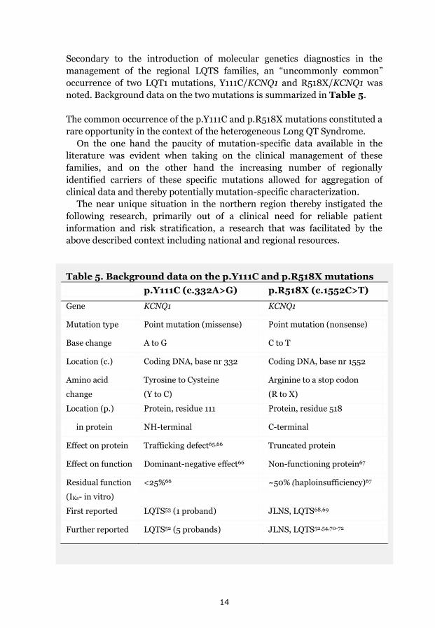
Secondary to the introduction of molecular genetics diagnostics in the
management of the regional LQTS families, an “uncommonly common”
occurrence of two LQT1 mutations, Y111C/KCNQ1 and R518X/KCNQ1 was
noted. Background data on the two mutations is summarized in Table 5.
The common occurrence of the p.Y111C and p.R518X mutations constituted a
rare opportunity in the context of the heterogeneous Long QT Syndrome.
On the one hand the paucity of mutation-specific data available in the
literature was evident when taking on the clinical management of these
families, and on the other hand the increasing number of regionally
identified carriers of these specific mutations allowed for aggregation of
clinical data and thereby potentially mutation-specific characterization.
The near unique situation in the northern region thereby instigated the
following research, primarily out of a clinical need for reliable patient
information and risk stratification, a research that was facilitated by the
above described context including national and regional resources.
Table 5. Background data on the p.Y111C and p.R518X mutations p.Y111C (c.332A>G) p.R518X (c.1552C>T)
Gene KCNQ1 KCNQ1
Mutation type Point mutation (missense) Point mutation (nonsense)
Base change A to G C to T
Location (c.) Coding DNA, base nr 332 Coding DNA, base nr 1552
Amino acid
change
Tyrosine to Cysteine
(Y to C)
Arginine to a stop codon
(R to X)
Location (p.) Protein, residue 111 Protein, residue 518
in protein NH-terminal C-terminal
Effect on protein Trafficking defect65,66 Truncated protein
Effect on function Dominant-negative effect66 Non-functioning protein67
Residual function
(IKs- in vitro)
<25%66 ~50% (haploinsufficiency)67
First reported LQTS53 (1 proband) JLNS, LQTS68,69
Further reported LQTS52 (5 probands) JLNS, LQTS52,54,70-72

15
Overall Aim
To increase the knowledge regarding the occurrence and clinical phenotype
of LQTS in Sweden, by investigating two major causes of LQTS identified in
the regional clinical practice (KCNQ1 mutations p.Y111C and p.R518X), as
well as all national cases of the uncommon and clinically severe recessive
LQTS variant associated with congenital hearing loss (JLNS), aiming
towards identifying possible risk indicators and improving clinical
management of individuals and families with LQTS.
Specific Aims
To describe the occurrence of KCNQ1 mutations p.Y111C and
p.R518X, as well as the prevalence of JLNS, in the Swedish
population.
To investigate the possible founder nature of the Swedish p.Y111C
and p.R518X populations, as well as the geographic origin of
mutations identified in ≥ 2 alleles in Swedish JLNS probands.
To investigate the cardiac phenotype associated with p.Y111C,
p.R518X, and JLNS, including electrocardiographic findings (QTc),
symptoms (debut, type, frequency and triggers), with a special focus
on life-threatening cardiac events.
To evaluate the possible association between previously published
genetic modifiers and QT duration and/or cardiac events in p.R518X
heterozygotes sharing a common founder, in order to seek an
explanation of the marked intra-familial variability seen in clinical
phenotype.

16
Overview of aims and methods
Aims
Study group
To describe the occurrence of two common
KCNQ1 mutations (p.Y111C and p.R518X),
as well as the prevalence of recessive LQTS
including congenital hearing loss (JLNS),
in the Swedish population.
Regional and national cases with
ascertained p.Y111C or p.R518X
genotype and/or ascertained
JLNS genotype and/or clinical
JLNS diagnosis.
To investigate the possible founder nature
of the p.Y111C and p.R518X mutations, as
well as the origin of mutations identified in
≥ 2 alleles in Swedish JLNS probands.
p.Y111C; 170 cases, 37 probands
p.R518X; 101 cases, 19 probands
JLNS; 19 cases, 13 probands
p.Y111C (2 alleles),
p.R518X (12 alleles),
c.572_576del (4 alleles),
p.Q530X (2 alleles).
To investigate the cardiac phenotype
associated with p.Y111C, p.R518X, and
JLNS, including electrocardiographic
findings (QTc), symptoms (debut, type,
frequency and triggers), with a special
focus on life-threatening cardiac events.
p.Y111C; 80 cases, 15 probands
p.R518X; 101 cases, 19 probands
JLNS; 19 cases, 13 probands
To evaluate the possible association
between previously published genetic
modifiers and QT duration and/or cardiac
events in p.R518X heterozygotes sharing a
common founder, in order to seek an
explanation of the marked intra-familial
variability seen in clinical phenotype.
p.R518X; 8o cases
(founder heterozygotes)

17
Methods
Paper
* Inclusion of regional clinical LQTS and JLNS cases (Pediatric
Cardiology Clinic and/or LQTS Family Clinic, Umeå, Sweden) as
well as national LQTS and JLNS cases that were identified via
national referrals to the laboratory of Clinical Genetics and/or
Centre for Cardiovascular Genetics, Umeå, Sweden and/or a
national inventory (primarily JLNS cases)
* Molecular genetics diagnostics were consistently used for
case/genotype ascertainment
I-IV
* Genealogic investigation (census records)
* Analysis of geographic clustering of cases and/or ancestors
(p.Y111C, p.R518X, c.572_576del, p.Q530X)
* Haplotype analysis (14-15 microsatellite markers) in p.Y111C
and p.R518X index families and controls (n=84)
* Estimation of mutation age (p.Y111C and p.R518X) and
associated prevalence (ESTIAGE and DMLE computer software)
II-IV
* Clinical data, including the following variables, were collected
via medical records and/or a questionnaire, and in symptomatic
LQTS cases and all JLNS cases an interview:
Symptoms (debut, frequency, triggers)
Beta-blockers (duration and compliance), other interventions
LQTS family history
* ECGs were recorded at the LQTS Family Clinic or obtained from
medical records and measured manually
I-IV
* Literature review and selection of sequence variants with
proposed modifying properties
* Sequence variant analysis by bi-directional sequencing (3’UTR
of KCNQ1), uni-directional sequencing (KCNH2 exons 11 and 13,
KCNE1 exon 4 and SCN5A exon 12) and MGB-probes (NOS1AP)
in p.R518X heterozygotes sharing a common haplotype
* Association studies (QTc and cardiac events) for variants with
an allele-frequency >0.1, including relatedness correction via a
kinship matrix and an adjusted significance level (p<0.01)
III

18
Overview of study populations papers I-IV
p.Y111C
JLNS
p.R518X
II
I
III IV

19
Materials and Methods
Cases and families Participating cases and families with p.Y111C or p.R518X KCNQ1 mutations
were identified as a part of ordinary health care, either via the regional
Pediatric Cardiology Clinic (1967-), the regional LQTS Family Clinic (2005-),
or via national clinical referrals to the Centre for Cardiovascular Genetics
and/or the accredited laboratory of the Department of Clinical Genetics
(2006-), all at Umeå University Hospital, Umeå, Sweden. For cases and
families with JLNS a national inventory of clinical JLNS cases was
performed between 2007 and 2010.
All participants were first approached by their tending clinician and asked
for permission of first contact. All participants or their legal guardian signed
informed consents and the studies were approved by the Regional Ethical
Review Board, Umeå University, Umeå, Sweden.
A national inventory of JLNS cases
Being an uncommon, relatively unknown disease with a most severe
phenotype, we aimed to identify all clinical JLNS cases in Sweden and to
perform a prevalence estimate.
Identification of JLNS cases
In Sweden, no specific diagnostic code for JLNS has been available,
precluding the possibility of identifying cases via the National Diagnosis
Register (www.socialstyrelsen.se). The national inventory of JLNS cases,
designed primarily to identify now living JLNS cases, was performed
between 2007 and 2010, by mail, email and phone calls, approaching all
Swedish clinics or departments with potential knowledge of cases (clinics
and/or departments of pediatric cardiology, cardiology, medicine, clinical
genetics, otorhinolaryngology, audiology and cochlear implantation).
Furthermore, all Swedish schools teaching deaf children were similarly
approached. An advertisement requesting contact with clinicians with
knowledge of JLNS cases, using also the additional synonyms of “recessive
LQTS” and “Surdocardiac Syndrome”, was run in two national medical
journals, and similar information was spread at several national meetings in
pediatric cardiology, cardiology and molecular genetics. Prevalent cases were
reported by gender and birth year. Of these, cases meeting the inclusion
criteria, i.e. congenital bilateral hearing loss (incomplete or complete) and
clinical findings of either a personal history of syncopal attacks, a family
history of LQTS or electrocardiographic findings of a prolonged QT interval
corrected for heart rate (QTc), were invited to participate in the study.

20
A JLNS prevalence estimate in preadolescent children
Due to the high mortality before adulthood,23,73 a JLNS prevalence estimate
in preadolescent children was deemed appropriate. Moreover, during the
last decade the awareness of the disorder has increased and routine ECG
screening has been implemented in the national cochlear implant clinic and
the Swedish schools teaching deaf children, reducing the risk of JLNS non-
detection in children below 10 years of age.
The prevalence of JLNS in children below 10 years of age was calculated as
the number of now living JLNS cases with ascertained genotype/ the
population of interest (estimated using the annual birth-count during the
last decade; ~100 000 x 10= 1 x 10⁶, using population demographic data
from Statistics Sweden, www.scb.se).
Case ascertainment LQTS and/or JLNS diagnosis in living participants was ascertained by
molecular genetics methods throughout the studies, mainly within the
context of clinical routine diagnostic testing. To that end, DNA was extracted
from peripheral lymphocytes, typically obtained from a sample of 2.5 mL
whole blood, using standard salting out methods. For each analysis, DNA
was amplified by polymerase chain reaction using appropriate forward and
reverse primers.
Diagnostic strategies for index cases and family members
Diagnostic strategies for inherited monogenetic disorders typically entail
primary mutation identification in index cases and subsequent, voluntary,
ascertainment of mutation-carrier status in family members of index cases
with identified mutations via cascade-screening (Figure 6).
Figure 6. An index case or proband (left) constitutes the first clinically identified case in a new family
without known connection to any other family where the disease has previously been detected. After mutation
identification in the index case, the cascade-screening process (middle) comprises the consecutive testing of
the probands’ first-degree relatives (parents, siblings and offspring). Whenever an additional mutation-carrier
is identified in the pedigree, the screening process may expand concentrically by testing the first degree
relatives of that mutation-carrier, resulting in the ascertainment of carrier-status in members of the extended
family pedigree (right). The concept index family thereby includes the index case and all members of the
extended family identified via the cascade-screening process. Information regarding the possibility of
cascade-screening was spread in the extended family by index cases, on a voluntary basis. (filled symbol-
mutation-carrier, empty symbol- non-carrier or not tested, circle- female, square- male)

21
Molecular genetics diagnostic methods and screening strategies used
Referrals for molecular genetics diagnostics of participants, performed
during the time period 2002-2011, mainly occurred within clinical routine
praxis, and therefore methods and strategies used have not been uniform.
The earliest molecular genetics diagnostics in study participants were
performed at the laboratory of Clinical Genetics, Amsterdam, Netherlands,
between 2002 and 2006, including sequencing of the KCNQ1 gene in 14
index cases.
In May 2006 the Department of Clinical Genetics laboratory in Umeå,
Sweden started up molecular genetics diagnostics of LQTS index cases, and
subsequently received a substantial proportion of both regional and national
referrals.
Since the start in 2006, several changes in the methods and screening
strategies used have been implemented as a part of regular method
development and quality control (Table 6). A certain degree of overlap
between methods and strategies occurred during transitions and also
occasionally due to laboratory logistics or specific requests of referents.
Table 6. Screening strategies regarding index cases referred for clinical
molecular genetics LQTS diagnostics to the Clinical Genetics laboratory,
Umeå University Hospital from the start in 2006 and onwards
Time
period
Method Screening Strategy/Ordera
2006-2010b Pre-screeningc Y111C/KCNQ1 and R518X/KCNQ1
2006-2008 DHPLC-screeningd KCNQ1, KCNH2, SCN5A, KCNE1, KCNE2
2008-2009 DHPLC-screening KCNQ1-KCNE1, KCNH2-KCNE1, SCN5A
2009 Sequencinge KCNQ1-KCNE1, KCNH2-KCNE1, SCN5A
2009-2010 Pre-screening, TaqMan
+ Sequencing
followed by sequencing, in the order
Y111C+R518X, if negative:
KCNH2-KCNE1, KCNQ1-KCNE1, SCN5A
2011- Sequencing KCNQ1-KCNE1, KCNH2-KCNE1, SCN5A
a For all screening strategies in LQTS index cases: if a definitely putative mutation was identified in a gene,
the screening of subsequent genes was discontinued. If a sequence variant of unknown significance was
identified the screening continued and all identified possible mutations evaluated after the screening was
completed b Strategy intermittently used, together with the below stated methods, during the time period c Targeted mutation analysis, TaqMan, MGB-probes, ABI 7000, AppliedBiosystems, Inc, Foster City, Calif. d Denaturing high-performance liquid chromatography (DHPLC), Wave 3500 HT, Transgenomic, Inc,
Omaha, Neb. e Bi-directional, CEQ 8000, Beckman Coulter, Inc, Fullerton, Calif.

22
Investigating the possible founder nature of a mutation The methods for investigating the origin of potential founder mutations
included genealogical investigations, assessment of geographical clustering
of ancestors, haplotype analysis in index families and controls as well as
computer statistics regarding mutation dating and associated prevalence.
Genealogical investigations
In Sweden near-unique prerequisites for genealogical investigations exist via
comprehensive preserved population records that were initiated by the
Crown in 1686, annually describing the births, deaths, marriages and
migration of the inhabitants in Swedish parishes. Genealogical investigations
were performed, either via accessing original records and digital scans
(Figure 7) of local parish registers and catechetical examination records at
the Umeå University research archive, or in digital form via private
genealogical databases and the digital genealogical and census archives
available at the Swedish archive information homepage (www.svar.ra.se).
The genealogical investigations were performed tracing all maternal and
paternal ancestral lines of probands throughout as many generations as
possible, and at least to the year 1750. Found connections between lineages
were assessed and the most likely founder/connecting couple was defined as
the couple that connected the highest number of probands in the least
generations. The hypothetical route of inheritance suggested by the
identification of a common founder was assumed to be constituted of
obligate mutation-carriers.
Figure 7. Example of a scanned census record

23
Geographic origin and clustering of ancestors
During the genealogical investigation of potential founder populations, the
birth-places of ancestors were noted on regional maps. The geographic
clustering and migration of ancestors, between the 17th and the 20th
centuries were traced and visually assessed. In p.Y111C ancestors the origin
of spouses, defined as from within or without the river valley region of
interest, was noted in order to investigate the prevailing marriage patterns.
Handling and presenting genealogical and geographic data
For handling genealogical data, the DISGEN computer software (Lakewood,
CO) was used. Kinship structures were presented in pedigrees constructed
using Cyrillic 2.1 (Cyrillic Software, Oxfordshire, United Kingdom) and/or
Progeny 7 (Progeny Software, South Bend, Ind., US) software (paper I). In
paper II, III and IV pedigrees and geographical maps were constructed using
Open Source software; Inkscape (vector graphics editor) and GIMP (GNU
Image Manipulation Program).
Haplotype analysis using microsatellite markers
In order to discern whether the occurrence of population prevalent
mutations were due to founder effects, a haplotype analysis using
microsatellite markers was performed in index cases, family members and
healthy controls of northern Swedish origin. Ideally, two mutation-carriers
in separate generations from each LQTS index family (three in homozygous
JLNS families) were included in the analysis in order to identify the
mutation-associated allele. The analysis included 14-15 markers flanking the
KCNQ1 gene (5-6 upstream and 9 downstream) and spanning over a total
distance of approximately 8 x 10⁶ base pairs (Figure 8).
Figure 8. An overview of the selected microsatellite markers used for haplotype analysis (indicated by bold
arrows) flanking the KCNQ1 gene over a distance of approximately 8 x 10⁶ base pairs (8 cM). Markers are
denominated “D11S-“ followed by the number under each arrow. The vertical line represents the location of
the mutation of interest (here p.R518X) and physical distances calculated between the specific mutation and
each marker, for each separate analysis (p.Y111C and p.R518X). The marker D11S-1318, located upstream and
adjacent to the KCNQ1 gene (gray arrow), was analyzed in paper II (p.Y111C) but excluded in paper III
(p.R518X) due to a generally poor quality of peaks and high frequency of background noise.
24
The microsatellite markers were chosen from the National Centre for
Biotechnology Information Entrez Gene database with respect to distance
from the KCNQ1 gene, using deCODE, Généthon and Marshfield genetic
maps, selecting markers with the highest heterogeneity according to the
CEPH Genotype database. For each marker, appropriate forward and reverse
primers were used (Sigma-Aldrich Inc.). Fragment analysis was performed
according to the manufacturers’ instructions, and the PCR product was
analyzed using an automated capillary electrophoresis based DNA Sequencer
(Wave® 3500 HT, Transgenomic Inc, Omaha, Nebraska). Solutions and
material for PCR mix were manufactured by GE Healthcare (United
Kingdom) and Applied Biosystems (Foster City, California). Microsatellite
data was analyzed using GeneMapper software version 3.7 (Applied
Biosystems Inc, Foster City, California). The pattern of mutation-associated
alleles in all included index families was assessed and the most likely
ancestral haplotype reconstructed. The frequencies of the mutation-
associated alleles in a sample of control chromosomes (n=168) obtained
from northern Swedish military recruits, were used as weights in the
assessment of the probability that the pattern seen was the result of a
founder effect.
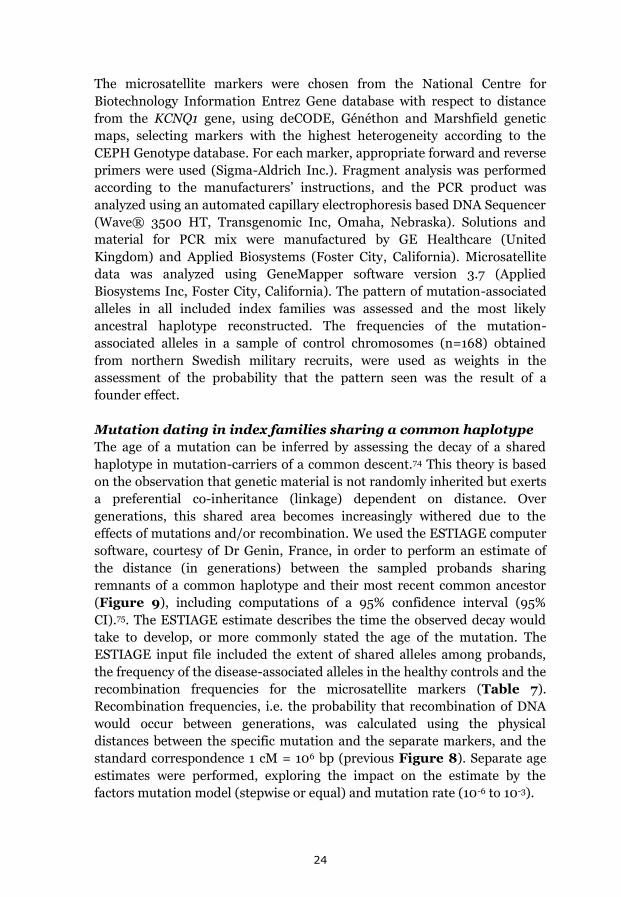
Mutation dating in index families sharing a common haplotype
The age of a mutation can be inferred by assessing the decay of a shared
haplotype in mutation-carriers of a common descent.74 This theory is based
on the observation that genetic material is not randomly inherited but exerts
a preferential co-inheritance (linkage) dependent on distance. Over
generations, this shared area becomes increasingly withered due to the
effects of mutations and/or recombination. We used the ESTIAGE computer
software, courtesy of Dr Genin, France, in order to perform an estimate of
the distance (in generations) between the sampled probands sharing
remnants of a common haplotype and their most recent common ancestor
(Figure 9), including computations of a 95% confidence interval (95%
CI).75. The ESTIAGE estimate describes the time the observed decay would
take to develop, or more commonly stated the age of the mutation. The
ESTIAGE input file included the extent of shared alleles among probands,
the frequency of the disease-associated alleles in the healthy controls and the
recombination frequencies for the microsatellite markers (Table 7).
Recombination frequencies, i.e. the probability that recombination of DNA
would occur between generations, was calculated using the physical
distances between the specific mutation and the separate markers, and the
standard correspondence 1 cM = 106 bp (previous Figure 8). Separate age
estimates were performed, exploring the impact on the estimate by the
factors mutation model (stepwise or equal) and mutation rate (10-6 to 10-3).
25
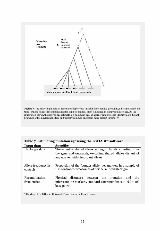
Figure 9. By analysing mutation-associated haplotypes in a sample of related probands, an estimation of the
time to the most recent common ancestor can be obtained, often simplified to signify mutation age. As the
illustration shows, the derived age estimate is a minimum age, as a larger sample could identify more distant
branches of the phylogenetic tree and thereby common ancestors more distant in time (t).
Table 7. Estimating mutation age using the ESTIAGE* software
Input data Specifics Haplotype data The extent of shared alleles among probands, counting from
the gene and outwards, excluding shared alleles distant of
any marker with discordant alleles
Allele-frequency in
controls
Proportion of the founder allele, per marker, in a sample of 168 control chromosomes of northern Swedish origin
Recombination
frequencies
Physical distances between the mutation and the
microsatellite markers, standard correspondence 1 cM = 106
base pairs
* Courtesy of Dr E Genin, Université Paris Diderot, Villejuif, France

26
Prevalence estimations derived from mutation age
An approximation of mutation-specific prevalence as a function of mutation
age was estimated using the DMLE software, including 95% confidence
intervals (www.dmle.org). The DMLE input file included the full haplotype
of probands and controls for all analyzed markers, regional population
growth rate estimates and an estimate of the proportion sampled (Table 8).
By viewing the proportion of population sampled as the unknown variable,
an estimate of the size of the total founder population can be obtained from
the size of the sample (included probands). Using the 95% confidence
interval of the ESTIAGE mutation age estimate as upper and lower limits of
mutation age, iterations over the interval 0.0001-0.5 were performed, and
the upper limit for acceptable values of proportion sampled corrected for the
number of known specific mutation-carriers. A best-fit approach was used to
identify the magnitude of the proportion of population sampled, within a
range of possible values for mutation age and population growth rate.
Table 8. Estimating mutation prevalence using DMLE* software
Input data Specifics Haplotype data Full haplotype of both probands and controls for all markers
Population growth
rates
Analyzed as a discrete variable, per generation = e^(ln [end
population/start population]/number of generations) -1)
using regional population demographics data available from
Statistics Sweden (www.scb.se)
Proportion of
population sampled
Viewed as the unknown variable, iterations were performed
over the interval 0.0001-0.5, within a range of possible
values for mutation age (overlap between the 95% confidence
intervals of the ESTIAGE and DMLE estimates)
* Available at www.dmle.org
Prevalence estimates using Hardy-Weinberg statistics
The Hardy-Weinberg equilibrium or Hardy–Weinberg principle states that,
barring disturbing influences, population allele and genotype frequencies
remain in equilibrium from generation to generation. Using the Hardy-
Weinberg equilibrium in its simplest form, assessing a single locus with two
possible alleles (dominant allele “A” and recessive allele “a”, i.e. LQTS
mutation and no LQTS mutation), the allele frequencies for “A” and “a”
(denoted p and q, respectively) may be calculated as
q; freq(A)=p; freq(a)=q; p+q=1. If allele frequencies are static in the
population (Hardy Weinberg equilibrium) the frequencies of the different
27
genotypes are calculated as: freq(AA)=p2 for the AA homozygotes in the
population, freq(aa)=q2 for the aa homozygotes, and freq(Aa)=2pq for the
heterozygotes.
For a constant allele frequency between two generations the following is
required: no change of alleles (mutations), no new alleles or loss of alleles
(immigration or emigration), an infinite population size, no selective
pressure for or against any alleles, and a random mating pattern.
Consequently, the Hardy-Weinberg principle is based on assumptions that
are not fulfilled outside a laboratory setting. Nevertheless, Harry-Weinberg
statistics are useful for assessing the distribution of alleles in a population,76
and with the above reservations Hardy-Weinberg statistics were used in
order to roughly estimate the prevalence of LQTS-mutation carriership in
the Swedish population, inferred from the number of homozygous and/or
compound heterozygous mutation-carriers identified (paper III, IV).
Clinical cardiac phenotype Clinical phenotype in JLNS and LQTS typically includes presence or absence
of symptoms of arrhythmia, presence or absence of electrocardiographic
findings of a prolonged QT interval, and in JLNS variable congenital hearing
loss.
Data collection
Within the concept of the LQTS Family Clinic, each family participated in an
informal interview with special focus on family history and symptoms. Each
individual also answered a questionnaire regarding personal LQTS history
(occurrence of symptoms and beta-blocker therapy duration and
compliance). The same questionnaire (Appendix) was distributed to all
study participants that did not attend the Family Clinic. In cases that in the
questionnaire had reported symptoms suspect of LQTS (loss-of-
consciousness or pre-syncope), the circumstances regarding the possible
cardiac event (debut, type, frequency and triggers) were investigated either
by questionnaire, medical records or interview. Interviews were performed
by one researcher, either by telephone or e-mail. In JLNS families a personal
interview ± interpreter (in sign) was performed. The JLNS family interviews
were semi-structured, lasted on average two to four hours and focused on
symptoms and experiences throughout life, living with JLNS. During the
interviews, anamnestic information regarding first-degree family members
(occurrence of symptoms) and any deceased LQTS and/or JLNS family
members was specifically asked for. In JLNS cases, full medical records were
additionally reviewed, including data on medical therapies and interventions
(such as beta-blocker therapy, Left Cardiac Sympathetic Denervation (LCSD)
and Implantable Cardioverter-Defibrillator (ICD) therapy) and hearing loss
ascertained by review of audiograms.
28
Electrocardiograms (ECGs) were systematically recorded via the LQTS
Family Clinic. Electrocardiograms recorded before beta-blocker therapy and
from participants not attending the Family Clinic were obtained from
medical records.
Definitions of cardiac events
Among the symptoms reported by the participants (spells of dizziness,
palpitations, near fainting/pre-syncope etc.) symptomatic LQTS/JLNS was
defined as occurrence of transient but complete loss of consciousness
(syncope), in the presence or absence of potential triggers of arrhythmia. The
term life-threatening cardiac event was used for cardiac arrests, aborted by
resuscitation (ACA) or resulting in sudden cardiac death (SCD). Implantable
cardioverter defibrillator therapy was mainly reported separately, and not
per se regarded as a life-threatening event.
Electrocardiographic assessment of QT duration
QT duration was assessed in standard 12-lead surface electrocardiograms,
recorded at paper speed 50 mm/s and a gain of 1 mV/10 mm, preferably
recorded before the initiation of beta-blocker therapy. Beta-blocker therapy
was not discontinued in any participant in order to obtain a non-medicated
recording. QT duration, manually measured from the start of the QRS
complex to the end of the T wave, was consequently assessed on coded ECGs
by one observer as the mean of three consecutive beats and corrected for
heart rate by Bazett’s formula (QT/√R-R)15, using the mean of the R-R
intervals (lead III) preceding the measured beats. Measurement strategies
differed somewhat between the studies (Table 9). In studies I, II and IV the
QT intervals were measured once by one observer to obtain the highest mean
QTc, but in study III, as QTc was the major variable used for association
testing regarding genetic modifiers, ECGs were assessed twice by the same
observer in order to reduce the impact of the intra-observer measurement
error/variability.
Table 9. QT measurement strategies used in the studies
Studies QT in lead Specifics
I, II, IV II, V5, V6 The lead with the highest mean QTc was used
III II QT intervals were measured twice by one observer on
two separate occasions and the mean QTc used
Goldenberg’s age and gender-adjusted cut-offs were used to define QTc
levels as normal, borderline or prolonged.16 For comparison with other
studies, the definition of a prolonged QTc if over 440 ms in males and if over
460 ms in women, and the additional cutoffs of a QTc below 440 ms or over
500 ms were also used.

29
Assessments of mutation-specific phenotypes
Investigation of natural history in a pedigree
In order to assess the natural history of a mutation (Y111C/KCNQ1) in
absence of beta-blocker therapy, the survival beyond 40 years of age in non-
medicated ancestors along the hypothesized line of mutation descent was
analyzed in families connected by a common founder. The analysis included
obligate mutation-carriers, born between 1873 and 1968, along the direct
line of inheritance and their siblings (interpreted as constituting 50%
carriers, Mendelian laws of inheritance; Figure 10). The time frame 1968-
1873 was derived from the prerequisites that complete data from records of
census should be available, and that the observation time from birth and
onwards should be at least 40 years (in 2008). In included ancestors, the
date of birth, date of death, and, if occurring before 40 years of age, the cause
of death was investigated by review of medical records or records of census.
The proportion (%) of ancestors that died before 40 years of age was
summarized and presented in age categories. Infant death rates and overall
death rates in ancestors were compared to regional population demographics
data on age-stratified mortality rates in the Swedish population (Statistics
Sweden, www.scb.se).
Figure 10. The survival beyond 40 years of age was analyzed in obligate-mutation carriers (as defined by the
direct line of inheritance between the probands and the identified common ancestor) and their siblings, born
between 1968-1873. The pedigree illustrates the chosen time-frame of the analysis (shaded gray) and the
included ancestors (obligate-mutation carriers) born within the time-frame. All siblings (not shown) of
obligate-mutation carriers were included in the analysis. Adapted from Circ Cardiovasc Genet. 2009
Dec;2(6):558-64.
30
Comparisons to other LQT1 and/or founder populations
To assess the apparent benign phenotypes of the population prevalent LQTS
mutations p.Y111C and p.R518X, the annual incidence rates of life-
threatening cardiac events before therapy were compared to previously
published summarized data from a large LQT1 population, derived from the
consecutive testing of 193 LQTS families, whereof 104 families with mixed
KCNQ1 mutations (n=38677). The clinical characteristics of the p.Y111C
population was further compared to that of a malignant LQT1 population
carrying the same mutation (A341V/KCNQ1, n=244 individuals, whereof 166
founder individuals78) and a mixed LQT1 population where carriers of the
p.A341V mutation had been excluded (n=20578).
Statistics: Clinical phenotype
Data was summarized and presented as mean including standard deviation
(±SD), median, range, interquartile range (IQR), or percentage. For all tests,
a value for p <0.05 was considered statistically significant.
Comparisons within or between populations or groups
For comparisons within or between p.Y111C and p.R518X populations
and/or JLNS cases, between numerical and nominal variables Mann-
Whitney U Test or t-test were used, depending on group size.
Correspondingly, for comparisons between nominal variables Fisher’s Exact
Test or the x2 test was used. All tests were two-tailed. Statistical analyses
were performed with SPSS versions 16.0 and 19.0 (Statistical Package for
Social Sciences, SPSS Inc., Chicago, Ill).
Comparisons between populations or groups using summarized data
Comparisons between LQT1 populations regarding numerical variables were
performed using the 2-sample t test or the Wilcoxon signed-rank test, and
for nominal variables Fisher’s Exact Test was used. All tests were two-tailed,
except the tests of the hypothesis that the incidence of life-threatening
cardiac events was lower in the p.Y111C population, which were lower tailed.
Statistical analyses on summarized data were performed using software from
Minitab, Inc. (version 15, State College, Pa).
Repeated electrocardiographic measurements
The intra-observer coefficient of variation at repeated QTc measurements,
expressed in percent, was calculated by dividing the standard deviation of
the mean by the mean standard error, multiplied by 100.
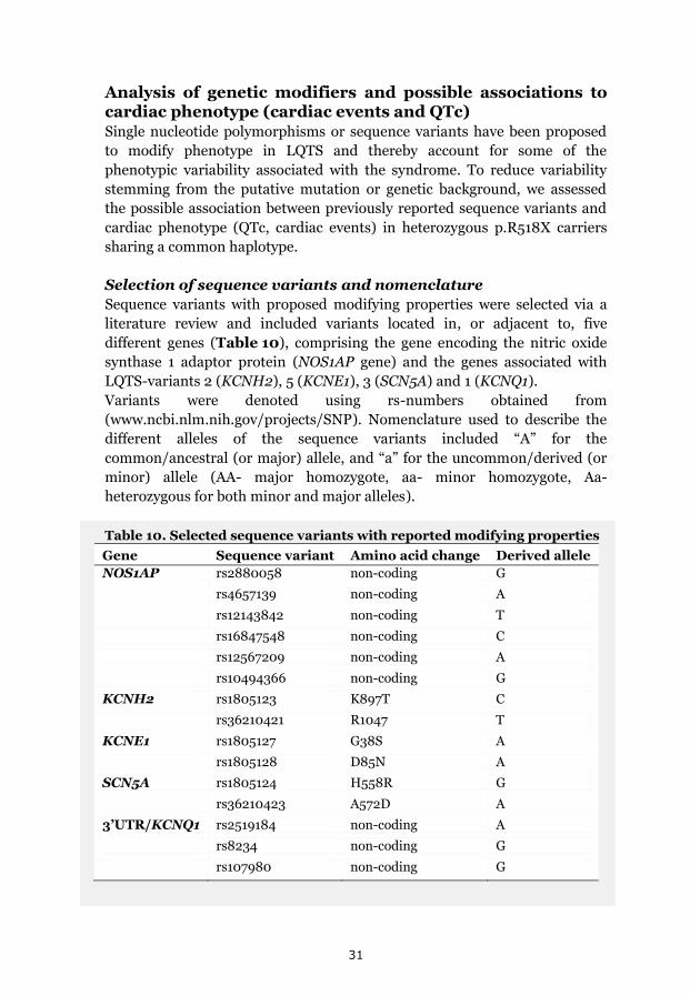
31
Analysis of genetic modifiers and possible associations to cardiac phenotype (cardiac events and QTc) Single nucleotide polymorphisms or sequence variants have been proposed
to modify phenotype in LQTS and thereby account for some of the
phenotypic variability associated with the syndrome. To reduce variability
stemming from the putative mutation or genetic background, we assessed
the possible association between previously reported sequence variants and
cardiac phenotype (QTc, cardiac events) in heterozygous p.R518X carriers
sharing a common haplotype.
Selection of sequence variants and nomenclature
Sequence variants with proposed modifying properties were selected via a
literature review and included variants located in, or adjacent to, five
different genes (Table 10), comprising the gene encoding the nitric oxide
synthase 1 adaptor protein (NOS1AP gene) and the genes associated with
LQTS-variants 2 (KCNH2), 5 (KCNE1), 3 (SCN5A) and 1 (KCNQ1).
Variants were denoted using rs-numbers obtained from
(www.ncbi.nlm.nih.gov/projects/SNP). Nomenclature used to describe the
different alleles of the sequence variants included “A” for the
common/ancestral (or major) allele, and “a” for the uncommon/derived (or
minor) allele (AA- major homozygote, aa- minor homozygote, Aa-
heterozygous for both minor and major alleles).
Table 10. Selected sequence variants with reported modifying properties
Gene Sequence variant Amino acid change Derived allele
NOS1AP rs2880058 non-coding G
rs4657139 non-coding A
rs12143842 non-coding T
rs16847548 non-coding C
rs12567209 non-coding A
rs10494366 non-coding G
KCNH2 rs1805123 K897T C
rs36210421 R1047 T
KCNE1 rs1805127 G38S A
rs1805128 D85N A
SCN5A rs1805124 H558R G
rs36210423 A572D A
3’UTR/KCNQ1 rs2519184 non-coding A
rs8234 non-coding G
rs107980 non-coding G
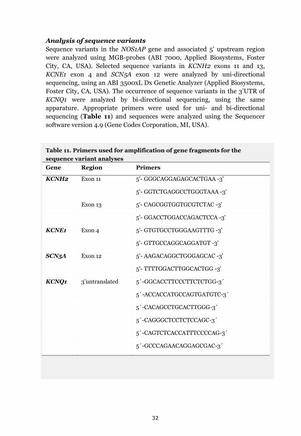
32
Analysis of sequence variants
Sequence variants in the NOS1AP gene and associated 5’ upstream region
were analyzed using MGB-probes (ABI 7000, Applied Biosystems, Foster
City, CA, USA). Selected sequence variants in KCNH2 exons 11 and 13,
KCNE1 exon 4 and SCN5A exon 12 were analyzed by uni-directional
sequencing, using an ABI 3500xL Dx Genetic Analyzer (Applied Biosystems,
Foster City, CA, USA). The occurrence of sequence variants in the 3’UTR of
KCNQ1 were analyzed by bi-directional sequencing, using the same
apparature. Appropriate primers were used for uni- and bi-directional
sequencing (Table 11) and sequences were analyzed using the Sequencer
software version 4.9 (Gene Codes Corporation, MI, USA).
Table 11. Primers used for amplification of gene fragments for the
sequence variant analyses
Gene Region Primers
KCNH2 Exon 11 5'- GGGCAGGAGAGCACTGAA -3'
5'- GGTCTGAGGCCTGGGTAAA -3'
Exon 13 5'- CAGCGGTGGTGCGTCTAC -3'
5'- GGACCTGGACCAGACTCCA -3'
KCNE1 Exon 4 5'- GTGTGCCTGGGAAGTTTG -3'
5'- GTTGCCAGGCAGGATGT -3'
SCN5A Exon 12 5'- AAGACAGGCTGGGAGCAC -3'
5'- TTTTGGACTTGGCACTGG -3'
KCNQ1 3’untranslated 5´-GGCACCTTCCCTTCTCTGG-3´
5´-ACCACCATGCCAGTGATGTC-3´
5´-CACAGCCTGCACTTGGG-3´
5´-CAGGGCTCCTCTCCAGC-3´
5´-CAGTCTCACCATTTCCCCAG-3´
5´-GCCCAGAACAGGAGCGAC-3´
33
Assessment of allelic phase
Of the selected sequence variants, derived variants in the 3’UTR of the
KCNQ1 gene have proposed allele-specific modifying effects. Allele-specific
effects imply a possibility for both aggravating and ameliorating effects
dependent on the location of the derived variant in relation to the mutation.
Allelic-phase of the derived variants, i.e. whether they occurred on the
mutated or the healthy allele, was therefore ascertained by analysis of trios,
including parents with ascertained genotype (one mutation-carrier and one
non-carrier) and their mutual offspring (mutation-carrier). Allelic phase
may be expressed from the point of view of the mutation, i.e. if the derived
variant is on the same allele as the mutation it is said to occur in cis and if it
occurs on the healthy allele, thus opposite of the mutation, the derived
variant is said to occur in trans.
Statistics: Associations between sequence variants and cardiac
phenotype in founder heterozygotes
For the association analyses, analyzed sequence variants with a minor allele
frequency >0.1 were included. Variants with a lower frequency in the study
population were excluded in order to reduce the risk of a type 1 error.
Associations between sequence variants and scalar QTc were evaluated by
linear regression analysis, associations related to age and gender adjusted
QTc levels16 and cardiac events were analyzed by logistic regression.
For linear and logistic regression analysis of non-allele specific effects, a
dominant model was used (genotypes coded as AA= 0, Aa or aa=1). For
analysis of allele-specific effects, the association between derived variants
and prevalent haplotypes, dependent on allelic phase, were analyzed
individually and in combination. The impact of possible confounders,
including gender, age and beta-blocker therapy, was assessed by stratified
analyses (i.e. no possible confounders were included in the models and no
model fitting was performed).
Correction for relatedness, i.e. correction for type 1 errors in allelic
association testing, was performed for associations at the adjusted
significance level using a kinship matrix (corrected x2 test).79 The kinship
matrix included data derived from the genealogical investigation, including
detailed data on interrelation between all included cases.
The significance level of the association-analyses was adjusted (correction
for multiple testing) for the number of independent tests used (5) to 0.01=1-
exp(ln(0.95)/5), where linked sequence variants (p<0.01 by x2 test) were
counted as interdependent. Data summarization and statistical analyses
were performed with SPSS version 19.0 (Statistical Package for Social
Sciences, SPSS Inc, Chicago, Ill).

34
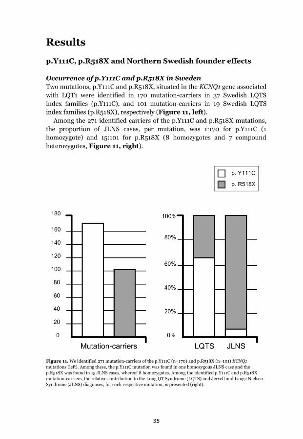
35
Results
p.Y111C, p.R518X and Northern Swedish founder effects
Occurrence of p.Y111C and p.R518X in Sweden
Two mutations, p.Y111C and p.R518X, situated in the KCNQ1 gene associated
with LQT1 were identified in 170 mutation-carriers in 37 Swedish LQTS
index families (p.Y111C), and 101 mutation-carriers in 19 Swedish LQTS
index families (p.R518X), respectively (Figure 11, left).
Among the 271 identified carriers of the p.Y111C and p.R518X mutations,
the proportion of JLNS cases, per mutation, was 1:170 for p.Y111C (1
homozygote) and 15:101 for p.R518X (8 homozygotes and 7 compound
heterozygotes, Figure 11, right).
Figure 11. We identified 271 mutation-carriers of the p.Y111C (n=170) and p.R518X (n=101) KCNQ1
mutations (left). Among these, the p.Y111C mutation was found in one homozygous JLNS case and the
p.R518X was found in 15 JLNS cases, whereof 8 homozygotes. Among the identified p.Y111C and p.R518X
mutation-carriers, the relative contribution to the Long QT Syndrome (LQTS) and Jervell and Lange Nielsen
Syndrome (JLNS) diagnoses, for each respective mutation, is presented (right).
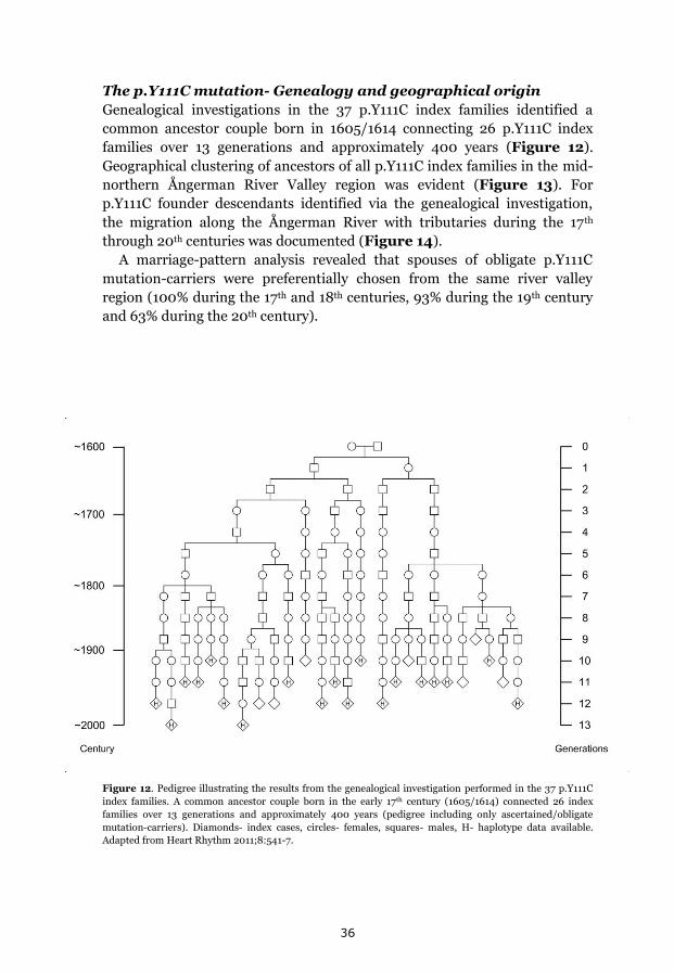
36
The p.Y111C mutation- Genealogy and geographical origin
Genealogical investigations in the 37 p.Y111C index families identified a
common ancestor couple born in 1605/1614 connecting 26 p.Y111C index
families over 13 generations and approximately 400 years (Figure 12).
Geographical clustering of ancestors of all p.Y111C index families in the mid-
northern Ångerman River Valley region was evident (Figure 13). For
p.Y111C founder descendants identified via the genealogical investigation,
the migration along the Ångerman River with tributaries during the 17th
through 20th centuries was documented (Figure 14).
A marriage-pattern analysis revealed that spouses of obligate p.Y111C
mutation-carriers were preferentially chosen from the same river valley
region (100% during the 17th and 18th centuries, 93% during the 19th century
and 63% during the 20th century).
Figure 12. Pedigree illustrating the results from the genealogical investigation performed in the 37 p.Y111C
index families. A common ancestor couple born in the early 17th century (1605/1614) connected 26 index
families over 13 generations and approximately 400 years (pedigree including only ascertained/obligate
mutation-carriers). Diamonds- index cases, circles- females, squares- males, H- haplotype data available.
Adapted from Heart Rhythm 2011;8:541-7.

37
.
Figure 13. (right) Map delimiting the
Ångerman/Indal Rivers with tributaries
(depicted as lines within the shaded
area) in the mid-northern part of
Sweden (shown in magnification in the
left upper hand box), illustrating
geographical clustering of p.Y111C
ancestors (all families, birthplaces
depicted as dots) during the 19th century.
Figure 14. (below) The same area as
shown in magnification in Figure 13,
illustrating the historical migration of
p.Y111C obligate mutation-carriers
included in the founder pedigree
(Figure 12), within the specific river
valley region. Dots represent the
birthplaces of founder descendants born
during the 17th through 20th centuries.
Figure 13 and 14 were adapted from
paper II, published in Heart Rhythm
2011;8:541-7.
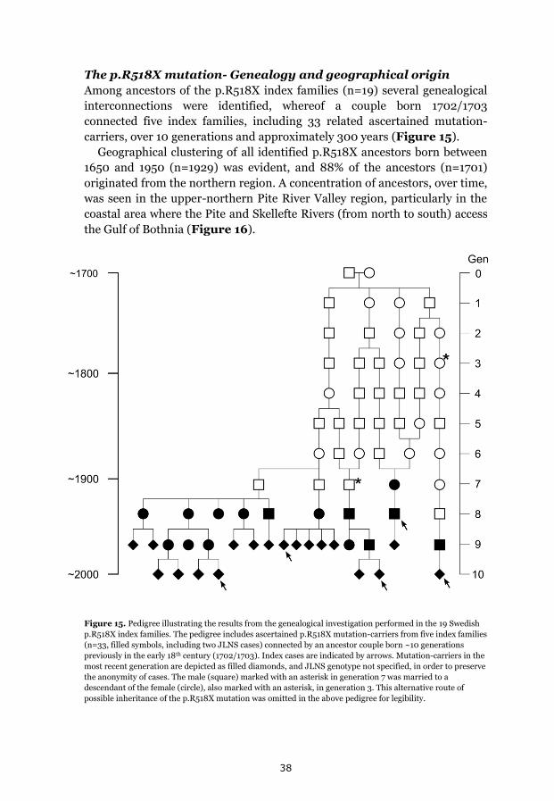
38
The p.R518X mutation- Genealogy and geographical origin
Among ancestors of the p.R518X index families (n=19) several genealogical
interconnections were identified, whereof a couple born 1702/1703
connected five index families, including 33 related ascertained mutation-
carriers, over 10 generations and approximately 300 years (Figure 15). Geographical clustering of all identified p.R518X ancestors born between
1650 and 1950 (n=1929) was evident, and 88% of the ancestors (n=1701)
originated from the northern region. A concentration of ancestors, over time,
was seen in the upper-northern Pite River Valley region, particularly in the
coastal area where the Pite and Skellefte Rivers (from north to south) access
the Gulf of Bothnia (Figure 16).
Figure 15. Pedigree illustrating the results from the genealogical investigation performed in the 19 Swedish
p.R518X index families. The pedigree includes ascertained p.R518X mutation-carriers from five index families
(n=33, filled symbols, including two JLNS cases) connected by an ancestor couple born ~10 generations
previously in the early 18th century (1702/1703). Index cases are indicated by arrows. Mutation-carriers in the
most recent generation are depicted as filled diamonds, and JLNS genotype not specified, in order to preserve
the anonymity of cases. The male (square) marked with an asterisk in generation 7 was married to a
descendant of the female (circle), also marked with an asterisk, in generation 3. This alternative route of
possible inheritance of the p.R518X mutation was omitted in the above pedigree for legibility.
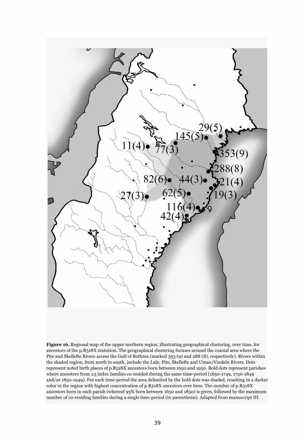
39
Figure 16. Regional map of the upper northern region, illustrating geographical clustering, over time, for
ancestors of the p.R518X mutation. The geographical clustering focuses around the coastal area where the
Pite and Skellefte Rivers access the Gulf of Bothnia (marked 353 (9) and 288 (8), respectively). Rivers within
the shaded region, from north to south, include the Lule, Pite, Skellefte and Uman/Vindeln Rivers. Dots
represent noted birth places of p.R518X ancestors born between 1650 and 1950. Bold dots represent parishes
where ancestors from ≥3 index families co-resided during the same time-period (1650-1749, 1750-1849
and/or 1850-1949). For each time-period the area delimited by the bold dots was shaded, resulting in a darker
color in the region with highest concentration of p.R518X ancestors over time. The number of p.R518X
ancestors born in each parish (whereof 93% born between 1650 and 1850) is given, followed by the maximum
number of co-residing families during a single time-period (in parenthesis). Adapted from manuscript III.

40
In summary, tracing of the bloodlines of the p.Y111C and p.R518X index
families to at least 1750 revealed several genealogical interconnections and
clustering of ancestors in two separate northern river valley regions, for each
respective mutation (Figure 17).
Figure 17. Genealogical investigation regarding the p.Y111C and p.R518X index families revealed
geographical clustering of ancestors in two separate northern river valley regions, focusing around the coastal
parts of the Ångerman/Indal River valleys (p.Y111C, left) and the Pite/Skellefte River valleys (p.R518X, right).
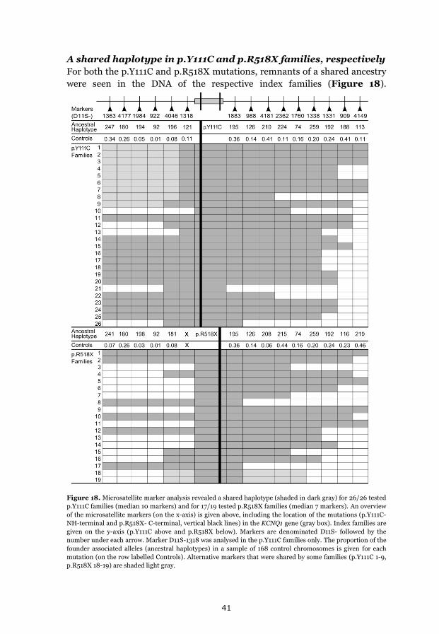
41
A shared haplotype in p.Y111C and p.R518X families, respectively
For both the p.Y111C and p.R518X mutations, remnants of a shared ancestry
were seen in the DNA of the respective index families (Figure 18).
Figure 18. Microsatellite marker analysis revealed a shared haplotype (shaded in dark gray) for 26/26 tested
p.Y111C families (median 10 markers) and for 17/19 tested p.R518X families (median 7 markers). An overview
of the microsatellite markers (on the x-axis) is given above, including the location of the mutations (p.Y111C-
NH-terminal and p.R518X- C-terminal, vertical black lines) in the KCNQ1 gene (gray box). Index families are
given on the y-axis (p.Y111C above and p.R518X below). Markers are denominated D11S- followed by the
number under each arrow. Marker D11S-1318 was analysed in the p.Y111C families only. The proportion of the
founder associated alleles (ancestral haplotypes) in a sample of 168 control chromosomes is given for each
mutation (on the row labelled Controls). Alternative markers that were shared by some families (p.Y111C 1-9,
p.R518X 18-19) are shaded light gray.
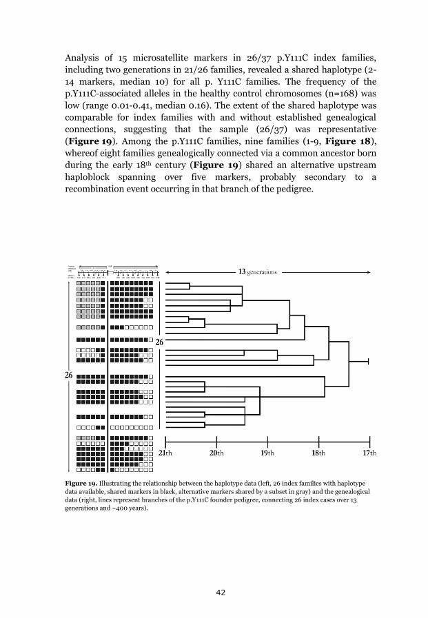
42
Analysis of 15 microsatellite markers in 26/37 p.Y111C index families,
including two generations in 21/26 families, revealed a shared haplotype (2-
14 markers, median 10) for all p. Y111C families. The frequency of the
p.Y111C-associated alleles in the healthy control chromosomes (n=168) was
low (range 0.01-0.41, median 0.16). The extent of the shared haplotype was
comparable for index families with and without established genealogical
connections, suggesting that the sample (26/37) was representative
(Figure 19). Among the p.Y111C families, nine families (1-9, Figure 18),
whereof eight families genealogically connected via a common ancestor born
during the early 18th century (Figure 19) shared an alternative upstream
haploblock spanning over five markers, probably secondary to a
recombination event occurring in that branch of the pedigree.
Figure 19. Illustrating the relationship between the haplotype data (left, 26 index families with haplotype
data available, shared markers in black, alternative markers shared by a subset in gray) and the genealogical
data (right, lines represent branches of the p.Y111C founder pedigree, connecting 26 index cases over 13
generations and ~400 years).
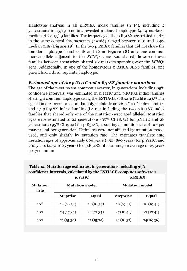
43
Haplotype analysis in all p.R518X index families (n=19), including 2
generations in 15/19 families, revealed a shared haplotype (4-14 markers,
median 7) for 17/19 families. The frequency of the p.R518X-associated alleles
in the same control chromosomes (n=168) ranged between 0.01 and 0.46,
median 0.18 (Figure 18). In the two p.R518X families that did not share the
founder haplotype (families 18 and 19 in Figure 18) only one common
marker allele adjacent to the KCNQ1 gene was shared, however these
families between themselves shared six markers spanning over the KCNQ1
gene. Additionally, in one of the homozygous p.R518X JLNS families, one
parent had a third, separate, haplotype.
Estimated age of the p.Y111C and p.R518X founder mutations
The age of the most recent common ancestor, in generations including 95%
confidence intervals, was estimated in p.Y111C and p.R518X index families
sharing a common haplotype using the ESTIAGE software (Table 12).75 The
age estimates were based on haplotype data from 26 p.Y111C index families
and 17 p.R518X index families (i.e not including the two p.R518X index
families that shared only one of the mutation-associated alleles). Mutation
ages were estimated to 24 generations (95% CI 18;34) for p.Y111C and 28
generations (95% CI 19;41) for p.R518X, assuming a mutation rate of 10-6 per
marker and per generation. Estimates were not affected by mutation model
used, and only slightly by mutation rate. The estimates translate into
mutation ages of approximately 600 years (450; 850 years) for p.Y111C, and
700 years (475; 1025 years) for p.R518X, if assuming an average of 25 years
per generation.
Table 12. Mutation age estimates, in generations including 95%
confidence intervals, calculated by the ESTIAGE computer software75
p.Y111C p.R518X
Mutation
rate
Mutation model Mutation model
Stepwise Equal Stepwise Equal
10-6 24 (18;34) 24 (18;34) 28 (19;41) 28 (19;41)
10-4 24 (17;34) 24 (17;34) 27 (18;41) 27 (18;41)
10-3 21 (15;30) 21 (15;29) 24 (16;37) 24(16; 36)
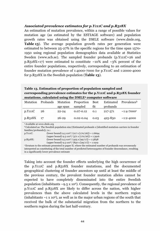
44
Associated prevalence estimates for p.Y111C and p.R518X
An estimation of mutation prevalence, within a range of possible values for
mutation age (as estimated by the ESTIAGE software) and population
growth rates was obtained using the DMLE software (www.dmle.org,
Table 13). The average population growth rates per generation were
estimated to between 25-27% in the specific regions for the time span 1570-
1950 using regional population demographics data available at Statistics
Sweden (www.scb.se). The sampled founder probands (p.Y111C=26 and
p.R518X=17) were estimated to constitute ~10% and ~3% percent of the
entire founder populations, respectively, corresponding to an estimation of
founder mutation prevalence of 1:4000-7000 for p.Y111C and 1:2000-4000
for p.R518X in the Swedish population (Table 13).
Table 13. Estimation of proportion of population sampled and
corresponding prevalence estimates for the p.Y111C and p.R518X founder
mutations, calculated using the DMLEa computer software
Mutation Probands Mutation
age span
Proportion
sampled
Best
fit
Estimated
probands
Prevalenceb
p.Y111C 26 22-24 0.07-0.12 0.1 217-371 ~1:4-7000c
p.R518X 17 26-29 0.02-0.04 0.03 425-850 ~1:2-4000
a Available at www.dmle.org b Calculated as: The Swedish population size/(Estimated probands x [identified mutation-carriers in founder
families/probands]), i.e.:
p.Y111C: (lower bound) 9.5 x10⁶/ (217 x [170/26]) = 1:6694
(upper bound) 9.5 x10⁶/ (371 x [170/26]) = 1:3916
p.R518X: (lower bound) 9.5 x10⁶/ (425 x [99/17]) = 1:3838
(upper bound) 9.5 x10⁶/ (850 x [99/17]) = 1:1919 c Erratum to the estimate presented in paper II, where the estimated number of probands was erroneously
interpreted as constituting of the total number of predicted heterozygotes of founder descendance, resulting
in a significantly lower prevalence estimate
Taking into account the founder effects underlying the high occurrence of
the p.Y111C and p.R518X founder mutations, and the documented
geographical clustering of founder ancestors up until at least the middle of
the previous century, the prevalent founder mutation alleles cannot be
expected to have completely disseminated into the entire Swedish
population (inhabitants ~9.5 x 10⁶). Consequently, the regional prevalence of
p.Y111C and p.R518X are likely to differ across the nation, with higher
prevalences than the above calculated levels in the northern region
(inhabitants ~1 x 10⁶), as well as in the major urban regions of the south that
received the bulk of the substantial migration from the northern to the
southern region during the last half-century.

45
Prevalence and mutation spectrum of JLNS in Sweden Via the national inventory, five now living preadolescent JLNS cases (mean
age 6 years, range 5-8 years) were identified, corresponding to an
approximate incidence of 1 new case every other year and a minimum JLNS
prevalence of 1:200 000 in Swedish children below 10 years of age.
In total, 19 clinical JLNS cases, 11 females and 8 males, in 13 Swedish
families were identified, whereof five previously identified regional cases
were deceased.
Genotype was ascertained in all living index cases (12/13 families), revealing
that the mutation spectrum for JLNS in Sweden consisted of 8 KCNQ1
mutations, and no KCNE1 mutations. The identified KCNQ1 mutations were
all previously reported as associated with LQTS and/or JLNS and included
two nonsense mutations (14/24 alleles), two frame-shift mutations (5/24
alleles), three missense mutations (4/24 alleles) and one splice site mutation
(1/24 alleles). Four of the mutations, p.Y111C, p.R190W, p.S349W and
p.S277del were for the first time, to our knowledge, reported as associated
with JLNS (Table 14).
Table 14. Inventory of KCNQ1 mutations in 12 Swedish JLNS probands
Nucleotide
change
Amino acid
change
Region Mutation
type
Alleles
identifieda c.1522C>T68,69 p.R518X C-terminal Nonsense 12 (3)
c.572_576del41 p.R192Cfs91X S2-S3 Frame-shift 4 (2)
c.332A>G53 p.Y111Cb N-terminal Missense 2 (1)
c.1588C>T70 p.Q530X C-terminal Nonsense 2
c.477+1G>A40,80 p.M159sp S2 Splice error 1
c.568C>T26 p.R190Wb S2-S3 Missense 1
c.828_830del81 p.S277delb S5 Frame-shift 1
c.1046C>G53 p.S349Wb S6 Missense 1
a Number of homozygous probands is given in parenthesis b Mutation identified for the first time in association with JLNS (this study)
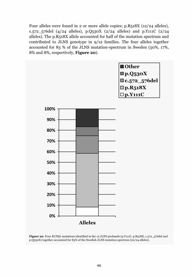
46
Four alleles were found in 2 or more allele copies; p.R518X (12/24 alleles),
c.572_576del (4/24 alleles), p.Q530X (2/24 alleles) and p.Y111C (2/24
alleles). The p.R518X allele accounted for half of the mutation spectrum and
contributed to JLNS genotype in 9/12 families. The four alleles together
accounted for 83 % of the JLNS mutation-spectrum in Sweden (50%, 17%,
8% and 8%, respectively, Figure 20).
0%
10%
20%
30%
40%
50%
60%
70%
80%
90%
100%
Alleles
Other
p.Q530X
c.572_576del
p.R518X
p.Y111C
Figure 20. Four KCNQ1 mutations identified in the 12 JLNS probands (p.Y111C, p.R518X, c.572_576del and
p.Q530X) together accounted for 83% of the Swedish JLNS mutation spectrum (20/24 alleles).

47
In the Swedish index families, JLNS was equally caused by homozygosity
and compound heterozygosity (in 6/12 families, respectively, Table 15).
Table 15. Genotype in 13 Swedish JLNS families carrying KCNQ1
mutations, including 19 cases
Familya Amino acid change
(KCNQ1 gene)
Gender Case nrb
JLN1 p.S349W/p.R518X M 6
JLN2 p.R518Xc F 5
JLN3 p.R518Xc F/M 7, 8
JLN4 p.S277del/p.R518X M 11
JLN5 p.R518Xc 2F/3M 14, 16-19d
JLN6 p.Q530X/p.R518X M 4
JLN7 p.R518X/p.R190W F 2
JLN8 p.M159sp/p.R518X 2 F 1, 3
JLN9 p.R518X/p.Q530X F 10
JLN10 p.R192Cfs91Xc F 9
JLN11 p.R192Cfs91Xc F 13
JLN12 p.Y111Cc F 12
JLN13 DNA not available M 15d
Abbreviations: F- female, M- male a Family number in accordance with paper III b Case number (nr) in accordance with paper IV (living cases 1-14 according to age at study, deceased cases 15-
19 according to birth year) c Homozygous for the given allele d Cases deceased at the time of the study
The number of JLNS index cases with ascertained genotype (n=12) predicts a
rough estimated prevalence of heterozygous mutation-carriers in the
Swedish population of 1:300-600, whereof the prevalence of the specific
p.R518X allele was estimated to 1:1000, using Hardy-Weinberg statistics.
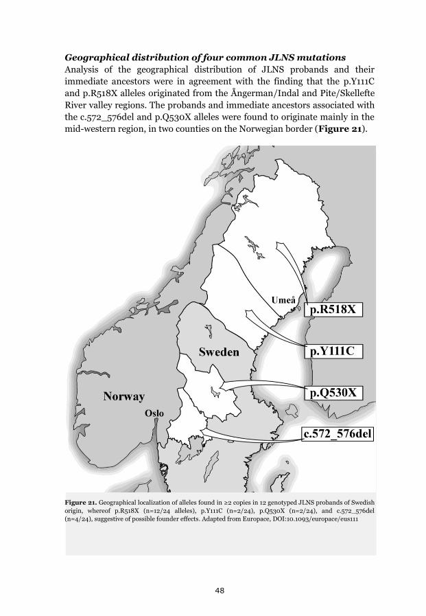
48
Geographical distribution of four common JLNS mutations
Analysis of the geographical distribution of JLNS probands and their
immediate ancestors were in agreement with the finding that the p.Y111C
and p.R518X alleles originated from the Ångerman/Indal and Pite/Skellefte
River valley regions. The probands and immediate ancestors associated with
the c.572_576del and p.Q530X alleles were found to originate mainly in the
mid-western region, in two counties on the Norwegian border (Figure 21).
Figure 21. Geographical localization of alleles found in ≥2 copies in 12 genotyped JLNS probands of Swedish
origin, whereof p.R518X (n=12/24 alleles), p.Y111C (n=2/24), p.Q530X (n=2/24), and c.572_576del
(n=4/24), suggestive of possible founder effects. Adapted from Europace, DOI:10.1093/europace/eus111
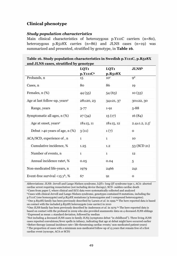
49
Clinical phenotype
Study population characteristics
Main clinical characteristics of heterozygous p.Y111C carriers (n=80),
heterozygous p.R518X carries (n=86) and JLNS cases (n=19) was
summarized and presented, stratified by genotype, in Table 16.
Table 16. Study population characteristics in Swedish p.Y111C, p.R518X
and JLNS cases, stratified by genotype
LQT1
p.Y111Ca
LQT1
p.R518X
JLNSb
Probands, n 15 10c 9d
Cases, n 80 86 19
Females, n (%) 44 (55) 54 (63) 10 (53)
Age at last follow-up, yearse 28±20, 23 34±21, 37 30±22, 30
Range, years 3-77 1-91 5-88
Symptomatic all ages, n (%) 27 (34) 15 (17) 16 (84)
Age at onset, yearse 18±15, 11 18±15, 12 2.4±1.2, 2.5f
Debut >40 years of age, n (%) 3 (11) 1 (7) 0
ACA/SCD, experience of , n 1 1 10
Cumulative incidence, % 1.25 1.2 53 (SCD 21)
Number of events, n 1 1 12
Annual incidence rateg, % 0.05 0.04 5
Non-medicated life-years, n 1979 2466 241
Event-free survival <15 yh, % 67 91 0
Abbreviations: JLNS- Jervell and Lange-Nielsen syndrome, LQT1- long QT syndrome type 1, ACA- aborted
cardiac arrest requiring resuscitation (not including device therapy), SCD- sudden cardiac death a Cases from paper I, where clinical and ECG data were systematically collected and analyzed b Cases with clinical Jervell and Lange-Nielsen syndrome, genotypes contained 8 mutations, including the
p.Y111C (one homozygote) and p.R518X mutations (4 homozygotes and 7 compound heterozygotes). c One p.R518X family has been previously described by Larsen et al. in 1999.69 The here reported data is based
on contact with the included p.R518X heterozygote (one carrier) in 2010 d One JLNS family has been previously described by Andersson et al. in 1979.64 The here reported data is
based on contact with the proband in 2009 who also provided anamnestic data on 4 deceased JLNS siblings e Expressed as mean ± standard deviation, followed by median f Not including 4 deceased JLNS cases in family JLN5 (symptoms debut “in childhood”). Three living JLNS
cases reported convulsions/fever spells in infancy, indicating that age at debut might have occurred earlier g Before therapy (annual incidence rate= life-threatening cardiac events/ non-medicated patient-years)
h The proportion of cases with a minimum non-medicated follow-up of 15 years that remain free of a first
cardiac event (syncope, ACA or SCD)
50
A range of clinical severity evident
The cardiac phenotype ranged from mild in p.Y111C and p.R518X
heterozygotes to severe in JLNS (Table 16). The most benign phenotype
was seen in p.R518X heterozygotes, where only 17% presented with a first
possible cardiac event and 91% of non-medicated individuals remained
event-free up to 15 years of age. The corresponding numbers for p.Y111C
heterozygotes was 34% for symptomatic phenotype and 67% for non-
medicated event-free survival up to 15 years of age. On the other end of the
scale, 84% of JLNS cases had experienced a first cardiac event and the three
cases that were as yet asymptomatic were all pediatric cases, between 4-6
years of age, and on beta-blockers since birth.
With regards to life-threatening cardiac events, one non-medicated
p.Y111C heterozygous male experienced an aborted cardiac arrest at 13 years
of age (he had experienced previous syncopal attacks but these had been
interpreted as seizures), and one p.R518X heterozygous female suffered a
first fatal cardiac event at 56 years of age (in relation to a combination of
physical exercise and hypokalemia). This corresponds to a cumulative
incidence of life-threatening cardiac events of 1.25% and 1.2% in the p.Y111C
and p.R518X populations respectively. Among JLNS cases 10 cases
experienced a life-threatening cardiac event, whereof 4 cases died suddenly
at ages 5, 20, 27 and 37 years. This corresponds to a cumulative incidence of
life-threatening cardiac events of 53%, whereof sudden cardiac death 21%.
The severity of the JLNS phenotype may be underestimated as “appropriate
shocks” from ICDs were not per se counted as life-threatening cardiac
events. If regarding experience of ICD-therapy as a proxy for a life-
threatening event, the cumulative incidence would be 63% in the JLNS
subgroup (12/19).
Since the time at risk and also the presence of beta-blocker therapy are
factors of interest, the annual incidence of life-threatening cardiac events
was calculated (number of events/ sum of non-medicated patient-years) to
0.05% in p.Y111C heterozygotes and 0.04% in p.R518X heterozygotes. In the
JLNS subgroup, the combination of a high number of life-threatening events
(n=12) and few non-medicated life-years (n=241) rendered a 5% annual
incidence of life-threatening cardiac events before therapy (Table 16).
Triggers and cardiac events
Similar circumstances surrounding the cardiac events were reported for both
LQT1 heterozygotes (p.Y111C and p.R518X) and JLNS cases (Table 17). As is
common for KCNQ1-associated LQTS and JLNS, the most common type of
triggers reported were within the range of physical activities, not limited to
exercise specifically. For p.R518X heterozygotes and JLNS cases, the trigger
swimming (included in the category “Exercise” in Table 17) was reported
separately and accounted for 2% and 12% of triggers, respectively. Three of
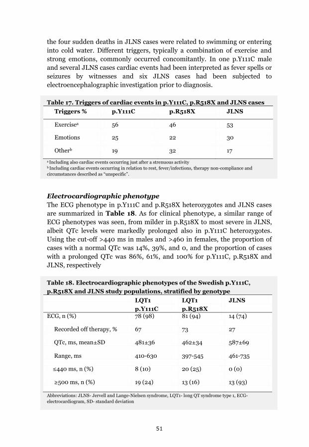
51
the four sudden deaths in JLNS cases were related to swimming or entering
into cold water. Different triggers, typically a combination of exercise and
strong emotions, commonly occurred concomitantly. In one p.Y111C male
and several JLNS cases cardiac events had been interpreted as fever spells or
seizures by witnesses and six JLNS cases had been subjected to
electroencephalographic investigation prior to diagnosis.
Table 17. Triggers of cardiac events in p.Y111C, p.R518X and JLNS cases
Triggers % p.Y111C p.R518X JLNS
Exercisea 56 46 53
Emotions 25 22 30
Otherb 19 32 17
a Including also cardiac events occurring just after a strenuous activity b Including cardiac events occurring in relation to rest, fever/infections, therapy non-compliance and
circumstances described as “unspecific”.
Electrocardiographic phenotype
The ECG phenotype in p.Y111C and p.R518X heterozygotes and JLNS cases
are summarized in Table 18. As for clinical phenotype, a similar range of
ECG phenotypes was seen, from milder in p.R518X to most severe in JLNS,
albeit QTc levels were markedly prolonged also in p.Y111C heterozygotes.
Using the cut-off >440 ms in males and >460 in females, the proportion of
cases with a normal QTc was 14%, 39%, and 0, and the proportion of cases
with a prolonged QTc was 86%, 61%, and 100% for p.Y111C, p.R518X and
JLNS, respectively
Table 18. Electrocardiographic phenotypes of the Swedish p.Y111C,
p.R518X and JLNS study populations, stratified by genotype LQT1
p.Y111C
LQT1
p.R518X
JLNS
ECG, n (%) 78 (98) 81 (94) 14 (74)
Recorded off therapy, % 67 73 27
QTc, ms, mean±SD 481±36 462±34 587±69
Range, ms 410-630 397-545 461-735
≤440 ms, n (%) 8 (10) 20 (25) 0 (0)
≥500 ms, n (%) 19 (24) 13 (16) 13 (93)
Abbreviations: JLNS- Jervell and Lange-Nielsen syndrome, LQT1- long QT syndrome type 1, ECG-
electrocardiogram, SD- standard deviation

52
Genotype and gender-specific assessment of cardiac phenotypes
p.Y111C heterozygotes
Among the symptomatic p.Y111C heterozygotes (n=27, 18 (67%) females) 24
developed symptoms before, and 3 cases after, 40 years of age. All
individuals with a symptoms debut after 40 years of age were female.
When assessing the proportion of cases symptomatic by different ages, males
experienced a first cardiac event earlier; 33% were symptomatic by 10 years
of age, 89% were symptomatic by 20 years of age and all were symptomatic
by 25 years of age, as compared to females, where the corresponding
proportion was 33%, 67% and 72%.
Females presented with longer mean QTc levels (490±40 ms vs. 470±28
ms, p=0.012, as compared to males). QTc levels in the 3 females with an age
at onset >40 years were markedly prolonged (473 ms, 532 ms and 630 ms).
QTc prolongation related to symptomatic phenotype (symptomatic carriers
vs. asymptomatic carriers: 494±38 ms vs. 474±33 ms, p=0.019), however
female gender was not significantly associated with symptomatic phenotype.
p.R518X heterozygotes
Among the symptomatic p.R518X heterozygotes (n=15, 10 (67%) females)
one female suffered a first (fatal) event after 40 years of age. Age at debut
(ranging from 1.5 years of age to 56 years of age) did not significantly differ
between the genders.
QTc in symptomatic cases varied between 402-545 ms, and 33% of
symptomatic cases had a QTc below 440 ms. QTc in the female with a first
(fatal) cardiac event at 56 years of age was 506 ms. The variability regarding
ECG phenotype was evident also when considering the entire p.R518X
population; QTc ranged between 397-545 ms in all p.R518X heterozygotes,
and between 402-545 ms and 397-521 ms in females and males, respectively.
Females presented with overall longer mean QTc levels (473±33 vs.443±29
ms, p<0.001, as compared to males).
No significant correlations between the probability of a symptomatic
phenotype and the variables scalar QTc prolongation, heart rate and gender
were found (data not shown).
JLNS cases
Among JLNS cases all but three pediatric girls on beta-blockers since birth
had experienced a first cardiac event. With regards to clinical presentation
over time, a difference between the genders during adulthood was suggested.
While cardiac events typically were frequent during childhood for both
genders, their frequency tended to abate for males, but not females, when
reaching adulthood (Figure 22).

53
Figure 22. Figure depicting the timing of cardiac events and interventions during the lifetime of 19
Swedish JLNS cases. The age of the included cases at cardiac events and/or interventions is given on the
x-axis. Cases are numbered on the y-axis according to age at study (youngest-oldest, case 1-14). Deceased
cases are numbered according to birth year (youngest-oldest, case 15-19). After 45 years the time-scale on
the x-axis is disrupted (⸗) and the age at study/death of cases > 45 years of age given in the right hand
margin. Case 17 died from malignancy at 59 years of age (†). F= female, M= male. Symbols occurring
above each cases’ line (see also figure legend) represent cardiac events (first cardiac event, most recent
cardiac event, time-period during which at least one cardiac event per year were reported, time-period
during which a first cardiac event was reported to occur, aborted cardiac arrest and sudden (suspected)
cardiac death). Symbols occurring below each cases’ line (see also figure legend) represent interventions
(start of beta-blocker pharmacotherapy, implantation of a cardioverter-defibrillator and/or the surgical
performance of a left cardiac sympathetic denervation). Reprinted with permission: Europace 2012. DOI:
10.1093/europace/eus111.
Mean QTc levels were significantly longer in symptomatic cases (mean
605±62 ms, median 584 ms) vs. asymptomatic cases (mean 518±50 ms,
median 540 ms, p=0.016), however no differences between the genders were
evident.

54
Beta-blocker therapy and other interventions
At the time of the studies, the reported proportion of cases on beta-blocker
therapy was 63% for p.Y111C heterozygotes, 43% for p.R518X heterozygotes
and 79% for JLNS (all now living JLNS cases were on beta-blocker therapy,
Table 19).
In accordance with the severe phenotype in JLNS, age at therapy start was
younger (median 2 years of age) and additional therapies, such as LCSD
(16%) and ICD-therapy (32%), were more often utilized.
Table 19. Preventive medical interventions in the p.Y111C, p.R518X and JLNS populations, stratified by genotype
LQT1
p.Y111C
LQT1
p.R518X
JLNS
Mutation-carriers/probands, n/n 80/15 86/10 19/9
Beta-blocker therapy, n (%) 50 (63) 37 (43) 15 (79)a
Age at therapy start, yearsb 17±17, 12 25±19, 17 7±13, 2
Time before therapy, yearsb 25±20, 20 31±21, 33 13±18, 5
LCSD, n (%) 0 0 3 (16)
Age at procedure, years, (per individual)
- - 2, 24, 21+24
Mean time to recurrence, years - - 1
ICD, n (%) 1 0 6 (32)
Age at implantation, years >30 - 22±13, 21
Experience of ICD-therapy, n (%) 0c - 6 (100)
Abbreviations: JLNS- Jervell and Lange-Nielsen syndrome, LQT1- long QT syndrome type 1, LCSD- Left
Cardiac Sympathetic Denervation (indication: post aborted cardiac arrest or symptoms in spite of beta-
blocker therapy ± too young for ICD-implantation), ICD- Implantable Cardioverter-Defibrillator (indication:
post aborted cardiac arrest or symptoms in spite of beta-blocker therapy ±LCSD) a All now living JLNS cases and one deceased JLNS case
b Expressed as mean ± standard deviation, followed by median c 2-year follow-up

55
Assessment of the “benign” p.Y111C and p.R518X phenotypes
We made an attempt to investigate the natural history of the p.Y111C
mutation, as well as perform comparisons with other LQT1 populations.
Benign phenotype verified in non-medicated p.Y111C ancestors
We analyzed the survival beyond 40 years of age for non-medicated p.Y111C
ancestors connected by a common founder. The study group consisted of 107
non-medicated individuals born between 1873 and 1968, whereof 24 were
ascertained p.Y111C mutation-carriers, 16 obligate mutation-carriers born
along the hypothetical line of descent of the p.Y111C mutation and 67 were
siblings to the obligate mutation-carriers. Based on the assumption that the
direct bloodline between the common founder and the probands constitute
the true route of the mutation, the study group consisted of ~73 mutation-
carriers (24+16+ [0.5 x 67] =73.5). The results from the mortality
investigation are summarized in Table 20.
Among the 107 ancestors investigated, 4 individuals (all siblings) died
before reaching 40 years of age; 3 infants between 1873 and 1915
(corresponding to an infant death rate of 7%) and one case of acute nephritis
after 1915. Age at death could not be ascertained in 4 individuals (all
siblings) born during the period of interest (late 19th century).
Overall death rates before 40 years of age varied between 4% (4/107) and
7% (8/107) if assuming that all missing cases had died before reaching 40
years of age. In the general Swedish population the age-stratified mortality
rate in 1900 was 13% for infants, 4-11% for children ≤14 years of age, and 6-
7% for individuals 15-39 years old,82 i.e. in all strata higher than among the
p.Y111C ancestors.
Table 20. Survival >40 years of age of 107 non-medicated p.Y111C
founder pedigree ancestors, born 1873-1968 Non-medicated ancestors
N 107
Ascertained mutation-carriers 24
Obligate carriers 16
Siblings 67
Survival >40 yrs, n (%) 99 (93)
Death <40 yrs, n (%) 4a,b (4)
Unknown, n (%) 4a (4)
a All siblings (50% carriership) b One case of acute nephritis, three infant deaths during the period 1873-1915

56
Phenotypic comparisons between LQT1 populations
The cumulative incidence (1.25% and 1.2%) and annual incidence rate before
therapy (0.05% and 0.04%) associated with heterozygous p.Y111C and
p.R518X carriership was apparently low. When comparing these levels to
previously published data on 386 non-medicated carriers of mixed KCNQ1
mutations77 the annual incidence of life-threatening cardiac events under the
age of 40 and before therapy was lower in p.Y111C heterozygotes as
compared to the mixed LQT1 population77 (0.05%/year vs. 0.3%/year,
p=0.025). The sudden cardiac death in the p.R518X population occurred in a
female >40 years of age, but even if including her in the analysis, the annual
incidence rate was significantly lower as compared to the mixed LQT1
population77 (p=0.007, Table 21).
Table 21. Incidence rate of life-threatening cardiac events before therapy,
comparison between LQTS populations
Y111Ca R518X LQT1a,b p
Mutation-carriers, n 80 86 386 …
ACA/SCD, n before therapy
1 1c 37 …
Incidence rate, %/y 0.05 0.04 0.3 0.025d
0.007e Mean follow-up before therapy, y
25±20 31±21 29±20 …
Abbreviations: ACA- aborted cardiac arrest, SCD- sudden cardiac death, y- years a in individuals below 40 years of age b Priori et al. N. Engl. J. Med. 2003;348(19):1866-1874. c The SCD occurred in a non-medicated female mutation-carrier 56 years of age d p.Y111C as compared to LQT1 by Fisher’s exact test
e p.R518x as compared to LQT1 by Fisher’s exact test
When comparing the clinical phenotype of the p.Y111C population (n=80) to
that of 244 carriers of the “malignant” p.A341V mutation78 and 205 carriers
of mixed KCNQ1 mutations, not including p.A341V78 a discrepancy between
QTc levels and cardiac phenotype was evident (Table 22). While mean QTc
levels were comparable for the p.Y111C and p.A341V mutations, the
proportion of carriers that had suffered a life-threatening cardiac event
significantly differed (1.25% vs. 30%, p<0.001).
The mean QTc levels in the KCNQ1/non-A341V population, on the other
hand, were significantly lower than in the p.Y111C population (465±38 ms
vs. 481±36 ms, p<0.001), and similar to that of the p.R518X population
(462±34 ms).
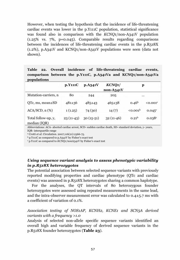
57
However, when testing the hypothesis that the incidence of life-threatening
cardiac events was lower in the p.Y111C population, statistical significance
was found also in comparison with the KCNQ1/non-A341V population
(1.25% vs. 7%, p=0.045). Comparable results regarding comparisons
between the incidences of life-threatening cardiac events in the p.R518X
(1.2%), p.A341V and KCNQ1/non-A341V populations were seen (data not
shown).
Table 22. Overall incidence of life-threatening cardiac events,
comparison between the p.Y111C, p.A341Va and KCNQ1/non-A341Va
populations
p.Y111C p.A341V KCNQ1/
non-A341V
p
Mutation-carriers, n 80 244 205 …
QTc, ms, mean±SD 481±36 485±43 465±38 0.46b <0.001c
ACA/SCD, n (%) 1 (1.25) 74 (30) 14 (7) <0.001b 0.045c
Total follow-up, y,
median (IQR)
23 (11-43) 30 (15-51) 32 (11-46) 0.21b 0.038c
Abbreviations: ACA- aborted cardiac arrest, SCD- sudden cardiac death, SD- standard deviation, y- years,
IQR- interquartile range a Crotti et al. Circulation. 2007;116(21):2366-75. b p.Y111C as compared to p.A341V by Fisher’s exact test c p.Y111C as compared to KCNQ1/nonA341V by Fisher’s exact test
Using sequence variant analysis to assess phenotypic variability
in p.R518X heterozygotes
The potential association between selected sequence variants with previously
reported modifying properties and cardiac phenotype (QTc and cardiac
events) was assessed in p.R518X heterozygotes sharing a common haplotype.
For the analyses, the QT intervals of 80 heterozygous founder
heterozygotes were assessed using repeated measurements in the same lead,
and the intra-observer measurement error was calculated to 0.4±5.7 ms with
a coefficient of variation of 0.1%.
Association testing of NOS1AP, KCNH2, KCNE1 and SCN5A derived
variants with a frequency >1.0
Analysis of selected non-allele specific sequence variants identified an
overall high and variable frequency of derived sequence variants in the
p.R518X founder heterozygotes (Table 23).

58
Table 23. Frequency of sequence variants with previously reported
modifying properties in Swedish p.R518X founder heterozygotes
Gene Sequence
variant
Amino acid
change
Allelea Genotype N (%) MAF
NOS1AP rs2880058
non-coding A/G AA AG GG
34 (43) 36 (45) 10 (13)
0.350
rs4657139
non-coding T/A TT AT AA
32 (40) 37 (46) 11 (14)
0.369
rs12143842
non-coding C/T CC CT TT
36 (45) 37 (46)
7 (9)
0.319
rs16847548
non-coding T/C TT CT CC
37 (46) 38 (48)
5 (6)
0.300
rs12567209
non-coding G/A GG AG AA
74 (93) 6 (8)
0
0.038
rs10494366
non-coding T/G TT GT GG
36 (45) 35 (44) 9 (11)
0.331
KCNH2 rs1805123 K897T A/C AA AC CC
50 (63) 24 (30)
6 (8)
0.225
rs36210421 R1047 G/T GG TG TT
67 (84) 12 (15)
1 (1)
0.088
KCNE1 rs1805127 G38S A/G AA AG GG
32 (40) 37 (46) 11 (14)
0.369
rs1805128 D85N G/A GG AG AA
75 (94) 5 (6)
0
0.031
SCN5A rs1805124 H558R A/G AA AG GG
45 (56) 32 (40)
3 (4)
0.238
rs36210423
A572D C/A CC AC AA
80 (100) 0 0
0
Abbreviations: MAF- Minor Allele Frequency (derived allele) a Ancestral/derived allele
Inclusion criteria for association analysis (previously reported modifying
properties and a minor allele frequency (for the derived allele) of at least 0.1)
were met by 5/6 sequenced NOS1AP variants, KCNH2-variant rs1805123
(p.K897T), KCNE1-variant rs1805127 (p.G38S) and SCN5A-variant rs1805124
(p.H558R) (Table 23).

59
In p.R518X heterozygous males, a significant association between NOS1AP
variants rs12143842 and rs16847548 and QT prolongation was detected,
constituting a 29 ms prolongation of QTc (p=0.004), explaining 27% of QTc
variability (Table 24). The association remained at the adjusted significance
level (p<0.01) after adjusting for relatedness using a kinship matrix
(adjusted p-value p=0.007). Similar results (although not significant) were
seen for all NOS1AP variants. Findings remained performing separate
analyses in adult males and non-medicated males only.
No significant associations to cardiac events or QTc were found for any of
the analyzed sequence variants when analyzed in the study population as a
whole, or when analyzed in females only (data not shown).
Table 24. QT duration in male p.R518X heterozygotes by sequence
variants in the NOS1AP gene
Sequence
variant
A/a AA a-carriers pa Betab R2c
QTc males
n QTc males
n
rs2880058
A/G
429±22 11 452±29 18 0.026 23.8 (0.41)
17.0
rs4657139
T/A
429±24 10 451±29 19 0.053 21.4 (0.36)
13.2
rs12143842
C/T
426±23 12 456±26 17 0.004
29.3 (0.51)
26.5
rs16847548
T/C
426±23 12 456±26 17 0.004 29.3 (0.51)
26.5
rs10494366
T/G 429±22 13 455±29 16 0.013 25.8 (0.46)
20.9
AA- homozygous for the major allele, a-carriers- carrier of at least one minor (derived) allele a as calculated by linear regression, adjusted significance level 0.01 b the effect on QTc, in ms, if having an a-allele, as compared to AA genotype, followed by the Beta coefficient
in parenthesis c the proportion of the QTc variance, in %, explained by the variable (sequence variant)
Adapted from Manuscript III.

60
Frequency and allelic phase of 3’UTR derived sequence variants
A high frequency of five derived 3’UTR/KCNQ1 sequence variants was
identified in the heterozygous p.R518X mutation-carriers via bi-directional
sequencing (Table 25). Analysis of allelic phase revealed that these five
3’UTR-KCNQ1 derived alleles segregated with the p.R518X mutation in the
founder population (Figure 23). Of these, rs2519184 (A), rs8234 (G) and
rs107980 (G) have previously reported modifying properties63, including
allele-specific repression of gene expression when, as in the p.R518X founder
heterozygotes, occurring in cis with the mutation.
Table 25. Frequency of derived variants identified by bi-directional
sequencing of the 3’UTR of the KCNQ1 gene, including allelic phase, in
p.R518X founder heterozygotes
Sequence
variant
Allelea Genotype N (%) MAF p.R518X-
associated allele rs45460605 C/T CC
CT TT
2 (2) 79 (98) 0
0.488 T
rs2519184 G/A GG AG AA
2 (2) 69 (85) 10 (12)
0.549 A
rs146324627 G/A GG AG AA
2 (2) 79 (98) 0
0.488 A
rs8234 A/G AA AG GG
1 (1) 55 (68) 25 (31)
0.648b G
rs107980
A/G AA AG GG
1 (1) 55 (68) 25 (31)
0.648b G
Abbreviations: MAF- Minor Allele Frequency (derived allele) a Ancestral/derived allele b 3’UTR/KCNQ1 sequence variants rs8234 and rs107980 were in complete linkage disequilibrium
The possible association between a milder phenotype and the repressive
AGG haplotype in cis could not be formally assessed as the majority of cases
(78/80, 98%) shared this configuration.

61
The possible (aggravating) effect of having the AGG (10/80, 13%) or GGG
haplotype (25/80, 31%) also in trans was analyzed by linear and logistic
regression, revealing no significant associations to QTc, QTc on the extremes
of the scale (≥500 ms vs. ≤440ms) or cardiac phenotype.
In summary, two derived NOS1AP sequence variants explained 27% of
QTc variance in heterozygous p.R518X males. No significant contribution to
QTc variance in p.R518X females (or in the population as a whole) was found
when analyzing selected sequence variants in the NOS1AP, KCNH2, KCNE1
and SCN5A genes. Sequence variants with previously documented allele-
specific effects on gene expression were found to segregate with the p.R518X
mutation in the founder population, potentially affecting overall phenotype.
Figure 23. In the p.R518X
founder population, allelic phase
analysis of sequence variants
located in the 3’untranslated
region of the KCNQ1 gene revealed
that five derived sequence variants;
rs45460605 (T), rs2519184 (A),
rs146324627 (A), rs8234 (G) and
rs10798 (G), segregated with the
mutation, i.e. within the shared
haplotype. As exemplified to the
right, allelic phase in the p.R518X
founder families was ascertained
by sequence variant analysis in
trios, here constituting two parents
and mutual offspring (a father
carrying the p.R518X mutation, a
non-carrier mother and their
daughter carrying the p.R518X
mutation). Three of the five
derived variants; rs2519184 (A),
rs8234 (G) and rs10798 (G), have
previously been associated with
repression of gene expression,
specific to the allele on which they
reside (Amin et al Eur Heart J,
2011). Square- male, circle- female,
black half of symbols- allele
carrying the p.R518X mutation, N-
normal/non-carrier, + and –
indicate alleles with/without the
p.R518X mutation

62

63
Discussion
In the current clinical context, characterized by a dawning realisation that
the phenotypic manifestations traditionally associated with LQTS may well
represent the tip of the proverbial iceberg, information regarding carriership
prevalence, mutation-specific phenotype and potential risk indicators may
be of significant importance for clinical management.
This thesis includes the description of the first two Swedish LQTS founder
populations, the to date largest compiled mutation-specific data on the
p.Y111C and p.R518X mutations, novel findings regarding unusually benign
clinical manifestations and the potential role of modifier genes in Swedish
founder heterozygotes, and adds to the knowledge regarding the spectrum of
mutations contributing to JLNS as well as their associated phenotypes in
Swedish JLNS cases.
LQTS in Sweden and the impact of founder effects
In this thesis, the KCNQ1/Y111C and KCNQ1/R518X mutations were
identified as major causes of LQTS in Sweden, with 169 and 86 heterozygous
mutation-carriers ascertained, respectively. The high occurrences of the
specific p.Y111C and p.R518X mutations in the Swedish population were
revealed to be secondary to strong founder effects associated with the
population development of the northern region, relating to aspects such as
geographical isolation, limited migration/emigration, and a preference to
marry within the same region. Common origins for all p.Y111C and the
majority of p.R518X families were identified in two northern river valley
regions. Shared haplotypes were identified in the DNA of founder index
families, and the dating of the most recent common ancestors of the
respective founder populations coincided with the early phase of the
“colonization” of the northern regions.83 Consequently, in a parallel manner
and within a temporospatial context, northern Swedish founder effects were
associated with the concomitant enrichment of the p.Y111C and p.R518X
alleles in two separate river valley populations.
The impact of the specific p.Y111C and p.R518X mutations on the Swedish
LQTS mutation spectrum is further supported by the finding that these
mutations were the most commonly identified specific mutations,
accounting for over 25% of index cases with ascertained genotype, in a
cohort of Swedish LQTS index cases, referred for LQTS diagnostics
(excluding JLNS probands) at the laboratory of Clinical Genetics, Umeå
University Hospital, between 2006 and 2009 (Figure 24).84 The cohort
comprised 200 consecutive index cases, whereof 78% from without the
northern region.
Figure 20. Allelic phase analysis of 3’UTR-
KCNQ1 sequence variants revealed that 5
derived variants segregated with the
mutation in the p.R518X founder families.

64
0%
20%
40%
60%
80%
100%
Swedish LQTS index cases
with identifed mutations
(2006-2009)
Other
Private
p.Y111C+p.R518X
Figure 24. The proportion of mutations (founder, private and other mutations identified in >1 index case)
contributing to the Swedish LQTS mutation spectrum is described (as exemplified by the results from the first
200 consecutive index cases referred to the Clinical Genetics laboratory in Umeå for diagnostic testing,
between 2006 and 2009).84 Among 61 identified LQTS-causing mutations defined as definitely pathogenic, 13
mutations occurred in more than one index case while 48 were “private” and occurred in a single index case.84
The p.Y111C and p.R518X alleles contributed significantly to the Swedish LQTS mutation spectrum,
accounting for over 25% of index cases with identified LQTS mutations (n=102). Referrals came from all six
Swedish health care regions, whereof 22% from the northern region.
JLNS in Sweden and the impact of founder effects
A minimum JLNS prevalence in Swedish preadolescent children of
1:200 000 was revealed, significantly exceeding the prevalence of 1 per
million extrapolated from the established LQTS prevalence12, and equal to
the hitherto highest reported national JLNS prevalence estimate from
Norway.70 The incidence of JLNS in Sweden might be considerably higher, as
the inventory was not designed to identify deceased cases.
The p.R518X mutation was identified as the major cause of JLNS in
Sweden, accounting for 50% of alleles present in 12 Swedish genotyped JLNS
index families. In total 8 KCNQ1 mutations were identified in the Swedish
JLNS families, whereof four mutations (p.Y111C, p.R518X, c.572_576del and
p.Q530X) were present in more than one allele copy and together accounted
for 83% of the Swedish JLNS mutation spectrum.

65
Findings suggestive of Scandinavian LQTS founder effects
Historically, there has been a substantial admixture between the northern
Scandinavian populations. Apart from the geographical proximity (Figure
25) and cultural similarities, Sweden has been in a political union with
western neighboring country Norway between 1814 and 1905, and occupied
eastern neighboring country Finland from the 13th century until 1809.
Figure 25. Map illustrating the location of Scandinavia (including Iceland, Norway, Denmark, Sweden and
Finland) in the northern part of Europe. Sweden, in white, shares the entirety of its western border with
Norway. Along the north-eastern border and across the Gulf of Bothnia, the northernmost part of the Baltic
Sea, Sweden borders to Finland. In the south-west and across the Skagerrak/Kattegat strait, Sweden borders
to Denmark. In contrast to Norway and Finland, no founder mutations have, to our knowledge, been
reported in Denmark.

66
In Norway, the occurrence of the p.R518X mutation has previously partly
been contributed to founder effects.70 A comparison between the Swedish85
and Norwegian72 JLNS mutation spectra revealed marked similarities
regarding the impact of the specific p.R518X, c.572_576del and p.Q530X
mutations (Table 26). The similarities between the JLNS mutation spectra,
as well as the geographical clustering of both the Swedish (Figure 20) and
the Norwegian70 JLNS cases of c.572_576del and p.Q530X genotype, jointly
were suggestive of Scandinavian founder effects.
Table 26. Comparison between the JLNS mutation spectra, including
associated prevalence estimates regarding heterozygotes, in Sweden and
Norway
Mutation (KCNQ1) 12 Swedish probands
(alleles=24)
12 Norwegian
probandsa (alleles=24) p.R518X, n (%) 12 (50) 5 (21)
c.572_576del, n (%) 4 (17) 12 (50)
p.Q530X, n (%) 2 (8) 6 (25)
Sum, n (%) 18 (75) 23 (96)
Estimation of heterozygotesb
1:300-600 (95% CI) 1:317a
a Berge et al. 200872 b Calculated using the Hardy-Weinberg equilibrium of allele frequencies76
In Finland, no p.R518X index cases have been described, however, four
Finnish founder mutations (KCNQ1/G589D, KCNQ1/IVS7-2A>G,
KCNH2/L552S and KCNH2/R176W) explain over 70% of LQTS.86 In
accordance with the geographical and historical closeness between Sweden
and Finland, three of the four Finnish founder mutations were identified
among the 200 Swedish LQTS index cases referred for genetic diagnostics to
the Clinical Genetics laboratory in Umeå (Stattin, manuscript 2012).
LQTS prevalence estimates in Sweden, Norway and Finland
As exemplified above in Table 26, a high prevalence of heterozygous
carriers of the p.R518X, c.572_576del and p.Q530X mutations in Sweden
and Norway was suggested by their contribution to the respective JLNS
mutation spectrum, as calculated using Hardy-Weinberg statistics.
Intuitively, for non-consanguineous homozygosity or compound
heterozygosity to occur, a certain number of heterozygous mutation-carriers
in the population would be inferred. Accordingly, the four alleles accounting

67
for 83% of the JLNS mutation spectrum (p.Y111C, p.R518X, c.572_576del
and p.Q530X) were all among the six most commonly identified mutations
in the cohort of Swedish LQTS index cases (Stattin, manuscript 2012).
Together the four JLNS-associated alleles accounted for 31% of the Swedish
LQTS mutation spectrum (32/102 index cases with ascertained mutations).
With regards to the specific p.R518X mutation, accounting for 50% of
JLNS-associated alleles and 3 homozygous index cases, Hardy-Weinberg
statistics infer a corresponding prevalence estimate around 1:1000 in
Sweden. As calculated by the DMLE computer software, based on mutation
dating and population growth rates, the prevalence of founder-associated
p.R518X heterozygotes specifically was predicted to ~1:2000-4000.
While there are obvious methodological limitations associated with
prevalence estimates derived from observational, genealogical, genetic and
epidemiological data, it is
notable that the resultant
estimates generally were in
rough agreement, both
between methods used and
also between the neighboring
countries of Norway, Sweden
and Finland (Figure 26).
The LQTS prevalence in
Norway has, as mentioned,
been estimated to ~1:100-300
based on the number of
identified JLNS index cases
(Hardy-Weinberg statistics),
and in Finland a population
survey including >6000
individuals, using molecular
genetics methods, reported
carriership of one of the four
Finnish founder mutations in
~1:250, constituting the
highest ascertained LQTS
prevalence to date.86,87
Jointly, the combined data
from Norway, Sweden and
Finland suggest a high
prevalence of heterozygous
LQTS mutation-carriers in
the northern Scandinavian
population.
1:100-300
1:300-600
1:250
Figure 26. Prevalence estimates in Norway, Sweden and
Finland, regarding heterozygous carriership of LQTS
mutations. Prevalence estimates in Norway and Sweden
were derived from the number of identified JLNS index
cases (Hardy-Weinberg statistics) whereas the Finnish
estimate was based on a population based screening
using molecular genetics methods

68
LQTS founder populations in Sweden and world wide
In the international context, the Swedish KCNQ1/Y111C (n=170) and
KCNQ1/R518X (n=101) founder populations are amongst the largest LQTS
founder populations reported worldwide, together with the 4 Finnish
founder populations KCNQ1/G589D (n=508), KCNQ1/IVS7-2A>G (n=55),
KCNH2/L552S (n=90), and KCNH2/R176W (n=30)86 and the Boer
immigrants segregating the KCNQ1/A341V mutation in South Africa
(n=205) 62,78,88
Between the Swedish, Finnish and South African founder populations
penetrance (proportion of symptomatic individuals) ranged between 30%
and 17% in the Swedish KCNQ1/Y111C and KCNQ1/R518X populations, 21%
and 34% in the Finnish founder populations86,87, and exceeded 75% in the
South African KCNQ1/A341V population78.
The severe phenotype associated with the South African p.A341V founder
mutation, in spite of only a weak dominant-negative effect in vitro88, clearly
contradicts the popular notion that a founder mutation per se would be
benign. Indeed, carriership of the KCNQ1/A341V mutation was associated
with a staggering 30% cumulative incidence of life-threatening cardiac
events.78 As is evident in the Boer progeny, if the factors driving the founder
effects are strong enough (such as during cultural/ethnic and geographic
isolation and a concomitantly high nativity), a clearly deleterious allele (such
as the KCNQ1/A341V allele) may still become enriched in a subpopulation.
In addition to their occurrences as founder mutations, both the p.R518X and
the p.A341V mutations are commonly found worldwide, presumably as “hot
spot” mutations.52,54,72,78 The Finnish founder mutations on the other hand
seem mainly to occur within the founder context, with the exception of
KCNH2/R176W that is found in 0.3%–0.5% of Caucasians and may
therefore possibly be classified as a common polymorphism.87,89 Similarly,
reports of index cases carrying the p.Y111C mutation are limited to
Sweden90,91 and Northern America.52,53 We have previously theorized that
descendants of the p.Y111C founder might exist in Northern America, due to
the many emigrants identified among the p.Y111C ancestors.91 Since the
publications on the p.Y111C founder mutation by our group,90,91 we have
been contacted by a Northern American family segregating the p.Y111C
mutation that upon interrogation revealed a Swedish emigrant heritage.
Future studies will reveal whether the occurrence of p.Y111C in Northern
American probands is actually, as previously theorized, the result of an
extended founder effect.

69
Main observed clinical phenotypes related to genotype In this thesis, the main observed clinical phenotypes ranged from veritably
unmistakable in JLNS (presenting little difficulty regarding detection or risk
stratification) to diffuse and variable in p.R518X heterozygotes. Specifically,
the studies included the expected severe phenotype of the JLNS cases, the
unexpected benignancy of the p.Y111C and p.R518X founder mutations when
occurring in the heterozygous form, and the frustrating remaining
phenotypic variability, observed in general, but most pronounced in p.R518X
founder heterozygotes.
As all included cases (JLNS and LQTS cases) carried KCNQ1 mutations,
some features were shared across the genotypes. As an example, the triggers
of cardiac events reported in all included symptomatic cases typically
constituted situations with increased sympathetic activation (during/after
exercise and/or emotional stress), in accordance with the literature39, and
possibly correlating with the previously discussed failure to adapt the
repolarization during increasing heart rates.
Severe phenotype associated with JLNS
As has been previously described in larger JLNS cohorts23,73, the overall
clinical phenotype in JLNS cases was severe, with an early age at debut, a
high cumulative incidence of life-threatening cardiac events, including four
sudden deaths (with a trend towards a more severe phenotype in
homozygous JLNS cases), and a limited efficacy of therapeutic interventions.
The severe phenotype in the JLNS cases corresponded to the ascertained
genotypes, typically constituting of either homozygous or compound
heterozygous carriership of mutations resulting in non-functioning Kv7.1 ion
channels (nonsense, frame-shift and splice mutations), as is common in
JLNS.71 Missense mutations occurred in three JLNS cases, whereof one
female carried the p.Y111C mutation in homozygous form (predicted effect:
total loss of Kv7.1 function). In one of the compound heterozygous cases
carrying a missense mutation on one allele (p.S349W/p.R518X), residual
hearing was present, constituting another example of the previously reported
finding that a mere 10% residual IKs function may rescue hearing in JLNS.49
The other case with compound heterozygosity including a missense mutation
(p.R190W/p.R518X) is deaf but as yet asymptomatic with regards to a first
cardiac event, jointly supportive of the concept that the clinical phenotype in
JLNS/LQTS correlates to the level of Kv7.1 dysfunction.

70
A benign phenotype seen in founder heterozygotes
A surprisingly benign overall clinical phenotype was seen in heterozygous
p.Y111C and p.R518X mutation-carriers, possibly contributing to the
enrichment of these mutations in the population.
With regards to the p.Y111C mutation, the finding of a surprisingly low
0.05% annual incidence rate of life-threatening cardiac events before
therapy “in vivo” was particularly unexpected as the specific mutation was
predicted by in vitro electrophysiological studies to exert a dominant-
negative effect on the wild-type ion channels coded for by the healthy allele,
leading to a less than 25% residual IKs current when expressed in equal
amounts.60, 61 The finding of a marked QT prolongation in p.Y111C cases
could be seen as related to this predicted Kv7.1 function loss, however the
QTc prolongation was not accompanied by a correspondingly high incidence
of life-threatening cardiac events. On the contrary, the incidence of life-
threathening cardiac events was lower among Swedish p.Y111C carriers as
compared to both “malignant” (p.A341V78) and “benign” (non-
p.A341V/KCNQ178) LQT1 populations, the latter associated with significantly
lower QTc levels. Furthermore, the benign p.Y111C founder phenotype was
corroborated in p.Y111C founder ancestors, where no increased LQTS-
suspect mortality before the age of 40 was noted, in spite of the absence of
beta-blocker therapy and available resuscitation therapy91. Typically,
functional studies have been viewed as predictive of clinical phenotype.48
Our discrepant findings constituted a first description of a surprisingly
benign clinical phenotype in carriers of a dominant-negative missense
mutation, and contributed to the debate regarding the generalizability of in
vitro data.91
With regards to the p.R518X mutation, 39% of mutation-carriers had a
normal QTc and the proportion of cases with a minimum non-medicated
follow-up of 15 years that remained free of a first cardiac event was 91%.
Even by the standards of being a nonsense mutation predicted to cause a
non-functioning truncated protein and therefore a relatively benign clinical
phenotype when occurring in the heterozygous form92, the clinical phenotype
of the Swedish p.R518X founder heterozygotes, including the 0.04% annual
incidence rate of life-threatening cardiac events before therapy, appeared to
be milder than expected. Nonsense mutations typically do not cause
dominant-negative effects as they do not co-assemble with wild type
subunits to form heteromeres, and are consequently associated with a
residual Kv7.1 function around 50% (causing haplo-insufficiency).62, 106
Nonetheless, in comparison to heterozygous carriers of other KCNQ1
mutations associated with the same level of predicted residual Kv7.1 function
(n=169)48, the p.R518X founder heterozygotes still stood out as more benign
(syncope 17% vs. 27%, aborted cardiac arrest 0 vs. 3%, sudden death 1% vs.
2.4%).

71
Remaining phenotypic variability in LQTS heterozygotes
Intuitively, a more homogeneous expression of clinical phenotype would be
expected in founder populations carrying identical mutations and sharing a
similar genetic background. Frustratingly, with the exception of the overall
benign phenotype seen in p.Y111C and p.R518X heterozygotes, phenotypic
variability remained, especially regarding QT duration, most pronounced in
p.R518X founder heterozygotes. Phenotypic variability within the founder
population context had previously been noted in the KCNQ1/A341V founder
population.88
Compound heterozygosity as a potential confounder
A potential source of phenotypic variability that we, due to the study design,
could not assess or control for constitutes the possibility of additional
mutations occurring among the included cases (compound heterozygosity
without congenital hearing loss). In the clinical screening strategy used,
additional mutations in LQTS index cases were not screened for after a first
putative mutation had been identified in a LQTS-associated gene. Due to the
mainly mild phenotype in p.Y111C and p.R518X mutation-carriers, a “second
hit” could certainly account for some of the more severe phenotypes seen.
The occurrence of compound heterozygosity in LQTS (index) cases,
previously proposed as explaining the phenomenon of phenotypic
heterogeneity,93 has been reported as high as 9-12% in LQTS.52,54,94 However,
it is not unusual that population prevalent derived sequence variants such as
the KCNE1/D85N (rs1805128) or SCN5A/A572D (rs36210423) are reported
as the “second hit”, thereby confounding what the concept “compound
heterozygosity” entails.54,94 In the Swedish cohort of LQTS index cases,
systematically screened for mutations in the KCNQ1, KCNH2, SCN5A,
KCNE1 and KCNE2 genes, compound heterozygosity of pathogenic
mutations without hearing loss was identified in 4/102 index cases with
identified mutations (4%), whereof two homozygous cases (Stattin,
manuscript 2012).
Among the heterozygous p.R518X and p.Y111C index cases included in the
thesis (n=47), 26 were screened for additional mutations in the KCNQ1,
KCNH2, SCN5A, KCNE1 and KCNE2 genes, and 1/26 (4%) had an additional
mutation (in the SCN5A gene). Intuitively, index cases would be more likely
to have a compound heterozygous genotype as compared to screening-
detected family members, suggesting that the prevalence of double putative
mutations could be well below the previously reported levels in the included
Swedish LQTS cases. Nevertheless, this remains a major concern.

72
The contribution of sequence variants to phenotypic expression
and variability in LQTS founder populations
As QT interval duration has been identified as a heritable trait in the general
population95, and most monogenetic diseases have revealed themselves as
“complex traits” when it comes to factors influencing clinical expression51,96,
it was tempting to search for a source of the remaining phenotypic variability
within the concept of additional genetic modifiers.
LQTS founder populations segregating mutations identical by descent
have previously been identified as unique “in vivo” research models, where
elusive concepts such as the mechanism underlying phenotypic variability
may be studied in a milieu with reduced variability related to common
confounders (causative gene, mutation type/function and genetic
background noise).97
With regards to modifier genes and founder populations, as we have
previously theorized90,91, a modifying allele could either be independently
inherited and thereby affect the phenotype of individuals and as such
contribute to the phenotypic variability of the population, or could be co-
inherited with the putative mutation (i.e. within the founder haploblock) and
therefore affect the overall phenotype in the entire founder population.
Sequence variants with non-allele-specific effects
In the South African KCNQ1/A341V population (n=205), thoroughly
investigated regarding genetic modifiers and their potential association with
LQTS and arrhythmia62,88,98, a significant association between two derived
NOS1AP variants (rs4657139 and rs16847548) and cardiac phenotype was
seen (symptomatic phenotype p=0.019 and p=0.003, respectively; life-
threatening cardiac events p=0.028 and p=0.014, respectively) as well as the
probability of having a QTc in the top 40% of values (rs4657139, p=0.03;
rs16847548, p=0.03).62 No significant association between derived NOS1AP
sequence variants and scalar QTc prolongation was seen, a finding that was
interpreted as secondary to the sample size.62
In the Swedish p.R518X founder heterozygotes (n=80), the NOS1AP
variant with the strongest association to clinical phenotype in the p.A341V
population, and also associated with QTc in LQTS cases of mixed
genotypes99, rs16847548, was associated with a 29 ms scalar prolongation of
QTc in males (n=29, p=0.004), explaining ~27% of male QTc variability
(based on a R2 value of 26.5). Identical findings were seen for the rs12143842
sequence variant, previously identified as the NOS1AP variant most strongly
associated with QT duration in the general population.100,101
Similarly, in the Finnish p.G589D founder population, a significant
association between the occurrence of the KCNE1/D85N derived sequence
variant (rs1805128) and QT prolongation (26 ms, p=0.003) was seen in
KCNQ1/G589D males (n=213) but not in KCNQ1/G589D females
73
(n=279).102 A significant interaction between D85N genotype and sex was
seen by linear regression (p=0.028). D85N genotype additionally showed
association to measures of clinical phenotype severity, such as the
probability of being an index case or being on beta-blocker therapy. The
frequency of the D85N sequence variant in the Swedish founder population
was below 0.1, and no significant association was seen when performing
separate tests of association for D85N (data not shown).
It remains unclear why significant phenotypic effects of genetic modifiers
were seen only in males in the Swedish and Finnish founder populations and
further studies regarding gender-specific effects of genetic modifiers and the
underlying mechanisms are needed.
In the study on the Finnish founder populations, no significant
associations between D85N and phenotype was seen in the smaller
populations constituting KCNQ1/IVS7-2A>G (n=66), KCNH2/L552S
(n=73), and KCNH2/R176W (n=88), again indicating the importance of
sample size.102
As the probability to detect an association is related to both the sample
size and the effect size, it is worth mentioning that the effects on QTc of non-
allele specific sequence variants described in the literature, while robust,
typically are modest (often below 3 ms and the majority well below 10 ms)
both in the general population and in LQTS cases.59,99,103 Based on an “a
posteriori” power assessment (including the sample size (n=80), mean QTc
and standard deviation (462±34 ms), and a statistical power of 80%) the
study was powered to detect effects on QTc >10 ms. Therefore, with regards
to the association analyses in the p.R518X founder heterozygotes, it is
plausible that the analyzed sequence variants do have an effect on overall QT
duration, but that the effect size is too small to detect in a sample of this size
(or as in the case of D85N that the frequency of the variant is too low).
Sequence variants with allele-specific effects
The finding of the three 3’UTR-KCNQ1 derived variants with allele-specific
repressive properties63 segregating within the haploblock of the p.R518X
founder heterozygotes represented an intriguing possible explanation to the
benign overall phenotype seen in the founder population. The predicted
effect of derived variants rs2519184 (A), rs8234 (G) and rs10798 (G) in cis
would be a decreased proportion of mutated ion channels being expressed
and thus an attenuated clinical phenotype.63 Preliminary, unpublished
findings suggest that at least two of the repressive derived 3’UTR-KCNQ1
variants segregate also within the p.Y111C haploblock. Repressive modifier
genes segregating with the founder mutation(s) could by such a mechanism
potentially constitute a novel explanation of the benign founder phenotypes
seen, as we previously hypothesized regarding the p.Y111C founder
mutation.90,91

74
Main methodological considerations Below, the main methodological concerns regarding the studies are
addressed, followed by an overview of the methodological approaches used,
including an assessment of their possible evidential strength.
As mentioned in the introduction, observations are typically biased
towards that which is present and/or notable. In the context of
methodological considerations, this concept is relevant with regards to the
question: “Which cases do we detect and therefore include in our studies?”
On the one hand, clinical observation studies typically bias towards more
severe phenotypic manifestations (i.e. severe cases tend to be detected). On
the other hand, individuals with a more severe phenotype may have died
previous to the time of observation and/or diagnosis, thereby remaining
undetected. In the context of the studies included in the thesis, I hold the
latter probable with regards to the JLNS inventory. As JLNS is associated
with a high mortality before adulthood, and since the inventory was not
designed to detect deceased cases, it is likely that the presented data
underestimates both JLNS incidence and phenotype severity. Furthermore,
the JLNS prevalence estimate was based on the results from an inventory
dependent on reporting, indicating that disease prevalence may be
underestimated, since failure to report even a few cases would potentially
have a high impact on the estimate due to the rarity of the disease.
With regards to the benign LQTS-related phenotype seen in the founder
populations, I conclude that the bias towards detecting more severe
manifestations probably strengthens our findings. By that I mean that it
seems improbable that the clinical phenotype of the prevalent but non-
detected cases would be more severe than that of the diagnosed (and thereby
included) cases. Furthermore, the thorough penetration of the genealogy and
family histories of the founder families did not indicate that the overall mild
phenotypes observed were secondary to early deaths of more severely
afflicted cases (nor an isolated effect of beta-blocker therapy).
Clinically detected LQTS cases are typically categorized as having a
“prolonged”, “borderline” or “normal” QT interval corrected for heart rate,
and discussed in the terms “symptomatic” or “asymptomatic”. As these are
central concepts to the studies and constitute primary variables in the
analyses, there are some main considerations that I would like to address,
the first concerning the quality of our ECG data and the second relating to
the risk of misclassification, applying to clinical LQTS studies in general due
to the nature of the disease and its management.
Regarding the quality of the ECG data in the included studies, on the plus
side all ECGs were manually measured by the same observer (for the
sequence variant association analysis additionally by repeated
measurements). However, the ECGs were recorded using different software

75
over several decades and were not uniform with regards to confounders such
as beta-blocker pharmacotherapy and age at registration.
Potential misclassifications regarding clinical phenotype include:
failure to report a possible cardiac event
(i.e. misclassification as asymptomatic)
the interpretation of a non-cardiac fainting as “syncope”
(i.e. misclassification as symptomatic)
confounding secondary to age/timing of assessment
(i.e. a pre-symptomatic individual that later develops symptoms)
confounding secondary to prophylactic beta-blocker therapy
(i.e. successful prevention of phenotype expression)
In the context of methodological considerations, our definition of a cardiac
event as “occurrence of transient but complete loss of consciousness
(syncope), in the presence or absence of potential triggers of arrhythmia”
inherently conveys a substantial risk for misclassification (low specificity).
As an example, the finding that cardiac events in p.R518X heterozygotes
more often occurred in absence of known triggers and showed no correlation
with QTc could possibly relate to misclassification of non-cardiac loss-of-
consciousness as syncope. However, the definition of cardiac events it is thus
set to maintain an adequate sensitivity, since the nature of LQTS-related
cardiac events entail arrhythmias that typically are non-recorded while
occurring and non-verifiable after the fact. Previous syncope is a major risk
factor in LQTS, and symptomatic disease associated with an untreated 10-
year mortality of approximately 50%.104-106 Consequently, it seems prudent
to adopt a strategy to rather err on the side of caution when assessing a loss-
of-consciousness-event in a LQTS mutation-carrier, especially as cardiac
syncope, at least in young individuals, may present with symptoms and signs
often associated with non-cardiac loss-of-consciousness.107
While the potential misclassification of asymptomatic cases as
symptomatic conveys a risk of overestimating the severity of a mutation, the
severity may in turn be underestimated by inclusion of pediatric cases, often
on beta-blocker therapy since diagnosis. In order to limit the impact of this
important confounder, constructs such as “annual incidence before therapy”
were used for descriptive and comparative purposes and age stratified
analyses included in the association analysis.
Taken together, the quality of the ECG data and the risk of clinical
misclassifications probably limited the possibility of finding significant
correlations between genotype (including sequence variants) and clinical
phenotype (QTc and cardiac events), as well as contributed to the phenotypic
variability seen.
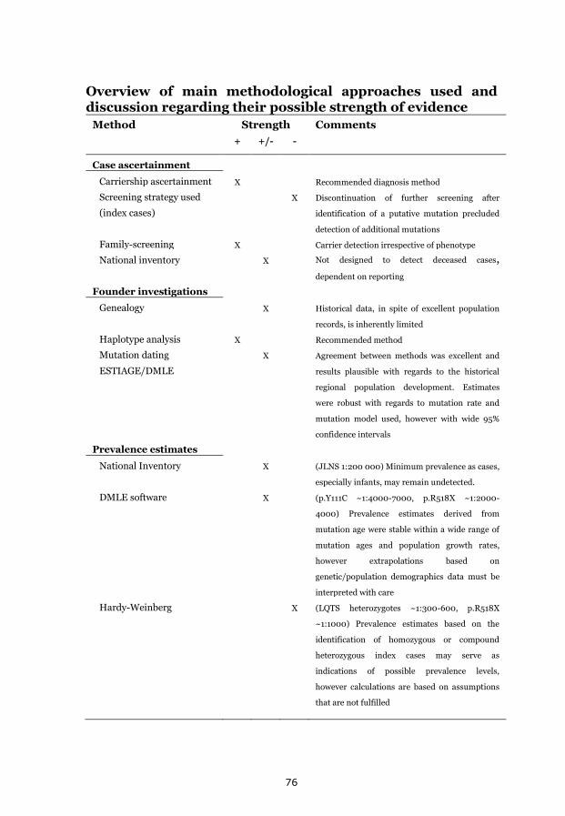
76
Overview of main methodological approaches used and discussion regarding their possible strength of evidence
Method Strength Comments
+ +/- -
Case ascertainment
Carriership ascertainment X Recommended diagnosis method
Screening strategy used
(index cases)
X Discontinuation of further screening after
identification of a putative mutation precluded
detection of additional mutations
Family-screening X Carrier detection irrespective of phenotype
National inventory X Not designed to detect deceased cases,
dependent on reporting
Founder investigations
Genealogy X Historical data, in spite of excellent population
records, is inherently limited
Haplotype analysis X Recommended method
Mutation dating
ESTIAGE/DMLE
X Agreement between methods was excellent and
results plausible with regards to the historical
regional population development. Estimates
were robust with regards to mutation rate and
mutation model used, however with wide 95%
confidence intervals
Prevalence estimates
National Inventory X (JLNS 1:200 000) Minimum prevalence as cases,
especially infants, may remain undetected.
DMLE software X (p.Y111C ~1:4000-7000, p.R518X ~1:2000-
4000) Prevalence estimates derived from
mutation age were stable within a wide range of
mutation ages and population growth rates,
however extrapolations based on
genetic/population demographics data must be
interpreted with care
Hardy-Weinberg X (LQTS heterozygotes ~1:300-600, p.R518X
~1:1000) Prevalence estimates based on the
identification of homozygous or compound
heterozygous index cases may serve as
indications of possible prevalence levels,
however calculations are based on assumptions
that are not fulfilled

77
Overview continued
Method Strength Comments
+ +/- -
Clinical severity
Pedigree mortality
Comparison between
populations
X
X
Historical data, in spite of excellent population
records, is inherently limited
Findings must be interpreted within the context of
the obvious limitations related to comparisons
based on summarized data published by other
authors, including variations in methodology used
Modifier genes
Sequence variant
analysis
X Detection methods were appropriate and the
frequency of analysed sequence variants generally
high (>0.1) and variable
Association analysis X The study was not powered to detect effects on QTc
≤10 ms (limited sample size). The possibility to
detect an association with cardiac events was
limited by the highly asymptomatic phenotype and
prophylactic beta-blocker therapy
Correction for
relatedness
(Kinship matrix)
X Genealogical interrelations among the p.R518X
founder heterozygotes were largely well established
and significant findings could be adjusted in order
to limit the risk of type 1 errors secondary to
relatedness
Significance of 3’UTR-
KCNQ1 variants in cis
X A lower expression of the KCNQ1 gene when the
derived variants rs2519184 (A), rs8234 (G) and
rs10798 (G) occur in cis has been demonstrated by
Luciferase reporter assays in vitro.63 However, the
significance of these variants, found to segregate
with the p.R518X mutation in the founder
population, could not be formally assessed.

78
Clinical management of LQTS within a temporal context In a historical perspective, the ability to detect and diagnose the LQTS
variants has had a major impact on the interpretation of disease occurrence
and associated phenotypes. In accordance with the bias towards detection of
more severe manifestations, it cannot be seen as a coincidence that the JLNS
form, though far less common, was the first to be described.108
With the introduction of molecular genetic testing, including pre-
symptomatic cascade-screening of family members, the view of LQTS has
changed from that of a rare disorder estimated to affect ~1: 20 000 with a
untreated annual mortality of 1o%, to a more common disorder found in
1: 100089-500024 associated with an overall annual mortality of ~1%109 as
well as a marked variability regarding penetrance.50 The typical LQTS
patient has concomitantly changed from a highly symptomatic individual
with a markedly prolonged QTc to an often asymptomatic mutation-carrier
with a borderline QTc.
These changes constitute new and substantial challenges to the clinical
management of an increasingly asymptomatic patient group. Reliable
predictions regarding which mutation-carriers are at risk of life-threatening
cardiac events remain of utmost clinical importance, not only to prevent
LQTS-related morbidity and mortality- but also in order to minimize adverse
effects due to unnecessary treatment.
Unfortunately, while we have some tools to identify high risk individuals
(JLNS phenotype, compound heterozygosity, QTc >500 ms, previous
syncope or aborted cardiac arrest, early age at debut and possibly also risk-
conferring sequence variants)23,62,63,73,77,99 the negative predictive value of
these markers remain unsatisfactory.
However, what we do know is that beta-blocker pharmacotherapy
constitutes a highly effective prophylactic treatment in LQT1, with the caveat
that treatment efficacy is dependent on compliance.110
With regards to JLNS, being the most severe form of the disease, risk
stratification typically presents no difficulties (JLNS= high risk).
Consequently, an “all in” strategy, including complemental treatments such
as LSCD and/or ICD is more often warranted in the management of JLNS.
However, the rarity of the disorder limits the availability of validated data
regarding current treatment strategies and associated outcomes, whereby
prospective studies are needed (if nothing else to attenuate the bleak
statistics regarding reported outcome in JLNS).

79
When LQTS mutation prevalence in the general population, as in parts of
Scandinavia, comes within the range of 1%- 1‰86,87 these mutations
constitute a population prevalent substrate for the development of triggered
arrhythmia. As endogenous and/or environmental factors such as increased
sympathetic activation, hypokalemia and/or commonly prescribed QT
prolonging drugs may further challenge the repolarization reserve51 in
genetically susceptible individuals, possibly life-threatening arrhythmias
may suddenly be triggered in previously “healthy” individuals. In this
context, pre-symptomatic identification of mutation-carriers via molecular
genetics methods, detailed patient information (including protective
measures and life-style adaptations) and prophylactic treatment of identified
mutation-carriers currently remains the best strategy to prevent sudden
cardiac death from LQTS.
Direct clinical implication of the studies in the thesis
As a result of the studies included in the thesis (and foremost of course the
underlying clinical practice), the patient management of LQTS index families
in Sweden carrying the identified founder mutations has changed to include
mutation-specific information. While a mutation-specific differentiation
regarding patient management would be ideal, this still lies in the future.
Transiently, the findings described also affected the screening strategies at
the then primary LQTS diagnostics laboratory in the country.
The realization that LQTS mutation-carriership probably is relatively
common in the population emphasizes the need for LQTS Family Clinics,
such as the one in Umeå, to handle diagnostics, family screening and patient
management. With the realization that LQTS is not a “simple” Mendelian
disorder, the need for a close collaboration between clinicians (pediatric and
“adult” cardiologists) and clinical geneticists becomes the more evident.
With regards to the national inventory of JLNS cases, the interviews with the
Swedish JLNS families generated clinical data extending beyond the
Surdocardiac Syndrome. Based on what is known about the diversity of the
KCNQ1 gene and the Kv7.1 channel30 it should not have surprised us (as it
did) that our observations led to the first report linking the iron-deficiency
anemia often reported in JLNS cases70,108,111,112 with Kv7.1 dysfunction in the
gastrointestinal tract, and specifically to loss of gastric acid secretion.113
These findings and other as yet unpublished observations regarding the
JLNS families jointly expand on the genotype-phenotype correlations
associated with severe KCNQ1 function loss.

80
In Summary
Thesis Aims Results To describe the occurrence
of p.Y111C and p.R518X
and the prevalence of
JLNS Sweden
We identified 170 p.Y111C mutation-carriers,
in 37 index families. p.Y111C prevalence was
estimated to 1:4000-7000 (by DMLE).
We identified 101 p.R518X mutation-carriers
in 19 index families. p.R518X prevalence was
estimated to between 1:2000-4000 (by DMLE)
and 1:1000 (Hardy-Weinberg).
We identified 19 JLNS cases in 13 families and
reported an absolute prevalence of >1:2oo ooo
in children below 10 years of age.
To investigate the possible
founder nature of p.Y111C
and p.R518X in Sweden,
as well as the geographic
origin of mutations
identified in ≥ 2 alleles in
Swedish JLNS probands
p.Y111C and p.R518X are Swedish founder
mutations, by congruent genealogic,
geographic and haplotype evidence. Mutation
age was estimated to ~600 years (p.Y111C) and
~700 years (p.R518X, by ESTIAGE).
Geographical clustering of p.Y111C and
p.R518X ancestors in two northern river valley
regions was shown. JLNS alleles p.Q530X and
c.572_576del showed localization towards the
Norwegian border.
To investigate the cardiac
phenotype associated with
p.Y111C, p.R518X and
JLNS, with a special focus
on life-threatening cardiac
events
A benign phenotype was associated with
heterozygous carriership of p.Y111C and
p.R518X alleles, with a low incidence rate of
life-threatening cardiac events before therapy
(0.05 and 0.04%/year, respectively).
A severe phenotype was seen in JLNS cases
with an annual incidence of life-threatening
cardiac events before therapy of 5%, and a
cumulative incidence of 53%.
To evaluate the possible
association between
previously published
genetic modifiers and
phenotype in p.R518X
heterozygotes sharing a
common founder
Intra-familial variability was partly explained
as two NOS1AP derived variants were
associated with a 29 ms increase in QTc in
males, explaining 27% of the male QTc
variance. Three repressive 3’UTR-KCNQ1
derived variants segregated with the mutation
in the p.R518X founder population, possibly
explaining the benign phenotype seen.
81
Conclusions
We have established that carriership of LQTS mutations (and specifically the
p.Y111C and p.R518X alleles) is more commonly occurring in the Swedish
population than previously thought. Accordingly, the prevalence of JLNS in
Sweden is among the highest reported worldwide. This finding motivates an
increased awareness among clinicians (on the regional and national level)
regarding LQTS (and JLNS) as possible differential diagnoses, with regards
to unexplained syncope or seizures.
By aggregation of clinical data from the p.Y111C and p.R518X populations,
mutation-specific characterization of these alleles have been accomplished,
enabling mutation-specific risk stratification and patient information in
p.Y111C and p.R518X families. The spectrum of clinical manifestations of the
p.R518X heterozygotes additionally adds to the increasing awareness
regarding the occurrence of atypical/weak phenotypes in LQTS.
Regarding the p.Y111C mutation, our findings confirmed that the clinical
phenotype in LQTS does not necessarily correlate with in vitro studies
predicting ion-channel function, and that additional elements are needed to
perform valid genotype-phenotype correlations.
Regarding modifier genes, we have presented a possible explanation to how
sequence variants located within a shared haplotype (here in the 3’UTR of
the KCNQ1 gene) could contribute to an overall benign phenotype in a
founder population. Moreover, we have presented data in support of gender-
specific effects of modifying genes, specifically derived sequence variants in
the NOS1AP gene, warranting further research.
Finally, in the context of founder effects and associated cardiac phenotypes,
we have demonstrated that even in two homogeneous founder populations
sharing mutations identical by descent, substantial phenotypic variability
remains, not readily explainable by factors such as age, gender and/or
previously identified modifying genes.
While the mechanisms underlying the phenotypic variability in LQTS remain
elusive, mutation-carrier identification using the cascade-screening
approach remains imperative in order to identify individuals at risk of
environmentally induced life-threatening arrhythmia, thereby enabling
prophylactic measures and treatment of identified mutation-carriers.
82
Where do we go from here? In the context of population prevalent LQTS mutations and a relatively high
probability of JLNS conceptions, further research in this field is warranted.
Due to the rarity of most mutations, as well as the JLNS genotype,
international collaborations are necessary in order to compile
comprehensive data sets, and to further elucidate the mechanisms
underlying the development of life-threatening arrhythmia in some LQTS
mutation-carriers.
In the clinical setting collaboration across different clinical specialties
constitutes a prerequisite for adequate patient management.
Among questions I would want to see investigated, I would currently name:
Can we further explain the phenotypic variability in LQTS?
Can we identify better predictors of arrhythmia risk, and specifically
reliable predictors of significant risk reduction?
Can we better determine the clinical significance of modifying
sequence variants in LQTS? (single- /combinations of variants)
Can we clarify the relationship between gender and arrhythmia risk?
(over time, within and between genders, also including possible
associations with genetic modifiers)
Can we further relate the clinical phenotype to different levels of
Kv7.1 dysfunction? (JLNS/compound heterozygosity/LQTS)
Does severe Kv7.1 dysfunction relate to further extracardiac
manifestations?
Can we produce validated data on treatment strategies for JLNS?
Regarding the Swedish p.Y111C founder mutation:
o Why are there so few p.Y111C alleles among the JLNS cases?
o Is the occurrence of p.Y111C in Northern America due to
emigrant founder effects?
o What is the role of genetic modifiers on p.Y111C founder
phenotype?
And so on…

83
Contribution statement Paper Main contributor(s)
Initiating overall LQTS research AR, ELS, SJ
Patient recruitment, main data collection I-III AR, ELS, SJ, UBD
IV AR, AW, HW
Data collection, complemental I-II AW
III AW, KE
Overall study design I-III AR, AW, ELS, SJ
IV AR, AW
Interviews, analysis of symptoms data I-IV AR, AW
ECG measurements and analyses I-IV AW
Genealogic and geographic data I-II UBD
III CN
Analysis of geographical data I-IV AW
Pedigree mortality data and analysis I AW
Design, haplotype analysis II, III AW, ELS
Laboratory work, haplotype analysis II AW
III SH
Data analysis, haplotype analysis II, III AW
Design, sequence variants III AN, ELS, JJ, KC
Laboratory work, sequence variants III CA, CB, EE
Data analysis, sequence variants III AW
Statistical design and analyses I-IV AW, JP
Design of Figures I-IV AW, EW
First draft of manuscripts I-IV AW
Manuscript design and approval I-IV All co-authors
Correspondence with scientific journals I-IV AW
AR- Annika Rydberg1, AW- Annika Winbo1, AN- Anna Norberg2, CA- Charlotte Andersson2, CB- Christina
Bergsdorf2, CN- Charlotte Nordin1, EE- Elin Ek2, ELS- Eva-Lena Stattin2, EW- Erik Winbo*, HW- Hans
Wendel**, JJ- Jenni Jonasson2, JP- Johan Persson***, KE- Katarina Englund3, KC- Kristina Cederqvist2, SH-
Susann Haraldsson2, SJ- Steen Jensen3, UBD- Ulla-Britt Diamant3
1Department of Clinical Sciences, Pediatrics, 2Department of Medical Biosciences, Medical and Clinical
Genetics, 3Department of Public Health and Clinical Medicine, Heart Centre, all at Umeå University, 90187
Umeå, Sweden, *Illustrator, erikwinbo.artworkfolio.com, **Former Pediatric Cardiologist, Pediatric Cardiology
Clinic, Umeå University Hospital, 1967-1991***Consultant in Mathematics and Computer Science

84
Acknowledgements
During the years that led up to the completion of this thesis, I hold the
continuous presence and support of three individuals as the main
explanation of the fact that it has been foremost a lust-driven process on my
part; namely: my supervisor Annika Rydberg, my PhD student colleague
Anna Myléus, and my husband Erik Winbo.
I hereby want to express my sincerest gratitude:
To my supervisor Annika, for throughout the PhD process making me feel
like a colleague, while never shirking her responsibilities as a supervisor. For
gradually relinquishing control and transferring responsibility while
remaining present. For mildly reining in my at times excessive enthusiasm.
For introducing me into the intriguing world of clinical research (and
researchers) and never losing track of the things that really matter. For
always making it seem like she had time for my questions (although she
might at the time be loading a bull fiddle onto a train set or wheeling in her
fishing nets). For understanding, advocating and demonstrating, that a
balance between research, clinical practice, family life and extracurricular
activities makes us better at all that we do. For an extraordinary research
partnership characterized by curiosity and the identification of possibilities
rather than limitations.
To my PhD student colleague Anna, for the endless hours over the phone
and/or over glasses of wine, revising manuscripts and discussing the ins and
outs of study design, presentation, and reflecting over the overall experience
of being a PhD student. No detail, no success or failure was too small or
insignificant to warrant lengthy discussions in our forum of two, not only
contributing to the preservation of our mutual sanity, but probably sparing
that of our immediate surroundings as well…
To my husband, companion and best friend Erik, not primarily for his
fantastic illustrations (and his apparent limitless patience with my calls for
alterations), but for his discrete communication of incessant support via the
fresh coffee thermos appearing by my computer each morning, and for his
stable presence keeping the family on track when I was otherwise (physically
or mentally) engaged.
Also, I want to acknowledge the invaluable support of my extended family
and friends, throughout successes and setbacks related to the PhD process.
85
As this thesis primarily is the result of the joint efforts of our LQTS research
group, as well as that of several co-workers, I want to thank:
The LQTS research group for a challenging, educational and ultimately
rewarding collaboration; in addition to Annika Rydberg, specifically:
Steen M Jensen for taking on the role as co-supervisor of this headstrong
PhD student, at the moment when it was most needed
Eva-Lena Stattin for introducing me to the intriguing field of molecular
genetics and for a consistently rapid and valuable feedback
Ulla-Britt Diamant for patiently teaching me all there is to know about the
elusive end of the T-wave, for opening up the doors to the past through an
obvious passion for genealogical research, as well as for adding a well needed
shot of “norrländsk-down-to-earth realism” to our research team
Charlotte Nordin for her flexibility and methodical dedication to the
sometimes challenging genealogical investigation into the p.R518X families
Anna Norberg for a truly inspirational, effective and enjoyable cooperation
regarding genetic modifiers
Johan Persson for venturing into the obscure field of founder effect
statistics with us, for applying his innovative effort and systematic approach
to elevating the quality of our analyses, and for taking the time to make sure
we understood what we were doing
Illustrator Erik Winbo (erikwinbo.artworkfolio.com) for providing original
artwork (figures, maps, pedigrees etc.) that elevated the data and the thesis
For expert technical assistance: Charlotte Andersson, Christina
Bergsdorf, Elin Ek, Jenni Jonasson, Kristina Cederqvist and
Susann Haraldsson at the Department of Medical Biosciences, Medical
and Clinical Genetics; and Katarina Engqvist at the Centre for
Cardiovascular Genetics, Heart Centre, all at Umeå University, Sweden.
Furthermore:
I want to thank Barbro, Ina, Mona, Jenny, Antti and Dag at the
Pediatric Cardiology Clinic in Umeå, (together with Annika) for providing
me with a “home away from home” during these years.
For all their invaluable help with assorted practical issues related to the fine
art of juggling a PhD thesis, a medical internship and parenthood I also want
to thank: Karin Moström, Administrator, Pediatrics; Ulla M Norrman,
Economic Administrator, Pediatrics and Margareta Backman (Maggan),
Intern-Recruiter for the Northern County Council, all at Umeå University
Hospital, Umeå, Sweden.

86
For support during the latter part of my PhD student process, I specifically
thank Olle Hernell at the Institution of Pediatrics, Umeå University, as
well as Irina Golovleva at the Institution of Medical and Clinical Genetics,
Umeå University, Stellan Mörner at the Institution of Medicine, Umeå
University, and Thomas Gilljam at the Institution of Medicine, University
of Gothenburg, for their constructive critique during my mid-term seminar.
For their support and contribution of clinical cases I thank: Håkan
Eliasson, Pediatric Cardiologist, and Anna-Karin Hammarstedt,
research nurse, at Queen Silvias Hospital for Sick Children, Stockholm,
Sweden, and Margit Qvist, research nurse, and her colleagues at the ICD-
clinic at Örebro Hospital, Örebro, Sweden.
For her valuable input and advice with regards to mutation dating and for
gracefully providing us with her ESTIAGE software, I thank Dr
Emmanuelle Genin, Université Paris Diderot, Villejuif, France.
I thank also Professor Arthur Wilde, Department of Cardiology and Heart
Failure Research Center, Academic Medical Center, University of
Amsterdam, Amsterdam, the Netherlands for his instigative support.
For providing the scaffolding within which the research encompassed by this
thesis was conducted I acknowledge the Centre for Cardiovascular
Genetics, Umeå University Hospital and the Institution of Pediatrics,
Department of Clinical Sciences, Umeå University, both in Umeå,
Sweden.
For funding throughout the years leading up to the completion of this thesis,
I primarily want to thank the Swedish Heart-Lung Foundation, and
also acknowledge the support of the Northern County Councils
Cooperation Committee as well as the Heart Foundation of
Northern Sweden.
Lastly, I want to thank all cases and families that we came in contact with
during the course of the studies, for their participation, contribution and
confidence. Ultimately, they constitute the reason for performing this
research.

87
References
1. Einarsdottir E, Egerbladh I, Beckman L, Holmberg D, Escher SA. The genetic population structure of northern Sweden and its implications for mapping genetic diseases. Hereditas 2007;144:171-80.
2. Bittles AH, Egerbladh I. The influence of past endogamy and
consanguinity on genetic disorders in northern Sweden. Annals of Human Genetics 2005;69:549-58.
3. Burstedt MS, Sandgren O, Holmgren G, Forsman-Semb K. Bothnia dystrophy caused by mutations in the cellular retinaldehyde-binding protein gene (RLBP1) on chromosome 15q26. Invest Ophthalmol Vis Sci 1999;40:995-1000.
4. Holmgren G, Costa PM, Andersson C, et al. Geographical distribution of TTR met30 carriers in northern Sweden: discrepancy between carrier frequency and prevalence rate. J Med Genet 1994;31:351-4.
5. Iselius L, Gustavson KH. Spatial distribution of the gene for
infantile genetic agranulocytosis. Hum Hered 1984;34:358-63.
6. Jagell S, Gustavson KH, Holmgren G. Sjogren-Larsson syndrome in Sweden. A clinical, genetic and epidemiological study. Clin Genet 1981;19:233-56.
7. Floderus Y, Shoolingin-Jordan PM, Harper P. Acute intermittent porphyria in Sweden. Molecular, functional and clinical consequences of some new mutations found in the porphobilinogen deaminase gene. Clin Genet 2002;62:288-97.
8. Romano C. Congenital Cardiac Arrhythmia. Lancet
1965;1:658-9.
9. Ward OC. A New Familial Cardiac Syndrome in Children. J Ir Med Assoc 1964;54:103-6.
10. Jervell A, Lange-Nielsen F. Congenital deaf-mutism, functional heart disease with prolongation of the Q-T interval and sudden death. American Heart Journal 1957;54:59-68.
11. Schwartz PJ, Stramba-Badiale M, Crotti L, et al. Prevalence of
the congenital long-QT syndrome. Circulation 2009;120:1761-7.

88
12. Ackerman MJ, Priori SG, Willems S, et al. HRS/EHRA expert consensus statement on the state of genetic testing for the channelopathies and cardiomyopathies this document was developed as a partnership between the Heart Rhythm Society (HRS) and the European Heart Rhythm Association (EHRA). Heart Rhythm 2011;8:1308-39.
13. Fridericia LS. The duration of systole in an electrocardiogram
in normal humans and in patients with heart disease. 1920. Ann Noninvasive Electrocardiol 2003;8:343-51.
14. Sagie A, Larson MG, Goldberg RJ, Bengtson JR, Levy D. An improved method for adjusting the QT interval for heart rate (the Framingham Heart Study). Am J Cardiol 1992;70:797-801.
15. Bazett HC. The time relations of the blood-pressure changes after excision of the adrenal glands, with some observations on blood volume changes. J Physiol 1920;53:320-39.
16. Goldenberg I, Moss AJ, Zareba W. QT interval: how to
measure it and what is "normal". J Cardiovasc Electrophysiol 2006;17:333-6.
17. Schwartz PJ, Moss AJ, Vincent GM, Crampton RS. Diagnostic criteria for the long QT syndrome. An update. Circulation 1993;88:782-4.
18. Schwartz PJ. Pharmacological and non-pharmacological management of the congenital long QT syndrome: the rationale. Pharmacol Ther 2011;131:171-7.
19. Curran ME, Splawski I, Timothy KW, Vincent GM, Green ED,
Keating MT. A molecular basis for cardiac arrhythmia: HERG mutations cause long QT syndrome. Cell 1995;80:795-803.
20. Wang Q, Curran ME, Splawski I, et al. Positional cloning of a novel potassium channel gene: KVLQT1 mutations cause cardiac arrhythmias. Nat Genet 1996;12:17-23.
21. Wang Q, Shen J, Splawski I, et al. SCN5A mutations associated with an inherited cardiac arrhythmia, long QT syndrome. Cell 1995;80:805-11.
22. Schulze-Bahr E. Long QT Syndromes: Genetic Basis. Cardiac
Electrophysiology Clinics 2012;4:1-16.
89
23. Schwartz PJ, Spazzolini C, Crotti L, et al. The Jervell and Lange-Nielsen syndrome: natural history, molecular basis, and clinical outcome. Circulation 2006;113:783-90.
24. Hofman N, Wilde AA, Kaab S, et al. Diagnostic criteria for
congenital long QT syndrome in the era of molecular genetics: do we need a scoring system? Eur Heart J 2007;28:575-80.
25. Hofman N, Tan HL, Alders M, van Langen IM, Wilde AA. Active cascade screening in primary inherited arrhythmia syndromes: does it lead to prophylactic treatment? J Am Coll Cardiol 2010;55:2570-6.
26. Napolitano C, Priori SG, Schwartz PJ, et al. Genetic testing in the long QT syndrome: development and validation of an efficient approach to genotyping in clinical practice. JAMA 2005;294:2975-80.
27. Tester DJ, Ackerman MJ. Genetic testing for potentially
lethal, highly treatable inherited cardiomyopathies/channelopathies in clinical practice. Circulation 2011;123:1021-37.
28. Lee MP, Hu RJ, Johnson LA, Feinberg AP. Human KVLQT1 gene shows tissue-specific imprinting and encompasses Beckwith-Wiedemann syndrome chromosomal rearrangements. Nat Genet 1997;15:181-5.
29. Splawski I, Shen J, Timothy KW, Vincent GM, Lehmann MH, Keating MT. Genomic structure of three long QT syndrome genes: KVLQT1, HERG, and KCNE1. Genomics 1998;51:86-97.
30. Jespersen T, Grunnet M, Olesen SP. The KCNQ1 potassium
channel: from gene to physiological function. Physiology (Bethesda) 2005;20:408-16.
31. Yang WP, Levesque PC, Little WA, Conder ML, Shalaby FY, Blanar MA. KvLQT1, a voltage-gated potassium channel responsible for human cardiac arrhythmias. Proc Natl Acad Sci U S A 1997;94:4017-21.
32. Barhanin J, Lesage F, Guillemare E, Fink M, Lazdunski M, Romey G. K(V)LQT1 and lsK (minK) proteins associate to form the I(Ks) cardiac potassium current. Nature 1996;384:78-80.
33. Sanguinetti MC, Curran ME, Zou A, et al. Coassembly of
K(V)LQT1 and minK (IsK) proteins to form cardiac I(Ks) potassium channel. Nature 1996;384:80-3.
90
34. Kramer DB, Zimetbaum PJ. Long-QT syndrome. Cardiol Rev 2011;19:217-25.
35. Akar FG, Yan GX, Antzelevitch C, Rosenbaum DS. Unique
topographical distribution of M cells underlies reentrant mechanism of torsade de pointes in the long-QT syndrome. Circulation 2002;105:1247-53.
36. Di Diego JM, Belardinelli L, Antzelevitch C. Cisapride-induced transmural dispersion of repolarization and torsade de pointes in the canine left ventricular wedge preparation during epicardial stimulation. Circulation 2003;108:1027-33.
37. Roden DM. Repolarization reserve: a moving target. Circulation 2008;118:981-2.
38. Roden DM, Abraham RL. Refining repolarization reserve.
Heart Rhythm 2011;8:1756-7.
39. Schwartz PJ, Priori SG, Spazzolini C, et al. Genotype-phenotype correlation in the long-QT syndrome: gene-specific triggers for life-threatening arrhythmias. Circulation 2001;103:89-95.
40. Neyroud N, Tesson F, Denjoy I, et al. A novel mutation in the potassium channel gene KVLQT1 causes the Jervell and Lange-Nielsen cardioauditory syndrome. Nat Genet 1997;15:186-9.
41. Tyson J, Tranebjaerg L, Bellman S, et al. IsK and KvLQT1:
mutation in either of the two subunits of the slow component of the delayed rectifier potassium channel can cause Jervell and Lange-Nielsen syndrome. Hum Mol Genet 1997;6:2179-85.
42. Schulze-Bahr E, Wang Q, Wedekind H, et al. KCNE1 mutations cause jervell and Lange-Nielsen syndrome. Nat Genet 1997;17:267-8.
43. Casimiro MC, Knollmann BC, Ebert SN, et al. Targeted disruption of the Kcnq1 gene produces a mouse model of Jervell and Lange-Nielsen Syndrome. Proc Natl Acad Sci U S A 2001;98:2526-31.
44. Lee MP, Ravenel JD, Hu RJ, et al. Targeted disruption of the
Kvlqt1 gene causes deafness and gastric hyperplasia in mice. Journal of Clinical Investigation 2000;106:1447-55.
45. Vetter DE, Mann JR, Wangemann P, et al. Inner ear defects induced by null mutation of the isk gene. Neuron 1996;17:1251-64.

91
46. Rivas A, Francis HW. Inner ear abnormalities in a Kcnq1 (Kvlqt1) knockout mouse: a model of Jervell and Lange-Nielsen syndrome. Otol Neurotol 2005;26:415-24.
47. Friedmann I, Fraser GR, Froggatt P. Pathology of the ear in
the cardioauditory syndrome of Jervell and Lange-Nielsen (recessive deafness with electrocardiographic abnormalities). J Laryngol Otol 1966;80:451-70.
48. Moss AJ, Shimizu W, Wilde AA, et al. Clinical aspects of type-1 long-QT syndrome by location, coding type, and biophysical function of mutations involving the KCNQ1 gene. Circulation 2007;115:2481-9.
49. Bhuiyan ZA, Momenah TS, Amin AS, et al. An intronic mutation leading to incomplete skipping of exon-2 in KCNQ1 rescues hearing in Jervell and Lange-Nielsen syndrome. Prog Biophys Mol Biol 2008;98:319-27.
50. Priori SG, Napolitano C, Schwartz PJ. Low penetrance in the
long-QT syndrome: clinical impact. Circulation 1999;99:529-33.
51. Dipple KM, McCabe ER. Modifier genes convert "simple" Mendelian disorders to complex traits. Mol Genet Metab 2000;71:43-50.
52. Kapplinger JD, Tester DJ, Salisbury BA, et al. Spectrum and prevalence of mutations from the first 2,500 consecutive unrelated patients referred for the FAMILION long QT syndrome genetic test. Heart Rhythm 2009;6:1297-303.
53. Splawski I, Shen J, Timothy KW, et al. Spectrum of mutations
in long-QT syndrome genes. KVLQT1, HERG, SCN5A, KCNE1, and KCNE2. Circulation 2000;102:1178-85.
54. Tester DJ, Will ML, Haglund CM, Ackerman MJ. Compendium of cardiac channel mutations in 541 consecutive unrelated patients referred for long QT syndrome genetic testing. Heart Rhythm 2005;2:507-17.
55. Keating MT, Sanguinetti MC. Molecular and cellular mechanisms of cardiac arrhythmias. Cell 2001;104:569-80.
56. Newton-Cheh C, Guo CY, Larson MG, et al. Common genetic
variation in KCNH2 is associated with QT interval duration: the Framingham Heart Study. Circulation 2007;116:1128-36.
92
57. Gouas L, Nicaud V, Berthet M, et al. Association of KCNQ1, KCNE1, KCNH2 and SCN5A polymorphisms with QTc interval length in a healthy population. Eur J Hum Genet 2005;13:1213-22.
58. Gouas L, Nicaud V, Chaouch S, et al. Confirmation of
associations between ion channel gene SNPs and QTc interval duration in healthy subjects. Eur J Hum Genet 2007;15:974-9.
59. Marjamaa A, Newton-Cheh C, Porthan K, et al. Common candidate gene variants are associated with QT interval duration in the general population. J Intern Med 2009;265:448-58.
60. Arking DE, Pfeufer A, Post W, et al. A common genetic variant in the NOS1 regulator NOS1AP modulates cardiac repolarization. Nat Genet 2006;38:644-51.
61. Crotti L, Lundquist AL, Insolia R, et al. KCNH2-K897T is a
genetic modifier of latent congenital long-QT syndrome. Circulation 2005;112:1251-8.
62. Crotti L, Monti MC, Insolia R, et al. NOS1AP is a genetic modifier of the long-QT syndrome. Circulation 2009;120:1657-63.
63. Amin AS, Giudicessi JR, Tijsen AJ, et al. Variants in the 3' untranslated region of the KCNQ1-encoded Kv7.1 potassium channel modify disease severity in patients with type 1 long QT syndrome in an allele-specific manner. Eur Heart J 2011. doi: 10.1093/eurheartj/ehr473
64. Andersson P, Lundkvist L. The Q-T syndrome--a family
description. Acta Med Scand 1979;206:73-6.
65. Jespersen T, Rasmussen HB, Grunnet M, et al. Basolateral localisation of KCNQ1 potassium channels in MDCK cells: molecular identification of an N-terminal targeting motif. J Cell Sci 2004;117:4517-26.
66. Dahimene S, Alcolea S, Naud P, et al. The N-terminal juxtamembranous domain of KCNQ1 is critical for channel surface expression: implications in the Romano-Ward LQT1 syndrome. Circ Res 2006;99:1076-83.
67. Huang L, Bitner-Glindzicz M, Tranebjaerg L, Tinker A. A
spectrum of functional effects for disease causing mutations in the Jervell and Lange-Nielsen syndrome. Cardiovasc Res 2001;51:670-80.
93
68. Wei J, Fish FA, Myerburg RJ, Roden DM, George AL, Jr. Novel KCNQ1 mutations associated with recessive and dominant congenital long QT syndromes: evidence for variable hearing phenotype associated with R518X. Human Mutation 2000;15:387-8.
69. Larsen LA, Fosdal I, Andersen PS, et al. Recessive Romano-
Ward syndrome associated with compound heterozygosity for two mutations in the KVLQT1 gene. European Journal of Human Genetics 1999;7:724-8.
70. Tranebjaerg L, Bathen J, Tyson J, Bitner-Glindzicz M. Jervell and Lange-Nielsen syndrome: a Norwegian perspective. Am J Med Genet 1999;89:137-46.
71. Tyson J, Tranebjaerg L, McEntagart M, et al. Mutational spectrum in the cardioauditory syndrome of Jervell and Lange-Nielsen. Hum Genet 2000;107:499-503.
72. Berge KE, Haugaa KH, Fruh A, et al. Molecular genetic
analysis of long QT syndrome in Norway indicating a high prevalence of heterozygous mutation carriers. Scandinavian Journal of Clinical and Laboratory Investigation 2008;68:362-8.
73. Goldenberg I, Moss AJ, Zareba W, et al. Clinical course and risk stratification of patients affected with the Jervell and Lange-Nielsen syndrome. J Cardiovasc Electrophysiol 2006;17:1161-8.
74. Slatkin M, Rannala B. Estimating allele age. Annu Rev Genomics Hum Genet 2000;1:225-49.
75. Genin E, Tullio-Pelet A, Begeot F, Lyonnet S, Abel L.
Estimating the age of rare disease mutations: the example of Triple-A syndrome. Journal of Medical Genetics 2004;41:445-9.
76. Mayo O. A century of Hardy-Weinberg equilibrium. Twin Res Hum Genet 2008;11:249-56.
77. Priori SG, Schwartz PJ, Napolitano C, et al. Risk stratification in the long-QT syndrome. N Engl J Med 2003;348:1866-74.
78. Crotti L, Spazzolini C, Schwartz PJ, et al. The common long-
QT syndrome mutation KCNQ1/A341V causes unusually severe clinical manifestations in patients with different ethnic backgrounds: toward a mutation-specific risk stratification. Circulation 2007;116:2366-75.
94
79. Choi Y, Wijsman EM, Weir BS. Case-control association testing in the presence of unknown relationships. Genet Epidemiol 2009;33:668-78.
80. Chouabe C, Neyroud N, Richard P, et al. Novel mutations in
KvLQT1 that affect Iks activation through interactions with Isk. Cardiovasc Res 2000;45:971-80.
81. Gouas L, Bellocq C, Berthet M, et al. New KCNQ1 mutations leading to haploinsufficiency in a general population; Defective trafficking of a KvLQT1 mutant. Cardiovasc Res 2004;63:60-8.
82. Cohort mortality in Sweden. Stockholm: National Central Bureau of Statistics; 1997:93.
83. Westin G, Olofsson SI. Övre Norrlands historia. D. 1, Tiden till
1600 /The History of The Upper Northern Regions. part 1, Time before 1600. Umeå : Norrbottens och Västerbottens läns landsting; 1962.
84. A Norberg KC, J. Jonasson, B-A. Jonsson, A. Rydberg, S. Jensen, E. Stattin. Genetic screening of Long QT syndrome in Sweden. (Abstract/T371). Presented at the 12th International Congress of Human Genetics/61st Annual Meeting of The American Society of Human Genetics, October 12, 2011, Montreal, Canada.
85. Winbo A, Stattin EL, Diamant UB, Persson J, Jensen SM, Rydberg A. Prevalence, mutation spectrum, and cardiac phenotype of the Jervell and Lange-Nielsen syndrome in Sweden. Europace 2012. doi: 10.1093/europace/eus111
86. Fodstad H, Swan H, Laitinen P, et al. Four potassium channel
mutations account for 73% of the genetic spectrum underlying long-QT syndrome (LQTS) and provide evidence for a strong founder effect in Finland. Annals of Medicine 2004;36:53-63.
87. Marjamaa A, Salomaa V, Newton-Cheh C, et al. High prevalence of four long QT syndrome founder mutations in the Finnish population. Annals of Medicine 2009;41:234-40.
88. Brink PA, Crotti L, Corfield V, et al. Phenotypic variability and unusual clinical severity of congenital long-QT syndrome in a founder population. Circulation 2005;112:2602-10.

95
89. Ackerman MJ, Tester DJ, Jones GS, Will ML, Burrow CR, Curran ME. Ethnic differences in cardiac potassium channel variants: implications for genetic susceptibility to sudden cardiac death and genetic testing for congenital long QT syndrome. Mayo Clin Proc 2003;78:1479-87.
90. Winbo A, Diamant UB, Rydberg A, Persson J, Jensen SM,
Stattin EL. Origin of the Swedish long QT syndrome Y111C/KCNQ1 founder mutation. Heart Rhythm 2011;8:541-7.
91. Winbo A, Diamant UB, Stattin EL, Jensen SM, Rydberg A. Low incidence of sudden cardiac death in a Swedish Y111C type 1 long-QT syndrome population. Circ Cardiovasc Genet 2009;2:558-64.
92. Wilde AA, Escande D. LQT genotype-phenotype relationships: patients and patches. Cardiovasc Res 2001;51:627-9.
93. Schwartz PJ, Priori SG, Napolitano C. How really rare are rare
diseases?: the intriguing case of independent compound mutations in the long QT syndrome. J Cardiovasc Electrophysiol 2003;14:1120-1.
94. Westenskow P, Splawski I, Timothy KW, Keating MT, Sanguinetti MC. Compound mutations: a common cause of severe long-QT syndrome. Circulation 2004;109:1834-41.
95. Newton-Cheh C, Larson MG, Corey DC, et al. QT interval is a heritable quantitative trait with evidence of linkage to chromosome 3 in a genome-wide linkage analysis: The Framingham Heart Study. Heart Rhythm 2005;2:277-84.
96. Dipple KM, McCabe ER. Phenotypes of patients with "simple"
Mendelian disorders are complex traits: thresholds, modifiers, and systems dynamics. Am J Hum Genet 2000;66:1729-35.
97. Schwartz PJ. Sudden cardiac death, founder populations, and mushrooms: what is the link with gold mines and modifier genes? Heart Rhythm 2011;8:548-50.
98. Schwartz PJ, Vanoli E, Crotti L, et al. Neural control of heart rate is an arrhythmia risk modifier in long QT syndrome. J Am Coll Cardiol 2008;51:920-9.
99. Tomas M, Napolitano C, De Giuli L, et al. Polymorphisms in
the NOS1AP gene modulate QT interval duration and risk of arrhythmias in the long QT syndrome. J Am Coll Cardiol 2010;55:2745-52.

96
100. Eijgelsheim M, Aarnoudse AL, Rivadeneira F, et al. Identification of a common variant at the NOS1AP locus strongly associated to QT-interval duration. Hum Mol Genet 2009;18:347-57.
101. Nolte IM, Wallace C, Newhouse SJ, et al. Common genetic
variation near the phospholamban gene is associated with cardiac repolarisation: meta-analysis of three genome-wide association studies. PLoS One 2009;4:e6138.
102. Lahtinen AM, Marjamaa A, Swan H, Kontula K. KCNE1 D85N polymorphism--a sex-specific modifier in type 1 long QT syndrome? BMC Med Genet 2011;12:11.
103. Pfeufer A, Jalilzadeh S, Perz S, et al. Common variants in myocardial ion channel genes modify the QT interval in the general population: results from the KORA study. Circ Res 2005;96:693-701.
104. Jons C, Moss AJ, Goldenberg I, et al. Risk of fatal arrhythmic
events in long QT syndrome patients after syncope. J Am Coll Cardiol 2010;55:783-8.
105. Sauer AJ, Moss AJ, McNitt S, et al. Long QT syndrome in adults. J Am Coll Cardiol 2007;49:329-37.
106. Schwartz PJ, Locati E. The idiopathic long QT syndrome: pathogenetic mechanisms and therapy. Eur Heart J 1985;6 Suppl D:103-14.
107. MacCormick JM, Crawford JR, Chung SK, et al. Symptoms
and signs associated with syncope in young people with primary cardiac arrhythmias. Heart Lung Circ 2011;20:593-8.
108. Jervell A, Lange-Nielsen F. Congenital deaf-mutism, functional heart disease with prolongation of the Q-T interval and sudden death. Am Heart J 1957;54:59-68.
109. Gussak I. Electrical diseases of the heart: genetics, mechanisms, treatment, prevention. London: Springer; 2008.
110. Vincent GM, Schwartz PJ, Denjoy I, et al. High efficacy of
beta-blockers in long-QT syndrome type 1: contribution of noncompliance and QT-prolonging drugs to the occurrence of beta-blocker treatment "failures". Circulation 2009;119:215-21.
97
111. Fraser GR, Froggatt P, Murphy T. Genetical Aspects of the Cardio-Auditory Syndrome of Jervell and Lange-Nielsen (Congenital Deafness and Electrocardiographic Abnormalities). Ann Hum Genet 1964;28:133-57.
112. Langslet A, Sorland SJ. Surdocardiac syndrome of Jervell and
Lange-Nielsen, with prolonged QT interval present at birth, and severe anaemia and syncopal attacks in childhood. Br Heart J 1975;37:830-2.
113. Winbo A, Sandstrom O, Palmqvist R, Rydberg A. Iron-deficiency anaemia, gastric hyperplasia, and elevated gastrin levels due to potassium channel dysfunction in the Jervell and Lange-Nielsen Syndrome. Cardiol Young 2012:1-10. doi: 10.1017/S1047951112001060

98
1
Appendices
I. LQTS Questionnaire, page 1 and 2 (Swedish)
II. LQTS Questionnaire, translated

2
LQTS Questionnaire, page 1

3
LQTS Questionnaire, page 2
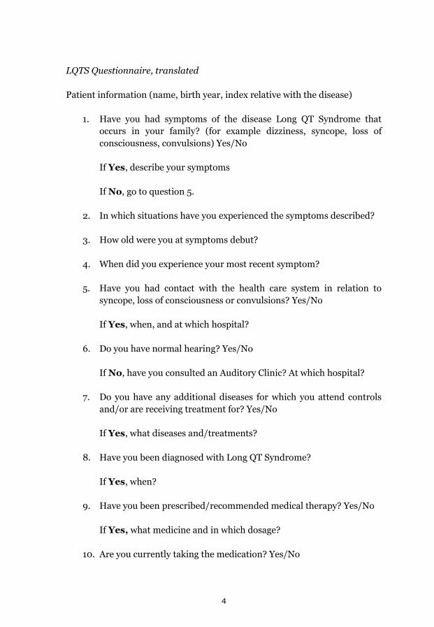
4
LQTS Questionnaire, translated
Patient information (name, birth year, index relative with the disease)
1. Have you had symptoms of the disease Long QT Syndrome that
occurs in your family? (for example dizziness, syncope, loss of
consciousness, convulsions) Yes/No
If Yes, describe your symptoms
If No, go to question 5.
2. In which situations have you experienced the symptoms described?
3. How old were you at symptoms debut?
4. When did you experience your most recent symptom?
5. Have you had contact with the health care system in relation to
syncope, loss of consciousness or convulsions? Yes/No
If Yes, when, and at which hospital?
6. Do you have normal hearing? Yes/No
If No, have you consulted an Auditory Clinic? At which hospital?
7. Do you have any additional diseases for which you attend controls
and/or are receiving treatment for? Yes/No
If Yes, what diseases and/treatments?
8. Have you been diagnosed with Long QT Syndrome?
If Yes, when?
9. Have you been prescribed/recommended medical therapy? Yes/No
If Yes, what medicine and in which dosage?
10. Are you currently taking the medication? Yes/No

5
If Yes, since when are you taking the medication? Are you taking the
prescribed dosage?
If No, why are you not taking the medication?
11. When taking the medication, do you experience any adverse effects?
Yes/No
If Yes, describe the adverse effects
12. Is there anything you would like to add with regards to the disease,
symptoms, controls, medication or other aspects?