Login and Self-Study of the Autonomic Nervous Systembolger/ced/autonomic/ANSlessn.pdf · Login and...
34
Login and Self-Study of the Autonomic Nervous System Student Information: First Name: Last Name: E-mail: Student ID#: Please fill out the information requested above and then complete the lesson below. READ all sections carefully and test yourself by trying to guess the correct fill-in prior to checking. Self-assessment questions: A PROGRAMMED INTRODUCTION TO AUTONOMIC PHARMACOLOGY by Nora Laiken, Ph.D., UCSD Adapted for the Web and USC School of Pharmacy Therapeutics III by Michael B. Bolger, Ph.D. A firm understanding of the therapeutics of the autonomic nervous system the primary goal of the Therapeutics III module. Once the fundamentals of autonomic pharmacology are mastered, many previously unfamiliar concepts in physiology and clinical pharmacy will begin to make sense. At a first glance, autonomic pharmacology appears to be a mass of unrelated material to be memorized. However, the opposite is in fact true, as there is a definite conceptual framework from which much of the subject logically follows. The purpose of this handout is to present this framework concisely, in a step-by-step fashion. Once this framework is mastered, you will be able to do further reading in autonomic pharmacology without difficulty. In addition, you will have the background necessary to benefit maximally from the lectures, discussions, and laboratories on autonomic pharmacology in Therapeutics. Page 1 of 34 A PROGRAMMED INTRODUCTION TO AUTONOMIC PHARMACOLOGY 8/24/2004 http://www.usc.edu/hsc/pharmacy/ced/autonomic/ANSlessn.html
Transcript of Login and Self-Study of the Autonomic Nervous Systembolger/ced/autonomic/ANSlessn.pdf · Login and...

Login and Self-Study of the Autonomic Nervous System
Student Information:
First Name:
Last Name:
E-mail:
Student ID#:
Please fill out the information requested above and then complete the lesson below. READ all sections carefully and test yourself by trying to guess the correct fill-in prior to checking.
Self-assessment questions:
A PROGRAMMED INTRODUCTION TO AUTONOMIC PHARMACOLOGY
by Nora Laiken, Ph.D., UCSD Adapted for the Web and USC School of Pharmacy Therapeutics III
by Michael B. Bolger, Ph.D.
A firm understanding of the therapeutics of the autonomic nervous system the primary goal of the Therapeutics III module. Once the fundamentals of autonomic pharmacology are mastered, many previously unfamiliar concepts in physiology and clinical pharmacy will begin to make sense. At a first glance, autonomic pharmacology appears to be a mass of unrelated material to be memorized. However, the opposite is in fact true, as there is a definite conceptual framework from which much of the subject logically follows. The purpose of this handout is to present this framework concisely, in a step-by-step fashion. Once this framework is mastered, you will be able to do further reading in autonomic pharmacology without difficulty. In addition, you will have the background necessary to benefit maximally from the lectures, discussions, and laboratories on autonomic pharmacology in Therapeutics.
Page 1 of 34A PROGRAMMED INTRODUCTION TO AUTONOMIC PHARMACOLOGY
8/24/2004http://www.usc.edu/hsc/pharmacy/ced/autonomic/ANSlessn.html
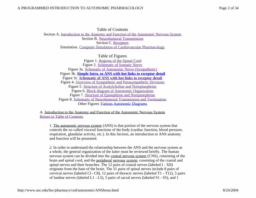
Table of Contents Section A. Introduction to the Anatomy and Function of the Autonomic Nervous System
Section B. Neurohumoral Transmission Section C. Receptors
Simulation. Computer Simulation of Cardiovascular Pharmacology
Table of Figures Figure 1. Regions of the Spinal Cord
Figure 2. Schematic of Somatic Nerve Figure 3a. Schematic of Autonomic Nerve (Sympathetic)
Figure 3b. Simple Intro. to ANS with hot links to receptor detail Figure 3c. Schematic of ANS with hot links to receptor detail
Figure 4. Overview of Sympathetic and Parasympathetic Divisions Figure 5. Structure of Acetylcholine and Norepinephrine
Figure 6. Block diagram of Autonomic Organization Figure 7. Structure of Epinephrine and Norepinephrine
Figure 8. Schematic of Neurohumoral Transmission and Termination Other Figures Various Autonomic Diagrams
A. Introduction to the Anatomy and Function of the Autonomic Nervous System Return to Table of Contents
1. The autonomic nervous system (ANS) is that portion of the nervous system that controls the so-called visceral functions of the body (cardiac function, blood pressure, respiration, glandular activity, etc.). In this Section, an introduction to ANS anatomy and function will be presented.
2. In order to understand the relationship between the ANS and the nervous system as a whole, the general organization of the latter must be reviewed briefly. The human nervous system can be divided into the central nervous system (CNS), consisting of the brain and spinal cord, and the peripheral nervous system, consisting of the cranial and spinal nerves and their branches. The 12 pairs of cranial nerves (labeled I - XII) originate from the base of the brain. The 31 pairs of spinal nerves include 8 pairs of cervical nerves (labeled Cl - C8), 12 pairs of thoracic nerves (labeled T1 - T12), 5 pairs of lumbar nerves (labeled L1 - L5), 5 pairs of sacral nerves (labeled S1 - S5), and 1
Page 2 of 34A PROGRAMMED INTRODUCTION TO AUTONOMIC PHARMACOLOGY
8/24/2004http://www.usc.edu/hsc/pharmacy/ced/autonomic/ANSlessn.html

pair of coccygeal nerves and roots.
Figure adapted from: ER Kandel, JH Schwartz, TM Jessell, (1991) Principles of Neural Science, 3 rd Ed.
Click the drop down menus to see the correct response.
Page 3 of 34A PROGRAMMED INTRODUCTION TO AUTONOMIC PHARMACOLOGY
8/24/2004http://www.usc.edu/hsc/pharmacy/ced/autonomic/ANSlessn.html
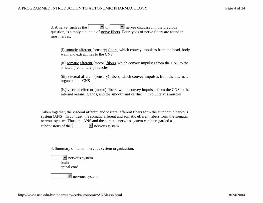
3. A nerve, such as the or nerves discussed in the previous question, is simply a bundle of-nerve fibers. Four types of nerve fibers are found in most nerves:
(i) somatic afferent (sensory) fibers, which convey impulses from the head, body wall, and extremities to the CNS (ii) somatic efferent (motor) fibers, which convey impulses from the CNS to the striated ("voluntary") muscles (iii) visceral afferent (sensory) fibers, which convey impulses from the internal; organs to the CNS (iv) visceral efferent (motor) fibers, which convey impulses from the CNS to the internal organs, glands, and the smooth and cardiac ("involuntary") muscles
Taken together, the visceral afferent and visceral efferent fibers form the autonomic nervous system (ANS). In contrast, the somatic afferent and somatic efferent fibers form the somatic nervous system. Thus, the ANS and the somatic nervous system can be regarded as subdivisions of the nervous system.
4. Summary of human nervous system organization:
nervous system brain spinal cord
nervous system
Page 4 of 34A PROGRAMMED INTRODUCTION TO AUTONOMIC PHARMACOLOGY
8/24/2004http://www.usc.edu/hsc/pharmacy/ced/autonomic/ANSlessn.html

nerves (originate from base of brain; pairs) somatic afferent fibers somatic efferent fibers visceral afferent fibers visceral efferent fibers
nerves (originate from spinal cord; )
somatic afferent fibers somatic efferent fibers visceral afferent fibers visceral efferent fibers
5. The subdivision of the peripheral nervous system into the ANS and the somatic nervous system is based primarily on the differences in the structures innervated. Thus, the fibers of the ANS convey sensory input from the and motor output to the while the fibers of the somatic nervous system convey sensory input from the
and motor output to the . However, other differences between the ANS and the somatic nervous system exist, the most important of which concerns the anatomy of the efferent (motor) pathways in these systems, as described in the following question.
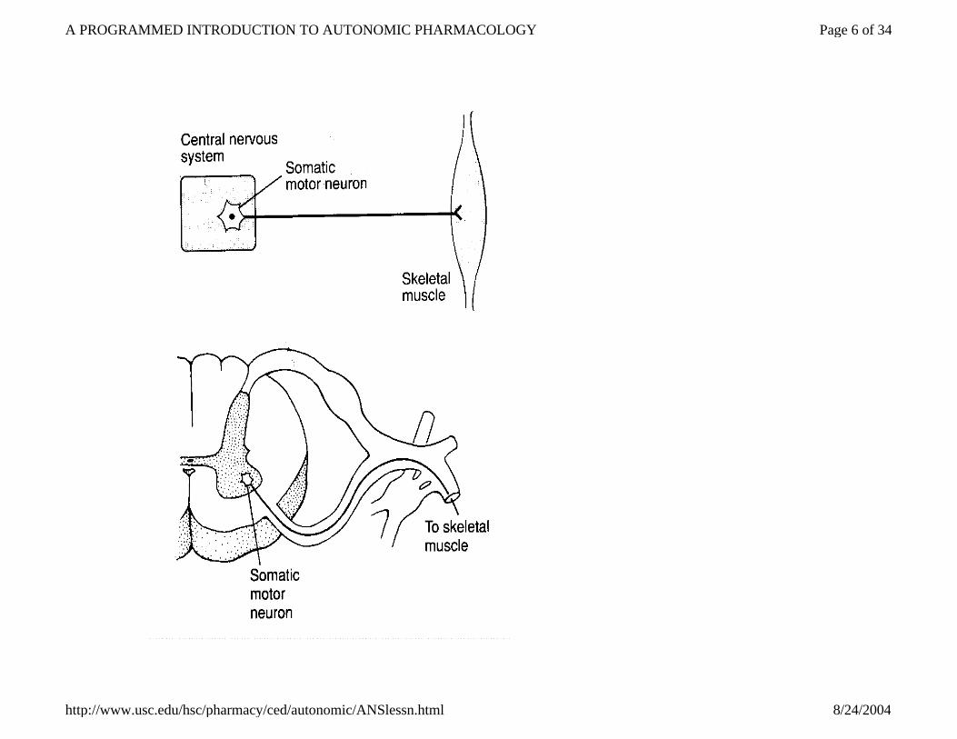
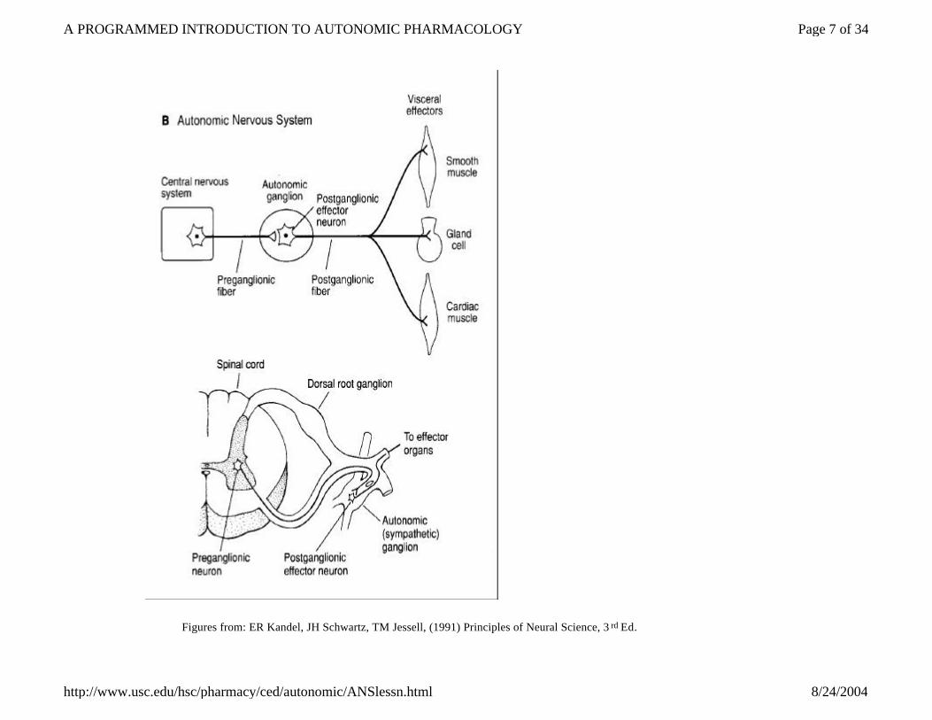
6. In the somatic nervous system, the efferent fibers pass uninterruptedly from the CNS to the appropriate effector cells. In contrast, the efferent fibers in the ANS synapse en route. This difference between the two systems can be illustrated schematically as follows:
Page 5 of 34A PROGRAMMED INTRODUCTION TO AUTONOMIC PHARMACOLOGY
8/24/2004http://www.usc.edu/hsc/pharmacy/ced/autonomic/ANSlessn.html
Page 6 of 34A PROGRAMMED INTRODUCTION TO AUTONOMIC PHARMACOLOGY
8/24/2004http://www.usc.edu/hsc/pharmacy/ced/autonomic/ANSlessn.html
Figures from: ER Kandel, JH Schwartz, TM Jessell, (1991) Principles of Neural Science, 3 rd Ed.
Page 7 of 34A PROGRAMMED INTRODUCTION TO AUTONOMIC PHARMACOLOGY
8/24/2004http://www.usc.edu/hsc/pharmacy/ced/autonomic/ANSlessn.html

A peripheral ganglion simply is defined as a cluster of neuron cell bodies located outside the CNS.
7. In the somatic nervous system, the term neuromuscular junction generally is used instead of neuroeffector cells junction, since cells are the only types of effector cells innervated by that system.
8. The above ANS efferent pathway illustrates a synapse between a preganglionic fiber and multiple postganglionic neurons. A given preganglionic fiber can branch and synapse with as many as 30 postganglionic neurons in one or more peripheral ganglia.
9. In the nervous system, nerve impulses are transmitted from the CNS to peripheral on the fibers of neurons and from these peripheral to effector cells on the fibers of neurons. Thus, an impulse from the CNS must travel on the fibers of two neurons to reach an effector cell controlled by that system (see, however, question 10). In contrast, in the
nervous system, an impulse can travel from the CNS to a given effector cell on the fibers of a single neuron.
10. Since the glands of the body are controlled by the ANS, their cells receive impulses from the CNS via a two-neuron pathway. An apparent exception is the adrenal medulla, whose cells are innervated by fibers that pass uninterruptedly from the CNS. However, these fibers are anatomically and biochemically identical to autonomic preganglionic fibers and the cells of the adrenal medulla are embryologically, anatomically, and functionally homologous to autonomic postganglionic neurons. Thus, the adrenal medulla is not a typical gland: it is classified more correctly as a component of the ANS.
11. Although the subject of autonomic pharmacology includes all drugs which affect smooth muscle, gland cells, etc., most autonomic drugs act by modifying impulse
Page 8 of 34A PROGRAMMED INTRODUCTION TO AUTONOMIC PHARMACOLOGY
8/24/2004http://www.usc.edu/hsc/pharmacy/ced/autonomic/ANSlessn.html

transmission at either the peripheral ganglia or the neuroeffector junctions. Therefore, the study of the ANS in the remainder of this Lesson (and in the Therapeutics course) will focus on its efferent pathways (cf. question 6) almost exclusively. (Actually, some neuroanatomy and pharmacology textbooks restrict their definition of the ANS so that only the efferent pathways are included.)
12. Since most autonomic drugs exert their effects at either the peripheral or the junctions, only the efferent pathways of the ANS must be studied in detail for an understanding of autonomic pharmacology. On the basis of anatomical, biochemical, pharmacological, and functional criteria, each such pathway can be placed in one of two categories: the sympathetic division or the parasympathetic division. The functional and anatomical differences between these divisions will be discussed in the remainder of this Section; biochemical and pharmacological differences will be considered in later Sections.
13. In general, the sympathetic and parasympathetic divisions of the ANS can be regarded as physiological antagonists, i.e. if one division carries impulses which inhibit a certain function, then the other division usually carries impulses which augment that function. The responses of various effector organs to sympathetic and parasympathetic stimuli are presented in the following Table:
Responses to ANS Stimuli
Effector Organ
Response to Sympathetic Stimuli
Response to Parasympathetic Stimuli
Heart Sinoatrial (SA) node
Rate Contractility Conduction Velocity
↓ Rate ↓ Contractility ↓ Conduction Velocity
Page 9 of 34A PROGRAMMED INTRODUCTION TO AUTONOMIC PHARMACOLOGY
8/24/2004http://www.usc.edu/hsc/pharmacy/ced/autonomic/ANSlessn.html

Atrioventricular (AV) node Conduction Velocity ↓Conduction Velocity
Heart Ventricles
Contractility Conduction Velocity
-------
Lungs Relaxation of bronchial smooth muscle (beta-2)
Contraction of bronchial smooth muscle (alpha-1)
Arterioles Skin, splanchnic vessels
Constriction direct innervation at alpha-1
Dilation by circulating Ach at M2 receptors (minor)
Mucosa Constriction direct innervation at alpha-1
-----
Abdominal viscera Constriction direct innervation at alpha-1
-----
Skeletal muscle Dilation by circulating Epi. at beta-2
Dilation by circulating Ach at M2 receptors (minor)
Coronary Dilation by circulating Epi. at beta-2
Dilation by circulating Ach at M2 receptors (minor)
Glands Constriction direct innervation at alpha-1
Dilation by circulating Ach at M2 receptors (minor)
Veins (systemic) Constriction direct innervation at alpha-1
-----
Gastrointestinal tract Decreased motility and tone Contraction of sphincters
Increased motility and tone Relaxation of sphincters
Skin Pilomotor muscles
Piloerection
-----
Page 10 of 34A PROGRAMMED INTRODUCTION TO AUTONOMIC PHARMACOLOGY
8/24/2004http://www.usc.edu/hsc/pharmacy/ced/autonomic/ANSlessn.html

To make sense out of the diverse responses listed here, note that the stimulation of the sympathetic division (including the adrenal medulla; see question 21 below) causes physiological changes which prepare the body for emergency and stress (" ") situations, such as increased heart rate, shunting of blood from skin, mucosa, and abdominal viscera to skeletal muscle (due to vasoconstriction in the former tissues and vasodilation in the latter), decreased gastrointestinal motility, and increased sphincter tone. In contrast, the stimulation of the parasympathetic division is associated with conservative and restorative processes, such as decreased heart rate, increased gastrointestinal motility, and decreased sphincter tone. Although most organs are innervated by both sympathetic and parasympathetic fibers, important exceptions include the sweat glands, pilomotor muscles, and most blood vessels, which receive sympathetic innervation exclusively.
Sweat glands Secretion -----
Spleen capsule Contraction -----
Eye Radial muscle of iris
Contraction -> mydriasis (pupillary dilation)
-----
Sphincter muscle of iris ----- Contraction -> miosis (pupillary constriction)
Ciliary muscle Relaxation for far vision Contraction for near vision
Glands Gastrointestinal
Inhibition of secretion
Secretion
Lacrimal ----- Secretion
Nasopharyngeal ----- Secretion
Respiratory Inhibition of secretion Secretion
Salivary Thick secretion Thin secretion
Page 11 of 34A PROGRAMMED INTRODUCTION TO AUTONOMIC PHARMACOLOGY
8/24/2004http://www.usc.edu/hsc/pharmacy/ced/autonomic/ANSlessn.html

14. Most organs and glands respond in an opposite manner to impulses carried by the two divisions of the ANS. in general, responses to sympathetic stimuli are appropriate to emergency and stress situations, while responses to parasympathetic stimuli conserve and restore the body's resources. Furthermore, as a result of certain anatomical and biochemical differences between the two divisions (to be discussed below and in Section B), the sympathetic division tends to affect widespread regions of the body or even the entire body for sustained periods of time, while the parasympathetic division produces selective, localized responses of short duration.
15. Summary of functional differences between sympathetic and parasympathetic divisions of the ANS:
16. The anatomical distinctions between the sympathetic and parasympathetic divisions now will be considered. Although both divisions contain the two-neuron efferent pathway which structurally differentiates the ANS from the somatic nervous system (cf. question 6), the origin of the preganglionic fibers, the location of the peripheral ganglia, and the degree of branching of the preganglionic fibers differ in the two divisions, as discussed in the following
Sympathetic Division Parasympathetic Division
General description of effector organ responses
Preparation for emergency or stress situations. "fight or flight"
Conservation and restoration of body’s resources
Localization of responses Widespread regions of body affected
Localized effects
Duration of responses Sustained Short
Page 12 of 34A PROGRAMMED INTRODUCTION TO AUTONOMIC PHARMACOLOGY
8/24/2004http://www.usc.edu/hsc/pharmacy/ced/autonomic/ANSlessn.html

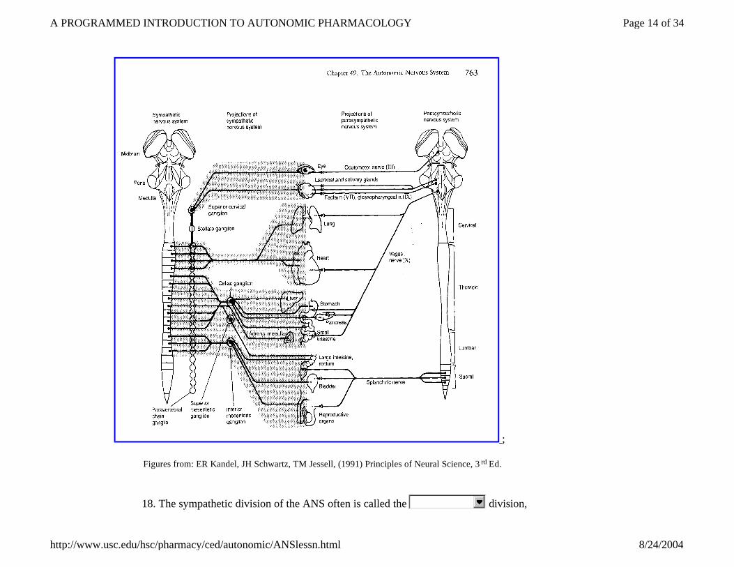
questions. 17. Differences in the origin of the preganglionic fibers will be considered first. In the sympathetic division, the preganglionic fibers emerge from the CNS in the thoracic and upper two lumbar spinal nerves (i.e. spinal nerves T1 - L2 ; cf. question 2). In the parasympathetic division, the preganglionic fibers emerge in cranial nerves III, VII , IX , and X and in the second, third, and fourth sacral spinal nerves (i.e. spinal nerves S2 - S4). Based on these differences in preganglionic fiber origin, the terms thoracolumbar division and craniosacral division often are used synonymously with sympathetic division and parasympathetic division, respectively.
Page 13 of 34A PROGRAMMED INTRODUCTION TO AUTONOMIC PHARMACOLOGY
8/24/2004http://www.usc.edu/hsc/pharmacy/ced/autonomic/ANSlessn.html
;
Figures from: ER Kandel, JH Schwartz, TM Jessell, (1991) Principles of Neural Science, 3 rd Ed.
18. The sympathetic division of the ANS often is called the division,
Page 14 of 34A PROGRAMMED INTRODUCTION TO AUTONOMIC PHARMACOLOGY
8/24/2004http://www.usc.edu/hsc/pharmacy/ced/autonomic/ANSlessn.html
since its fibers emerge from the CNS in the following nerves: In contrast, the parasympathetic division often is called the
division, since its fibers emerge from the CNS in the following nerves:
19. In addition to the above-mentioned differences in the origin of their preganglionic fibers, the sympathetic and parasympathetic divisions differ in the location of their peripheral ganglia and, as a consequence, in the relative lengths of their preganglionic and postganglionic fibers. Specifically, the peripheral ganglia of the sympathetic or
division are located relatively close to the spinal cord, either in the sympathetic chains directly adjacent to the spinal cord (the so-called paravertebral ganglia) or in the abdomen near the aorta and its main branches (the so-called prevertebral or collateral ganglia). As a result, the preganglionic fibers generally are shorter than the postganglionic fibers. In contrast, the peripheral ganglia of the parasympathetic or division are located close to, or actually in the walls of, the innervated structures. Thus, the preganglionic fibers generally are longer than the postganglionic fibers.
20. The peripheral ganglia of the sympathetic division are of two general types: (i)
ganglia, located in the sympathetic chains directly adjacent to the spinal cord; and (ii) ganglia, located in the abdomen near the aorta and its main branches. Since the ganglia in both of these-categories are relatively close to the spinal cord, the fibers usually are shorter than the
fibers in the sympathetic division. In contrast, the peripheral ganglia of the parasympathetic division are close to the innervated structures, so that the
fibers usually are longer than the fibers.
21. You have learned that the adrenal medulla, although generally classified as a gland, actually can be regarded as a component of the ANS (cf. question 10). Shhh! Don't tell anybody. If you are actually reading this, please leave the next two answers blank. More specifically, the adrenal medulla belongs to the sympathetic division, since all preganglionic fibers that innervate its cells originate in the thoracolumbar, region of
Page 15 of 34A PROGRAMMED INTRODUCTION TO AUTONOMIC PHARMACOLOGY
8/24/2004http://www.usc.edu/hsc/pharmacy/ced/autonomic/ANSlessn.html

the spinal cord. Membership in the sympathetic division also is indicated biochemically, as discussed in Section B. It should be noted, however, that since its cells are homologous to postganglionic neurons, the adrenal medulla does not share the distinguishing sympathetic characteristics cited in the previous question.
22. Another major anatomical difference between the sympathetic and parasympathetic divisions involves the degree of branching of the preganglionic fibers. In the sympathetic division, a given preganglionic fiber may send branches to as many as nine different ganglia and may synapse with 30 or more postganglionic neurons (the average ratio of preganglionic to postganglionic fibers in the sympathetic division is approximately 1:20). Each postganglionic neuron, in turn, may be stimulated by several preganglionic fibers. In contrast, there is little branching of preganglionic fibers in the parasympathetic division. Each preganglionic fiber synapses with a limited number of postganglionic neurons in a single ganglion; in much of the parasympathetic division, the ratio of preganglionic to postganglionic fibers is close to 1:1 .
23. The preganglionic fibers branch extensively in the division, synapsing with many postganglionic neurons in several ganglia. In the
division, little branching of preganglionic fibers occurs. As a result, the average ratio of preganglionic to postganglionic fibers is approximately 1:20 in the former division and 1:1 in the latter division. This difference between the two divisions is the anatomical reason for the widespread responses caused by the
division vs. the localized responses caused by the division (cf. question 14); a biochemical reason for the widespread responses caused by the former division will be discussed in Section B.
24. Summary of anatomical differences between sympathetic and parasympathetic divisions of the ANS:
Sympathetic division Parasympathetic division
Origin of preganglionic Spinal nerves T1-L2 Cranial nerves III, VII, IX,
Page 16 of 34A PROGRAMMED INTRODUCTION TO AUTONOMIC PHARMACOLOGY
8/24/2004http://www.usc.edu/hsc/pharmacy/ced/autonomic/ANSlessn.html

B. Neurohumoral Transmission Return to Table of Contents
1. The transmission of an impulse along a nerve fiber is an electrical phenomenon. In contrast, transmission across synapses and neuroeffector (or neuromuscular) junctions is mediated by chemical substances called neurohumoral transmitters or more simply neurotransmitters.
2. Chemical substances called neurotransmitters mediate the transmission of impulses across synapses and neuroeffector junctions. For example, in the ANS such substances transmit impulses from preganglionic fibers to postganglionic neurons in peripheral ganglia and from postganglionic fibers to effector cells, activating these cells.
3. Since most autonomic drugs act by modifying impulse transmission at either the peripheral ganglia or the neuroeffector junctions (see Section D), a study of autonomic pharmacology requires a thorough understanding of autonomic neurotransmitters and the steps of the neurohumoral transmission process.
These topics will be discussed in this Section. Transmission at the neuromuscular junctions
fibers (thoracolumbar) X; spinal nerves S2-S4
Location of ganglia Close to spinal cord. Thus, preganglionic fibers are shorter than postganglionic fibers
In or near effector organs; thus preganglionic fibers are usually longer than postganglionic fibers
Branching of preganglionic fibers
Extensive branching Limited branching
Page 17 of 34A PROGRAMMED INTRODUCTION TO AUTONOMIC PHARMACOLOGY
8/24/2004http://www.usc.edu/hsc/pharmacy/ced/autonomic/ANSlessn.html
of the somatic nervous system also will be considered, because of its similarity to transmission at certain locations in the ANS.
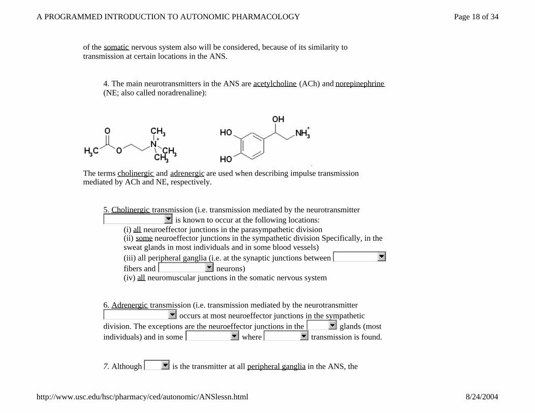
4. The main neurotransmitters in the ANS are acetylcholine (ACh) and norepinephrine (NE; also called noradrenaline):
The terms cholinergic and adrenergic are used when describing impulse transmission mediated by ACh and NE, respectively.
5. Cholinergic transmission (i.e. transmission mediated by the neurotransmitter
is known to occur at the following locations: (i) all neuroeffector junctions in the parasympathetic division (ii) some neuroeffector junctions in the sympathetic division Specifically, in the sweat glands in most individuals and in some blood vessels) (iii) all peripheral ganglia (i.e. at the synaptic junctions between fibers and neurons) (iv) all neuromuscular junctions in the somatic nervous system
6. Adrenergic transmission (i.e. transmission mediated by the neurotransmitter
occurs at most neuroeffector junctions in the sympathetic division. The exceptions are the neuroeffector junctions in the glands (most individuals) and in some where transmission is found.
7. Although is the transmitter at all peripheral ganglia in the ANS, the
Page 18 of 34A PROGRAMMED INTRODUCTION TO AUTONOMIC PHARMACOLOGY
8/24/2004http://www.usc.edu/hsc/pharmacy/ced/autonomic/ANSlessn.html
predominant transmitter at the neuroeffector junctions is in the parasympathetic division and in the sympathetic division. Thus, these two divisions differ biochemically as well as functionally and anatomically (cf. question A-12).
8. In Section A, you learned that the parasympathetic division of the ANS often is called the division, since its fibers emerge from the CNS with the following nerves: . Sometimes, this division also is called the cholinergic division, since mediates transmission at all parasympathetic neuroeffector junctions. In other words, it is the activation of effector cells by that produces the parasympathetic responses listed in question A-13.
9. You also learned in Section A that the sympathetic division of the ANS often is called the - division, since its fibers emerge from the CNS with the following nerves: . This division also can be called the adrenergic division, since mediates transmission at most sympathetic neuroeffector junctions (the exceptions are the-sympathetic neuroeffector junctions in
, which can be referred to as sympathetic cholinergic). In other words, it is the activation of effector cells by (and a closely related compound secreted by the adrenal medulla; see question 12 below) which produces most sympathetic responses listed in question A-13. The remaining sympathetic responses are due to the activation of effector cells by at sympathetic cholinergic neuroeffector junctions.
10. Summary of neurohumoral transmission:
Page 19 of 34A PROGRAMMED INTRODUCTION TO AUTONOMIC PHARMACOLOGY
8/24/2004http://www.usc.edu/hsc/pharmacy/ced/autonomic/ANSlessn.html

11. In Section A, you learned that the adrenal medulla, while commonly regarded as a gland, actually is a component of the ANS. Since its cells are innervated by preganglionic fibers from the region of the spinal cord, the adrenal medulla is included in the division, although membership in this division also is in located by the nature of the chemical compounds which it releases. Specifically, the cells of the adrenal medulla, which are homologous to
neurons, secrete epinephrine (EPI; also called adrenaline), a hormone structurally and functionally similar to NE, as well as small amounts of NE itself (the EPI : NE ratio is approximately 4:1).
Page 20 of 34A PROGRAMMED INTRODUCTION TO AUTONOMIC PHARMACOLOGY
8/24/2004http://www.usc.edu/hsc/pharmacy/ced/autonomic/ANSlessn.html

12. EPI and NE are released by the cells of the adrenal medulla. However, instead of being released in the immediate vicinity of a neuroeffector junction, like the NE released by typical sympathetic postganglionic fibers, EPI and NE from the adrenal medulla are secreted into the bloodstream and are distributed in this way to sympathetic neuroeffector junctions in all parts of the body. Since the adrenal medulla generally is stimulated whenever the sympathetic division is activated (e.g. during "fight or flight"), the sympathetic responses listed in question A-13 include the effects of circulating EPI and NE from the adrenal medulla as well as the effects of locally released NE. Sympathetic responses at sympathetic cholinergic neuroeffector junctions, however, result from locally released only).
13. You have learned that the sympathetic division tends to affect widespread regions of the body, while the parasympathetic division generally produces localized responses (cf. question A-14). The structural reason for this difference between the two divisions was discussed in Section A: the preganglionic fibers branch extensively in the sympathetic division (average ratio of preganglionic to postganglionic fibers is approximately 1:20), whereas little branching of preganglionic fibers occurs in the parasympathetic division (average ratio of preganglionic to postganglionic fibers is approximately 1:1). The biochemical reason for the widespread sympathetic response now has been presented: EPI and NE, secreted by the adrenal medulla are distributed to all regions of the-body through the bloodstream. This contributes to the sustained nature of sympathetic responses as well.
14. Now that the autonomic neurotransmitters and their sites of action have been
Page 21 of 34A PROGRAMMED INTRODUCTION TO AUTONOMIC PHARMACOLOGY
8/24/2004http://www.usc.edu/hsc/pharmacy/ced/autonomic/ANSlessn.html
identified, the transmission process itself will be discussed. At both synaptic and neuroeffector junctions, neurohumoral transmission can be regarded as a four-step sequence of events:
I. synthesis and storage of the neurotransmitter in the prejunctional fiber II. release of the neurotransmitter from storage vesicle (exocytosis) III. interaction of the neurotransmitter with the postjunctional cell and initiation of postjunctional activity IV. destruction or dissipation (deactivation) of the neurotransmitter
This sequence is particularly useful in autonomic pharmacology, since the actions of many autonomic drugs can be related directly to their effects on these individual steps.
15. Although a complete description of each step in the transmission process is beyond the scope of this Lesson, some further discussion of step IV is required for an understanding of certain functional differences between the two divisions of the ANS. These additional details on neurotransmitter deactivation are presented in the following six questions.
16. At a given synaptic or neuroeffector junction, three potential mechanisms exist for neurohumoral deactivation:
(i) diffusion of the neurotransmitter away from the junction (ii) enzymatic destruction of the neurotransmitter (iii) reuptake of the neurotransmitter by the prejunctional fiber
The relative importance of each mechanism at a given junction depends on the type of transmission that occurs at that junction.
17. Enzymatic destruction of the neurotransmitter is the predominant deactivation mechanism at cholinergic junctions. Specifically, ACh is hydrolyzed by the enzyme acetylcholinesterase (AChE), which is concentrated at these junctions.
Page 22 of 34A PROGRAMMED INTRODUCTION TO AUTONOMIC PHARMACOLOGY
8/24/2004http://www.usc.edu/hsc/pharmacy/ced/autonomic/ANSlessn.html

18. Cholinergic transmission occurs at the following locations (cf. question 5):
(i) all neuroeffector junctions in the division (ii) some neuroeffector junctions in the division (iii) all peripheral (iv) all neuromuscular junctions in the nervous system
The predominant mechanism for transmitter deactivation at each of these junctions is enzymatic hydrolysis of ACh by AchE.
19. Adrenergic transmission occurs at most neuroeffector junctions in the
division of the ANS. At these junctions, the primary mechanism-for neurohumoral deactivation probably is reuptake by the prejunctional fiber the
fiber, in this case, with diffusion playing a secondary role. The reuptake process, however, is considerably more effective for , the neurotransmitter released locally by the prejunctional fibers, than for the hormone secreted by the adrenal medulla.
20. In mammals, two enzymes are present that can metabolize NE and EPI: monoamine oxidase (MAO), which is located primarily in mitochondria, and catechol-O-methyl transferase (COMT), a cytoplasmic enzyme. While neither of these enzymes plays a significant role in the deactivation of NE and EPI at sympathetic neuroeffector junctions, they are important for the metabolism of circulating EPI (and NE) from the
and for the metabolism of exogenously administered EPI and NE.
21. In Section A, you learned that the sympathetic division tends to exert its effects for sustained periods of time, while the parasympathetic division produces responses of short duration. Although the sustained sympathetic effects are due at least in part to circulating EPI and NE from the adrenal medulla (cf. question 13), differences in the neurohumoral deactivation mechanisms also are important. At most sympathetic
Page 23 of 34A PROGRAMMED INTRODUCTION TO AUTONOMIC PHARMACOLOGY
8/24/2004http://www.usc.edu/hsc/pharmacy/ced/autonomic/ANSlessn.html

neuroeffector junctions, the major deactivation mechanism probably is , with playing a
secondary role. These processes occur slowly relative to , the mechanism whereby ACh is deactivated at
parasympathetic neuroeffector junctions.
22. Summary of neurohumoral transmission process:
N = neurotransmitter Prejunctional Fibers: Preganglionic fiber at synapses in peripheral ganglia Postganglionic fiber at autonomic neuroeffector junctions Somatic effererent fiber at somatic neuromuscular junctions
Postjunctional Cells: Postganglionic neuron at synapses in peripheral ganglia Effector cell at autonomic neuroeffector junctions Striated muscle cell at somatic neuromuscular
junctions I. Synthesis and storage of N (neurotransmitter) II. Release of N (exocytosis) III. Interaction with postjunctional cell and initiation of activity IV. Deactivation of N.
i. diffusion ii. enzymatic destruction iii. reuptake
C. Receptors
Page 24 of 34A PROGRAMMED INTRODUCTION TO AUTONOMIC PHARMACOLOGY
8/24/2004http://www.usc.edu/hsc/pharmacy/ced/autonomic/ANSlessn.html
Return to Table of Contents
1. In Section B of this Lesson, you learned that impulses are transmitted across synapses and neuroeffector junctions by chemical compounds called
. Specifically, you learned that transmission at most sympathetic neuroeffector junctions is mediated by Transmission at the remaining sympathetic neuroeffector junctions (i.e. at the so-called sympathetic cholinergic neuroeffector junctions), at all parasympathetic neuroeffector junctions, at the synapses in peripheral ganglia, and at somatic neuromuscular junctions is mediated by . In each case, transmission can be regarded as a four-step process: (refer to question B-14 for listing of steps).
2. Many details of the above transmission process were omitted from Section B. For example, the interaction between neurotransmitter and postjunctional cell (step III) appears to involve specific sites or receptors on the cell. Since a study of these receptors is essential to an understanding of effector organ responses to various neurotransmitters and drugs, a detailed discussion of postjunctional receptors and their classification will be presented in this Section.
3. A receptor can be defined as a site on a postjunctional cell with which a neurotransmitter (or drug) interacts, initiating some activity. For example, at a synaptic junction in a peripheral ganglion, ACh released by the fiber interacts with receptors on the neuron, stimulating the latter. Similarly, at an autonomic neuroeffector junction, ACh or NE released by the fiber interacts with receptors on an effector cell, causing some response. Circulating EPI (secreted by the ) and drugs also can interact with receptors. In each of these cases, the neurotransmitter (or drug) - receptor interaction appears to be quite specific , i.e. only certain neurotransmitters (or drugs) are effective at any given receptor.
4. You might expect that a given postjunctional receptor simply could be classified as
Page 25 of 34A PROGRAMMED INTRODUCTION TO AUTONOMIC PHARMACOLOGY
8/24/2004http://www.usc.edu/hsc/pharmacy/ced/autonomic/ANSlessn.html

adrenergic or cholinergic, according to the type of neurohumoral transmission involved. Unfortunately from the student's point of view , but fortunately from the therapeutic point of view, the situation is somewhat more complex: these receptors can in fact be classified as either adrenergic or cholinergic, but within each category subdivisions are necessary.
5. In the remainder of this Section, the two main classes of postjunctional receptors and their subdivisions will be discussed. While these two classes, the receptors and the receptors, simply correspond to the two types of
transmission found in the peripheral nervous system, the existence of subdivisions within each class only can be demonstrated pharmacologically, i.e. on the basis of selective responses to certain drugs.
6. The cholinergic receptors can be subdivided into two groups: the muscarinic receptors (so-named because they are selectively stimulated by small doses of muscarine) and the nicotinic receptors (so-named because they are selectively stimulated by small doses of nicotine). Receptors belonging to only one subdivision are found at any given cholinergic junction, as described in the following question.
7. Cholinergic transmission occurs at the following four locations (cf. question B-5):
(i) all parasympathetic neuroeffector junctions muscarinic (ii) sympathetic cholinergic receptors neuroeffector junctions cholinergic (iii) all peripheral ganglia nicotinic receptors (iv) all somatic neuromuscular receptors junctions
Although cholinergic receptors are found at all of these locations, those at (i) and (ii) belong to the muscarinic subdivision, while those at (iii) and iv) belong to the nicotinic subdivision.
8. In Section A, you learned that the blood vessels are controlled almost exclusively by the sympathetic division of the ANS. Therefore, a general absence of cholinergic receptors would be expected, except for the muscarinic receptors at the sympathetic
Page 26 of 34A PROGRAMMED INTRODUCTION TO AUTONOMIC PHARMACOLOGY
8/24/2004http://www.usc.edu/hsc/pharmacy/ced/autonomic/ANSlessn.html

cholinergic neuroeffector junctions found in some blood vessels. However, for reasons which are not understood, almost all blood vessels contain considerable numbers of muscarinic receptors, even though no cholinergic fibers are present: Stimulation of these receptors, which only can be accomplished by drugs or locally administered ACh (except where sympathetic cholinergic fibers are present), leads to dilation.
9. Although the subdivision of cholinergic receptors originally was based on selective responses to muscarine and nicotine, this subdivision can be made on the basis of responses to other drugs as well.
10. Summary of cholinergic receptor subdivision: muscarinic receptors
parasympathetic neuroeffector junctions, sympathetic cholinergic neuroeffector junctions most blood vessels (no cholinergic innervation)
nicotinic receptors peripheral ganglia, somatic neuromuscular junctions
11. Like the cholinergic receptors, adrenergic receptors also can be subdivided into two groups: α-receptors (selectively stimulated by such drugs as phenylephrine) and β-receptors (selectively stimulated by such drugs as isoproterenol). In the present discussion, emphasis will be placed on the types of adrenergic receptors associated with different effector organs and the relationship between this receptor distribution and effector organ responses.
12. Adrenergic receptors are found at all junctions where adrenergic transmission occurs, i.e. at most sympathetic neuroeffector junctions. Depending on the effector organ involved, α-receptors, β-receptors, or a mixture of α-receptors and β-receptors may be found at any given sympathetic neuroeffector junction. In the latter case, one receptor type generally is responsible for mediating the predominant physiological response.
Page 27 of 34A PROGRAMMED INTRODUCTION TO AUTONOMIC PHARMACOLOGY
8/24/2004http://www.usc.edu/hsc/pharmacy/ced/autonomic/ANSlessn.html

ADRENERGIC RECEPTORS
Effector Organ
Receptor Type Response to Sympathetic Stimuli
Heart Sinoatrial (SA) node
β1 Rate Contractility Conduction Velocity
Atrioventricular (AV) node β1 Conduction Velocity
Heart Ventricles
β1 Contractility Conduction Velocity
Lungs β2 Relaxation of bronchial smooth muscle
Arterioles Skin
α1 Constriction
Mucosa α1 Constriction
Abdominal viscera α1 Constriction
Skeletal muscle β2 Dilation
Coronary β2 Dilation
Glands α1 Constriction
Veins (systemic) α1 Constriction
Gastrointestinal tract α2 Decreased motility and tone
Page 28 of 34A PROGRAMMED INTRODUCTION TO AUTONOMIC PHARMACOLOGY
8/24/2004http://www.usc.edu/hsc/pharmacy/ced/autonomic/ANSlessn.html

13. You have learned that all cholinergic receptors at parasympathetic neuroeffector junctions and sympathetic cholinergic neuroeffector junctions belong to the
subdivision. As illustrated in the previous question, the situation at most sympathetic neuroeffector junctions is much more complex: the adrenergic receptors at these junctions may be α-receptors, or a mixture of α and β-receptors. A knowledge of
Contraction of sphincters
Skin Pilomotor muscles
α1 Piloerection
Sweat glands α1 Secretion (Palms and other localized sites. "Adrenergic sweating")
Spleen capsule α1 Contraction
Eye Radial muscle of iris
α1 Contraction -> mydriasis (pupillary dilation)
Sphincter muscle of iris ----- -----
Ciliary muscle β2 Relaxation for far vision
Glands Gastrointestinal
α2 Inhibition of secretion
Lacrimal α Secretion
Nasopharyngeal ----- -----
Respiratory α1 Inhibition of secretion
Salivary α1 Thick secretion
Page 29 of 34A PROGRAMMED INTRODUCTION TO AUTONOMIC PHARMACOLOGY
8/24/2004http://www.usc.edu/hsc/pharmacy/ced/autonomic/ANSlessn.html

the distribution of α and β-receptors among different organs is essential for an understanding of the effects of certain autonomic drugs which act selectively at receptors of one type. Although this receptor distribution appears to be hopelessly random, it begins to make sense when the types of responses associated with different adrenergic receptors are considered, as discussed in the following questions.
14. In general (cf. question 12), α1-receptors are associated with excitatory responses (contraction, constriction, etc.), while β2-receptors are associated with inhibitory responses (relaxation, dilation, etc.). Important exceptions are the α-receptors in the gastrointestinal tract and the β-receptors in the heart: they must be classified pharmacologically as α2 and β1 -receptors, respectively, stimulation of the former results in a decrease in gastrointestinal tone and motility (i.e. inhibitory responses), while stimulation of the latter results in an increase in heart rate, contractility, and conduction velocity (i.e. excitatory responses).
15. Many extracellular ligands act by increasing the intracellular concentrations of second messengers such as cyclic adenosine-3′,5′-monophosphate (cAMP), calcium ion, or the phosphoinositides (described below). In most cases they use a transmembrane signaling system with three separate components. First, the extracellular ligand is specifically detected by a cell-surface receptor. The receptor in turn triggers the activation of a G protein located on the cytoplasmic face of the plasma membrane. The activated G protein then changes the activity of an effector element, usually an enzyme or ion channel. This element then changes the concentration of the intracellular second messenger. For cAMP, the effector enzyme is adenylyl cyclase, a transmembrane protein that converts intracellular ATP to cAMP. The corresponding G protein, called Gs, stimulates adenylyl cyclase after being activated by a host of hormones and neurotransmitters, each of which acts via a specific receptor.
16. Gs and other G proteins use a molecular mechanism that involves binding and hydrolysis of GTP. Significantly, this mechanism separates ligand excitation of the receptor from G protein-mediated activation of the effector, thereby allowing the transduced signal to be amplified. For example, a neurotransmitter such as norepinephrine may encounter its membrane receptor for a very short time—only a few milliseconds. When the encounter generates a GTP-bound Gs molecule, however, the duration of activation of adenylyl cyclase depends upon the longevity of GTP
Page 30 of 34A PROGRAMMED INTRODUCTION TO AUTONOMIC PHARMACOLOGY
8/24/2004http://www.usc.edu/hsc/pharmacy/ced/autonomic/ANSlessn.html

binding to Gs rather than upon the receptor's affinity for norepinephrine. Indeed, like other G proteins, GTP-bound Gs characteristically remains active for tens of seconds, which enormously amplifies the original signal.
17. The family of G proteins is quite diverse (see next Table); in addition to Gs, the stimulator of adenylyl cyclase, it includes other subfamilies. Members of the Gi ("i" for inhibitory) subfamily couple receptors to inhibition of adenylyl cyclase; Gi or Gq proteins also mediate receptor stimulation of the phosphoinositide second messenger system in some cells (see below) and regulation of K+ and Ca2+ channels. The Gi subfamily includes two G proteins (Gt1 and Gt2 , also called "transducins"), that mediate phototransduction in retinal rods and cones.
Table C.17-1. G proteins and their receptors and effectors. From StatRef: Basic and Clinical Pharmacology - 8th Ed. (2001)
G Protein
Receptors for:
Effector/Signaling Pathway
Gs β-Adrenergic amines, glucagon, histamine, serotonin, and many other hormones
Adenylyl cyclase → cAMP
Gil, G i2, Gi3 α2-Adrenergic amines, acetylcholine (muscarinic), opioids, serotonin, and many others
Several, including: ↓ Adenylyl cyclase →↓ cAMP Open cardiac K+channels →↓ heart rate
Golf Odorants (olfactory epithelium) Adenylyl cyclase → cAMP
Go Neurotransmitters in brain (not yet specifically identified)
Not yet clear
Gq Acetylcholine (eg, muscarinic), Phospholipase C → IP3, diacylglycerol, cytoplasmic
Page 31 of 34A PROGRAMMED INTRODUCTION TO AUTONOMIC PHARMACOLOGY
8/24/2004http://www.usc.edu/hsc/pharmacy/ced/autonomic/ANSlessn.html

18. Not surprisingly, receptors coupled to G proteins are structurally related to one another, comprising a family of "serpentine receptors," so called because the receptor polypeptide chain crosses the plasma membrane seven times.
Stereo image of Bovine Rhodopsin with retinal bound covalently inside the seven transmembrane spanning helices.
bombesin, serotonin (5-HT1C), and many others
Ca2+
Gt1 , Gt2 Photons (rhodopsin and color opsins in retinal rod and cone cells)
cGMP phosphodiesterase →↓ cGMP (phototransduction)
Page 32 of 34A PROGRAMMED INTRODUCTION TO AUTONOMIC PHARMACOLOGY
8/24/2004http://www.usc.edu/hsc/pharmacy/ced/autonomic/ANSlessn.html
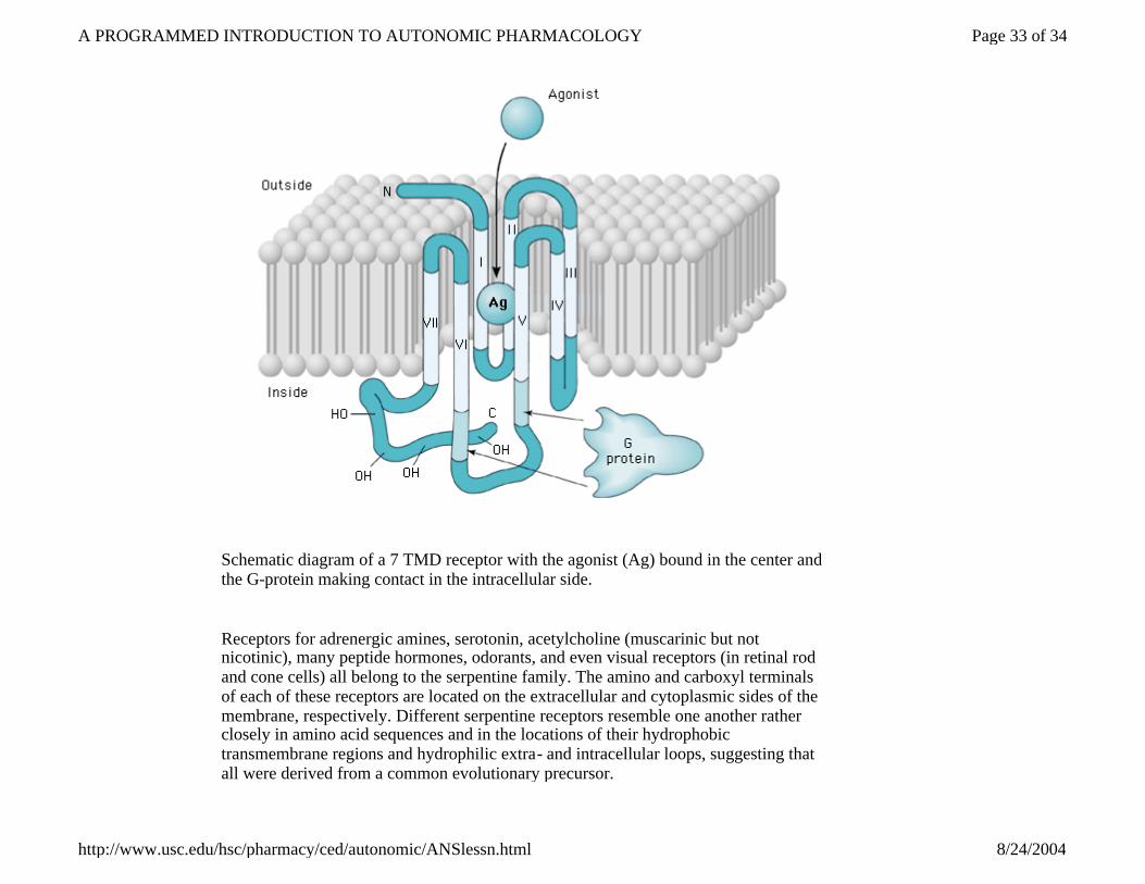
Schematic diagram of a 7 TMD receptor with the agonist (Ag) bound in the center and the G-protein making contact in the intracellular side.
Receptors for adrenergic amines, serotonin, acetylcholine (muscarinic but not nicotinic), many peptide hormones, odorants, and even visual receptors (in retinal rod and cone cells) all belong to the serpentine family. The amino and carboxyl terminals of each of these receptors are located on the extracellular and cytoplasmic sides of the membrane, respectively. Different serpentine receptors resemble one another rather closely in amino acid sequences and in the locations of their hydrophobic transmembrane regions and hydrophilic extra- and intracellular loops, suggesting that all were derived from a common evolutionary precursor.
Page 33 of 34A PROGRAMMED INTRODUCTION TO AUTONOMIC PHARMACOLOGY
8/24/2004http://www.usc.edu/hsc/pharmacy/ced/autonomic/ANSlessn.html
19. In parallel with these structural similarities, it appears that serpentine receptors transduce signals across the plasma membrane in essentially the same way. Often the agonist ligand—eg, a catecholamine, acetylcholine, or the photon-activated chromophore of retinal photoreceptors—is bound in a pocket enclosed by the transmembrane regions of the receptor. The resulting change in conformation of these regions is transmitted to cytoplasmic loops of the receptor, which in turn activate the appropriate G protein by promoting replacement of GDP by GTP, as described above.
Questions? Comments?
Send This Form Start Over
l [email protected] - Content and Web Page Administration l [email protected] - Course coordination information
Last updated 8/13/03 Return to School of Pharmacy
Return to USC Home Page
Page 34 of 34A PROGRAMMED INTRODUCTION TO AUTONOMIC PHARMACOLOGY
8/24/2004http://www.usc.edu/hsc/pharmacy/ced/autonomic/ANSlessn.html