LOCOMOTION OF THE LIMPET, PATELLA VULGATA L.jeb.biologists.org/content/jexbio/52/1/201.full.pdf ·...
18
J. Exp. Biol. (1970), 5a, 201-316 201 With 1 plate and 10 text-figures Printed in Great Britain LOCOMOTION OF THE LIMPET, PATELLA VULGATA L. BY H. D. JONES* AND E. R. TRUEMAN* Department of Zoology, The University, Hull (Received 19 June 1969) INTRODUCTION The foot of Patella, in common with that of many gastropods, is adapted for loco- motion over, and adhesion to, hard substrates. In some snails propulsion is effected by cilia on the ventral surface of the foot, but in the majority a wave-like phenomenon of muscular contraction and relaxation passes along the sole and propels the animal. The pedal waves may pass along the foot in the same direction as the animal is moving, in which case they are termed 'direct' waves, or may pass in the opposite direction to the movement of the snail, these being termed ' retrograde' waves. These definitions are those of Vies (1907), who defined them in relation to the direction of movement rather than to the anterior and posterior of the animal. Where one set of locomotory waves covers the whole width of the foot the wave pattern is termed ' monotaxic' and when one set occurs on each side of the mid-line of the sole they are termed 'ditaxic'. Ditaxic waves are usually alternate. The reviews of Morton (1964) and Gray (1968) both give valuable summaries of previous work and details and examples of further patterns of locomotory activity. There have been few attempts, e.g. Trappmann (1916), Rotarides (1936) (Un- traceable, source: Rotarides, 1945), Elves (1961), to relate the type of locomotion with the structure of the foot, many of the earlier papers being simply records of visual observations. Limited experiments were first performed on Helix by Ten Cate (1922) and Bonse (1935) and their techniques were subsequently considerably modi- fied and improved by Lissmann (1945, 1946). The latter remain the only published detailed investigations of gastropod locomotion. Lissmann studied three species, Helix pomatia, Haliotis tuberculata and Pomatias elegans, all of which exhibit direct waves, Helix being monotaxic and the others ditaxic. Unfortunately Lissmann did not study retrograde locomotion, nor did he attempt to relate his results to the disposition of the muscle fibres in the foot. Previous observations on Patella vulgata by Fretter & Graham (1962) indicated that Patella is of the direct ditaxic type of forward locomotion, whereas the present in- vestigation has shown it to have retrograde ditaxic forward locomotion but direct ditaxic backward locomotion. Neither Vies (1907) nor Weber (1925) could distinguish any waves on the sole of Patella during locomotion. The purpose of this work is to investigate the process of locomotion in P. vulgata by the use of modern recording techniques, to relate these observations to the anatomy of the foot and to attempt to explain the locomotory mechanism. • Present address: Department of Zoology, The University, Manchester.
Transcript of LOCOMOTION OF THE LIMPET, PATELLA VULGATA L.jeb.biologists.org/content/jexbio/52/1/201.full.pdf ·...
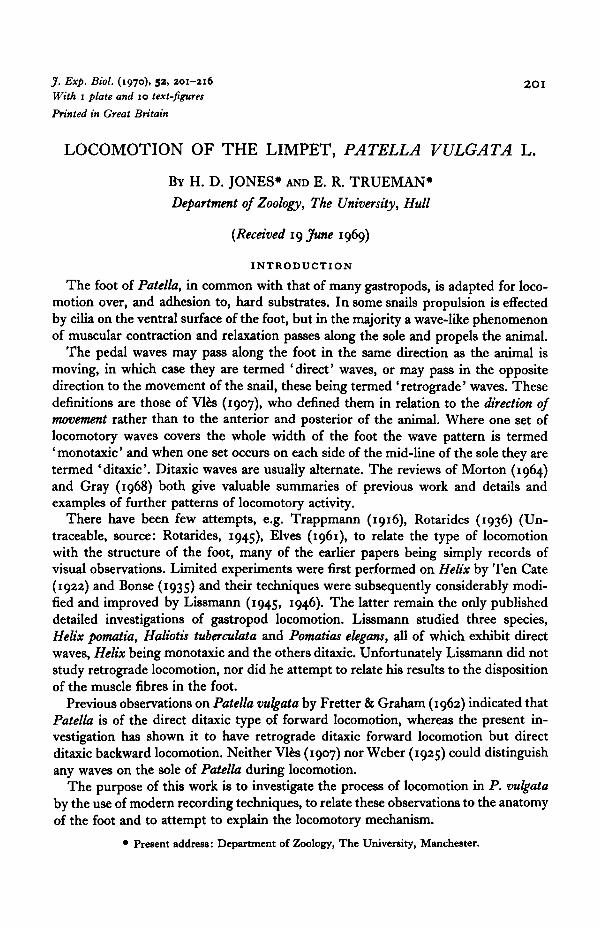
J. Exp. Biol. (1970), 5a, 201-316 201With 1 plate and 10 text-figures
Printed in Great Britain
LOCOMOTION OF THE LIMPET, PATELLA VULGATA L.
BY H. D. JONES* AND E. R. TRUEMAN*
Department of Zoology, The University, Hull
(Received 19 June 1969)
INTRODUCTION
The foot of Patella, in common with that of many gastropods, is adapted for loco-motion over, and adhesion to, hard substrates. In some snails propulsion is effectedby cilia on the ventral surface of the foot, but in the majority a wave-like phenomenonof muscular contraction and relaxation passes along the sole and propels the animal.
The pedal waves may pass along the foot in the same direction as the animal ismoving, in which case they are termed 'direct' waves, or may pass in the oppositedirection to the movement of the snail, these being termed ' retrograde' waves. Thesedefinitions are those of Vies (1907), who defined them in relation to the direction ofmovement rather than to the anterior and posterior of the animal. Where one set oflocomotory waves covers the whole width of the foot the wave pattern is termed' monotaxic' and when one set occurs on each side of the mid-line of the sole they aretermed 'ditaxic'. Ditaxic waves are usually alternate. The reviews of Morton (1964)and Gray (1968) both give valuable summaries of previous work and details andexamples of further patterns of locomotory activity.
There have been few attempts, e.g. Trappmann (1916), Rotarides (1936) (Un-traceable, source: Rotarides, 1945), Elves (1961), to relate the type of locomotionwith the structure of the foot, many of the earlier papers being simply records ofvisual observations. Limited experiments were first performed on Helix by Ten Cate(1922) and Bonse (1935) and their techniques were subsequently considerably modi-fied and improved by Lissmann (1945, 1946). The latter remain the only publisheddetailed investigations of gastropod locomotion. Lissmann studied three species,Helix pomatia, Haliotis tuberculata and Pomatias elegans, all of which exhibit directwaves, Helix being monotaxic and the others ditaxic. Unfortunately Lissmann did notstudy retrograde locomotion, nor did he attempt to relate his results to the dispositionof the muscle fibres in the foot.
Previous observations on Patella vulgata by Fretter & Graham (1962) indicated thatPatella is of the direct ditaxic type of forward locomotion, whereas the present in-vestigation has shown it to have retrograde ditaxic forward locomotion but directditaxic backward locomotion. Neither Vies (1907) nor Weber (1925) could distinguishany waves on the sole of Patella during locomotion.
The purpose of this work is to investigate the process of locomotion in P. vulgataby the use of modern recording techniques, to relate these observations to the anatomyof the foot and to attempt to explain the locomotory mechanism.
• Present address: Department of Zoology, The University, Manchester.
202 H. D. JONES AND E. R. TRUEMAN
METHODS
(i) Anatomical
Longitudinal and transverse serial sections, 12/t thick, were cut of juvenile andadult specimens of Patella. The animals were narcotized with propylene phenoxytol(Runham, Isarankura & Smith, 1965), fixed in Bouin's fixative in sea water, doubleembedded in celloidin/ester wax, and stained in Mallory's triple stain. Narcotizationwas considered to be essential prior to fixing in order to minimize distortion. A limitednumber of histochemical tests were carried out on freshly frozen sections.
(2) Visual and photographic
16 mm. black and white films were taken and analysed using conventional tech-niques. The lighting requirements were critical, strong lateral light sources beingnecessary to film the movement of the locomotory waves. The animals were allowedto settle and crawl on Perspex plates on which a 1 cm.2 grid was scratched. The platewas then clamped vertically in a small tank containing running cooled sea water.
(3) Surgical
The sole of the foot of Patella has a very uniform coloration which makes detailedfilm analysis of the locomotion difficult. In an attempt to overcome this various vitaldyes were used in an attempt to mark the foot. This was unsuccessful, so sub-parallelcuts were made on the sole of the foot. By cutting in different directions differentgroups of muscle fibres could be eliminated, or at least their effect during locomotioncould be considerably reduced. The depth of cut could not be ascertained accuratelythough in all cases it exceeded the depth of the highly vascular area adjacent to thesole. One such cut animal was subsequently narcotized, fixed and sectioned.
(4) Physical
These involved a method essentially similar to one of Lissmann's (1945) and pressurerecordings from beneath the foot. It proved impossible to record the pressure fromwithin the pedal haemocoel of Patella.
Lissmann (1945) used two thin glass rods, the ends of which were arranged to beside by side in contact with the sole of the foot. One rod was pivoted so that the endmoved vertically and the other so as to move antero-posteriorly in the horizontal plane.The animals were made to crawl over a glass plate with two small perforations, onefor each rod. The other ends of each rod were made to write on a smoked drum,enabling movements of the sole of the foot to be recorded during locomotion. Essen-tially the same apparatus was used in this investigation except that the rods were ofaluminium and the recording end of each lever moved an isotonic myograph whichwas connected to a pen-recorder. Such recordings will be referred to as lever records.
This apparatus was calibrated by the use of dial calipers. These were rigidlyclamped so that the free arm moved the sensitive end of each rod a known distancein the appropriate direction.
Pressure records from beneath the foot of Patella involved the use of a smallPerspex plate through which one or two small (1 mm. diameter) holes were drilled.
Locomotion of the limpet 203The holes were connected by short lengths of rigid plastic tubing to Statham P23BBpressure gauges. Recordings obtained in this manner are referred to as sub-pedalpressure records.
Correlation of physical changes taking place in the foot was carried out by visualobservations associated with manual marking of the record.
THE ANATOMY OF THE FOOT
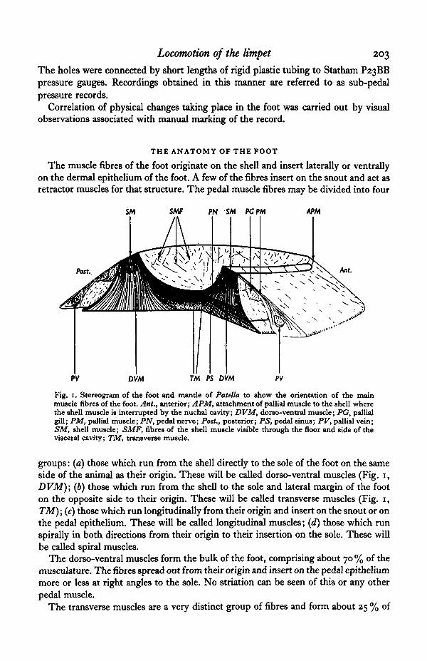
The muscle fibres of the foot originate on the shell and insert laterally or ventrallyon the dermal epithelium of the foot. A few of the fibres insert on the snout and act asretractor muscles for that structure. The pedal muscle fibres may be divided into four
SMF PN SM PGPM APM
DVM TM PS DVM PV
Fig. 1. Stereogram of the foot and mantle of Patella to show the orientation of the mainmuscle fibres of the foot. Ant., anterior; APM, attachment of pallial muscle to the shell wherethe shell muscle is interrupted by the nuchal cavity; DVM, dorso-ventral muscle; PG, pallialgill; PM, pallial muscle; PN, pedal nerve; Post., posterior; PS, pedal sinus; PV, pallial vein;SM, shell muscle; SMF, fibres of the shell muscle visible through the floor and side of thevisceral cavity; TM, transverse muscle.
groups: (a) those which run from the shell directly to the sole of the foot on the sameside of the animal as their origin. These will be called dorso-ventral muscles (Fig. 1,DVM); (b) those which run from the shell to the sole and lateral margin of the footon the opposite side to their origin. These will be called transverse muscles (Fig. i,TM); (c) those which run longitudinally from their origin and insert on the snout or onthe pedal epithelium. These will be called longitudinal muscles; (d) those which runspirally in both directions from their origin to their insertion on the sole. These willbe called spiral muscles.
The dorso-ventral muscles form the bulk of the foot, comprising about 70% of themusculature. The fibres spread out from their origin and insert on the pedal epitheliummore or less at right angles to the sole. No striation can be seen of this or any otherpedal muscle.
The transverse muscles are a very distinct group of fibres and form about 25 % of
204 H. D. JONES AND E. R. TRUEMAN
the pedal musculature. They run as a band immediately below the visceral sinus^forming the ventral boundary of the sinus. They remain as a compact group until theycross the mid line of the foot, from where they spread out to their insertions. Themid line of the foot consists entirely of crossing transverse muscle fibres. The trans-verse fibres pass dorsally to the pedal nerve cord on the same side as their origin andthe thickness between the nerve cord and the visceral sinus is made up entirely oftransverse fibres (Fig. i).
The spiral and longitudinal muscles together form the other 5 % of the foot. Thespiral muscles retain their identity as bundles for some two-thirds of their length,becoming diffuse as they near the sole. Ansell (personal communication and 1969)has noted that Patella has a defence reaction to predators which has been studiedusing Nucella lapillus as a stimulus. The animal raises its shell on the side of contactwith the predator and repeatedly brings the shell down on to the substrate with someforce. After two or three of these violent contractions the animal will sometimes spinits shell through as much as 900. As Weber (1925) suggested, it would seem that thespiral muscles rotate the shell.
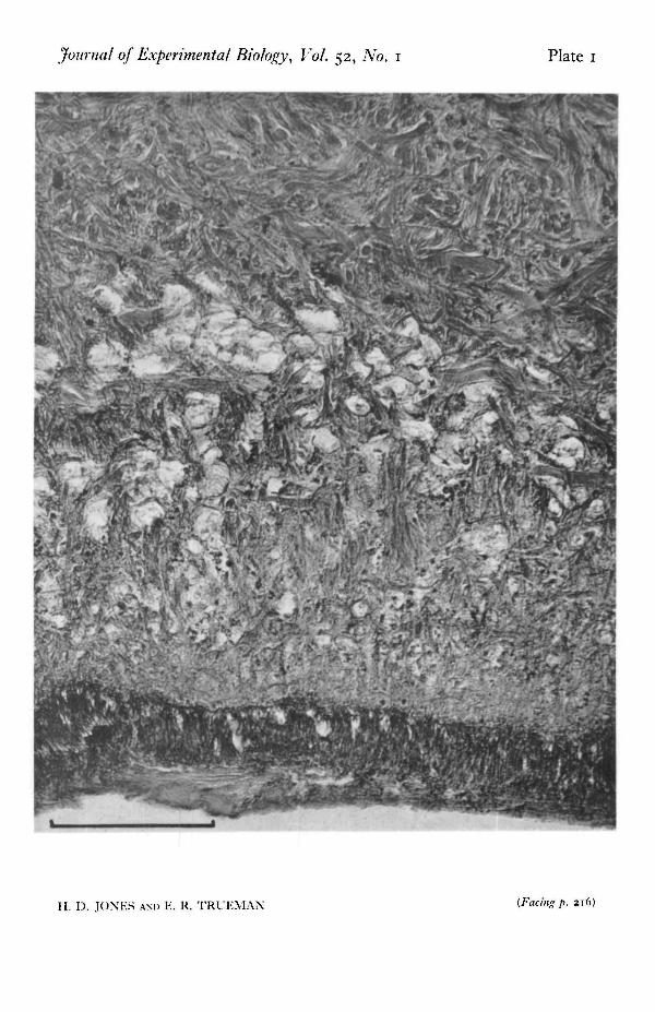
In the foot of Patella there are very few muscle fibres which run longitudinally.Some of these fibres run from the posterior end of the shell muscle, through thetransverse muscle and insert on the snout or on the anterior margin of the foot.Some fibres run from the anterior end of the shell muscle and insert on the pos-terior margin of the foot. Morton (1964) shows a diagram of the foot of a 'generalizedprosobranch gastropod' which includes a very thin layer of longitudinal muscleimmediately above the sole. No such layer is visible in Patella (Plate 1).
The haemocoel spaces of the foot receive blood from the cephalic sinus (Fretter &Graham, 1962). Blood flows into two pedal sinuses, each adjacent to the two pedalnervecords (Fig. 1), whence it diffuses into the remaining pedal haemocoel. There isa region extending for approximately 0-5 mm. above the sole which contains manydistinct, more or less spherical, spaces each about 10/t in diameter. The rest of thefoot has very few blood spaces except for several channels running up the side of thefoot which may be for the venous return of the blood.
The importance of the blood in the foot should not be underestimated, for, asClark (1964) points out, the blood forms virtually the only skeletal support for the foot.
Tests for elastic tissue using the orcinol/new fuchsin technique (Pearse, i960) werecarried out on the foot of Patella but with negative results.
EXPERIMENTAL OBSERVATIONS
(1) Visual and photographic
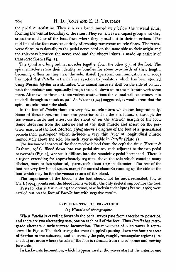
When Patella is crawling forwards the pedal waves pass from anterior to posterior,and there are two alternating sets, one on each half of the foot. Thus Patella has retro-grade alternate ditaxic forward locomotion. The movement of such waves is repre-sented in Fig. 2. The dark triangular areas (stippled) passing down the foot are areasof fixation to the substrate, and conversely the pale, roughly rectangular regions (un-shaded) are areas where the sole of the foot is released from the substrate and movingforwards.
In backwards locomotion, which happens rarely, the waves start at the anterior end
Locomotion of the limpet 205
of the foot as in forward locomotion and are alternate ditaxic. However, as they arenow travelling in the same direction as the animal is moving then they are directwaves. The darker waves now represent moving areas of the foot and numerouscreases can be seen in these dark regions, indicating that the foot is here compressedlongitudinally. The paler areas thus represent fixed and stationary regions of the foot.
35
55
75
1 cm. \
Direction of movement
Fig. 2. Diagrams drawn from cine film of the normal forward locomotion of a limpet whenturning slightly to its left. The stippled areas are regions in which the foot is attached to the sub-strate; the unshaded areas indicate where the foot is released from the substrate and movingforwards. The numbers give time in sixteenths of a second.
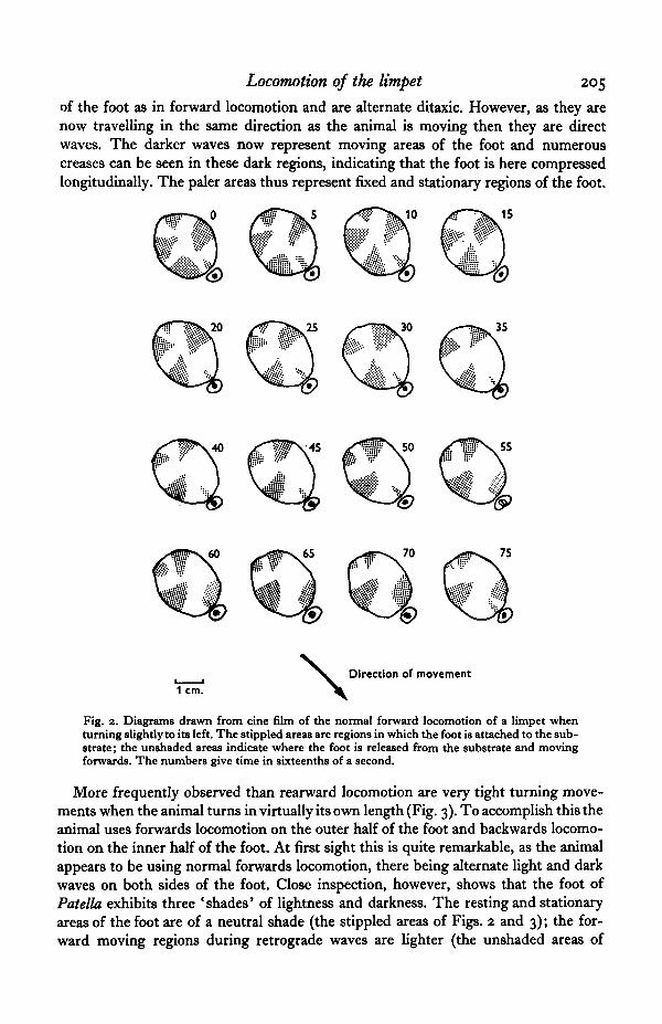
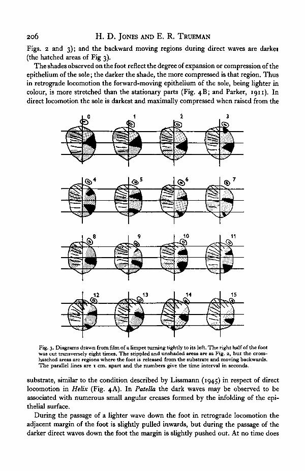
More frequently observed than rearward locomotion are very tight turning move-ments when the animal turns in virtually its own length (Fig. 3). To accomplish this theanimal uses forwards locomotion on the outer half of the foot and backwards locomo-tion on the inner half of the foot. At first sight this is quite remarkable, as the animalappears to be using normal forwards locomotion, there being alternate light and darkwaves on both sides of the foot. Close inspection, however, shows that the foot ofPatella exhibits three 'shades' of lightness and darkness. The resting and stationaryareas of the foot are of a neutral shade (the stippled areas of Figs. 2 and 3); the for-ward moving regions during retrograde waves are lighter (the unshaded areas of
206 H. D . JONES AND E. R. TRUEMAN
Figs. 2 and 3); and the backward moving regions during direct waves are darkei(the hatched areas of Fig 3).
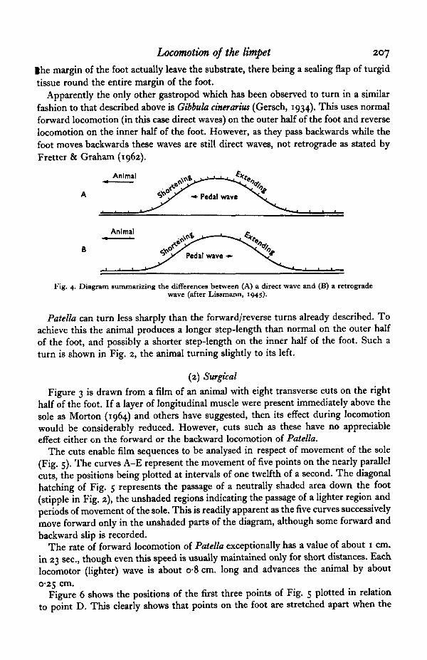
The shades observed on the foot reflect the degree of expansion or compression of theepithelium of the sole; the darker the shade, the more compressed is that region. Thusin retrograde locomotion the forward-moving epithelium of the sole, being lighter incolour, is more stretched than the stationary parts (Fig. 4B; and Parker, 1911). Indirect locomotion the sole is darkest and maximally compressed when raised from the
Fig. 3. Diagrams drawn from film of a limpet turning tightly to its left. The right half of the footwas cut transversely eight times. The stippled and unshaded areas are as Fig. 2, but the cross-hatched areas are regions where the foot is released from the substrate and moving backwards.The parallel lines are I cm. apart and the numbers give the time interval in seconds.
substrate, similar to the condition described by Lissmann (1945) in respect of directlocomotion in Helix (Fig. 4A). In Patella the dark waves may be observed to beassociated with numerous small angular creases formed by the infolding of the epi-thelial surface.
During the passage of a lighter wave down the foot in retrograde locomotion theadjacent margin of the foot is slightly pulled inwards, but during the passage of thedarker direct waves down the foot the margin is slightly pushed out. At no time does
Locomotion of the limpet 207
Ihe margin of the foot actually leave the substrate, there being a sealing flap of turgidtissue round the entire margin of the foot.
Apparently the only other gastropod which has been observed to turn in a similarfashion to that described above is Gibbula cinerarias (Gersch, 1934). This uses normalforward locomotion (in this case direct waves) on the outer half of the foot and reverselocomotion on the inner half of the foot. However, as they pass backwards while thefoot moves backwards these waves are still direct waves, not retrograde as stated byFretter & Graham (1962).
Animal
Animal
Fig. 4. Diagram summarizing the differences between (A) a direct wave and (B) a retrogradewave (after Lissmann, 1945).
Patella can turn less sharply than the forward/reverse turns already described. Toachieve this the animal produces a longer step-length than normal on the outer halfof the foot, and possibly a shorter step-length on the inner half of the foot. Such aturn is shown in Fig. 2, the animal turning slightly to its left.
(2) Surgical
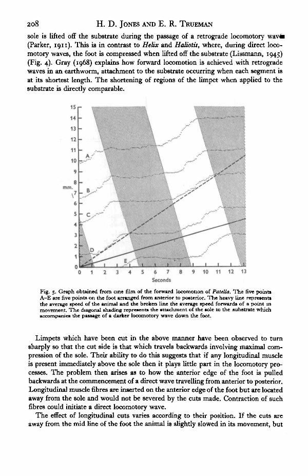
Figure 3 is drawn from a film of an animal with eight transverse cuts on the righthalf of the foot. If a layer of longitudinal muscle were present immediately above thesole as Morton (1964) and others have suggested, then its effect during locomotionwould be considerably reduced. However, cuts such as these have no appreciableeffect either en the forward or the backward locomotion of Patella.
The cuts enable film sequences to be analysed in respect of movement of the sole(Fig. 5). The curves A-E represent the movement of five points on the nearly parallelcuts, the positions being plotted at intervals of one twelfth of a second. The diagonalhatching of Fig. 5 represents the passage of a neutrally shaded area down the foot(stipple in Fig. 2), the unshaded regions indicating the passage of a lighter region andperiods of movement of the sole. This is readily apparent as the five curves successivelymove forward only in the unshaded parts of the diagram, although some forward andbackward slip is recorded.
The rate of forward locomotion of Patella exceptionally has a value of about 1 cm.in 23 sec, though even this speed is usually maintained only for short distances. Eachlocomotor (lighter) wave is about o-8 cm. long and advances the animal by about0-25 cm.
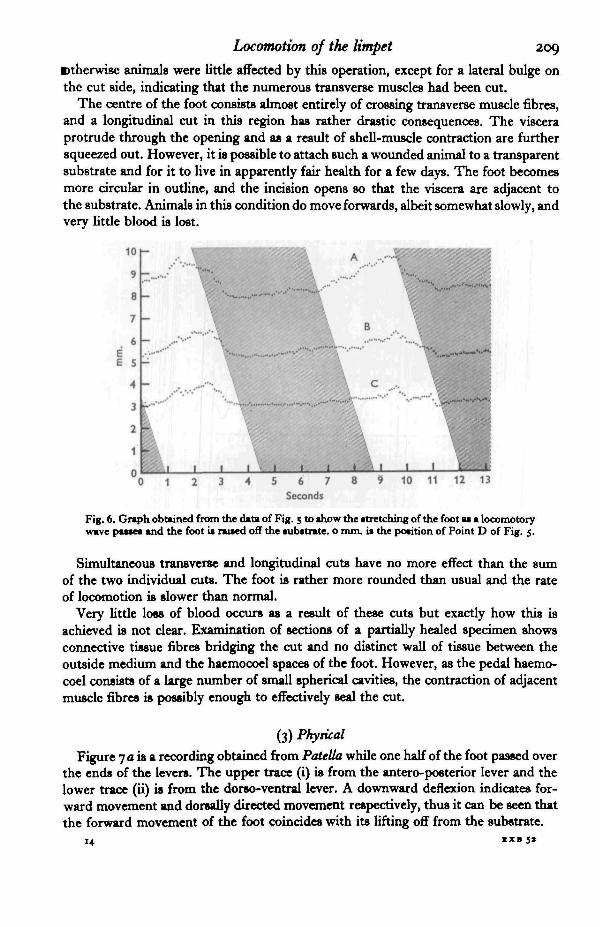
Figure 6 shows the positions of the first three points of Fig. 5 plotted in relationto point D. This clearly shows that points on the foot are stretched apart when the
208 H. D. JONES AND E. R. TRUEMAN
sole is lifted off the substrate during the passage of a retrograde locomotory(Parker, 1911). This is in contrast to Helix and Haliotis, where, during direct loco-motory waves, the foot is compressed when lifted off the substrate (Lissmann, 1945)(Fig. 4). Gray (1968) explains how forward locomotion is achieved with retrogradewaves in an earthworm, attachment to the substrate occurring when each segment isat its shortest length. The shortening of regions of the limpet when applied to thesubstrate is directly comparable.
13
Fig. 5. Graph obtained from cine film of the forward locomotion of Patella. The five point*A-E are five points on the foot arranged from anterior to posterior. The heavy line represent*the average speed of the animal and the broken line the average speed forwards of a point inmovement. The diagonal shading represents the attachment of the sole to the substrate whichaccompanies the passage of a darker locomotory wave down the foot.
Limpets which have been cut in the above manner have been observed to turnsharply so that the cut side is that which travels backwards involving maximal com-pression of the sole. Their ability to do this suggests that if any longitudinal muscleis present immediately above the sole then it plays little part in the locomotory pro-cesses. The problem then arises as to how the anterior edge of the foot is pulledbackwards at the commencement of a direct wave travelling from anterior to posterior.Longitudinal muscle fibres are inserted on the anterior edge of the foot but are locatedaway from the sole and would not be severed by the cuts made. Contraction of suchfibres could initiate a direct locomotory wave.
The effect of longitudinal cuts varies according to their position. If the cuts areaway from the mid line of the foot the animal is slightly slowed in its movement, but
Locomotion of the limpet 209
Btherwise animals were little affected by this operation, except for a lateral bulge onthe cut side, indicating that the numerous transverse muscles had been cut.
The centre of the foot consists almost entirely of crossing transverse muscle fibres,and a longitudinal cut in this region has rather drastic consequences. The visceraprotrude through the opening and as a result of shell-muscle contraction are furthersqueezed out. However, it is possible to attach such a wounded animal to a transparentsubstrate and for it to live in apparently fair health for a few days. The foot becomesmore circular in outline, and the incision opens so that the viscera are adjacent tothe substrate. Animals in this condition do move forwards, albeit somewhat slowly, andvery little blood is lost.
10
9
8
7
6
E 5
4
3
2
1
0 1 t 1 1 1
0 1 2 3 4 5 6 7 8 9 10 11 12 13Seconds
Fig. 6. Graph obtained from the data of Fig. 5 to show the stretching of the foot as a locomotorywave passes and the foot is rawed off the substrate, o mm. is the position of Point D of Fig. 5.
Simultaneous transverse and longitudinal cuts have no more effect than the sumof the two individual cuts. The foot is rather more rounded than usual and the rateof locomotion is slower than normal.
Very little loss of blood occurs as a result of these cuts but exactly how this isachieved is not clear. Examination of sections of a partially healed specimen showsconnective tissue fibres bridging the cut and no distinct wall of tissue between theoutside medium and the haemocoel spaces of the foot. However, as the pedal haemo-coel consists of a large number of small spherical cavities, the contraction of adjacentmuscle fibres is possibly enough to effectively seal the cut.
(3) Physical
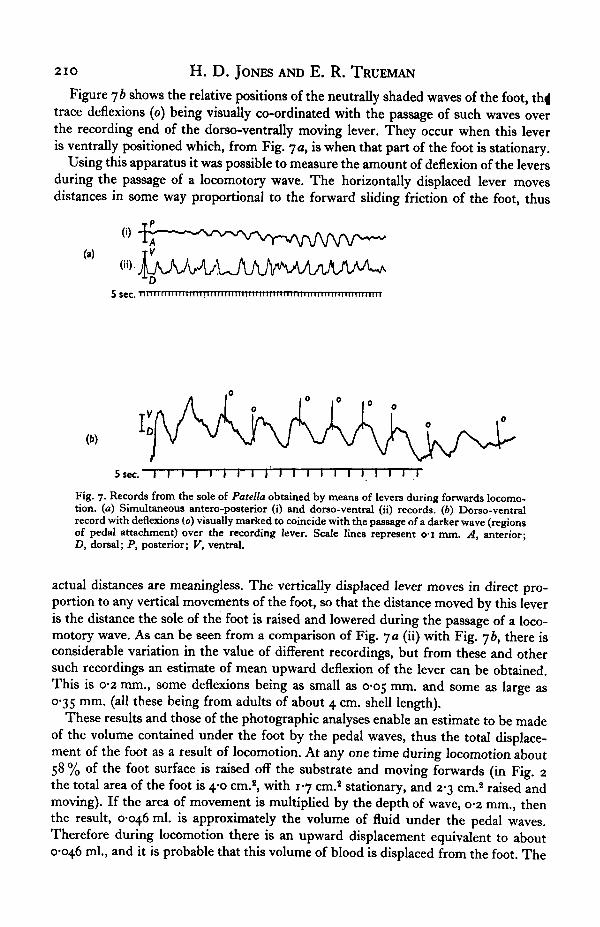
Figure 7 a is a recording obtained from Patella while one half of the foot passed overthe ends of the levers. The upper trace (i) is from the antero-posterior lever and thelower trace (ii) is from the dorso-ventral lever. A downward deflexion indicates for-ward movement and dorsally directed movement respectively, thus it can be seen thatthe forward movement of the foot coincides with its lifting off from the substrate.
U ixssa
210 H. D . JONES AND E. R. TRUEMAN
Figure jb shows the relative positions of the neutrally shaded waves of the foot, thi|trace deflexions (o) being visually co-ordinated with the passage of such waves overthe recording end of the dorso-ventrally moving lever. They occur when this leveris ventrally positioned which, from Fig. ja, is when that part of the foot is stationary.
Using this apparatus it was possible to measure the amount of deflexion of the leversduring the passage of a locomotory wave. The horizontally displaced lever movesdistances in some way proportional to the forward sliding friction of the foot, thus
5 sec. i
(b)
5 sec. I I I I I I I I I I I I I I I I IFig. 7. Records from the sole of Patella obtained by means of levers during forwards locomo-tion, (a) Simultaneous antero-posterior (i) and dorso-ventral (ii) records. (6) Dorso-ventralrecord with deflexions (0) visually marked to coincide with the passage of a darker wave (regionsof pedal attachment) over the recording lever. Scale lines represent o-i mm. A, anterior;D, dorsal; P, posterior; V, ventral.
actual distances are meaningless. The vertically displaced lever moves in direct pro-portion to any vertical movements of the foot, so that the distance moved by this leveris the distance the sole of the foot is raised and lowered during the passage of a loco-motory wave. As can be seen from a comparison of Fig. ya (ii) with Fig. yb, there isconsiderable variation in the value of different recordings, but from these and othersuch recordings an estimate of mean upward deflexion of the lever can be obtained.This is 0-2 mm., some deflexions being as small as 0-05 mm. and some as large as0-35 mm. (all these being from adults of about 4 cm. shell length).
These results and those of the photographic analyses enable an estimate to be madeof the volume contained under the foot by the pedal waves, thus the total displace-ment of the foot as a result of locomotion. At any one time during locomotion about58 % of the foot surface is raised off the substrate and moving forwards (in Fig. 2the total area of the foot is 4-0 cm.2, with 17 cm.2 stationary, and 2-3 cm.2 raised andmoving). If the area of movement is multiplied by the depth of wave, 0-2 mm., thenthe result, 0-046 ml. is approximately the volume of fluid under the pedal waves.Therefore during locomotion there is an upward displacement equivalent to about0-046 ml., and it is probable that this volume of blood is displaced from the foot. The
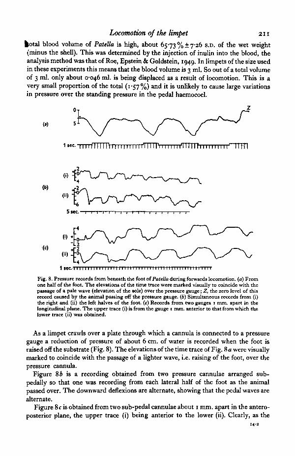
Locomotion of the limpet 211
fcotal blood volume of Patella is high, about 6573 % ± 7-26 S.D. of the wet weight(minus the shell). This was determined by the injection of inulin into the blood, theanalysis method was that of Roe, Epstein & Goldstein, 1949. In limpets of the size usedin these experiments this means that the blood volume is 3 ml. So out of a total volumeof 3 ml. only about 0-046 ml. is being displaced as a result of locomotion. This is avery small proportion of the total (1*57%) and it is unlikely to cause large variationsin pressure over the standing pressure in the pedal haemocoel.
1 $ec-~TTTTTIi11 n 1111111111iTTTTPi 111111 I'I 1111 r 111111 i 111• u r n
( I I ) .
1 seel 1 1 1 1 1 1 1 1 1 1 1 1 1 1 1 1 1 1 1 1 1 1 1
Fig. 8. Pressure records from beneath the foot of Patella during forwards locomotion, (a) Fromone half of the foot. The elevations of the time trace were marked visually to coincide with thepassage of a pale wave (elevation of the sole) over the pressure gauge; Z, the zero level of thisrecord caused by the animal passing off the pressure gauge. (6) Simultaneous records from (i)the right and (ii) the left halves of the foot, (c) Records from two gauges i mm. apart in thelongitudinal plane. The upper trace (i) is from the gauge i mm. anterior to that from which thelower trace (ii) was obtained.
As a limpet crawls over a plate through which a cannula is connected to a pressuregauge a reduction of pressure of about 6 cm. of water is recorded when the foot israised off the substrate (Fig. 8). The elevations of the time trace of Fig. 8 a were visuallymarked to coincide with the passage of a lighter wave, i.e. raising of the foot, over thepressure cannula.
Figure 8 A is a recording obtained from two pressure cannulae arranged sub-pedally so that one was recording from each lateral half of the foot as the animalpassed over. The downward deflexions are alternate, showing that the pedal waves arealternate.
Figure 8 c is obtained from two sub-pedal cannulae about i mm. apart in the antero-posterior plane, the upper trace (i) being anterior to the lower (ii). Clearly, as the
14-2
212 H. D. JONES AND E. R. TRUEMAN
animal crawls forward over the cannulae the posterior one will be reached first andFig. Sc (ii) shows a deflexion before (i). Thereafter both traces have a very similarshape, the anterior one (i) showing negative pressure slightly sooner than the posteriortrace (ii) due to the rearward direction of the locomotory wave.
DISCUSSION
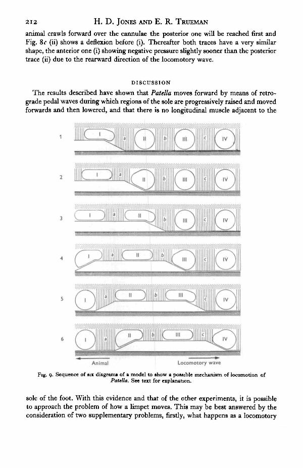
The results described have shown that Patella moves forward by means of retro-grade pedal waves during which regions of the sole are progressively raised and movedforwards and then lowered, and that there is no longitudinal muscle adjacent to the
Animal Locomotory wave
Fig. 9. Sequence of six diagrams of a model to show a possible mechanism of locomotion ofPatella. See text for explanation.
sole of the foot. With this evidence and that of the other experiments, it is possibleto approach the problem of how a limpet moves. This may be best answered by theconsideration of two supplementary problems, firstly, what happens as a locomotory
Locomotion of the limpet 213
jvave passes to cause forward movement, and secondly, how is the anterior edge of thetoot moved forwards at the start of a locomotory wave.
Figure 9 shows six diagrams to represent the sequence of events in a possiblemodel which answers the first of the two problems. This produces forward movementutilizing the dorso-ventral muscles as the main propulsive agents, with the haemocoelspaces of the foot and the transverse muscles as essential factors. The pedal haemocoelconsists largely of spherical cavities, I-IV, immediately above the epithelium of thesole, these are assumed to be of constant volume and to have no lateral displacement.The latter is not strictly true as the foot is slightly narrowed as the foot passes, butthis complements the following explanation and does not detract from it. That a con-stant volume of blood is retained in the haemocoel cavities is suggested by the absenceof bleeding when incisions are made in the foot.
Locomotory wave Animal
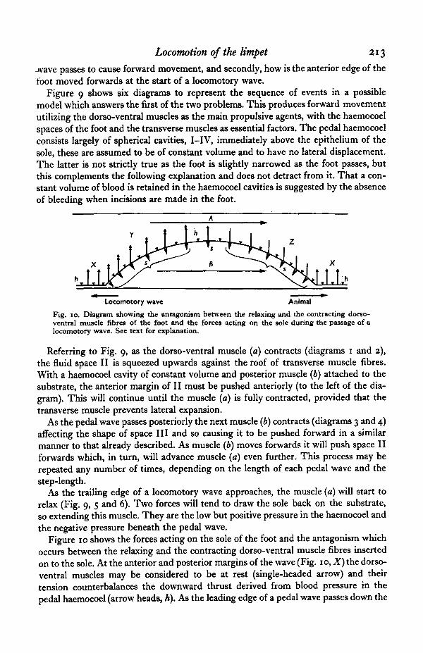
Fig. 10. Diagram showing the antagonism between the relaxing and the contracting dorso-ventral muscle fibres of the foot and the forces acting on the sole during the passage of alocomotory wave. See text for explanation.
Referring to Fig. 9, as the dorso-ventral muscle (a) contracts (diagrams 1 and 2),the fluid space II is squeezed upwards against the roof of transverse muscle fibres.With a haemocoel cavity of constant volume and posterior muscle (b) attached to thesubstrate, the anterior margin of II must be pushed anteriorly (to the left of the dia-gram). This will continue until the muscle (a) is fully contracted, provided that thetransverse muscle prevents lateral expansion.
As the pedal wave passes posteriorly the next muscle (b) contracts (diagrams 3 and 4)affecting the shape of space III and so causing it to be pushed forward in a similarmanner to that already described. As muscle (b) moves forwards it will push space IIforwards which, in turn, will advance muscle (a) even further. This process may berepeated any number of times, depending on the length of each pedal wave and thestep-length.
As the trailing edge of a locomotory wave approaches, the muscle (a) will start torelax (Fig. 9, 5 and 6). Two forces will tend to draw the sole back on the substrate,so extending this muscle. They are the low but positive pressure in the haemocoel andthe negative pressure beneath the pedal wave.
Figure 10 shows the forces acting on the sole of the foot and the antagonism whichoccurs between the relaxing and the contracting dorso-ventral muscle fibres insertedon to the sole. At the anterior and posterior margins of the wave (Fig. 10, X) the dorso-ventral muscles may be considered to be at rest (single-headed arrow) and theirtension counterbalances the downward thrust derived from blood pressure in thepedal haemocoel (arrow heads, A). As the leading edge of a pedal wave passes down the
214 H. D. JONES AND E. R. TRUEMAN
foot the dorso-ventral muscles contract ( Y), exerting an upward force on the sole of thdfoot (double-headed arrows). This upward force would cause a slight local increase inthe pressure in the pedal haemocoel and a decrease in pressure under the foot (arrowheads, s). The relaxing dorso-ventral muscle (at Z) will be drawn down and restoredto its resting length both by the positive pressure in the haemocoel (Fig. 10, arrow A)and by the negative pressure under the foot (arrow 5).
All that is needed to produce movement of the wave down the foot is for the succes-sive contraction and relaxation of the dorso-ventral muscles to proceed posteriorly,forward movement of the foot could then be produced solely by the hydraulic proper-ties of the foot.
Chapman (1958) points out that the primary function of a skeleton is to providea means by which opposing muscles may be antagonized or brought to bear on eachother for the restitution of their relaxed state. Since the enclosed cavity of the pedalwave of Patella would seem to act to restore the fibres at the trailing edge of the pedalwave to their resting length by the contraction of the fibres at the leading edge of thewave, then it is possible to regard the cavity as a rather novel type of external hydro-static skeleton. In this case the fibres of the leading edge of the wave are antagonisticagainst fibres at the trailing edge of the wave.
Some of the work done by the contracting dorso-ventral muscle fibres is thereforeexpended in the restitution of other muscle fibres to their resting length, but most ofthe remainder may be turned into a propulsive force by the hydraulic properties of thefoot. This system, as already stressed, requires that the lateral extension of the footbe strictly controlled, and in Patella there exist numerous transverse muscle fibreswhich may perform this function. This is evidenced by the reduction of speed whenthe transverse muscles are cut.
The other main problem concerning the limpet's locomotion is how the anteriormargin of the foot is advanced at the start of a pedal wave. The solution is a continua-tion of the preceding paragraphs. If the pedal haemocoel near the front of the foot isvertically narrowed and little or no blood escapes then either a lateral or forwardextension of the foot must occur. The lateral extent of the foot is controlled by thetransverse muscles so that forward movement must follow and the process describedabove may then proceed.
The backward locomotion of Patella using direct waves may be similarly explaineddespite the lack of longitudinal muscle next to the sole. The contraction of the longi-tudinal muscle that inserts on the anterior margin of the foot (but runs for most of itslength away from the sole) could cause a compression of the anterior edge of the foot.Such a compression is observed as a series of angular inpushings of the sole of the footin this region. Once this initial compression has been achieved then all that is necessaryis for the dorso-ventral muscles behind the compressed area to contract so as tocause this area to automatically pass backwards, assuming that the epithelium ofthe foot is not entirely rigid. Relaxation of the dorso-ventral muscles would occurat the trailing edge of the wave in a similar manner to that described for the retrogradelocomotory wave, except that slight increase in pressure under the foot may be ex-pected. It would seem that the epithelium of the sole of the foot of Patella is not able tobe compressed beyond its resting length as it is thrown into angular folds by the initialcontraction of the longitudinal muscle. This can be compared to Lissmann's (1945)
Locomotion of the limpet 215
examples of direct locomotory waves, where in Helix the foot has relatively smoothbut apparently deep waves, and in Haliotis, where the foot has arched and creasedwaves. The latter enables a longer step-length (10 mm.) than either Helix (1 mm.) orPatella (2-5 mm.).
The above explanation requires no longitudinal muscle in the foot except for somefibres inserted on the anterior margin of the foot of Patella or the rear margin of thefeet of Helix and Haliotis. Helix has longitudinal muscle in the foot which inserts onthe posterior margin of the foot (Trappmann, 1916; Weber, 1925; Elves, 1961). Weber(1925) could find no longitudinal muscle in the foot of Haliotis but there are musclesrunning down from the shell to the posterior edge of the foot and which could pull thelatter forwards at the commencement of a direct pedal wave.
These remarks concerning the locomotion of Haliotis and Helix require furtherexperimental elucidation, but in Patella the observations discussed show that the dorso-ventral muscles alone are capable of producing forward locomotion, some of the workdone being converted into forward locomotion by the hydraulic properties of the foot.Rearward locomotion may also be produced solely by the action of the dorso-ventralmuscles but only after some longitudinal muscle has pulled back the anterior edge ofthe foot.
SUMMARY
1. The locomotion of Patella has been studied by histological, photographic andexperimental techniques. The foot consists principally of dorso-ventral and transversemuscles and has no longitudinal muscle fibres near to its sole. The pedal haemocoel islimited to a region of small spherical cavities, extending for 0-5 mm. above the sole,two pedal sinuses and several lateral vertical channels.
2. Patella moves forward by means of retrograde alternate ditaxic pedal locomotorywaves, but during rarely observed backward movement the waves are direct. Duringtight turns the limpet uses forward locomotion on one half of the foot and backwardlocomotion on the other.
3. During the passage of a retrograde locomotory wave the foot is lifted off thesubstrate by about 0-2 mm. and the pressure beneath the sole falls by about 6 cm. ofwater.
4. A model is proposed to account for locomotion utilizing the dorso-ventralmuscles as the sole propulsive agents in the hydraulic system of the foot. This systemconsists principally of the dorso-ventral and transverse muscles, the spherical cavitiesof the pedal haemocoel and the fluid enclosed beneath each locomotory wave. Bothfluid systems may be utilized during contraction and relaxation of different groupsof dorso-ventral muscles.
We thank Professors P. G. Espinasse and J. G. Phillips for the provision of facilitiesand the Science Research Council for a research studentship (H. D. J.).
2i6 H. D. JONES AND E. R. TRUEMAN
REFERENCES
ANSELL, A. D. (1969). Defensive adaptations to predation in the Mollusca. Symp. Mar. Biol. Ass., India(in the Press).
BONSE, H. (1935). Ein Beitrag zum Problem der Schneckenbewegung. Zool. Jb. (Zool. Physiol.) 54,349-84-
CATE, J. TEN (1922). Quelques observations sur la locomotion de l'escargot des vignes. Archs neerl.Physiol. 7, 103-111.
CHAPMAN, G. (1958). The hydrostatic skeleton in the invertebrates. Biol. Rev. 33, 338-71.CLARK, R. B. (1964). Dynamics in Metazoan Evolution. Oxford: Clarendon Press.ELVES, M. W. (1961). The histology of the foot of Discus rotundatus and the locomotion of gastropod
molluscs. PTOC. malac. Soc. Land. 34, 346-55.FRETTER, V. & GRAHAM, A. (1962). British Prosobranch Molluscs. London: Ray Society.GERSCH, M. (1934). Zur experimentellen VerSnderung der Richtung der Wellenbewegung auf der
Kriechsohle von Schnecken und zur Ruckwartsbewegung von Schnecken. Biol. Zbl. 54, 511-18.GRAY, J. (1968). Animal Locomotion. London: Weidenfeld and Nicholson.LISSMANN, H. W. (1945). The mechanism of locomotion in gastropod molluscs. I. Kinematics. J. exp.
Biol. 21, 58-69.LISSMANN, H. W. (1946). The mechanism of locomotion in gastropod molluscs. II. Kinetics. J. exp.
Biol. 22, 37-50-MORTON, J. E. (1964). Locomotion. In Physiology of Molluscs, vol. 1, pp. 383-423. Eds. K. M. Wilbur
and C. M. Yonge, New York: Academic Press.PARKER, G. H. (1911). The mechanism of locomotion in gastropods. J. Morph. 22, 155-70.PEARSE, A. G. E. (i960). Histochemistry. London: J. and A. Churchill.ROE, J. H., EPSTEIN, J. H. & GOLDSTEIN, N. P. (1949). A photometric method for the determination
of inulin in plasma and urine. J. biol. Chem. 178, 839-45.ROTARIDES, M. (1936). Die morphologischen Grundlagen der Lokomotion der Schnecken. M. Tud.
Akad. Mat. dis Termeszettud. 66.ROTARIDES, M. (1945). Zur Micromorphologie des Fusses der Patelloiden Schnecken. Atmls Hist. Nat.
Mus. Nat. Hung. 38, 1-36.RUNHAM, N. W., ISARANKURA, K. & SMITH, B. J. (1965). Methods for narcotising and anaesthatising
gastropods. Malacologia 3, 231-8.TRAPPMANN, W. (1916). Die Muskulatur von Helix pomatia L. Z. tviss. Zool. 115, 489-585.VLES, F. (1907). Sur les ondes pldieuses des mollusques reptateurs. C. R. Acad. Set., Paris 145, 276-8.WEBER, H. (1925). Ober die Umdrehreflexe einiger Prosobranchier des Golfs von Neapel. Ein Beitrag
zur Bewegungsphysiologie und Reflexbiologie der Gastropoden. Z. vergl. Physiol. 3, 389-474.
EXPLANATION OF PLATE
Micrograph of longitudinal section of Patella showing the epithelium of the sole and the region im-mediately dorsal to it. Note the lack of longitudinal muscle near the sole and the numerous sphericalcavities of the pedal haemocoel. Scale line = 0-1 mm.
Journal of Experimental Biology, Vol. 52, No. 1 Plate 1
II. D. JONKS AND K. R. TRUKMAN (Facing p. 216)