
Localized Pulmonary Hemorrhage Associated With Massive ...
5
113 INTRODUCTION Localized pulmonary hemorrhage is a diagnostic term referring to hemorrhage localized in one portion of lung, usually a lobe, and is markedly different from diffuse alveolar hemorrhage. Diffuse alveolar hemorrhage is always associated with pulmonary capillaritis initiated by an immune mechanism [1]. The etiology of localized pulmonary hemorrhage is quite variable, and includes localized tumor, ulceration, and cavitary lesion. In many cases, there is no gross abnormality (other than blood), which poses a difficult and often frustrating problem for the pathologist. In some authors' experience, the source of bleeding in such cases is almost always the bronchi, and serial sections should be taken from the bronchial tree to secure the underlying etiology. In a significant number of cases, however, the source of bleeding was not identified [2]. CASE REPORT A previously healthy 48-year-old man presented with massive hemoptysis and profound fatigue. One month prior to admission, he had his first episode of coughing-up blood. In the subsequent weeks, he had intermittent bloody cough about once a week. There was no fever, dyspnea, arthralgia, weight loss, or bleeding diathesis. He had no systemic disease such as liver cirrhosis, renal insufficiency or hematology disease. He smoked about 25 packs per year. His occupational history included 10 years' employment at a plastic factory. There was no history of tuberculosis. On admission, he presented with cough with blood amounting Massive hemoptysis is a rare but emergent condition which requires prompt attention. In this case study, a 48-year-old man presented with massive hemoptysis. Chest computerized tomography obtained after admission demonstrated focal density in the right-middle lobe and presence of nodulointerstitial infiltrate in both lungs. Active bleeding limited fibrobronchoscopy. Therapeutic bronchial artery embolization was attempted prior to surgery. An uneventful resection of the right-middle lobe was performed and the pathological diagnosis was localized pulmonary hemorrhage. ( Mid Taiwan J Med 2002;7:113-7) Key words hemoptysis, pulmonary hemorrhage Received : September 26, 2001. Revised : November 23, 2001. Accepted : November 26, 2001. Address reprint requests to : Yang-Hao Yu, Devision of Pulmonary and Critical Care, China Medical College Hospital, No 2, Yuh-Der Road, Taichung 404, Taiwan, R.O.C. Localized Pulmonary Hemorrhage Associated With Massive Hemoptysis: Report of a Case Yang-Hao Yu, Te-Chun Hsia, Liang-Wen Hang, Tze-Yi Lin 1 , Nan-Yung Hsu 2 , Der-Yuan Wang 3 Devision of Pulmonary and Critical Care, 1 Department of Pathology; 2 Department of Chest Surgery, China Medical College Hospital; 3 Department of Internal Medicine, Lin-Shin Hospital, Taichung, Taiwan, R.O.C.
Transcript of Localized Pulmonary Hemorrhage Associated With Massive ...
113
IINNTTRROODDUUCCTTIIOONN
Localized pulmonary hemorrhage is adiagnostic term referring to hemorrhagelocalized in one portion of lung, usually alobe, and is markedly different from diffusealveolar hemorrhage. Diffuse alveolarhemorrhage is always associated withpulmonary capillaritis initiated by an immunemechanism [1] . The etiology of localizedpulmonary hemorrhage is quite variable, andincludes localized tumor, ulceration, andcavitary lesion. In many cases, there is nogross abnormality (other than blood), whichposes a difficult and often frustrating problemfor the pathologist . In some authors'experience, the source of bleeding in suchcases is almost always the bronchi, and serial
sections should be taken from the bronchialtree to secure the underlying etiology. In asignificant number of cases, however, thesource of bleeding was not identified [2].
CCAASSEE RREEPPOORRTT
A previously healthy 48-year-old manpresented with massive hemoptysis andprofound fatigue. One month prior toadmission, he had his first episode ofcoughing-up blood. In the subsequent weeks,he had intermittent bloody cough about oncea week. There was no fever, dyspnea,arthralgia, weight loss, or bleeding diathesis.He had no systemic disease such as livercirrhosis, renal insufficiency or hematologydisease. He smoked about 25 packs per year.His occupational history included 10 years'employment at a plastic factory. There was nohistory of tuberculosis. On admission, hepresented with cough with blood amounting
Massive hemoptysis is a rare but emergent condition which requires prompt attention. In this
case study, a 48-year-old man presented with massive hemoptysis. Chest computerized
tomography obtained after admission demonstrated focal density in the right-middle lobe and
presence of nodulointerstitial infiltrate in both lungs. Active bleeding limited
fibrobronchoscopy. Therapeutic bronchial artery embolization was attempted prior to surgery.
An uneventful resection of the right-middle lobe was performed and the pathological diagnosis
was localized pulmonary hemorrhage. ( Mid Taiwan J Med 2002;7:113-7)
KKeeyy wwoorrddss
hemoptysis, pulmonary hemorrhage
Received : September 26, 2001. Revised : November 23, 2001.Accepted : November 26, 2001.Address reprint requests to : Yang-Hao Yu, Devision of Pulmonary and Critical Care, China Medical College Hospital, No 2, Yuh-DerRoad, Taichung 404, Taiwan, R.O.C.
Localized Pulmonary HemorrhageAssociated With Massive Hemoptysis:
Report of a Case
Yang-Hao Yu, Te-Chun Hsia, Liang-Wen Hang, Tze-Yi Lin1,
Nan-Yung Hsu2, Der-Yuan Wang
3
Devision of Pulmonary and Critical Care, 1Department of Pathology;
2Department of
Chest Surgery, China Medical College Hospital; 3Department of Internal Medicine,
Lin-Shin Hospital, Taichung, Taiwan, R.O.C.

114 Localized Pulmonary
to 250 mL.On examination, he was acutely ill and
weak. His vital signs were: BP 130/70 mmHg,
PR 100/min, RR 18/min, BT 36.8 C. There wasno respiratory distress or labored breathing.Some inspiratory coarse crackles were audiblefrom the right-lower anterior chest wall. Hehad neither clubbing digit nor palpabletelangiectasis. Cervical and supraclavicularlymph nodes were palpated but normal. The
chest roentgenogram (Figs. 1,2) remainedunchanged during the one month follow-up.His hemoglobin was 110 g/L and his white
blood cell count was 7,200/mm3. Results of his
coagulating profile and biochemical laboratorysurvey, including antinuclear antibody, werenormal.
Bronchoscopy was performed butlimited by profound hemoptysis during theprocedure. Emergent bronchial arteryembolization was attempted but notperformed due to multiple supply of thelesion and common supply of the intercostaland spinal artery. He received surgicalintervention with RML resection.
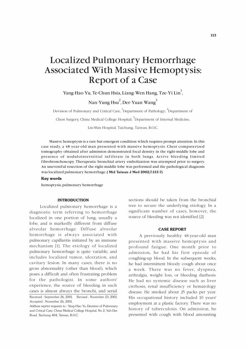
The surgical specimen (Fig. 3) revealedparenchymal hemorrhage with firm palpation.There were some blood clots within the smallbronchus, but no change of bronchiectasis,bronchial mural thickening, or other mucosalabnormalities. The pulmonary arterial andvenous systems were unremarkable.
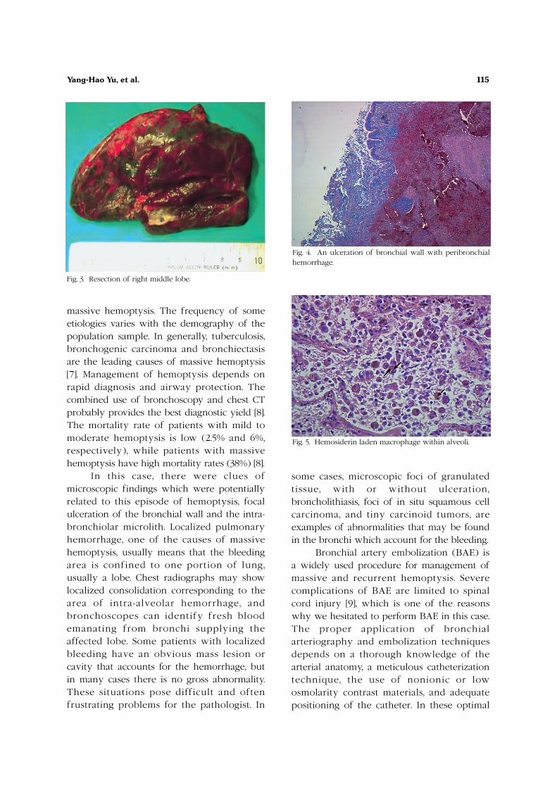
Microscopically, the alveolar parenchymashowed marked hemorrhage with organizedhematoma. Sections from the grosshemorrhage area showed intrabronchiolar andintra-alveolar hemorrhage with a secondarycompensatory emphysematous appearance inthe adjacent lung tissue. There was evidenceof a focal ulcerative area in the bronchial walland peribronchial hemorrhage (Fig. 4). Amicrolith was also noted within thebronchiole. Chronic inflammatory cellsinfiltrated the peribronchiolar region, andsome hemosiderrin-laden macrophages (Fig. 5)were present. No evidence of dysplasia ormitotic cells were noted in the ulcerativeepithelium, and no granulation tissue wasdemonstrated.
DDIISSCCUUSSSSIIOONN
The definition of massive hemoptysisvaries widely in the literature, from 200 to1000 mL/24 hrs [3,4], but more than 600 mL in24 hours is what most authors use in clinicalreports to define massive hemoptysis [5],which accounts for approximately 1.5% of allhemoptysis [6]. There are many causes of
Fig. 1. CXR of PA view showed non-specific findings withfocal infiltrate over Rt heart border compatible with RMLlesion.
Fig. 2. Chest CT demonstrated focal density adjacent toright heart border, indicating RML lesion.
115Yang-Hao Yu, et al.
massive hemoptysis. The frequency of someetiologies varies with the demography of thepopulation sample. In generally, tuberculosis,bronchogenic carcinoma and bronchiectasisare the leading causes of massive hemoptysis[7]. Management of hemoptysis depends onrapid diagnosis and airway protection. Thecombined use of bronchoscopy and chest CTprobably provides the best diagnostic yield [8].The mortality rate of patients with mild tomoderate hemoptysis is low (2.5% and 6%,respectively), while patients with massivehemoptysis have high mortality rates (38%) [8].
In this case, there were clues ofmicroscopic findings which were potentiallyrelated to this episode of hemoptysis, focalulceration of the bronchial wall and the intra-bronchiolar microlith. Localized pulmonaryhemorrhage, one of the causes of massivehemoptysis, usually means that the bleedingarea is confined to one portion of lung,usually a lobe. Chest radiographs may showlocalized consolidation corresponding to thearea of intra-alveolar hemorrhage, andbronchoscopes can identify fresh bloodemanating from bronchi supplying theaffected lobe. Some patients with localizedbleeding have an obvious mass lesion orcavity that accounts for the hemorrhage, butin many cases there is no gross abnormality.These situations pose difficult and oftenfrustrating problems for the pathologist. In
some cases, microscopic foci of granulatedtissue, with or without ulceration,broncholithiasis, foci of in situ squamous cellcarcinoma, and tiny carcinoid tumors, areexamples of abnormalities that may be foundin the bronchi which account for the bleeding.
Bronchial artery embolization (BAE) isa widely used procedure for management ofmassive and recurrent hemoptysis. Severecomplications of BAE are limited to spinalcord injury [9], which is one of the reasonswhy we hesitated to perform BAE in this case.The proper application of bronchialarteriography and embolization techniquesdepends on a thorough knowledge of thearterial anatomy, a meticulous catheterizationtechnique, the use of nonionic or lowosmolarity contrast materials, and adequatepositioning of the catheter. In these optimal
Fig. 3. Resection of right middle lobe.
Fig. 4. An ulceration of bronchial wall with peribronchialhemorrhage.
Fig. 5. Hemosiderin laden macrophage within alveoli.

116 Localized Pulmonary
conditions of safety, BAE is the treatment ofchoice for severe and recurrent hemoptysis.
RREEFFEERREENNCCEESS
1. Green RJ, Ruoss SJ, Kraft SA, et al. Pulmonarycapillaritis and alveolar hemorrhage. Update ondiagnosis and management. [Review] Chest 1996;110:1305-16.
2. Anna LA, Katzenstein. Katzenstein and Askin'sSurgical Pathology of Non-Neoplastic Lung Disease.1997:159.
3. Colice GL. Hemoptysis. Three questions that candirect management. [Review] Postgrad Med 1996;100:227-36.
4. Corey R, Hla KM. Major and massive hemoptysis:reassessment of conservative management. Am J
Med Sci 1987;294:301-9.5. Baum GL, Wolinsky E. Textbook of Pulmonary
Diseases. Boston, Little, Brown and Co, 1994:248-50.6. Wyngaarden JB, Smith LH, Bennett JC. Ceci l
Textbook of Medicine. 9th ed. Philadelphia: WBSaunders, 1992:370.
7. Jean-Baptiste E. Clinical assessment and managementof massive hemoptysis. [Review] Crit Care Med 2000;28:1642-7.
8. Hirshberg B, Biran I, Glazer M, et al. Hemoptysis:etiology, evaluation, and outcome in a tertiaryreferral hospital. Chest 1997;112:440-4.
9. Mesuro l l e B , Lacombe P, Qanad l i S , e t a l.Angiographic identification of spinal cord arteriesbefore bronchial artery embolization. J Radiol1997;78:377-80.

1 2 3
1 2 3
48
2002;7:113-7
404 2
9/26/2001 11/23/2001
11/26/2001
117


