Localization of Na+,K+-ATPase t -Subunit to the Sinusoidal...
10
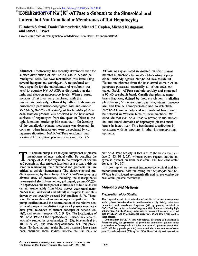
Localization of Na+,K+-ATPase t -Subunit to the Sinusoidal and Lateral but Not Canalicular Membranes of Rat Hepatocytes Elizabeth S. Sztul, Daniel Biemesderfer, Michael J. Caplan, Michael Kashgarian, and James L. Boyer Liver Center,YaleUniversitySchool of Medicine, New Haven, Connecticut 06510 Abstract. Controversy has recently developed over the surface distribution of Na+,K+-ATPase in hepatic pa- renchymal cells. We have reexamined this issue using several independent techniques. A monoclonal anti- body specific for the endodomain of tx-subunit was used to examine Na+,K+-ATPase distribution at the light and electron microscope levels. When cryostat sections of rat liver were incubated with the monoclonal antibody, followed by either rhodamine or horseradish peroxidase-conjugated goat anti-mouse secondary, fluorescent staining or horseradish peroxi- dase reaction product was observed at the basolateral surfaces of hepatocytes from the space of Disse to the tight junctions bordering bile canaliculi. No labeling of the canalicular plasma membrane was detected. In contrast, when hepatocytes were dissociated by col- lagenase digestion, Na+,K+-ATPase ~t-subunit was localized to the entire plasma membrane. Na+,K+- ATPase was quantitated in isolated rat liver plasma membrane fractions by Western blots using a poly- clonal antibody against Na÷,K÷-ATPase ~t-subunit. Plasma membranes from the basolateral domain of he- patocytes possessed essentially all of the cell's esti- mated Na÷,K+-ATPase catalytic activity and contained a 96-kD ~t-subunit band. Canalicular plasma mem- brane fractions, defined by their enrichment in alkaline phosphatase, 5' nucleotidase, gamma-glutamyl transfer- ase, and leucine aminopeptidase had no detectable Na÷,K÷-ATPase activity and no ~t-subunit band could be detected in Western blots of these fractions. We conclude that Na+,K÷-ATPase is limited to the sinusoi- dal and lateral domains of hepatocyte plasma mem- brane in intact liver. This basolateral distribution is consistent with its topology in other ion-transporting epithelia. T HE sodium pump is an integral component of plasma membranes of most animal cells. By coupling the energy of ATP hydrolysis to the transport of sodium and potassium, this enzyme functions as a primary driving force in maintaining the differential ion gradients that are critical to cellular homeostasis. The electrochemical gra- dient generated by the activity of Na+,K+-ATPase governs a diverse array of processes, including the transepithelial movement of electrolytes, water, and organic solutes (10, 20). In hepatocytes, the transport of solutes such as bile acids and certain amino acids from blood across basolateral mem- branes (i.e., sinusoidal and lateral) is coupled to and thus driven by the inwardly directed Na ÷ gradient (3, 7). There- fore, the resolution of membrane-specific patterns of Na÷ pump localization and the determination of the relative den- sities of pumps along distinct regions of plasma membrane have great relevance to current concepts of hepatic ion, HzO, and solute transport (3, 7, 9, 13). The localization of Na+,K÷-ATPase on the hepatocyte cell surface has been ex- tensively studied by cytochemical (2, 23), biochemical (4, 40, 30, 5, 28), and immunocytochemical (24, 39) proce- dures. To date, variant results (further discussed later) have been obtained; some studies indicate that the bulk of Na+,K÷-ATPase activity is localized to the basolateral sur- face (2, 23, 30, 5, 28), whereas others suggest that the en- zyme is present on both basolateral and bile canalicular domains (24, 39). In this report we present immunocytochemical and im- munobiochemical data indicating that hepatocyte Na+,K÷- ATPase is distributed asymmetrically and is restricted to the basolateral plasma membrane. Materials and Methods Preparation of Antibodies The preparation and characterization of anti-Na+,K+-ATPase monoclonal antibody have been described in detail elsewhere (22). Briefly, mice were immunized with membrane fragments (100 lag protein) enriched in Na+,K+-ATPase by the method of Jorgensen (19). Animals exhibiting high antibody titer (in ELISA assay) were used for fusion. Clones were screened both by ELISA and by a functional assay (22). Clone C62.4 was used in these studies. Canine kidney Na+,K+-ATPase was purified, according to the method of Jorgensen (19), for generation of polyclonal antibodies. Sodium pump preparations with enzymatic activities indicative of significant enrichment (,'020 mM P/mg protein per min) were mixed with equal volumes of com- plete Freund's adjuvant (200 ~tg Na+,K÷-ATPase/150 ~tl) and injected in- © The Rockefeller University Press, 0021-9525/87/05/1239/10 $1.00 The Journal of Cell Biology, Volume 104, May 1987 1239-1248 1239 on May 6, 2018 jcb.rupress.org Downloaded from http://doi.org/10.1083/jcb.104.5.1239 Published Online: 1 May, 1987 | Supp Info:
Transcript of Localization of Na+,K+-ATPase t -Subunit to the Sinusoidal...
Localization of Na+,K+-ATPase t -Subunit to the Sinusoidal and Lateral but Not Canalicular Membranes of Rat Hepatocytes Elizabeth S. Sztul, Daniel Biemesderfer, Michael J. Caplan, Michael Kashgarian, and James L. Boyer Liver Center, Yale University School of Medicine, New Haven, Connecticut 06510
Abstract. Controversy has recently developed over the surface distribution of Na+,K+-ATPase in hepatic pa- renchymal cells. We have reexamined this issue using several independent techniques. A monoclonal anti- body specific for the endodomain of tx-subunit was used to examine Na+,K+-ATPase distribution at the light and electron microscope levels. When cryostat sections of rat liver were incubated with the monoclonal antibody, followed by either rhodamine or horseradish peroxidase-conjugated goat anti-mouse secondary, fluorescent staining or horseradish peroxi- dase reaction product was observed at the basolateral surfaces of hepatocytes from the space of Disse to the tight junctions bordering bile canaliculi. No labeling of the canalicular plasma membrane was detected. In contrast, when hepatocytes were dissociated by col- lagenase digestion, Na+,K+-ATPase ~t-subunit was localized to the entire plasma membrane. Na+,K +-
ATPase was quantitated in isolated rat liver plasma membrane fractions by Western blots using a poly- clonal antibody against Na÷,K÷-ATPase ~t-subunit. Plasma membranes from the basolateral domain of he- patocytes possessed essentially all of the cell's esti- mated Na÷,K+-ATPase catalytic activity and contained a 96-kD ~t-subunit band. Canalicular plasma mem- brane fractions, defined by their enrichment in alkaline phosphatase, 5' nucleotidase, gamma-glutamyl transfer- ase, and leucine aminopeptidase had no detectable Na÷,K÷-ATPase activity and no ~t-subunit band could be detected in Western blots of these fractions. We conclude that Na+,K÷-ATPase is limited to the sinusoi- dal and lateral domains of hepatocyte plasma mem- brane in intact liver. This basolateral distribution is consistent with its topology in other ion-transporting epithelia.
T HE sodium pump is an integral component of plasma membranes of most animal cells. By coupling the energy of ATP hydrolysis to the transport of sodium
and potassium, this enzyme functions as a primary driving force in maintaining the differential ion gradients that are critical to cellular homeostasis. The electrochemical gra- dient generated by the activity of Na+,K+-ATPase governs a diverse array of processes, including the transepithelial movement of electrolytes, water, and organic solutes (10, 20). In hepatocytes, the transport of solutes such as bile acids and certain amino acids from blood across basolateral mem- branes (i.e., sinusoidal and lateral) is coupled to and thus driven by the inwardly directed Na ÷ gradient (3, 7). There- fore, the resolution of membrane-specific patterns of Na ÷ pump localization and the determination of the relative den- sities of pumps along distinct regions of plasma membrane have great relevance to current concepts of hepatic ion, HzO, and solute transport (3, 7, 9, 13). The localization of Na+,K÷-ATPase on the hepatocyte cell surface has been ex- tensively studied by cytochemical (2, 23), biochemical (4, 40, 30, 5, 28), and immunocytochemical (24, 39) proce- dures. To date, variant results (further discussed later) have been obtained; some studies indicate that the bulk of
Na+,K÷-ATPase activity is localized to the basolateral sur- face (2, 23, 30, 5, 28), whereas others suggest that the en- zyme is present on both basolateral and bile canalicular domains (24, 39).
In this report we present immunocytochemical and im- munobiochemical data indicating that hepatocyte Na+,K ÷- ATPase is distributed asymmetrically and is restricted to the basolateral plasma membrane.
Materials and Methods
Preparation of Antibodies The preparation and characterization of anti-Na+,K+-ATPase monoclonal antibody have been described in detail elsewhere (22). Briefly, mice were immunized with membrane fragments (100 lag protein) enriched in Na+,K+-ATPase by the method of Jorgensen (19). Animals exhibiting high antibody titer (in ELISA assay) were used for fusion. Clones were screened both by ELISA and by a functional assay (22). Clone C62.4 was used in these studies.
Canine kidney Na+,K+-ATPase was purified, according to the method of Jorgensen (19), for generation of polyclonal antibodies. Sodium pump preparations with enzymatic activities indicative of significant enrichment (,'020 mM P/mg protein per min) were mixed with equal volumes of com- plete Freund's adjuvant (200 ~tg Na+,K÷-ATPase/150 ~tl) and injected in-
© The Rockefeller University Press, 0021-9525/87/05/1239/10 $1.00 The Journal of Cell Biology, Volume 104, May 1987 1239-1248 1239
on May 6, 2018jcb.rupress.org Downloaded from http://doi.org/10.1083/jcb.104.5.1239Published Online: 1 May, 1987 | Supp Info:

tradermally at multiple sites along the backs and shoulders of 5-lb. female New Zealand white rabbits. 1 mo later the initial injection serum was col- lected and tested for immunoreactivity to Jorgensen-purified Na+,K +- ATPase by ELISA (32) assay. Positively responding rabbits were boosted with antigen mixed with incomplete Freund's adjuvant (200 ng/150 ~tl) by injection at multiple intradermal sites. Boosting and bleeding continued at 2-wk intervals. The serum used in the studies described in this paper was collected 2 wk after the third boost injection and was stored frozen at -20°C.
Antibodies to gamma-glutamyl transferase were a kind gift of Dr. David J. Castle (Department of Cell Biology, Yale University School of Medicine). Details of procedures used for antigen purification and antibody generation as well as characterization of obtained immune sera have been published (7).
Preparation of Isolated Hepatocytes
Isolated rat hepatocyte couplets and single cells were prepared by a modification of a collageuase perfusion technique as previously described (6, 15). The amount of collagenase (Sigma Chemical Co., St. Louis, MO) was decreased to 0.05 % and the dissociated cells were filtered through gauze and resuspended in Leibovitz-15 tissue culture media (Gibco, Grand Is- land, NY). This preparation currently results in 31 + 6% of the cells iso- lated as couplets with initial viabilities of >90% (as assessed by trypan blue exclusion).
lmmunocytochemical Localization of NW, K+-ATPase a-Subunit in Intact Rat Liver and in Isolated Hepatocytes Sprague-Dawley male rats were anesthetized with Inactin (10 mg/100 g body wt). A cannula was inserted into the aorta via the left ventricle, the vena cava was cut, and the animal was perfused with mammalian Ringers for ~-,1 min. Subsequently, the animal was perfused for 5 min with fixative consist- ing of 0.01 M Na metaperiodate, 0.75 M lysine, 2% paraformaldehyde in 0.0375 M NaHPO4 buffer (25). The final pH of the fixative was 6.2. This fixative provided adequate structural preservation with excellent retention of antigenicity (22). The liver was removed and pieces (2-4 mm 3) were postfixed in the same fixative for 6 h at 4°C. The tissue was washed five times in 0.1 M NaHPO4 buffer, incubated in 10% DMSO for 1 h, and then frozen by plunging into swirling liquid Freon 22 (Presto, New Haven, CT) cooled in liquid nitrogen. Frozen tissue was stored in liquid nitrogen until used.
Hepatocytes, isolated from rat liver as described above, were maintained on glass coverslips for 1-4 h. The cells were then fixed by immersing the coverslip in the fixative described above and incubating for 6 h. The cells were cryoprotected and frozen by the same techniques used for whole tissue.
For antigen localization at the light level, sections (0.5-1.0-ttm thick) were cut with an Ultracut equipped with an FC-4 cryounit (Reichert Scientific Instruments, Buffalo, NY). Sections were incubated in 50 mM ammonium chloride for 15 min to quench free aldehyde groups. The sec- tions were incubated for 1 h in a solution containing PBS, 1% BSA, and a primary antibody (either sera or ascites fluid) at 1:100 dilution. We used either monoclonal antibodies to a-subunit of Na+,K+-ATPase or, for a double-label experiment, a mixture of monoclonal antibodies to a-subunit of Na+,K+-ATPase and polyclonal antibodies to gamma-glutamyl transfer- ase. The sections were washed in PBS and incubated with either rhodamine- labeled goat anti-mouse IgG (Cappel Laboratories, Malvern, PA) to detect adsorbed monoclonal antibodies, or, for a double-label experiment, with a mixture of rhodamine-labeled goat-anti mouse IgG and fluorescein-labeled goat anti-rabbit IgG (Cappel Laboratories). In the latter case, both Q-sub- unit of Na+,K+-ATPase and gamma-glutamyl transferase will be detected on the same section. After washing, sections were examined with a Zeiss incident light fluorescence microscope.
The method for immunocytochemical labeling at the ultrastructural level was essentially that described by Brown and Farquhar (8) and recently ap- plied to the ultrastructural localization of Na+,K+-ATPase in the kidney (22). 16-gm thick cryostat sections, or pieces of coverslip with attached he- patocytes, were incubated overnight at 4°C in antibody against the a-subunit of the sodium pump. The tissue was washed five times with PBS followed by a 2-h incubation in sheep anti-mouse (Fab) conjugated to horseradish peroxidase (HRP)) After washing, the tissue was fixed for 1 h in 1.5%
1. Abbreviations used in this paper: blLPM, basolateral liver plasma mem- brane; cLPM, canalicular liver plasma membrane; HRP, horseradish per- oxidase; NC, nitrocellulose.
glutaraidehyde in 0.1 M Na cacodylate buffer (pH 7.4) containing 5% su- crose. After three washes in 0.1 M Na cacodylate buffer with 7.5% sucrose and three washes in 50 mM Tris-HCl (pH 7.4) with 7.5% sucrose, peroxi- dase reaction product was developed in a solution of 0.2% diaminobenzi- dine in the Tris-sucrose buffer to which H202 had been added to yield a final concentration of 0.01%. The reaction was stopped after 10-20 min by washing in cold Tris-sucrose buffer. Osmium fixation was carried out using the reduced osmium method of Karnovsky (21). The tissue was dehydrated and embedded in Epon 812, and unstained thin sections were examined with a Zeiss 10B electron microscope.
Isolation and Characterization of Canalicular and Basolateral Liver Plasma Membrane Fractions
The procedure for isolation of canalicuiar and basolateral liver plasma membrane (cLPM and blLPM, respectively) subfractions has been pre- viously described in detail (28). Briefly, livers from 160-180-g male Sprague-Dawley rats (Charles River Breeding Laboratories, Inc., Wilming- ton, MA) were homogenized and the homogenate was spun down (7,000 g for 10 rain) to give a nuclear pellet fraction. A mixed LPM subfraction was isolated from the nuclear pellet by rate zonal flotation. This material was tightly homogenized and the vesiculated LPM elements were separated on a three-step sucrose gradient (31, 34, and 38% wt/wt). The membranes col- lected at each interphase (cLPM at 31/34 and blLPM at 34/38) were then spun down (105,000 g for 60 rain) and resuspended in 0.25 M sucrose, 0.2 mM CaCI2, 5 m M MgSO,, 10 mM Hepes-Tris, pH 7.5. The degree of purification of cLPM and blLPM was analyzed extensively by measuring intracellular and plasma membrane marker enzyme activities as in reference (28). These studies have indicated minimal contamination of both LPM fractions by intracellular organelles and virtually complete separation of blLPM from cLPM as indicated by the absence of glucagon-stimulatable adenylate cyclase or secretory component in cLPM (28). BILPM subfrac- tion was contaminated with cLPM elements by ~10%.
Western Blots
Madin-Darby canine kidney (MDCK) cells were grown and lysed, and cel- lular membranes were prepared as described in reference 22. MDCK mem- branes and LPM subfractions were processed for SDS PAGE and subjected to electrophoresis as described previously (38). Upon completion of elec- tropboresis, separated proteins were transferred to nitrocellulose (NC) filters for 4 h at room temperature and constant current (150 rnA). The fillers were immediately quenched in 12.5 mg/ml hemoglobin (2 h at room temper- ature) and then incubated overnight at room temperature with either nonim- mune serum or a polyclonal anti-Na+,K+-ATPase ct-subunit serum (1:100 dilutions in 12.5 mg/ml hemoglobin). The filters were washed (2 x 10 rain) with PBS, then with PBS containing 0.05 % Nonidet P-40 (2 x 10 rain), and finally rinsed (2 x 5 rain) with PBS. Adsorbed IgGs were detected by in- cubating the filters in nSI-labeled protein A (r~20 x 106 cpm in 200 ml of 12.5 mg/ml hemoglobin) for 4 h at room temperature, followed by washing (as above), drying, and autoradiography.
Inhibition of ATPase Activity in Isolated Membrane Subfraction
Rat liver basolateral membranes were isolated as described (28). Membrane samples (25 gl) containing 50-70 gg protein were added to 25 gl bovine serum albumin fraction V (Sigma Chemical Co.) and 2.5 I~l 1% SDS (14). All membrane fractions were soluhilized in 0.05% SDS (Bio-rad Laborato- ries, Richmond, CA) and incubated at room temperature for 10 min before enzyme analysis, since preliminary studies determined that concentrations of 0.06% SDS and above decreased enzyme activity. 10 I.tl of either deionized water, preimmune rat serum, control rat ascites, polyclonal or monoclonal (C62.4) antibodies were added to the membranes. Na+,K +- ATPase and Mg+2-ATPase activities were assayed after a 30-rain room temperature incubation by a standard spectrophotometric assay (34).
Results
Characterization of Polyclonal and Monoclonal Antibodies against Na+,K ÷-ATPase The monoclonal antibody C62.4 has been characterized pre- viously (22). This antibody inhibits Na+-dependent but not
The Journal of Cell Biology, Volume 104, 1987 1240

K+-dependent Na+,K+-ATPase activity. Immunoprecipita- tion of MDCK cells, biosynthetically labeled with [35S]me- thionine, demonstrated that the antibody recognizes a 96-kD protein. Furthermore, this antibody precipitated a 96-kD protein labeled in vitro with [3H]NAB ouabain. Immunocy- tochemical localization revealed that the antigenic site recog- nized by this antibody is on the cytoplasmic domain of basolateral plasma membranes of renal tubular epithelium.
Presence of polyclonal antibodies m sera ot immunized rabbits was tested by Western blots. As shown in Fig. 5, the antibodies reacted with ¢t-subunit on NC transfers of purified canine Na+,K+-ATPase. To test whether antibodies to con- taminating ( n o n - N a + , K + - A ~ ) antigens were perhaps also generated, we used the immune sera in Western blots of membranes from MDCK cells and from rat liver. As shown in Fig. 1, only a single polypeptide with a molecular mass of 96 kD was detected in both MDCK and rat liver mem- brane fractions. No reactive band was seen when non- immune serum was used. This polyclonal antibody also immunoprecipitated ct-subunit labeled with a photoaffinity derivative of ouabain, a highly specific inhibitor of sodium pump (Smith, Z., M. J. Caplan, and J. Jamieson, manuscript submitted for publication), thus indicating that it recognizes the Na+,K+-ATPase. Because no immunoreactivity to the I]-subunit of Na+,K+-ATPase was observed with either monoclonal or polyclonal antibody, we conclude that both were monospecific for the a-subunit of the enzyme.
Both antibodies were tested for their ability to inhibit en- zymatic activity of two hepatic ATPases. As shown in Table I, the polyclonal serum inhibited Na+,K+-ATPase activity by ~50%, while having no effect on Mg+2-ATPase. The monoclonal C62.4 antibody inhibited Na+,K+-ATPase ac- tivity of hepatocyte plasma membrane even further (19.7 % of normal activity), and also had no effect on Mg+2-ATPase. Both antibodies showed inhibition only when intact right- side out LPM vesicles were permeabilized with SDS. (The slight decline in ATPase activity in the permeabilized mem- brane is caused by SDS inactivation). Since the vesicles re- tain their right-side out orientation during preparation, the results indicate that the antibodies recognize a cytoplasmic domain of the ct-subunit, not accessible to the antibodies in the nonpermeabilized vesicles. The same level of Na+,K +- ATPase inhibition was found when C62.4 was incubated with dog kidney membranes (22).
Figure 1. Western blot analysis of polyclonal antibodies to the a-sub- unit of canine Na+,K+-ATPase. Total cell membranes and a mixed LPM fraction were prepared from MDCK ceils and from rat liver, respectively. Membrane proteins were separated by SDS PAGE electrophoresis and then trans- ferred to NC filters. The filters were incubated with either anti- Na+,K+-ATPase or nonimmune
sera, followed by t25I-protein A. Shown are autoradiograms of filters. (Lane A) Western blot of MDCK ceil membrane proteins with immune sera; (lane B) western blot of MDCK cell membrane proteins with nonimmune sera; (lane C) western blot of liver mem- brane proteins with immune sera; (lane D) western blot of liver membrane proteins with nonimmune sera.
Table L Inhibition of Enzymatic Activities of Na +, K+-ATPase and Mg+Z-ATPase by Polyclonal and Monoclonal Antibodies
Nonimmune Polyclonal Normal Monoclonal scram antibody ascites C62.4
Na+,K÷-ATPase Nonpermeabil ized
vesicles 12.25 10.96 12.41 8.22 SDS-permeabilized
vesicles 9.51 4.76 10.24 2.02
Mg+2-ATPase Nonpermeabilized
vesicles 24.50 23.05 23.05 23.05 SDS-permeabilized
vesicles 25.22 25.22 23.20 23.20
Isoh~ed LPM vesicles were incubated with control or immune antibodies as described in Materials and Methods. Na÷,K÷-ATPas¢ and Mg+2-ATPase ac- tivities were then assayed and are represented as I.tmoi P released per nag pro- rein per rain. Polyclonal antibodies inhibited Na÷,K+-ATPase activity by 50.1%, while mon~lomd antibodies inlu'bi~l by 80%. In both cases activity of Mg+2-ATPase was unaffected. The data represent averages of three determi- nations.
lmmunolocalization of a-Subunit of Na+,K +-ATPase in Intact Rat Liver
lmmunofluorescent Localization. When. the monoclonal anti-Na+,K+-ATPase antibodies were used to label rat liver cryosections, sinusoidal and lateral plasma membranes of hepatocytes were uniformly labeled (Fig. 2 A). Bile canalic- uli (arrows) were consistently negative, thus suggesting that Na+,K+-ATPase is distributed asymmetrically, with a high concentration of a-subunit per surface area on sinusoidal and lateral membranes and no detectable pumps on the biliary domain of the plasmalemma.
To determine whether the lack of bile canalicular staining represents bonafide distribution of the enzyme, or perhaps is caused by inaccessibility of the biliary region of the hepa- tocyte to antibodies, we performed a double-label experi- ment in which we localized ¢t-subunit of Na+,K+-ATPase and gamma-glutamyl transferase (a known bile canalicular marker) on the same section. Liver sections were incubated with a mixture of mouse anti-Na+,K+-ATPase antibodies and rabbit anti-gamma-glutamyl transferase antibodies, fol- lowed by a mixture of rhodamine-conjugated goat anti-mouse antibodies and fluorescein-conjugated goat anti-rabbit anti- bodies. As shown in Fig. 2 B, when a rhodarnine-detecting filter was used, fluorescence was observed along sinusoidal and lateral membranes up to the tight junctions delineating bile canaliculi (arrows). When a fluorescein-detecting filter was used (Fig. 2 C), a distinct pattern was observed. Sinu- soidal and lateral membranes were not stained while bile canaliculi (arrows) showed strong fluorescent signals. In ad- dition, a number of intracellular organelles was stained. This distribution of label is compatible with the previously ob- served (7) apical distribution of gamma-glutamyl transferase in liver and in other epithelia. These data indicate that the bile canalicular domain of hepatocytes is readily accessible for immunolabeling and suggest that Na÷,K+-ATPase ¢t-sub- unit is not present in detectable amounts in that membrane region.
UItrastructural Localization. Cryostat sections were in- cubated with monoclonal antibody C62.4 followed by goat
Sztul et al. Hepatic Na+-Pump Is Localized to Basolateral Domain 1241

Figure 2. Immunofluorescent localization of Na+,K+-ATPase in rat liver. In A sections were incubated with anti-Na+,K+-ATPase monoclonal antibodies. In B and C, sections were incubated with a mixture of anti-Na+,K+-ATPase monoclonal antibodies (labeling visualized in B) and anti-gamma-glutamyl transferase antibodies (labeling visualized in C). A clearly indicates that staining is restricted to the sinusoidal and lateral domains of the bepatocyte and is absent from bile canaliculi (arrows). B shows sinusoidal and lateral staining of hepatocytes and absence of staining from bile canliculi (arrows). C shows staining of bile canaliculi (arrows) and neighboring intracellular organdies. Bars, (A) 12.5 gin; (B and C) 6.3 gm.
anti-mouse F(ab) conjugated to HRP. As seen in Fig. 3 A, HRP reaction product was restricted to the cytoplasmic do- main of basolateral plasma membranes of hepatocytes. On the sinusoidal surface (Fig. 3 B), the distribution of reaction product appeared to be uniform over the entire plas- malemma, a finding in accord with the distribution of Na+,K+-ATPase at the basolateral invaginations of the kid- hey epithelium (22).: A similar lack of clustering of Na+,K +- ATPase has been previously reported in dog hepatic tissue by Takemura et al. (39). Lateral plasma membranes (Fig. 3 A, double arrows) were uniformly labeled with HRP reaction product up to tight junctions defining the bile canaliculus. It is important to note that the bile canalicular membrane with its microvilli was consistently unlabeled with reaction prod- uct. While we realize that the irnmunoperoxidase method is
2. Flat regions ofplasmalemma directly opposed to the basement membrane were devoid of reaction product, suggesting a microdomain sorting of Na+,K+-ATPase in the kidney (22).
not very quantitative and has a finite level of detection, the lack of reaction product clearly indicates that Na ÷,K÷-ATPase concentration in apical domain is significantly below that in basolateral membrane. Reaction product was not observed in other intraceUular organelles in our sections. This may be the result of low levels of intracellular antigen (below detec- tion level of the antibody HRP method) or a change in the conformation of the a-subunit in some intracellular mem- branes. (The C62.4 antibody reacted with the membanes of the medial Golgi cisternae in cells of the thick ascending limb of rat kidney medulla [22].) The specificity of the im- munoreaction was established by the lack of immunolabeling when sections were incubated with control monoclonal anti- body (Fig. 3 C).
Immunolocalization of Na÷,K÷-ATPase in Isolated Rat Hepatocytes and in Hepatocyte Couplets
Rat hepatocytes and hepatocyte couplets were isolated after collagenase disruption, fixed, and processed for immuno-
The Journal of Cell Biology, Volume 104, 1987 1242
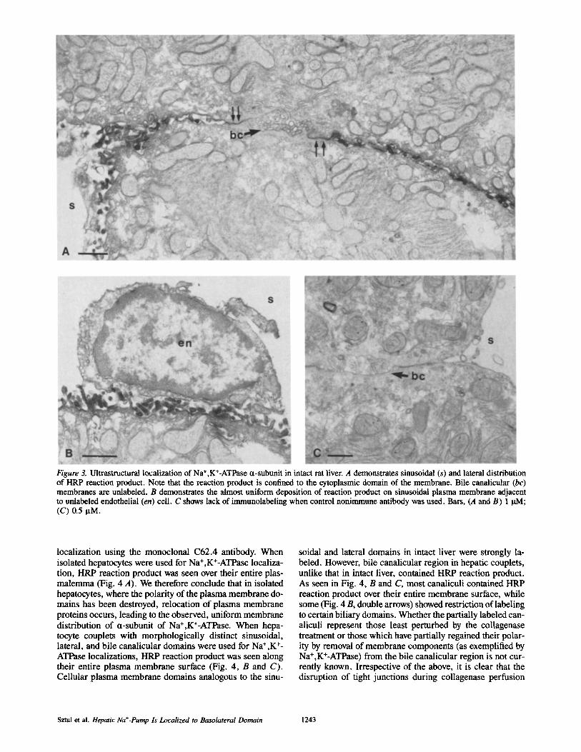
Figure 3. Ultrastructural localization of Na +,K+-ATPase ¢t-subunit in intact rat liver. A demonstrates sinusoidal (s) and lateral distribution of HRP reaction product. Note that the reaction product is confined to the cytoplasmic domain of the membrane. Bile canalicular (bc) membranes are unlabeled. B demonstrates the almost uniform deposition of reaction product on sinusoidal plasma membrane adjacent to unlabeled endothelial (en) cell. C shows lack of immunolabeling when control nonimmune antibody was used. Bars, (A and B) 1 BM; (C) 0.5 BM.
localization using the monoclonal C62.4 antibody. When isolated hepatocytes were used for Na+,K+-ATPase localiza- tion, HRP reaction product was seen over their entire plas- malemma (Fig. 4 A). We therefore conclude that in isolated hepatocytes, where the polarity of the plasma membrane do- mains has been destroyed, relocation of plasma membrane proteins occurs, leading to the observed, uniform membrane distribution of ¢t-subunit of Na+,K+-ATPase. When hepa- tocyte couplets with morphologically distinct sinusoidal, lateral, and bile canalicular domains were used for Na+,K +- ATPase localizations, HRP reaction product was seen along their entire plasma membrane surface (Fig. 4, B and C). Cellular plasma membrane domains analogous to the sinu-
soidal and lateral domains in intact liver were strongly la- beled. However, bile canalicular region in hepatic couplets, unlike that in intact liver, contained HRP reaction product. As seen in Fig. 4, B and C, most canaliculi contained HRP reaction product over their entire membrane surface, while some (Fig. 4 B, double arrows) showed restriction of labeling to certain biliary domains. Whether the partially labeled can- aliculi represent those least perturbed by the collagenase treatment or those which have partially regained their polar- ity by removal of membrane components (as exemplified by Na+,K+-ATPase) from the bile canalicular region is not cur- rently known. Irrespective of the above, it is clear that the disruption of tight junctions during collagenase perfusion
Sztul et al. Hepatic Na+-Pump Is Localized to Basolateral Domain 1243

The Journal of Cell Biology, Volume 104, 1987 1244
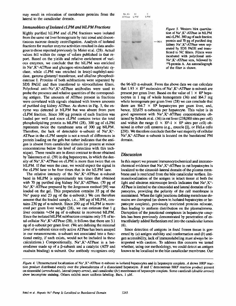
may result in relocation of membrane proteins from the lateral to the canalicular domain.
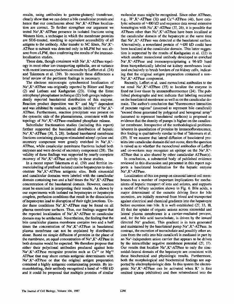
Immunoblo ts o f Isolated c L P M and bILPM Fractions
Highly purified blLPM and cLPM fractions were isolated from the same rat liver homogenate by rate zonal and discon- tinuous sucrose density centrifugation. Analysis of isolated fractions for marker enzyme activities resulted in data analo- gous to those reported previously by Meier et al. (28). Actual values fell within the range of values published in that re- port. Based on the yields and relative enrichment of vari- ous enzymes, we conclude that the blLPM was enriched in Na+,K+-ATPase and glucagon-stimulatable adenylate cy- clase, while cLPM was enriched in leucyl-naphthyl-ami- dase, gamma-glutamyl transferase, and alkaline phosphodi- esterase I. Proteins of both subfractions were separated by SDS PAGE and then transferred to nitrocellulose filters. Polyclonal anti-Na+,K+-ATPase antibodies were used to probe the presence and relative quantities of the correspond- ing antigen. The amounts of ATPase present in fractions were correlated with signals obtained with known amounts of purified dog kidney ATPase. As shown in Fig. 5, the en- zyme was detected in blLPM but was absent from pure cLPM fraction. Since 300 Ixg protein of each fraction was loaded per well and since cLPM contains twice the total phospholipid/mg protein as blLPM (28), 300 [tg of cLPM represents twice the membrane area of 300 ~tg blLPM. Therefore, the lack of detectable ct-subunit of Na+,K ÷- ATPase in the cLPM sample is not a result of differences in protein loading on the gels but rather indicates that the anti- gen is absent from canalicular domain (or present at minor concentrations below the level of detection with this tech- nique). These results are in direct contrast to those obtained by Takemura et al. (39) in dog hepatocytes, in which the den- sity of Na+,K+-ATPase on cLPM is more than twice that on blLPM. If that were the case, we would expect the signal in the cLPM lane to be four times that in the blLPM lane.
The relative intensity of the Na+,K+-ATPase ¢t-subunit band in blLPM is approximately ten times that obtained with 70 pg purified dog kidney Na+,K+-ATPase. (70 lag of Na+,K+-ATPase prepared by the Jorgenson method [19] was loaded on the gel. This preparation contains 35 pg of the Na + pump and 23 pg of the a-subunit.) We can therefore assume that the loaded sample, i.e., 300 lxg of blLPM, con- tains 230 pg of ¢t-subunit. Since 200 ~tg of blLPM is recov- ered per gram liver weight (28), we can estimate that 1 g liver contains ~154 pg of ~t-subunit in recovered blLPM. Since the isolated blLPM subfraction contains only 5 % of to- tal cellular Na+,K+-ATPase (28), it follows that there are 3.1 ng of ct-subunit per gram liver. (We are defining the minimal level of a-subunit since only active ATPase has been assayed in our measurements. ¢t-subunit not associated into a func- tional entity, if such exists, would not be included in these calculations.) Compositionally, Na+,K+-ATPase is a bet- erodimer made up of a 13-subunit and a catalytic (ATP and ouabain binding) ¢t-subunit. Our antibody recognizes only
Figure 5. Western blot quantita- don of Na+,K+-ATPase in blLPM and cLPM. 300 ttg of each fraction protein and 70 pg of purified dog kidney Na+,K+-ATPase were sep- arated by SDS PAGE and trans- ferred to NC filters. Filters were incubated with polyclonal anti- Na+,K+-ATPase scra, followed by mI-protein A. An autoradiograph of the filter is shown.
the 96-kD ct-subunit. From the above data we can calculate that 1.93 x 10 u molecules of Na÷,K+-ATPase a-subunit are present per gram liver. Based on the value of 1 x 10⁶ hepa- tocytes in 1 nag of whole homogenate (36) and 164.7 mg whole homogenate per gram liver (28) we can conclude that there are 164.7 x 10⁶ hepatocytes per gram liver, and, hence, 115,675 a-subunits per hepatocyte. This value is in good agreement with Na+,K÷-ATPase concentrations ob- tained by Schenk et al. (36) in rat liver (238,000 sites per cell) and within the range (1 x l(P-1 x 10⁶ sites per cell) ob- tained in other cell systems (e.g., muscle [1], or HeLa cells [29]). We therefore conclude that the vast majority of cellular Na÷,K÷-ATPase ct-subunit is located on the basolateral PM domain.
Discussion
In this report we present irnmunocytochemical and immuno- chemical evidence that Na÷,K÷-ATPase in rat hepatocytes is localized to the sinusoid-lateral domain of the plasma mem- brane and is restricted from the bile canaiicular surface. Im- munolocalization of the antigen in intact tissue at both the light and electron microscope levels indicates that Na÷,K ÷- ATPase is limited to the sinusoidal and lateral domains ofhe- patocytes, providing the polarity of the cell membrane is maintained. When the tight junctions defining membrane do- mains are disrupted (as shown in isolated hepatocytes or he- patocyte couplets), previously restricted proteins relocate, thus leading to uniform distribution on the plasmalemma. Disruption of the junctional complexes in hepatocyte coup- lets has been previously demonstrated by penetration of ex- tracellularly added Ruthenium red into the canalicular lumen (6).
Since detection of antigens in fixed frozen tissue is gov- erned by (a) antigen stability and conformation and (b) anti- gen accessibility, lack of immunolabeling must always be in- terpreted with caution. To address this concern we tested whether, using our methodology, we could detect an antigen known to be localized to the bile canalicular membrane. Our
Figure 4. Ultrastructural localization of Na+,K+-ATPase ct-subunit in isolated hepatocytes and in hepatocyte couplets. A shows HRP reac- tion product distributed evenly over the plasmalemma of a dissociated hepatocyte. B and C demonstrate HRP reaction product present on sinusoidal (arrowheads), lateral (empty arrow), and canalicular (bc) membranes of hepatocyte couplets. Some canaliculi (double arrows) show incomplete staining. Others exhibit more uniform labeling. Bars, 1 p.M.
Sztul et al. Hepatic Na*-Pump la Localized to Basolateral Domain 1245
results, using antibodies to gamma-glutamyl transferase, clearly show that we can detect a bile canalicular protein and hence that our conclusions about Na+,K+-ATPase localiza- tion are correct. To further strengthen our argument we tested Na+,K+-ATPase presence in isolated fractions using Western blots, a technique in which the membrane proteins are SDS-treated, resulting in equivalent accessibility of all antigens to the antibody. After transfer to NC filters, Na+,K +- ATPase a-subunit was detected only in blLPM but was ab- sent from cLPM, thus confirming the results of the immuno- localization studies.
These data, though consistent with Na+,K+-ATPase topol- ogy in most other ion-transporting epithelia, are at variance with recent immunocytochemical results by Leffert et al. (24) and Takemura et al. (39). To reconcile these differences a brief review of the pertinent findings is necessary.
The electron microscopic cytochemical localization of Na+,K+-ATPase was originally reported by Blitzer and Boyer (2) and Latham and Kashgarian (23). Using the Ernst nitrophenyl phosphatase technique (12) both groups indepen- dently reported basolateral distribution of this enzyme. Reaction product deposition was K + and Mg +2 dependent and was inhibited by ouabain, a specific inhibitor of Na +,K ÷- ATPase. Furthermore, reaction precipitate was present on the cytosolic side of the plasmalemma, consistent with the topology of Na+,K+-ATPase-mediated phosphate release.
Subcellular fractionation studies (including this report) further supported the basolateral distribution of hepatic Na+,K+-ATPase (30, 5, 28). Isolated basolateral membrane fractions containing glucagon-stimulated adenyl cyclase and secretory component were greatly enriched in Na+,K +- ATPase, while canalicular membrane fractions lacked both enzymes and were devoid of secretory component (28). The validity of these findings was supported by nearly complete recovery of Na+,K+-ATPase activity in these studies.
In a recent report Takemura et al. (39) used ferritin im- munolabeling of prefixed isolated canine hepatocytes to dem- onstrate Na+,K+-ATPase antigenic sites. Both sinusoidal and canalicular domains were labeled with the canalicular domain containing two and a half times the Na +,K+-ATPase concentration of the basolateral domain. However, caution must be exercised in interpreting their results. As shown by our experiments with isolated rat hepatocytes or hepatocyte couplets, perfusion conditions that result in the dissociation of hepatocytes lead to disruption of their tight junctions. Un- der these conditions Na+,K+-ATPase may be found on all plasma membrane surfaces. Thus, our findings suggest that the reported localization of Na+,K+-ATPase to canalicular domain may be artifactual. Nevertheless, the finding that the bile canalicular plasma membrane contains two and a half times the concentration of Na+,K+-ATPase in basolateral plasma membrane can not be explained by distribution alone. Based on simple diffusion of proteins in the plane of the membrane, an equal concentration of Na+,K+-ATPase in both domains would be expected. We therefore propose that either their polyclonal antibodies produced against holo Na+,K+-ATPase recognize proteins such as Ca +2 or Mg +2 ATPase that may share certain antigenic determinants with Na+,K+-ATPase or that the original antigen preparation contained a highly antigenic contaminating protein. By im- munoblotting, their antibody recognized a band of ,,~100 kD and it could be proposed that multiple proteins of similar
molecular mass might be recognized. Since other ATPases, e.g., H+,K+-ATPase (31) and Ca+2-ATPase (41), have cata- lytic subunits of'x,100 kD and sequence data reveal extensive homologies with Na+,K÷-ATPase (37, 26), it is possible that ATPases other than Na+,K+-ATPase have been localized at the canalicular domain of the hepatocyte at the same time that Na+,K+-ATPase was detected at the basolateral surface. Alternatively, a nonrelated protein of '~100 kD could have been localized at the canalicular domain. This latter sugges- tion is supported by the results of Kashgarian et al. (22) in which another monoclonal antibody developed to enriched Na÷,K+-ATPase and immunoprecipitating a 96-kD band from biosynthetically labeled rat kidney membranes local- ized exclusively to brush borders of rat kidney cells, indicat- ing that the original antigen preparation contained a non- Na+,K+-ATPase component.
Recently, Leffert et al. used monoclonal antibodies to the rat renal Na+,K÷-ATPase (35) to localize the enzyme in fixed rat liver tissue by immunofluorescence (24). The pub- lished photographs and text indicate that labeling occurred on the basolateral membrane as well as on the canalicular do- main. The author's conclusion that "fluorescence intensities of punctate regions" (assumed to represent bile canaliculi) "exceed those generated by polygonal and hexogonal array" (assumed to represent basolateral surfaces) is proposed as evidence that the density of pumps is higher on the canalicu- lar membrane. Irrespective of the methodological problems inherent in quantitation of proteins by immunofluorescence, this finding is qualitatively similar to that of Takemura et al. (39). If we assume that lateral diffusion of basolateral pro- teins into canalicular domain did not occur, then the question is raised as to whether the monoclonal antibodies of Leffert and co-workers may recognize an epitope on the Na+,K +- ATPase that is also shared by other canalicular ATPase(s).
In conclusion, a substantial body of published evidence reviewed in this discussion and presented in this report sup- ports a basolateral localization for the hepatic enzyme Na +,K+-ATPase.
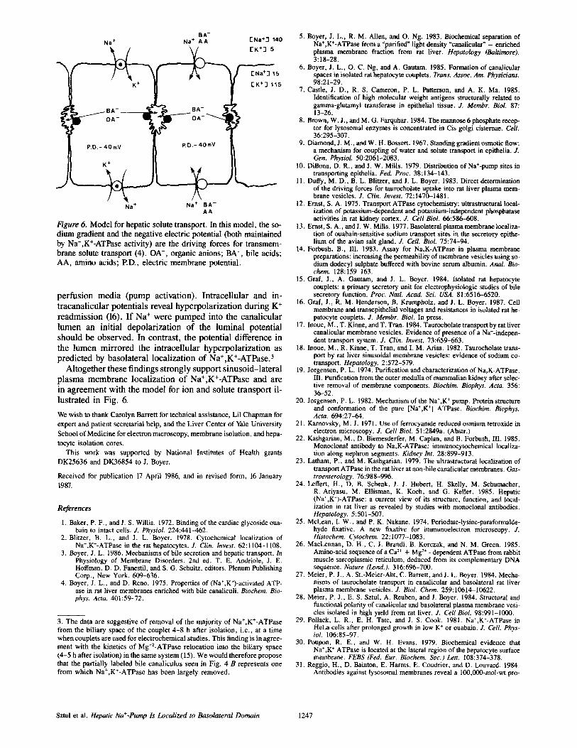
Localization of this ion pump on sinusoid lateral cell mem- branes has a number of important implications for mecha- nisms of hepatic transport of ions and solutes, and supports a model of biliary secretion shown in Fig. 6. Bile acids, a major determinant of the osmotic driving force for bile secretion, are initially removed from blood and transported against electrical and chemical gradients into the hepatocyte before excretion into bile. It is well established (27, 33, 18, 11) that the uptake of organic anions across sinusoidal and lateral plasma membranes is a carrier-mediated process, and, for the bile acid taurocholate, is driven by the inward directed Na ÷ gradient. This gradient is in turn generated and maintained by the basolateral pump Na+,K+-ATPase. In contrast, the excretion of taurocholate and possibly other an- ions from the cells into bile canaliculi is mediated in part by an Na+-independent anion carrier that appears to be driven by the intracellular negative membrane potential (27, 17). Our results that localize Na+,K+-ATPase to only the sinu- soidal-lateral domain of the hepatocyte are consistent with these biochemical and physiologic results. Furthermore, both the morphological and biochemical findings are sup- ported by electrophysiologic data. In this system the electro- genic Na+,K+-ATPase can be activated when K ÷ is first omitted (pump inhibition) and then reintroduced into the
The Journal of Cell Biology, Volume 104, 1987 1246
"N
BA- Na + Na + A A
K +
P.D.- 40 mV
K +
N a ÷
B A - ¢~ "
P.D,- 4OmV
Na + B A - A A
I- Na+J 140
[K+3 5
~ 5 [K +-I 115
Figure 6. Model for hepatic solute transport. In this model, the so- dium gradient and the negative electric potential (both maintained by Na+,K+-ATPase activity) are the driving forces for transmem- brane solute transport (4). OA-, organic anions; BA-, bile acids; AA, amino acids; P.D., electric membrane potential.
perfusion media (pump activation). Intracellular and in- tracanalicular potentials reveal hyperpolarization during K ÷ readmission (16). If Na ÷ were pumped into the canalicular lumen an initial depolarization of the luminal potential should be observed. In contrast, the potential difference in the lumen mirrored the intracellular hyperpolarization as predicted by basolateral localization of Na+,K÷-ATPase. 3
Altogether these findings strongly support sinusoid-lateral plasma membrane localization of Na+,K÷-ATPase and are in agreement with the model for ion and solute transport il- lustrated in Fig. 6,
We wish to thank Carolyn Barrett for technical assistance, Lil Chapman for expert and patient secretarial help, and the Liver Center of Yale University School of Medicine for electron microscopy, membrane isolation, and hepa- tocyte isolation cores.
This work was supported by National Institutes of Health grants DK25636 and DK36854 to J. Boyer.
Received for publication 17 April 1986, and in revised form, 16 January 1987.
References
1. Baker, P. F., and J. S. Willia. 1972. Binding of the cardiac glycoside oua- bain to intact cells. J. Physiol. 224:441-462.
2. Blitzer, B. L., and J. L. Boyer. 1978. Cytochemical localization of Na*,K+-ATPase in the rat hepatocytes. J. Clin. Invest. 62:1104-1108.
3. Boyer, J. L. 1986. Mechanisms of bile secretion and hepatic transport. In Physiology of Membrane Disorders. 2nd ed. T. E. Andriole, J. E. Hoffman, D. D. Fanestil, and S. G. Schultz, editors. Plenum Publishing Corp., New York. 609-636.
4. Boyer, J. L., and D. Reno. 1975. Properties of (Na+,K*)-activated ATP- ase in rat liver membranes enriched with bile canaliculi. Biochem. Bio- phys. Acta. 401:59-72.
3. The data are suggestive of removal of the majority of Na+,K+-ATPase from the biliary space of the couplet 4-8 h after isolation, i.e., at a time when couplets are used for electrochemical studies. This finding is in agree- ment with the kinetics of Mg÷2-ATPase relocation into the biliary space (4-5 h after isolation) in the same system (15). We would therefore propose that the partially labeled bile canaliculus seen in Fig. 4 B represents one from which Na+,K÷-ATPase has been largely removed.
5. Boyer, J. L., R. M. Allen, and O. Ng. 1983. Biochemical separation of Na+,K+-ATPase from a "purified" light density "canalicular" - enriched plasma membrane fraction from rat liver. Hepatology (Baltimore). 3:18-28.
6. Boyer, J. L., O. C. Ng, and A. Gautam. 1985. Formation of canalicular spaces in isolated rat hepatocyte couplets. Trans. Assoc. Am. Physicians. 98:21-29.
7. Castle, J. D., R. S. Cameron, P. L. Patterson, and A. K. Ma. 1985. Identification of high molecular weight antigens structurally related to gamma-glutamyl transferase in epithelial tissue. J. Membr. Biol. 87: 13-26.
8. Brown, W. J., and M. G. Farquhar. 1984. The mannose 6 phosphate recep- tor for lysosomal enzymes is concentrated in Cis golgi cisternae. Cell. 36:295-307.
9. Diamond, J. M., and W. H. Bossert. 1967. Standing gradiem osmotic flow: a mechanism for coupling of water and solute transport in epithelia. J. Gen. Physiol. 50:2061-2083.
10. DiBona, D. R., and J. W. Mills. 1979. Distribution of Na+-pump sites in transporting epithelia. Fed. Proc. 38:134-143.
11. Duffy, M. D., B. L. Blitzer, and J. L. Boyer. 1983. Direct determination of the driving forces for taurocholate uptake into rat liver plasma mem- brane vesicles. J. Clin. Invest. 72:1470-1481.
12. Ernst, S. A. 1975. Transport ATPase cytochemistry: ultrastrnctural local- ization of potassium-dependent and potassium-independent phosphatase activities in rat kidney cortex. J. Cell Biol. 66:586-608.
13. Ernst, S. A., andL W. Mills. 1977. Basolateral plasma membrane localiza- tion of ouabaln-sensitive sodium transport sites in the secretory epithe- lium of the avian salt gland. J. Cell. Biol. 75:74-94.
14. Forbush, B., Ill. 1983. Assay for Na,K-ATPas¢ in plasma membrane preparations: increasing the permeability of membrane vesicles using so- dium dodecyl sulphate buffered with bovine serum albumin. Anal. Bio- chem. 128:159-163.
15. Graf, J., A. Gautam, and J. L. Boyer. 1984. Isolated rat hepatocyte couplets: a primary secretory unit for electrophysiologic studies of bile secretory function. Proc. Natl. Acad. Sci. USA. 81:6516-6520.
16. Graf, J., R. M. Henderson, B. Krnmpholz, and J. L. Boyer. 1987. Cell membrane and transepithelial voltages and resistances in isolated rat he- patocyte couplets. J. Membr. Biol. In press.
17. Inoue, M., T. Kinne, and T. Tran. 1984. Taurocholate transport by rat liver canalicular membrane vesicles. Evidence of presence of a Na+-indepe n- dent transport system. J. Clin. Invest. 73:659-663.
18. Inoue, M., R. Kinne, T. Tran, and I. M. Arias. 1982. Taurocholate trans- port by rat liver sinusoidal membrane vesicles: evidence of sodium co- transport. Hepatology. 2:572-579.
19. Jorgensen, P. L. 1974. Purification and characterization of Na,K-ATPase. III. Purification from the outer medulla of mammalian kidney after selec- tive removal of membrane components. Biochim. Biophys. Acta. 356: 36-52.
20. Jorgensen, P. L. 1982. Mechanism of the Na*,K + pump. Protein structure and conformation of the pure [Na+,K +] ATPase. Biochim. Biophys. Acta. 694:27-64.
21. Karnovsky, M. J. 1971. Use of ferrocyanide reduced osmium tetroxide in electron microscopy. J. Cell Biol. 51:2849a. (Abstr.)
22. Kashgarian, M., D. Biemesderfer, M. Caplan, and B. Forbush, III. 1985. Monoclonal antibody to Na,K-ATPase: immunocytochemical localiza- tion along nephron segments. Kidney Int. 28:899-913.
23. Latham, P., and M. Kashgarian. 1979. The ultrastructural localization of transport ATPase in the rat liver at non-bile canalicular membranes. Gas- troenterology. 76:988-996.
24. Leffert, H., D. B. Schenk, J. J. Hubert, H. Skelly, M. Schumacher, R. Ariyasu, M. Ellisman, K. Koch, and G. Keller. 1985. Hepatic (Na+,K+)-ATPase: a current view of its structure, function, and local- ization in rat liver as revealed by studies with monoclonal antibodies. Hepatology. 5:501-507.
25. McLean, I. W., and P. K. Nakane. 1974. Periodate-lysine-paraformalde- hyde fixative. A new fixative for immunoelectron microscopy. J. Histoehem. Cytoehem. 22:1077-1083.
26. MacLennan, D. H., C. J. Brandl, B. Korczak, and N. M. Green. 1985. Amino-acid sequence of a Ca 2+ + Mg 2+ - dependent ATPas¢ from rabbit muscle sarcoplasmic reticulum, deduced from its complementary DNA sequence. Nature (Lond.). 316:696-700.
27. Meier, P. J., A. St.-Meier-Abt, C. Barrett, andJ. L. Boyer. 1984. Mecha- nisms of taurocholate transport in canalicular and basolateral rat liver plasma membrane vesicles. J. Biol. Chem. 259:10614-10622.
28. Meier, P. J., E. S. Sztul, A. Reuben, and L Boyer. 1984. Structural and functional polarity of canalicular and basolateral plasma membrane vesi- cles isolated in high yield from rat liver. J. Cell Biol. 98:991-1000.
29. Pollack, L. R., E. H. Tate, and J. S. Cook. 1981. Na+,K+-ATPase in HeLa cells after prolonged growth in low K + or ouabain. J. Cell. Phys- ioL 106:85-97.
30. Poupon, R. E., and W. H. Evans. 1979. Biochemical evidence that Na~,K + ATPase is located at the lateral region of the hepatocyte surface membrane. FEBS (Fed. Eur. Biochem. Soc.) Lett. 108:374-378.
31. Reggio, H., D. Bainton, E. Harms, E. Coudrier, and D. Louvard. 1984. Antibodies against lysosomal membranes reveal a 100,000-mol-wt pro-
Sztul et al. Hepatic Na+-Pump Is Localized to Basolateral Domain 1247

rein that cross-reacts with purified H+,K + ATPase from gastric mucosa. J. Cell Biol. 99:1511-1526.
32. Rennard, S. I., G. R. Martin, J. M. Foidart, and P. G. Robey. 1980. Enzyme-linked-immunoassay (ELISA) for connective tissue compo- nents. Anal. Biochem. 104:205-214.
33. Scharschmidt, B. F., and J. E. Stepbens. 1981. Transport of sodium, chlo- ride, and taurocholate by cultured rat hepatocytes. Proc. Natl. Acad. Sci. USA. 78:986-990.
34. Scharschmidt, B. F., E. B. Keeffe, N. M. Blankenship, and R. K. Ockner. 1979. Validation of a recording spectrophotometric method for measure- merit of membrane-associated Mg- and NaK-ATPase activity. J. Lab. Clin. Med. 93:790-799.
35. Scbenk, D. B., and H. L. Leffert. 1983. Monoclonal antibodies to rat Na+,K+-ATPase block enzymatic activity~ Proc. Natl. Acad. Sci. USA. 80:5281-5285.
36. Schenk, D. B., J. J. Hubert, and H. L. Leffert. 1984. Use of monoclonal antibody to quantify (Na+,K÷)-ATPase activity and sites in normal and
regenerating rat liver. J. Biol. Chem. 259:14941-14951. 37. Shull, G. E., A. Schwartz, and J. B. Lingrel. 1985. Amino-acid sequence
of the catalytic subunit of the (Na + + K +) ATPase deduced from a com- plementary DNA. Nature (Lond.). 316:691-695.
38. Sztul, E. S., K. E. Howell, and G. E. Palade. 1983. Intracellular and trans- cellular transport of secretory component and albumin in rat hepatocytes. J. Cell Biol. 97:1582-1591.
39. Takemura, S., K. Omori, K. Tanaka, K. Omori, S. Matsuura, and Y, Tashiro. 1984. Quantitative immunoferritin localization of [Na+,K ÷] ATPase on canine hepatocyte cell surface. J. Cell Biol. 99:1502-1510.
40. Toda, G., H. Oka, T. Oda, and Y. Tashiro. 1975. Subfractionation of rat liver plasma membranes. Uneven distribution of plasma membrane bound enzymes on the liver cell surface. Biochem. Biophys. Acta. 413:52-64.
41. Warren, G. B., P. A. Toon, N. Y. Birdsall, A. G. Lee, andJ. C. Metcalfe. 1974. Re, constitution of a calcium pump using defined membrane compo- nents. Proc. Natl. Acad. Sci. USA. 71:622-626.
The Journal of Cell Biology, Volume 104, 1987 1248