Localization and analysis of natriuretic peptide receptors ... · Localization and analysis of...
8
Localization and analysis of natriuretic peptide receptors in the gills of the toadfish, Opsanus beta (teleostei) JOHN A. DONALD, TES TOOP, AND DAVID H. EVANS Department of Zoology, University of Florida, Gainesville, Florida 32611 Donald, John A., Tes Toop, and David H. Evans. Localization and analysis of natriuretic peptide receptors in the gills of the toadfish, Opsanus beta (teleostei). Am. J. Physiol. 267 (Regulatory Integrative Comp. Physiol. 36): R1437-R1444, 1994.-The distribution and nature of natri- uretic peptide binding sites was determined in the gills of the toadfish, Opsanus beta. Specific 1251-labeled rat atria1 natri- uretic peptide (rANP) and 1251-labeled porcine C-type natri- uretic peptide (pCNP) binding sites were observed on the afferent and efferent filamental arteries and lamellar arteri- oles, and on the marginal channels of the secondary lamellae. In both section autoradiography and competition assays, the binding of both ligands was completely displaced by 1 FM rANP and 1 PM pCNP, but residual binding was observed with 1 FM of the type C natriuretic peptide receptor (NPR-C) -specific ligand C-ANF. Electrophoresis of gill membranes cross- linked with 1251-rANP showed a major band at 75 kDa and a fainter band at 140 kDa. Both rANP and pCNP significantly stimulated the production of cGMP above basal levels; C-ANF had no stimulatory effect. These data show that the intrafila- mental gill vasculature of toadfish contains a major population of natriuretic peptide receptors very similar to mammalian clearance receptors and a smaller population of receptors that are linked to a membrane-bound guanylate cyclase. atria1 natriuretic peptide; C-type natriuretic peptide; bran- chial vasculature; cardiovascular; osmoregulation THE FAMILY OF natriuretic peptides (NPs) has now been shown to contain four structurally different peptides: atrial natriuretic peptide (ANP; Ref. 8), brain natriuretic peptide (BNP; Ref. 27), C-type natriuretic peptide (CNP; Ref. Zs), and ventricular natriuretic peptide, a peptide isolated from the ventricle of the Japanese eel, AnguiZZa japonica (33). ANP and BNP are predominantly found in the heart of mammals, but CNP appears to be confined to the central nervous system (34). Several NPs have been sequenced from the hearts and brains of fish. ANP is present in the heart ofA. japonica (31), and CNP is found in the brains of A. japonica (32) and the killifish, Fund&s heteroczitus (20). In contrast to mammals, CNP is the native peptide in both the hearts and brains of elasmobranchs (26,29,30). The mammalian receptors for NPs are now well characterized (11). To date, three types of NP receptor (NPR) have been described: NPR-A, which is primarily activated by ANP and BNP; NPR-B, which is primarily activated by CNP; and NPR-C, which is a unique receptor thatbinds all NPs including the ring-deleted NP C-ANF, a peptide that does not bind to either NPR-A or NPR-B and is, therefore, a diagnostic indicator of NPR-C. Both NPR-A and NPR-B are members of the family of receptor guanylate cyclases in which binding of ligand to the extracellular domain stimulates the intra- cellular production of guanosine 3’,5’-cyclic monophos- phate (cGMP) (11). NPR-C is not a guanylate cyclase receptor and until very recently was thought not to be linked to an intracellular signal transduction pathway. However, recent data suggest that the NPR-C may be coupled to adenosine 3’,5’-cyclic monophosphate (CAMP) and inositol trisphosphate (IP,) second messenger sys- tems (22). Before these recent discoveries, it was consid- ered that the NPR-C receptor functions as a mechanism for clearing NPs from the circulation and tissues, since the receptor is distributed extensively in the vascular endothelium and most tissues in which NPR-A or NPR-B occurs (22). Although it is now well documented that NPs are present in the heart and brain of teleost, elasmobranch, and cyclostome fish (see Ref. 4), information on the distribution and characterization of their receptors is only now emerging. The bulk of the information on the presence of NPR in teleost fish has been derived with in vitro and in vivo physiological techniques, which have shown that NPs affect vascular smooth muscle and epithelial function (7). More specific studies have demon- strated NP receptors coupled to guanylate cyclase in branchial cells (2) and nephrons (19) of AnguiZZa an- guilla and the nephron only of trout (19). Furthermore, receptors characteristic of mammalian NPR-C are pre- sent in the vascular system of the trout Oncorhynchus mykiss (5, 18) and chondrocytes of A. japonica gill cartilage (23). The toadfish, Opsanus beta, is a euryhaline teleost in which NPs are found in the heart and brain (3) and are inhibitory in the ventral aorta (6). In the present study we have examined the distribution and nature of NP binding sites in the gills of toadfish. The gills are an important putative target organ for NPs, since they are the main site of monovalent ion exchange, and blood flow through the gills acts together with epithelial function to regulate plasma osmolality (21). The studies of NP binding sites in the gills were performed using the following techniques: tissue section autoradiography to determine anatomic locations, in vitro receptor binding assays to determine kinetics and specificity of the sites, affinity cross-linking followed by sodium dodecyl sulfate- polyacrylamide gel electrophoresis (SDS-PAGE) to deter- mine molecular weights of receptor proteins, and guanyl- ate cyclase assays to determine if binding sites in the gills are guanylate cyclase-linked receptors. Because the sequences of toadfish NPs are unknown, iodinated heterologous rat ANP (rANP) and porcine CNP (pCNP) were used for these studies. MATERIALS AND METHODS Animals Specimens of the gulf toadfish, Opsanus beta (20-150 g), were purchased from Gulf Specimen Supply (Panacea, FL) 0363-6119/94 $3.00 Copyright o 1994 the American Physiological Society R1437
Transcript of Localization and analysis of natriuretic peptide receptors ... · Localization and analysis of...

Localization and analysis of natriuretic peptide receptors in the gills of the toadfish, Opsanus beta (teleostei)
JOHN A. DONALD, TES TOOP, AND DAVID H. EVANS Department of Zoology, University of Florida, Gainesville, Florida 32611
Donald, John A., Tes Toop, and David H. Evans. Localization and analysis of natriuretic peptide receptors in the gills of the toadfish, Opsanus beta (teleostei). Am. J. Physiol. 267 (Regulatory Integrative Comp. Physiol. 36): R1437-R1444, 1994.-The distribution and nature of natri- uretic peptide binding sites was determined in the gills of the toadfish, Opsanus beta. Specific 1251-labeled rat atria1 natri- uretic peptide (rANP) and 1251-labeled porcine C-type natri- uretic peptide (pCNP) binding sites were observed on the afferent and efferent filamental arteries and lamellar arteri- oles, and on the marginal channels of the secondary lamellae. In both section autoradiography and competition assays, the binding of both ligands was completely displaced by 1 FM rANP and 1 PM pCNP, but residual binding was observed with 1 FM of the type C natriuretic peptide receptor (NPR-C) -specific ligand C-ANF. Electrophoresis of gill membranes cross- linked with 1251-rANP showed a major band at 75 kDa and a fainter band at 140 kDa. Both rANP and pCNP significantly stimulated the production of cGMP above basal levels; C-ANF had no stimulatory effect. These data show that the intrafila- mental gill vasculature of toadfish contains a major population of natriuretic peptide receptors very similar to mammalian clearance receptors and a smaller population of receptors that are linked to a membrane-bound guanylate cyclase.
atria1 natriuretic peptide; C-type natriuretic peptide; bran- chial vasculature; cardiovascular; osmoregulation
THE FAMILY OF natriuretic peptides (NPs) has now been shown to contain four structurally different peptides: atrial natriuretic peptide (ANP; Ref. 8), brain natriuretic peptide (BNP; Ref. 27), C-type natriuretic peptide (CNP; Ref. Zs), and ventricular natriuretic peptide, a peptide isolated from the ventricle of the Japanese eel, AnguiZZa japonica (33). ANP and BNP are predominantly found in the heart of mammals, but CNP appears to be confined to the central nervous system (34). Several NPs have been sequenced from the hearts and brains of fish. ANP is present in the heart ofA. japonica (31), and CNP is found in the brains of A. japonica (32) and the killifish, Fund&s heteroczitus (20). In contrast to mammals, CNP is the native peptide in both the hearts and brains of elasmobranchs (26,29,30).
The mammalian receptors for NPs are now well characterized (11). To date, three types of NP receptor (NPR) have been described: NPR-A, which is primarily activated by ANP and BNP; NPR-B, which is primarily activated by CNP; and NPR-C, which is a unique receptor thatbinds all NPs including the ring-deleted NP C-ANF, a peptide that does not bind to either NPR-A or NPR-B and is, therefore, a diagnostic indicator of NPR-C. Both NPR-A and NPR-B are members of the family of receptor guanylate cyclases in which binding of ligand to the extracellular domain stimulates the intra- cellular production of guanosine 3’,5’-cyclic monophos-
phate (cGMP) (11). NPR-C is not a guanylate cyclase receptor and until very recently was thought not to be linked to an intracellular signal transduction pathway. However, recent data suggest that the NPR-C may be coupled to adenosine 3’,5’-cyclic monophosphate (CAMP) and inositol trisphosphate (IP,) second messenger sys- tems (22). Before these recent discoveries, it was consid- ered that the NPR-C receptor functions as a mechanism for clearing NPs from the circulation and tissues, since the receptor is distributed extensively in the vascular endothelium and most tissues in which NPR-A or NPR-B occurs (22).
Although it is now well documented that NPs are present in the heart and brain of teleost, elasmobranch, and cyclostome fish (see Ref. 4), information on the distribution and characterization of their receptors is only now emerging. The bulk of the information on the presence of NPR in teleost fish has been derived with in vitro and in vivo physiological techniques, which have shown that NPs affect vascular smooth muscle and epithelial function (7). More specific studies have demon- strated NP receptors coupled to guanylate cyclase in branchial cells (2) and nephrons (19) of AnguiZZa an- guilla and the nephron only of trout (19). Furthermore, receptors characteristic of mammalian NPR-C are pre- sent in the vascular system of the trout Oncorhynchus mykiss (5, 18) and chondrocytes of A. japonica gill cartilage (23).
The toadfish, Opsanus beta, is a euryhaline teleost in which NPs are found in the heart and brain (3) and are inhibitory in the ventral aorta (6). In the present study we have examined the distribution and nature of NP binding sites in the gills of toadfish. The gills are an important putative target organ for NPs, since they are the main site of monovalent ion exchange, and blood flow through the gills acts together with epithelial function to regulate plasma osmolality (21). The studies of NP binding sites in the gills were performed using the following techniques: tissue section autoradiography to determine anatomic locations, in vitro receptor binding assays to determine kinetics and specificity of the sites, affinity cross-linking followed by sodium dodecyl sulfate- polyacrylamide gel electrophoresis (SDS-PAGE) to deter- mine molecular weights of receptor proteins, and guanyl- ate cyclase assays to determine if binding sites in the gills are guanylate cyclase-linked receptors. Because the sequences of toadfish NPs are unknown, iodinated heterologous rat ANP (rANP) and porcine CNP (pCNP) were used for these studies.
MATERIALS AND METHODS
Animals
Specimens of the gulf toadfish, Opsanus beta (20-150 g), were purchased from Gulf Specimen Supply (Panacea, FL)
0363-6119/94 $3.00 Copyright o 1994 the American Physiological Society R1437

R1438 NATRIURETIC PEPTIDE RECEPTORS IN TOADFISH GILLS
after netting or trapping in the Gulf of Mexico. Fifteen animals were used for the study. The fish were maintained at 23-25°C in seawater in glass aquariums with charcoal-cotton filtration and were fed weekly. Atlantic sea water (32-34 ppt) was collected from the running seawater system at Marineland, Florida. Fish were anesthetized in tricaine (MS-222; ethyl-m- aminobenzoate; 1: 7,500) before use. In preliminary experi- ments, the rat kidney was used as a control tissue to verify the autoradiography protocols. Male Sprague-Dawley rats were kept at constant room temperature and controlled dark-light periodicity with ad libitum accessto water and pelleted chow. Rats were killed by a sharp blow to the head and exsanguination.
Autoradiography
For autoradiography, tissues were dissected free and freeze mounted in Tissue Tek (Miles; Elkhart, IN) in a microtome cryostat (Minotome, IEC; Needham Heights, MA), and 20-pm sections were cut and mounted on gelatin-chromium alumi- num-coated slides and dried overnight under vacuum at 4°C. The sections were used immediately or were stored in sealed boxes at -20°C until used.
Preincubation of the tissue sections was performed at room temperature (22-24°C) for 15 min in 50 mM tris(hydroxymeth- yl)aminomethane (Tris) l HCl buffer (pH 7.4), 50 mM NaCl, 5 mM MgClz, 0.1% bovine serum albumin (BSA), and 0.05% bacitracin. This was followed by incubation for 60 min in the same buffer supplemented with 4 pg/ml of leupeptin, 2 Fg/ml chymostatin, 2 pg/ml pepstatin, low6 M phenylmethylsulfonyl- fluoride (PMFS), and either 1251-labeled rat ANP (called 1251- rANP here) or porcine 1251-labeled TyrO-CNP (termed 1251- pCNP here). Displacement of specific binding for 1251-rANP was examined in consecutive sections in the presence of 1 PM rANP-(3-28) (rANP), 1 PM pCNP, and 1 FM rat C-ANF, a truncated ANP that binds only to NPR-C of mammals (15). Displacement of specific binding for 1251-pCNP was similarly examined in consecutive sections in the presence of 1 FM rANP, 1 FM pCNP, 1 FM TyrO-CNP (the peptide used for iodination), and 1 FM C-ANF. After incubation the slides were washed (2 x 10 min at 4°C) in preincubation buffer, fixed for 20 min in 4% formaldehyde in 0.1 M phosphate buffer (pH 7.4, 4’0, washed in 0.1 M phosphate buffer (pH 7.4,4”C) and then in distilled water (1 min), and dehydrated in alcohol and dried overnight at 60°C. The sections were then apposed to Hyper- film-pmax for 5-21 days (depending on specific activity) at room temperature. The film was developed in Kodak GBX developer for 4 min, washed in water for 2 min, and fixed in Kodak GBX fixer for 5 min.
For higher resolution autoradiography, sections were dipped in nuclear track emulsion (Kodak NTB.2) at 43°C. After drying the sections were stored at 4°C for 7-14 days and then developed in Kodak D 19 for 3 min, washed in water, and fixed in Kodak Rapid Fixer diluted 1:l for 7 min. Subsequently they were stained in 1% toluidine blue. Sections were viewed under an Olympus BH-2 microscope, and photomicrographs were made with a Wild Leitz MPS 46 Photoautomat camera using Kodak Plus-X 125 ASA black and white film.
In Vitro NP Binding Assays
The gills were removed from anesthetized fish and placed in ice-cold phosphate-buffered saline (pH 7.4). The bony arch was dissected off and the gill filaments were placed in a 50-ml centrifuge tube in 10 volumes of 50 mM Tris and 1 mM NaHC03 (pH 7.4) and homogenized with a Tissue-Tearor (Biospec; Bartlesville, OK) at setting 7 for 1 min. The homog- enate was diluted 1:l with 50 mM Tris, 1 mM EDTA, and 1 mM MgCl2 (pH 7.4) and centrifuged at 800 g for 10 min at 4°C. The supernatant was collected and centrifuged at 30,000 g for
20 min. The pellet was washed with 50 mM Tris (pH 7.4) and 250 mM sucrose and resuspended in the same buffer, and the protein concentration was determined with a BCA protein assay kit, calibrated against BSA standards. Membranes were stored at - 70°C until use.
For competition binding assays, 25-50 pg protein of the gill membrane fraction were incubated in the same buffer used for sections and lo-25 pM of 1251-rANP or 20-35 pM of 1251- pCNP. Competition for the iodinated ligands was performed using the unlabeled peptides rANP, pCNP, or C-ANF in increasing concentrations (lo-l2 to 10s6 M). The incubation volume for the assays was either 0.125 or 0.250 ml. Binding reactions were performed for 2 h at 23°C; preliminary experi- ments showed that maximal binding was achieved after 60 min. Binding was terminated by dilution with 2 ml of ice-cold 50 mM Tris HCl (pH 7.4), and bound ligand was separated from free ligand by filtration through 1% polyethylenimine- treated Whatman GF/C filters. Filters were washed with 5 ml of ice-cold 50 mM Tris l HCl (pH 7.4), and the radioactivity on the filter was determined in a Beckman Gamma counter with 78% efficiency.
Afinity Cross-Linking and Electrophoresis of Gill Membranes
Membranes were isolated as previously described and incu- bated in binding assay buffer with 1251-rANP or 1251-pCNP alone or in combination with either 1 PM rANP, 1 FM pCNP, or 1 PM C-ANF for 2 h. Affinity cross-linking was performed according to Ref. 16. After incubation, the covalent cross- linking agent disuccinimidyl suberate in dimethyl sulfoxide was added to a final concentration of 1 mM and the incubation continued for 20 min at 4°C. The cross-linking reaction was stopped by addition of an equal volume of quench buffer containing 400 mM EDTA and 1 M Tris HCl (pH 6.8). Membranes were centrifuged in an Eppendorf centrifuge at 13,000 g for 20 min to separate unbound hormone, and the resulting pellet was suspended for SDS-PAGE in 100 ~1 of sample buffer containing 62.5 mM Tris base, 2% SDS, 5% glycerol, 0.01% bromophenol blue, 2% P-mercaptoethanol, pH 6.8, and then boiled for 3 min. The solubilized membranes were again centrifuged at 13,000 g to remove particulate matter, electrophoresis was performed on a 7.5% unidimen- sional polyacrylamide slab gel using a mini-gel apparatus, and proteins were stained with Coomassie brilliant blue (Bio-Rad; Richmond, CA). The gels were dried and apposed to Hyperfilm MP for l-2 wk. Identification of the molecular weights of the labeled bands was done by use of reference standards (30,000- 200,000) on the Coomassie blue-stained gel. A linear relation- ship between the relative mobility (Rf) and the log relative molecular mass of the standards was constructed and the unknown values calculated.
Guanylate Cyclase Assays
Gill membranes were isolated and the protein concentration was determined. Membranes were kept on ice until needed. For determination of guanylate cyclase activity, 50 pg of gill protein were added. to 50 mM Tris HCl, 2 mM isobutyl methylxanthine (IBMX), 10 mM creatinine phosphate, 1,000 U/ml creatine phosphokinase, 4 mM MnC12, 1 mM GTP, and increasing concentrations of rANP, pCNP, and C-ANF in a final volume of 100 ~1. The basal rate of cGMP generation was determined in tubes without ligand. The incubations were performed for 15 min at 24°C and were terminated by the addition of 4 mM EDTA. The tubes were boiled for 3 min and centrifuged at 2,300 g for 15 min. The supernatant was collected and frozen until the cGMP content was determined with a cGMP radioimmunoassay kit.
NATRIURETIC PEPTIDE RECEPTORS IN TOADFISH GILLS R1439
Data analysis binding data were analyzed using the EBDA and LIGAND computer programs (13). Analysis of variance was performed using Statview SE on a Macintosh computer. Data are expressed as means 2 SE.
Materials
Rat (3-[1251]iodotyrosy12R) atria1 natriuretic peptide (1,800- 2,000 Ciimmol), Hyperfilm-pmax, Hyperfilm MP, and the cGMP 3H assay system were purchased from Amersham (Arlington Heights, IL). Porcine 1251-Tyro-CNP (1,400-1,600 Ciimmol) and porcine TyrO-CNP were obtained from Penin- sula (Belmont, CA). rANP-(3-281, pCNP, and C-ANF [rat des[Gln1*,Ser1g,Gly2o,Leuz1,Gly22lANP-~4-23)-NH2~ were ob- tained from Bachem (Torrance, CA). The BCA protein assay kit and disuccinimidyl suberate were purchased from Pierce (Rockford, IL). GTP and IBMX were purchased from Sigma. All other chemicals were reagent grade.
RESULTS
Autoradiographic Studies
In preliminary experiments we verified the autoradio- graphical procedure by visualizing lz51-rANP binding sites on glomeruli of the rat kidney as previously reported (10). Specific binding was determined as the difference in binding between sections incubated with radiolabeled ligand alone and sections incubated with radiolabeled ligand and 1 FM unlabeled rANP (data not shown).
Autoradiograms of toadfish gill sections showed spe- cific lz51-rANP binding sites along the efferent (relative to blood flow) and afferent sides of the gill filaments (Figs. la, 2a). Specific binding was also observed on the lateral margins of the secondary lamellae in the region
Fig. 1. Autoradiographs of toadfish gill exposed to X-ray film showing the distribution of natriuretic peptide (NP) binding sites. Transverse section to the gill arch of a holobranch. Magnification x 16; scale bar = 1 mm. la: Specific ‘““I-labeled rat atria1 natriuretic peptide (rANP) binding sites in the gill filaments. Binding is confined to the afferent edge (small arrowhead) and the efferent edge (large arrowhead). F, filament; A, gill arch. lb: Adjacent section to la showing displacement of specific binding by 1 uM cold rANP. Binding was also displaced by 1 p,M porcine C-type natriuretic peptide (pCNP).
Fig. 2. Autoradiographs of toadfish gill exposed to X-ray film showing the distribution of NP binding sites. Transverse section to the gill filaments of a holobranch. Magnification x 16; scale bar = 1 mm. 2~: Specific i2”I-rANP binding sites in paired rows of gill filaments. Binding is visible on the efferent (large arrowhead) and afferent (small arrowhead) edge of the filaments. 26: Adjacent section to la showing displacement of specific binding by 1 )LM rANP. 2c: Adjacent section to lb showing partial displacement of specific binding by 1 FM C-ANF. Residual binding is observed mainly on the efferent edge of the filaments (large arrowhead).
of the marginal channels, but not over the entire lamellar body. No specific binding was observed on any gill arch structures, and little, if any, specific binding could be discerned on the epithelium of the filament body. The specific binding was displaced by incubation of sections with 1 ~..LM rANP (Figs. lb, 2b) and 1 FM pCNP. Compared with the rANP-displaced sections, a small amount of residual specific binding was present in sections incubated with 1 FM C-ANF (Fig. 2~).
The locations of binding sites within the complex gill microvasculature were determined by light-microscopic examination of sections coated with X-ray-sensitive emulsion. Specific binding was determined by compar- ing the silver grain densities in the presence of lz51- rANP alone (Figs. 3a, 4a) with the grain densities in sections incubated with 1251-rANP and 1 p,M rANP (Figs. 36, 4b). Specific binding sites were concentrated on the efferent filamental artery (Fig. 4a) and were very dense on efferent lamellar arterioles (Fig. 4a). Binding sites were also present on the afferent filamental artery and afferent lamellar arterioles (Fig. 3a). The binding sites on the lamellar arterioles extended along the marginal channel of the secondary lamellae, but little specific binding was present on the body of the lamellae. The binding on the blood vessels was mainly observed on the tunica intima and tunica media. The grain density on the filament body epithelium and the afferent and efferent branchial arteries did not appear to differ between experimental and control sections. In sections incubated with 1 p,M C-ANF, binding sites had the same distribution but with reduced density.
R1440 NATRIURETIC PEPTIDE RECEPTORS IN TOADFISH GILLS
Fig. 3. Light micrographs of gill sections dipped in X-ray-sensitive emulsion to show the distribution of specific binding in the filaments, as determined by silver grain densities. Transverse sections to the gill arch. Magnification x420; scale bar = 25 pm. 3a: Binding on the afferent edge of the filament was found to be associated with afferent
filamental arteries (AF) and afferent lamellar arterioles (AL). L, secondary lamellae. Arrows indicate glancing sections through lamellar arteriole walls. 3b: Adjacent section incubated with both radiolabel and 1 JLM of cold rANP. Most of the silver grains are displaced from the blood vessels.
The distribution of 1251-pCNP binding sites in the gills matched that observed for 1251-rANP. The binding was displaced by 1 FM rANP and 1 PM pCNP. Some residual binding was observed in sections incubated with 1 FM C-ANF. This binding had the same distribution as that in the sections incubated with 1251-rANP and 1 PM C-ANF.
Characterization of NP Binding Sites
The binding sites in the gills were analyzed by competi- tive radioligand assays, electrophoresis of membrane protein affinity cross-linked to radioligand, and determi- nation of guanylate cyclase activity.
Competition experiments. The specificity and kinetics of the 1251-rANP and 1251-pCNP binding sites was exam- ined by determining the ability of rANP, pCNP, and C-ANF to compete for binding sites in isolated mem- brane preparations.
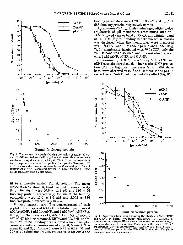
12s~-~p BINDING SITE. The concentration of each peptide that displaced 50% of the labeled ligand was 0.1 nM for rANP, 0.2 nM for pCNP, and 4 nM for C-ANF (Fig. 5, top). In the presence of 1 FM C-ANF, 7 f 1% of specific 1251-rANP binding remained. Scatchard analysis of unlabeled rANP competing for the 1251-rANP binding data showed a nonlinear plot (EBDA), and subsequent curve-fitting analysis of the data by LIGAND gave a best
1
Fig. 4. Light micrographs of gill sections dipped in X-ray-sensitive emulsion to show the distribution of specific binding in the filaments, as determined by silver grain densities. Transverse sections to the gill arch. Magnification x420; scale bar = 25 pm. 4o: Binding on the efferent edge of the filament was found to be associated with efferent filamental arteries (EF) and efferent lamellar arterioles (EL). L, secondary lamellae. 46: Adjacent section incubated with both radiolabel and 1 FM of cold rANP. Most of the silver grains are displaced from the blood vessels.
NATRIURETIC PEPTIDE RECEPTORS IN TOADFISH GILLS R1441
2 80
rANP - C-ANF - pCNP
1o-l2 1o-ll lo-lo 1o-9 1o-8 1o-7 10 -6 1o-5
[peptide] M
Q
binding parameters were 1.28 t 0.35 nM and 1,332 t 588 fmol/mg protein, respectively (n = 4).
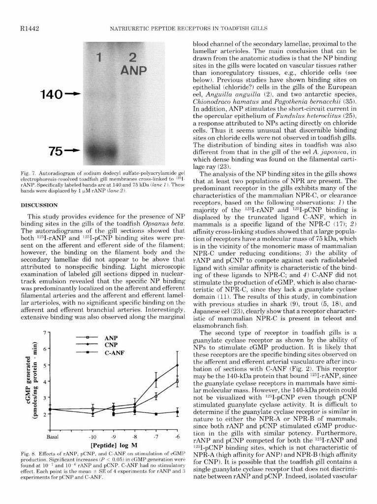
Afinity cross-linking. Under reducing conditions, elec- trophoresis of gill membranes cross-linked with 1251- rANP showed a major band at 75 kDa and a fainter band at 140 kDa (Fig. 7). Binding at both molecular masses was displaced when the membranes were incubated with 1251-rANP and 1 PM rANP, pCNP, and C-ANF (Fig. 7). In membranes incubated with 1251-pCNP, only the 75kDa band was discerned, and this was also displaced with 1 PM rANP, pCNP, and C-ANF.
Stimulation of cGMP production by NPs. rANP and pCNP caused a dose-dependent increase in cGMP produc- tion (Fig. 8). Significant increases (P < 0.05) above basal were observed at low7 and 10s6 rANP and pCNP, respectively. C-ANF had no stimulatory effect (Fig. 8).
u.v 1 . I
0 lob0 2ooo 3& 4&o
Bound fmoles/mg protein Fig. 5. Top: competition study showing the ability of rANP, pCNP, and C-ANF to bind to toadfish gill membranes. Membranes were incubated to equilibrium with 25 pM 1251-rANP in the presence of increasing concentrations of cold peptide. Each point is the mean + SE of 5 experiments. Bottom: representative Scatchard plot from 1 experiment of rANP competing for the 1251-rANP binding site. The plot is consistent with a 2-site model.
fit to a two-site model (Fig. 5, bottom). The mean dissociation constant (&) and maximal binding capacity (B,,) for site 1 were 56.6 t 11.2 pM and 240 t 54 fmol/mg protein, respectively; for site 2 the binding parameters were 11.0 t 8.9 nM and 2,052 t 636 fmol/mg protein, respectively (n = 5).
12%-pc~p BINDING SITE. The concentration of each peptide that displaced 50% of the labeled ligand was 4 nM for pCNP, 1 nM for rANP, and 7 nM for C-ANF (Fig. 6, top). In the presence of C-ANF, 15 t 5% of specific 1251-pCNP binding remained. EBDA and LIGAND analy- sis of 1251-pCNP binding data showed a nonlinear plot consistent with a two-site model (Fig. 6, bottom). The mean & and B,, for site 1 were 0.65 t 0.24 nM and 297 + 104 fmol/mg protein, respectively; for site 2 the -
110
100
90
80
70
60
50
40
30
20
10
0
- rANP
- C-ANF
lo-‘? 1o-ll 1o-lo 1o-9 1o-8 1o-7 1o-6 1o-5
[peptide] M
0.03 1
0.02 ! I . I . I 1 1
0 loo0 1500 2ooo
Bound fmoles/mg protein Fig. 6. Top: competition study showing the ability of rANP, pCNP, and C-ANF to displace 1251-pCNP. Membranes were incubated to equilibrium with 35 pM 12!jI-labeled pCNP in the presence of increas- ing concentrations of cold peptide. Each point is the mean + SE of 5 experiments. Bottom: representative Scatchard plot from 1 experi- ment of pCNP competing for the 1251-pCNP binding site. The plot is consistent with a two-site model.
R1442 NATRIURETIC PEPTIDE RECEPTORS IN TOADFISH GILLS
140
75
Fig. 7. Autoradiogram of sodium dodecyl sulfate-polyacrylamide gel electrophoresis-resolved toadfish gill membranes cross-linked to lzsI- rANP. Specifically labeled bands are at 140 and 75 kDa (lane 1). These bands were displaced by 1 pM rANP (lane 2).
DISCUSSION
This study provides evidence for the presence of NP binding sites in the gills of the toadfish Opsanus beta. The autoradiograms of the gill sections showed that both lz51-rANP and lz51-pCNP binding sites were pre- sent on the afferent and efferent side of the filament; however, the binding on the filament body and the secondary lamellae did not appear to be above that attributed to nonspecific binding. Light microscopic examination of labeled gill sections dipped in nuclear- track emulsion revealed that the specific NP binding was predominantly localized on the afferent and efferent filamental arteries and the afferent and efferent lamel- lar arterioles, with no significant specific binding on the afferent and efferent branchial arteries. Interestingly, extensive binding was also observed along the marginal
1 - ANP - CNP - C-ANF
I I - I ’ I . I * 8 * I - 1
Basal -10 -9 -8 -7 -6
[Peptide] log M Fig. 8. Effects of rANP, pCNP, and C-ANF on stimulation of cGMP production. Significant increases (P < 0.05) in cGMP generation were found at l0-7and 10.” rANP and pCNP. C-ANF hadno stimulatory effect. Each ooint is the mean t SE of 4 exneriments for rANP and 3 experiments&for pCNP and C-ANF. _
blood channel of the secondary lamellae, proximal to the lamellar arterioles. The main conclusion that can be drawn from the anatomic studies is that the NP binding sites in the gills were located on vascular tissues rather than ionoregulatory tissues, e.g., chloride cells (see below). Previous studies have shown binding sites on epithelial (chloride?) cells in the gills of the European eel, AnguiLla anguilla (21, and two antarctic species, Chionodraco hamatus and Pagothenia bernacchii (35). In addition, ANP stimulates the short-circuit current in the opercular epithelium of Fundulus heteroclitus (251, a response attributed to NPs acting directly on chloride cells. Thus it seems unusual that discernible binding sites on chloride cells were not observed in toadfish gills. The distribution of binding sites in toadfish was also different from that in the gill of the eel A. japonica, in which dense binding was found on the filamental carti- lage ray (23).
The analysis of the NP binding sites in the gills shows that at least two populations of NPR are present. The predominant receptor in the gills exhibits many of the characteristics of the mammalian NPR-C, or clearance receptors, based on the following observations: 1) the majority of the lz51-rANP and 1251-pCNP binding is displaced by the truncated ligand C-ANF, which in mammals is a specific ligand of the NPR-C (17); 2) affinity cross-linking studies showed that a large popula- tion of receptors have a molecular mass of 75 kDa, which is in the vicinity of the monomeric mass of mammalian NPR-C under reducing conditions; 3) the ability of rANP and pCNP to compete against each radiolabeled ligand with similar affinity is characteristic of the bind- ing of these ligands to NPR-C; and 4) C-ANF did not stimulate the production of cGMP, which is also charac- teristic of NPR-C, since they lack a guanylate cyclase domain (11). The results of this study, in combination with previous studies in shark (91, trout (5, 181, and Japanese eel (231, clearly show that a receptor character- istic of mammalian NPR-C is present in teleost and elasmobranch fish.
The second type of receptor in toadfish gills is a guanylate cyclase receptor as shown by the ability of NPs to stimulate cGMP production. It is likely that these receptors are the specific binding sites observed on the afferent and efferent arterial vasculature after incu- bation of sections with C-ANF (Fig. 2). This receptor may be the 140-kDa protein that bound lz51-rANP, since the guanylate cyclase receptors in mammals have simi- lar molecular mass. However, the 140-kDa protein could not be visualized with 1251-pCNP even though pCNP stimulated guanylate cyclase activity. It is difficult to determine if the guanylate cyclase receptor is similar in nature to either the NPR-A or NPR-B of mammals, since both rANP and pCNP stimulated cGMP produc- tion in the gills with similar potency. Furthermore, rANP and pCNP competed for both the 1251-rANP and i251-pCNP binding sites, which is not characteristic of NPR-A (high affinity for ANP) and NPR-B (high affinity for CNP). It is possible that the toadfish gill contains a single guanylate cyclase receptor that does not discrimi- nate between rANP and pCNP. Indeed, isolated vascular
NATRIURETIC PEPTIDE RECEPTORS IN TOADFISH GILLS R1443
smooth muscle rings of toadfish respond equally to ANP and CNP, further suggesting that-distinct NPR-A and NPR-B receptor proteins are not found in the branchial vasculature of this species (20). Molecular studies exam- ining the homology of gill receptor protein genes with NPR-A and NPR-B genes may provide additional infor- mation on this point.
The presence of NPR-C in the branchial vasculature of toadfish is consistent with the recent findings of a physiological study in the trout (18). These authors showed that the single-pass extraction of 1251-ANP by the gills was 60% and that this process was mediated by an NPR-C type of receptor, since it was markedly reduced when C-ANF (K-46542) was injected into the circulation, which would occupy the NPR-C. Further- more, the extraction of ANP occurred nearly exclusively in the arterioarterial (respiratory) pathway, which corre- lates well with the anatomic locations of NP binding sites in the gills of toadfish. The ability of C-ANF to compete for the binding sites in toadfish strongly indi- cates that the gills of this species have a similar clear- ance function. Because in fish all blood must pass through the gills, variable expression of clearance recep- tors in vascular smooth muscle cells of the intrafilamen- tal circulation could be an important mechanism for regulating the levels of plasma NPs by differential clearance of the peptide from the circulation (18). Alternatively, it has recently been suggested that NPR-C may regulate the local concentration of NPs vicin .ity of the guanylate cyclase receptors (14).
in the
The absence of discernible binding sites on the bran- chial epithelium does not exclude the possibility of the mitochondria-rich chloride cells, or any other epithelial cell type, being a target for NPs. There are several examples in mammals where potent cGMP stimulation has been shown in cells in which binding sites were not detected (12). This situation may apply to cells that do not express the low-molecular-weight NPR-C, which is often the predominant NPR in many tissues. In the present study , it is possible that a small number of binding sites would not be detected above the level attributed to nonspecific binding. However, since two studies have clearly shown NP binding sites on gill epithelial cells, including chloride cells, using techniques similar to those employed in this study, the absence of binding on the epithelium of toadfish is initial evidence that the epithelium is not targeted by circulating NPs. Interestingly, no detectable binding sites can be found in the kidney of 0. beta (Donald, unpublished observa- tions), further suggesting that direct regulation of ion- transporting tissues in toadfish is limited.
Perspectives
Although NPs have been to shown to mediate a number of osmoregulatory effects in fish, the data are not substantial and are somewhat conflicting (e.g., effects in eel vs. other species; Ref. 7). Current data show that the plasma level of immunoreactive NP is higher in seawater-adapted rather than freshwater-adapted ani- mals, suggesting that the peptide is more important in the regulation of salt load rather than volume load. In
contrast, the vasodilatory actions of NPs have been observed in all species studied, suggesting that the NPs are involved in cardiovascular regulation in fish. The results of the present study provide evidence that the arterioarterial branchial vasculature of 0. beta is regu- lated by circulating NPs. The presence of guanylate cyclase receptors on both the afferent and efferent lamellar arterioles and filamental arteries indicates that NPs could be important in the control of pre- and postlamellar blood flow. Dilation of the branchial vascu- lature would facilitate changes in intrafilamental hemo- dynamics that could alter the functional surface area of the gills by increasing or decreasing the number of perfused secondary lamellae (see Refs. 1, 23). Adjust- ments in the functional surface area affect both osmo- regulation and gas exchange, and therefore it is relevant to consider that the physiological effects of NPs could extend to gill functions other than the regulation of salt and water balance and blood flow. For example, if stretching of the atria1 wall stimulates the release of NPs into the blood, then the peptide will be released during increases in cardiac output associated with activi- ties such as exercise, since this is facilitated primarily by an increase in stroke volume rather than heart rate (24). Many studies have shown that the branchial circulation is regulated by both neural and hormonal mechanisms (17). Clearly, NPs are an important new hormone that may interact with other neural and hormonal systems to control blood flow in the branchial vasculature.
This research was supported by National Science Foundation Grant DCB-8916413 to D. H. Evans.
Present address of J. A. Donald: School of Biological and Chemical Sciences, Deakin Univ., Geelong, Victoria, Australia 32 17.
Address reprint requests to J. A. Donald.
Received 4 November 1993; accepted in final form 16 April 1994.
REFERENCES
1.
2.
3.
4.
5.
6.
7.
8.
9.
Booth, J. H. The distribution of blood flow in the gills of fish: application of a new technique to rainbow trout (Salmo gaird- neri). J. Exp. Biol. 73: 119-129, 1978. Broadhead, C. L., U. T. O’Sullivan, C. F. Deacon, and I. W. Henderson. Atrial natriuretic peptide in the eel, Anguilla an- guilla L.: its cardiac distribution, receptors and actions on isolated branchial cells. J. MOL. Endocrinol. 9: 103- 114,1992. Donald, J. A., and D. H. Evans. Immunohistochemical localisa- tion of natriuretic peptides in the heart and brain of the gulf toadfish, Opsanus beta. CeZZ Tissue Res. 269: 151-158, 1992. Donald, J. A., A. J. Vomachka, and D. H. Evans. Immunohis- tochemical locahsation of natriuretic peptides in the brains and hearts of the spiny dogfish Squalus acanthias and the Atlantic hagfkh Myxine glutinosa. CeZZ Tissue Res. 270: 535-545,1992. Duff, D. W., and K. R. Olson. Atrial natriuretic peptide clear- ance receptors in trout: effects of receptor inhibition in vivo. J. Exp. ZooZ. 262: 343-346,1992. Evans, D. H., E. Chipouras, and J. A. Payne. Immunoreactive atriopeptin in plasma of fishes: its potential role in gill hemodynam- its. Am. J. Physiol. 257 (Regulatory Integrative Comp. Physiol. 26): R939-R945,1989. Evans, D. H., and Y. Takei. A putative role for natriuretic peptides in fish osmoregulation. News PhysioZ. Sci. 7: 15-19, 1992. Flynn, T. G., M. L. deBold, and A. J. deBold. The amino acid sequence of an atrial peptide with potent diuretic and natriuretic properties. Biochem. Biophys. Res. Commun. 117: 859-865, 1983. Gunning, M., C. Cuero, R. Solomon, and P. Silva. C-type natriuretic peptide receptors and signaling in rectal gland of

R1444 NATRIURETIC PEPTIDE RECEPTORS IN TOADFISH GILLS
10.
11.
12.
13.
14.
15.
16.
17.
18.
19.
20.
21.
22.
23.
Squalus acanthias. Am. J. Physiol. 264 (Renal Fluid Electrolyte Physiol. 33): F300-F305, 1993. Healy, D. P., and D. D. Fanestil. Localization of atria1 natri- uretic peptide binding sites within the rat kidney. Am. J. Physiol. 250 (RenaZ FZuid Electrolyte Physiol. 19): F573-F578, 1986. Koller, K. J., and D. V. Goeddel. Molecular biology of the natriuretic peptides and their receptors. Circulation 86: 1081- 1088,1992. Leitman, D. C., and F. Murad. Structure and function of atria1 natriuretic peptide receptor subtypes. In: AtriaZ Natriuretic Pep- tides, edited by W. K. Samson and R. Quirion. Boca Raton, FL: CRC, 1990, p. 78-93. McPherson, G. Kinetic, EBDA, Ligand, Lowry. A CoLLection of Radioligand Binding Analysis Programs. Amsterdam: Elsevier, 1985. McQueen, J., J. C. Kingdom, M. J. Whittle, and J. M. C. Connell. Characterization of atrial natriuretic peptide receptors in human fetoplacental vasculature. Am. J. Physiol. 264 (Heart Circ. Physiol. 33): H798-H804, 1993. Maack, T. Receptors of atria1 natriuretic factor. Annu. Rev. Physiol. 54: 11-27, 1992. Martin, E. R., J. A. Lewicki, R. M. Scarborough, and B. J. Ballerman. Expression and regulation of ANP receptor subtypes in rat renal glomeruli and papillae. Am. J. Physiol. 257 (RenaZ FZuid Electrolyte Physiol. 26): F649-F657, 1989. Nilsson, S., and S. Holmgren. Cardiovascular control by purines, 5-hydroxytryptamine, and neuropeptides. In: Fish Physi- ology, edited by W. S. Hoar, D. J. Randall, and A. P. Farrell. San Diego, CA: Academic, 1992, vol. 12B, p. 301-341. Olson, K. R., and D. W. Duff. Single-pass gill extraction and tissue distribution of atrial natriuretic peptide in trout. Am. J. Physiol. 265 (Regulatory Integrative Comp. Physiol. 34): R124- R131,1993. Perrott, M. N., R. J. Sainsbury, and R. J. Balment. Peptide hormone-stimulated second messenger production in the tel- eostean nephron. Gen. Comp. EndocrinoZ. 89: 387-395,1993. Price, D. A., K. E. Doble, T. D. Lee, S. M. Galli, B. M. Dunn, B. Parten, and D. H. Evans. The sequencing, synthesis and biological actions of an ANP-like peptide isolated from the brain of the killifish, FunduZus heteroczitus. BioZ. BUZZ. Woods HoZe 178: 279-285,199l. Randall, D. Blood flow through gills. In: Gills, edited by D. F. Houlihan, J. C. Rankin, and T. J. Shuttleworth. Cambridge, UK: Cambridge Univ. Press, 1982, p. 173-191. Ruskoaho, H. Atrial natriuretic peptide: synthesis, release, and metabolism. Physiol. Rev. 44: 479-602, 1992. Sakaguchi, H., T. Katafuchi, H. Hagiwara, Y. Takei, and S. Hirose. High-density localization of ANP receptors in chondro-
24.
25.
26.
27.
28.
29.
30.
31.
32.
33.
34.
35.
cytes of eel gill cartilage. Am. J. Physiol. 265 (Regulatory Integra- tive Comp. Physiol. 34): R474-R479,1993. Satchell, G. H. Physiology and Form of Fish CircuZation. Cambridge, UK: Cambridge Univ. Press, 1991. Scheide, J. I., and J. A. Zadunaisky. Effect of atriopeptin II on isolated opercular epithelium of Fundulus heteroclitus. Am. J. Physiol. 254 (Regulatory Integrative Comp. Physiol. 23): R37- R32,1988. Schofield, J. P., D. S. C. Jones, and J. N. Forrest. Identification of C-type natriuretic peptide in heart of spiny dog&h shark (Squalus acanthias). Am. J. Physiol. 261 (Renal Fluid EZectroZyte Physiol. 30): F734-F739, 1991. Sudoh, T., K. Kangawa, N. Minamino, and H. Matsuo. A new natriuretic peptide in porcine brain. Nature Lond. 332: 78-81,1988. Sudoh, T., N. Minamino, K. Kangawa, and H. Matsuo. C-type natriuretic peptide (CNP): a new member of the natri- uretic peptide family identified in porcine brain. Biochem. Bio- phys. Res. Commun. 168: 863-870,199O. Suzuki, R., A. Takahashi, N. Hazon, and Y. Takei. Isolation of high-molecular-weight C-type natriuretic peptide from the heart of a cartilaginous fish (European dogfish, Scyliorhinus canicula). FEBS Lett. 282: 321-325, 1991. Suzuki, R., A. Takahashi, and Y. Takei. Different molecular forms of C-type natriuretic peptide isolated from the brain and heart of an elasmobranch, Triakis scyllia. J. EndocrinoZ. 135: 317-323,1992. Takei, Y., A. Takahashi, T. X. Watanabe, K. Nakajima, S. Sahakibara. Amino acid sequence and relative biological activity of eel atrial natriuretic peptide. Biochem. Biophys. Res. Commun. 164: 537-543,1989. Takei, Y., A. Takahashi, T. X. Watanabe, K. Nakajima, S. Sahakibara, T. Takao, and Y. Shimonishi. Amino acid se- quence and relative biological activity of a natriuretic peptide isolated from eel brain. Biochem. Biophys. Res. Commun. 170: 883-891,199O. Takei, Y., A. Takahashi, T. X. Watanabe, K. Nakajima, and S. Sahakibara. A novel natriuretic peptide isolated from eel cardiac ventricles. FEBS Lett. 282: 317-320, 1991. Ueda, S., N. Minamino, M. Aburaya, K. Kangawa, S. Mat- sukura, and H. Matsuo. Distribution and characterisation of immunoreactive porcine C-type natriuretic peptide. Biochem. Biophys. Res. Commun. 175: 759-767,199l. Uva, B. M., M. A. Masini, L. Napoli, and M. Devecchi. Immunoreactive atrial natriuretic- like peptide in antarctic tele- osts. Comp. Biochem. Physiol. A Comp. Physiol. 104: 291-297, 1993.