Local Hyperthermia in Oncology – To Choose or not
83
3,300+ OPEN ACCESS BOOKS 107,000+ INTERNATIONAL AUTHORS AND EDITORS 113+ MILLION DOWNLOADS BOOKS DELIVERED TO 151 COUNTRIES AUTHORS AMONG TOP 1% MOST CITED SCIENTIST 12.2% AUTHORS AND EDITORS FROM TOP 500 UNIVERSITIES Selection of our books indexed in the Book Citation Index in Web of Science™ Core Collection (BKCI) Chapter from the book Hyperthermia Downloaded from: http://www.intechopen.com/books/hyperthermia PUBLISHED BY World's largest Science, Technology & Medicine Open Access book publisher Interested in publishing with InTechOpen? Contact us at [email protected]
Transcript of Local Hyperthermia in Oncology – To Choose or not

3,300+OPEN ACCESS BOOKS
107,000+INTERNATIONAL
AUTHORS AND EDITORS113+ MILLION
DOWNLOADS
BOOKSDELIVERED TO
151 COUNTRIES
AUTHORS AMONG
TOP 1%MOST CITED SCIENTIST
12.2%AUTHORS AND EDITORS
FROM TOP 500 UNIVERSITIES
Selection of our books indexed in theBook Citation Index in Web of Science™
Core Collection (BKCI)
Chapter from the book HyperthermiaDownloaded from: http://www.intechopen.com/books/hyperthermia
PUBLISHED BY
World's largest Science,Technology & Medicine
Open Access book publisher
Interested in publishing with InTechOpen?Contact us at [email protected]
Chapter 1
© 2013 Szasz et al., licensee InTech. This is an open access chapter distributed under the terms of the Creative Commons Attribution License (http://creativecommons.org/licenses/by/3.0), which permits unrestricted use, distribution, and reproduction in any medium, provided the original work is properly cited.
Local Hyperthermia in Oncology – To Choose or not to Choose?
Andras Szasz, Nora Iluri and Oliver Szasz
Additional information is available at the end of the chapter
http://dx.doi.org/10.5772/52208
1. Introduction
1.1. Historical way – To go or not to go?
Hyperthermia means the overheating of the living object completely (systemic) or partly (regionally or locally). If overheating can be identical with higher temperature then a/the question arises: could overheating be identical with higher temperature or could “higher temperature” be caused only by “overheating”?
Hyperthermia is one of the most common therapies in “house” applications. It is applied according to unwritten traditions in every culture and in every household. It can be applied simply to prevent the common cold but it is also good for its treatment moreover it is effective against various pains (joints, muscle-spasms, etc.), it can be applied for better overall conditions and simply for relaxation, or sometimes for spiritual reasons. The various heat therapies are commonly used complementary with natural drugs (teas, herbs, oils, aromas, etc.) or with natural radiations (sunshine, red-hot iron radiation, etc.) This popular medicine is sometimes connected with ritual, cultural and social events (ritual hot bath cultures), or ancient healing methods (like special spa treatments, hot-spring natural drinks, etc.).
Hyperthermia has two certainly different fields in medicine: hyperthermia as a treatable disease or as a therapeutic method for various diseases (see Figure 1.)
The source of whole body temperature increase as a disease can be internal, having fever by reaction to infections [1] or pyrogens [2] or malignant hyperthermia [3] as well. These whole-body “heating” processes differ also in their systemic reaction. The natural fever is induced by the living system [4], while the system works against the compulsory metabolic heating and tries to keep the temperature normal. Whole-body hyperthermia can be induced by external heating as well [5]. This happens in hot environments when the body is not able to cool itself down, although the system fights against the increase of its

Hyperthermia 2
temperature. This whole-body heating can be unwanted, mainly accidental from environmental sources, and it might be accelerated by additional heavy muscular work or by extra metabolic activity. This is a life-threatening situation with the danger of a heat-stroke [6]. Hyperthermia requests definite and effective medical treatment when it appears as a disease, having elevated body temperature and the patient suffers from its serious consequences. This medical intervention applies therapies to reestablish the normal temperature of the body keeping the healthy homeostasis (overall stability) and handling the consequences of the unwanted high body temperature.
Figure 1. Main categories of hyperthermia
The popular heat-treatment applications are types of “kitchen medicine”: the old recipes are “sure”, the patient follows them, and is cured when everything is done according to the auricular traditional regulations. The meaning of “kitchen medicine” is: do it like in the kitchen, reading the process from the cookery-book: “heat it on the prescribed temperature for the prescribed time and the success is guaranteed”. This type of thinking has its origin in the ancient cultures, when the Sun - the fire, the heat - was somehow in the centre of the religious beliefs and philosophical focus.
This idea of “take it for sure” is the disadvantage of the popular wisdom. It interprets this heating method as a simple causal process, “do it, get it”, however, hyperthermia is not as simple as traditions interpret it.
1.2. Medical history of hyperthermia in oncology
Hyperthermia is really an ancient tradition of human medicine. It is one of the very first known medical therapies, more than 5000 years old, [7].It has been historically recognized that malignant and infectious diseases can be successfully treated by raising the body temperature.
It has been historically recognized that malignant and infectious diseases can be successfully treated by raising the body temperature. The first known oncotherapy by heat was made by an Egyptian priest/physician, Imhotep, in the 5th century BC. He exposed the tumors to “natural heat” (fever) before surgically removing them, which was in fact the very first immune approach as well.
Hyperthermia
Disease caused by various
functional irregularities or by accidental whole-body heating
Therapy applied curatively or palliative
for various diseases in households and in medicine

Local Hyperthermia in Oncology – To Choose or not to Choose? 3
Many ancient cultures used heat to treat diseases and to maintain health. The thermal baths for their curative properties were used by the Greeks, the pre-Christian Jews and the Romans too [8]. The Chinese treated many diseases including syphilis and leprosy by hot spring baths [9]. Taking regular, extremely hot baths from infancy is the block of developing rheumy, according to the ancient Japanese medical notes.
Naturally, this treatment had a sacral meaning in historic times, regarding the Sunlight, the fire and the heat as parts of the leading spiritual object in all ancient religions. This belief was behind when the heat was applied to locally affected parts of the body and to its entirety by means of hot water, steam, hot sand and hot mud baths. Natural hot air caverns connected with volcanic sources were also used. By the development of the medical knowledge, more and more heat applications were applied in practice. Later Hippocrates documented it [10], and he was convinced of its overall efficacy, telling when hyperthermia (fire) does not help, then the disease has to be declared as incurable. Hippocrates said: “Give me the power to produce fever and I will cure all diseases”. His followers in line were Aurelius Cornelius Celsus and Rufus of Ephesus, who believed in the curative effect of fever. The progress continued in the Middle Ages [11], when the ablation techniques (burn out the tumor) and hot-bathes dominated the hyperthermia practices, while the temperature measurement was worked out step by step.
The first clinical thermometer was introduced only later by Sir Clifford Allbutt in 1868. This was the start of the modern history of heat-therapies. The controllable era of hyperthermia was started. The temperature measurement made it possible to control the homogeneous heating and to make correlations with various physiological changes, like:
Increased rate of nerve conduction, Elevation of pain threshold, altering muscle strength, Possible changes in enzyme reactions, Increased soft tissue extensibility, Increased heat- and field-stress reactions (mainly the developments of heat-shock-
proteins), Increased venous and lymphatic flow, Changes in physical properties of tissues, Increased tissue extensibility, Supporting muscular relaxation, reduced muscle spasm, Lymphedema reduction, Superficial wound healing, Treatment of venous ulcers, Assistance in removal of cellular debris and toxins, Alteration of diffusion rate across the cell membrane, Increased intramuscular metabolism, Relieving pain, analgesia,
Hyperthermia 4
Increased metabolic rate of contracted joints using heat and stretch techniques, Need of stretching during and/or immediately following the treatment, Alterations of collagen properties, allowing it to elongate, Increased rate of phagocytosis, Increased ATP activity (assisting wound regeneration), Psycho-feedback (pleasant sensing) [positive placebo effect], Ability to control the chronic infection by increasing the circulation, Heat increases the extensibility of fibrous tissues such as tendons, joint capsules and
scars.
It was applied for many various diseases like:
rheumy, gout, pain-management, arthritis, some dermatological disorders, muscle spasms, supportive therapies in sport, some gynecological disorders, some allergies, rhinitis, common cold, pediatric ear diseases, wound healing, supporting the general rehabilitation process.
A Nobel Prize was also granted for hyperthermia in Physiology & Medicine in 1927 "for his discovery of the therapeutic value of malaria inoculation in the treatment of dementia paralytica" to Julius Wagner-Jauregg, (1857-1940, Austria).
The application of heat in oncology has been restarted with huge intensity. Among the first modern curative applications in oncology, Busch [12] and Coley [13] were successful at the end of the 19th century with artificial fever generated by infection and toxins, respectively. These systemic applications were soon followed by local and regional heating by Westermark F. [14], Westermark N. [15], and Overgaard K. [16]. The leading German surgeon at that time, Bauer KH’s opinion in his monograph “Das Krebsproblem” about the oncologic hyperthermia is typical: “All of these methods impress the patient very much; they do not impress their cancer at all.” However, very early, in 1912, a controlled Phase II clinical study was published, 100 patients showing the benefit of the thermo-radiation therapy [17]. Tremendous number of publications were prepared in the first quarter of the 20th century, expecting fantastic development in the topic, [18], [19], [20], [21], [22], [23], [24], [25], [26], [27], [28], [29], [30], [31], [32], [33], [34], [35], [36], [37], [38], [39], [40], [41], [42], [43], [44], [45], [46], [47], [48], [49].

Local Hyperthermia in Oncology – To Choose or not to Choose? 5
1.3. Heating methods – local and systemic
As it was shown before, there are two, basically different hyperthermia processes: the systemic (heats the complete body, whole-body treatment), and the local/regional heating (heats only a part of the organism). The two basic kinds of the heating methods also differ in their physiological limitations: the systemic treatment, of course, modifies the entire physiology of the organism, and that could limit the applied energy-absorption and body-temperature. There is a possibility to absorb energy in large volume equally or having a layer-by-layer changing front (heat-diffusion, heat-flow flux) depending on the penetration depth of the actually applied energy. Nevertheless, the old direct heating methods (hot solids or liquids in the area or in the nearest body cavity) were not effective enough for local deep heating without skin injury.
Thermodynamically the systemic and local/regional treatments differ by their energy-intake. The whole body treatment is based on the blood-heating (mostly heats up the subcutaneous capillary bed, or heats the mainstream of the blood directly with extracorporeal heater), while the local hyperthermia is definitely a tissue heating approach. This difference drastically divides the two methods from thermal point of view. In whole body treatment the blood is a heating media, it delivers the heat to the tumor and heats it up; while in local treatment, the blood remains on body temperature during the local heating, so it is a cooling media (heat-sink) for the locally heated tumor, (see Figure 3).
Figure 2. Opposite thermodynamic mechanisms of whole-body, systemic (a) and local (b) heating methods. The blood-heated tumor in whole body treatment reaches thermal equilibrium after a certain time, while the local treatment is always in non-equilibrium state, because the body temperature is lower than the heated tissue, creating intensive heat-flow from the target to the neighborhood.
The systemic (whole-body) treatment uses the blood-circulation to heat-up the body. (see Figure 3.)
The artificially elevated body-temperature is the source of heating in fever inducing methods. Fever inducing can be solved with various drugs [50], as well as with special inflammation-inducing toxins.

Hyperthermia 6
The oldest whole body heating was the contact method, immersing the patient into the hot bath, but due to its numerous disadvantages of this method, it is rarely in use any more. Mainly two direct methods are available in the modern medicine to make systemic hyperthermia. The less frequently used is the extracorporeal (the blood is heated outside the body), or the intracorporeal (the blood is heated in-situ in the body). In most cases the capillary bed of the subcutane area is heated by conductive (e.g. hot-bath) or radiative (e.g. infrared) way, using extra-corporal blood-heating. This method takes out the blood from the continuous flow by a definite arterial outlet and the outside heated hot blood is pumped back to the patient.
Figure 3. Categories of whole-body hyperthermia
The whole-body heating could be solved in various ways, like steam, water or radiation heating (see Figure 4.). There are other possibilities as well (e.g. wax heating, hot-air heating, etc.) but the limited possible heat-flux and the poor technical realizations hinder these solutions. By all of these whole-body heating forms the patient’s safety has to be seriously considered. These are based on the blood-heating in the subcutaneous capillary bed, and the physiological reactions (vasodilatation and sweating) work well against the huge heat-flux into the body. The long heating time is also challenging (over an hour) moving the body away from the healthy homeostasis. The heat-flux through the skin is limited by the heat injuries (~1 W/cm2 is the limit) so the contact heating with steam and water has definite problems. The radiation heating can be solved by special infrared wave (Infrared A) which penetrates deeper (~1-2 mm) into the subcutaneous layer, and can manage higher energy-flux without burn injuries. The method has many early descriptions [51], [52], [53], [54]; but the dominant systemic hyperthermia method is based on the infra-red radiation by multi-reflecting filtering [55], [56] or by water-filtering [57], [58], [59], [60].

Local Hyperthermia in Oncology – To Choose or not to Choose? 7
Figure 4. Various main methods of the whole-body heating: (a) steam, (b) water, (c) electromagnetic radiation
When the whole body is systemically heated, it has a strict physiological limit: 42 ºC in humans. The thermal distribution till this level is homogeneous and well controllable. No hot-spots exist, no question arises about the isotherms; the physiologically extreme temperature can be fixed all over the body. Suppressing the risk; a decreased treatment temperature (moderate WBH/whole-body fever-range thermal-therapy) is also applied. The application of lower temperatures for longer time period (fever-therapy, or mild-hyperthermia) also showed surprisingly good efficacy for whole-body hyperthermia treatments [61], [62], [63], [64]. The whole body hyperthermia and even its fever-range versions mean effective immune support, [65], [66], [67], [68], which might be a very important factor for patients with weak immune-system.
The local/regional hyperthermia has also large categories (see Figure 5.) and various technical choices (see Figure 6.). The widely applied technical solutions were available only after the discovery of the electromagnetic heating. The electromagnetic waves can penetrate deeply into the body. Dominantly, the local/regional systems work by radiative [69], [70], [71], or by capacitive [72], [73], [74] technical solutions.
Both heating categories form large groups of treating options having special subcategories, which are involved in different physiological actions and support different reaction mechanisms in the organism.
Technically, a huge variety of heating can be applied by heat therapies. Its energy-production, its selectivity, locality, kind of energy-delivery, invasivity control, frequency of the electromagnetic waves, as well as their medical applications and combination with other methods make the heat-therapies different, (see Figure 7.).

Hyperthermia 8
Figure 5. Categories of local heat-treatments
Figure 6. Main technical solutions of local heating: (a) contact heat, (b) deep-heating, (c) radiative heating, (d) immersing a part of the body in hot water
These combinations involve more than a hundred solutions of hyperthermia used as treatment, which make the therapy indefinite in its applications. This emphasizes the reason why the present review tries to summarize the main categories.
The thermodynamic situation, and in consequence of the physiological effects of the systemic and local/regional heating modalities are entirely different. There is a definite difference between the temperature of the blood and the inter-capillary volume in the microscopic structure of the target: the blood-arteries are hot-sources in case of systemic treatment, they deliver the heat. However, in case of local/regional hyperthermia it is entirely the opposite: the blood is relatively cold (remaining on unchanged body temperature), the arteries are heat-sinks which are cooling the tumor down, in fact, these work against the local heating.
One question automatically arises: when the heat, the energy-flow has a central role in hyperthermia, then what does the temperature do in the living systems?
The temperature as the average of kinetic energy in the system has a double role in the control of the heat-absorption. It characterizes the heat-absorption, when the heating is homogeneous,
Local Hyperthermia in Oncology – To Choose or not to Choose? 9
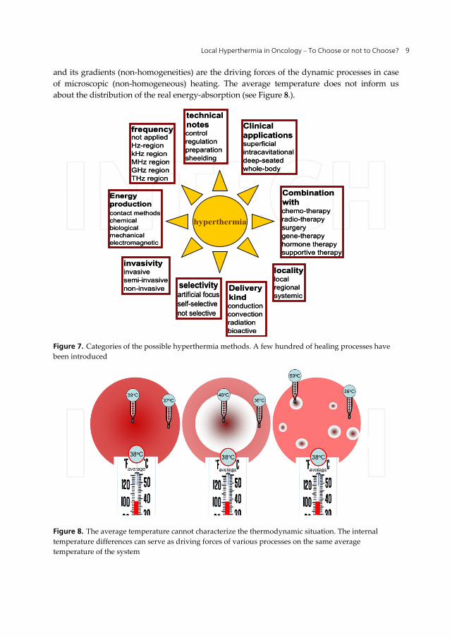
and its gradients (non-homogeneities) are the driving forces of the dynamic processes in case of microscopic (non-homogeneous) heating. The average temperature does not inform us about the distribution of the real energy-absorption (see Figure 8.).
Figure 7. Categories of the possible hyperthermia methods. A few hundred of healing processes have been introduced
Figure 8. The average temperature cannot characterize the thermodynamic situation. The internal temperature differences can serve as driving forces of various processes on the same average temperature of the system
Energyproductioncontact methodschemicalbiologicalmechanicalelectromagnetic
Energyproductioncontact methodschemicalbiologicalmechanicalelectromagnetic
frequencynot appliedHz-regionkHz regionMHz regionGHz regionTHz region
frequencynot appliedHz-regionkHz regionMHz regionGHz regionTHz region
invasivityinvasivesemi-invasivenon-invasive
invasivityinvasivesemi-invasivenon-invasive selectivity
artificial focusself-selectivenot selective
selectivityartificial focusself-selectivenot selective
localitylocalregionalsystemic
localitylocalregionalsystemic
technicalnotescontrolregulationpreparationsheelding
technicalnotescontrolregulationpreparationsheelding
Deliverykindconductionconvectionradiationbioactive
Deliverykindconductionconvectionradiationbioactive
Clinicalapplicationssuperficialintracavitationaldeep-seatedwhole-body
Clinicalapplicationssuperficialintracavitationaldeep-seatedwhole-body
Combinationwithchemo-therapyradio-therapysurgerygene-therapyhormone therapysupportive therapy
Combinationwithchemo-therapyradio-therapysurgerygene-therapyhormone therapysupportive therapy
hyperthermia
Hyperthermia 10
Herewith we do not discuss the extreme temperature facilities in local treatments, the ablative techniques with high temperature (heat-ablation, [75]) or low-temperature (cryo-ablation, [76]).
1.4. Technical history of local hyperthermia in oncology
Despite the hyperthermia was among the very first medical treatments in human medicine, this approach has ambivalent evaluation as a therapy. While it is popular in households to “treat” many diseases like common cold, pain, various orthopedic problems, etc., it is on the periphery of the serious medical therapies. This frustration characterizes the complete history of hyperthermia in medicine, and explains why hyperthermia has no well- deserved place in the medical armory to treat various diseases.
At the end of the 19th century, energy delivery by electromagnetic fields became possible; but the real technical revolution of the heat therapies was when the modern microwave heating was developed, and applied in medicine from the middle of the 20th century. This focused, temperature-based deep heating became one of the line (focused local/regional heating), while the other was the whole body heating with various methods. In local heating, the paradigm is to reach the appropriate temperature locally.
Nevertheless, the intensive use of hyperthermia in oncology began in the last third of the 20th century. The first symposium on oncological hyperthermia was held in Washington DC, USA in 1975; and the second one in Essen, Germany in 1977. Both conferences were supported by the local scientific communities. We may consider the birth of the modern oncological hyperthermia from this time on, and take it as a strong candidate and a member of the acknowledged tumor therapies.
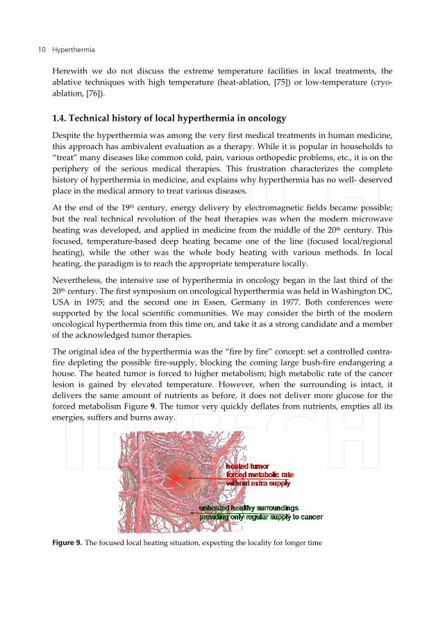
The original idea of the hyperthermia was the “fire by fire” concept: set a controlled contra-fire depleting the possible fire-supply, blocking the coming large bush-fire endangering a house. The heated tumor is forced to higher metabolism; high metabolic rate of the cancer lesion is gained by elevated temperature. However, when the surrounding is intact, it delivers the same amount of nutrients as before, it does not deliver more glucose for the forced metabolism Figure 9. The tumor very quickly deflates from nutrients, empties all its energies, suffers and burns away.
Figure 9. The focused local heating situation, expecting the locality for longer time
Local Hyperthermia in Oncology – To Choose or not to Choose? 11
Indeed, there are many factors showing the validity of this assumption, but the breakthrough to accept the method widely hasn’t been reached in the long historic period. The appropriate temperature selectively administered in the tumor still cannot be reached. This fact forms a lot of questions in the published literature:
Is the community of radiation oncologist ready for clinical hyperthermia? [77]; What happened to hyperthermia and what is its current status in cancer treatment? [78]; Where there’s smoke, is there fire? [79]; Should interstitial thermometry be used for deep hyperthermia? [80]; If we can’t define the quality, can we assure it? [81]; Is there a future for hyperthermia in cancer treatment? [82]; What is against the acceptance of hyperthermia? [83]; Progress in hyperthermia? [84]; Prostate cancer: hot, but hot enough? [85]; Is heating the patient a promising approach? [86]; Hyperthermia: has its time come? [87].
Many of the researchers evaluating the capabilities of oncological hyperthermia share the opinion expressed in the editorial comment of the European Journal of Cancer in 2001: the biological effects are impressive, but physically the heat delivery is problematic. The hectic results are repulsive for the medical community. The opinion, to blame the “physics” (means technical insufficiency) for inadequate treatments is general in the field of oncological hyperthermia, formulated the following statement: “The biology is with us, the physics are against us [82]. In the latest oncological hyperthermia consensus meeting the physics was less problematic. However, in accordance with the many complex physiological effects, a modification was proposed: “The biology and the physics are with us, but the physiology is against us” [88].
The most problematic issues have always been technical: how to heat in depth, locally focused, being selective for malignant cells, and the other side: how to control it and how to measure the efficacy of the treatment? Even when the local treatment is focused well, the temperature by its way tends to be equalized; the focus is extended by time, due to the very effective heat-exchanger – the blood-stream. The heated tumor strongly exchanges its heat with its healthy surrounding, extending the focus gradually and increasing the local blood-flow. This unwanted effect has some problematic consequences:
1. The heating focus is growing, it is not correct any more, so the healthy tissue is also heated up,
2. The distributed spots of the tumour (local metastases) can be covered only by on average, covering the area completely with the intermediate healthy parts,
3. The natural movements of the patients (i.e. due to the breathing) cannot be followed by the focus,
4. The blood-flow is increased locally, supplying the tumor with nutrients (first of all with glucose) and the higher temperature gains the local metabolic rate as well,
5. The intensive tumor-metabolism produces high level of lactic acid in the volume, which lowers the pH and forces the blood to buffer it. The blood starts to shift to alkaline and

Hyperthermia 12
the alkaline blood delivers more oxygen to the volume, by positive feedback mechanism,
6. The intensive blood-flow has a risk of the further disseminations and metastases, 7. The heat flow to the surroundings can damage the healthy neighbourhood, 8. Enlarging the sphere having certain temperature gradients increases the area of the
injury current which supports the cell-proliferation, 9. The growing heated volume is uncontrolled, the vigilance of the process becomes
complicated, 10. The incident energy might burn the skin, so surface cooling is necessary. The heat-sink
of the surface decreases the incident power, but its quantity has no measurable parameters. This is the reason why only the temperature in the target will orientate the control of the treatment process,
11. The necessary temperature measurement is mostly invasive, which could cause many complications, including inflammation, bleeding, infection, dissemination of the cells, etc.
12. The microwave heating might do harm not only by its unwanted hot-spots, but also with its ability to create carcinogens in situ.
These technical challenges are definitely complex, and can make the actual hyperthermia treatment uncontrolled. This branch of problems could be the reason for some controversial results and the weak acceptance of the conventional hyperthermia among medical experts.
2. Malignant diseases – To heat or not to heat?
The popular terms “heat-dose”, “temperature-gain”, “thermal dose”, “energy-intake”, “energy-dose” may have different definitions in various individuals who use them. Our first task is to clarify the differences between these (in popular literature many times used as synonyms) terms.
Nevertheless, the temperature is not heat, not even energy. Temperature, in this sense, is a hypothetical value of the average energy. Particles have various energies and it could happen no any individual particle has such definite energy as the average (the temperature) indicates.The temperature is an average; we can define it only on a large number of participating units (particles). The average energy of the various particles at normal human body temperature (ideal thermodynamic model) is ~ 2.5 kJ/mol. This relatively large energy is embedded and blocked in the actual system. (It is so large, that if it could be liberated within one second, the obtained power would be 2.5 kW/mol.) This average thermal energy limits the internal bonds and interactions, because any lower energy bond will be destroyed by this thermal background. This internal energy could make abrupt changes by such chemical reactions, by which activation energy is smaller (or equal) than the actual thermal average energy. The weakest bonds in life are the hydrogen-bridges, having 18 kJ/mol in ice [89] and ranging 3-30 kJ/mol in various compounds in living objects [90].
Pumping heat into a system can increase its temperature. The result of heating is not a definite temperature. The resulting temperature always greatly depends on how the heat is consumed in the system, and how the system transfers this energy to its environment.

Local Hyperthermia in Oncology – To Choose or not to Choose? 13
Pumping the same energy into identical volumes, but having different surfaces, the temperature increase will be definitely different, because the volume is differently cooled down by the environmental conditions. Furthermore, the heat can make structural or other rearrangement in the material, it could be transformed into work without temperature increase of the system. For example: melting the ice absorbs energy without temperature increase, still it is completely transformed into liquid. The heat, which is pumped in to the system at this phase transition does not increase the temperature; its energy is used completely to change the structure of the material from solid to liquid.
Other clear example is the Sun radiation to the Earth. A huge part of the energy of the Sun’s radiation is converted to the meteorology (like wind or rain), their mechanical effects (like waves in the ocean, like distortion of the rocks). The Sun’s energy is the solely energy-source of the life processes, and this energy of the Sun makes our oil reserves, allowing us to use this energy for various applications in our everyday life. So the simple electromagnetic radiation (the spectrum of the Sun) is converted to the various kinds of energies in the Earth. Only a fraction of the radiation is realized as heat and rising temperature.
Other simple example is the human energy-intake: a female adult eats ~1600 kcal/day. When she takes more energy a day, it will not change her body temperature at all. However, when she gets this energy from radiation, she will starve, despite of the fact that she gets more energy than she could take from nutrients.
When there are so serious differences between the heat and temperature, why do we confuse them? The main reason is: we fix our attention to simple situations when we heat such materials which distribute the energy immediately all over the system, making thermal equilibrium without internal work (reactions, dilatation, etc.) in the system. In such cases, of course, all the heat energy increases the internal energy of the system, and so it is distributed in it, and proportionally increases its temperature.
When we fix our attention to such systems, like heating water in its liquid form, the applied heat and temperature are strictly proportional in a certain interval (when the water is definitely liquid). However, we have a problem even in this simple system. When a phase-transition occurs, we lose this proportionality completely. At the/a study of hyperthermia in living systems, we have to well distinguish the overall energy, the heat energy and the temperature (which is not energy) from each other.
“The use of thermodynamics in biology has a long history rich in confusion.” [91]. The main complication is the fact that life cannot be studied isolated from its environment, and so the energetically open system could lead to numerous uncertainties, leading sometimes to mystification as well.
2.1. The heating paradigm
The idea of the local heating effect to burn-out the energy sources of the malignancy has dominated the hyperthermia applications from the time of Hippocrates. Its operation measured by modern methods [92], shows definite decrease of ATP and increase of lactic

Hyperthermia 14
acid in tumors after hyperthermia treatment. The ATP depletion makes a heavy ionic-imbalance in cells [93]. Furthermore, the increased temperature can slow down or even arrest DNA replication, [94], [95].
Higher tissue temperature stimulates the immune system [94] with observed increase in natural killer cell activity [96]. Moreover, the elevated temperature distributes tumor-specific antigens on the surface of various tumor cells [97] and assists in their secretion into the extracellular fluid [98], triggering the immune reactions against the malignant cell [94].
Hyperthermia has shown significant pain-reduction during treatments [99]. This makes this method excellent to improve the quality of the patient’s life and it can be applied as palliative treatment when the curative solution does not work.
Multiple effects can be counted by homogeneous heating the living organisms completely or locally. One of the decisional factors is the vasodilatation. The induced vasodilation increases:
Blood flow Capillary filtration Capillary pressure Oxygen perfusion
These induce the following actions:
Increases fibroblastic activity and capillary growth, Increases the oxygenation in the volume, Increases drug-delivery in the volume, when the drug is administered systemically, Increases the nutrition concentration in the volume, Increases the metabolic activity in the volume (higher quantity of nutrition, oxygen and
higher local temperature), Increases the field-dependent effects, (membrane excitation, activation of signal
pathways, etc.), Increases the effects on the blood-structure in the volume, Increases the micro vascular perfusion (circulation), nutrients, and phagocytes.
The chemo-drugs are delivered to the tumor by the blood-stream. Higher local temperature increases the microcirculation in the heated volume, [100], [101], [102], [103], [104], [105], [106], [107], [108], [109]; and with this it enhances the efficacy of the conventional chemo-therapies [110]. The synergy between heat-treatment and many of the chemotherapies is well-established [111], [112].
Further support is that the hot drug is more reactive [113], providing excellent possibility to synergy, which is even more effective when we consider the accelerated drug metabolism and gained pharmacokinetic parameters. The thermo-chemotherapy results in a better therapeutic effect and it increases the target specificity as well as it reduces the systemic side effects [114], [115]. In some cases the low-dose chemotherapy could be used [116], [117] with the hyperthermia promotion, it is also applied in low-dose metronomic chemo-regulation,

Local Hyperthermia in Oncology – To Choose or not to Choose? 15
[118]. Furthermore, hyperthermia acts in G0 phase of cell-division which makes the action of conventional therapies possible on these cells too.
For radiotherapies, the increased microcirculation sensitizes the effect of ionizing radiation [119]. The primarily applied heat can enhance the effect of ionizing radiation because of the higher oxygen concentration in the area. Furthermore, the most efficient action of hyperthermia is in S-phase [120], which well completes the weak effect of radiotherapy in this phase of the cell-cycle. The heat-induced decrease of the DNA-dependent protein-kinase (DNA-PK), [121] is also a radio-sensitizer. Sensitizing the classical ionizing-radiation by hyperthermia has been well-known [122], [123] for a long time, and different review articles have summarized this knowledge [124], [125], [126], [127]. The advantage of combining heat-treatment with the classical ionizing-radiation is unambiguous, [128], [129], [84], the synergy between the methods is well known [130], [122] and successfully applied [131], [132], [106].
Hyperthermia has also been found to have pronounced advantages for surgical interventions. Through hyperthermia induced inhibition of angiogenesis and heat entrapment, the outline of the tumor often becomes pronounced and the size of the tumor often shrinks, making previously dangerous operations possible [133]. The feasibility of the preoperative application for locally advanced rectal cancer is well shown in a Phase II clinical trial [134]. Postoperative application of hyperthermia has also been thought to prevent relapses and metastatic processes [132]. Intraoperative radiofrequency ablation [135] and local hyperthermia [136] have also been used to improve surgical outcomes.
The combination of hyperthermia with gene-therapy also looks very promising, as shown by the successful combination of hyperthermia with HSP-promoter mediated gene therapy in cases of patients with advanced breast cancer [137]. Hyperthermia improved the results of the HSP-promoter gene therapy by inducing local HSP production and by enhancing the local rate of release from liposome [138]; it was also helpful in the double suicide gene transfer into prostate carcinoma cells [139]. It was proven that this combination therapy was highly selective for mammary carcinoma cells. Also the heat-induced gene expression could be an excellent tool in the targeted cancer gene therapy [140].
Combination with hormone therapies is also a vivid method, applied for prostate [141]; and in melanoma treatment [112]. Enzyme-therapy [142], photodynamic therapy [143], gene therapy [144], immune- [145] and other supportive-therapies [146] are used in combination with hyperthermia.
All these factors position hyperthermia to one of the most effective treatments in oncology. Since these are mostly temperature dependent effects, an accelerated race has been started for the rising temperature and providing the highest available thermal support for the conventional therapies. Most of the applied electromagnetic techniques started to gain their power over 1 kW, providing powerful radiative, capacitive and magnetic effects to increase the temperature in depth in the targeted tissue.

Hyperthermia 16
2.2. Targeting complications
The large heating energy heats up the healthy surrounding as well, see Figure 10. The blood-flow will be enhanced, the nutrients supply will be higher and the result is the opposite of our aim. The situation becomes even worse by continuing the high-energy heating: the high blood-flow helps the dissemination [147], [148], [149] and could gain the metastases: Figure 11. With this, we can definitely worsen the survival and the quality of life of the patient. This problem gained the official policy in many oncological departments: avoid application of hyperthermia in oncotherapies.
Figure 10. The real situation heats up the surroundings, the local heating does not remain locally focused
Figure 11. The large heated volume is not controlled from the focus, and makes the malignant dissemination possible by the high blood-flow in the healthy surroundings
It is plausible: the temperature spreads to the neighboring volumes independently of how precise the focus of the energy is. The energy can be focused, but the temperature seek is to be equalized and the focused energy-intake will heat up the tissue out of the focus too. This process is very rapid anyway in such good heat-conduction material as the living tissue. The cooling becomes even more emphasized, considering the physiological feedback

Local Hyperthermia in Oncology – To Choose or not to Choose? 17
mechanisms, which tries to reestablish the local homeostatic equilibrium by intensified blood-flow. The blood is on body temperature and this way it is an effective cooling media. This is the reason why huge energy is necessary to compensate the heat-loss of the tumor, and keeps the tumor temperature actually higher than its healthy environment.
A typical capacitive coupling solution pumps enormous energy, [150]. The rise of temperature, applying 1200 W energy after 45 min, was 4.8 oC but the reached focus differs with about 1 oC only from its untargeted neighborhood. In case of radiative applications the situation is the same. The temperature elevation in the tumor after 57 min was 4.2 oC; reached by as high power as 1300 W [151]. The overall heating obviously shows unwanted hot-spots. The elapsed time smears the relatively focused temperature. The temperature increase in the tumor was 4.2 oC on average, while in the surrounding muscle it was only 3.8 oC [151]. Is this the focus we expected? (Note, a standard speedy electric tea kettle uses 1300 W to boil a cup of water within a couple of minutes. The increase of the temperature for the ~ 0.3 liter water is ~75 ºC. We apply, in these cases, the same power reaching a temperature increase ~7 ºC during 60 minutes).
Further problems occur by a huge surface energy-density pumping high energy dose in depth. Due to the huge thermal load on the skin, more and more sophisticated methods have been/are being developed to cool the skin and to avoid its burn. A new technical race started: cooling the surface together with the increase of the incident power.
The maximal surface power density could be 1 W/cm2 (10 kW/m2), for ~ 42 min, (see Figure 12. [152]), which is a definite limit of the energy intake.
Figure 12. Threshold of surface blisters by power density through the dermal layers
In general, the injury level (threshold of blisters) depends on the full integral of the heat-flux, consequently, a longer treatment-time has lower power limit of use and for example, the 60 min treatment allows maximum 0.5 W/cm2 without surface toxicity. Cooling is applied in most of the technical solutions to avoid the surface burn from overheating, see Figure 13.
Threshold of blisters
Tim
e o
f h
eat
exp
osi
ng
[m
in]
y = 42.599x -0.4089
R2 = 0.9943
30
35
40
45
50
55
60
65
0.4 0.6 0.8 1 1.2 1.4 1.6 1.8
Power density [W/cm2]
y = 42.599x -0.4089
R2 = 0.9943
30
35
40
45
50
55
60
65
0.4 0.6 0.8 1 1.2 1.4 1.6 1.8
Power density [W/cm2]

Hyperthermia 18
Figure 13. The incident power can overcome the threshold of blisters (a). Appropriate surface cooling is introduced to avoid this burn (b)
The surface cooling has a double effect: it cools down the surface taking away the surface energy to avoid the burn and to keep the heat-sensors (which are located in the near-surface subcutaneous area) of the body in the pleasant range of feeling.
However, the surface cooling creates serious problems too:
It makes the control of the energy intake ambiguous, no precise dose can be measured by the forwarded power. When the forwarded dose is 100 W, the cooling is 50W and the intensive cooling of the blood circulation on the surface is 30W and then maximum 20W can be absorbed in the target (see Figure 14.).
Figure 14. The forwarded power and the cooling power work oppositely. No idea about the real power pumped into the body
The high energy loss in the surface area is mainly due to the adipose layers, which are good electric- and heat-isolators [153]. Their isolation ability depends on their thickness, and it is controlled by the blood perfusion. When we cool down the surface, the homeostatic control will increase the isolating layer (see Figure 15.) by reducing the blood-flow in the area.
There is a misleading competition on the power, which is applied for local hyperthermia. The subcutane adipose tissue is an electric- and heat-flow blockade, and its conductivity decides the current transport at a fixed power transmission. The applied voltage depends on the contact area and on the applied frequency as well. The available devices for local heating range from 150W to 2000W and, in fact, the local temperature in the tumor ranges between

Local Hyperthermia in Oncology – To Choose or not to Choose? 19
40-45 oC. The local maximal temperature in most cases depends more on the patient than on the incident power. At the end, the origin of the heat-flow will not exceed the 1 W/cm2 on average. The higher isolation will absorb more energy, and so we need more cooling for safety and so on, see Figure 16. It is a positive feedback regulation, requesting enormous energies.
Figure 15. The blood-perfusion changes the conductivity and the relative permittivity as well
Figure 16. The physiologically regulated optimum is necessary for energy control. The large cooling grows the physiological reaction, creates a layer of isolation, which accelerates the dangerous surface overheating by positive feedback
Further complication in the control is the value of blood-flow, which is temperature dependent. The physiologic effects connected with the blood-flow are considered to be important and it is studied in details, [154], [155], [156], [157], [158].
The cooling energy is indefinite. The applied definite heating is modified with this energy-factor, making the really applied energy immeasurable. The hyperthermia dose must be the physically correct, accepted specific-energy absorption rate in J/kg, as we do it in the case of ionizing radiation too. The dosing in this case requests other, deep-inside measurement for indication of the process: the temperature as the character of the really absorbed energy. However, it is again problematic from a technical point of view: in fact, the absorbed energy
Electric parameters of the skin, depending on the blood-perfusion
de
vepe
eb
y
0 0.1 0.2 0.3 0.4 0.5 0.6 0.70.1
1
10
100
Conductivity [S/m]
Relative permittivity
de
vepe
eb
y
0 0.1 0.2 0.3 0.4 0.5 0.6 0.70.1
1
10
100
Conductivity [S/m]
Relative permittivity
Blood perfusion (ratio)
Relative permittivity
Conductivity (S/m)
Water-bolusWater-bolusWater-bolusWater-bolus Water-bolusWater-bolusPhysiologically regulated larger isolation layerHigh drop of field
Water-bolusWater-bolusIntensive cooling
Regulated temperatureNo intensive cooling
Physiologically regulated good heat- and electric conduction (optimized for low drop of field)
High drop of field burns the subcutaneous layer under the electrode
Intensive cooling continues
Regulated optimumOvercooled surface Positive feedback → burn

Hyperthermia 20
distribution and the gained temperature are very different [159]. The reason is simple: provide the same energy to identical volume but the blood-flow is different in every case so it will result in different temperature. The physiology modifies the temperature! It is not possible to match the specific absorption rate (SAR) and the developed temperature.
The measurement of the power is missing, due to the fact that cooling is immeasurable and has a modifying effect on the temperature. This makes the temperature measurement in the target important, as this is the only parameter which gives us some idea about the absorbed energy, (see Figure 17.). This (generally invasive, measurement) makes it possible to have orienting value of the absorbed power in general, and sometimes it is important for safety to avoid the unwanted hot spots as well. For the successful energy dose control we have to know the energy taken away by the cooling. This underlines the importance of the control of the surface cooling triggered by the actual physiological conditions in the subcutaneous capillary bed.
Figure 17. The control of both the energies together with the physiological parameters make oncothermia safe and effective
The full unsuccessful temperature focusing together with the intensive cooling process in conventional hyperthermia is, borrowing the words from Shakespeare, “much ado about nothing”.
Dr. Storm a recognised specialist of hyperthermia formulated [78] a general opinion: “The mistakes made by the hyperthermia community may serve as lessons, not to be repeated by investigators in other novel fields of cancer treatment.”
2.3. What to learn from nano-scale energy-liberation?
The general idea of microscopic heating is simple: the heating energy is not liberated in a sudden single step, but regulated and multiple small energy liberation does the same job, (see Figure 18). In our case, the forwarded energy selectively targets the most influential areas. Instead of the high, general energy pumping into the lesion, the energy is liberated at the membranes of the malignant cells.
The microscopic effects, instead of the large energy liberation, is one of the most update thinking in energy source developments.
The conventional engines in vehicles use the energy of explosion of different chemicals (e.g. petrol, diesel, kerosene). The explosion by a spark or heating over their activation energy

Local Hyperthermia in Oncology – To Choose or not to Choose? 21
liberates large energy in a short time, and only a small fraction of this could be applied beneficially, most of the energy is radiated, conducted or lost in various other ways. One of the largest losses is the heat-exchange by the high temperature, which somehow has to be used again (e.g. intercooler, turbo). The latest solution, however, is the set of microscopic explosions, promoting the chemical reactions individually by a membrane control (i.e. fuel cell solution) and using the energy step-by-step as a sum of the micro-reactions. The relatively low efficacy combusting engines are intended to be replaced by the fuel-cell energy-sources combined with electric motors, which are based on the membrane regulated microscopic reactions of gases. (Mostly hydrogen and oxygen gases are in use.)
In fact, life “invented” the controlled energy-liberation by micro-processes, blocking the sudden, explosion-like energy liberation, driving the processes small subsequent energy-conversion steps instead. In the living objects the energy is liberated gradually in a “ladder” of multistep processes, and this is also moderated by surface reactions.
Figure 18. The difference between macro- and micro-liberation of energy. The latter is much more efficient.
The applied power and its efficacy are usually not connected. Good examples can be found in our everyday life, in systems like the standard light bulbs and the energy safe ones using a fraction of the power for the same light; or the various power-consumptions of cars, having equal performance, or the various fuel consumption of them having the same engine-power. The incandescent bulb creates light by high-temperature filament, which heats up the environment, having only 10% efficacy.
Fluorescent technology solves this task more smartly: it makes the energy-liberation selective where the effect radiates light only (see Figure 19.). Fluorescent particles turn the UV from mercury excitation to visible light. The full process has approx. 45% efficacy.
The LED technology is even more effective, because no intermediate mercury-plasma is used, direct annihilation of the electrons and electron-holes emit the light with over 90% efficacy! (see Figure 20.)

Hyperthermia 22
Figure 19. Fluorescent technology. The energy is liberated by micro-”explosions”, excited by an UV radiation of the mercury-plasma
Figure 20. LED technology. The energy is liberated by micro-”explosions” by using the energy of the excited electrons
This way, the more intensive light (virtually higher temperature micro-explosions) need less energy by the block of the wasted energy to heat up the environment instead of the visible light emission: the same light-emission needs 60W, 13 W and 5 W in cases of incandescent, fluorescent or LED light-sources.
The present main-stream thinking of oncological hyperthermia is a typical loss of aims by illusions: the temperature only makes conditions that are implements and not the aim. The question “Tool or goal?” has become relevant to study the temperature alone. By a simple example of mixing the tool and the goal in our everyday life: the graduation is a tool for our

Local Hyperthermia in Oncology – To Choose or not to Choose? 23
professional life, however, when somebody regards the certificate of studies as a goal, its application, the aim of the study is lost.
Mixing the tool with the action creates false goal in hyperthermia application: increase the temperature alone. This “auto-suggestion” creates such a situation when magnetic resonant imaging (MRI) is applied to control the temperature during the treatment, instead of using this capable imaginary method to see what is happening in the tumor indeed.
3. Bioelectromagnetic selection – To select or not to select?
In hyperthermia applications the macroscopic heating centers on the equal (homogeneous) temperature of the entire targeted volume, irrespective of its content and the ratio of the tumor-cells in the target, (see Figure 21./a.). However, the target volume has only a small fraction of active malignant cells, and the heating process would be enough for those alone, avoiding heating (and wasting energy) to the other part of the target-volume. The micro-heating concept works differently, and heats the selected malignant cells only absorbing the energy non-equally in the target (see Figure 21/b.).
Figure 21. The schematics of macroscopic and microscopic heating of tumor
In this case, the lost energy is minimal; the efficacy of the energy utilization and its control is maximal. The energy is concentrated in this case directly to the chemical reactions and does not involve the above listed losses. The energy liberated by the micro reactions is used for the desired job in full, while the explosion-like, huge energy-supply in short time cannot be used optimally, due to the intensity of the immediate offer for the available energy is too much for prompt use. This causes a large demand of waste and a low efficacy of the desired effect. The problem of the heating, however, is that it shows a false, specious effect of applications in biology. When the liberated energy is not used as active biochemical or biophysical driving-force then the waste appears as a simple growth of the temperature in the target. This deceptive illusion seems to have higher efficacy. This, of course, heats everything in the target. The excess energy is wasted in the neighborhood of the malignancy and gaining the average energy of all the parts in the target, irrespective its malignancy or its electrolyte state. This is a typical wasting of energy, using it for the actually not-necessary energizing of healthy parts. This massive heating forces the homeostatic feedback mechanisms acting against the growth of drastic temperature growths.

Hyperthermia 24
The oncological hyperthermia application, which uses the nano-scale heating technology (called oncothermia, [160]); the radiofrequency (RF) current flows through the chosen volume of the body (see Figure 22.), heating up the cell-membrane individually (see Figure 23.). The cell-membrane is a good isolator and so the current is most dense at the extracellular electrolyte in the immediate vicinity of the cells. Of course, when the absorbed energy is too much, the individual cellular-heating does not work, all the volume will be equally heated. This is again the declaration of the well- known rule: “the difference between the poison and the medicament is only their dose”.
Figure 22. Both electrodes are always active, independently of its size or form. The current starts in one and ends on the other. The energy density is different, and many safety functions differ
Figure 23. The selection mechanism of the optimally applied RF-current targets the cellular membrane, concentrates the energy in nano-range of the cell
Generally, a certain power interval is necessary for optimal efficacy, both the too high and the too low are non-optimal. The cars form a trivial example: the cold engine needs more fuel to be heated up for its optimal use, but it must be cooled down and kept in a definite range of temperature by a cooling system for its optimal work.
The average heating cannot produce high-efficacy. The high efficacy requests high selectivity for the accurate control of the process. The simple control of the average wastes a part of energy. This “waste” is expended energizing the particles, which are not involved in the desired process. The particles in the targeted process, which would like to have more power for the actual effect, have also the average only. Simple examples could be quoted from the everyday life again: when I would like to honor somebody’s excellent work, it would be inefficient to honor everybody in average, being sure that the person whom the honor is due is also among the members of the group.
The proper selection has to choose not only the cells in general from the heated volume, but especially the malignant cells have to be selected from the target. This task could be solved using the specialties of malignant cells in comparison with their healthy counterparts.

Local Hyperthermia in Oncology – To Choose or not to Choose? 25
3.1. Selection by Warburg’s effect (conductivity selection)
As Otto Warburg discovered, the malignant cells behave completely differently from their healthy counterparts, [161], having mitochondrial dysfunction to produce ATP. (For this discovery a Nobel-prize was granted for him.) Warburg’s work nowadays has its renaissance [162], [163]; showing the validity of the dominance of non-mitochondrial (fermentative) way of ATP production. The fermentative way of the metabolic ATP production is anyway “ancient” chemical reaction, which was characteristic at the beginning of the evolution of life, when the oxygen, the general electron acceptor was available only in a small amount in the atmosphere. It is the fermentative way to utilize the energy of glucose converting it into lactic acid (CH3CHOHCOOH), producing only 2 ATPs in one cycle.
The metabolism in healthy cells is mainly governed by the convertible energy-source of ATP. The citrate (Krebs) cycle by mitochondria, the “energy plant” in cells, produces 36 ATPs with excellent efficacy with the help of oxygen (see Figure 24/a.). The fermentative ATP production is a low efficacy process in malignant cells, (see Figure 24/b.), however, (due to its simplicity) it can occur in large amount, its overall energy-flux can be higher than obtained from the high efficacy process.
Figure 24. Differences in healthy and malignant cells
The malignant cells are in frequent and permanent cellular-division. The energy-consumption for the intensive division is higher than the energy requirement for the healthy cells in homeostasis. This is available only when the glucose intake is at least 18 times higher, because its ATP production is 18 times less than normal. This allows the cell to supply energetically all the normal processes and make the differentiation and development, the adaptation and evolution possible. This is a huge additional part of the glucose influx to the anyway high Warburg process. The higher glucose metabolism can be measured by positron emission tomography (PET) [164]. When we take the higher
Healthy cell Malignant cellATP2acidlactic2glucose ATP36CO6O6glucose 22
glucose
glucose
•Low glucose influx•Normal ECM•High membrane potential
•High glucose influx •High ion-concentration in ECM•Low membrane potential

Hyperthermia 26
reproduction (proliferation) rate of malignant cells into account, (which requests more energy than for the cells in homeostasis), the final products (“waste”) are produced intensively. Hence, these cells are surrounded by “waste” compounds, their extracellular electrolyte is denser in ions, and the pH in their vicinity is lower. Consequently, the higher metabolism increases the ion-transport and the ion-concentration in the area of the malignant cell, which lowers the impedance (gains the conductivity) of that volume. This irregular behavior can be measured and imaged by the Electric Impedance Tomography (EIT), [165]. One of the applications of this effect can be applied even in the prophylactics like mammography [166].
In consequence of the physical differences, the malignant cells are distinguishable by their biophysical parameters; their electric properties differ from normal. The main differences are:
The efficacy of the ATP production in the cancerous cell is low. The large ATP demand for the proliferative energy-consumption allows less ATP for active membrane stabilization by K+ & Na+ transport, so the membrane potentiating weakens [167].
The cellular membrane of cancerous cells differ electrochemically also from the normal and its charge-distribution also deviates [168].
The membrane of the cancerous cell differs in its lipid and sterol content from their healthy counterpart [169].
The membrane-permeability is changed by the above differences. In consequence of these, the efflux of the K+, Mg++ and Ca++ ions increase, while the efflux of Na+ decreases together with the water-transport from the cell. Therefore, the cell swallows, and its membrane potential decreases further [170]. (The efflux of K+ regulates the pH of the cell, takes the protons out from the cytosol). The concentration of Na+ increases in the cytosol, and parallel to this, the negative ion-concentration also grows on the glycocalix shell, decreasing the membrane potential and the tumor will be negatively polarized on average, [171]. This fact was well used for direct current treatment (electro-chemical cancer therapy (ECT)) by Nordenstrom and others.
The conductivity (σ) of the tumor tissue will be higher than normal, [172].
The conductance (as a self-selective factor choosing optimal current path, see Figure 25.) ranges from 20% to 4000% difference between the healthy and malignant tissues. The data sporadically fluctuate, but generally the tumor has lower impedance than its healthy counterpart does! This is exactly what is used in the oncothermia technique.
There are numerous proofs of the conductive selection.
In a simple theoretical investigation [173] an elliptical “tumor” is introduced into an otherwise homogeneous body. Making use of the appropriate Green’s function, the changes in conductivity between the tumor and the surrounding region can be determined.
A precise diagnostics has been established by careful calculation of electric impedance of human thorax as well [174], and the 3-D electrical impedance tomography is intensively studied, [165].

Local Hyperthermia in Oncology – To Choose or not to Choose? 27
The increase of the current density in the tumor can be visualized by the RF-CDI (radiofrequency current density image), which is a definite, MRI-conducted measurement of the real processes, [175], [176], [177], [178].
Figure 25. Effective and automatic focusing of oncothermia is a strong selective factor of the tumor and the malignant cells
Together with these effects, further add-ons are expected by oncothermia: the intensive heat transfer on the cellular membrane intensifies the ionic-transports [179], which (in positive feedback) changes the ionic motility and conductivity. In addition, the gain of the blood perfusion by the increasing temperature (below 39 oC) will lower the impedance (increase the conductance) [92], [180] which is an additional positive feedback selectivity at the beginning of the treatment. In the advanced cases, the blood-perfusion is increased by the neo-angio-genesis [181]. This extra perfusion (generally till T=39 oC) lowers the impedance (increases the conductivity), which is again a selectivity factor.
The positive feedback of growing temperature effectively increases the conductivity, [182]. The measured gain of the selectivity is 2% in oC, [183], which is in the range of 3643 oC, that is 14% increase.
3.2. Selection by Szent-Gyorgyi’s effect (dielectric selection)
The living material is not an ordered solid. Contrary to the crystals [184], it is hard to introduce the co-operation. The living matter is in aqueous solution, which is mostly well ordered, nearly crystalline (semi-crystalline, [185]) in the living state. This relative order formed the "dilute salted water" into the system having entirely different mechanical, chemical, physical, etc. behaviors from the normal aqueous solutions. Indeed, the important role in the living systems of the so-called ordered water was pointed out in the middle of the

Hyperthermia 28
sixties, and later it was proven, [186]. At first the ordered water was suggested as much as 50% of the total amount of the water in the living bodies [187]. The systematic investigations showed more ordered water [188], [189] than it was expected before. Probably the ordered water bound to the membrane is oriented (ordered) by the membrane potential, which probably decreases the order of the connected water. Consequently, it increases the electric permeability of the water [190], and so decreases the cell-to-cell adhesion and causing cell-division and proliferation [190].
According to the Warburg’s effect the metabolism gradually favors the fermentation in malignancy. The end-products of both the metabolic processes are ions in the aqua-based electrolyte. The oxidative cycle products dissociate like 6CO2+6H2O � 12H+ + 6CO32- while the lactate produced by fermentation dissociates: 2CH3CHOHCOOH � 2CH3CHOHCOO- + 2H+. Assuming the equal proton production (by more intensive fermentation energy-flux) the main difference is in the negative ions. The complex lactate-ion concentration grows rapidly and increases its osmotic pressure. Keep the pressure normal, the dissolvent (the monomer water) has to be increased as well, seeking to solvent by non-ordered water. Indeed, it is measured in various malignancies that the water is changed to be disordered, [191], [192], [193], so in these cases the ordered water concentration in cancerous cells is smaller than in their healthy counterpart. Consequently, the hydrogen ionic transmitter becomes weak, the removal of the hydrogen ions becomes less active. This decreases the intracellular pH and the proton gradient in mitochondria, which directly worsens the efficacy of ATP production. To compensate the lowered proton-gradient, the membrane potential of mitochondria grows. This lowers the permeability of the membrane, decreases the mitochondrial permeability transition (MPT), which have a crucial role in apoptosis, [194], [195]. (The high mitochondrial membrane potential and low K-channel expression were observed in cancerous processes, [196].). These processes lead to apoptosis resistance, and for the cell energizing the ATP production of the host cell (fermentation) becomes supported. The free-ion concentration increases in the cytoplasm, and so the HSP chaperone stress proteins start to be produced. This process needs more ATP as well as it is anti-apoptotic agent, so the process could lead to the complete block of apoptosis. Rearranging (disordering) the water structure needs energy [197]. It is similar to the way the ice is melted with latent heat from zero centigrade solid to liquid with unchanged temperature conditions. This drastic change (phase transition) modifies the physical properties (like the dielectric constant) of the material without changing the composition (only the microscopic ordering) of the medium itself.
The decisional role of the two metabolic pathways (the oxidative and the fermentative) was studied by Szent-Gyorgyi [190], having an etiology approach and using additional formulation. His interpretation describes the cellular states by two different stages. The alpha-state of the cell is the fermentative status, (see Figure 26.).
This was general in the early development of life, when free oxygen was not available. The aggressive electron acceptor was not present [198]. In this stage, only simple, primitive life forms could exist. The main task was to maintain life with their unlimited multiplication. This state was only reproduction oriented, to develop complex structures and complicated

Local Hyperthermia in Oncology – To Choose or not to Choose? 29
work-division was not possible. All the living objects in alpha state are autonomic, they compete with each other and cooperative communication does not exist between them. With the later presence of free oxygen beta-state of life was developed. The oxygen made it possible to exchange higher value of electric charges, the unsaturated protein allowed more complex interactions and started the diversity of life. The cells, in this state, are cooperative, the task from the only multiplication became more complex, including the optimal energy-consumption, the diversity for optimal adjustment to life. This is the phase, which integrated the mitochondria for oxidative ATP production, and so produced the energy in high efficacy.
Figure 26. The dielectric (order) differences between healthy and malignant cell-environments
The historical development of the life from alpha- to beta-stages has been generalized [190], introducing the same states for the actual stage of the cells in developed complex living systems. (Further on Greek letters α and β will be used to denote those states.)
The highly organized living objects are mainly built up from cells in β-state. Their cell-division becomes controlled. This control is mandatory, because the division needs autonomic actions, the cooperative intercellular forces slack, a part of the structure has to be dissolved and rearranged, so the cell in division state is again in non-differentiate state, similar to the α one.
The α state is the basic status of life. In this status the highest available entropy is accompanied by the lowest available free-energy. All complex living systems could easily be transformed into this basic state when it becomes instable. Then by the simple physical constrains (seek to low free energy and to high entropy) the cells try (at least partly) realize the α-state again. Once more, the system (or a part of it) contains cells with high autonomy and proliferation-rate. By simple comparison of Szent-Gyorgyi’s states and Warburg’s metabolic pathways are common: the α- and β-states correspond with the fermentative and oxidative metabolism, respectively. In other words, α-state prefers the host cell ATP production (anaerobic) but when the perfect mitochondria function works, that is β-state.
Hyperthermia 30
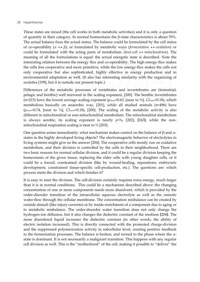
These states are mixed (the cell works in both metabolic activities) and it is only a question of quantity in their category. In normal homeostasis the β-state characteristics is about 70%. The actual balance fixes the actual status. The balance could be formulated by the cell status of co-operability (α β); or formulated by metabolic ways (fermentation oxidation) or could be formulated with the acting parts of metabolism: (host-cell mitochondrion). The meaning of all the formulations is equal: the actual energetic state is described. Note the interesting relation between the energy flux and co-operability. The high energy-flux makes the cells less cooperative and more primitive, while the low energy-flux makes the cells not only cooperative but also sophisticated, highly effective in energy production and in environmental adaptation as well. (It also has interesting similarity with the organizing of societies [199], but it is outside our present topic.)
Differences of the metabolic processes of vertebrates and invertebrates are (terrestrial, pelagic and benthic) well mirrored in the scaling exponent, [200]. The benthic invertebrates (n=215) have the lowest average scaling exponent (pmean=0.63, [near to ⅔], CImean=0.18), which metabolizes basically on anaerobic way, [201], while all studied animals (n=496) have (pmean=0.74, [near to ¾], CImean=0.18), [200]. The scaling of the metabolic activity is also different in mitochondrial or non-mitochondrial metabolism. The mitochondrial metabolism is always aerobic, its scaling exponent is nearly p=¾, [202], [243], while the non-mitochondrial respiration scaling is near to ⅔ [203].
One question arises immediately: what mechanism makes control on the balance of β-and α-states in the highly developed living objects? The electromagnetic behavior of electrolytes in living systems might give us the answer [204]. The cooperative cells mostly run on oxidative metabolism, and their division is controlled by the cells in their neighborhood. There are two basic reasons for normal cellular division, and it could be a regular division keeping the homeostasis of the given tissue, replacing the elder cells with young daughter cells, or it could be a forced, constrained division (like by wound-healing, reparations, embryonic development, constrained tissue-specific cell-production, etc.). The questions are: which process starts the division and which finishes it?
It is easy to start the division. The cell-division certainly requires extra energy, much larger than it is in normal conditions. This could be a mechanism described above: the changing concentration of one or more components needs more dissolvent, which is provided by the order-disorder transition of the intracellular aqueous electrolyte as well as the osmotic water-flow through the cellular membrane. The concentration misbalance can be created by outside stimuli (like injury currents) or by inside enrichment of a component due to aging or to metabolic misbalance. The order-disorder water transition does not only change the hydrogen-ion diffusion, but it also changes the dielectric constant of the medium [204]. The more disordered liquid increases the dielectric constant (in other words, the ability of electric isolation increased). This is directly connected with the promoted charge-division and the suppressed polymerization activity in subcellular level, creating positive feedback to the fermentation processes. The balance is broken, and turned to the phase where the α-state is dominant. It is not necessarily a malignant transition. This happens with any regular cell division as well. This is the “motherhood” of the cell, making it possible to “deliver” the
Local Hyperthermia in Oncology – To Choose or not to Choose? 31
daughter cells. The “individualism” of the mother-cell is explainable with the extreme high energy demand of the division process. When the daughter cells appear, they must accept the previous order. Their “infancy” is normal, as the “babyhood” is normal after the deliveries. The “babyhood” period has to be limited in time, and the newly born cell has to find its normal collective function. Consequently, the process might go wrong, if after finishing the division, the daughter cells do not find the way of the co-operability and the β-state again. When it is not the case, the cells are blocked in the α-state, their proliferation becomes uncontrolled. This unfortunate case, however, is not a simple process originated from one single defect. It is a disturbance of a complex controlling mechanism [190], which well correlates anyway with the single “renegade cell” concept [205], showing a long process to produce “a renegade cell” as the ancestor of the billion-cell group called cancer. According to the epidemiological research, for a complex damage to occur and for the cancer to develop at least five different mutations have to be coincidently present to be malignant. [206].
Again we are back to the main question: what is the mechanism to re-establish the β-state after the division of the cell. We think, that the down-regulation of the energy-flux has the same active elements as the up-regulation had at the start of the division. The clue is again the order-disorder transformation in the aqueous solution. As we told, at the beginning of the division, a huge energy has to be ready to supply the process, a large number of proteins and other cellular elements (lipids, enzymes, etc.) have to be produced, and all need ATP desperately. In α-sate the conditions are ready for that. When the division is over, two new daughter cells appear, the energy-consumption drastically drops to the normal level of the two cells. The doubled cytoplasm and all the cellular elements had enough dissolvent capacity even in the ordered water case. The hydrogen-bridge proton bifurcation can be reorganized, there are no opposite environmental driving forces. The sudden doubling of the cellular elments cools down the liquid to solid. It goes through the same phase transition (disorder-order transition) as it was (only the opposite direction) when the division started. This again (like in the liquid phase transitions) lowers the free energy, and in all (together with the environment, where the extra heat is radiated) increases the entropy. Note, the entropy apparently decreases (information build up) in the local cellular level, the overall conditions have to be considered for the full picture.
As we showed, the metabolic pathways could drastically modify the development of the cell, and it could be the primary source of the malignant deviations. The balance of the oxidative and fermentative metabolism tunes the cellular ability to behave collectively or constrict autonomy, being individual. These conditions of course well depend on the energy (and signaling) exchange of the cell with its actual environment. The intracellular transport properties also have to be different at changing metabolic pathway. The intensive energy flux of the fermentative metabolism increases the liberated heat in the cell, and so the temperature gradient between the extra- and intracellular compartments. The growing temperature difference could reach a critical threshold, when the heat flow turns from conductive to convective [207]. (This phenomenon works like the well-known Benard instability, [208].) The convective way promotes the ionic flows through the cellular
Hyperthermia 32
membrane increasing the glucose permeability and so supports the fermentation way of metabolism together with the changes of the intracellular circulations, [209], [210]. This complex change could down-regulate the mitochondrial oxidative metabolism.
At the divisional processes the intracellular flows and all pathway activities are probably higher both at regular and at malignant cell-division. Possibly the order-disorder transition of the aqueous solution also has a role in the changes [211].
However, finishing the division, the daughter cells are separated, a higher surface suddenly appears and the separated volumes limit the intracellular flows and change the order of structure as well. It decreases the gradient through the membrane. This regulates the heat-flow through the cellular membrane and changes the energy-exchange from convective to the conductive one again [207]. The conductive heat-exchange does not support the intensive diffusion of the large-molecule glucose, so the oxidative way becomes necessary and regular. The two daughter cells have less than half energy-consumption (each) than it was requested by the mother cell. It is because the mother cell was large (doubled its volume) and was intensively producing various elements to complete the daughter cells. Instead of the division conditions where the high energy-request gained the energy-demand and preferred the high-energy flux fermentative metabolism, the normal homeostatic conditions will dominate again.
The significantly larger permittivity and conductivity in tumor-tissue in vitro is explained on this basis, [212]. Both the conductivity and dielectric differences between the healthy- and tumor-tissues at the applied 13.56 MHz frequency in most cases of the malignancies are over 15% [213], [172]. It is clinically proven, that the cancerous and healthy tissues of the hepatic tumors are significantly different [214]. Also the VX-2 carcinoma can be measured [215]: rabbit-liver at low frequency in vivo had a conductivity 6-7.5 times higher, permittivity 2-5 times lower than in the healthy parts (impedance difference is about 600%), while for 10 MHz region it is 200%. With impedance tomography we can make a distinction between the living and necrotic malignancy as well, [214]; both the conductivity and permittivity are higher in the malignant liver, but the frequency dependence of necrotic tissue differs.
The dielectric properties are also distinguishable by the water content of the malignant tissue, which is higher than that of their healthy counterpart. The proliferating cells control their cell-volume by their water content, in the malignant growth [216], and this effect increases the conductivity and generally the dielectric properties in the given tissue.
The high dielectric constant allows the additional selection (focusing): the higher dielectric constant absorbs more from the RF-energy, (see Figure 27.).
3.3. Selection by order-disorder effect (fractal physiology selection)
The non-equilibrium phenomena is well formulated in the description of the Avrami-equation [217], pioneered by Kolmogorov [218], by Johnson & Mehl [219] and by others, [220], [221]. The description could serve as a mathematical model for different biological processes, [222], [223]. Experimental data which were collected by Cope [224], [225], and by others [226], [227], show a definite universality to describe the real processes.
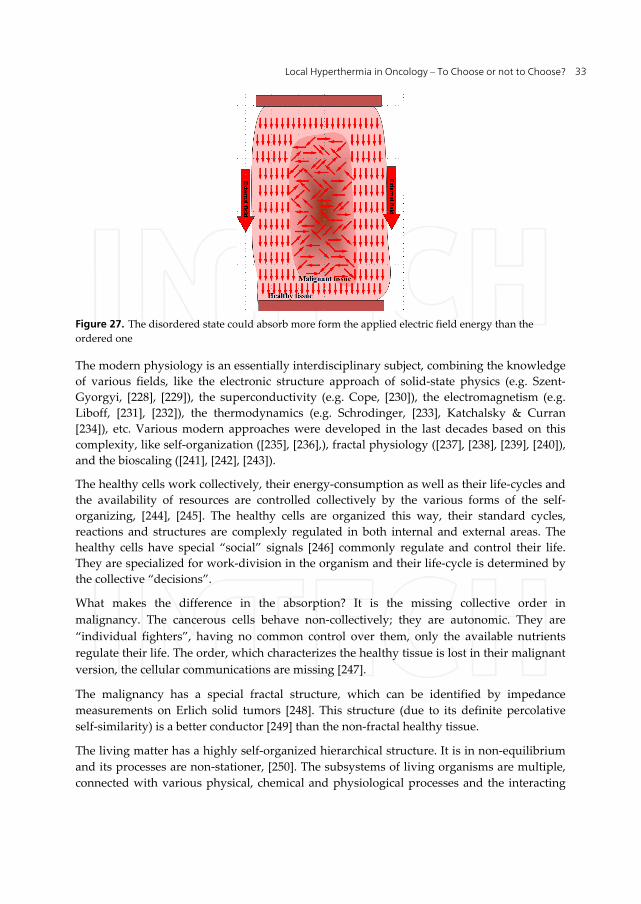
Local Hyperthermia in Oncology – To Choose or not to Choose? 33
Figure 27. The disordered state could absorb more form the applied electric field energy than the ordered one
The modern physiology is an essentially interdisciplinary subject, combining the knowledge of various fields, like the electronic structure approach of solid-state physics (e.g. Szent-Gyorgyi, [228], [229]), the superconductivity (e.g. Cope, [230]), the electromagnetism (e.g. Liboff, [231], [232]), the thermodynamics (e.g. Schrodinger, [233], Katchalsky & Curran [234]), etc. Various modern approaches were developed in the last decades based on this complexity, like self-organization ([235], [236],), fractal physiology ([237], [238], [239], [240]), and the bioscaling ([241], [242], [243]).
The healthy cells work collectively, their energy-consumption as well as their life-cycles and the availability of resources are controlled collectively by the various forms of the self-organizing, [244], [245]. The healthy cells are organized this way, their standard cycles, reactions and structures are complexly regulated in both internal and external areas. The healthy cells have special “social” signals [246] commonly regulate and control their life. They are specialized for work-division in the organism and their life-cycle is determined by the collective “decisions”.
What makes the difference in the absorption? It is the missing collective order in malignancy. The cancerous cells behave non-collectively; they are autonomic. They are “individual fighters”, having no common control over them, only the available nutrients regulate their life. The order, which characterizes the healthy tissue is lost in their malignant version, the cellular communications are missing [247].
The malignancy has a special fractal structure, which can be identified by impedance measurements on Erlich solid tumors [248]. This structure (due to its definite percolative self-similarity) is a better conductor [249] than the non-fractal healthy tissue.
The living matter has a highly self-organized hierarchical structure. It is in non-equilibrium and its processes are non-stationer, [250]. The subsystems of living organisms are multiple, connected with various physical, chemical and physiological processes and the interacting
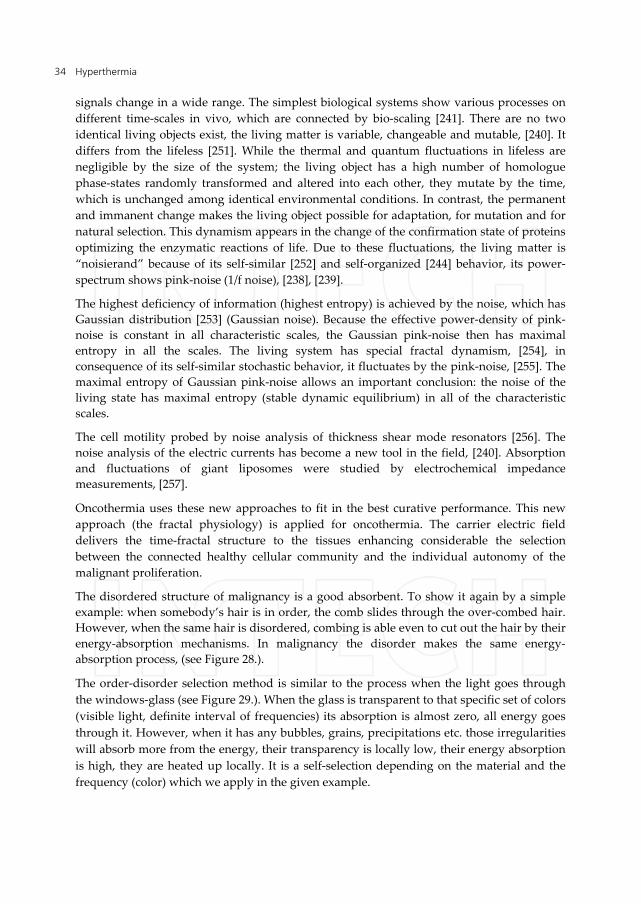
Hyperthermia 34
signals change in a wide range. The simplest biological systems show various processes on different time-scales in vivo, which are connected by bio-scaling [241]. There are no two identical living objects exist, the living matter is variable, changeable and mutable, [240]. It differs from the lifeless [251]. While the thermal and quantum fluctuations in lifeless are negligible by the size of the system; the living object has a high number of homologue phase-states randomly transformed and altered into each other, they mutate by the time, which is unchanged among identical environmental conditions. In contrast, the permanent and immanent change makes the living object possible for adaptation, for mutation and for natural selection. This dynamism appears in the change of the confirmation state of proteins optimizing the enzymatic reactions of life. Due to these fluctuations, the living matter is “noisierand” because of its self-similar [252] and self-organized [244] behavior, its power-spectrum shows pink-noise (1/f noise), [238], [239].
The highest deficiency of information (highest entropy) is achieved by the noise, which has Gaussian distribution [253] (Gaussian noise). Because the effective power-density of pink-noise is constant in all characteristic scales, the Gaussian pink-noise then has maximal entropy in all the scales. The living system has special fractal dynamism, [254], in consequence of its self-similar stochastic behavior, it fluctuates by the pink-noise, [255]. The maximal entropy of Gaussian pink-noise allows an important conclusion: the noise of the living state has maximal entropy (stable dynamic equilibrium) in all of the characteristic scales.
The cell motility probed by noise analysis of thickness shear mode resonators [256]. The noise analysis of the electric currents has become a new tool in the field, [240]. Absorption and fluctuations of giant liposomes were studied by electrochemical impedance measurements, [257].
Oncothermia uses these new approaches to fit in the best curative performance. This new approach (the fractal physiology) is applied for oncothermia. The carrier electric field delivers the time-fractal structure to the tissues enhancing considerable the selection between the connected healthy cellular community and the individual autonomy of the malignant proliferation.
The disordered structure of malignancy is a good absorbent. To show it again by a simple example: when somebody’s hair is in order, the comb slides through the over-combed hair. However, when the same hair is disordered, combing is able even to cut out the hair by their energy-absorption mechanisms. In malignancy the disorder makes the same energy-absorption process, (see Figure 28.).
The order-disorder selection method is similar to the process when the light goes through the windows-glass (see Figure 29.). When the glass is transparent to that specific set of colors (visible light, definite interval of frequencies) its absorption is almost zero, all energy goes through it. However, when it has any bubbles, grains, precipitations etc. those irregularities will absorb more from the energy, their transparency is locally low, their energy absorption is high, they are heated up locally. It is a self-selection depending on the material and the frequency (color) which we apply in the given example.
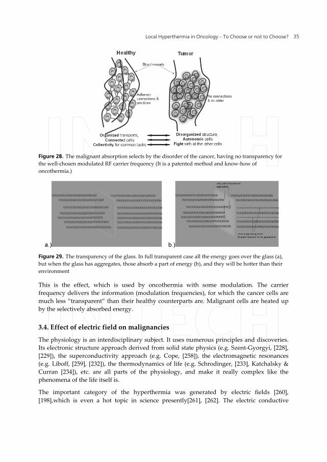
Local Hyperthermia in Oncology – To Choose or not to Choose? 35
Figure 28. The malignant absorption selects by the disorder of the cancer, having no transparency for the well-chosen modulated RF carrier frequency (It is a patented method and know-how of oncothermia.)
Figure 29. The transparency of the glass. In full transparent case all the energy goes over the glass (a), but when the glass has aggregates, those absorb a part of energy (b), and they will be hotter than their environment
This is the effect, which is used by oncothermia with some modulation. The carrier frequency delivers the information (modulation frequencies), for which the cancer cells are much less “transparent” than their healthy counterparts are. Malignant cells are heated up by the selectively absorbed energy.
3.4. Effect of electric field on malignancies
The physiology is an interdisciplinary subject. It uses numerous principles and discoveries. Its electronic structure approach derived from solid state physics (e.g. Szent-Gyorgyi, [228], [229]), the superconductivity approach (e.g. Cope, [258]), the electromagnetic resonances (e.g. Liboff, [259], [232]), the thermodynamics of life (e.g. Schrodinger, [233], Katchalsky & Curran [234]), etc. are all parts of the physiology, and make it really complex like the phenomena of the life itself is.
The important category of the hyperthermia was generated by electric fields [260], [198],which is even a hot topic in science presently[261], [262]. The electric conductive
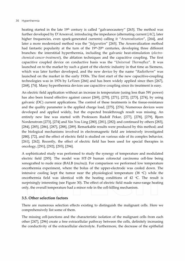
Hyperthermia 36
heating started in the late 19th century is called “galvanocautery” [263]. The method was further developed by D’Arsonval, introducing the impedance (alternating current [AC], later higher frequencies, even spark-generated currents) calling it “Arsonvalization”, [264], and later a more modernized method was the “fulguration” [265]. The Arsonvalization method had fantastic popularity at the turn of the 19th-20th centuries, developing three different branches: the interstitial hyperthermia, including the galvanic heat-stimulation (electro-chemical-cancer-treatment), the ablation techniques and the capacitive coupling. The first capacitive coupled device on conductive basis was the “Universal Thermoflux”. It was launched on to the market by such a giant of the electric industry in that time as Siemens, which was later further developed, and the new device by the name “Radiotherm” was launched on the market in the early 1930s. The first start of the new capacitive-coupling technologies was in 1976 by LeVeen [266] and has been widely applied since then [267], [268], [74]. Many hyperthermia devices use capacitive coupling since its treatment is easy.
An electric field application without an increase in temperature (using less than 5W power) has also been found effective against cancer [269], [270], [271], [272], [273], [274], by using galvanic (DC) current applications. The control of these treatments is the tissue-resistance and the quality parameter is the applied charge load, [275], [276]. Numerous devices were developed and applied widely, but the expected breakthrough result was missing. An entirely new line was started with Professors Rudolf Pekar, [277], [278], [279], Bjorn Nordenstrom [273], [274] and Xin You Ling [280], [281], [282]; and continued by others [283], [284], [285], [286], [287], [288], [289]. Remarkable results were produced by this method; and the biological mechanisms involved in electromagnetic field are intensively investigated [290], [72], and the effect of electric field is studied on various side of its complex behavior, [261], [262]. Recently, the effect of electric field has been used for special therapies in oncology, [291], [292], [293], [294].
A sophisticated study was performed to study the synergy of temperature and modulated electric field [295]. The model was HT-29 human colorectal carcinoma cell-line being xenografted to nude mice (BALB (nu/nu)). For comparison we performed low temperature oncothermia experiment, where the bolus of the upper-electrode was cooled down. The intensive cooling kept the tumor near the physiological temperature (38 oC) while the oncothermia field was identical with the heating conditions of 42 oC. The result is surprisingly interesting (see Figure 30). The effect of electric field made nano-range heating only, the overall temperature had a minor role in the cell-killing mechanism.
3.5. Other selection factors
There are numerous selection effects existing to distinguish the malignant cells. Here we comprehensively list some of them.
The missing cell-junctions and the characteristic isolation of the malignant cells from each other [247], [296] create a free extracellular pathway between the cells, definitely increasing the conductivity of the extracellular electrolyte. Furthermore, the decrease of the epithelial
Local Hyperthermia in Oncology – To Choose or not to Choose? 37
barrier function (tight-junction permeability changes) can be measured by electric impedance measurement, [297].
Figure 30. Oncothermia was 3 times more effective than hyperthermia on the identically high (42 oC) temperature. However, cooling down the tumor during the treatment, the death-rate decreased only slightly, exceeding more than 2 times the classic hyperthermia on high temperature
The special membrane effect (rectification on membranes, [298]), is also a factor of the cellular selectivity of cancer by RF electric field. The impedance measurement is useful for the control of other treatment modalities. It adequately measures the distortion, made by irradiation [299], and the drug-effect can also be controlled, [300]. Such usual practice, like following the wound healing, is also objectively traceable [301]. Bioimpedance vector pattern can distinguish cancer patients without disease versus locally advanced or disseminated diseases [302].
The impedance measures selectively, differentiates between the cancerous and healthy tissue, and is able to distinguish the extra- and intra-cellular electrolyte. Selective impedance measurements are provided clinically as well. Many comparative studies have been provided for malignant tissues, however, the results are not identical; the measurements very much depend on the conditions. (This is a trivial consequence of many factors, which we listed above.) However, all the studies measured lower impedance in the tumor than in their healthy counterpart in all of the tissue and staging of the tumor, in vitro and in vivo as well.
Hyperthermia increases biochemical reaction rates [303] and therefore the metabolic rate as well. The metabolic heat production of tumor depends on the doubling time of its volume [304]. The high metabolic rate keeps the temperature for tumor tissues higher than its neighborhood, [305]. This works as selection factor for heating of tumor tissue. Therefore, in case of 6 oC increase the amount of growth will be 1.8-times higher [306] than its healthy counterpart.

Hyperthermia 38
It has been long known that hyperthermia can cause the softening or melting of the lipid bilayer [307], [308], [155], it can change lipid-protein interactions [309], and it can denature proteins [310]. All of these events can arrest the cell-cycle of the tumor.
Heat-treatment cause structural alteration in transmembrane proteins, causing a change in the active membrane transport and membrane capacity [311] leading to substantial changes in potassium, calcium, and sodium ion gradients [312], membrane potential [313], [314], cellular function [315], [316], and causing thermal block of electrically excitable cells [317], [303]. The thermo tolerable cells have significantly higher (~15%) membrane potential than the naïve cells [314], and the difference rapidly grows by the elapsed time at 45°C.
In chemo-thermo-therapies the role of chaperone proteins is important. Chaperones (stress- or heat-shock-proteins) are highly conserved proteins, which are vital in almost every living cell and on their surfaces during their whole lifetime, regardless of their stage in the evolution, [318]. Any kind of change in the dynamic equilibrium of the cell life (environmental stresses, various pathogen processes, diseases, etc.) activates their synthesis [319]. Excretion of the chaperones is the ‘stress-answer’ of the cells to accommodate themselves to the new challenges. As a consequence of the stressful ‘life’ of malignant cells, the molecular chaperones are present in all cancerous cells [320], [321], [322] to adapt the actual stress to help tumor-cell survival. Moreover, the shock-proteins are induced by every oncological treatment-method, which are devoted to eliminate the malignancy: after conventional hyperthermia [323], after chemotherapy [324], after radiotherapy [325] or even after photo-therapy [326] intensive HSP synthesis was shown. By the stress adaptation the induction or over-expression of the stress proteins generally provide effective protection of the cell against apoptosis [327], but their extracellular expression acts oppositely: it makes a signal to the immune system on the defect of the actual cell [328]. Furthermore, induction of various HSPs (HSP27, HSP70, and HSP90) was observed in numerous metastases and the HSP90 homologue, GRP94 may act as a mediator of metastasis generation. HSP generally degrades the effect of the hyperthermia therapy because it may increase the tumor cell survival, and its massive induction may generate the tumor thermo-tolerance and in parallel drug- and radio-tolerance. Heat treatment can also lead to multi-drug resistance [329].
Non-temperature dependent effects (mainly field stresses) can also produce chaperone-synthesis [330]. The HSP manifestation in the biopsies might give a good clinical indication for the treatment response [331].
On the other hand, the chaperone HSP70 assists to freeze the actual dynamic equilibrium (the “status-quo”) and so tries to re-establish the cellular communication in the extra-cellular electrolyte [328]. It is shown that their expression on the cell-membrane gains the apoptotic signals and enhances the immune reactions, [328]. HSP participates in the activation of the p53 tumor-suppresser [332] and has been associated with the tumor-suppresser retinoblastoma protein [333].
Recently, numerous scientific theories have also concentrated on the significance of thermally induced non-thermal effects, such as heat-shock protein (HSP) production [334],

Local Hyperthermia in Oncology – To Choose or not to Choose? 39
[335]. Development of the thermo-tolerance is one of the suppressors of hyperthermia efficacy [323]. From the point of view of the thermo-tolerance, one of the most prominent chaperone proteins is the HSP72. The concentration of this HSP is 5-10 times lower in the healthy cells than in the malignant ones, and both grow by the heat-treatment [336]. However, responding to the heat treatment, their concentration in healthy cells is: multiplied by 8 or 10, while during the same heat treatment the HSP72 multiplication creates only 1.2-1.5 times higher concentration in malignant cells, [323].
4. Cellular effects – To kill or not to kill?
Like Nobel-laureate A. Szent-Gyorgyi formulated: from the point of view of life, it is not interesting how the monkey goes through the forest, but it is essential how the forest goes through the monkey [as nutrition]. In hyperthermia we have to have the same change of paradigm: it is not important how the incident energy increases the temperature, but that is essential how the incident energy changes the structure and the chemical bonds in the targeted tissue.
4.1. Apoptotic cell killing
The above described mechanism targets the membrane of the malignant cells, and excites numerous pathways essential for the cellular fate, (see Figure 31.). The excitations have serious consequences on membrane:
Induces apoptotic signal Forms membrane-HSP Makes the membrane more transparent Forms higher motility of membrane domains Rebinds the E-cadherin adherent connections Damages the cell-membrane Rectificates, demodulates the modulated signal (stochastic effects are involved)
The effects in the cytoplasm are also remarkable. The transmembrane temperature gradient
Dilutes the cytoplasm Develops higher intracellular pressure Activates apoptotic pathways Activates death receptors Suppresses the proliferation Arrests the DNA replication
These effects were shown in multiple, different experiments. The basic repeated phenomena are the time-delay of the action, selection and tumor destruction of oncothermia became trivial hours after the treatment (see Figure 32., [337]). The numerous apoptotic bodies show the character of the process.
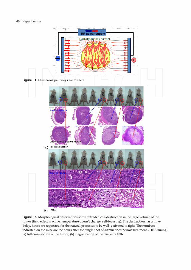
Hyperthermia 40
Figure 31. Numerous pathways are excited
Figure 32. Morphological observations show extended cell-destruction in the large volume of the tumor (field effect is active, temperature doesn’t change, self-focusing). The destruction has a time-delay, hours are requested for the natural processes to be well- activated to fight. The numbers indicated on the mice are the hours after the single shot of 30 min oncothermia treatment, (HE Staining). (a) full cross section of the tumor, (b) magnification of the tissue by 100x

Local Hyperthermia in Oncology – To Choose or not to Choose? 41
The time delay indicates the long-duration processes, which were identified as programmed cell-death (apoptosis), by various investigations: macro- and micro-morphology, enhanced activity of p53 tumor-suppressor, cleaved caspase 3 involvement, Tunel reaction, DNA fragmentation (laddering), etc. (see Figure 33., [337]). The rebuilt external apoptotic pathways are shown by reestablished E-cadherin connections, by enreachment of beta-catenin and its relocalization into the nuclei, by connexin induction, by activation of death-toll receptors, like FADD, FAS, DR-5, etc.
Figure 33. External electromagnetic field activates signal transduction pathways, concluding to apoptotic cell-death. (Oncothermia treatment (HT29 colorectal xenograft model)
This apoptotic process is non-toxic (no inflammatory reactions afterwards) and promotes the immune reactions (works parallel with these).
The cellular selection is proven by various experiments. One of these is the definite selection of malignant cells in co-cultures, [338]. The effect on healthy human keratinocytes (HCK) co-cultured with human fibroblasts (see Figure 34.) was marginal, and was regenerated by the time. The co-culture with immortalized (but not malignant) HaCaT keratinocytes is affected slightly, but the malignant A431 cell-line was selected from the co-culture (see Figure 35.).
The time-delay and the relocalization of the beat-catenin to the cell-nuclei is shown in vitro (HepG2 cell-line) and in vivo (HT29 xenograft) shown in Figure 36. [339].
The time delay is shown in the Tunel reactions (see Figure 37.), indicating the late apoptotic phases dominantly after 24 h of the single shot treatment.
An interesting measurement was provided by Ki67 proliferation marker, measured in the living tumor part after the single-shot treatment of oncothermia, (see Figure 37, [340]). The surviving, living malignant cells in the treated tumor had definitely and significantly

Hyperthermia 42
suppressed Ki67 marker compared to its untreated counterpart in all the time-scale investigated.
Figure 34. Co-culture with normal human skin fibroblasts as a model of a squamous carcinoma growing within connective tissue cells (100.000/ml) were exposed to oncothermia. Samples were incubated for 24 h at 37°C, fixed and stained with crystal violet. Cellular metabolic activity was measured using the MTT assay (standard colorimetric test) and quantitated at 630 nm. (Data represent the mean value ± S.E.M. of 4-6 separate experiments assayed in triplicate, but some experiments were repeated up to 12 times to obtain reliable data.)
Figure 35. The selection of the malignant cells is effective in the co-culture of the malignant A431 human keratinocytes and healthy human fibroblasts. [a – before oncothermia, b – after oncothermia]. The healthy cells remain intact, while the malignant cells are damaged and cleared. (Experiment is described in Figure 34.)

Local Hyperthermia in Oncology – To Choose or not to Choose? 43
Figure 36. The beta-catenin relocalization into the nuclei
Figure 37. TUNEL assay: enzymatically label the DNA fragments resulted by apoptotic process
Figure 38. Colon26 (murine colorectal cancer) cell line derived allograft (CID mice), single shot. (Ki-67: proliferation marker protein, expressed in the nuclear membrane only in the dividing cells.)

Hyperthermia 44
The cleaved Caspase-3 (as important part of the apoptotic pathway) was activated (see Figure 39., [341]), as well as the death toll receptors became overexpressed (see Figure 40., [341]).
Figure 39. HT29 human colorectal carcinoma cell-line xenograft model in nude mice. Caspases, or cysteine-aspartic proteases or cysteine-dependent aspartate-directed proteases are a family of cysteine proteases that play essential roles in apoptosis (programmed cell death), necrosis, and inflammation (the red line is for the reference value) [Reference: untreated control]
Figure 40. HT29 human colorectal carcinoma cell-line xenograft model in nude mice. Death receptor activity (the red line is for the reference value) [Reference: untreated control]
4.2. Suppressing the dissemination of malignant cells
Oncothermia is different from this point of view as well. It blocks the dissemination, avoids their motility due to the lazy connections with the tumor. Oncothermia makes it by the reestablishing the cellular connections (see Figure 41. [337]), which is also great success to save life. The built up connections can force not only the sticking together, but make bridges between the cells for information exchange to limit the individuality and the competitive behavior of the malignant cells.

Local Hyperthermia in Oncology – To Choose or not to Choose? 45
Figure 41. Oncothermia can reestablish the adherent cell connections (E-cadherin and β-catenin) as well as the gap-junctions (Connexin-43). These could reestablish the cellular communication and can stop the dissemination of tumor cells
This “gluing mechanism” is not only important for the external apoptotic signal, but makes the malignant cells less mobile, their motility is reduced and the dissemination risk is decreased.
4.3. Action on far distant metastases
The main danger of malignancies is the metastases, attacking the organs which are crucial for life. When the tumor grows somewhere without endangering the important systems like the respiratory system, central nervous system, cardiovascular system, etc., it is not life-threatening. These tumors are local (benign or early malignant), their elimination is possible. The real life-threatening danger is the malignancy, when the cells are disseminated from the tumor-lesion by the various transport systems (lymph, blood), or their effect becomes systemic by one of the general mechanisms of the organism.
The heavy life-threatening effect of metastases has been observed on statistical basis on colorectal adenocarcinoma collecting data for 15 years [342]. The long- term (10 years) survival was around 90% when no metastases were present, 60% in case of regional metastases and only 15% when distant metastases were developed in the patient. The blood-transported cells can be blocked easily by the brain, lung, kidney, liver, etc., causing fatality. The challenge of the treatments is to recognize the tumor early, to avoid the metastases, and/or to block the dissemination as much as possible.
In veterinarian clinical trial of dogs having osteosarcomas without evidences of metastases, the local radiation combined complementary with whole body hyperthermia was studied. The result was surprisingly bad [343], [344]: the combined treatment was not effective on the
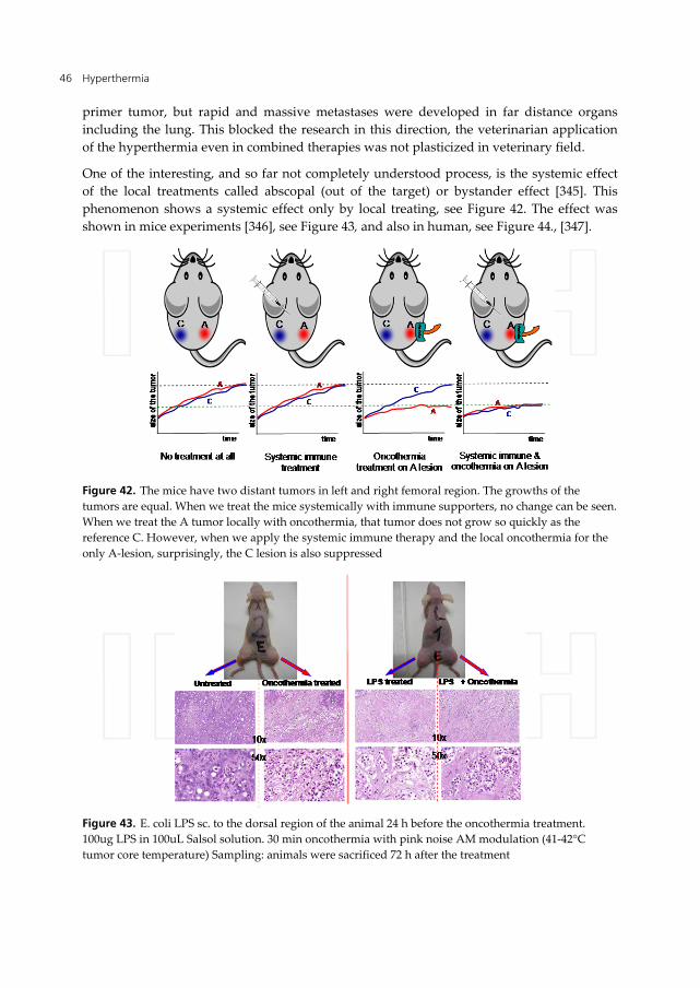
Hyperthermia 46
primer tumor, but rapid and massive metastases were developed in far distance organs including the lung. This blocked the research in this direction, the veterinarian application of the hyperthermia even in combined therapies was not plasticized in veterinary field.
One of the interesting, and so far not completely understood process, is the systemic effect of the local treatments called abscopal (out of the target) or bystander effect [345]. This phenomenon shows a systemic effect only by local treating, see Figure 42. The effect was shown in mice experiments [346], see Figure 43, and also in human, see Figure 44., [347].
Figure 42. The mice have two distant tumors in left and right femoral region. The growths of the tumors are equal. When we treat the mice systemically with immune supporters, no change can be seen. When we treat the A tumor locally with oncothermia, that tumor does not grow so quickly as the reference C. However, when we apply the systemic immune therapy and the local oncothermia for the only A-lesion, surprisingly, the C lesion is also suppressed
Figure 43. E. coli LPS sc. to the dorsal region of the animal 24 h before the oncothermia treatment. 100ug LPS in 100uL Salsol solution. 30 min oncothermia with pink noise AM modulation (41-42°C tumor core temperature) Sampling: animals were sacrificed 72 h after the treatment

Local Hyperthermia in Oncology – To Choose or not to Choose? 47
Figure 44. Investigator: Prof. Dr. Seong Min Yoon, Institute: Division of Hematology-Oncology, Department of Internal Medicine, Samsung Changwon Hospital, Sungkyunkwan University, S.Korea Patient: SAsc, 72 y, male, Primer-tumor: Non-small cell lung cancer; Size: 9.5 cm right middle lobe, Metastases: in sentinel and distant lymph-nodes, Tumor-classification: cT2 cN2 M0, stage IIIB, Treatment: trimodal protocol: 28x1.7 Gy; support: 250 microgram Leukine and Oncothermia 6x; Only the primer tumor was locally treated
4.4. Some clinical results
Oncothermia has a long-time history with large number of documented case reports and clinical trials [160]. During more than 20 years 43 studies were performed involving more than 2000 patients altogether from 14 clinics in 4 countries (see Table 1.) Details of the clinical effects are summarized in the publications as well as in the specialized monograph [160].
The clinical trials of oncothermia are dominantly retrospective. To develop randomized clinical trials is a challenge for patients. Patients do not agree to be in the control-arm at any case. In most of the cases they are registered for oncothermia because the other methods (conventional gold standards) failed. In these cases progression could occur anyway due to drug-resistance, organ-overload (kidney, liver, etc.), tumor-relapses, psycho-resistance, etc.
The advanced cases at the conditions described above emphasize not only the complexity of the individual situation of patients, but also underlines the fact that oncothermia is applied as the facility of the “no other is possible” many time hopeless cases providing over 3rd line treatment approach. This high-line treatment process is in general palliation (the first goal is to provide acceptable quality of life), which is an important factor for oncothermia as well. However oncothermia even in these advanced situations has curative value, and makes curative therapy in 3rd-line or over. The professional literature clearly shows the rare facility of the evidence-based clinical trials for these high-line treatments. Other evidences have to

Hyperthermia 48
be shown when randomized controlled trials are not possible, [348]. The challenges of evaluation appear forcefully in case of patients with advanced stages having inoperable (or partly resected) tumors, having relapsed malignancies, patients who are resistant on the gold-standard treatments, etc. Oncothermia is facing to this challenge as well.
We have to make special attention evaluating of the clinical results performed by oncothermia. The complications make definite challenges to objective evaluation. The main challenges are:
Oncothermia is applied in higher (usually third and subsequent) treatment-lines, boosting or resensitizing the effect of the conventional therapies. Oncothermia is mostly applied in cases when the conventional therapies fall. In most of the cases oncothermia therapy is applied when the patient has/is - Inoperable lesion - Radio-resistant - Chemo-resistant (refractory) - Improper blood-counts - Liver-failure - Kidney failure - Psycho-resistance In most of the cases the complex combination of the above problems occurs. Due to these conditions, oncothermia is applied in higher line of the therapies. This sequence of the treatments is mostly determined by the individual decisions of the physicians [349], usually without having help from any evidence based statistical approvals.
Usually it is applied in palliative care; many patients are in terminal phase. This patient care has very limited statistical evidence based trials; the medical decision-making processes are usually well tailored to the individual patients [350], [351].
Only few controlled randomized clinical trials are available for oncothermia. The results have to be concluded from observational studies and from the historical and data-base comparisons. USA- and EU-databases (SEER [352], Eurocare [353]) are mostly used. Due to the not solved problem between the hypothesis check confidence of evidence based medicine and the observational studies [354], [355], this data-comparison is acceptable. Make the result as objective as they could be, we compared the collected results of the same localizations and the same protocols from various clinics. The common significant difference from the databases can be accepted as evidence.
In the case of long-survivals, we have to consider, that oncothermia is taken only in a small fraction of the overall survival. The patients are treated by oncothermia in their definite late stages, after prognosis “no curative help” by continuing the gold-standards alone. In consequence, the long overall survival generally is not the result of the oncothermia, but the selection of the patients in their end. Oncothermia effect in these cases can be negligible, irrespective of its real efficacy. The chance to measure the efficacy is the first year survival rate (%), when the patients with the most aggressive kind of the given cancer do not survive the following year after the diagnosis. If they start the oncothermia in the first year, the survival-rate result can be an objective sign of the efficacy.

Local Hyperthermia in Oncology – To Choose or not to Choose? 49
Special cases are treated on Intend-to-Treat population. This makes the patient selection not objective enough, but the dominantly metastatic patient’s spectrum compensates for this lack of selection. In this case, we have an automatic selection of advanced (many times terminal) cases.
The generally low quality of life (QoL) in combination of supportive therapies weaken the measurability of oncothermia alone. However, due to the fact that oncothermia is not proposed as monotherapy, the combination objectively measures the benefit, if the supportive therapies alone would not be successful at all.
The patients are treated with oncothermia in their very advanced, metastatic states. The local oncothermia treatment, of course, concentrates on definite localizations, (primary or metastatic) which again lowers the full measurability of the oncothermia in the development of the cancer. This is the main reason why oncothermia measures first of all the overall survival rates, which are good objective parameters of the treatment efficacy in general.
The quality of life (QoL) of the patients is an important characteristic of such a method as oncothermia. In a generally controlled and randomized study a trial effect exists [356], which is not the case in oncothermia applications. However, it could be negative outcome, that in a strict competitive market the opinions are not independent and objective [357] and of course the conflict of interest could make considerable bias [358], [359].
On the above bias structure the characteristics of the clinical studies could be described as – Single arm, open label, observational for intention-to-treat (ITT) population, dominantly for the patients in late/advanced stages, where the conventional methods have failed. Mostly the survival rate was the studied endpoint. The inclusion criteria were the inoperable and in progression after chemo- and/or radio-therapy. Exclusions were only the well-known, above described contraindications of oncothermia.
The oncothermia challenge is its use when the conventional treatments are unsuccessful. In consequence, its effect could be active only in a small (last) fraction of the overall survival. When patients have long overall survival by the conventional treatments alone, oncothermia is applied only in the last stage of the disease so objectively the life-elongation by oncothermia can not be observed.
To make an objective evaluation, we have special considerations how to get the evidences from the available information pool and find the objective evidences. It is a complex challenge, having four basic approaches. These methods highlight the objective information and their parallel results make the obtained data evidence-based. The five legs are:
1. Fast course case comparison. Use the survival of the rapid, fast course cases (most advanced, drastically quickly developing cases) in comparison with large databases. (Only the survival is considered as relevant parameter, the clinical outcomes (responses) are not studied as evidences.)
2. Comparison of clinics. Compare the obtained data of the independent clinics, using the same protocol for the same cohort.
3. Quality of life comparison. Collect the data about the quality of life and the adverse effects limiting the application of oncothermia.
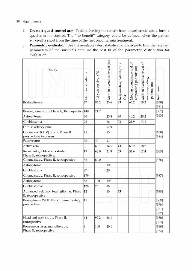
Hyperthermia 50
4. Create a quasi-control arm. Patients having no benefit from oncothermia could form a quasi-arm for control. The “no benefit” category could be defined when the patient survival is short from the time of the first oncothermia treatment.
5. Parametric evaluation. Use the available latest statistical knowledge to find the relevant parameters of the survivals and use the best fit of the parametric distribution for evaluation.
Study
Num
ber o
f pat
ient
s
1st y
ear s
urvi
val (
%)
Med
ian
over
all s
urvi
val (
m)
Resp
osnd
ing
patie
nts/
ratio
(%
)
Med
ian
over
all s
urvi
val o
f re
spon
ding
pat
ient
s (m
)
Med
ian
over
all s
urvi
val o
f no
n-re
spon
ding
p
atie
nts
(m)
Refe
renc
e
Brain gliomas 27 86.2 23.6 43 66.2 18.2 [360], [361]
Brain-glioma study Phase II, Retrospective 140 71.7 [362], [363]
Astrocytoma 40 25.8 80 40.2 20.2 Glioblastoma 92 16 73 21.9 13.1 Diffuse astrocytoma 8 52.9 Glioma (WHO IV) Study, Phase II, prospective, two arms
45 15 [160], [364]
Passive arm 36 40 11 Active arm 9 65 14.5 43 66.2 18.2 Recurrent glioblastoma study, Phase II, retrospective,
19 68.0 21.8 59 32.6 12.4 [365]
Glioma study, Phase II, retrospective 36 60.0 [366] Astrocytoma 9 106 Glioblastoma 27 20 Glioma study, Phase II, retrospective 179 [367]
Astrocytoma 53 100 103 Glioblastoma 126 76 16 Advanced, relapsed brain gliomas, Phase II, retrospective
12 10 25 [368]
Brain glioma WHO III-IV, Phase I, safety prospective
15 [369], [370], [371], [372]
Head and neck study, Phase II. retrospective
64 92.2 26.1 [160], [373]
Bone-metastases, monotherapy, Phase II, retrospective
6 100 40.1 [160], [373]

Local Hyperthermia in Oncology – To Choose or not to Choose? 51
Study
Num
ber o
f pat
ient
s
1st y
ear s
urvi
val (
%)
Med
ian
over
all s
urvi
val (
m)
Resp
osnd
ing
patie
nts/
ratio
(%
)
Med
ian
over
all s
urvi
val o
f re
spon
ding
pat
ient
s (m
)
Med
ian
over
all s
urvi
val o
f no
n-re
spon
ding
p
atie
nts
(m)
Refe
renc
e
Refractory bone-metastases study, Phase II, retrospective
11 90.9 [374]
Breast cancers 103 97.1 52.1 45 274.8 10.9 [160] , [373]
Colorectal cancer () 218 84.9 28.5 [160] , [373]
Sigma 12 34.1 Rectum 92 57.1 58 21 Colon 114 44.2 109.8 23.2
Colon cancer study, , Phase II, prospective, three arms, randomized
154 [375] [376]
Clifford TCM 53 75
Monotherapy 50 81
Combined therapy 51 91
Esophagus study, Phase II, retrospective 12 41.7 28.5 35 29.4 8.5 [160], [377]
Rectum cancer study, Inoperable→operable, Phase II, retrospective
7 71 [378]
Kidney cancer study, Phase II, retrospective
39 84.6 35.9 48 78.4 33.7 [160], [373]
Liver metastases from various origin, Phase II, retrospective
25 20.5 [379]
Liver metastases from various origin, Comparative study, Phase II, retrospective
28 [374]
With radiotherapy 16 81 With chemotherapy 8 38 Monotherapy 4 25 Liver metastasis form colorectal origin, Phase II, retrospective
80 86.0 24.1 [380]
Passive arm 53 11 Active arm 80 91 24.1 With chemotherapy 30 80 21.5 Monotherapy 50 92 24.4 Liver metastasis form colorectal origin, Phase II, retrospective
15 23 80 [381]
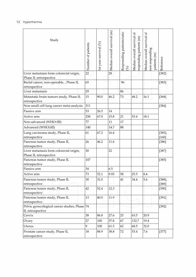
Hyperthermia 52
Study
Num
ber o
f pat
ient
s
1st y
ear s
urvi
val (
%)
Med
ian
over
all s
urvi
val (
m)
Resp
osnd
ing
patie
nts/
ratio
(%
)
Med
ian
over
all s
urvi
val o
f re
spon
ding
pat
ient
s (m
)
Med
ian
over
all s
urvi
val o
f no
n-re
spon
ding
p
atie
nts
(m)
Refe
renc
e
Liver metastasis form colorectal origin, Phase II, retrospective
22 28 [382]
Rectal cancer, non-operable, , Phase II, retrospective
65 96 [383]
Liver metastasis 29 86 Metastatic brain tumors study, Phase II, retrospective
15 90.0 46.2 73 48.2 16.1 [368]
Non-small cell lung cancer meta-analysis 311 [384]
Passive arm 53 26.5 14 Active arm 258 67.0 15.8 21 53.4 18.1 Non-advanced (WHO<III) 77 11 17 Advanced (WHO≥III) 140 14.7 88 Lung carcinoma study, Phase II, retrospective
61 67.2 16.4 [385], [160]
Pancreas tumor study, Phase II, retrospective
26 46.2 11.6 [386]
Liver metastasis form colorectal origin, Phase II, retrospective
30 22 [387]
Pancreas tumor study, Phase II, retrospective
107 [385] Passive arm 34 6.5
Active arm 73 52.1 9.93 58 25.5 8.4 Pancreas tumor study, Phase II, retrospective
30 31.0 41 34.4 5.6 [388], [389]
Pancreas tumor study, Phase II, retrospective
42 52.4 12.3 [390]
Pancreas tumor study, Phase II, retrospective
13 40.0 11.9 [391]
Pelvic gynecological cancer studies, Phase II, retrospective
74 [392] Cervix 38 86.8 27.6 25 63.5 20.9
Ovary 27 100 37.8 67 132.7 19.4 Uterus 9 100 61.5 62 68.5 32.0 Prostate cancer study, Phase II, retrospective
18 88.9 38.8 72 53.4 7.6 [377]

Local Hyperthermia in Oncology – To Choose or not to Choose? 53
Study
Num
ber o
f pat
ient
s
1st y
ear s
urvi
val (
%)
Med
ian
over
all s
urvi
val (
m)
Resp
osnd
ing
patie
nts/
ratio
(%
)
Med
ian
over
all s
urvi
val o
f re
spon
ding
pat
ient
s (m
)
Med
ian
over
all s
urvi
val o
f no
n-re
spon
ding
p
atie
nts
(m)
Refe
renc
e
Esophagus study, Phase II, retrospective 7 6.8 100 [378] Soft tissue sarcoma study, Phase II, retrospective
16 100 35.9 31 115.3 31.3 [160]
Stomach cancer study, Phase II, retrospective
68 58.9 14.4 [160]
Urinary bladder cancer study, Phase II, retrospective
18 85.0 36.5 73 42.0 22.6 [160]
Table 1. Summary of the studies made by oncothermia treatment
We show, as spectacular examples, two important categories in comparison with the literature: the sensitive extra-cranial (non-invasive) brain glioma treatments and the treatments of the aggressive pancreas tumors.
4.4.1. Comparison of brain studies
Some more open-label, single arm, monocentric, retrospective and intention-to-treat frame oncothermia studies were published at professional conferences, [393], [394], [395], [396], [397] as well.
The comparison of the median survivals for anaplastic astrocytoma and for glioblastoma multiform obtained by different clinics shows good correlations. Their glioblastoma multiform (WHO IV) results are ranging from 14-25.2 months median survival (weighted mean is 19.1 months), while the literature ([352], [398], [399], [400]), uses only 10.5 months weighted mean.
According to the RTOG classifications [401], we divided the patients into two groups: age under- and over-50 years. By this division in cases of glioblastoma oncothermia has 14.4 and 19 months for over and under 50 years of ages, [397]; while RTOG has 9.7 and 13.7 months, [401]; respectively. The method shows successful applications in pediatric cases as well, [402].
The results are pretty coherently above the statistical values of the large databases SEER [352] and the gold-standard radiotherapy (RT) and RT+PCV [398]. The results of oncothermia show advantages in comparison with the publications on Temozolomide [399], [400], too.
The first-year survival rates compared to SEER [352] and EUROCARE [353] databases as well as to the recent chemotherapy of Temozolomide also show significant advantages (more than 25% increase) of oncothermia.

Hyperthermia 54
No serious side effects were observed [403]. Patients tolerated the treatments well during the whole treatment period. Most of the patients were well relaxed, some even fell asleep during the treatment. Patients reported better quality of life, but this information was not objectively measured.
The results well indicate the feasibility and the benefit of the oncothermia showing a valid treatment potential and safe application. Oncothermia is a potential way to escape from the present impasse situation and could treat brain gliomas successfully. Question of Editorial of JAMA “Where to go from here?” [404] could be answered with the help of the oncothermia way.
4.4.2. Comparison of pancreas efficacy studies I. & II
For clear evidence of the results, let us compare the first year survivals and the median survivals obtained in various clinics in comparison to the historical control and the large databases. The weighted average of five clinics of the first year survival percentages and the median survival times are 47.5 % and 12.4 months for oncothermia, while the references (large databases SEER and EUROCARE) show 15.6 % and 7.3 months, respectively.
5. Future – To join or not to join?
Oncothermia formulated a new paradigm: [72], and made it clear “What is against the acceptance of hyperthermia?” [83]. The problem is the misleading aim of getting uncontrollable temperature as dose and ignoring the physiological reaction of the patients.
The philosophy of oncothermia simply follows the line of Hypocrates: “Nil Nocere” (“Do not harm”). Of course this has to be understood as “be natural as much as possible”. Oncothermia supports the natural processes of living organisms, applying the normal physiological and biophysical reactions of the body, using these to fight against the malignancy on standard way. The basic problem of the malignancy is that the immune system and all the protective mechanisms of the organism do not fight against this lesion, do not recognize the danger, they handle it as a normal tissue, supply and try to solve the irregularities, if it was a wound. By the appropriate selective mechanism, oncothermia is able to “discover” these irregularities, showing their differences from the normal tissue and activate the standard protective, defense mechanisms in the body to fight.
5.1. Natural treatment paradigm
The natural therapy must help the body’s internal corrective actions to reestablish the healthy state. In normal healthy state the body is in homeostasis, which is controlled by numerous negative feedback loops, making the actual state definitely “constant” despite its energetically open status, (see Figure 45.). The disease breaks up the relative equilibrium, and the body tries to reestablish the homeostasis. For this enhanced negative feedback control is enforced (see Figure 46.). Recognizing the disease, we act with our medical knowledge, and in many cases, we work against the natural homeostasis, the constrained

Local Hyperthermia in Oncology – To Choose or not to Choose? 55
action induces new homeostatic negative feedback. The body starts to fight against our constraints together with the disease (see Figure 47.). This controversial situation happens with classical hyperthermia, when the constrained massive temperature change is physiologically down-regulated (or at least the physiology works against it by the systemic [like blood-flow] and local [like HSP] reactions). Oncothermia disclaims the old approach, introducing a new paradigm: with the application of micro-heating, it induces considerably less physiological feedback to work against the action, and with the application of the electric field it uses such effect, for which the body has no physiological answer. With this new paradigm, oncothermia helps the natural feedback mechanisms to reestablish the healthy state (see Figure 48.).
Figure 45. The natural healthy state is stabilized by the negative feedback loops of physiology
Figure 46. The disease breaks the homeostasis, so the physiology tries to compensate and correct the damage
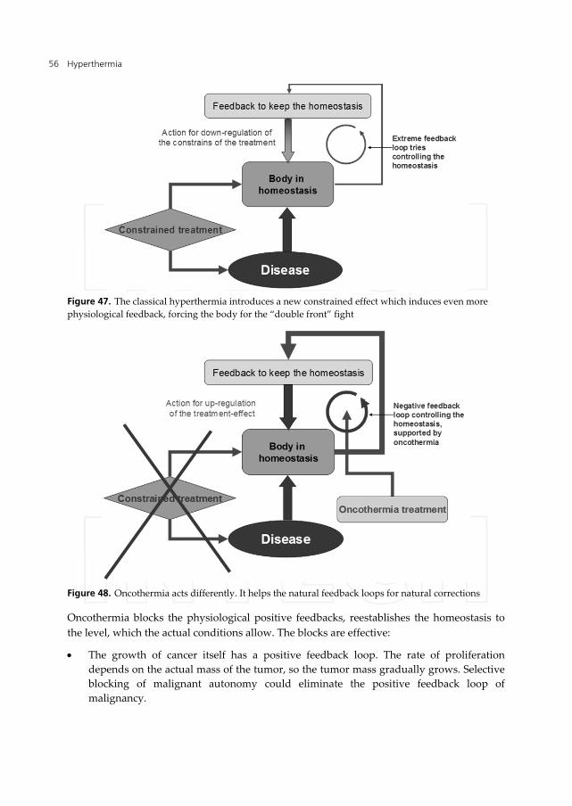
Hyperthermia 56
Figure 47. The classical hyperthermia introduces a new constrained effect which induces even more physiological feedback, forcing the body for the “double front” fight
Figure 48. Oncothermia acts differently. It helps the natural feedback loops for natural corrections
Oncothermia blocks the physiological positive feedbacks, reestablishes the homeostasis to the level, which the actual conditions allow. The blocks are effective:
The growth of cancer itself has a positive feedback loop. The rate of proliferation depends on the actual mass of the tumor, so the tumor mass gradually grows. Selective blocking of malignant autonomy could eliminate the positive feedback loop of malignancy.

Local Hyperthermia in Oncology – To Choose or not to Choose? 57
The lowering of the pH of the tumor is also a positive feedback loop. By the blocking of the high metabolic rate, the pH-loop is eliminated as well.
The injury current loop also has positive feedback. This is broken by the flow of the electric current density.
The neoangiogenesis is also a positive feedback, supporting the tumor by its accelerated nutrition demand. The electric current prefers the new vessels for conduction, blocking their actions.
The growing temperature decreases the impedance (increases the conductance by the mobility of the ions) in the tumor. This positive feedback loop is used till the temperature does not increase too much. The step-up heating uses this effect and controls this loop.
The growing temperature blocks the blood-flow in the tumor, which traps the heat in the center of the tumor. The heat-trap makes necrosis in the center of the tumor, which is anyway very acidic, and in most cases it is necrotized naturally. In artificial focusing, this necrotized volume remains the main target, however it does not need more energy. The real danger of the tumor is the vividly living outer “shell” which is responsible for metastases and for the volume-occupying rapid growth as well. The oncothermia current flows through this “shell” irrespective of how large the necrotic center is, and makes the oncothermia effective. It is shown, that after oncothermia the proliferation activity is drastically suppressed even in the possible remaining tumor tissue, which of course was massively targeted by the RF current.
5.2. Epilogue
Hyperthermia is an ancient treatment. It was the very first one in oncology, but it could not find its established place among the “gold standards” of the oncotherapies. The controversial results were originated from the paradigm to constrain the temperature growth in the process. The constrained forces made physiological contra-reactions keep the homeostasis, which unfortunately contains the malignant tissue as well. The actions have to be selective, and have to be gentle enough to work together with the natural processes, and not against them.
Oncothermia selects the malignant cells and acts differently from the physiological homeostatic reactions (heat-flow on the membrane supported by the electric field effects). It is natural, it is not against the homeostasis and physiology does not work against the action. The main task is to direct the physiology in the standard way, and act on such normal line. The positive feedback loops (the avalanche effects), which may destroy the normal homeostatic equilibrium have to be stopped.
Oncothermia follows the update demands of the modern oncology:
It is a personalized therapy, It is non- toxic, It elongates the survival time of the patients, It completes the curative actions with increased quality of life It has good cost/benefit ratio

Hyperthermia 58
The introduced new paradigm by oncothermia solved the classical challenges:
Challenge (1): “The biology is with us while the physics is against us” (Overgard J., [405]).
- Oncothermia solution: “The biophysics is with us” Challenge (2): “The biology and the physics are with us while the physiology is against
us” (Osinsky S., [88]). - Oncothermia solution: “The fractal physiology is with us” Challenge (3): “Reference point is needed!” (Fatehi D. van der Zee J., et. al. [406]). - Oncothermia solution: “Back to the gold standards, use the energy instead of
temperature”
The task for the future is challenging, and we are expecting professionals to repeat our results and come with us to fight in the war against cancer [407].
Author details
Andras Szasz, Nora Iluri and Oliver Szasz Department of Biotechnics, St.Istvan University, Hungary
6. References
[1] Fischler M.P., Reinhart W.H. (1997). "Fever: friend or enemy?". Schweiz Med Wochenschr 127 (20): 864–70
[2] Christian Raetz and Chris Whitfield (2002) Lipopolysaccharide Endotoxins Annu. Rev. Biochem. 71:635-700
[3] Rosenberg H, Davis M, James D, Pollock N, Stowell K (2007). "Malignant hyperthermia". Orphanet J Rare Dis 2: 21.
[4] Su F, Nguyen ND, Wang Z, Cai Y, Rogiers P, Vincent JL (2005). "Fever control in septic shock: beneficial or harmful?". Shock (Augusta, Ga.) 23 (6): 516–20
[5] Milligan AJ (1984) Whole-Body Hyperthermia Induction Techniques. Cancer Res 44:4869s-4872s
[6] Galvagno SM (2003) Emergency Pathophysiology: Clinical Applications for Prehospital Care. Teton New Media, Jackson, USA, Ch.30, p.361
[7] Smith E (2002) Egyptian Surgical Papyrus dated around 3000 B.C. (cited by: van der Zee J: Heating the patient: a promising approach? Ann. Oncol. 13:1173-1184
[8] Bruch W (1866) Verhandlungen ärztlicher Gesellschafter. Berl Klin Wschr. 3:245-246 [9] Schamberg J, Tseng H (1927) Experiments on the therapeutic value of hot baths with
special reference to the treatment of syphilis, and some physiologic observations. Am J of Syphilis XI:337-397
[10] Seegenschmiedt MH, Vernon CCA (1995) Historical Perspective on Hyperthermia in Oncology. Seegenschmiedt MH, Fessenden P, Vernon CC (eds): Thermoradiotherapy and thermochemotherapy Vol 1, Springer/Verlag Berlin Heidelberg

Local Hyperthermia in Oncology – To Choose or not to Choose? 59
[11] Medieval literature – 1. Medieval Turkish Surgical manuscript from Charaf ed-Din, 1465 (Paris, Bibliotheque Nationale), 2. Armamentarium chirurgicum of Johann Schultes, Amsterdam 1672 (Paris, Bibliotheque de Faculte Medicine), cited by: Seegenschmiedt MH, Vernon CC:A Historical Perspective on Hyperthermia in Oncology, In: Seegenschmiedt MH, Fessenden P, Vernon CC (eds): Thermoradiotherapy and thermochemotherapy Vol 1, Springer/Verlag Berlin Heidelberg 1995
[12] Busch W (1866) Uber den Einfluss welche heftigere Erysipeln zuweilig auf organisierte Neubildungenausuben. Vrh. Naturhist. Preuss Rhein Westphal 23:28-30
[13] Coley WB (1893) The treatment of malignant tumors by repeated inoculations of erysipelas with a report of ten original cases. Am J Med Sci 105:487-511
[14] Westermark F (1898) Uber die Behandlung des ulcerierenden Cervixcarcinoms mittels konstanter Warme, Zentralbl. Gynaekol. 22:1335-1337
[15] Westermark N (1927) The effect of heat on rat tumors. Skand. Arch. Physiol. 52:257-322 [16] Overgaard K (1934) Uber Warmeterapie bosartiger Tumoren. Acta Radiol. [Ther.]
(Stockholm) 15:89-99 [17] Muller C (1912) Therapeutische Erfahrungen an 100 mit kombination von
Rontgenstrahlen un Hochfrequenz, resp. Diathermie behandeleten bosartigen Neubildungen, Munchener Medizinische Wochenschrift 28:1546-1549
[18] Adam J (1923) Carcinoma of right tonsil and soft palate treated by diathermy. Journ. of laryngol. a. otol. No. 4
[19] Barney (1969) Efficiency of the high frequenz current on tumors of the bladder. Boston med. a. surg. journ. No. 1.
[20] Bordier (1924) Puissance de la diathermie dans le cancer. Paris Medical, No. 10. [21] Bruni (1921) La cura dei tumori benigni della vesicae con la diathermia. Rif med. Vol.
37. [22] Corbus Budd (1921) Presentation of a case of prickle celled carcinoma of the penis
treated by diathermy and radium. Urol. and. cut. rev. p. 204. [23] Czerny (1911) Therapie des Krebses. Münch. med. Wochenschr. Nr. 58. [24] Czerny (1910) Methoden der Krebsbehandlung. Münch. med. Wochenschr. Nr. 17. [25] Davies Colley (1922) Periostal sarcoma of the temporal bone treated by diathermy. Proc.
of the Royal. Soc. of Med. Vol. 15. [26] Grenouville et Lacaille (1921)Behandlung der Blasentumoren mittels Diathermie. Journ.
D’urol. p. 316. [27] Gould u. Pearce (1913) The Treatment of inoperable cancers. The Lancet [28] Johnson (1922) Diathermy in the treatment of malignant growths. Americ. Journ. of
Electrotherapeut A. Radiol. Nr. 11. [29] Müller Chr (1913) Die Krebskrankheit und ihre Behandlung mit Röntgenstrahlen und
hochfrequenter Elektrizität Resp. Diathermie. Strahlentherapie. Bd. 2., H. 1. [30] Müller Chr (1913) Die Röntgenbehandlung der malignen Tumoren und ihre
Kombinationen. Strahlentherapie. Bd. 3. [31] Novak (1922) The treatment of malignant tumors of the pharynx and larynx by
diathermy Illinois Med. Journ. Vol. 41.

Hyperthermia 60
[32]Patterson (1923) Diathermy for malignant diseases of the month, pharynx and nose. With notes on seventeen successful cases. Brit. Med. Journ. p. 56
[33]Renner (1914) Behandlung der Blasentumoren mit Hochfrequenzströmen. Berl. Klin. Wochenschr. Nr. 37.
[34] Schmidt L. u. Kollischer (1916) Diathermy in malignant tumours of the bladder. Surg. Gynecol. A. Obstetr, V. 23.
[35] Stephan (1912) Histologische Untersuchungen über die Wirkung der Thermopenetration auf normale Gewebe und Carcinom. Beitr. z. klin. Chirurg. Bd. 77. H. 2.
[36] Steward (1922) Diathermy in the treatment of malignant disease. Practit. V. 108. [37] Syne (1923) Surgical diathermy in the treatment of malignant disease of the troat.
Glasgow. Med. Journ. No. 4. [38] Syne (1923) Cases of malignant disease of the pharynx treated by surgical diathermy.
Journ. of Laryngol. A. Otol. No. 4. [39] Telemann (1911) Hochfrequenzströme in der Medizin. Dtsch. med. Wochenschr. Nr. 18. [40] Theilhaber (1912) Behandlung der Carcinomkranken nach der Operation. 6. Internat.
Kongr. f. Geburtsh. u. Gynäk. in Berlin [41] Theilhaber (1913) Operationslose Behandlung des Carcinoms. Berl. klin. Wochenschr.
Nr. 8. [42] Theilhaber (1913) Die Verhütung der Rezidive nach Krebsbehandlung. Krebskongr. in
Brüssel [43] Theilhaber (1918) Die Behandlung der Krebskranken nach Entfernung der Geschwülste.
Jahreskurse f. ärztl. Fortbild [44] Theilhaber (1919) Die akute Entzündung als Heilmittel. Wien. Klin. Wochenschr. Nr. 29. [45] Theilhaber (1919) Der Einfluss der Diathermiebehandlung auf das Carcinomgewebe.
Münch. med. Wochenschr. Nr. 44. [46] Theilhaber (1923) Der Einfluss der cellulären Immunität auf die Heilung der
Carcinome, (insbesondere der Mamma und des Uterus). Arch. f. Gynäkol. S. 237. [47] Werner: Die Rolle der Strahlentherapie bei der Behandlung maligner Tumoren.
Strahlentherapie. Bd. 1. H. 1. u. 2. [48] Werner u. Caan (1911) Elektro und Radiochirurgie im Dienste der Behandlung maligner
Tumoren. Münch. med. Wochenschr. Nr. 23. [49] Wyeth (1923) Die Diathermie bei der Behandlung maligner Tumoren. Americ. Journ. of.
Electrotherapeut. a Radiol. No. 5. [50] Kluger MJ (1991) Fever: Role of Pyrogens and Cryogens. Physiological Reviews 71:93-
127 [51] Devrient W (1950) Überwärmungsbäder. A.Marcus&E.Weber's Verlag Berlin [52] Hoff F (1957) Fieber, Unspezifische Abwehrvorgänge, Unspezifische Therapie. Georg
Thieme Stuttgart [53] Lampert H (1948) Überwärmung als Heilmittel. Hippokrates Stuttgart [54] Schmidt KL (1987) Hyperthermie und Fieber. Hippokrates Stuttgart
Local Hyperthermia in Oncology – To Choose or not to Choose? 61
[55] Heckel M (1990) Ganzkörperhyperthermie und Fiebertherapie – Grundlagen und Praxis. Hippokrates Stuttgart
[56] Heckel M (1992) Fiebertherapie und Ganzkörper-HT, Bessere Verträglichkeit und Effizienz durch thermoregulatorisch ausgewogene, kombinierte Anwendung beider Verfahren. ThermoMed 14-19
[57] Vaupel P, Kruger W (1992) Warmetherapie mit wassergefilterter Infrarot-A-Strahlung, Grundlagen und Anwendungsmoglichkeiten. Hippocrates, Verlag Stuttgart
[58] Hildebrandt B, Drager J, Kerner T et al (2004) Whole-body hyperthermia in the scope of von Ardenne's systemic cancer multistep therapy (sCMT) combined with chemotherapy in patients with metastatic colorectal cancer: a phase I/II study. Int. J. Hyperthermia, 20:317-333
[59] Ardenne A. Von, Wehner H (2005) Extreme Whole-Body Hyperthermia with Water-Filtered Infrared-A Radiation. Eurekah Bioscience Collection, Oncology, Landes Bioscience
[60] Wust P, Riess H, Hildebrandt B (2000) Feasibility and analysis of thermal parameters for the whole-body hyperthermia system IRATHERM 2000. Int. J. Hyperthermia 4:325-339
[61] Kraybill W, Olenki T (1998) A phase I study of fever-range whole body hyperthermia (FR-WBH) in patients with advanced solid tumors: correlation with mouse models, Int. J. Hyperthermia, 2002, Vol. 18, No. 3, 253-266 and Burd R, Dziedzic ST: Tumor Cell Apoptosis, Lymphocyte Recruitment and Tumor Vascular Changes Are Induced by Low Temperature, Long Duration (Fever-Like) Whole Body Hyperthermia, Journal of Cellular physiology 177:137-147
[62] Toyota, Strebel, Stephens, Matsuda, Bull (1997) Long Duration - Mild Whole Body Hyperthermia with Cisplatin: Tumour Response and Kinetics of Apoptosis and Necrosis in a Metastatic at Mammary Adenocarcinoma, Int. J. Hyperthermia, 13:497-506
[63] Sakaguchi Y, Makino M, Kaneko T et al (1994) Therapeutic Efficacy of Long Duration – Low Temperature Whole Body Hyperthermia When Combined with Tumor Necrosis Factor and Carboplatin in Rats. Cancer Research 54:2223-2227
[64] Ostberg R (2000) Use of mild, whole body hyperthermia in cancer therapy. Immunological Invest. 29:139-142
[65] Appenheimer MM, Chen Q, Girard RA, Wang WC, Evans SS (2005) Impact of Fever-Range Thermal Stress on Lymphocyte-Endothelial Adhesion and Lymphocyte Trafficking. Immunological Inestigations, 34:295-323
[66] Shah A, Unger E, Bain MD et al (2002) Cytokine and adhesion molecule expression in primary human endothelial cells stimulated with fever-range hyperthermia. Int. J. Hyperthermia, 18:534-551
[67] Ostberg JR, Gellin C, Patel R, Repasky EA (2001) Regulatory Potential of Fever-Range Whole Body Hyperthermia on Langerhans Cells and Lymphocytes in an Antigen-Dependant Cellular Immune Response. The Journal of Immunology; 167:2666-2670

Hyperthermia 62
[68] Burd R, Dziedzic TS, Xu Y et al (1998) Tumor Cell Apoptosis, Lymphocyte Recruitment and Tumor Vascular Changes Are Induced by Low Temperature, Long Duration (Fever-Like) Whole Body Hyperthermia. J. Cellular physiology, 177:137-147
[69] Dahl O, Dalene R, Schem BC, Mella O (1999) Status of clinical hyperthermia. Acta Oncol. 38(7):863-873
[70] Senior K (2001) Hyperthermia and hypoxia for cancer-cell destruction. Lancet Oncology, 2:524-525
[71] Wust P, Hildebrandt B, Sreenivasa G, Rau B et al (2002) Hyperthermia in combined treatment of cancer. Lancet 3:487-497
[72] Szasz A, Szasz O, Szasz N (2001) Electrohyperthermia: a new paradigm in cancer therapy. Wissenschaft & Forschung, Deutsche Zeitschrift für Onkologie, 33:91-99
[73] Szasz A, Szasz N, Szasz O (2003) Hyperthermie in der Onkologie mit einem historischen Uberblick. Wissenschaft & Forschung, Deutsche Zeitschrift für Onkologie; 35:140-154
[74] Abe M, Hiraoka M, Takahashi M et al (1986) Multi-institutional studies on hyperthermia using an 8-MHz radiofrequency capacitive heating device (Thermotron RF-8) in combination with radiation for cancer therapy. Cancer. 58:1589-1595
[75] Pacing Clin Electrophysiol. 2000 Jan;23(1):8-17. Tissue temperatures and lesion size during irrigated tip catheter radiofrequency ablation: an in vitro comparison of temperature-controlled irrigated tip ablation, power-controlled irrigated tip ablation, and standard temperature-controlled ablation. Petersen HH, Chen X, Pietersen A, Svendsen JH, Haunsø S., http://www.ncbi.nlm.nih.gov/pubmed/10666748
[76] Percutaneous cryoablation techniques and clinical applications, Servet Tatlı, Murat Acar, Kemal Tuncalı, Paul R. Morrison, Stuart Silverman
http://www.dirjournal.org/pdf/DIREPUB_1922_online.pdf [77] Hornbach NB (1987) Is the community radiation oncologist ready for clinical
hyperthermia? RadioGraphics 7:139-141 [78] Storm FK (1993) What happened to hyperthermia and what is its current status in
cancer treatment? J Surg Oncol 53:141-143 [79] Brizel DM (1998) Where there’s smoke, is there fire? Int J Hyperthermia 14:593-594 [80] Sneed PK, Dewhirst MW, Samulski T et al (1998) Should interstitial thermometry be
used for deep hyperthermia? Int. J. Radiat. Oncol Biol. Phys. 40:1205-1212 [81] Oleson JR (1989) If we can’t define the quality, can we assure it? Int. J. Radiat. Oncol
Biol. Phys 16:879 [82] Nielsen OS, Horsman M, Overgaard J (2001) A future of hyperthermia in cancer
treatment? (Editorial Comment), European Journal of Cancer, 37:1587-1589 [83] Szasz A (2006) What is against the acceptance of hyperthermia? Die Naturheilkunde
Forum-Medizine 83:3-7 [84] Oleson JR (1991) Progress in hyperthermia? Int. J. Radiat. Oncol Biol. Phys 20:1147-1164 [85] Oleson JR (1993) Prostate cancer: hot, but hot enough? Int. J. Radiat. Oncol Biol. Phys.
26: 369-370
Local Hyperthermia in Oncology – To Choose or not to Choose? 63
[86] van der Zee J (2002) Heating the patient: a promising approach? Annals of Oncology 13:1173-1184
[87] Smythe WR, Mansfield PF (2003) Hyperthermia: has its time come? Ann Surg Oncol 10:210-212
[88] Osinsky S, Ganul V, Protsyk V et al (2004) Local and regional hyperthermia in combined treatment of malignant tumors: 20 years experience in Ukraine, The Kadota Fund International Forum 2004, Awaji Japan, June 15-18
[89] Markovitch, O., Agmon, N.: Structure and energetics of the hydronium hydration shells. J. Phys. Chem. A. 111(12), 2253–2256 (2007)
[90] Jeffrey GA (1997) An Introduction to Hydrogen Bonding (Topics in Physical Chemistry). Oxford University Press, USA
[91] Morowitz HJ (1992) Beginnings of Cellular Life: Metabolism Recapitulates Biogenesis. Yale University Press, p 69
[92] Vaupel PW, Kelleher DK (1996) Metabolic status and reaction to heat of normal and tumor tissue, In: Seegenschmiedt MH., Fessenden P., Vernon CC. (Eds.) Thermo-radiotherapy and Thermo-chemotherapy, Vol. 1. Biology, physiology and physics, Springer Verlag, Berlin Heidelberg, pp. 157-176
[93] Kabakov AE, Gabai VL (1997) Heat Shock Proteins and cytoprotection: ATP-deprived mammalian cells. (Series: Molecular Biology Intelligence Unit), Springer Verlag, New York, Berlin, Heidelberg
[94] Keszler G, Csapo Z, Spasokoutskaja T et al (2000) Hyperthermy increase the phosporylation of deoxycytidine in the membrane phospholipid precursors and decrease its incorporation into DNA. Adv Exper Med Biol 486:333-337
[95] Dikomey E, Franzke J (1992) Effect of heat on induction and repair of DNA strand breaks in X-irradiated CHO cells. Int J Radiat Biol 61(2):221-233
[96] Shen, R.N., Lu, L., Young, P., et, al.: Influence of elevated temperature on natural killer cell activity, lymphokine-activated killer cell activity and lecitin-dependent cytotoxicity of human umbilical cord blood and adult blood cells. Int. J. Radiat. Oncol. Biol. Phys. 29, 821-26 (1994)
[97] Srivastava PK, DeLeo AB, Old LJ (1986) Tumor Rejection Antigens of Chemically Induced Tumors of Inbred Mice. Proc Natl Acad Sci USA 38(10):3407-3411
[98] Csermely P, Schnaider T, Soti C et al (1998) The 90 kDa Molecular Chaperone Family: Structure, Function and Clinical Applications A Comprehensive Review. Pharmacol Ther 79(2):129-168
[99] Gonzalez-Gonzalez, D.: Thermo-radiotherapy for tumors of the lower gastro-instenstinal tract. In: M.H. Seegenschmiedt, M.H., Fessenden, P., Vernon, C.C. (eds.) Thermo-radiotherapy and Thermo-chemiotherapy. Biology, physiology and physics, Vol. 1, pp. 105-119, Springer Verlag, Berlin, Heidelberg (1996)
[100] Reinhold HS (1987) Tumor microcirculation, In:Field SB and Franconi C (Eds): Physics and Technology of hyperthermia, NATO ASI Series, Series E: Applied Sciences, No. 127, Martinus Nijhoff Publishers, Dordrecht/Bosto/Lanchester, pp.448-457

Hyperthermia 64
[101] Reinhold HS (1987) Effects of hyperthermia on tumor microcirculation, In:Field SB and Franconi C (Eds): Physics and Technology of hyperthermia. NATO ASI Series, Series E: Applied Sciences, No. 127, Martinus Nijhoff Publishers, Dordrecht/Bosto/Lanchester, pp.458-469
[102] Tanaka Y (2001) Thermal responses of microcirculation and modification of tumor blood flow in treating the tumors, In: Kosaka M, Sugahara T, Schmidt KL, Simon E (Eds.): Thermotherapy for Neoplasia, Inflammation, and Pain. Springer Verlag Tokyo, pp. 407-419
[103] Reinhold HS, Blachiewicz, Berg-Blok (1978) Decrease in tumor microcirculation during hyperthermia, In: Streffer C, vanBeuningen D, Dietzel F, Röttinger E, Robinson JE, Scherer E, Seeber S, Trott K-R (Eds): Cancer Therapy by hyperthermia and radiation. Urban & Schwarzenberg, Baltimore-Munich, pp.231-232
[104] Dudar TE, Jain RK (1984) Differential response of normal and tumor microcirculation to hyperthermia. Cancer Research, 44:605-612
[105] Hudlicka O, Tyler KR (1984) The effect of long-term high-frequency stimulation on capillary density and fibre types in rabbit fast muscles. J. Physiol. 353:435-445
[106] Endrich B, Hammersen F (1986) Morphologic and hemodynamic alterations in capillaries during hyperthermia, In: Anghileri LJ, Robert J. (Eds.): Hyperthermia in Cancer Treatment. CRC Press, Inc. Boca Raton Florida, US, Vol II. pp. 17-47, (Ch. 2)
[107] Zant G. van (1986) Effects of hyperthermia on hematopoietic tissues, In: Anghileri LJ, Robert J. (Eds.): Hyperthermia in Cancer Treatment. CRC Press, Inc. Boca Raton Florida, US, Vol II. Pp. 59-73, (Ch. 4)
[108] Hales JRS, Hirata K (1986) Aspects of circulatory responses in animals pertinent to the use of hyperthermia in cancer treatment, In: Anghileri LJ, Robert J. (Eds.): Hyperthermia in Cancer Treatment. CRC Press, Inc. Boca Raton Florida, US, Vol II. Pp. 49-58, (Ch. 3)
[109] Ingram DL, Mount LE (1975) Man and animals in hot environments, Topics in environmental physiology and medicine. Springer Verlag, Berlin, Heidelberg, New York,
[110] Urano M (1994) Thermochemotherapy: from in vitro and in vivo experiments to potential clinical application, In: Urano & Douple (eds.) Hyperthermia in Oncology 4:169-204
[111] Ohno T, Sakagami T, Shiomi M et al (1993) Hyperthermai therapy for deep-regional cancer: thermochemotherapy, a combination of hyperthermia with chemotherapy. In: Matsuda T (ed) Cancer treatment by hyperthermia, radiation and drugs, Taylor&Francis, London-Washington DC, pp 303-316
[112] Piantelli M, Tatone D, Castrilli G et al (2001) Quercetin and tamoxifen sensitize human melanoma cells to hyperthermia. Melanoma Research 11:469-476
[113] Pilling MJ, Seakins PW (1995) Reaction kinetics. Oxford Science Publications, Oxford University Press, Oxford

Local Hyperthermia in Oncology – To Choose or not to Choose? 65
[114] Wiedermann GJ, Feyerabend T, Mentzel M et al (1994) Thermochemotherapie: grunde fur die kombinationsbehandlung mit hyperthermia und chemotherapie. Focus Mul 11:44-50
[115] Issels RD, Abdel-Rahman S, Salat C et al (1998) Neoadjuvant chemotherapy combined with regional hyperthermia (RHT) followed by surgery and radiation in primary recurrent high-risk soft tissue sarcomas (HR STS) of adults (updated report), J. Cancer Res. Clin. Oncol. 124:R105
[116] LeVeen HH, Rajagopalan PR, Vujic I et al (1984) Radiofrequency thermotherapy, local chemotherapy, and arterial Occlusion in the treatment of non-resectable cancer. Am Surg 50(2):61-65
[117] Okamura K, Nakashima K, Fukushima Y et al Hyperthermia with low dose chemotherapy for advanced non-small-cell lung cancer.
http://www.isshin.or.jp/okamura/awaji2004/awaji1.html [118] Franchi F, Grassi P, Ferro D et al (2007) Antiangiogenic metronomic chemotherapy
and hyperthermia in the palliation of advanced cancer. European Journal of Cancer Care 16(3):258-262
[119] Streffer C, (1995) Molecular and cellular mechanism of hyperthermia In: Seegenschmiedt MH., Fessenden P., Vernon CC. (Eds.) Thermo-radiotherapy and Thermo-chemotherapy, Vol. 1. Biology, physiology and physics, Springer Verlag, Berlin Heidelberg, pp. 47-74
[120] Roti JL, Laszlo A (1988) The effects of hyperthermia on cellular macromolecules. In: Urano M, Douple E (eds) Hyperthermia and Oncology Vol 1, Thermal effects on cells and tissues, VSP Utrecht, The Netherlands, pp 13-56
[121] Okumura Y, Ihara M, Shimasaki T, Takeshita S, Okaichi K (2001) Heat inactivation of DNA-dependent protein kinase: possible mechanism of hyperthermic radio-sensitization, in: Thermotherapy for Neoplasia, Inflammation, and Pain, (Kosaka M, Sugahara T, Schmidt KL, Simon E (Eds.)), Springer Verlag Tokyo pp. 420-423
[122] Urano M, Douple E. (eds) (1992) Hyperthermia and Oncology: Volume .2. Biology of thermal potentiation of radiotherapy. VSP BV Utrecht The Netherlands, VSP BV Utrecht, The Netherlands
[123] Perez CA, Brady LW, Halperin EC et al (2004) Principles and Practice of Radiation Oncology. 4th edition, Lippincott Williams and Wilkins, Philadelphia
[124] Molls M (1992) Hyperthermia – the actual role in radiation oncology and future prospects. Strahlenterapie und Oncologie 168:183-190
[125] Seegenschmiedt MH, Feldmann HJ, Wust P (1995) Hyperthermia – its actual role is radiation oncology. Strahlentherapie und Oncologie 171:560-572
[126] Emami B, Scott C, Perez CA et al (1996) Phase III study of interstitial thermoradiotherapy compared with interstitial radiotherapy alone in the treatment of recurrent or persistent human tumors: a prospectively controlled randomized study by radiation therapy oncology group. Int J Rad Oncol Biol Phys 34(5):1097-1104
[127] Wust P, Rau B, Gemmler M et al (1995) Radio-thermotherapy in multimodal surgical treatment concepts. Onkologie 18(2):110-121
Hyperthermia 66
[128] Hentschel M, Wust P, (2000), Hyperthermia: bald eine entablierte Therapie? MTA Spectrum, 12:623-628
[129] Hager ED, Birkenmeier J, Popa C, (2006), Hyperthermie in der Onkologie: Eine viel versprechende neue Methode? Deutsche Zeitschrift fur Onkologie, 38:100-107
[130] Hehr T, Wust P, Bamberg M et al (2003) Current and potential role of thermoradiotherapy for solid tumors. Onkologie 26(3):295-302
[131] van der Zee J, Gonzalez Gonzalez D, van Rhoon GC et al (2000) Comparison of radiotherapy alone with radiotherapy plus hyperthermia in locally advanced pelvic tumors: a prospective, randomised, multicentre trial. Dutch Deep Hyperthermia Group. Lancet 355(9210):1119-1125
[132] Kodama K, Doi O, Higashyama M et al (1993) Long-term results of postoperative intrathoracic chemo-thermotherapy for lung cancer with pleural dissemination. Cancer 72(2):426-431
[133] Masunaga S, Hiraoka M, Akuta K et al (1990) Non-Randomized Trials of Thermoradiotherapy versus Radiotherapy for Preoperative Treatment of Invasive Urinary Bladder Cancer. J Jpn Soc Ther Radiol Oncol 2: 313-320
[134] Rau B, Wust P, Hohenberger P et al (1998) Preoperative Hyperthermia Combined with Radiochemotherapy in Locally Advanced Rectal Cancer – A Phase II Clinical Trial. Annals of Surgery 227(3):380-389
[135] Pearson AS, Izzo F, Fleming RYD et al (1999) Intraoperative radiofrequency ablation of cryoablation for hepatic malignances. Amer J Surg 178(6):592-598
[136] Kouloulias VE, Kouvaris JR, Nikita KS et al (2002) Intraoperative hyperthermia in conjunction with multi-schedule chemotherapy (pre- intra- and post operative), by-pass surgery, and post-operative radiotherapy for the management of unresectable pancreatic adenocarcinoma. Int.J Hyperthermia 18:233-252
[137] Ohtsuru A, Braiden V, Cao Y (2001) Cancer Gene Therapy in Conjunction with Hyperthermia Under the Control of Heat-Inducible Promoter. In: Kosaka M, Sugahara T, Schmidt KL (eds) Thermotherapy for Neoplasia, Inflammation, and Pain, Springer Verlag .Tokyo, pp 464-470
[138] Gaber MH, Wu NZ, Hong K et al (1996) Thermosensitive liposomes: extravasation and relase of contents in tumor microvascular networks. Int J Radiat Oncol Biol Phys 36(5):1177-1187
[139] Balckburn LV, Galoforo SS, Corry PM et al (1998) Adenoviral-mediated transfer of heat-inducible double suicide gene into prostate carcinoma cells. Cancer Res 58(7):1358-1362
[140] Huang Q, Hu JK, Zhang L et al (2000) Heat-induced gene expression as a novel targeted cancer gene therapy strategy. Cancer Res 60(13):3435-3439
[141] Yerushalmi A, Shani A, Fishelovitz Y et al (1986) Local microwave hyperthermia in the treatment of carcinoma of the prostate. Oncology 43(5):299-305
[142] Oleson JR. Calderwood SK. Coughlin CT. Dewhirst MW. Gerweck LE. Gibbs FA. Kapp DS. (1988), Biological and Clinical Aspects of Hyperthermia in Cancer Therapy. Am. J. Clin. Oncology 11:368-380
Local Hyperthermia in Oncology – To Choose or not to Choose? 67
[143] Henderson BW, Waldow SM, Potter WR, Dougherty TJ. (1985) Interaction of Photodynamic Therapy and Hyperthermia: Tumor Response and Cell Survival Studies after Treatment of Mice in Vivo. Cancer Research 45:6071-6077
[144] Lohr F. Hu K, Huang Q. Zhang L. Samulski T. Dewhirst M. Li C. (2000) Enhancement of radiotherapy by hyperthermia-regulated gene therapy International Journal of Radiation OncologyBiologyPhysics, 48:1513-1518
[145] Skitzki JJ. Repasky EA. Evans SS. (2009) Hyperthermia as an immunotherapy strategy for cancer, Current Opinion in Investigational Drugs 10:550-558
[146] Vertrees RA. Jordan JM. Zwischenberger JB (2007) Hyperhtermia and Chemotherapy: The Science. In: Current Clinical Oncology, Intraperitoneal Cancer Therapy, Hlem CW. Edwards RP. (Eds.) Humana Press, Totowa NJ, USA
[147] Oliveira RS, Bevilacqua FRG, Chammas R (1997) Hyperthermia increases the metastatic potential of murine melanoma. Braz J Med Biol Res, 30:941-945
[148] Shah SA, Jain RK, Finney PL (1983) Enhanced metastasis formation by combined hyperthermia and hyperglycemia in rats bearing Walker 256 carcinosarcoma. Cancer Lett. 19(3):317-23
[149] Nathanson SD, Nelson L, Anaya P, Havstad S, Hetzel FW (1991) Development of lymph node and pulmonary metastases after local irradiation and hyperthermia of footpad melanomas, Clinical and Experimental Metastasis 9:377-392
[150] Brochure of Thermotron RF-8. (Yamamoto Vinita, Osaka, Japan) [151] Gellermann J, Wlodarczyk W, Hildebrandt B, Ganter H, Nicolau A, Rau B, Tilly W,
Fähling H, Nadobny J, Felix R, Wust P, (2005) Noninvasive Magnetic Resonance Thermography of Recurrent Rectal Carcinoma in a 1.5 Tesla Hybrid System Cancer Res 65:5872-5880
[152] Stoll AM (1967) Heat transfer in Biotechnology, in: Advances in heat Transfer, (Eds.: Hartnett JP, Irvine TF.) 4:65-139, Academic Press Inc. New York, London
[153] Cooper TE, Trezek GJ (1971) Correlation of thermal properties of some human tissue with water content. Aerospace Med. 42:24-27
[154] Ardenne M. von, Reitnauer PG (1977) Krebs-Mehrschritt-Therapie und Mikrozirkulation. Krebsgeschehen 9:134-149
[155] Streffer C (1990) Biological basis of thermotherapy, In: Gautherie M (Ed.): Methods of hyperthermia control, Clinical Thermology. Springer Verlag, Berlin, Heidelberg, New York, London, Paris, Tokyo, Hong Kong
[156] Law MP (1988) The response of normal tissues to hyperthermia, In: Urano M, Douple E. (Eds.) Hyperthermia and Oncology: Vol.1. Thermal effects on cells and tissues. VSP BV Utrecht The Netherlands, pp. 121-159
[157] Prescott DM (1996) Manipulation of physiological parameters during hyperthermia, In: Seegenschmiedt MH., Fessenden P., Vernon CC. (Eds.) Thermo-radiotherapy and Thermo-chemotherapy, Vol. 1. Biology, physiology and physics. Springer Verlag, Berlin Heidelberg, pp. 177-189

Hyperthermia 68
[158] Muftuler TL, Hamamura MJ, Birgul O, Nalcioglu O (2006) In Vivo MRI Electrical Impedance, Tomography (MREIT) of Tumors. Technology in Cancer Research and Treatment, 5(4):381-387
[159] van der Zee J, (2005) Presentation on Conference in Mumbai India (http://www.google.com/#sclient=psy&hl=en&site=&source=hp&q=%22van+der+Zee%22+Mumbai+ext:ppt&btnG=Google+Search&aq=&aqi=&aql=&oq=&pbx=1&bav=on.2,or.r_gc.r_pw.&fp=e7df6ea8d325b7b2, accessed Apr. 2011)
[160] Szasz A, Szasz N, Szasz O (2010) Oncothermia – Principles and Practices, Springer Verlag, Heidelberg
[161] Warburg O (1966) Oxygen, The Creator of Differentiation, Biochemical Energetics, Academic Press, New York, 1966; (Warburg O: The Prime Cause and Prevention of Cancer, Revised lecture at the meeting of the Nobel-Laureates on June 30, 1966 at Lindau, Lake Constance, Germany)
[162] Garber K (2004) Energy boost: The Warburg effect returns in a new theory of cancer. Journ. Nat. Canc. Inst. USA 96:1805-1806
[163] Hsu PP, Sabatini DM (2008) Cancer metabolism: Warburg and Beyond. Cell 134(5):703-707
[164] Oehr P, Biersack HJ, Coleman RE (Eds) (2004) PET and PET-CT in Oncology. Springer Verlag, Berlin-Heidelberg
[165] Babaeizadeh S (2007) 3-D Electrical Impedance Tomography of piecewise contant domains with known internal boundaries. IEEE Trans. Biomed. Eng. 54:2-10
[166] TransCan TS (2000) Transcan Medical Ltd. Migdal Ha’Emek, Israel, distributed by Siemens AG, Germany,
[167] Marino AA, Iliev IG, Schwalke MA, Gonzalez E, Marler KC, Flanagan CA (1994) Association between cell membrane potential and breast cancer. Tumour Biol, 15:82-89
[168] Cure JC (1995) Ont he electrical charateristics of cacer. II> International Congress of Electrochemical Treatment of Cancer. Jupiter, Florida
[169] Revici E (1961) Research in Pathophysiology as Basis Guided Chemotherapy, with Special Application to Cancer. Princeton, NJ> D. Van Nostrand Compani
[170] Seeger PG, Wolz S (1990) Succesful Biological Contol of Cancer. Neuwieder Verlagsgesellschaft Gmbh
[171] Cure JC. (1991) Cancer an electrical phenomenon. Resonant 1(1) [172] Foster KR, Schepps JL (1981) Dielectric properties of tumor and normal tissues at
radio through microwave frequencies. J. Microwave Power 16:107-119 [173] Peirce A (Tutor): IAM-CSC-PIMS senior undergraduate math modeling workshop,
The Institute of Applied Mathematics (IAM), The University of British Columbia [174] Metherall P (1998) Three-dimensional electrical impedance tomography of the human
thorax. PhD thesis, University of Sheffield [175] Mikac U, Demsar F, Beravs K, Sersa I (2001) Magnetic Resonance Imaging of
alternating electric currents. Magnetic Resonance Imaging 19:845-856 [176] Joy M, Scott G, Henkelman M (1989) In vivo detection of applied electric currents by
magnetic resonance imaging. Magnetic Resonance Imaging, 7:49-54

Local Hyperthermia in Oncology – To Choose or not to Choose? 69
[177] Seersa I, Beravs K, Dodd NJF et al (1997) Electric current Imaging of Mice tumors. MRM 37:404-409
[178] Scott GC, Joy MLG, Armstrong RL, Henkelman RM (1995) Electromagnetic considerations for RF current density imaging. IEEE Trans. Med. Imaging 14:515-524
[179] Szasz A, Vincze Gy, Szasz O, Szasz N (2003) An energy analysis of extracellular hyperthermia. Magneto- and electro-biology, 22:103-115
[180] Pence DM, Song CW (1986) Effect of heat on blood-flow, In: Anghileri LJ, Robert J. (Eds.): Hyperthermia in Cancer Treatment, Vol. II. CRC Press, Inc. Boca Raton Florida, US, pp.1-17
[181] Häfner H-M, Bräuer K, Eichner M et al (2005) Analysis of Cutaneous Blood Flow in Melanocytic Skin Lesions. J. vascular Res. 42:38-46
[182] McRae DA, Esrick MA, Mueller SC (1997) Non-invasive, in-vivo electrical impedance of EMT-6 tumors during hyperthermia: correlation with morphology and tumour-growth delay. Int. J. Hyperthermia, 13:1-20
[183] Esrick MA, McRae DA (1994) The effect of hyperthermia induced tissue conductivity changes on electrical impedance temperature mapping. Phys. Med. Biol. 39:133-144
[184] Mott NF, Jones H (1958) The theory of the properties of metals and alloys. Dover Publ Inc, New York
[185] Cope FW (1969) Nuclear magnetic resonance evidence using D2O for structured water in muscle and brain. Biophys J 9(3):303-319
[186] Damadian R (1971) Tumor detection by nuclear magnetic resonance. Science 171(3976):1151-1153
[187] Cope, F.W.: A review of the applications of solid state physics concepts to biological systems. J. Biol. Phys. 3(1), 1-41 (1975)
[188] Hazlewood CF, Nichols BL, Chamberlain NF (1969) Evidence for the existence of a minimum of two phases of ordered water in skeletal muscle. Nature 222(195):747–750
[189] Hazlewood CF, Chang DC, Medina D et al (1972) Distinction between the Preneoplastic and Neoplastic State of Murine Mammary Glands. Proc Natl Acad Sci USA 69(6):1478-1480
[190] Szentgyorgyi, A.: The living state and cancer. Physiological Chemistry and Physics 12, 99-110 (1980)
[191] Gniadecka M, Nielsen OF, Wulf HC (2003) Water content and structure in malignant and benign skin tumors. Journal of Molecular Structure 661-662:405-410
[192] Beall PT et al (1979) Water-relaxation times of normal, preneoplastic, and malignant primary cell cultures of mouse mammary gland. In: 23rd Annual Meeting of the Biophysical Society, Atlanta, Georgia, USA, 26-28 February 1979
[193] Chung, S.H., Cerussi, A.E., Klifa, C., et. al.: In vivo water state measurements in breast cancer using broadband diffuse optical spectroscopy. Phys. Med. Biol. 53, 6713-6727 (2008)
[194] Fiskum G (2000) Mitochondrial participation in ischemic and traumatic neural cell death. Journal of Neurotrauma 17(10):843–855

Hyperthermia 70
[195] Ichas F, Mazat JP (1998) From calcium signaling to cell death: two conformations for the mitochondrial permeability transition pore. Switching from low- to high- conductance state. Biochimica et Biophysica Acta 1366:33–50
[196] Bonnet S, Archer SL, Allalunis-Turner J et al (2007) A Mitochondria-K+ Channel Axis Is Suppressed in Cancer and Its Normalization Promotes Apoptosis and Inhibits Cancer Growth. Cancer Cell 11(1):37–51
[197] Chidanbaram, R., Ramanadham, M.: Hydrogen bonding in biological molecules-an update. Physica B 174(1-4), 300-305 (1991)
[198] Szentgyorgyi A (1960) Introduction to a Submolecular Biology. Academic Press, New York, London
[199] Akerlof GA, Shiller RJ (2009) Animal spirits. Princeton University Press, Princeton and Oxford
[200] Moses ME, Hou C, Woodruff WH et al (2008) Revisiting a model of oncogenic growth: estimating model parameters from theory and data. Am Nat 171(5):632-645
[201] Pamatmat MM (2005) Measuring aerobic and anaerobic metabolism of benthic infauna under natural conditions. Journal of Experimental Zoology 228(3):405-413
[202] West GB, Woodruf WH, Born JH (2002) Allometric scaling of metabolic rate from molecules and mitochondria to cells and mammals. Proc Natl Acad Sci U S A 99(1):2473–2478
[203] Lane N (2006) Mitochondria: Key to Complexity. In: Martin W (ed) Origins of Mitochondria and Hydrogenosomes, Chapter 2, Springer, Heidelberg, Germany
[204] Szentgyorgyi A (1998) Electronic Biology and Cancer. Marcel Dekkerm New York [205] Weinberg RA (1998) One renegade cell. Basic Books, A Member of the Perseus Books
Group, New York [206] Cairns J (1975) The Cancer Problem. Scientific American 233(5):64-72, 77-78 [207] Kurakin, A.: Scale-free flow of life: on the biology, economics, and physics of the cell.
Theor. Biol. Med. Model. 6:6 (2009) [208] Getling AV, Rayleigh-Benard C (1998) Structures and Dynamics. World Scientific,
Singapore [209] Hochachka PW (1999) The metabolic implications of intracellular circulation. Proc
Natl Acad Sci USA 96(22):12233-12239 [210] Coulson RA (1986) Metabolic rate and the flow theory: a study in chemical
engineering. Comp Biochem Physiol A Comp Physiol 84(2):217-229 [211] Szentgyorgyi A (1968) Bioelectronics, A Study on Cellular Regulations, Defense and
Cancer. Acad. Press, New York, London [212] Blad B, Baldetorp B (1996) Impedance spectra of tumour tissue in comparison with
normal tissue; a possible clinical application for electric impedance tomography. Physiol. Meas. 17Suppl.4A:A105-15
[213] Stoy RD, Foster KR, Schwan HP (1982) Dielectric properties of mammalian tissues form 0.1 to 100 MHz: a summary of recent data, Phys.Med.Biol. 27:501-513
[214] Haemmerich D, Staelin ST, Tsai JZ et al (2003) In vivo electrical conductivity of hepatic tumors. Physiol. Meas. 24:251-260

Local Hyperthermia in Oncology – To Choose or not to Choose? 71
[215] Smith SR, Foster KR, Wolf GL (1986) Dielectric properties of VX-2 carcinoma versus normal liver tissue. IEEE Trans. Biomed. Eng. BME-33:522-524
[216] Dubois J-M, Rouzaire-Dubois B (2004) The influence of cell-volume changes on tumor cell proliferation. Eur. Biophys. J, 33:227-232
[217] Avrami MA.: Kinetics of phase change Parts: I. J. Chem. Phys. 7, 1103 (1939); Avrami. M.A.: Kinetics of phase change Parts: II. J. Chem. Phys. 8, 212 (1940); Avrami, M.A.: Kinetics of phase change Parts: III. J. Chem. Phys. 9, 1103 (1941)
[218] Kolmogorov NN (1937) On the statistical theory of the crystallization of metals. Bull Acad Sci UssR Math Ser 1:355-359
[219] Joshnson WA, Mehl PA (1939) Reaction kinetics in processes of nucleation and growth. Trans Amer Inst Mining (Metal) Engrs 135:416
[220] Augis JA, Bennett JE (1978) Calculation of the Avrami parameters fer heterogeneous solid state reactions using a modification of the Kissinger method. J Therm Anal 13:285-291
[221] Levine LE, Narayan KL, Kelton KF (1997) Finite size corrections for the Johnson-Mehl-Avrami-Kolmogorov equation. J Mater Res 12(1):124-131
[222] Cope FW (1977) Detection of phase transitions and cooperative interactions by Avrami analysis of sigmoid biological time curves for muscle, nerve, growth, firefly, and infrared phosphorescence, of green leaves, melanin and cytochrome C. Physiol. Chem. Phys. 9(4-5):443-459
[223] Cope FW (1977) Solid State physical replacement of Hodgkin-Huxley theory. Phase transformation kinetics of axonal potassium conductance. Physiol. Chem. Phys. 9(2):155-160
[224] Cope FW (1977) The kinetics of biological phase transitions manifested by sigmoid time curves: a review of approaches. Physiol. Chem. Phys. 8(6):519-527
[225] Cope FW (1980) Avrami analysis of electrical switching in hydrated melanin suggest dependence on a phase transition. Physiol. Chem. Phys. 12:537-538
[226] Suckjoon J, Bechhoefer J (2005) Nucleation and growth in one dimension. II. Application to DNA replication kinetics. Phys. Rev. E. 71(1), 011909
[227] Haiyang Z (2005) Reconstructing DNA replication kinetics from small DNA fragments. Simon Fraser University, China
[228] Szent-gyorgyi A. (1941) Towards a new biochemistry? Science 93(2426):609-611 [229] Szent-gyorgyi A (1946) Internal photo-electric effect and band spectra in proteins.
Nature 157:875-875 [230] Cope FW (1971) Evidence from activation energies for superconductive tunneling in
biological systems at physiological temperatures. Physiol Chemistry & Physics 3:403-410
[231] Liboff AR (1985) Geomagnetic cyclotron resonance in living cells. J. Biol. Phys. 13(4):99-102
[232] Liboff AR (2003) Ion Cyclotron Resonance in Biological Systems: Experimental Evidence. In: Stavroulakis P (ed) Biological Effects of Electromagnetic Fields, Springer Verlag, Berlin-Heidelberg, pp 6-113

Hyperthermia 72
[233] Schrodinger E (1967) What is life? Cambridge University Press, Cambridge, United Kingdom
[234] Katchalsky A, Curran PF (1967) Non-equilibrium thermodynamics in biophysics. Harward University Press, Cambridge, MA, USA
[235] Haken H (1987) Self-Organization and Information. Phys. Script. 35(3):247-254 [236] Sornette D (2000) Chaos, Fractals, Self-Organization and Disorder: Concepts and
Tools. Springer Verlag, Berlin-Los Angeles [237] Deering W, West BJ (1992) Fractal physiology. IEEE Engineering in Medicine and
Biology 11(2):40-46 [238] West BJ (1990) Fractal Physiology and Chaos in Medicine. World Scientific,
Singapore, London [239] Bassingthwaighte JB, Leibovitch LS, West BJ (1994) Fractal Physiology. Oxford Univ.
Press, New York, Oxford [240] Musha T, Sawada Y (eds.) (1994) Physics of the living state. IOS Press, Amsterdam [241] Brown JH, West GB (eds) (2000) Scaling in Biology. Oxford University Press, Oxford [242] Brown JH, West GB, Enquis BJ (2005) Yes, West, Brown and Enquist’s model of
allometric scaling is both mathematically correct and biologically relevant. Functional Ecology 19(4):735 –738
[243] West GB, Brown JH (2005) The origin of allometric scaling laws in biology from genomes to ecosystems: towards a quantitative unifying theory of biological structure and organization. Journal of Experimental Biology 208:1575-1592
[244] Camazine S, Deneubourg JL, Franks NR et al (2003) Self-organization in biological systems. Princeton Studies in Complexity, Princeton Univ. Press, Princeton, Oxford
[245] Bak P, Tang C, Wieserfeld K (1988) Self-organized criticality. Phys. Rev. A. 38:364-373 [246] Raff MC (1992) Social controls on cell survival and death. Nature 356(6368):397-400 [247] Loewenstein WR, Kanno Y (1967) Intercellular communication and tissue growth.
The Journal of Cell Biology 33(2):225-234 [248] Dissado LA (1990) A fractal interpretation of the dielectric response of animal tissues.
Phys. Med. Biol. 35:1487-1503 [249] El-Lakkani A (2001) Dielectric response of some biological tissues.
Bioelectromagnetics 22:272-279 [250] Walleczek J (ed) (2000) Self-organized biological dynamics & nonlinear control.
Cambridge Univ. Press, Cambridge [251] Marjan MI, Szasz A (2000) Self-organizing processes in non-crystalline materials:
from lifeless to living objects. OncoTherm Kft., Budapest [252] West GB, Brown JH, Enquist BJ (1999) The Four Dimension of Life: Fractal Geometry
and Allometric Scaling of Organisms. Science 284:4 [253] Hegyi G, Vincze G, Szasz A (2007) Axial-vector interaction with bio-systems. Electr.
Biol. Med. 26:107-118 [254] Goldberger AL, Amaral LAN, Hausdorff JM, Ivanov PCh, Peng C-K: Fractal
dynamics in physiology: Alterations with disease and aging, PNAS Colloquium,

Local Hyperthermia in Oncology – To Choose or not to Choose? 73
99:2466-2472, Suppl.1., presented in Arthur M.Sackler Colloquium of NAS, March 23-24, 2001, Irvine, CA, USA
[255] Szendro P, Vincze G, Szasz A (2001) Bio-response on white-noise excitation. Electro-and Magnetobiology, 20:215-229
[256] Sapper A, Wegener J, Janshoff A (2006) "Cell motility probed by noise analysis of thickness shear mode resonators." Anal Chem. 15;78(14):5184-5191
[257] Sapper A, Reiss B, Janshoff A, Wegener J (2006) "Adsorption and fluctuations of giant liposomes studied by electrochemical impedance measurements." Langmuir. 22(2):676-680
[258] Cope, F.W.: Evidence from activation energies for superconductive tunneling in biological systems at physiological temperatures. Physiol Chemistry & Physics 3, 403-410 (1971)
[259] Liboff, A.R.: Geomagnetic cyclotron resonance in living cells. J. Biol. Phys. 13(4), 99-102 (1985)
[260] Schwan HP (1982) Nonthermal cellular effects of electromagnetic fields: AC-field induced ponderomotoric forces. Br. J. Cancer 45:220
[261] McCaig CD, Rajnicek AM, Song B, Zhao M (2005) Controlling cell behaviour electrically: current views and future potential. Physiol. Rev. 85:943-978
[262] Szasz N (2003) Electric field regulation of chondrocyte proliferation, biosynthesis and cellular signalling. PhD theses, MIT, Cambridge, USA
[263] Granmt D, (1904) The Galvano-Cautery in the Treatment of Intra-Laryngeal Growths. The Journal of Laryngology Rhinology and Ontology, 19 : 294-297
[264] Short History of Bioelectrics, http://www.pulsedpower.eu/bioelectrics/bio_02_main.html [265] Kratzer GL, Onsanit T. (2007) Fulguration of selected cancers of the rectum: Report of
27 cases. Diseases of Colon and Rectum, 15:431-435 [266] LeVeen HH, Wapnick S, Piccone V et al (1976) Tumor eradication by radiofrequency
therapy. JAMA 235(20):2198-2200 [267] Short JG, Turner PF (1980) Physical Hyperthermia and Cancer Therapy. Proc. IEEE
68:133-142 [268] Storm FK, Morton DL, Kaiser LR (1982) Clinical radiofrequency hyperthermia: a
review. Natl Cancer Inst Monogr 61:343-50 [269] Watson BW (1991) Reappraisal: The treatment of tumors with direct electric current.
Med. Sci. Res., 19:103-105 [270] Samuelsson L, Jonsson L, Stahl E (1983) Percutaneous treatment of pulmonary
tumors by electrolysis. Radiologie 23:284-287 [271] Miklavcic D, Sersa G, Kryzanowski M (1993) Tumor treatment by direct electric
current, tumor temperature and pH, electrode materials and configuration, Bioelectr. Bioeng. 30:209-211
[272] Katzberg AA (1974) The induction of cellular orientation by low-level electrical currents. Ann. New York Acad Sci. 238:445-450

Hyperthermia 74
[273] Nordenstrom BWE (1983) Biologically Closed Electric Circuits: Clinical experimental and theoretical evidence for an additional circulatory system. Nordic Medical Publications, Stockholm, Sweden
[274] Nordenstrom BWE (1998) Exploring BCEC-systems, (Biologically Closed Electric Circuits), Nordic Medical Publications. Stockholm, Sweden
[275] Matsushima Y, Takahashi E, Hagiwara K et al (1994) Clinical and experimental studies of anti-tumoral effects of electrochemical therapy (ECT) alone or in combination with chemotherapy. Eur. J. Surg. S-574:59-67
[276] Chou CK, Vora N, Li JR et al (1999) Development of Electrochemical treatment at he City of Hope (USA), Electricity and Magnetism in Biology and Medicine, Ed. Bersani, Kluwer Acad. Press/Plenum Publ., pp. 927-930,
[277] Pekar R, Korpan NN (2002) Krebs - Die medizinische und die biologische Tragödie. Vienna, Munich, Berne
[278] Pekar R (1996) Die Perkutane Galvano-Therapie bei Tumoren- Schwachstrombehandlung von zugänglichen Neoplasmen und ihre vitale Hybridisation in Theorie und Praxis. Verlag W. Maudrich, Vienna, Munich, Berlin
[279] Pekar R (2002) Die perkutane Bio-Elektrotherapie bei Tumoren (The percutaneous bio electrical therapy for tumors). Verlag W. Maudrich; Vienna – Munich - Berlin
[280] Ling X-Y. (1994) Advances in the treatment of malignant tumors by Electrochemical Therapy (ECT). Eur.J.Surgery, Suppl. 574:S31-36
[281] Xin Y, Xue F, Ge B et al (1997) Electrochemical treatment of lung cancer. Bioelectromagnetics 18(1):8-13
[282] Xin Y-L (1994) Organization and Spread electrochemical therapy (ECT) in China, Eur. J. Surg. S-574:25-30, 1994, and Xin Y-L: Advances in the treatment of malignant tumors by electrochemical therapy (ECT). Eur. J. Surg. S-574:31-36
[283] Quan K (1994) Analysis of the Clinical Effectiveness of 144 Cases of Soft Tissue and Superficial Maligniant Tumors Treated with Electrochemical Therapy The European Journal of Surgery Suppl. 574:S45-49, Scandinavian University Press
[284] Song Y (1994) Electrochemical Therapy in the Treatment of Malignant Tumors on the Body Surface. The European Journal of Surgery Suppl. 574:S41-43, Scandinavian University Press
[285] Senn E (1990) Elektrotherapie. Thieme Verlag, Stuttgart [286] Robertson GSM, Wemys-Holden SA, Dennisson AR, Hall PM, Baxter P, Maddern GJ
(1998) Experimental study of electrolysis-induced hepatic necrosis, British J. Surgery, 85:1212-1216
[287] Jaroszeski MJ, Coppola D, Pottinger C et al (2001) Treatment of hepatocellular carcinoma in a rat model, using electrochemotherapy. Eur. J. Cancer, 37:422-430
[288] Holandino C, Veiga VF, Rodrigues ML et al (2001) Direct current decreases cell viability but not P-glucoprotein expression and function in human multidrug resistant leukemic cells. Bioelectromagnetics 22:470/478
[289] Susil R, Semrov D, Miklavcic D (1998) Electric field-induced transmembrane potential depends on cell density and organization, Electro- and Magnetobiology, 17:391-399
Local Hyperthermia in Oncology – To Choose or not to Choose? 75
[290] Vaupel P, Hammersen F (Hrsg.) (1982) Mikrozirkulation in malignen Tumoren. Karger, Basel
[291] Kirson ED, Dbaly V, Tovary F et al (2007) Alterníting electric fields arrest cell rpliferation, in animal tumor models and human brain tumors. Proc Nat Acad Sci 104:10152-10157
[292] Barbault A, Costa FP, Bottger B, Munden RF, Bomholt F, Kuster N, Pasche B (2009) Amplitude-modulated electromagnetic fields for the treatment of cancer: Discovery of tumor specific frequencies and assessment of a novel therapeutic approach. Jornal of Experimental&Clinical Cancer Research 28:51-61
[293] Kirson ED, Dbal V, Rochlizt C, Tovarys F, Salzber M, Palti Y (2006) Treatment of locally advanced solid tumors using alternating electric fields (TTFields) – a translational study. Clinical Research 17: Phase II and III Adult Clinical Trials, Proceedings of American Association Cancer Research, 47. #5259.
http://www.aacrmeetingabstracts.org/cgi/content/abstract/2006/1/1233-b [294] National Institutes of Health US, Low Levels of Electromagnetic Fields to Treat
Advanced Cancer (ADLG3) http://clinicaltrials.gov/ct2/show/NCT00805337 [295] Andocs G, Renner H, Balogh L et al (2009) Strong synergy of heat and modulated
electromagnetic field in tumor cell killing, Study of HT29 xenograft tumors in a nude mice model. Strahlentherapie und Onkologie 185:120-126
[296] Loewenstein WR (1999) The touchstone of life, Molecular information, cell communication and the foundations of the life. Oxford University Press, Oxford, New York, pp 298-304
[297] Soler AP, Miller RD, Laughlin VK et al (1999) Increased tight junctional permeability is associated with the development of colon cancer. Carcinogenesis, 20:1425-1431
[298] Sabah NH (2000) Rectification in Biological Membranes. IEEE Engineering in Medicine and Biology, 19(1):106-113
[299] Osterman KS, Paulsen KD, Hoopes PJ (1999) Application of linear circuit models to impedance spectra in irradiated muscle, In: Electrical Bioimpedance Methods: Applications to Medicine and Biotechnology. Riu P, Rosell J, Bragos R, Casas O. (Eds.), Ann. New York Acad. Sci. 873:21-29
[300] Santini MT, Cametti C, Zimatore G et al (1995) A dielectric relaxation study on the effects of the antitumor drugs Lomidamineand Rhein on the membrane electrical properties of Erlich ascites tumour cells. Anticancer Res. 15:29-36
[301] Keese CR, Wegener J, Walker SR, Giaever I (2004) Electrical wound-healing assay for cells in vitro. PNAS, Proceedings Nat. Acad. Sci. USA 101:1554-1559
[302] Toso S, Piccoli A, Gusella M et al (2000) Altered tissue electric properties in lung cancer patients as detected by bioelectric impedance vector analysis. Nutrition, 16:120-124S
[303] Weiss TF (1996) Cellular Biophysics. Transport, Vol 1. MIT Press, Cambridge [304] Gautherie M (1982) Temperature and blood-flow patterns in breast cancer during
natural evolution and following radiotherapy. Biomedical Thermology, Alan R. Liss, New York, pp 21-24

Hyperthermia 76
[305] Head JF, Wang F, Lipari CA et al (2000) The important role of Infrared Imaging in Breast cancer. IEEE Engineering in Medicine and Biology Magazine 19(3):52-57
[306] Matay G, Zombory L (2000) Physiological effects of radiofrequency radiation and their application for medical biology. Muegyetemi Kiado, Budapest, p 80
[307] Heilbrunn LV (1923) The colloid chemistry of protoplasm. Am J Physiol 64:481-498 [308] Yatvin MB, Dennis WH (1978) Membrane lipid composition and sensitivity to killing
by hyperthermia, Procaine and Radiation. In: Streffer C, vanBeuningen D, Dietzel F et al (eds) Cancer Therapy by Hyperthermia and Radiation, Urban & Schwarzenberg, Baltimore, Munich, pp 157-159
[309] Bowler, K., Duncan, C.J., Gladwell, R.T., et. al.: Cellular heat injury. Comp. Biochem. Physiol. 45A, 441-450 (1973)
[310] Belehradek J (1957) Physiological aspects of heat and cold. Annu Rev Physiol 19:59-82 [311] Wallach DFH (1978) Action of Hyperthermia and Ionizing radiation on plasma
membranes In: Streffer C, vanBeuningen D, Dietzel F et al (eds) Cancer Therapy by Hyperthermia and Radiation, Urban & Schwarzenberg, Baltimore, Munich, pp 19-28
[312] Nishida T, Akagi K, Tanaka Y (1997) Correlation between cell killing effect and cell-membrane potential after heat treatment: analysis using fluorescent dye and flow cytometry. Int J Hyperthermia 13(2):227-234
[313] Weiss TF (1996) Cellular Biophysics. Electrical properties, Vol 2. MIT Press, Cambridge
[314] Ricardo R, Gonzalez-Mendez R, Hahn GM (1989) Effects of hyperthermia on the intercellular pH and membrane potential of Chinese hamster ovary cells. Int J Hyperthermia 5:69-84
[315] Mikkelsen RB, Verma SP, Wallach DFH (1978) Hyperthermia and the membrane potential of erythrocyte membranes as studied by Raman Spectroscopy. In: Streffer C, vanBeuningen D, Dietzel F et al (eds) Cancer Therapy by Hyperthermia and Radiation, Urban & Schwarzenberg, Baltimore, Munich, pp 160-162
[316] Hahn GM (1990) The heat-shock response: Effects before, during and after Gene activation. In: Gautherie M (ed) Biological Basis of Oncologic Thermotherapy, Springer verlag Berlin, pp 135-159
[317] Hodgkin, A.L., Katz, B.: The effect of temperature on the electrical activity of the giant axon of the squid. J. Physiol. 108, 37-77 (1949)
[318] Latchman DS (1999) Stress proteins. Springer Verlag, Berlin, Heidelberg [319] Soti C, Csermely P (1998) Molecular Chaperones in the Etiology and Therapy of
Cancer. Pathol Oncol Res 4(4):316-321 [320] Ferrarini M, Heltai S, Zocchi MR et al (1992) Unusual Expression and Localization of
Heat Shock Proteins in Human Tumor Cells. Int J Cancer 51(4):613-619 [321] Protti MP, Heltai S, Bellone M et al (1994) Constitutive Expression of the Heat Shock
Protein 72kDa in Human Melanoma Cells. Cancer Letters 85(2):211-216 [322] Gress TM, Muller-Pillasch F, Weber C et al (1994) Differenctial Expression of Heat
Shock Proteins in Pancreatic Carcinoma. Cancer Res 54(2):547-551
Local Hyperthermia in Oncology – To Choose or not to Choose? 77
[323] Xu M, Wright WD, Higashikubo R et al (1996) Chronic Thermotolerance with Continued Cell Proliferation. Int J Hyperthermia 12(5):645-660
[324] Pirity M, Hever-Szabo A, Venetianer A (1996) Overexpression of P-glycoprotein in Heta and/or Drug Resistant Hepatoma Variants. Cytotechnology 19(3):207-214
[325] Santin AD, Hermonat PL, Ravaggi A et al (1998) The Effects of Irradiation on the Expression of a Tumor Rejection Antigen (Heat Shock Protein GP96) in Human Cervical Cancer. Int Radiat Biol 76(6):699-704
[326] Morgan J, Whitaker JE, Oseroff AR (1998) GRP78 Induction by Calcium Ionophore Potentiates Photodynamic Therapy Using the Mitochondrial Targeting Dye Victoria Blue BO. Photocem Photobiol 67(1): 155-164
[327] Punyiczki M, Fesus L (1998) Heat Shock and Apoptosis: The Two Defense Systems of the Organisms May Have Overlapping Molecular Elements. Ann N Y Acad Sci 951:67-74
[328] Sapozhnikov AM, Ponomarev ED, Tarasenko TN et al (1999) Spontaneous apoptosis and expression of cell-surface het-shock proteins in cultured EL-4 lymphoma cells. Cell Proliferation 32(6):363-378
[329] Huot J, Roy G, Lambert H, Landry J (1992) Co-induction of HSP27 Phosphorylation and Drug Resistance in Chinese Hamster Cells. Inter J Oncology 1:31-36
[330] Goodman R, Blank M (1999) The induction of stress proteins for cytoprotection in clinical applications. First International Symposium on Nonthermal Medical/Biological Treatments Using Electromagnetic Fields and Ionized Gases, ElectroMed’99, Norfolk VA, USA, 12-14 April 1999
[331] Liu FF, Bezjak A, Levin W et al (1996) Assessment of palliation in women with recurrent breast cancer. Int J Hyperthermia 12(6):825-826
[332] Hupp TR, Meek DW, Midgley CA et al (1992) Regulation of the Specific DNA Binding Function of p53. Cell 71(5): 875-886
[333] Chen CF, Chen Y, Dai K et al (1996) A New Member of the HSP90 Family of Molecular Chaperones Interacts with the Retinoblastoma Protein During Mitosis and After Heat Shock. Mol Cell Biol 16(9):4691-4699
[334] de Pomarai D, Daniels C, David H et al (2000) Non-thermal heat-shock response to microwaves. Nature 405(6785):417-418
[335] Bukau B, Horwich AL (1998) The HSP70 and HSP60 chaperone machines. Cell 92(3):351-366
[336] Watanabe M, Suzuki K, Kodama S et al (1995) Normal human cells at confluence get heat resistance by efficient accumulation of hsp72 in nucleus. Carcinogenesis 16(10):2373-2380
[337] Andocs G, Szasz O, Szasz A (2009) Oncothermia Treatment of Cancer: From the laboratory to clinic, Electromagnetic Biology and Medicine, 28:148-165
[338] Brunner G, Klinik Hornheide, Munster Univ., Germany, Hyperthermia Symposium, Cologne, 2006

Hyperthermia 78
[339] Andocs G, Szasz O. Szasz A. (2009) In vitro and in vivo evidences of effects of modulated rf-conducting heating. 25th Annual Meeting of the European Society for Hyperthermic Oncology, ESHO, Verona, June 4-6.
[340] Okamoto Y, Andocs G, Kinya K. (2011) Oncothermia Research and Veterinary Application in Japan, Tottori University; Oncothermia Seminar, Toyama City, Japan
[341] Meggyeshazi N, Andocs G, Szasz A (2011) Possible immune-reactions with oncothermia. ESHO, Aarhus, Denmark, May 26-28.
[342] Konishi et al (2007) Prognosis and risk factors of metastasis in colorectal carcinoids: results of a nationwide registry over 15 years. GUT 56:863-868
[343] Lord PF, Kapp DS, Morrow D (1981) Increased Skeletal Metastases of Spontaneous Canine Osteosarcoma after Fractionated Systemic Hyperhtermia and Local X-Irradiation. Cancer Research 41:4331-4334
[344] Rice L, Urano M, Chu A, Suit HD. The influence of whole body hyperthermia on the frequency of metastasis in a murine tumor system (abstract). Proc Ann Soc Therap Radiol 21st Ann Meeting 1979:201
[345] Mole RH. Whole body irradiation-radiology or medicine? Br J Radio. 1953; 26: 234-241
[346] Saupe H (2010) Possible activation of neutrophiles by oncothermia. 1st International Oncothermia Symposium, Cologne, November 22-23
[347] Seong Min Yoon, Jung Suk Lee: (2011) Case of Abscopal effect with Metastatic Non-Small-Cell Lung Cancer , submitted to BMJ
[348] Kongsgaard UE, Werner MU (2009) Evidence-Based Medicine Works Best When There is Evidence: Challenges in Palliative Medicine When Randomized Controlled Trials are not Possible. Journal of pain and Palliative Care Pharmacotherapy 23:48-50
[349] Gardner MJ, Altman DG (1989) Statistics with Confidence, British Medical Journals Book, British Medical Journal, London
[350] Bruera E (2006) Process and content of Decision making by advanced cancer patients. J Clin Oncol 24:1029-1030
[351] Tassinari D, Montanari L, Maltoni M et al (2008) The palliative prognostic score and survival in patients with advanced solid tumors receiving chemotherapy. Supportive Care of Cancer 16:359-370
[352] Surveillance, Epidemiology, and End Results (SEER), National Cancer Institute. www.seer.cancer.gov. 2000
[353] EUROCARE-3 European Cancer Database. www.eurocare.org/profiles/index.html [354] Benson K, Hartz AJ (2000) A comparison of observational studies and randomized,
controlled trials. NEJM 342(25):1878-1886 [355] Concato J, Shah N, Horwitz RI (2000) Randomized, controlled trial, observational
studies, and the hierarchy of research design. NEJM 342(25):1887-1892 [356] Pappercorn JM, Weeks JC, Cook EF et al (2004) Comparison of outcomes in cancer
patients treated within and outside clinical trials: conceptual framework and unstructures review. The Lancet 363:263-270

Local Hyperthermia in Oncology – To Choose or not to Choose? 79
[357] Stelfox HT, Chua G, O’Rourke K et al (1998) Conflict of interests in the debate over calcium-channel antagonists. NEJM 338(2):101-106
[358] Choudhry NK, Stelfox HT, Detsky AS (2002) Relationships between authors of clinical practice guidelines and the pharmaceutical industry. JAMA 287(5):612-617
[359] Baird P, Downie J, Thompson J (2002) Clinical trials and industry. Science 297:2211 [360] Dani A, Varkonyi A: Electro-hyperthermia treatment of malignant brain tumors,
Results of hyperthermia, Seminar, St. Istvan University, Aug. 26-27., 2003. (in Hungarian)
[361] Szasz A, Dani A, Varkonyi A (2004) Az elektro-hipertermia eredményei nagyszámú beteg retrospektív kiértékelésének tükrében Magyarországon. Magyar Klinikai Onkológiai Társaság III. Kongresszusa, Budapest, Hungary, 17–20 November 2004
[362] Sahinbas H, Baier JE, Groenemeyer DHW, Boecher E, Szasz A. (2006) Retrospective clinical study for advanced brain-gliomas by adjuvant oncothermia (electro-hyperthermia) treatment.
www.gimt-online.de/uploads/media/Therapieergebnisse_Giloma_Studie_01.pdf [363] Sahinbas H, Groenemeyer D, Boecher E, Szasz A (2007) Retrospective clinical study of
adjuvant electrohyperthermia treatment for advanced braingliomas, Deutsche Zeitschrift fuer Onkologie, 39:154-160
[364] Szasz A (2009) Brain glioma results by oncothermia, a review. Expanding the Frontiers of Thermal Biology, Medicine and Physics Annual Meeting of Society of Thermal Medicine, Tucson, USA, 3–7 April 2009
[365] Douwes F, Douwes O, Migeod F, Grote C, Bogovic J (2006) Hyperthermia in combination with ACNU chemotherapy in the treatment of recurrent glioblastoma. http://www.klinikst-georg.de/pdf/hyperthermia_in_combination_with_ACNU_chemotherapy_in_the_treatment_of_recurrent_glioblastoma.pdf
[366] Hager ED et al (2003) The treatment of patients with high-grade malignant gliomas with RF-hyperthermia. Proc ASCO 22:118, #47;Proc Am Soc Clin Oncol 22: 2003
[367] Hager ED et al (2008) Prospective phase II trial for recurrent high-grade malignant gliomas with capacitive coupled low radiofrequency (LRF) deep hyperthermia. ASCO, J Clin ncol, Annual Meeting Proceedings (Post-Meeting Edition) 26:2047
[368] Fiorentini G, Giovanis P, Rossi S, Dentico P, Paola R, Turrisi G, Bernardeschi P (2006) A phase II clinical study on relapsed malignant gliomas treated with electro-hyperthermia. In Vivo, 20:721–724
[369] Wismeth C et al (2006) Loco-regional hyperthermia in patients with progressive astrocytoma WHO III or glioblastoma WHO IV (RNOP-10) – a prospective single arm phase I/II study; EANO
[370] Hau P (2010) Transcranial electro-hyperthermia combined with alkylating chemotherapy in patients with relapsed high-grade gliomas: phase I clinical results. 1st International Oncothermia Symposium, 22-23 November, Cologne, Germany
[371] Wismeth C et al (2009) Transcranial electro-hyperthermia combined with alkylating chemotherapy in patients with relapsing high-grade gliomas – Phase I clinical results.
Hyperthermia 80
Expanding the Frontiers of Thermal Biology, Medicine and Physics Annual Meeting of Society of Thermal Medicine, Tucson, USA, 3-7 April 2009
[372] Wismeth C, Dudel C, Pascher C et al (2010) Transcranial electro-hyperthermia combined with alkylating chemotherapy in patients with relapsed high-grade gliomas – Phase I clinical results. J Neurooncol 98(3):395–405
[373] Szasz A et al (2005) Retrospective analysis of 1180 oncological patients treated by electrohyperthermia in Hungary. Jahreskongress der Deutschen Gesellschaft für Radioonkologie, DEGRO 11, Karlsruhe, 26–29 May 2005
[374] Aydin H et al (2003) Strahlen-Hyperthermie bei Lebermetastasen und bei therapieresistenten Knochenmetastasen. Hyperthermia Symposium, Cologne, Germany, 25–26. October
[375] Pang C (2012) Clinical Research on Integrative Treatment of Colon Carcinoma with Oncothermia and Clifford TCM Immune Booster. Oncothermia Journal 5:24-41
[376] Pang C (2010) Clinical Research on Integrative Treatment of Colon Carcinoma with Oncothermia and Clifford TCM Immune Booster. 1st International Oncothermia Symposium, 22-23 November, Cologne, Germany
[377] Dani A, Varkonyi A, Magyar T, Szasz A (2010) A retrospective study of 1180 cancer patients treated by oncothermia. Forum Hyperthermia accepted (pp. 1–11)
[378] Renner H. (2003) Simultane RadioThermoTherapie bzw. RadioChemoThermoTherapie, Hyperthermia Symposium, Cologne, Germany, October
[379] Szasz A (2009) Clinical studies evidences of modulated rf-conductive heating (mEHT) method. Paper presented at the 25th Annual Meeting of the European Society for Hyperthermic Oncology, ESHO, Verona, Italy, 4–6 June
[380] Hager ED et al (1999) Deep hyperthermia with radiofrequencies in patients with liver metastases from colorectal cancer. Anticancer Res 19(4C):3403–3408
[381] Panagiotou P, Sosada M, Schering S, Kirchner H (2005) Irinotecan plus Capecitabine with regional electrohyperthermia of the liver as second line therapy in patients with metastatic colorectal cancer. ESHO, Jun.8–11, Graz, Austria
[382] Ferrari VD, De Ponti S, Valcamonico F et al (2007) Deep electro-hyperthermia (EHY) with or without thermo-active agents in patients with advanced hepatic cell carcinoma: phase II study. J Clin Oncol 25:18S, 15168
[383] Vigvary Z, Mako E, Dank M (2002) Combined radiological and interventional treatment of non-operable rectal tumors and their liver metastases. Regional Radiology Conference, Maribor, Sept. 19–20, Slovenia
[384] Dani A, Varkonyi A, Magyar T, Szasz A. (2011) Clinical study for advanced non-small-cell lung cancer treated by oncothermia; Oncothermia Journal, 3:39-49
[385] Szasz A, Dani A et.al (2005) Retrospective analysis of 1180 oncological patients treated by electro-hyperthermia, DEGRO 11. Jahreskongress der Deutschen Gesellschaft für Radioonkologie, 26-29 Mai 2005, Kongresszentrum, Karlsruhe
[386] Dani A, Varkonyi A, Nyiro I, Osvath M (2003) Clinical experience of electro-hyperthermia for advanced pancreatic tumors, Conference of European Society of Hyperthermic Oncology, (ESHO) Munich, 04-07. June
Local Hyperthermia in Oncology – To Choose or not to Choose? 81
[387] Fiorentini G, deGiorgi U, Turrisi G et al (2006) Deep electro-hyperthermia with radiofrequencies combined with thermoactive drugs in patients with liver metastases from colorectal cancer (CRC): a Phase II clinical study. ICACT 17th, Paris, France, Jan 30–Feb 2 2006
[388] Douwes F (2004) Thermo-Chemotherapie des fortgeschrittenen Pankreaskarzinoms. Ergebnisse einer klinischen Anwendungsstudie.
http://www.klinik-st-georg.de/publikationen/pdf/thermo-chemotherapie_des_fortgeschrittenen_pankreaskarzinoms.html
[389] Douwes F, Migeod F, Grote C (2006) Behandlung des fortgeschrittenen Pankreaskarzinoms mit regionaler Hyperthermie und einer Zytostase mit Mitomycin- C und 5-Fluorouracil/Folinsäure.
http://www.klinik-st-georg.de/pdf/pankreastherapien.pdf [390] VeraMed Clinic, Meshede, Germany; Dr.M. Kalden, unpublished data, private
information [391] Renner H, Albrecht I (2007) Analyse der Überlebenszeiten von Patienten mit
Pankreastumoren mit erfolgter kapazitativer Hyperthermiebehandlung. Internal Report of Praxis in Klinikum Nord, Nurnberg, Germany, (Prepared by: Mr. Mirko F; May 2007)
[392] Szasz A (2010) Oncothermia from laboratory to Clinical practice, 25th Annual Meeting of Korean Gynaecologic Oncology Group, Jeju, Korea, 29-30. April
[393] Szasz A., Sahinbas H., Dani A.: Electro- hyperthermia for anaplastic astrocytoma and gliobastoma multiforme ICACT 2004, Paris, 9-12. February, 2004
[394] Sahinbas H., Grönemeyer D.H.W., Böcher E., Lange S.: Hyperthermia treatment of advanced relapsed gliomas and astrocytoma, The 9th International Congress on hyperthermic oncology, St. Louis, Missuri, ICHO, April 24-27, 2004
[395] Hager E.D.: Response and survival of patients with gliomas grade III/IV treated with RF capacitive-coupled hyperthermia, ICHO Congress, St. Louis USA 2004
[396] Hager E.D.: Clinical Response and Overall Survival of Patients with Recurrent Gliomas Grade III/IV Treated with RF Deep Hyperthermia – An Update, ICHS Conference, Shenzhen, China, 2004
[397] Sahinbas H, Szasz A: Electrohyperthermia in brain tumors, Retrospective clinical study, Annual Meeting of Hungarian Oncology Society, Budapest November 3-5, 2005
[398] Medical Research Council Brain Tumor Working Party: Randomized Trial of Procarbazine, Lomustine, and Vincristine in the Adjuvant treatment of High-grade Astrocytoma: A Medical Research Council Trial, J. Clin. Oncol. 19:509-518, 2001
[399] Stupp R, Dietrich P-Y, Kraljevic SO et al (2002) Promising survival for patients with newly diagnosed clioblastoma multiforme treated with concomitant radiation plus temi=ozolomide followed by adjuvant temozolomide. J Clin Oncol 20:1375-1382
[400] Stupp R, Mason WP, van den Bent MJ et al (2005) Radiotherapy plus concomitant and adjuvant temozolomide for glioblastoma. The New England J. of Med (352)987-996
[401] Scott CB, Scarantino C, Urtasun R, et al: Validation and predictive power of Radiation Therapy Oncology Group (RTOG) recursive partitioning analysis classes for malignant glioma patients: A report using RTOG 90-06. Int J Radiat Oncol Biol Phys 40:51-55, 1998

Hyperthermia 82
[402] Sahinbas H.: EHT bei Kindern mit Hirntumoren und nicht-invasive Messverfahren am beispiel von Hirntumoren, Symposium Hyperthermie, Cologne, 15-16 Oktober 2004
[403] Sahinbas H et al (2006) Retrospective clinical study of adjuvant electro-hyperthermia treatment for advanced brain-gliomas. Deutche Zeitschrifts fuer Onkologie 39:154-160
[404] Fisher PG, Buffler PA: Malignant gliomas in 2005. Where to GO from here?, Editorials, JAMA 293:615-617, 2005
[405] Overgaard J, Nielsen OS, Lindegaard JC (1987) Biological basis for rational design of clinical treatment with combined hyperthermia and radiation. In: Physics and Technology of Hyperthermia, Field SB, Franconi C, (Eds.) NATO ASI Series, E: Applied Sciences, No.127. Martinus Nijhoff Publ. Dordrecht/Boston, pp. 54-79
[406] Fatehi D, van der Zee J, van der Wal E et al (2006) Temperature data analysis for 22 patients with advanced cervical carcinoma treated in Rotterdam using radiotherapy, hyperthermia and chemotherapy: a reference point is needed. Int J Hyperthermia 22:353-363
[407] US National Cancer Act of 1971 signed by then U.S. President Richard Nixon.