
Local and global deformations in a strain-stiffening...
11
This content has been downloaded from IOPscience. Please scroll down to see the full text. Download details: IP Address: 128.103.224.4 This content was downloaded on 25/08/2015 at 02:40 Please note that terms and conditions apply. Local and global deformations in a strain-stiffening fibrin gel View the table of contents for this issue, or go to the journal homepage for more 2007 New J. Phys. 9 428 (http://iopscience.iop.org/1367-2630/9/11/428) Home Search Collections Journals About Contact us My IOPscience
Transcript of Local and global deformations in a strain-stiffening...

This content has been downloaded from IOPscience. Please scroll down to see the full text.
Download details:
IP Address: 128.103.224.4
This content was downloaded on 25/08/2015 at 02:40
Please note that terms and conditions apply.
Local and global deformations in a strain-stiffening fibrin gel
View the table of contents for this issue, or go to the journal homepage for more
2007 New J. Phys. 9 428
(http://iopscience.iop.org/1367-2630/9/11/428)
Home Search Collections Journals About Contact us My IOPscience

T h e o p e n – a c c e s s j o u r n a l f o r p h y s i c s
New Journal of Physics
Local and global deformations in a strain-stiffeningfibrin gel
Qi Wen1,2, Anindita Basu 1, Jessamine P Winer 2, Arjun Yodh 1,3
and Paul A Janmey 1,2,3
1 Department of Physics and Astronomy, University of Pennsylvania,Philadelphia, PA 19104, USA2 Institute for Medicine and Engineering, University of Pennsylvania,Philadelphia, PA 19104, USAE-mail: [email protected]
New Journal of Physics 9 (2007) 428Received 31 May 2007Published 30 November 2007Online athttp://www.njp.org/doi:10.1088/1367-2630/9/11/428
Abstract. Extracellular matrices composed of filamentous biopolymers likecollagen and fibrin have viscoelastic properties that differ from those ofrubberlike elastomers or hydrogels formed by flexible polymers. Compared toflexible polymer gels, filamentous biopolymer networks generally have largerelastic moduli, a striking increase in elastic modulus with increasing strain,and a pronounced negative normal stress when deformed in simple shear. Allthree of these unusual features can be accounted for by a theory that extendsconcepts of entropic elasticity to a regime where the polymer chains are alreadysignificantly extended in the absence of external forces because of their finitebending stiffness. An essential assumption of the theories that relate microscopicstructural parameters such as persistence length and mesh size of biopolymergels to their macroscopic rheology is that the deformation of these materials isaffine: that is, the macroscopic strain of the bulk material is equal to the localstrain within the material at each point. The validity of this assumption for thedilute open meshworks of most biopolymer gels has been experimentally testedby embedding micron diameter fluorescent beads within the networks formedby fibrin and quantifying their displacements as the macroscopic samples aredeformed in a rheometer. Measures of non-affine deformation are small at smallstrains and decrease as strain increases and the sample stiffens. These resultsare consistent with the entropic model for non-linear elasticity of semiflexiblepolymer networks and show that strain-stiffening does not require non-affinedeformations.
3 Authors to whom any correspondence should be addressed.
New Journal of Physics 9 (2007) 428 PII: S1367-2630(07)51643-51367-2630/07/010428+10$30.00 © IOP Publishing Ltd and Deutsche Physikalische Gesellschaft
2
Contents
1. Introduction 22. Experimental methods 3
2.1. Sample preparation. . . . . . . . . . . . . . . . . . . . . . . . . . . . . . . . 32.2. Atomic force and scanning electron microscopy. . . . . . . . . . . . . . . . . 42.3. Rheology . . . . . . . . . . . . . . . . . . . . . . . . . . . . . . . . . . . . . 42.4. Non-affinity measurements. . . . . . . . . . . . . . . . . . . . . . . . . . . . 4
3. Results and discussion 53.1. Structure of fibrin gels and effect of embedded beads on fibrin viscoelasticity. 53.2. Non-linear elasticity. . . . . . . . . . . . . . . . . . . . . . . . . . . . . . . . 63.3. Negative normal stress. . . . . . . . . . . . . . . . . . . . . . . . . . . . . . 63.4. Displacement of beads in the gel. . . . . . . . . . . . . . . . . . . . . . . . . 73.5. Non-affine parameter. . . . . . . . . . . . . . . . . . . . . . . . . . . . . . . 7
4. Conclusions 9Acknowledgment 9References 9
1. Introduction
Networks of cytoskeletal filaments like F-actin and vimentin, and extracellular matrices likecollagen and fibrin have viscoelastic properties that are very different from those of rubberlikeelastomers or hydrogels formed by flexible polymers like polyacrylamide. Biopolymer gelsgenerally have much higher shear moduli at very low volume fractions compared to hydrogelsformed by synthetic polymers [1]; they often exhibit a striking increase in elastic moduluswith increasing strain over a range of strains where flexible polymer gels maintain a constantstiffness [2, 3]; and they show a pronounced negative normal stress when deformed in simpleshear that is not reported for other elastic materials [4]. The unique rheology of biologicalgels and tissues is thought to be due to the structure of most biopolymer filaments, which aresignificantly thicker (ranging from 6 to 25 nm in diameter) than synthetic polymers, and aremuch stiffer, with persistence lengths ranging from 300 nm [5] to 20µm [6], and are thereforenot approximated adequately as either rodlike or flexible molecules. All three of these unusualfeatures: high modulus at low volume fraction, strain-stiffening, and negative normal stress canbe accounted for by a theory that extends concepts of entropic elasticity to a regime where thepolymer chains are already significantly extended in the absence of external forces becauseof their finite bending stiffness [7]. Alternative models can also account for some of thesefeatures [8]–[11], but whether a single alternative model predicts all three features of biopolymergels is not yet determined.
An essential assumption of some theories that relate microscopic structural parameterssuch as persistence length and mesh size of biopolymer gels to their macroscopic rheologyis that the deformation of these materials is affine: that is, the macroscopic strain of the bulkmaterial is equal to the local strain within the material at each point. This affine assumptionhas been shown to be adequate for relatively dense rubberlike networks, but its validity for thedilute open meshworks of most biopolymer gels has not yet been extensively tested. One recent
New Journal of Physics 9 (2007) 428 (http://www.njp.org/)
3
study of crosslinked actin networks reported significant non-affinity at shear strains between10 and 30% [12], but systematic studies relating affinity measures to strains below and withinthe range of strain-stiffening have not yet been reported. At a length scale below that of thenetwork mesh size, generally in the range of 100–500 nm, this assumption must necessarilybreak down, but whether it holds for larger distances that might be biologically significant is notknown. Some simulations of non-thermal two-dimensional (2D) networks of filaments show thatthe local strains become highly non-affine at moderate strains (10–50%) where strain stiffeningbecomes significant [10], and the change in network geometry and non-affinity is essential tothe mechanism of strain stiffening in some models. Even at small strains isotropic materialssuch as the randomly crosslinked networks of the cytoskeleton and extracellular matrix arenecessarily non-affine and simulations, mostly in 2D, show that finite measures of non-affinedeformation decay as strains increase [13]. The magnitude of non-affinity depends strongly onmolecular parameters such as filament length, concentration, density of crosslinks and geometryof networks and is not yet possible to infer from the theory alone.
In order to determine whether the assumption of affine deformation is valid over a rangeof strain where the elastic response of a biopolymer gel is highly non-linear, we embeddedmicron-sized beads in a fibrin gel with mesh sizes ranging from 200 to 500 nm and visualizedthe movement of multiple beads within the network as it was deformed in a range of strainamplitudes over which the elastic modulus increased significantly. The experiments were madewith fibrin clots because these materials are intrinsically crosslinked and more reproduciblethan networks formed by cytoskeletal proteins with their associated crosslinkers [14]–[16]. Thestiffness, diameters and mesh size of fibrin filaments and gels very closely resemble those ofintermediate filaments, and therefore, the findings in this system are likely to be applicable tosemiflexible biopolymer networks in general.
2. Experimental methods
2.1. Sample preparation
Lyophilized fibrinogen [17] and thrombin [18] prepared from salmon blood plasma wereprovided by Sea-Run Holdings, Inc. (South Freeport, ME). Fibrinogen was rehydrated anddialyzed against 50 mM Tris, 150 mM NaCl, pH 7.4 at a concentration of 20 mg ml−1 and dilutedin the same buffer to the target concentration. Thrombin was rehydrated in 50 mM Tris, 1 MNaCl, pH 7.4 at a concentration of 1500 NIH U m−1. Fibrin gels were made by addition of1 NIH U ml−1 thrombin to 2.5 mg ml−1 fibrinogen polymerizedin situ between the rheometerplates. Under these conditions, fibrin makes optically clear gels containing a mixture of singleprotofibrils with diameters of 10 nm and persistence length of 500 nm, and thin bundles ofprotofibrils with diameters below 15 nm, as measured by atomic force microscopy. Fluorescentbeads with a mean diameter of 1µm were purchased from Molecular Probes (Invitrogen Corp.,Eugene, Oregon, USA), with an excitation wavelength of 580 nm and an emission wavelengthof 605 nm. To prepare gels with embedded fluorescent beads, a stock solution of beads wasdiluted into the thrombin solution before fibrinogen was added. The concentration of beads wasadjusted empirically to produce a sufficient bead density to allow tracking of multiple beadsin the observation volume without the overlapping of fluorescence intensities from adjacentbeads.
New Journal of Physics 9 (2007) 428 (http://www.njp.org/)
4
2.2. Atomic force and scanning electron microscopy
Fibrin fibers were imaged by a multimode atomic force microscope (Digital Instruments, Inc.,Santa Barbara, CA). Samples for AFM imaging were prepared on a mica surface. Beforeapplying the sample, the mica surface was submerged in a 0.1 mg ml−1 poly-lysine solutionfor 30 min. Blowing away the poly-lysine solution by a flow of nitrogen resulted in a micasurface with a thin layer of poly-lysine. A drop (∼3µl) of 100 NIH U ml−1 thrombin was thenapplied on the poly-lysine treated mica surface. After incubating for 2 min, a drop (∼50µl) of0.5 mg ml−1 fibrinogen in T7 buffer was added to the thrombin solution with gentle mixing.After incubating the sample for 10 min, the sample was flushed with DI water and then driedwith nitrogen. This step generates a mica surface with adsorbed fibrin fibers. The sample wasthen imaged on a Multimode AFM using contact mode.
For scanning electron microscopy, a 2.4 mg ml−1 fibrinogen solution in T7 buffer wasgelled at room temperature using 20 mM CaCl2 and 1.5 U ml−1 thrombin. The SEM samplewas fixed, dehydrated, critical point dried and coated with gold/palladium as describedpreviously [19]. The images were taken on a Philips XL20 scanning electron microscope(Philips Electron Optics, Eindhoven, The Netherlands) in the electron microscopy facility atthe Department of Cell Biology University of Pennsylvania, PA, USA.
2.3. Rheology
Rheologic measurements of fibrin networks were performed on a stress-controlled BohlinGemini rheometer (Malvern Instruments, UK). A 4◦ cone was used as the upper plate. To preventsample drying, a solvent trap was applied to cover the sample. The shear storage modulusG′ wasmeasured by oscillatory shear strain at a frequency of 0.1 Hz and a maximal strain amplitude of1% as a function of time during polymerization. Oscillatory amplitude sweep measurements ata frequency of 0.5 Hz were carried out to measure the non-linear elasticity of fibrin networkswith strain values varying from 1 up to 160%.G′, G′′, the shear loss modulus and the secondnormal stress difference were measured as functions of strain.
2.4. Non-affinity measurements
Local deformations in the gels were detected by measuring the displacements of fluorescentbeads embedded in the gel using the set-up shown in figure2(a). To visualize the fluorescentbeads, a Nikon TE 200 microscope was mounted below the rheometer with the lowermeasurement plate replaced by a microscope glass slide mounted on a home built microscopestage. An extensional rod was built to bring the upper plate of the rheometer down to themicroscope stage and enable the rheometer to apply stress to the gels. A 60× extra long workingdistance objective with a WD of 1.5–2.1 mm was controlled by a E-662 piezoelectric actuator(Physik Instrumente, Germany) to move up and down so that its focal point could sweep throughthe sample thickness. This arrangement enabled us to image beads at different depths within thesample. Positions of beads in the focal plan with shear on and off were recorded by a HamamatsuCCD camera (C4742-95). Images were then processed using a Matlab routine, which determinesthe beads’ positions with subpixel resolution [20], to quantify the displacements of beads witha resolution of 50 nm.
New Journal of Physics 9 (2007) 428 (http://www.njp.org/)
5
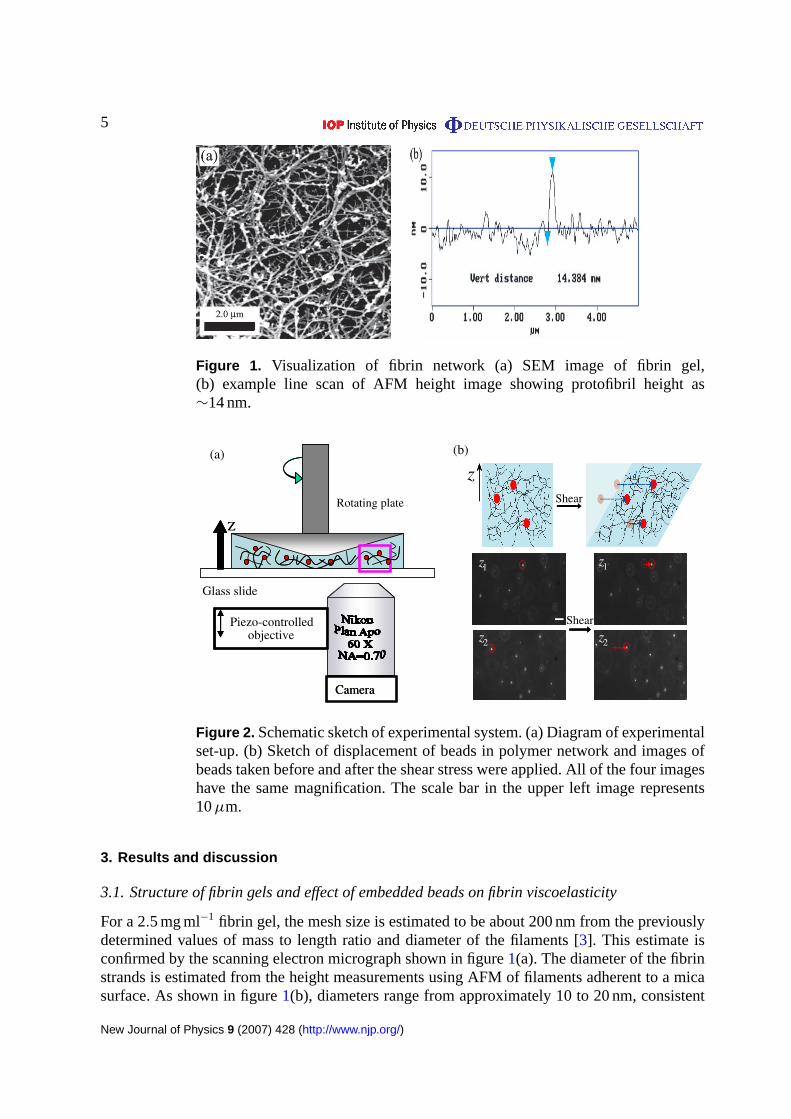
Figure 1. Visualization of fibrin network (a) SEM image of fibrin gel,(b) example line scan of AFM height image showing protofibril height as∼14 nm.
Z
Glass slide
Camera
(a)
Z
Rotating plate
Piezo-controlled objective
Camera
Shear
z1
z2
z1
z2
z(b)
z1
z2
z1
z2
z1z1
z2
z1
z2
z1z1
z2z2
Shear
Figure 2. Schematic sketch of experimental system. (a) Diagram of experimentalset-up. (b) Sketch of displacement of beads in polymer network and images ofbeads taken before and after the shear stress were applied. All of the four imageshave the same magnification. The scale bar in the upper left image represents10µm.
3. Results and discussion
3.1. Structure of fibrin gels and effect of embedded beads on fibrin viscoelasticity
For a 2.5 mg ml−1 fibrin gel, the mesh size is estimated to be about 200 nm from the previouslydetermined values of mass to length ratio and diameter of the filaments [3]. This estimate isconfirmed by the scanning electron micrograph shown in figure1(a). The diameter of the fibrinstrands is estimated from the height measurements using AFM of filaments adherent to a micasurface. As shown in figure1(b), diameters range from approximately 10 to 20 nm, consistent
New Journal of Physics 9 (2007) 428 (http://www.njp.org/)

6
Figure 3. G′ as functions of time and strain for gels with (empty circles)and without (solid circles) beads embedded. (a) Polymerization of fibrin leadsto increase inG′. (b) Stiffness of gels increase with increasing strain valuesindicating the nonlinear elasticity of the gels.
with a network of fibrin protofibrils and very small protofibril bundles. The thickness of thesalmon fibrin strands at physiological values of pH and ionic strength is significantly lowerthan that of fibrin formed from human or bovine fibrinogen as previously shown by turbiditymeasurements [17], and may account for the greater resistance of salmon fibrin to degradationin wound healing applicationsin vivo [21, 22].
The size of fluorescent beads was selected to be larger than the mesh size of the fibrinnetwork so that their Brownian motion is suppressed. Viscoelastic properties of fibrin gels withembedded beads were compared to those without beads. In figure3, G′ increases as a functionof time during polymerization. The added beads do not alter the polymerization rate, since theG′ for gel with beads increases at the same rate as that for the gel without beads. The finalG′
values at 1% strain taken after 1 h for both types of gels were measured to be: 45± 5 Pa. Theseresults suggest that the fluorescent beads are likely to act as tracers for fibrin gel displacementsand do not form defects or additional crosslinks in the network that would alter the gel rheology.
3.2. Non-linear elasticity
As the strain values increase from 1 to 160%, the elastic modulus increases from approximately50 Pa to a maximum value of 900 Pa at about 120% strain (see figure3(b)). The strainsweep curves for gels with and without embedded beads are not statistically distinguishable,confirming further that the beads do not alter gel structure. The sharp decrease inG′ at largerstrains is possibly due to sample failure, i.e. network disruption or network detachment from therheometer plates.
3.3. Negative normal stress
The second normal stress difference, which is in the direction perpendicular to the rheometerplate, has also been recorded. A negative normal stress, as has been reported for other biologicalgels [4], was also observed in the fibrin networks. Results in figure5 show that the magnitude
New Journal of Physics 9 (2007) 428 (http://www.njp.org/)
7
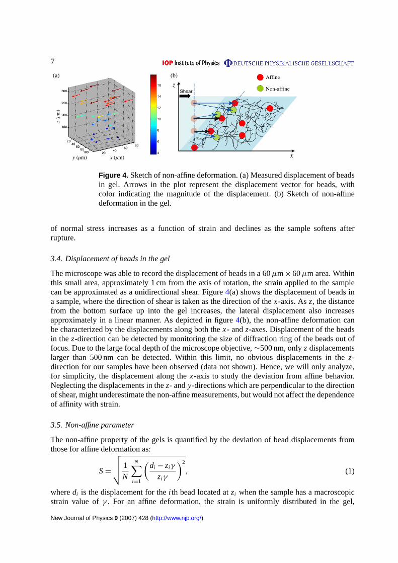
Figure 4. Sketch of non-affine deformation. (a) Measured displacement of beadsin gel. Arrows in the plot represent the displacement vector for beads, withcolor indicating the magnitude of the displacement. (b) Sketch of non-affinedeformation in the gel.
of normal stress increases as a function of strain and declines as the sample softens afterrupture.
3.4. Displacement of beads in the gel
The microscope was able to record the displacement of beads in a 60µm× 60µm area. Withinthis small area, approximately 1 cm from the axis of rotation, the strain applied to the samplecan be approximated as a unidirectional shear. Figure4(a) shows the displacement of beads ina sample, where the direction of shear is taken as the direction of thex-axis. Asz, the distancefrom the bottom surface up into the gel increases, the lateral displacement also increasesapproximately in a linear manner. As depicted in figure4(b), the non-affine deformation canbe characterized by the displacements along both thex- andz-axes. Displacement of the beadsin thez-direction can be detected by monitoring the size of diffraction ring of the beads out offocus. Due to the large focal depth of the microscope objective,∼500 nm, onlyz displacementslarger than 500 nm can be detected. Within this limit, no obvious displacements in thez-direction for our samples have been observed (data not shown). Hence, we will only analyze,for simplicity, the displacement along thex-axis to study the deviation from affine behavior.Neglecting the displacements in thez- andy-directions which are perpendicular to the directionof shear, might underestimate the non-affine measurements, but would not affect the dependenceof affinity with strain.
3.5. Non-affine parameter
The non-affine property of the gels is quantified by the deviation of bead displacements fromthose for affine deformation as:
S=
√√√√ 1
N
N∑i =1
(di − zi γ
zi γ
)2
, (1)
wheredi is the displacement for thei th bead located atzi when the sample has a macroscopicstrain value ofγ . For an affine deformation, the strain is uniformly distributed in the gel,
New Journal of Physics 9 (2007) 428 (http://www.njp.org/)
8
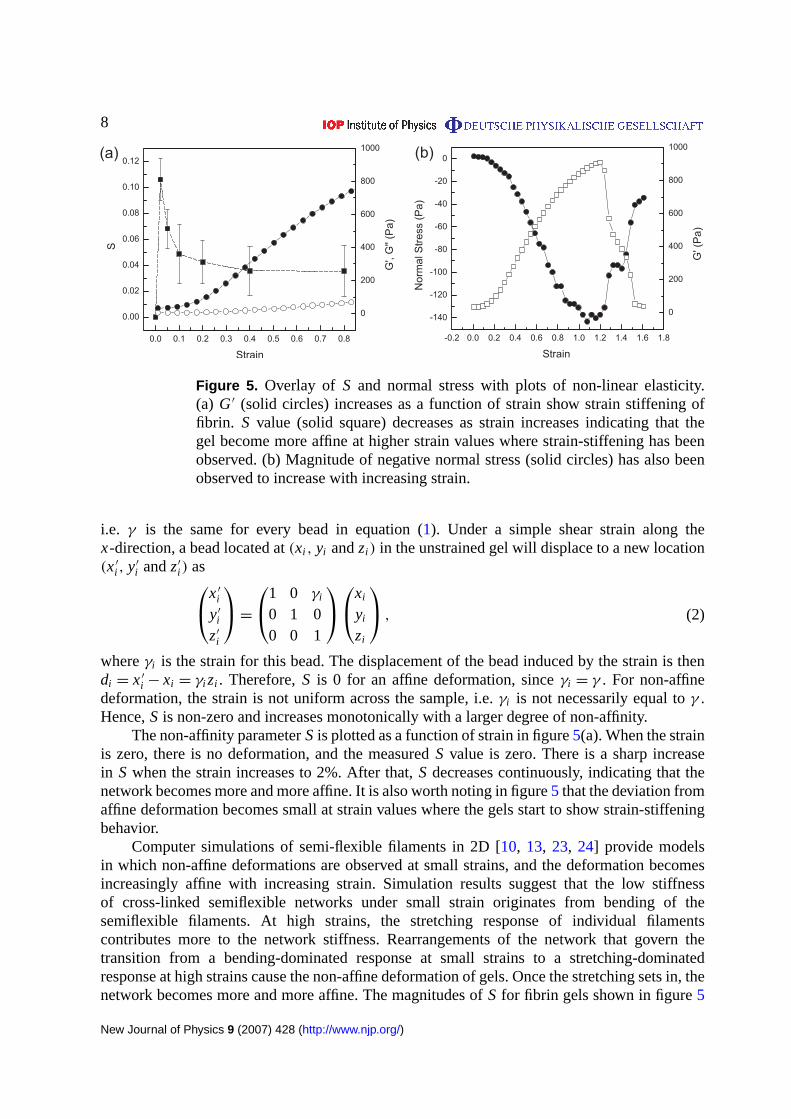
Figure 5. Overlay of S and normal stress with plots of non-linear elasticity.(a) G′ (solid circles) increases as a function of strain show strain stiffening offibrin. S value (solid square) decreases as strain increases indicating that thegel become more affine at higher strain values where strain-stiffening has beenobserved. (b) Magnitude of negative normal stress (solid circles) has also beenobserved to increase with increasing strain.
i.e. γ is the same for every bead in equation (1). Under a simple shear strain along thex-direction, a bead located at(xi , yi andzi ) in the unstrained gel will displace to a new location(x′
i , y′
i andz′
i ) asx′
i
y′
i
z′
i
=
1 0 γi
0 1 00 0 1
xi
yi
zi
, (2)
whereγi is the strain for this bead. The displacement of the bead induced by the strain is thendi = x′
i − xi = γi zi . Therefore,S is 0 for an affine deformation, sinceγi = γ . For non-affinedeformation, the strain is not uniform across the sample, i.e.γi is not necessarily equal toγ .Hence,S is non-zero and increases monotonically with a larger degree of non-affinity.
The non-affinity parameterS is plotted as a function of strain in figure5(a). When the strainis zero, there is no deformation, and the measuredS value is zero. There is a sharp increasein S when the strain increases to 2%. After that,S decreases continuously, indicating that thenetwork becomes more and more affine. It is also worth noting in figure5 that the deviation fromaffine deformation becomes small at strain values where the gels start to show strain-stiffeningbehavior.
Computer simulations of semi-flexible filaments in 2D [10, 13, 23, 24] provide modelsin which non-affine deformations are observed at small strains, and the deformation becomesincreasingly affine with increasing strain. Simulation results suggest that the low stiffnessof cross-linked semiflexible networks under small strain originates from bending of thesemiflexible filaments. At high strains, the stretching response of individual filamentscontributes more to the network stiffness. Rearrangements of the network that govern thetransition from a bending-dominated response at small strains to a stretching-dominatedresponse at high strains cause the non-affine deformation of gels. Once the stretching sets in, thenetwork becomes more and more affine. The magnitudes ofS for fibrin gels shown in figure5
New Journal of Physics 9 (2007) 428 (http://www.njp.org/)
9
are not simply related to similar values derived from the 2D simulations, but are small comparedto values derived for very disperse networks of very stiff filaments.
Negative normal stress has been ascribed to the asymmetric force-elongation relation ofsemi-flexible polymers that leads to an imbalance between forces resisting elongation comparedto compression of the end-to-end vectors of the filaments between network junctions [25]. Inan isotropic network, the number of filaments being stretched is approximately equal to thenumber of filaments being compressed. However, since bending contributes less resistance thanstretching, a net negative normal stress results. At small strains the normal stress is predicted todepend quadradically on strain, consistent with the data in figure5(b). At higher strain values,the negative normal stress increases more slowly with increasing strain until the sample fails.Within the range of strains where normal stress is negative, there was no evidence of verticaldisplacements of the fluorescent beads within the resolution of our measurements.
4. Conclusions
Fibrin networks formed under physiological conditions show properties of strain-stiffening andnegative normal stress in accordance with those reported [3, 25] for somewhat more uniformgels formed exclusively by fibrin protofilaments under non-physiological conditions. The non-linear viscoelastic response of fibrin gels at moderate strain coincides with very low magnitudesof non-affinity measures. The deviation from affine deformation for isotropic fibrin gels issignificantly different from zero, but less than 0.1 at small strains, and it decreases significantlyat higher strains, as the shear modulus increases. Although the relatively highS value at lowstrain could indicate that applied external stress can induce structural reorganization of thenetwork, which provides an explanation to the non-linear elasticity [10], the low S value atmoderate strain values suggests that the assumption of affine deformation is approximatelyapplicable for the strains where strain-stiffening is observed, and supports the use of entropictheories to account for this phenomenon. In a biological context these results also imply thatextracellular matrices like fibrin, which are the first scaffold set up during wound healing, haveisotropic responses to external deformation on the scale of microns measured in this study,even though the network strands have persistence lengths also near a micron. A typical celllike a fibroblast that would be imbedded in a fibrin gel would therefore be subjected to forcesconsistent with the macroscopic stress on the tissue. Highly non-affine stress fields within amatrix would appear to require stiffer filaments with larger mesh sizes such as recently shownfor collagen gels [26] or non-isotropic distributions in order to achieve spatially ordered stressesthat would dictate cell responses on a micron scale.
Acknowledgment
This work was supported by grant DMR05-20020 from the National Science Foundation.
References
[1] Kasai M, Kawashima H and Oosawa F 1960 Structure of f-actin solutionsJ. Polym. Sci.4451–69[2] Leterrier J F, Kas J, Hartwig J, Vegners R and Janmey P A 1996 Mechanical effects of neurofilament cross-
bridges modulation by phosphorylation, lipids, and interactions with f-actinJ. Biol. Chem.27115687–94
New Journal of Physics 9 (2007) 428 (http://www.njp.org/)

10
[3] Storm C, Pastore J J, MacKintosh F C, Lubensky T C and Janmey P A 2005 Nonlinear elasticity in biologicalgelsNature435191–4
[4] Janmey P A, McCormick M E, Rammensee S, Leight J L, Georges P C and MacKintosh F C 2007 Negativenormal stress in semiflexible biopolymer gelsNat. Mater.6 48–51
[5] Kreplak L, Bar H, Leterrier J F, Herrmann H and Aebi U 2005 Exploring the mechanical behavior of singleintermediate filamentsJ. Mol. Biol.354569–77
[6] Gittes F, Mickey B, Nettleton J and Howard J 1993 Flexural rigidity of microtubules and actin filamentsmeasured from thermal fluctuations in shapeJ. Cell. Biol.120923–34
[7] MacKintosh F C, Kas J and Janmey P A 1995 Elasticity of semiflexible biopolymer networksPhys. Rev. Lett.754425–8
[8] Hinner B, Tempel M, Sackmann E, Kroy K and Frey E 1998 Entanglement, elasticity and viscous relaxationof actin solutionsPhys. Rev. Lett.812614
[9] Kroy K and Frey E 1997 Dynamic scattering from solutions of semiflexible polymersPhys. Rev.E 553092[10] Onck P R, Koeman T, van Dillen T and van der Giessen E 2005 Alternative explanation of stiffening in
cross-linked semiflexible networksPhys. Rev. Lett.95178102[11] Satcher R L Jr and Dewey C F Jr 1996 Theoretical estimates of mechanical properties of the endothelial cell
cytoskeletonBiophys. J.71109–18[12] Liu J, Koenderink G H, Kasza K E, MacKintosh F C and Weitz D A 2007 Visualizing the strain field in
semiflexible polymer networks: strain fluctuations and nonlinear rheology of f-actin gelsPhys. Rev. Lett.98198304
[13] Didonna B and Lubensky T 2005 Nonaffinity and nonlinearity in random elastic networksPhys. Rev.E 72066619
[14] Weisel J W 2004 The mechanical properties of fibrin for basic scientists and cliniciansBiophys. Chem.112267–76
[15] Gerth C, Roberts W W and Ferry J D 1974 Rheology of fibrin clots. II. Linear viscoelastic behavior in shearcreepBiophys. Chem.2 208–17
[16] Shah J V and Janmey P A 1997 Strain hardening of fibrin gels and plasma clotsRheol. Acta36262–8[17] Wang L Z, Gorlin J, Michaud S E, Janmey P A, Goddeau R P, Kuuse R, Uibo R, Adams D and Sawyer E S
2000 Purification of salmon clotting factors and their use as tissue sealantsThromb. Res.100537–48[18] Michaud S E 2002 Purification of salmon thrombin and its potential as an alternative to mammalian thrombins
in fibrin sealantsThromb. Res.107245–54[19] Langer B G, Weisel J W, Dinauer P A, Nagaswami C and Bell W R 1988 Deglycosylation of fibrinogen
accelerates polymerization and increases lateral aggregation of fibrin fibersJ. Biol. Chem.26315056–63[20] Crocker J C and Grier D G 1996 Methods of digital video microscopy for colloidal studiesJ. Colloid Interface
Sci.179298[21] Rothwell S W, Reid T J, Dorsey J, Flournoy W S, Bodo M, Janmey P A and Sawyer E 2005 A salmon
thrombin-fibrin bandage controls arterial bleeding in a swine aortotomy modelJ. Trauma59143–9[22] Ju Y E, Janmey P A, McCormick M E, Sawyer E S and Flanagan L A 2007 Enhanced neurite growth from
mammalian neurons in three-dimensional salmon fibrin gelsBiomaterials282097–108[23] Head D A, Levine A J and MacKintosh F C 2003 Deformation of cross-linked semiflexible polymer networks
Phys. Rev. Lett.91108102[24] Head D A, Levine A J and MacKintosh F C 2003 Distinct regimes of elastic response and deformation modes
of cross-linked cytoskeletal and semiflexible polymer networksPhys. Rev.E 68061907[25] Levental I, Georges P C and Janmey P A 2007 Soft biological materials and their impact on cell functionSoft
Matter1 299–306[26] Chandran P L and Barocas V H 2006 Affine versus non-affine fibril kinematics in collagen networks:
theoretical studies of network behaviorJ. Biomech. Eng.128259–70
New Journal of Physics 9 (2007) 428 (http://www.njp.org/)