LncRNA MEG3 accelerates apoptosis of hypoxic myocardial ... · LncRNA MEG3 accelerates apoptosis of...
7
334 Abstract. – OBJECTIVE: To explore the ef- fects of long non-coding ribonucleic acid (ln- cRNA) maternally expressed gene 3 (MEG3) on the proliferation and apoptosis of hypoxic myo- cardial cells by regulating the expression of forkhead box O1 (FoxO1). MATERIALS AND METHODS: The myocar- dial A10 cell lines were divided into myocardi- al cell group (group A), hypoxic myocardial cell group (group B), and hypoxic myocardial cell + transfection with lncRNA MEG3 mimic group (group C). The correlations of the adenosine tri- phosphate (ATP) concentration, the degree of apoptosis, and the proliferation with FoxO1 and FoxO3a proteins in the cells were observed via ATP assay, Cell Counting Kit-8 (CCK-8) assay, terminal deoxynucleotidyl transferase-mediated dUTP nick end labeling (TUNEL) assay, West- ern blotting, and quantitative Reverse Transcrip- tion-Polymerase Chain Reaction (qRT-PCR), re- spectively. RESULTS: The ATP concentration in myocar- dial cells was the highest in group A ( p <0.05), and it was higher in group B than that in group C ( p <0.05). The results of the CCK-8 assay showed that the proliferation rate of the myo- cardial cells was the highest in group A and the lowest in group C ( p <0.05), and it was signifi- cantly increased in group B compared with that in group C ( p <0.05). The results of the TUNEL assay revealed that the normal cells displayed the purple color, and apoptotic cells displayed the green color. The myocardial cells were ar- ranged orderly, and the number of apoptot- ic cells was smaller in group A, the number of apoptotic cells was significantly larger in group B than that in group A, and it was the largest in group C ( p <0.05). Moreover, the re- sults of Western blotting manifested that the concentrations of FoxO1 and FoxO3 proteins in myocardial cells were the lowest in group A ( p <0.05), and they were significantly higher in group C than those in group B ( p <0.05). Ac- cording to the results of qRT-PCR, the mRNA expressions of FoxO1 and FoxO3 in myocar- dial cells were the lowest in group A ( p <0.05), and they were remarkably lower in group B than those in group C ( p <0.05). CONCLUSIONS: LncRNA MEG3 can increase the activity of FoxO1 to promote myocardial apoptosis in a hypoxic environment. Key Words: Hypoxic myocardial cells, FoxO1, LncRNA MEG3. Introduction Oxygen is one of the necessary factors to main- tain the normal vital movement of the human body, which can convert such nutrients as glucose and protein into energy. However, the heart, brain, kidney, and other organs will be damaged in var- ious degrees in the case of severe hypoxia. When the oxygen is insufficient to satisfy the demand of oxygen supply in myocardial cells, the myo- cardial necrosis and apoptosis will occur, and the weakened oxygen metabolism enhanced anaero- bic glycolysis and decreased cardiac output are the main characteristics of myocardial hypoxia 1,2 . These characteristics will, if they persist, aggra- vate the body’s damage and lead to common clin- ical complications, such as angina, coronary heart disease, and myocardial infarction. Therefore, it is extremely important to deeply understand and explore the causes and therapeutic mechanism of apoptosis of hypoxic myocardial cells, and it is also of great significance to control the myocar - dial apoptosis and increase the viability and sur- vival time of myocardial cells to the maximum degree in the increase of the survival rate of the myocardial cells in hypoxic environment 3 . The forkhead box O (FoxO) family widely existing in organisms is expressed in different organs, such as the brain, heart, and kidney. FoxO1 can move European Review for Medical and Pharmacological Sciences 2019; 23 (3 Suppl): 334-340 L.-Y. ZHAO 1 , X. LI 2 , L. GAO 3 , Y. XU 2 1 Medical Treatment Center, Weifang People’s Hospital Brain Hospital, Weifang, China 2 Department of Health Care, Weifang Second People’s Hospital, Weifang, China 3 Department of Emergency, Anqiu People’s Hospital, Weifang, China Corresponding Author: Ying Xu, MM; e-mail: [email protected] LncRNA MEG3 accelerates apoptosis of hypoxic myocardial cells via FoxO1 signaling pathway
Transcript of LncRNA MEG3 accelerates apoptosis of hypoxic myocardial ... · LncRNA MEG3 accelerates apoptosis of...
334
Abstract. – OBJECTIVE: To explore the ef-fects of long non-coding ribonucleic acid (ln-cRNA) maternally expressed gene 3 (MEG3) on the proliferation and apoptosis of hypoxic myo-cardial cells by regulating the expression of forkhead box O1 (FoxO1).
MATERIALS AND METHODS: The myocar-dial A10 cell lines were divided into myocardi-al cell group (group A), hypoxic myocardial cell group (group B), and hypoxic myocardial cell + transfection with lncRNA MEG3 mimic group (group C). The correlations of the adenosine tri-phosphate (ATP) concentration, the degree of apoptosis, and the proliferation with FoxO1 and FoxO3a proteins in the cells were observed via ATP assay, Cell Counting Kit-8 (CCK-8) assay, terminal deoxynucleotidyl transferase-mediated dUTP nick end labeling (TUNEL) assay, West-ern blotting, and quantitative Reverse Transcrip-tion-Polymerase Chain Reaction (qRT-PCR), re-spectively.
RESULTS: The ATP concentration in myocar-dial cells was the highest in group A (p<0.05), and it was higher in group B than that in group C (p<0.05). The results of the CCK-8 assay showed that the proliferation rate of the myo-cardial cells was the highest in group A and the lowest in group C (p<0.05), and it was signifi-cantly increased in group B compared with that in group C (p<0.05). The results of the TUNEL assay revealed that the normal cells displayed the purple color, and apoptotic cells displayed the green color. The myocardial cells were ar-ranged orderly, and the number of apoptot-ic cells was smaller in group A, the number of apoptotic cells was significantly larger in group B than that in group A, and it was the largest in group C (p<0.05). Moreover, the re-sults of Western blotting manifested that the concentrations of FoxO1 and FoxO3 proteins in myocardial cells were the lowest in group A (p<0.05), and they were significantly higher in group C than those in group B (p<0.05). Ac-cording to the results of qRT-PCR, the mRNA expressions of FoxO1 and FoxO3 in myocar-
dial cells were the lowest in group A (p<0.05), and they were remarkably lower in group B than those in group C (p<0.05).
CONCLUSIONS: LncRNA MEG3 can increase the activity of FoxO1 to promote myocardial apoptosis in a hypoxic environment.
Key Words:Hypoxic myocardial cells, FoxO1, LncRNA MEG3.
Introduction
Oxygen is one of the necessary factors to main-tain the normal vital movement of the human body, which can convert such nutrients as glucose and protein into energy. However, the heart, brain, kidney, and other organs will be damaged in var-ious degrees in the case of severe hypoxia. When the oxygen is insufficient to satisfy the demand of oxygen supply in myocardial cells, the myo-cardial necrosis and apoptosis will occur, and the weakened oxygen metabolism enhanced anaero-bic glycolysis and decreased cardiac output are the main characteristics of myocardial hypoxia1,2. These characteristics will, if they persist, aggra-vate the body’s damage and lead to common clin-ical complications, such as angina, coronary heart disease, and myocardial infarction. Therefore, it is extremely important to deeply understand and explore the causes and therapeutic mechanism of apoptosis of hypoxic myocardial cells, and it is also of great significance to control the myocar-dial apoptosis and increase the viability and sur-vival time of myocardial cells to the maximum degree in the increase of the survival rate of the myocardial cells in hypoxic environment3. The forkhead box O (FoxO) family widely existing in organisms is expressed in different organs, such as the brain, heart, and kidney. FoxO1 can move
European Review for Medical and Pharmacological Sciences 2019; 23 (3 Suppl): 334-340
L.-Y. ZHAO1, X. LI2, L. GAO3, Y. XU2
1Medical Treatment Center, Weifang People’s Hospital Brain Hospital, Weifang, China2Department of Health Care, Weifang Second People’s Hospital, Weifang, China3Department of Emergency, Anqiu People’s Hospital, Weifang, China
Corresponding Author: Ying Xu, MM; e-mail: [email protected]
LncRNA MEG3 accelerates apoptosis of hypoxic myocardial cells via FoxO1 signaling pathway
LncRNA MEG3 accelerates apoptosis of hypoxic myocardial cells via FoxO1 signaling pathway
335
freely inside and outside the cells and plays an im-portant role in myocardial cell growth, apoptosis, and oxidative stress4. In the hypoxic environment, FoxO1 can promote apoptosis, lead to massive autophagy, and induce the functional changes in the myocardial cells. Moreover, the increased activity of FoxO1 will aggravate the oxygen free radical injury in myocardial cells, resulting in the decline in the tolerance of them. The complete op-posite is the case in which the activity of FoxO1 declines. Long non-coding ribonucleic acid (ln-cRNA) maternally expressed gene 3 (MEG3) is a cancer suppressor gene constantly expressed in many normal organs and tissues, but its expres-sion is deleted or declines in a variety of tumor tissues5. Studies have demonstrated that lncRNA MEG3 can increase the activity of FoxO1 to pro-mote the apoptosis and destroy the cells under hypoxia-ischemia of myocardial cells. Besides, the decreased expression of lncRNA MEG3 can reduce the apoptosis of hypoxic myocardial cells6. In this paper, the effects of lncRNA MEG3 on the proliferation and apoptosis of hypoxic myocardial cells by regulating the expression of FoxO1 were explored in the hypoxic environment.
Materials and Methods
Materials, Reagents, and InstrumentsThe myocardial A10 cell lines were purchased
from American Type Culture Collection (ATCC) (Manassas, VA, USA), adenosine triphosphate (ATP) antibody, FoxO1 and FoxO3a antibodies, phosphorylated FoxO1 and FoxO3a and GAPDH antibodies from BD (Franklin Lakes, NJ, USA), trypsin and Dulbecco’s Modified Eagle’s Medi-um (DMEM) from Yiheng (Shanghai, China), RNA-iMAX from IKa (Staufen, Germany), and Cell Counting Kit-8 (CCK-8) kit from Proteintech (Wuhan, China).
Cell Culture and Grouping in Three Groups
The myocardial A10 cell lines were added with erythromycin and penicillin (80 mg/mL) and in-cubated in the complete medium containing 12% fetal bovine serum (FBS; Gibco, Rockville, MD, USA) at 37.5°C. The medium was replaced once every 24 h. When 90% of A10 cells were fused, a certain number of myocardial cells in the growth stage were taken and cultured under hypoxia, ac-cording to the literature3. The cells were divided into myocardial cell group (group A), hypoxic
myocardial cell group (group B), and hypox-ic myocardial cell + transfection with lncRNA MEG3 mimic (according to the literature7) group (group C).
ATP AssayA small number of myocardial cells were tak-
en, added with 150 μL of lysis buffer, fully stirred, and centrifuged for 10 min, and the supernatant was retained for later use. A small amount of ATP reagent was diluted at 5-fold to be the ATP assay working solution and stored under low tempera-ture. Then, 50 μL of ATP assay working solution was added into a 96-well plate and placed at 37°C for 10 min, and 30 μL of supernatant was added. After 10 s, the relative light unit (RLU) value was detected and recorded using a multifunctional mi-croplate reader, based on which the ATP concen-tration was calculated.
CCK-8 Assay of Cell ProliferationThe myocardial cells were inoculated into the
96-well plate (100 μL/well) and pre-incubated in the incubator with 8% CO2 at 37°C. After that, 15 μL of lncRNA MEG3 solution was added into each well, followed by incubation in the incuba-tor for 1-4 h. Then, the optical density (OD) val-ue was measured at 450 nm using the microplate reader. If the OD value was not measured tempo-rarily, 10 μL of 0.1 M HCl solution or 1% (w/v) SDS solution was added into each well, and the culture plate was stored at 37°C in a dark place. The OD value will not change if measured within 24 h.
TUNEL Assay of Myocardial ApoptosisThe myocardial cells were centrifuged and
re-paved into the 96-well plate. After adherent growth overnight, the cells were fixed with 4% paraformaldehyde for 20 min, permeabilized with Triton X-100 for 15 min and stained with TUNEL to detect myocardial apoptosis in each group in strict accordance with the instructions. The cells in each group were observed in five non-crossed and non-repeated fields randomly selected under a microscope. The myocardial apoptosis rate = apoptotic cells/total cells × 100%.
Analysis of FoxO1 and FoxO3a Protein Expressions Via Western Blotting
The cells in a 6-well plate were digested with 100 μg of trypsin extract, and then the digestion was terminated with 2 mL of medium. The myo-cardial cell extract was placed into the Eppendorf
L.-Y. Zhao, X. Li, L. Gao, Y. Xu
336
(EP) tube, mixed with trypsin extract at 1:100, and frozen in a refrigerator for 10 min. After com-plete lysis of cells, the E solution was obtained. The myocardial cells were placed into the EP tube, mixed with 2 mL of trypsin extract at 1:100. After complete lysis of cells, the F solution was obtained. Then, E solution and F solution were mixed evenly at 80:1, prepared into a working solution and placed in the incubator at 37.5°C for 20 min. After cooling, the protein concentration was calculated.
Detection of FoxO1 and FoxO3a Activity Via Quantitative Reverse Transcription-Polymerase Chain Reaction (qRT-PCR)
The myocardial cells were digested with trypsin, washed with Phosphate-Buffered Sa-line (PBS), and added with 1.5 mL of lncRNA MEG3 vector reagent to extract RNA. Then, 30 mg of cell suspension was placed into the EP tube, and the total mRNA was extracted using TRIzol (Invitrogen, Carlsbad, CA, USA) and reversely transcribed into cDNA, followed by RT-PCR amplification with glyceraldehyde 3-phosphate dehydrogenase (GAPDH) as an in-ternal reference (98°C for 6 min, 98°C for 28 s, 75°C for 30 s, and 80°C for 4 min, a total of 55 cycles). The primer sequences are shown in Table I.
Statistical AnalysisThe Statistical Product and Service Solutions
(SPSS) 17.0 software (SPSS Inc., Chicago, IL, USA) was used for the statistical analysis of the experimental data. The t-test was performed for the differences in the myocardial cell viability, cell proliferation rate, degree of apoptosis, FoxO1, and FoxO3a activity among group A, B, and C. The measurement data were expressed as mean ± standard deviation. The comparison between groups was done using the One-way ANOVA test followed by the post-hoc test (Least Signifi-
cant Difference). Spearman rank correlation co-efficient was adopted for the correlation analysis. p<0.01 suggested statistically significant differ-ences.
Results
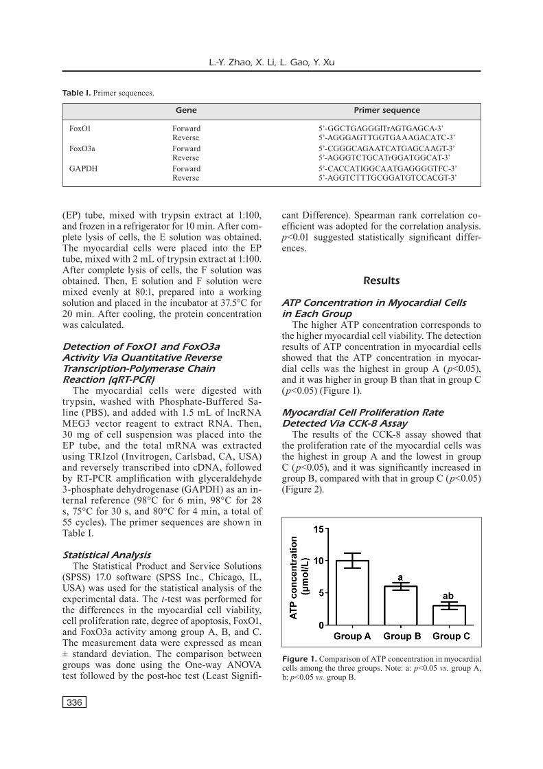
ATP Concentration in Myocardial Cells in Each Group
The higher ATP concentration corresponds to the higher myocardial cell viability. The detection results of ATP concentration in myocardial cells showed that the ATP concentration in myocar-dial cells was the highest in group A (p<0.05), and it was higher in group B than that in group C (p<0.05) (Figure 1).
Myocardial Cell Proliferation Rate Detected Via CCK-8 Assay
The results of the CCK-8 assay showed that the proliferation rate of the myocardial cells was the highest in group A and the lowest in group C (p<0.05), and it was significantly increased in group B, compared with that in group C (p<0.05) (Figure 2).
Table I. Primer sequences.
Gene Primer sequence
FoxO1 Forward 5’-GGCTGAGGGlTrAGTGAGCA-3’ Reverse 5’-AGGGAGTTGGTGAAAGACATC-3’FoxO3a Forward 5’-CGGGCAGAATCATGAGCAAGT-3’ Reverse 5’-AGGGTCTGCATrGGATGGCAT-3’GAPDH Forward 5’-CACCATIGGCAATGAGGGGTFC-3’ Reverse 5’-AGGTCTTTGCGGATGTCCACGT-3’
Figure 1. Comparison of ATP concentration in myocardial cells among the three groups. Note: a: p<0.05 vs. group A, b: p<0.05 vs. group B.

LncRNA MEG3 accelerates apoptosis of hypoxic myocardial cells via FoxO1 signaling pathway
337
Myocardial Apoptosis Detected Via TUNEL Assay
The results of the TUNEL assay revealed that apoptotic cells displayed the green color, and the blue color indicated the nuclei. The number of apoptotic cells was smaller in group A, the num-ber of apoptotic cells was significantly larger in group B than that in group A, and it was the larg-est in group C (p<0.05) (Figure 3).
FoxO1 and FoxO3a Protein Expressions in Myocardial Cells Detected Via Western Blotting
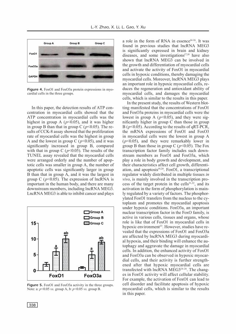
The results of the Western blotting manifest-ed that the concentrations of FoxO1 and FoxO3a proteins in myocardial cells were the lowest in group A (p<0.05), and they were significantly higher in group C than those in group B (p<0.05) (Figure 4).
FoxO1 and FoxO3a mRNA Expressions in Myocardial Cells Detected Via qRT-PCR
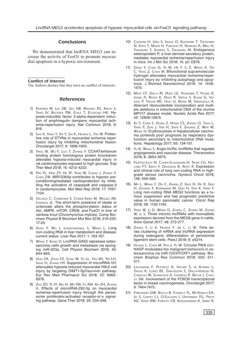
According to the results of qRT-PCR, the mRNA expressions of FoxO1 and FoxO3 in myo-cardial cells were the lowest in group A (p<0.05), and they were significantly lower in group B than those in group C (p<0.05) (Figure 5).
Discussion
Myocardial hypoxia refers to the pathologi-cal process of the myocardial tissues caused by the abnormal changes in tissue metabolism, cell function, and morphological structure in a hy-poxic environment due to the decline in oxygen supply in cells8,9. Hypoxia is not an independent disease but exists in a variety of diseases, whose pathological process is similar in the body. In the clinic, hypoxia is the direct cause of many dis-eases10, which may lead to death in severe cases. Myocardial hypoxia is one of the important caus-es of long-term progressive apoptosis of myocar-dial cells. According to the previous analysis, the persistent myocardial hypoxia will only result in cell necrosis, but there are many experimental data indicating that the long-term myocardial hy-poxia is a major cause of myocardial apoptosis11. Studies have demonstrated that the mitochondria are one of the most important elements in myo-cardial cells to maintain the body function, and they participate in the growth and metabolism of myocardial cells. However, the mitochondrial structure is susceptible to damage under hypoxic conditions, manifested as the rupture and disor-der inside, accelerating apoptosis12,13. Therefore, it is a priority to ensure the sufficient oxygen supply to the heart in time during treatment.
Figure 2. Myocardial cell proliferation rate in the three groups. Note: a: p<0.05 vs. group A, b: p<0.05 vs. group B.
Figure 3. Myocardial apoptosis in the three groups (magnification: 100×).
L.-Y. Zhao, X. Li, L. Gao, Y. Xu
338
In this paper, the detection results of ATP con-centration in myocardial cells showed that the ATP concentration in myocardial cells was the highest in group A (p<0.05), and it was higher in group B than that in group C (p<0.05). The re-sults of CCK-8 assay showed that the proliferation rate of myocardial cells was the highest in group A and the lowest in group C (p<0.05), and it was significantly increased in group B, compared with that in group C (p<0.05). The results of the TUNEL assay revealed that the myocardial cells were arranged orderly and the number of apop-totic cells was smaller in group A, the number of apoptotic cells was significantly larger in group B than that in group A, and it was the largest in group C (p<0.05). The expression of lncRNA is important in the human body, and there are many downstream members, including lncRNA MEG3. LncRNA MEG3 is able to inhibit cancer and plays
a role in the form of RNA in essence14-16. It was found in previous studies that lncRNA MEG3 is significantly expressed in brain and kidney diseases, and some investigations17,18 have also shown that lncRNA MEG3 can be involved in the growth and differentiation of myocardial cells and activate the activity of FoxO1 in myocardial cells in hypoxic conditions, thereby damaging the myocardial cells. Moreover, lncRNA MEG3 plays an important role in hypoxic myocardial cells, re-duces the regeneration and antioxidant ability of myocardial cells, and damages the myocardial cells, which is similar to the results in this paper.
In the present study, the results of Western blot-ting manifested that the concentrations of FoxO1 and FoxO3a proteins in myocardial cells were the lowest in group A (p<0.05), and they were sig-nificantly higher in group C than those in group B (p<0.05). According to the results of qRT-PCR, the mRNA expressions of FoxO1 and FoxO3 in myocardial cells were the lowest in group A (p<0.05), and they were remarkably lower in group B than those in group C (p<0.05). The Fox transcription factor family includes such down-stream members as FoxO1 and FoxO3a, which play a role in body growth and development, and their characteristics affect cell growth, differenti-ation, and apoptosis19,20. FoxO1, a transcriptional regulator widely distributed in multiple tissues in vivo, is mainly involved in the transcription pro-cess of the target protein in the cells21,22, and its activation in the form of phosphorylation is main-ly regulated by a variety of factors. The phosphor-ylated FoxO1 transfers from the nucleus to the cy-toplasm and promotes the myocardial apoptosis under hypoxic conditions. FoxO3a, an important nuclear transcription factor in the FoxO family, is active in various cells, tissues and organs, whose role is like that of FoxO1 in myocardial cells in hypoxic environment23. However, studies have re-vealed that the expressions of FoxO1 and FoxO3a are affected by lncRNA MEG3 during myocardi-al hypoxia, and their binding will enhance the au-tophagy and aggravate the damage in myocardial cells. In addition, the enhanced activity of FoxO1 and FoxO3a can be observed in hypoxic myocar-dial cells, and their activity is further strength-ened after that hypoxic myocardial cells are transfected with lncRNA MEG324-26. The chang-es in FoxO1 activity will affect cellular stability. For example, the activation of FoxO1 can lead to cell disorder and facilitate apoptosis of hypoxic myocardial cells, which is similar to the results in this paper.
Figure 4. FoxO1 and FoxO3a protein expressions in myo-cardial cells in the three groups.
Figure 5. FoxO1 and FoxO3a activity in the three groups. Note: a: p<0.05 vs. group A, b: p<0.05 vs. group B.
LncRNA MEG3 accelerates apoptosis of hypoxic myocardial cells via FoxO1 signaling pathway
339
Conclusions
We demonstrated that lncRNA MEG3 can in-crease the activity of FoxO1 to promote myocar-dial apoptosis in a hypoxic environment.
Conflict of InterestThe Authors declare that they have no conflict of interests.
References
1) Koeppen M, Lee JW, Seo SW, BrodSKy KS, Kreth S, yang IV, ButtrIcK pM, ecKLe t, eLtzSchIg hK. Hy-poxia-inducible factor 2-alpha-dependent induc-tion of amphiregulin dampens myocardial isch-emia-reperfusion injury. Nat Commun 2018; 9: 816.
2) Sun X, yang y, XIe y, ShI X, huang L, tan W. Protec-tive role of STVNa in myocardial ischemia reper-fusion injury by inhibiting mitochondrial fission. Oncotarget 2017; 9: 1898-1905.
3) yang W, Wu F, Luo t, zhang y. CCAAT/enhancer binding protein homologous protein knockdown alleviates hypoxia-induced myocardial injury in rat cardiomyocytes exposed to high glucose. Exp Ther Med 2018; 15: 4213-4222.
4) pan yL, han zy, he SF, yang W, cheng J, zhang y, chen zW. MiR133b5p contributes to hypoxic pre-conditioningmediated cardioprotection by inhib-iting the activation of caspase8 and caspase-3 in cardiomyocytes. Mol Med Rep 2018; 17: 7097-7104.
5) VeLaSco c, coMeSana S, conde-SIeIra M, MIguez JM, SoengaS JL. The short-term presence of oleate or octanoate alters the phosphorylation status of Akt, AMPK, mTOR, CREB, and FoxO1 in liver of rainbow trout (Oncorhynchus mykiss). Comp Bio-chem Physiol B Biochem Mol Biol 2018; 219-220: 17-25.
6) zhao y, Wu J, LIangpunSaKuL S, Wang L. Long non-coding RNA in liver metabolism and disease: current status. Liver Res 2017; 1: 163-167.
7) Wang y, Kong d. LncRNA GAS5 represses osteo-sarcoma cells growth and metastasis via spong-ing miR-203a. Cell Physiol Biochem 2018; 45: 844-855.
8) guo zX, zhou Fz, Song W, yu LL, yan WJ, yIn Lh, Sang h, zhang hy. Suppression of microRNA-101 attenuates hypoxia-induced myocardial H9c2 cell injury by targeting DIMT1-Sp1/survivin pathway. Eur Rev Med Pharmacol Sci 2018; 22: 6965-6976.
9) zhu zd, ye Jy, nIu h, Ma yM, Fu XM, XIa zh, zhang X. Effects of microRNA-292-5p on myocardial ischemia-reperfusion injury through the peroxi-some proliferator-activated receptor-α/-γ signal-ing pathway. Gene Ther 2018; 25: 234-248.
10) chadanI h, uSuI S, Inoue o, KuSayaMa t, taKaShIMa SI, Kato t, MuraI h, FuruSho h, noMura a, MISu h, taKaMura t, KaneKo S, taKaMura M. Endogenous selenoprotein P, a liver-derived secretory protein, mediates myocardial ischemia/reperfusion injury in mice. Int J Mol Sci 2018; 19. pii: E878.
11) deng y, chen g, ye M, he y, LI z, Wang X, ou c, yang z, chen M. Bifunctional supramolecular hydrogel alleviates myocardial ischemia/reper-fusion injury by inhibiting autophagy and apop-tosis. J Biomed Nanotechnol 2018; 14: 1458-1470.
12) MoSS cF, daLLa rI, hunt Le, yaSuKaWa t, young r, JoneS a, reddy K, deSaI r, VIrtue S, eLgar g, Vo-ShoL p, tayLor MS, hoLt IJ, reIJnS M, SpInazzoLa a. Aberrant ribonucleotide incorporation and multi-ple deletions in mitochondrial DNA of the murine MPV17 disease model. Nucleic Acids Res 2017; 45: 12808-12815.
13) Ke S, chen S, dong z, hong cS, zhang Q, tang L, yang p, zhaI J, yan h, Shen F, zhuang z, Wen W, Wang h. Erythrocytosis in hepatocellular carcino-ma portends poor prognosis by respiratory dys-function secondary to mitochondrial DNA muta-tions. Hepatology 2017; 65: 134-151.
14) yu B, Wang S. Angio-lncRs: lncRNAs that regulate angiogenesis and vascular disease. Theranostics 2018; 8: 3654-3675.
15) FILIppoV-LeVy n, cohen-SchuSSheIM h, trope cg, het-Land Ft, SMIth y, daVIdSon B, reIch r. Expression and clinical role of long non-coding RNA in high-grade serous carcinoma. Gynecol Oncol 2018; 148: 559-566.
16) Ma L, Wang F, du c, zhang z, guo h, XIe X, gao h, zhuang y, KornMann M, gao h, tIan X, yang y. Long non-coding RNA MEG3 functions as a tu-mour suppressor and has prognostic predictive value in human pancreatic cancer. Oncol Rep 2018; 39: 1132-1140.
17) yang W, LI d, Wang g, zhang c, zhang M, zhang W, LI S. Three intronic lncRNAs with monoallelic expression derived from the MEG8 gene in cattle. Anim Genet 2017; 48: 272-277.
18) zheng y, LI X, huang y, JIa L, LI W. Time se-ries clustering of mRNA and lncRNA expression during osteogenic differentiation of periodontal ligament stem cells. PeerJ 2018; 6: e5214.
19) huang L, chen M, pan J, yu W. Circular RNA circ-NASP modulates the malignant behaviors in os-teosarcoma via miR-1253/FOXF1 pathway. Bio-chem Biophys Res Commun 2018; 500: 511-517.
20) LaLLeMand F, petItaLot a, Vacher S, de KonIng L, taouIS K, Lopez BS, zInn-JuStIn S, daLLa-VenezIa n, cheMLaLI W, SchnItzLer a, LIdereau r, BIeche I, capu-to SM. Involvement of the FOXO6 transcriptional factor in breast carcinogenesis. Oncotarget 2017; 9: 7464-7475.
21) FernandeS gW, Bocco B, FonSeca tL, McanInch ea, Jo S, Lartey LJ, o-SuLLIVan I, unterMan tg, preIte nz, VoIgt rM, ForSyth cB, KeShaVarzIan a, SInKo r,
L.-Y. Zhao, X. Li, L. Gao, Y. Xu
340
goLdFIne aB, pattI Me, rIBeIro Mo, gereBen B, BI-anco ac. The Foxo1-inducible transcriptional re-pressor zfp125 causes hepatic steatosis and hy-percholesterolemia. Cell Rep 2018; 22: 523-534.
22) StaLLIngS ce, eLLSWorth BS. Premature expression of FOXO1 in developing mouse pituitary results in anterior lobe hypoplasia. Endocrinology 2018; 159: 2891-2904.
23) pan S, deng y, Fu J, zhang y, zhang z, ru X, QIn X. Decreased expression of ARHGAP15 promotes the development of colorectal cancer through PTEN/AKT/FOXO1 axis. Cell Death Dis 2018; 9: 673.
24) KInyua aW, Ko cM, doan KV, yang dJ, huynh M, Moh Sh, choI yh, KIM KW. 4-hydroxy-3-me-
thoxycinnamic acid regulates orexigenic pep-tides and hepatic glucose homeostasis through phosphorylation of FoxO1. Exp Mol Med 2018; 50: e437.
25) LI W, yI J, zheng X, LIu S, Fu W, ren L, LI L, hoon dSB, Wang J, du g. MiR-29c plays a suppres-sive role in breast cancer by targeting the TIMP3/STAT1/FOXO1 pathway. Clin Epigenetics 2018; 10: 64.
26) LIu t, Ma L, zheng z, LI F, LIu S, XIe y, LI g. Res-veratrol inhibits age-dependent spontaneous tumorigenesis by SIRT1-mediated post-trans-lational modulations in the annual fish Notho-branchius guentheri. Oncotarget 2017; 8: 55422-55434.