LN Urinalysis Final
138
LECTURE NOTES For Medical Laboratory Technology Students Urinalysis Assamenew Kassa, B.S c. Mistir Wolde, B.Sc. Belayhun Kibret, M.Sc. University of Gondar In collaboration with the Ethiopia Public Health Training Initiative, The Carter Center, the Ethiopia Ministry of Health, and the Ethiopia Ministry of Education November 2002
-
Upload
mohsen-haleem -
Category
Documents
-
view
236 -
download
0
Transcript of LN Urinalysis Final
8/13/2019 LN Urinalysis Final
http://slidepdf.com/reader/full/ln-urinalysis-final 1/138
LECTURE NOTES
For Medical Laboratory Technology Students
Urinalysis
Assamenew Kassa, B.Sc.Mistir Wolde, B.Sc.
Belayhun Kibret, M.Sc.
University of Gondar
In collaboration with the Ethiopia Public Health Training Initiative, The Carter Center,the Ethiopia Ministry of Health, and the Ethiopia Ministry of Education
November 2002
8/13/2019 LN Urinalysis Final
http://slidepdf.com/reader/full/ln-urinalysis-final 2/138
Funded under USAID Cooperative Agreement No. 663-A-00-00-0358-00.
Produced in collaboration with the Ethiopia Public Health Training Initiative, The Carter
Center, the Ethiopia Ministry of Health, and the Ethiopia Ministry of Education.
Important Guidelines for Printing and PhotocopyingLimited permission is granted free of charge to print or photocopy all pages of thispublication for educational, not-for-profit use by health care workers, students orfaculty. All copies must retain all author credits and copyright notices included in theoriginal document. Under no circumstances is it permissible to sell or distribute on acommercial basis, or to claim authorship of, copies of material reproduced from thispublication.
©2002 by Assamenew Kassa, Mistir Wolde, and Belayhun Kibret
All rights reserved. Except as expressly provided above, no part of this publication maybe reproduced or transmitted in any form or by any means, electronic or mechanical,including photocopying, recording, or by any information storage and retrieval system,without written permission of the author or authors.
This material is intended for educational use only by practicing health care workers orstudents and faculty in a health care field.
8/13/2019 LN Urinalysis Final
http://slidepdf.com/reader/full/ln-urinalysis-final 3/138
i
Preface
Books on Medical history are full of fascinating information on the
study of urine. It has been known for centuries that abnormalities in
urine may indicate disease and the analysis of urine are developed
from simple visual examination to the modern automated methods.The need for trained human resources in the field is, therefore, very
essential not only for patient care but also for preventive
measures. In this lecture material the routine urine test, physical,
chemical and microscopic examination of urine are briefly discussed
and it is a preparation which is intended to solve the critical shortage
of reference materials on the subject for students and health
professionals. This is also designed to make the training have a
practical application.
Each chapter is provided with introduction, objectives and exercise.
Authors from higher health teaching institutions are those who
compiled the lecture note. Books, and existing lecture manuscripts
have been mainly used to develop this first draft of lecture material.
Useful ideas of different instructors of the course were also
incorporated to standardize it to the present status. The authors
hope to improve the draft through further consultations, pretests and
revisions.
8/13/2019 LN Urinalysis Final
http://slidepdf.com/reader/full/ln-urinalysis-final 4/138
ii
Acknowledgments
The authors heart- felt gratitude shall go to The Carter Center,
Atlanta Georgia for its financial support to the subsequent workshops
conducted to develop the lecture notes. Special thanks are extended
to Professor Dennis Carlson for developing lecture notes and for his
technical and moral support.
The writers also express their special thanks and gratitude to
Ato Aklilu Mulugeta of the Administrative and Finance Service, The
Carter Center for his material and logistic support. Finally we thank
all individuals and institutions that have in some or another way
contributed to this lecture note.
8/13/2019 LN Urinalysis Final
http://slidepdf.com/reader/full/ln-urinalysis-final 5/138
iii
Contents
Preface i
Aknowledgements ii
Content iii
List of tables v
List of figures vi
Abrevations vii
Introduction viii
CHAPTER ONE
Anatomy and Physiology of the Kidney 1
1.1 Anatomy of the Kidney 3
1.2 Physiology of the Kidney & Formation of Urine 31.3 The Composition of Urine 4
1.4 Factors affecting the composition of urine 4
1.5 Renal clearance and renal threshold 5
CHAPTER TWO
Collection and Preservation of Urine Specimen
2.1 Collection of urine specimen 9
2.2 Preservation of urine specimen 13
2.3 Types of Examination in Routine Urinalysis 16
CHAPTER THREE
Physical Examination of urine 18
3.1 Volume 18
3.2 Odor 20
3.3 Foam 21
3.4 Color 22
3.5 Appearance (Transparency ) 253.6 pH 27
3.7 Specific Gravity of urine 31
8/13/2019 LN Urinalysis Final
http://slidepdf.com/reader/full/ln-urinalysis-final 6/138
iv
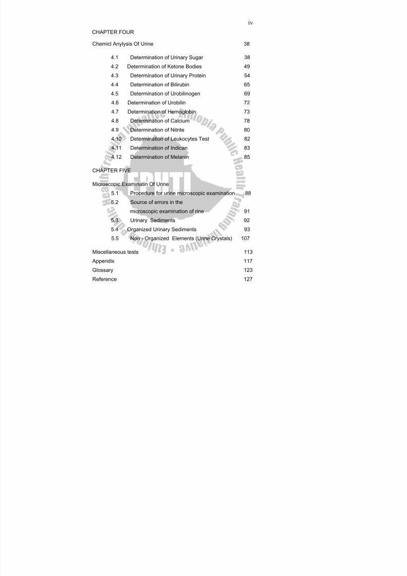
CHAPTER FOUR
Chemicl Anylysis Of Urine 38
4.1 Determination of Urinary Sugar 38
4.2 Determination of Ketone Bodies 49
4.3 Determination of Urinary Protein 544.4 Determination of Bilirubin 65
4.5 Determination of Urobilinogen 69
4.6 Determination of Urobilin 72
4.7 Determination of Hemoglobin 73
4.8 Determination of Calcium 78
4.9 Determination of Nitrite 80
4.10 Determination of Leukocytes Test 82
4.11 Determination of Indican 83
4.12 Determination of Melanin 85
CHAPTER FIVE
Microscopic Examinatin Of Urine
5.1 Procedure for urine microscopic examination 88
5.2 Source of errors in the
microscopic examination of rine 91
5.3 Urinary Sediments 92
5.4 Organized Urinary Sediments 93
5.5 Non - Organized Elements (Urine Crystals) 107 Miscellaneous tests 113
Appendix 117
Glossary 123
Reference 127
8/13/2019 LN Urinalysis Final
http://slidepdf.com/reader/full/ln-urinalysis-final 7/138
v
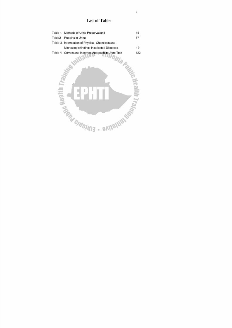
ist of Table
Table 1 Methods of Urine Preservation1 15
Table2 Proteins in Urine 57
Table 3 Interrelation of Physical, Chemicals andMicroscopic findings in selected Diseases 121
Table 4 Correct and Incorrect Approach in Urine Test 122

8/13/2019 LN Urinalysis Final
http://slidepdf.com/reader/full/ln-urinalysis-final 8/138
vi
List of Figures
Figure 1.1 Components of the Renal System 2
Figure 1.2 Anatomy of the Kidney 2
Figure 1.3 The structural and Functional
segments of the Nephrons 4Figure 3.1 Urinometer and Urinometer Cylinder 32
Figure 5 Formed Urine Elements 106
Figure 6 Urine Crystals 112
8/13/2019 LN Urinalysis Final
http://slidepdf.com/reader/full/ln-urinalysis-final 9/138
vii
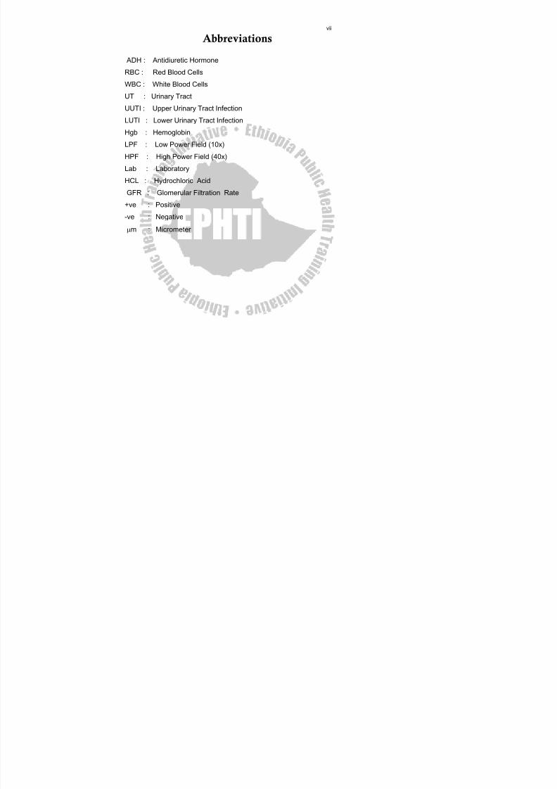
Abbreviations
ADH : Antidiuretic Hormone
RBC : Red Blood Cells
WBC : White Blood Cells
UT : Urinary Tract
UUTI : Upper Urinary Tract Infection
LUTI : Lower Urinary Tract Infection
Hgb : Hemoglobin
LPF : Low Power Field (10x)
HPF : High Power Field (40x)
Lab : Laboratory
HCL : Hydrochloric Acid
GFR : Glomerular Filtration Rate
+ve : Positive
-ve : Negative
μm : Micrometer
8/13/2019 LN Urinalysis Final
http://slidepdf.com/reader/full/ln-urinalysis-final 10/138
viii
Introduction
Examination of urine as an aid to diagnose a number of diseases is the
oldest among tests in the history of Medical Laboratory Technology. It
has been known for centuries that abnormalities in urine may indicate
disease. Perhaps, one of the earliest known record of urine test was the
technique of pouring urine on the ground and observing whether or not it
attracted insects. The attraction of insects indicates " honey urine "
which was known to be excreted by people with boils. Today checking
sugar in urine is a test to detect diabetes (And untreated diabetes still
suffer from boils). Around 1000 AD a Persian Physician named Ismail of
Jordan described seven different observations made on urine such as
Urine Consistency, Color, Odor, Transparency, Sediment and Froth. It
was Walter Ames Compton who ushered in the modern era of Urinalysis
in the early 1940's with the invention of 'CLINITEST'.Urinalysis is a group of tests performed most frequently on random
specimen. It is one of the most helpful indicators of health and
disease, especially, it is useful as a screening test for the detection
of various endocrine or metabolic abnormalities in which the
kidneys function properly but they will excrete abnormal amounts of
metabolic end-products specific of a particular disease. It is also
used to detect intrinsic conditions that may adversely affect the kidneys
or urinary tract. Diseased kidneys cannot function normally in regulating
the volume and composition of body fluids, and in maintaining
homeostasis. Consequently, substances normally retained by a kidney
or excreted in small amounts may appear in the urine in large quantities,
or substances normally excreted may be retained by kidney.
Generally, urinalysis provides useful information concerning the
presence or absence of renal and other diseases, and as a
routine test, i t is a very simple method for monitoring the course
of a disease as well as the efficacy of treatment.
8/13/2019 LN Urinalysis Final
http://slidepdf.com/reader/full/ln-urinalysis-final 11/138
ixThis lecture note is prepared for Diploma Medical Laboratory
Technology Students. It provides them with basic knowledge of Urine
Examination. It also helps the students as well as other health
professionals to understand and acquire the necessary procedures,
which are useful in the investigation of normal and abnormal urine
constituents and interpretation of the results.
8/13/2019 LN Urinalysis Final
http://slidepdf.com/reader/full/ln-urinalysis-final 12/138
1
CHAPTER ONE
Anatomy and Physiology of The Kidney
Objective :
This chapter is intended- To give a basic knowledge of the kidney structure and urine formation
as an important aid in understanding urinalysis and test interpretation.
Introduction
The Renal System is a system which is composed of two kidneys,
two ureters, one bladder and one urethra. As the components of the
renal system the kidneys have the following functions:
Regulation of water and electrolyte ( such as chloride,
potassium, calcium, hydrogen, magnessium, and phosphate ions )
balances. - Regulation of acid – base balance of the blood.
Regulation of body fluid osmolality and electrolyte
concentratiions.
Regulation of arterial pressure.
Excretion of metabolic waste products and forein chemicals. The
kidneys are the primary means for the eliminating waste products
of body metabolism that are no longer needed by the body.
These products include urea from the metabolism of amino acids,
uric acid from the nucleic acids , creatinine from muscle creatine,
bilirubin from the breakdown of hemoglobin.
Secretion of hormones such as renin.
Gluconeogenesis. The kidneys synthesize glucose from amino
acids and other precursors, like lactate and glycerol, during
prolonged fasting by the process called gluconeogenesis.
8/13/2019 LN Urinalysis Final
http://slidepdf.com/reader/full/ln-urinalysis-final 13/138
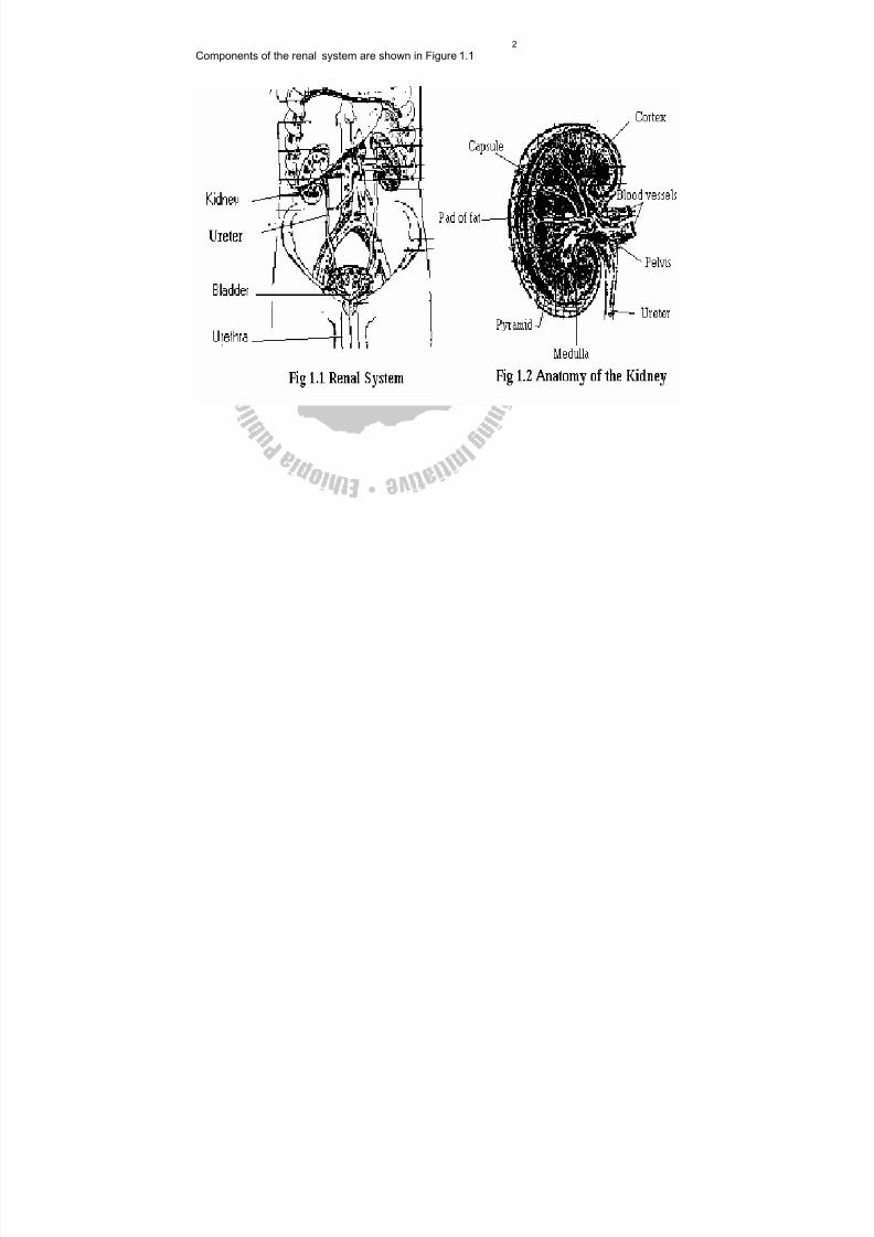
2Components of the renal system are shown in Figure 1.1
8/13/2019 LN Urinalysis Final
http://slidepdf.com/reader/full/ln-urinalysis-final 14/138
31.1 Anatomy of the Kidney
The kidneys are two bean shaped organs located under the lowermost
part of the ribs in the posterior abdominal cavity. Each human kidney
weighs 150 gms and measures 1x2x3 inches (thickness, width, and
length). A coronal section of the kidney shows an outer reddish granular
layer called renal medulla. In the renal medulla the triangular and wedge
shaped structure is called renal pyramids. The tips of the pyramids
found on the renal papillae at which urine is drained into cavities is called
Renal Calyces. Renal Calyces drain urine into renal pelvis, then to
ureter, which in turn drain to bladder and then through the urethra is
voided out.
The gross anatomy of the kidney is shown in Figure 1.2.
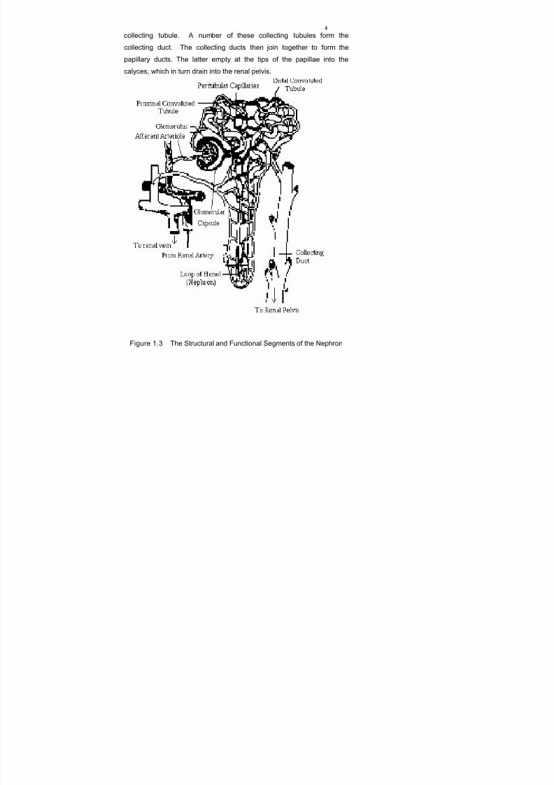
The functional unit of the kidney is the nephron (Figure 1. 3). There are
approximately one million nephrons in each kidney. Each nephron
consists of a glomerulus, which is essentially filtering system, and atubule through which the filtered liquid passes. Each glomerulus consists
of a network of capillaries surrounded by a membrane called
Bowman's ( Glomerular ) Capsule, which continues on to form Bowman's
Space and the beginning of the renal tubule. The afferent arteriole,
which carries blood from the renal artery into the glomerulus divides to
form a capillary network. These capillaries re-unite to form the efferent
arteriole, through which blood leaves the glomerulus. The blood
vessels thus follow the course of the tubule, forming a surrounding
capillary network. The tubular portion of each nephron has several
distinct structural and functional segments. The uppermost portion,
which continuous with the glomerulus, is the proximal convoluted tubule,
followed by the thin walled segment and the distal convoluted tubule
respectively. The descending limb of the proximal tubule (the thin-walled
segment) and the distal tubule form a loop known as the Loop of Henle.
The distal convoluted tubules from several nephrons drain into a
8/13/2019 LN Urinalysis Final
http://slidepdf.com/reader/full/ln-urinalysis-final 15/138
4collecting tubule. A number of these collecting tubules form the
collecting duct. The collecting ducts then join together to form the
papillary ducts. The latter empty at the tips of the papillae into the
calyces, which in turn drain into the renal pelvis.
Figure 1.3 The Structural and Functional Segments of the Nephron
8/13/2019 LN Urinalysis Final
http://slidepdf.com/reader/full/ln-urinalysis-final 16/138
5
1.2 Physiology of the Kidney and Formation of Urine
The kidney is a highly discriminating organ, which maintains the internal
environment by selectively excreting or retaining various substances
according to specific body needs. Approximately 1,200 mililiters of blood
flow through the kidneys each minute. This represents about one-fourth
of the total blood volume. The blood enters the glomerulus of eachnephron by passing through the afferent arteriole into the glomerular
capillaries. The capillary walls in the glomerulus are highly permeable to
water and the low molecular-weight components of the plasma. They
filter through the capillary walls and the closely adhering membrane of
Bowman's Capsule into Bowman's Space from where the plasma ultra
filtrate passes into the tubule where reabsorption of some substances,
secretion of others, and the concentration of urine occur. Many
components of the plasma filtrate such as glucose, water, and amino
acids, are partially or completely reabsorbed by the capillariessurrounding the proximal tubules. In the distal tubules, more water is
reabsorbed and potassium and hydrogen ions are secreted. The Loop
of Henle and the system of collecting tubules are the principal sites
where the urine is concentrated as a mechanism for conserving body
water. Urine formed by the three physiological processes that are by
glomerular filtration, tubular reabsorption, and tubular secretion, is
collected by the collecting duct and passes into bladder through
ureters and then comes out through urethra.
1.3 The Compos itio n of Urine
Normal Urine Constituents Abnormal Urine Constituents
- Water (about 95% of urine) - Glucose
- Urea - Protein
- Creatinine - Bile pigments
- Uric acid - Blood cells
8/13/2019 LN Urinalysis Final
http://slidepdf.com/reader/full/ln-urinalysis-final 17/138
6- Electrolytes - Cast parasites and
Bacterial microbes
1.4 The Factor s Affect ing The composi tion of Urine
Diet and nutritional status
Condition of body metabolism Ability of kidney function
Level of contamination with pathogenic microorganisms
( bacteria) or even non-pathogenic microflora
1.5 Renal Clearance and Renal Threshold
Renal Clearance
Renal Clearance value indicates the degree to which a substance is
removed from the blood by excretion in the urine. Clearance is usuallydefined as the blood volume that contains the quantity of a substance
excreted in the urine per minute.
About 120 ml of glomerular filtrate is produced per minute. The rate at
which the glomerular filtrate is formed is known as the glomerular
filtration rate (GFR).
Creatinine is a substance present in the filtrate, which is not reabsorbed
(however, this is some tubular secretion of creatinine). Therefore the
clearance of creatinine from the plasma is 120 ml per minute. Hence
creatinine clearance is used clinically to give an approximate indicationof glomerular filtrate rate and, therefore, as a test of kidney function.
When the filtration rate falls, the concentration of creatinine in the
plasma rises. The creatinine clearance test express the volume of blood
containing the amount of creatinine excreted by the kidney in one
minute.
8/13/2019 LN Urinalysis Final
http://slidepdf.com/reader/full/ln-urinalysis-final 18/138
7The creatinine clearance (Ccr) is calculated by collecting a 24 hr urine
specimen, and a blood sample as well within the urine collection time.
Creatinine is then determined in both urine and serum, and the
creatinine clearance calculated in milliliters per minute (ml / minute)
Ccr ml / minute = U x V
S
Where U= Urine Creatinine Concentration in mol/l
V= Volume of urine in ml per 24 hrs
S= Serum Creatinine Concentration in mol/l
Normal Range:
The normal Ccr value usually ranges between 110 – 140 ml / minute.
Renal Threshold
The renal threshold of a substance refers to the highest concentration of
a substance, which is present in the blood before it is found in the urine.
A substance such as glucose is a high threshold substance, because it
is completely absorbed from the glomerular filtrate and is only found in
the urine, when the blood glucose level is markedly raised. Urea and
creatinine, however, are always present in the urine independent of the
blood level because very little, if any, of these substance is reabsorbed.
8/13/2019 LN Urinalysis Final
http://slidepdf.com/reader/full/ln-urinalysis-final 19/138
8
Exercise 1.
Answer th e Fol lowing Ques tions:
1. Describe the functions of the Urinay System.
2. Explain how Urine is formed by the Nephrons.
3. Mention the factors that determine the selective passage of
molecules through the glomerular membrane.
4. Calculate the CcCr of a patient who voided 1500 ml of urine in
24 hrs. The serum and urine concentration of creatinine of the
patient are 0.28 mmol/l and 10.5mmol/l respectively.
8/13/2019 LN Urinalysis Final
http://slidepdf.com/reader/full/ln-urinalysis-final 20/138
9
CHAPTER TWO
Collection And Preservation Of Urine
Specimen
Objectives
It is expected that the information presented in this chapter will enable
the student to :
Identfy factors affecting the quality of a specimen.
List the basic rules of urine collection.
Describe types of urine specimens.
Identify the commonly used preservatives and know the advantages
and disadvantages of their use.
2.1 Collection of Urine Specimen
In order to make Urinalysis reliable the urine must be properly collected.
Improper collection may invalidate the results of the laboratory
procedures, no matter how carefully and skillfully the tests are
performed.
Urine Containers
There are many types of containers used for collecting urine. Before
specimens are collected, the containers must be cleaned andthoroughly dried. Disposable containers of plastic or coated paper are
available in many sizes and are provided with lids to reduce bacterial
and other types of contamination. Special polyethlene bags are
available for collectionn of urine from infants and children who are not
toilet trained.URIN-TEK disposable collection system is available for
use in collecting, storing, transporting, and testing specimens of urine.
The system consists of a flat-bottomed paper collection cup, a 15 ml
8/13/2019 LN Urinalysis Final
http://slidepdf.com/reader/full/ln-urinalysis-final 21/138
10plastic tube with a plastic snap-cap and self-adhesive identification
label.Disposable tube holders are available for handling ten tubes at a
time. The patient voids directly into the paper cup, transfers the
specimen to the URIN-TEK tube, and covers it with the plastic cap to
prevent contamination or spillage. After the label is filled out and
attached, the specimen is ready to be transported and analyzed.
Specific gravity can be run directly in the URIN-TEK tube if a colorimetricstrip or a urinometer is used. By using the convenient reagent strips,
many chemical tests can be performed directly in the URIN-TEK tube,
making additional laboratory glassware unnecessary. This procedure
also decreases the risk of identification errors because transferring and
relabeling of the specimens is not necessary. Large, wide-mouthed
plastic or glass containers with screw cap tops are used for cumulative
collection of urine over a long period of time. These bottles should be
kept refrigerated. When urine is to be cultured for bacterial content, the
specimen must be obtained under septic condition and collected in a
sterile glass container or a sterile disposable plastic container. In either
case, the receptacle should be equipped with a tight-fitting, sterile cap.
This cap is left in position until the actual time of urine collection, and
replaced immediately afterward.
Methods of Obtaining Specimens
A freshly voided urine specimen is adequate for most urinalysis except
the microbiological culture. The patient should be instructed to void
directly into a clean, dry container, or a clean, dry bedpan so that the
specimen can be transferred to an appropriate container. Specimens
from infants and young children can be collected in a disposable
collection apparatus. If a urine specimenn is likely to be contaminated
with vaginal discharge or menstrual blood, this period has to be
avoided and the patient must be informed to bring a clean-voided
8/13/2019 LN Urinalysis Final
http://slidepdf.com/reader/full/ln-urinalysis-final 22/138
11specimen. All specimens should be immediately covered and taken to
the laboratory.
Types of Specimen
First Morning Specimen - a speciemen obtained during the first
urination of the day.
Most concentrated
Bladder incubated
Best for:
Nitrite
Protein
Microscopic examination
Random Specimen - a spciemen obtained at any time during
examination.
Most convenient
Most common
Good for:
Chemical Screen
Microscopic examination
Second-voided Specimen - In this case first morning specimen is
discardedand the second specimen is collected and tested. Such
type of specimen is good for:
Reflection of blood glucose.
Keeping of formed elements intact.
Postprandial : a specimen obtained 2 hours after meal.
Good for glucose.
8/13/2019 LN Urinalysis Final
http://slidepdf.com/reader/full/ln-urinalysis-final 23/138
1224- Hour specimen - a specimen obtained within 24 hours.
Necessary for quantitative tests, especially for quantitative
determination of protein.
Procedure for Collection o f 24 hour Urine Specimen
1. Inform or Direct the patient to completely empty his bladder and
discard his urine exactly at the beginning of the 24 hour time
collection (let say at 6:00 a.m.).
2. Collect all urine voided during the following 24 hours, including that
voided exactly at the end of the 24 hour period in a container (at
6:00 a.m.) of the following (second) day.
3. All the urine collected must be preserved.
4. The container should be labeled with :
The test order
The patient’s name
Time of collection
The preservative added
Mid- stream Specimen - a specimen obtained from the middle part
of the first urine.
It is commonly used for routin e urinalysis.
It is also important for bacteriological urine culture.
Clean Catch Urine Specimen
Used for microbial culture and routine urinalysis. When specimens are
collected for bacteriological examination they should be collected by the
‘clean catch’ method or by catheterization into sterilized container.
Catheterization is the process of passing a tube through the urethra to
the bladder for the withdrawal of urine (it may introduce urinary tract
infection).
8/13/2019 LN Urinalysis Final
http://slidepdf.com/reader/full/ln-urinalysis-final 24/138
13The best method is properly collected ‘clean catch’ urine, which is
collected as follows :
a. The genital area should be cleaned with soap and water and
rinsed well. This is to keep off bacteria on the skin from
contaminating the urine specimen.
b. The patient should urinate a small amount and this is discarded.
c. The urine that comes next, the mid-stream specimen, should be
collected into a sterile container of 30 to 50 ml.
d. After obtaining the specimen the patient continues to urinate and
this is discarded.
Sources of Errors in the Collection of Urine
1. Bacteriologically or chemically contaminated specimen.
2. Wrong type/amount of preservative.
3. Partial loss of specimen or inclusion of two-morning specimen in
the 24 hr collection.
4. Inadequate mixing of specimen before examination.
5. Careless measuring of the 24 hr volume.
2.2 Preservation of Urine Specimen
Urine should be examined immeditely as much as possible after it is
passed, because some urinary components are unstable. If urine
specimen can not be examined immediately, it must be refrigerated or
preserved by using different chemical presevatives. The maximum timethat urinary contents to be maintained in urine specimen is one
hour.Long standing of urine at room temperature can cause :
Growth of bacteia
Break down of urea to ammonia by bacteria leading to an increase in
the pH of the urine and this may cause the precipitation of calcium
and phosphates.
Oxidation of urobilingen to urobilin.
8/13/2019 LN Urinalysis Final
http://slidepdf.com/reader/full/ln-urinalysis-final 25/138
14 Distruction of glucose by bacteria.
Lysis of RBCs, WBCs and casts.
Method of Preservation of Urine Specimen
a. Physic al Method
- Refrigeration
- Freezing
b. Chemical Method
Use of chemical preservatives such as :
- Thymol
- Toluene
- Formaldehyde
- Hydrochloric acid ( HCl)
- Chloroform
- Boric acid
- Chlorhexidine
- Sodium carbonate
8/13/2019 LN Urinalysis Final
http://slidepdf.com/reader/full/ln-urinalysis-final 26/138
15
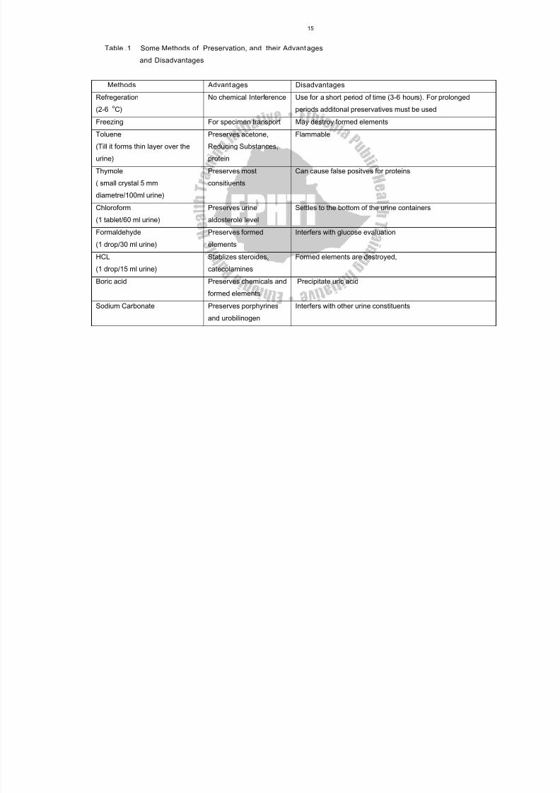
Table .1 Some Methods of Preservation, and their Advant ages
and Disadvantages
Methods Advantages Disadvantages
Refregeration
(2-6 oC)
No chemical Interference Use for a short period of time (3-6 hours). For prolonged
periods additonal preservatives must be used
Freezing For specimen transport May destroy formed elements
Toluene
(Till it forms thin layer over the
urine)
Preserves acetone,
Reducing Substances,
protein
Flammable
Thymole
( small crystal 5 mm
diametre/100ml urine)
Preserves most
consitiuents
Can cause false positves for proteins
Chloroform
(1 tablet/60 ml urine)
Preserves urine
aldosterole level
Settles to the bottom of the urine containers
Formaldehyde
(1 drop/30 ml urine)
Preserves formed
elements
Interfers with glucose evaluation
HCL
(1 drop/15 ml urine)
Stablizes steroides,
catecolamines
Formed elements are destroyed,
Boric acid Preserves chemicals and
formed elements
Precipitate uric acid
Sodium Carbonate Preserves porphyrines
and urobilinogen
Interfers with other urine constituents
8/13/2019 LN Urinalysis Final
http://slidepdf.com/reader/full/ln-urinalysis-final 27/138
16
2.3 Type of Examination in Routine Urinalysi s
Physical Examination of Urine
Volume
Color
Odor
Appearance
pH
Specific gravity
Chemical Examination of Urine
Glucose
Protein
Ketones
Bilirubin
Urobilinogen Blood
Nitrite
Leukocyte Esterase
Indican
Melanin
Microscopic Examination of Urine
RBCs
WBCs Epithelial cells
Casts
Bacteria
Yeasts
Parasites
Crystals
Artifacts
8/13/2019 LN Urinalysis Final
http://slidepdf.com/reader/full/ln-urinalysis-final 28/138
17
Categories of Urine Tests
According to their degree of accuracy urine tests are grouped into three
broad categories:
Screening tests
Qualitative tests
Quantitative test
Screening tests tell only whether a substance is present or absent, and
the results are reported as positive or negative. They are done on
random specimen. Qualitative tests give rough estimate of the amount of
substance present. They are also called semi-quantitative tests. The
results of qualitative tests can be graded as negative, trace, +1, +2, +3
or +4. Quantitative tests determine accurately the amount of the
substances to be tested. However, since they are time consuming, they
are not included in routine urinalysis. Most common quantitative tests
performed in urinalysis laboratory are those for sugar and for protein.
The results of a quantitative test are usually reported in milligrams per
deciliter, gram per deciliter, and per liter. For quantitative test, a
complete 24-hour urine specimen is needed. An appropriate
preservative should be added to the container or the specimen should
be stored in refrigerator.
Exercises :
Answer the following questions:
1. What type of specimen would be appropriate for both routine
urinalysis and bacteriological culture?
2. What essential supplies for collection of urine specimen should
be recommended?
3. What type of preservative(s) would be the most useful, and why ?
8/13/2019 LN Urinalysis Final
http://slidepdf.com/reader/full/ln-urinalysis-final 29/138
18
CHAPTER THREE
Physical Examination Of Urine
Objective
At the end of this chapter, the student shall be able to carry out physical
examination of urine such as odour, volume, color, transparency, foam,
specific gravity, pH of uirne and interprete the result of the
investigation so that to identify further the necessary type of
examination ( chemical or microscopic or both).
Introduction
Physical examination of urine is the first part of routine urinalysis. It isthe simplest procedure of all urine examination, but this simplicity does
not mean that any one can do it with out any background knowledge and
experience. Physical examination of urine usually gives hint for the
subsequent urinalysis. For example, white turbid urine sample may
suggest to the technician the presence of Leukocytes (pus cells) and/or
Epithelial cells in microscopic examination, and in chemical examination,
with positive result of Nitrite.
3.1 Volume
Normally, 600 – 2000 ml of urine is voided per 24 hr.
Volume of urine excreted is related to:
Individual fluid intake
Body temperature
Climate
Individual’s health status
8/13/2019 LN Urinalysis Final
http://slidepdf.com/reader/full/ln-urinalysis-final 30/138
19
Abnormally higher amount (greater than 2000 ml/24) or very low amount
i.e. less than 600 ml/24 occur mostly due to some pathological
conditions.
Test Procedure
For the measurement of the volume of urine, the patient should collect24 hr urine specimen.
The laboratory technician supplies the urine container, and it should be
Clean and dry.
Brown colored to avoid direct sunlight contact with the collected
urine and interaction of sunlight with the chemicals.
Contains appropriate preservative for the desired urine chemical
test, or that is kept after each urine collection within refrigerator.
Labeled on the wall, that indicates
o Name of patient
o Collection time and date
o Type of chemical test ordered
o Preservative used
* Using graduated cylinder does measurement of urine volume. The
amount is recorded in terms of ml/24 hr.
Clinical Significance
The Measurement of the volume of urine indicates the evaluation of fluidbalance and kidney function. When an individual excretes more than
2000 ml of urine/24 hr, consistently (for long period) it is called Polyuria.
It may occur due to:
Diabetic mellitus
Diabetic insipidus
Certain tumors of brain and spinal cord
Acromegaly
8/13/2019 LN Urinalysis Final
http://slidepdf.com/reader/full/ln-urinalysis-final 31/138
8/13/2019 LN Urinalysis Final
http://slidepdf.com/reader/full/ln-urinalysis-final 32/138
21
Test Procedure
The test is conducted by smelling of urine and the result is based on the
perception of the technician.
Clinical Significance
Abnormal urine odor may result from aging of urine, disease and diet.
If the urine specimen is old, i.e. after collection, left on the bench
with out preservative for more than 2 hrs, it will have ammonical
(pungent) odor. The ammonical odor result is due to break down and
conversion of urea in the urine into ammonia by the action of
bacteria.
Cystinuria and homocystinuria (type of amino acids, voided from
abnormal metabolism) have sulfurous odor.
Oasthouse urine disease has a smell associated with the smell of a
brewery (yeast).
Tyrosenemia is characterized by cabbage like or “fishy” urine odor.
The presence of ketone bodies in the urine, that may be due to
diabetes mellitus, vomiting, starvation, strenuous exercise,
characterized by “sweet fruity” odor.
Butyric / hexanoic acidemia produce a urine odor resembling that of
sweat.
Urine of infants, which has inherited amino acid metabolism
disorder, smells like “burnt sugar” or maple, hence the name, “maplesugar urine disease”.
Also due to some food stuff such as asparagus, characteristic, urine
odor is produced, which has no clinical significance.
3.2 Foam
Normally when urine specimen is voided in a container, it produces small
amount of white foam. But during certain abnormal physiological and
8/13/2019 LN Urinalysis Final
http://slidepdf.com/reader/full/ln-urinalysis-final 33/138
22
metabolic conditions, the color and amount of foam may be changed.
For example, when there is high bile pigment in the urine, the amount of
foam increases, and the color of foam becomes yellowish. This may
indicate the presence of bilirubin in the urine. But the presence of
yellowish foam should not be taken as a confirmatory test for the
presence of bilirubin in urine. Chemical analysis of urine for billirubinshould be done.
3.4 Color
Normally color of urine may vary within a day; in the morning it has dark
yellow color, while in the afternoon or evening, the color ranges from
light yellow to colorless. Normal urine color varies from straw (light
yellow color) to dark amber (dark yellow).
Light yellow indicate that the urine is more diluted, and has low
specific gravity. Such exceptional condition occurs in case of
diabetic mellitus. In this condition the color of urine is mostly light
yellow, but because of having high glucose content, its specific
gravity is high.
On the other hand, dark amber (dark yellow) color mostly indicates
that the urine is concentrated, and has high specific gravity. This
type of urine is seen normally in the first morning urination.
Normal urine color resultes from three pigments. They are:
- Urochrome, responsible for yellow color formation. This pigment
is found in high proportion than the other two.
- Uroerythrin, – responsible for red color formation.
- Urobilin, – responsible for the orange-yellow color formation.
Thus, normal urine gets its color from a combination of the
above-mentioned three pigments.
8/13/2019 LN Urinalysis Final
http://slidepdf.com/reader/full/ln-urinalysis-final 34/138
23
Procedure of the Test
Urine color is recorded, after looking at freshly voided urine specimen. If
the urine sample color is not recorded within 30 minutes after collection,
chemical changes will occur in it, and so its color will change, and will
result in false report.
Clinical Implication
By observing the color of freshly voided urine, an experienced laboratory
technician can forecast the possible findings in the chemical and
microscopical examination of urine. Depending up on the constituents of
urine, the abnormal color of urine varies as follows:
Pale to colorless urine may indicate:
• Large fluid intake
• Diabetic mellitus
• Diabetic insipidus
• Alcohol consumption
• Nervousness
Dark yellow or brown red urine may indicate:
• Concentrated urine
• Decreased fluid consumption
• Dehydration
• Fever
• Certain urinary tract medication (eg. phenazophyridine)
• Yellow brown or “beer brown” color may indicate the presence of
bilirubin.
This is also confirmed:
- By looking at the yellow foam or green foam by shaking the
sample.
8/13/2019 LN Urinalysis Final
http://slidepdf.com/reader/full/ln-urinalysis-final 35/138
24
- By letting it to stand for more than 30 minutes and looking at
the change of color into green, because of oxidation of
bilirubin into biliverdin.
- Due to bilirubin crystals, as mentioned in urine segment, the
urine samples have opalescent appearance.
- By doing chemical tests for bilirubin.
Clear red may indicate presence of Hemoglobinuria (presence of
hemoglobin in the urine). This hemoglobinuria may result from:
- Incompatible blood transfusion.
- Increased red blood cell destruction (intravascular
haemolysis) due to different hemoparasites, e.g. Malaria.
- Glucose – 6-phosphate dehydrogenase deficiency.
- Certain infections or disease.
Cloudy red / smoky red color may indicate hematuria (presence of
red blood cell in the urine).It differs from clear red by the presence of
RBC rather than Hgb alone. It is important to differentiate
hemoglobinuria from hematuria, because the cause of this abnormal
urine differs. On standing the red cell in hematuria may hemolize
and settle and so the urine becomes clear red (hemoglobin in urine).
To differentiate this the definition of specific gravity is important.
Hematuria has high specific gravity than helmoglobinuria.
Dark brown colored urine may contain porphyrines, melanin,
homogenstic acid, which is associated with an abnormal metabolism
of tyrosine. Milky urine may contain fat, cystine crystals, and many
WBC or amorphous phosphates. Dark reddish color may indicate
myoglobin (muscle Hgb), usually associated with extensive muscle
injury, hemoglobinuria and porphyrine.
8/13/2019 LN Urinalysis Final
http://slidepdf.com/reader/full/ln-urinalysis-final 36/138
25
Interfering Factors
It is usually important to consider, that on standing of urine for more than
30 minutes, the urobilinogen that is found in urine will oxidize and
change to urobilin. Thus due to this process, the color of urine becomes
dark. Therefore, the physical examination of urine should be done
immediately after the delivery of urine to the laboratory.
Other interfering factors that result in abnormal urine color formation are
certain foodstuff, and medications.
Food stuf, such as beets will give white red color.
Drugs such as Vitamin B 12 and riboflavin will give bright yellow color
to urine.
Rifampicilin will give red color to urine.
Iron salt will give dark color to urine.
Sulfonamides will give rusty yellow or brownish color.
Therefore, when abnormal colored urine is observed, it is important to
ask the patient, what kind of food he consumed in the last 36-24 hrs, and
also whether he used drugs or not. If so, it is important to know what
food and what drug he used.
3.5 App earance (Transparency)
Fresh voided urine specimen is normally clear and transparent. On long
standing, due to chemical changes that occur in normal constituents of
urine through time, as described in the introduction part of this lecturenote, it becomes turbid.
Procedure of the Test
Appearance (transparency) of urine can be measured only by
observation of fresh voided urine specimen.
8/13/2019 LN Urinalysis Final
http://slidepdf.com/reader/full/ln-urinalysis-final 37/138
26
Degree of cloudiness of the urine is described by using common
terms, starting by clear to turbid i.e. clear, hazy, cloudy, very cloudy
and turbid.
Clinical Implications
Freshly voided urine specimen appearance may indicate the presence of
some abnormal constituents in it. Causes of turbid urine, as it is freshly
voided includes:
• White blood cells (pus cells) that occur due to UTI
• Kidney stones
• RBC’s
• Yeast cells,
• High number of bacteria cells
• High number of epithelial cells
• Fat droplets in urine, which give opalescent appearance (rare
condition).
• Amorphous urates, in case of gout and leukemia.
• High number of mucus trades.
All the above findings are confirmed by urine microscopic examination.
Interfering Factors
High consumption of foodstuff that contains urates and phosphates may
produce cloudy urine. This is because of the precipitation of urates and
phosphates in the form of amorphus urate and phosphates respectively.
Semen, or vaginal discharge mixed with urine is other common causes
of urine turbidity. Urine specimen, stood for long period in the bench, will
become hazy or cloudy due to precipitation of crystals, mucus trades
etc., which normally occur in urine. The settlements of crystal and
mucus trades seen in urine sample are to be preserved in refrigerator.
Amorphous urates have “Brice red” precipitation, while amorphous
phosphates have white precipitations.
8/13/2019 LN Urinalysis Final
http://slidepdf.com/reader/full/ln-urinalysis-final 38/138
27
3.6 pH
A test that determine acidity, neutrality or alkalinity of a solution.
pH 7 indicates neutrality.
pH < 7 indicate acidity.
pH > 7 indicate alkalinity.
Normally, freshly voided urine pH range from 5-7 in healthy individuals,and average is pH 6.
Procedure of the Test
pH of urine can be measured by using different techniques, such as by
using:
Litmus paper
Nitrazine paper
Dipstick
Glass electrode
These different pH-measuring techniques vary in their sensitivity and
reading techniques.
Litmus Paper
In this technique pH measurement takes place by using blue, and red
litmus paper.
The procedure is:
- Collect a freshly voided well mixed urine.
- Tear small blue litmus paper.
- Dip the paper, in the urine and remove immediately.
- Look for color change of blue litmus paper. If the blue colors of
paper change to red, it indicates the acidity of the urine.
- Tear small red litmus paper.
- Dip the paper, in the urine and remove it immediately.
8/13/2019 LN Urinalysis Final
http://slidepdf.com/reader/full/ln-urinalysis-final 39/138
28
- Look for color change of red litmus paper. If the red litmus paper
change to blue, it indicates that the urine is alkaline.
The blue and red litmus paper technique is a less sensitive method. This
is because it indicates only the alkalinity or acidity of urine; it does not
tell the exact quantity or figure of pH.
Nitrazine Paper
This is also a paper that changes its color from yellow (for acidic urine)to blue (for alkaline urine). The paper is impregnated with sodiumdinitrophenolazo-naphthal disulphonate chemical. This chemical isresponsible for the color change in acidic and alkaline urine. Unlikelitmus paper, the color change is matched with reference color chart, andbased on the value of color change on the reference color chart; the pHof the urine is recorded.
Procedure
The procedure of the test is:
Tear small nitrazine paper
Dip the paper in well mixed freshly voided urine sample and remove
immediately
Compare the color change with that of reference color chart.
Record the value of color change form reference color chart.
Reference color chart -value range from 3 to 4 (for yellow color) to pH 9
(that is for deep blue color). The result of urine pH is usually reported by
saying acidic or alkaline and by indicating the figure.
Urine Dipstick Method
This is a reagent strip test impregnated with chemicals called methyl red
and bromethymol blue. These impregnated chemicals depending up on
the concentration of hydrogen ion in the urine change their color from
yellow (acid) to blue (alkaline).
8/13/2019 LN Urinalysis Final
http://slidepdf.com/reader/full/ln-urinalysis-final 40/138
29
The color change is interpreted by comparing the reference color chart
supplies with the reagent strip. pH indicator- strip is usually
manufactured together with other tests for urine constituents.
Procedure
The procedure of the test is:
Dip the reagent strip in the well mixed freshly voided urine and
remove immediately.
Remove the excess urine from the strip, by taping the strip at the
edge of urine container.
According to the time mentioned by the manufacturer to read the
result, wait for full color development in the strip.
Then read the color change by comparing within the reference color
chart and report the result.
The reference color chart value, range from 5 for acidic urine (yellow
color) to 9 for alkaline urine (blue color).
Glass Electrode
This is a very sensitive pH measurement technique. The measurement
is done by using small electronic instrument that has electronic pH value
indicator and glass electrode.
This instrument operates on battery or electricity.
Procedure
The procedure is to dip the glass rods in the freshly voided mixed urinespecimen, and then look for the figure in the instrument to find out the
value of pH.
Clinical Significance
As indicated in the chapter one, one of the functions of renal system is to
regulate pH of blood i.e. keep pH of blood at 7.4 + 0.05. This is done by
absorption or release of hydrogen ion, especially at distal convoluted
8/13/2019 LN Urinalysis Final
http://slidepdf.com/reader/full/ln-urinalysis-final 41/138
30
tubules of the nephron, depending on the pH of blood, i.e. hydrogen ion
absorbed from surrounding blood capillaries of nephrone when pH is
acidic (below 7.35), and release from nephron to the surrounding blood
vessels when pH of blood is alkaline (above 7.45).
pH measurement of urine, like other physical tests of urine, may indicate
the on- going process in body, mostly about the renal system.Normal pH of urine is 5-6.
* Persistent alkaline urine (pH > 6) may be caused by:
UTI
Renal failure
Vomiting
Anorexia nervosa
Alkalosis (metabolic or respiratory e.g. due to accumulation CO 2 in
our body.
Alkalizing drugs i.e. during intake of drugs such as streptomycin,
kanamycin etc. eg. for UTI.
It should also important to bear in mind that certain vegetables,
citrus fruits, and milk products also may cause alkaline urine, which
is not pathological
* Persistent acid urine (pH < 6) may be caused by:
Diarrhea
Malabsorption syndromes
Diabetic ketoacidosis
Dehydration
Fever
Starvation
And also certain drugs such as – Phenacetic
Here it is important to bear in mind that high protein diet may also
result in acidic urine, but this is not a pathological condition.
8/13/2019 LN Urinalysis Final
http://slidepdf.com/reader/full/ln-urinalysis-final 42/138
31
pH measurement is also important in the management of renal stone
patients, who are being treated for renal calculi and who are
frequently given diets or medications to change the pH of the urine
so that kidney stone will not form.
Calcium phosphates, calcium carbonate, and magnesium phosphate
stones develop in alkaline urine. In such instances the urine mustbe kept acidic (i.e. either by diet such as meat, or medication).
Uric acid, cystine, and calcium oxalate stones are precipitated in
acidic urine. Therefore, as part of treatment, the urine should be
kept alkaline (either by diet eg. leguminous plants, citrus fruits and
most vegetables or by medication).
Interfering Factors
If urine specimen is left on the bench for more than 2 hours, bacteria will
grow in it and by converting urea into ammonia, the pH will become
alkaline. This is false alkaline urine, and indicates the specimen in not-
fresh urine.
3.7 Specific Gravity of Urine
Specific gravity is defined as the ratio of the weight of a fixed volume of
solution to that of the same volume of water at a specified temperature,
usually 20 o C (in some books 25 oC). The specific gravity of urine has
been used for years as measure of the total amount of material
dissolved in it (total solids), and thus of the concentrating and excretory
power of the kidneys.
Measurement of Specific Gravity
The following methods are used to test the specific gravity of urine:
Urinometer
Refractometer
Reagent strip
Weighing technique
8/13/2019 LN Urinalysis Final
http://slidepdf.com/reader/full/ln-urinalysis-final 43/138
32
Specimen: It should be the first urine passed at the beginning of the day
with the patient having taken no fluid for 10 hours. The testing of random
urine specimen has little clinical value.
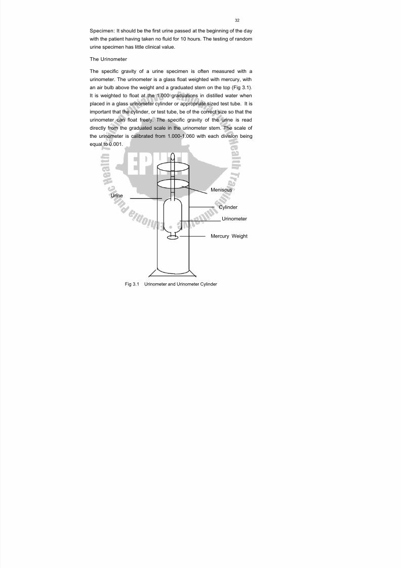
The Urinometer
The specific gravity of a urine specimen is often measured with a
urinometer. The urinometer is a glass float weighted with mercury, with
an air bulb above the weight and a graduated stem on the top (Fig 3.1).
It is weighted to float at the 1.000 graduations in distilled water when
placed in a glass urinometer cylinder or appropriate sized test tube. It is
important that the cylinder, or test tube, be of the correct size so that the
urinometer can float freely. The specific gravity of the urine is read
directly from the graduated scale in the urinometer stem. The scale of
the urinometer is calibrated from 1.000-1.060 with each division being
equal to 0.001.
MeniscusUrine
Cylinder
Urinometer
Mercury Weight
Fig 3.1 Urinometer and Urinometer Cylinder
8/13/2019 LN Urinalysis Final
http://slidepdf.com/reader/full/ln-urinalysis-final 44/138
33
Calibration
To obtain the correct specific gravity readings in urine, the urinometer
must be weighted to read exactly 1.000 in distilled water. The reading
on the urinometer scale should be exactly 1.000. If it is not, a correction
must be applied to all values obtained for urine specimens with the
urinometer. For Example, Suppose the urinometer reads 1.002 in
distilled water. The specific gravity of water is 1.000. Therefore the
urinometer correction is 0.002 must be subtracted from the subsequent
relative specific gravity. If a urine specimen has an apparent specific
gravity of 0.037, this value minus 0.002 results in the corrected specific
gravity of 1.035 for the urine specimen.
Temperature Correction
The specific gravity of a solution is dependent on temperature. Most
urinometers are calibrated for use at 15 oC. For each 3 oC difference
0.001 must be added if above, or subtracted if below than the calibration
temperature. For example, if the specific gravity of the urine is 1.022 at
23 oC, and the urinometer has been calibrated at 20 oC, the correct
reading is 1.022+0.001= 1.023.However, significant error will result if
the reading is taken on the urine specimen that has been refrigerated.
Instead of applying this correction, the urine specimen should be allowed
to warm up to room temperature before its specific gravity is determined.
Correction for abnor mal Dissolved SubstancesThe specific gravity increases by 0.004 for every 1% glucose in urine
and 0.003 for every 1% protein in solution. Therefore subtract 0.004 from
the specific gravity reading for every 1% glucose in urine. And subtract
0.003 from the specific gravity reading for every 1% protein in the urine.
It is not usual however for the Laboratory Technician to correct specific
gravity readings for the presence of sugar or protein when laboratory
results are reported. Instead, the clinician will be aware that the specific
8/13/2019 LN Urinalysis Final
http://slidepdf.com/reader/full/ln-urinalysis-final 45/138
34
gravity is elevated because of the presence of sugar or protein and
takes this into account in the assessment of kidney function.
Procedure for Using the Urinometer
1. Fill the urinometer cylinder or test tube to about 1 from the top
with well mixed urine being careful so as not to cause it to foam.
2. Float the urinometer in the by rotting it rapidly to prevent its
touching the bottom or side of the cylinder.
3. When it comes to rest, read the graduation on the stem of the
urinometer at the level of the lower part of the meniscus. When the
reading is taken, the urinometer must not be touching the sides of
the container.
4. Record the reading.
5. If the quantity of the urine is too small to float the urinometer, the
urine must be diluted with distilled water. The specific gravity is
read and the last two digits of the specific gravity are multiplied by
the amount of the dilution. This method is also used if the
urine specific gravity is greater than the calibration on the
urinometer.
Sample Calculation
If the urine is diluted 1:2 (one part of urine and two parts of water), the
last two digits of the urinometer reading are multiplied by the
dilution factor. If the reading of the specific gravity is 1.021, the last
digit 0.021 is multiplied by the dilution factor 2 ( 0.021 x 2 = 0.042 )
and added to 1.000 ( 1.000 + 0.042 = 1.042). Hence the corrected
specific gravity is 1.042.
Sources of Error :
Temperature differences
Proteinuria
8/13/2019 LN Urinalysis Final
http://slidepdf.com/reader/full/ln-urinalysis-final 46/138
35
Glycosuria
X-ray contrast media, it increases urine specific gravity
Chemical preservatives
Urinometer Controls :
The following solutions can be used to check urinometers:
Solutions Specific gravity
pure water 1.000
Sodium chloride solution (2.5 g/dl) 1.018
" " " (5 g/dl) 1.035
" " " (7.5 g/dl) 1.051
Refractometer
It is an instrument, which reads the refractive index of the urine. The
refractive index measurement depends on the number of dissolved
particles in the urine. The higher the concentration of the particles the
greater the refractive index, and so the specific gravity.
Reagent Strip Test of the Specific Gravity of Urine
A test area to determine specific gravity in urine can be found in the
multiple test strip of Ames called N-multistix. The reagent test area
responds to the concentration of ions in the urine. It contains certain
pretreated polyelectrolytes. The pKa of which changes depending up on
the ionic concentration of the urine .The indicator bromothymol blue is
used to detect the change.
Colors ranges from deep blue when the urine is of low specific gravity
through green to yellow- green when the urine is of high ionic
concentration.
8/13/2019 LN Urinalysis Final
http://slidepdf.com/reader/full/ln-urinalysis-final 47/138
36
Exercises
Say True or False
1. Urine color and urine concentration commonly vary together.
2. The normal yellow color of the urine is due primarily to uroblin,
uroerythrin and urochrome.3. A turbid urine specimen always indicates a pathologic condition.
4. The incidence of turbidity of the urine increases following
refrigeration..
5. The pH of the urine usually rises after collection due to the growth
of urea splitting bacteria, which produce ammonia.
8/13/2019 LN Urinalysis Final
http://slidepdf.com/reader/full/ln-urinalysis-final 48/138
37
CHAPTER FOUR
Chemical Analysis Of Urine
Introduction
Chemical analysis of urine is an important procedure, which is
impotant in the detection of many diseases. Urine contains normal
chemical compositions. But in abnormal ( pathological ) conditions its
composition varies in kind and quantities. So the chemical changes
of urine can indicate disease at the early stage. The composition
of urine varies because it is the principal route for soluble waste
material from body metabolism. Its composiotion therefore depends
greatly on how much and what specific waste material is to be
excreted. Urea, creatinine, uric acid, ammonium salts, chlorides,
sulphates and phosphates of sodium, potassium, calcium and
magnesiumn are the normal composition of urine. They are excreted
through the urine as a final body metabolism. Glucose, protein,
ketone bodies, bilirubin , bile salts etc. are the abnormal constituents
of urine. Normaly these substances do not appear in the urine in
detectable amount . So their appearance in the urine shows the
pathological condition. For example, glucose does not appear in the
urine in detectable amount . But during diabetes mellitus it appears in
the urine. Protein also appears in the urine during renal disease.
Generally the chemical examination of urine helps to investigate the
health condition of individual.
Objective :
Give basic knowledge to the students on how to perform chemical tests
and show the various types of method used to determine the chemical
8/13/2019 LN Urinalysis Final
http://slidepdf.com/reader/full/ln-urinalysis-final 49/138
38
constituents of urine.
4.1 Determination o f Urinary Sugar (Gluco se)
Introduction
Glucose, a monosaccharide, is the principal sugar in blood, serving thetissues as a major metabolic fuel. It is mainly the end- product of
carbohydrate digestion, which provides energy for life process. When
body requires energy glucose oxidized to pyruvate and then to acetyl-
CoA and enter cycle Krebs (tricarboxilic acid,TCA,cycle). Along these
metabolic processes it gives energy in the form of adenosine
triphosphate (ATP). ATP is very important energetic organic compound
used for proper body function. When glucose is not required for the
body’s immediate energy needs, it is converted to glycogen and stored
in liver and muscles by the metabolic process called glycogenesis. Whenthere is an excess glucose in the blood (specially after carbohydrate
meal), it can be also converted to fats. Glucose first oxidized to acetyl-
CoA through glycolysis. The formed excess acetyl-CoA and then
converted to fats to be stored in the tissue. When it is required to
maintain the blood glucose level, particularly during starvation, glycogen
is converted to glucose by glycogenolysis. For maintaining the blood
glucose level, it can be synthesized from non-carbohydrate precursors
like amino acids, glycerol, lactate and etc. by the metabolic process,
which is called gluconeogensis. The blood glucose level is controlled by
a hormone, insulin, which is produced by the beta-islets of Langerhans
of the pancreas. Insulin lowers the content of the glucose in the blood
and increases its utilization and storage in the liver and muscle as
glycogen. The absence or lower production of insulin resulted in
Diabetes mellitus, which is characterized by an elevated blood glucose
8/13/2019 LN Urinalysis Final
http://slidepdf.com/reader/full/ln-urinalysis-final 50/138
39
levels (hyperglycemia) and accompanying glycosuria and may be
accompanied by changes in fat metabolism.
Glucose is the sugar most commonly found in the urine, although
other sugars , such as lactose, fructose , galactose, and pentose, may
be found under certain condition. Normally, urine doesnot contain a
sufficient amount of sugar to react with any of the popular enzymeor reducing tests. When sugar appears in the urine, it shows the
abnormality caused by disease diabets mellitus.Hence urine sugar
tests are extremly usefull in monitoring the treatment of diabetes.
Clinical Significance
The presence of detectable amount of glucose in the urine is
known as glycosuria . Normally almost all the glucose, which passes
from the blood into the glomerular filtrate, is reabsorbed back into the
circulation by the kidney tubules
( proximal convoluted tubules ). Usually less than 15 - 20 mg/dl (0.8
mmol) is excreted in the urine. But this amount cannot be detected
by the routine laboratory tests. The term glycosuria is usually used to
describe the presence of more than the normal amount ( 15- 20 mg /dl )
of glucose in the urine.
The occurrence of glucose in the urine is not normal if more than 15 - 20
mg/dl. The blood glucose concentration normally lies between 65 and
110 mg/dl. After a meal it may increase to 120 - 160 mg/dl. If the blood
glucose concentration becomes too high (usually greater than 170 - 180mg/dl), the excess glucose will not be reabsorbed into the blood and
glucose start appearing in urine. The lowest blood glucose
concentration that will result in glycosuria is termed as the renal
threshold. The most common condition in which the renal threshold for
glucose exceeds is diabetes mellitus.
8/13/2019 LN Urinalysis Final
http://slidepdf.com/reader/full/ln-urinalysis-final 51/138
40
Causes of Glycosuria
• Physiological
• Pathological
Physiological
Sometimes under physiological situations, glycosuria can occur
a. After large ingestion of carbohydrates
b. Anything that stimulates sympathetic nervous system such as
excitement, stress etc.
c. 15 to 20% cases of pregnancy may be associated with
physiological glycosuria.
d. Renal Glycosuria: In some persons, glycosuria is found when
blood glucose is in normal range. This is known as renal
glycosuria. This is again due to lowered renal threshold. Usually
this is a benign condition .
Pathologi cal Glycosuria
A. Diabetes mel li tus
The most common condition for glycosuria is diabetes mellitus, a
metabolic disorder due to deficiencies of insulin. Glucose is not properly
metabolized and blood glucose concentration rises, and when it is in
range of 170 - 180 mg /dl , glucose starts appearing in urine.
B. Glycosuria due to other endocrine disorders
Deranged function of a number of endocrine disorders can cause
hyperglycemia and this may result in glycosuria,
e.g. - Hyperthyroidism
- Hyperadrenalism
- Hyperpitutarism
- Some diseases of pancreas
8/13/2019 LN Urinalysis Final
http://slidepdf.com/reader/full/ln-urinalysis-final 52/138
41
Types of Urinary Sugar ( Glucose )Tests
Test for urine sugar is used to detect diabets mellitus and also
used to monitor the effectiveness of diabetic control.
There are various tests for glucose which may be applied to
urine. The most frequently used are :
a. Non specific reduction tests based on the reduction ofcertain metal ions by glucose;
b. Enzymatic tests based on the action of glucose oxidase on
glucose.
Non- Specific Tests for Glucose
These tests are based on the ability of glucose to act as reducing
substances. Tests that are based on the reducing ability of glucose, are
not specific for glucose. In these tests, glucose is acting as a reducing
agent, and any compound with a free aldehyde or ketone group will givethe same reaction. Hence Glucose is not the only reducing substance
that may be found in urine. Urine contine nonglucose reducing
substance (NGRS) such as: uric acid, creatinine, galactose, fructose,
lactose, pentose, levulose, homogentisic acid, ascorbic acid, chloroform,
and formaldehyde.
Commonly used non-specific tests for urinary sugar are Benedict's
Qualitative Test and the Clinitest Tablet Test.
A. Benedict 's Qual itative Test
Benedict is a very sensetive copper reduction test and may give
positive reactions with non-specific non-glucose reducing substances
normally present in urine. Since glucose is the reducing agent, it is
oxidized to gluconic acid. The positive reaction is indicated by a color
change. It is a qualitative test in which the degree of color formation is
proportional to the amount of reducing substance present in the
8/13/2019 LN Urinalysis Final
http://slidepdf.com/reader/full/ln-urinalysis-final 53/138
42
specimen and the results are graded as negative, trace 1+, 2+, 3+, and
4+.
Principle
When boiled in an alkaline copper sulphate solution, glucose and other
reducing substances reduce (convert) the blue copper (II) in Benedict's
qualitative reagent to copper (I) oxide (Cu 2O), which is orange to red in
color. A positive reaction is graded as a change in color ranging from
blue to green, yellow, orange and finally red.
The overall reaction is:
Cupric ions + reducing sugar alkali Cuprous ions + Oxidized sugar
( CuSO 4 ) (eg. glucose) heat (Cu 2O)(e.g. gluconic acid)
(Blue) (Orange-red )
The copper (II) ions are supplied in Benedict's qualitative reagent in the
form of copper sulphate (CuS0 4). In the presence of a strong alkali this is
converted to copper ( I) oxide (Cu 2O ). The heat is supplied by means
of a boiling-water (100 OC) bath. The tubes are brought back to room
temperature, and the results are read when convenient.
Procedure:
1. Measure 8 to 10 drops or 0.5 ml of well-mixed urine in a test
tube.
2. Add 5 ml of Benedict's qualitative reagent. Mix well.
3. Place in boiling-water bath for exactly 5 minutes (or boil in naked
flame forexactly 2 minutes.
4. Remove from the boiling-water bath and immediately cool to room
temperature in a cold water bath (about 10 minutes).
5. Observe the color change.
A positive reaction depends on the presence of a fine yellow,
orange, or brick red precipitate.
The test is then graded on the basis of the color of the mixed solution.
8/13/2019 LN Urinalysis Final
http://slidepdf.com/reader/full/ln-urinalysis-final 54/138
43
Grade results according t o the following cri teria:
Negative: No change in the blue color of the reagent or the occurrence
of a white or green precipitate from phosphates in the urine.
Trace: Slight amount of yellow precipitate with a greenish blue to bluish
green mixed solution. (This represents less than 500mg/dl of
sugar).
+ : Moderate amount of yellow precipitate with green, often referred
to as apple green, mixed solution. (Approximately 500mg/dl of
sugar).
++: Large amount of yellow precipitate with a yellowish green, often
called muddy green mixed solution. (Appr. 750mg/dl of sugar).
+++: Large amount of yellow precipitate with green yellow, or muddy
orange, mixed solution. Some blue color remains in
supernatant.
(Appr. 1000mg/dl of sugar)
++++: Large amount of yellow to red precipitate with reddish yellow
to red mixed solution. No blue remains in the supernatant.
(Appr. 2000mg/dl)
Preparation of Benedict`s Reagent: Look at reagent numer 4
B. Clinit est Tablet Test
Principle:
This is a qualitative, non-specific test for sugar. The principle of clinitest
is essentially the same as that of Benedict's Qualitative Test. The
clinitest tablet may be taught of as a solid form of Benedict's Qualitative
reagent. In addition, the clinitest tablet contains anhydrous sodium
hydroxide, which results in moderate boiling when added to dilute urine
gives heat in its reaction with citric acid. In other words, the heat for the
reaction is also supplied in the tablet, making a boiling water bath
8/13/2019 LN Urinalysis Final
http://slidepdf.com/reader/full/ln-urinalysis-final 55/138
44
unnecessary.The reaction for clinitest is analogous to Benedict's
reaction.
Results are also graded as negative, trace, 1+, 2+, 3+, or 4+ by
comparison with a permanent color chart supplied with the tablets.
Colors are comparable to those described for Benedict's Qualitative
Test.
Procedure
Follow the directions supplied with the Clinitest tablets.
1. Place 5 drops of urine in a test tube and add 10 drops of distilled
water.
2. Add one clinitest tablet.
3. Watch while boiling takes place, but do not shake.
4. Wait 15 seconds after boiling stops; then shake the tube gently
and compare the color of the solution with the color scale.
5. Grade the results as negative, trace, 1+, 2+, 3+, or 4+. The results
correspond to the following concentrations (mg/dl): trace, 250mg;
1+, 500mg; 2+, 750mg; 3+, 1000mg; and 4+, 2000mg.
6. Watch the solution carefully while it is boiling. If it passes through
orange to a dark shade of greenish brown, the sugar concentration
is more than 2000mg/dl and the result should be recorded as 4+
without reference to the color scale.
Contents of the tablet
Clinitest tablet contains copper sulphate, citric acid, sodium carbonate,
and anhydrous sodium hydroxide.
Precautions
Observe the precautions in the literature supplied with clinitest tablets.
The bottle must be kept tightly closed at all times to prevent absorption
8/13/2019 LN Urinalysis Final
http://slidepdf.com/reader/full/ln-urinalysis-final 56/138
45
of moisture and must be kept in a cool, dry place, away from direct heat
and sunlight.
Sensitivity
Clinitest reagent tablets will detect as little as 250mg of sugar in 100ml of
urine.
Specific ( Enzymatic ) Tests
Enzymatic tests are specific tests for glucose. They are reagent strips
(dipsticks ), which are impregnated with enzymes glucose oxidases.
Glucose oxidase catalyzes only the oxidation glucose to gluconic acid
and hydrogen peroxide. The principle of all enzymatic, which is based
on the uses of glucose oxidase, is the same. They differ only on the
uses of different type of chromogen (a color indicator ).
A. Clinist ix Reagent Strip Test
Principle
This is a specific test for glucose based on the use of the enzyme
glucose oxidase, which is impregnated on a dip strip. In this test glucose
oxidase oxidizes glucose to gluconic acid and at the same time
reduces atmospheric oxygen to hydrogen peroxide. The hydrogen
peroxide formed , in the presence of the enzyme peroxidase,
oxidizes the reduced form of o-toluidine( a chromogen ) to oxidizedform of the indicator, wich produces a color change proportional to
the amount of glucose in the urine. Other sugars are not substrates
for the enzyme do not react with this test.
A positive reaction is seen as a change of color from red to blue,
depending on the amount of glucose present in the urine.
8/13/2019 LN Urinalysis Final
http://slidepdf.com/reader/full/ln-urinalysis-final 57/138
46
The overall reaction is :
Glucose + 0 2(air ) lucose oxidase Gluconic acid + Hydrogen Peroxide (H 2O 2)
H2O2 + o-tolidine peroxidase Oxidized o-tolidine + H 2O
(red ) ( blue )
Contents of the reagent strip
The clinistix, reagent strip contains glucose oxidase,peroxidase,and
0-toluidine.
Procedure
Follow the directions supplied with the strips.
1. Rapidly dip the test end of the strip in the urine.
2. Read the results after exactly 10 seconds, looking for the presence
of a purple color.
3. Record the results as positive or negative. If the test area remains
red, the result is negative. A positive result is indicated by the
appearance of a purple color in the test area.
Sensitivity:
Clinistix is more sensitive to the presence of glucose than Benedict's
Test or the Clinitest tablets and will detect 100mg/dl of glucose or less in
the urine.
Precautions:
Observe the precautions in the literature supplied with the clinistix
strips. The test area must be completely moistened, but excessive
contact with the specimen will dissolve the reagents from the strip.
The result must be read within 10 seconds.Falsely positive results
may be obtained.
8/13/2019 LN Urinalysis Final
http://slidepdf.com/reader/full/ln-urinalysis-final 58/138
47
Large concentrations of ascorbic acid (vitamin C) cause false
negative results or results that are delayed for 2 minutes or so, while
bleach or peroxide may cause falsely positive reactions.
B. Tes - Tape Test for Glucos e
Principle
Tes-Tape is also a screening test, specific for glucose. The principle of
the test and the reaction are virtually identical to those of clinistix; the
tests differ in the oxidation -reduction indicator employed, and the
material the reagents are impregnated on. In Tes-Tape the reagents are
impregnated on a tear strip of special paper, and the indicator is yellow
in its reduced form and green to blue in its oxidized form.
Therefore, a positive reaction is the appearance of a green to blue color.
Sensitivity
Like Clinistix, Tes-Tape is more sensitive to the presence of glucose
than the Benedict's and clinitest methods and will detect 100mg/dl of
glucose or less.
Contents of the test strip
Tes-Tape is impregnated with glucose oxidase, peroxidase, and an
oxidation-reduction indicator in its reduced form.
Precaution
Observe the precaution in the literature with the product.
Procedure
Follow the manufacture direction:
1. Tear off approximately 1 and 1/2 inch.
8/13/2019 LN Urinalysis Final
http://slidepdf.com/reader/full/ln-urinalysis-final 59/138
48
2. Dip part of the tape into the urine specimen; remove it
immediately.
3. Wait for 30 seconds; then observe the appearance of any green
color.
4. Record the result as positive or negative. If the test area remains
yellow after 30 seconds, the result is negative. If any green coloris present at this time, the result is positive.
C. Diastix Reagent Strip for Glucos e
Principle
Diastix is a specific test for glucose based on the use of glucose
oxidase, which is impregnated on the reagent strip. The chemical
reaction is the same as for clinistix, the difference being the chromogen
system used to indicate the presence of glucose. The reagent area
contains glucose oxidase, peroxidase, a blue background dye, and
potassium iodide as the chromogen. In a positive reaction oxidation of
potassium iodide results in the formation of free iodide, which blends
with the blue background dye to give shades of green through brown.
(The Boeringer dip-strip Test is also based on the same principle). As
with clinistix, large amounts of ascorbic acid may give falsely negative or
delayed results for glucose. This suppression is not as great as with
clinistix, but it may cause problems. Bleach and hydrogen peroxide
may cause falsely positive reactions, as with Clinistix.
Diastix has the advantage of being suitable as a screening test for the
presence of glucose in the urine, and giving a rough estimate of the
amount of glucose present. It detects as little as 100 mg of glucose per
100 ml of urine. However, urine specimens from pediatric patients must
be subjected to a non-specific test for urinary sugar (Clinitest or
Benedict's test) in addition to the specific glucose screening test in order
to detect the presence of sugars other than glucose.
8/13/2019 LN Urinalysis Final
http://slidepdf.com/reader/full/ln-urinalysis-final 60/138
49
Diastix is incorporated in the glucose test area in various other multiple-
reagent strips produced by the manufacturer, Ames Co. these other
tests include: Combistix, Ketodiastix, Labstix, Multistix, Uristix and the
like.
Procedure
Follow the directions supplied with the reagent strip.
1. Dip the reagent area of the strip briefly into the specimen.
2. Compare the test area with the colour chart after 10 seconds to
see whether the reaction is positive or negative for glucose.
3. Compare the test area with the color chart at 30 seconds, for a
semiquantitative result, and report the results as indicated on the
chart.
Sensitivity
Diastix reagent strip detects as little as 100mg of glucose in 100 ml of
urine.
4.2 Determination of Ketone Bodies
Introduction
Ketone bodies, also called Ketones, are a group of three related
substances such as, acetone, acetoacetate (acetoacetic acid or diacetic
acid), and β -hydroxybutyrate ( β -hydroxybutyric acid).
Ketone bodies are normal products of fat metabolism.They are normallynot detectable in the blood or urine. In normal metabolism, fat is broken
down in the tissues to glycerol and fatty acids. The free fatty acids are
transported by the plasma albumin to the liver where they are broken
down to acetyl coenzyme A (acetyl Co-A) molecules. These condense
with oxaloacetate in the Krebs cycle to produce citrate. The citrate is
then oxidized to produce heat and energy. Whenever there is
inadequate carbohydrate in the diet or a defect in carbohydrate
8/13/2019 LN Urinalysis Final
http://slidepdf.com/reader/full/ln-urinalysis-final 61/138
50
metabolism or absorption, the body metabolizes increasing amounts of
fatty acids, which is then converted into excessive amount of acetyl-CoA.
The extra acetyl-CoA molecules join up in pairs to form acetoacetic acid.
Most of this is reduced to β-hydroxybutyric acid while some is
decarboxylated to acetone. Acetoacetic and β-hydroxybutyric acids are
transported in the blood to the peripheral tissues to serve as analternative fuel for cells. In the peripheral tissues these ketone bodies
are reconverted to acetyl- CoA, and oxidized by the tricarboxylic acid
cycle to give energy. Acetone is excreted in the urine.
Clinical Significance
When the rate of formation of ketone bodies is greater than the rate of
their use, their levels begin to rise in the blood, which is called
ketonemia, and eventually in the urine, which is known as ketonuria.
These two conditions are seen most often in cases of starvation anddiabetes mellitus. Ketone bodies can be seen also in the urine during
prolonged vomiting, severe diarrhea, anesthesia, severe liver damage,
high fat intake and low carbohydrate diet.
The excessive production and accumulation of ketone bodies may lead
to ketosis.
Its physiological effect is serious because acetoacetic acid and β-
hydroxybutyric acid contribute excess hydrogen ions to the blood,
resulting in acidosis - a condition that tends to lower the blood pH. If not
corrected in time this may result in death.
Another physiological effect of ketone accumulation concerns the
substance acetone and acetoacetic acid. Both have been found to be
toxic to brain tissue when present in increased amounts in the blood.
So this condition can result in permanent brain damage.
When ketones accumulate in the blood and urine, they do not occur in
equal concentrations. β-hydroxybutric acid is present in the greatest
8/13/2019 LN Urinalysis Final
http://slidepdf.com/reader/full/ln-urinalysis-final 62/138
51
concentration and acetone in the smallest concentrations. However
most of the tests for ketonuria are most sensitive to the presence of
acetoacetate. There are no simple laboratory tests for β-hydroxybutyric
acid. Most tests react with acetone and acetoacetate or both.
Types of Tests for Ketone Bodies
A test for ketone bodies should be done routinely on any urine that is
positive for glucose because they appear in the urine of diabetics. Test
for ketones should be done with in 2 hours after collection
Some of the commonly used tests for ketone bodies are the following:-
- Acetest tablet test,
- Acetone powder test,
- Reagent strip tests (Ex. Ketostix),
- Lang's test,
- Rothera's test.
Principle of t he Tests
Both acetone and acetoacetate give a purple color with alkaline
sodium nitroprusside. This is the general principle for the tests
mentioned above.
Results - Report the test as positive or negative
A. Rothera's Test fo r Acetone and Acetoacetate
Procedure
1. To 5 ml of fresh urine, add ammonium sulphate crystals until
saturated (about 1 g.).
2. Add 2 drops of sodium nitroprusside reagent and mix thoroughly.
3. Overlay with ammonium hydroxide solution (28% full strength).
8/13/2019 LN Urinalysis Final
http://slidepdf.com/reader/full/ln-urinalysis-final 63/138
52
4. If acetone or acetoacetate is present, a red to purple color will
develop at the line of contact. The color may not appear for 10-15
minutes. Disregard any brown or orange colors.
5. Report the test as positive or negative.
Note : Urine collected after a big meal may give a purplish color within30 seconds but it fades within 3-4 minutes. This is not a positive
test.
Preparation of Sodium Nitroprusside Reagent (See Reagent Number15)
B. Lang' s Test for Aceton e and Acetoacetate
Procedure
1. Pour about 5 ml of fresh urine into a test tube.
2. Add 5 drops of glacial acetic acid and a few drops of saturated
solution of sodium nitroprusside mix.
3. Slowly overlay with ammonium hydroxide (28%, full strength)
4. If acetone is present, a purple or reddish purple ring will appear at
the meeting place of the two liquids.
5. Report the test as positive or negative.
Preparation of Saturated Sodium Nitroprusside (See Reagent Number
16)
C. Acetest Tablet Test
Procedure
1. Place an acetest tablet on a piece of white paper.
2. Place a drop of urine on the tablet.
3. If acetone or diacetic acid is present, a purple color will develop
within 30 seconds. (compare with the color chart that comes with
the reagent).
4. Report the test as positive or negative.
8/13/2019 LN Urinalysis Final
http://slidepdf.com/reader/full/ln-urinalysis-final 64/138
53
Contents of t ablet
The acetest tablet contains Sodium Nitroprusside (Nitroferricyanide),
Glycine, and a strongly Alkaline buffer.
D. Aceton e Powder Test
Procedure
1. Place a small amount of acetone powder on a clean sheet of
paper.
2. Add three or four drops of urine.
3. A purple color is a positive test for acetone and diacetic acid.
Preparation of Acetone Powder Reagent (See Reagent Number 2)
E. Ketost ix, Rreagent Strip Test
Procedure
1. Dip test- end of the strip in urine
2. At 15 seconds compare the color of dipped- end with the color
chart.
3. Report as indicated by the color chart.
Contents of Reagent Strip
It contains Sodium Nitroprusside, Glycine, and a strongly Alkaline buffer.
In alkaline medium, acetoacetic acid (diacetic acid) and acetone react
with nitroprusside in the presence of glycine to form a purple complex.
Gerhardt's Test for Acetoacetate (Diacetic Acid)
Gerhardt's test is specific for acetoacetate (diacetic acid); however it is
capable of detecting only large amounts of acetoacetate. The test has
been used as a means of determining the severity of ketosis. A positive
result indicates severe ketosis, and treatment must be started
8/13/2019 LN Urinalysis Final
http://slidepdf.com/reader/full/ln-urinalysis-final 65/138
54
immediately. For this reason, Gerhardt's Test is performed whenever a
positive reaction occurred with Rothera's or Lang's Test.
Principle of t he Test
When acetoacetate (diacetic acid) reacts with a ferric chloride (FeC1 3)
solution, Bordeaux red color is formed.
Procedure
1. To 5ml fresh urine in a test tube, add 10 % ferric chloride solution
drop by drop until any precipitate of ferric phosphate dissolves. This
generally takes 5 - 10 drops of ferric chloride.
Filter if necessary, and add more ferric chloride.
If a red- brown to Bordeaux red (dark red) color appears, it merely
indicates the possible presence of acetoacetate since other substances
(phenol, salicylates, salicylic acid and sodium bicarbonate) can give asimilar color.
To confirm the presence of acetoacetic acid, divide the test solution in
half and boil one portion for 5 minutes. If the color disappears or
becomes lighter after boiling, acetoacetic acid is present. If the color
remains unchanged after boiling, one of the interfering substances is
present.
Report the result as positive or negative for acetoacetic acid.
Preparation of 10 % Ferric Chloride Reagent (See Reagent Number 8)
4.3 Determin ation of Urinary Protein
Introduction
Protein is a macromolecule, composed of one or more polypeptide
chains, each possessing a characteristic amino acid sequence and
molecular weight. It has many biologically important functions. Some
8/13/2019 LN Urinalysis Final
http://slidepdf.com/reader/full/ln-urinalysis-final 66/138
55
of the functions are acting as enzyme(e.g trypsin), transport protein (
e.g hemoglobin, myoglobin ) nutrient and storage protein (e.g ovalbumin
(egg), casein (milk), contractile or motile protein (e.g actin, myosin )
structural protein ( e.g keratin, fibroin, collagen ), defense protein (e.g
antibodies, fibrinogen ), and regulatory protein (e.g insulin, growth
hormone ).Test for urinary protein is one of the most important and valuable parts
of the routine 7urinalysis. Albumin is one of the important proteins,
which appears in urine during a pathological condition. It often occurs
as a symptom of renal disease. Globulins are excreted less
frequently. Bence Jones protein is a specific type of globulin excreted
in multiple myeloma.
Clinical Significance
The presence of protein in the urine is called Proteinuria. It is one
of the most important indicatior of renal disease.Its presence in the
urine depends on the nature of the clinical and pathological disorder
and the severity of the specific disease.
Causes of Proteinuria
1. Increased permeability of the glomerulus
Normally, the glomerular membrane, the initial stage in the formation of
urine, is not permeable for protein molecules. If the glomerular
membrane is damaged these large protein molecules can pass through,
and end up in the urine.
2. A decrease in normal reabsorption in the tubules
Under normal conditions, the small amount of protein (with lower
molecular weight), which does filter through the glomerulus, is
reabsorbed back into the blood stream. Normal urine, therefore,

8/13/2019 LN Urinalysis Final
http://slidepdf.com/reader/full/ln-urinalysis-final 67/138
56
contains only traces of protein, insufficient for detection by routine
laboratory tests. However, the concentration of protein that normally
filters into the glomerular filtrate is extremely small, and only 1% of the
glomerular filtrate is eliminated from the body as urine;the rest is
reabsorbed. Failure to reabsorb any protein from this large volume of
glomerular filtrate will result in fairly large amounts of protein in the urine.
Types of Proteinuria
1. Accidental or false proteinuria
Accidental or False Proteinuria occurs when there is a mixture of urine
with a proteinous fluid such as pus, blood or vaginal discharge. These
can occur in infection of the kidney, bladder or vagina.
2. Physiological or functional proteinuria.
Physiological or functional proteinuria is protein excretion in association
with fever, exposure to heat or cold, excessive exercise, emotional
stress, and later stage of pregnancy. The underlying physiologic
mechanism that induces proteinuria in all of these, is renal
vasoconstriction.
3. Postural (orthostatic) proteinuria
Postural or orthostatic proteinuria is excretion of protein by patients, who
are standing or sitting for a longtime. The proteinuria is intermittent and
disappears when the individual lies down. It can also occur duringabnormal curvature of spinal cord.
4. Renal or true proteinuria
Renal or true proteinuria occurs when protein passes from the blood into
the urine because of some malfunction in the filtering system, either in
the glomerulus or tubules.
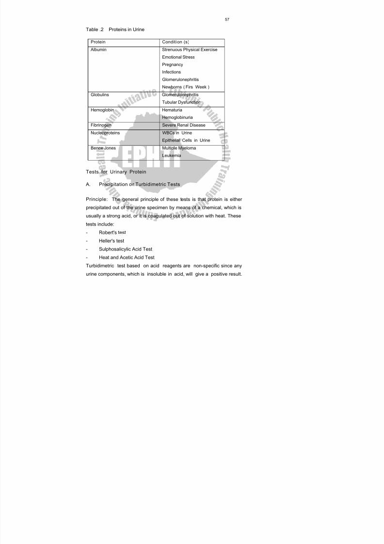
8/13/2019 LN Urinalysis Final
http://slidepdf.com/reader/full/ln-urinalysis-final 68/138
57
Table .2 Proteins in Urine
Protein Conditi on (s)
Albumin Strenuous Physical Exercise
Emotional Stress
Pregnancy
InfectionsGlomerulonephritis
Newborns ( Firs Week )
Globulins Glomerulonephritis
Tubular Dysfunction
Hemoglobin Hematuria
Hemoglobinuria
Fibrinogen Severe Renal Disease
Nucleoproteins WBCs in Urine
Epithelial Cells in Urine
Bence Jones Multiple Myeloma
Leukemia
Tests for Urinary Protein
A. Precipitation o r Turbidimetric Tests
Principle: The general principle of these tests is that protein is either
precipitated out of the urine specimen by means of a chemical, which is
usually a strong acid, or it is coagulated out of solution with heat. These
tests include:
- Robert's test
- Heller's test
- Sulphosalicylic Acid Test
- Heat and Acetic Acid Test
Turbidimetric test based on acid reagents are non-specific since any
urine components, which is insoluble in acid, will give a positive result.
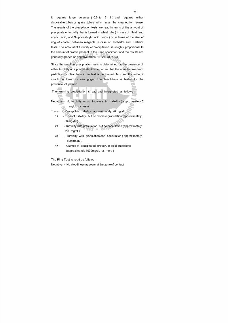
8/13/2019 LN Urinalysis Final
http://slidepdf.com/reader/full/ln-urinalysis-final 69/138
58
It requires large volumes ( 0.5 to 5 ml ) and requires either
disposable tubes or glass tubes which must be cleaned for re-use.
The results of the precipitation tests are read in terms of the amount of
precipitate or turbidity that is formed in a test tube ( in case of Heat and
acetic acid, and Sulphosalicylic acid tests ) or in terms of the size of
ring of contact between reagents in case of Robert`s and Heller`stests. The amount of turbidity or precipitation is roughly proportional to
the amount of protein present in the urine specimen, and the results are
generally graded as negative, trace, 1+, 2+, 3+, or 4+.
Since the result in precipitation tests is determined by the presence of
either turbidity or a precipitate, it is important that the urine be free from
particles or clear before the test is performed. To clear the urine, it
should be filtered or centriguged. The clear filtrate is tested for the
presence of protein.
The non-ring precipitation is read and interpreted as follows :
Negative - No turbidity, or no increase in turbidity ( approximately 5
mg/dL or less)
Trace - Perceptible turbidity ( approximately 20 mg /dL).
1+ - Distinct turbidity, but no discrete granulation (approximately
50 mg/dL ).
2+ - Turbidity with granulation, but no flocculation (approximately
200 mg/dL).3+ - Turbidity with granulation and flocculation ( approximately
500 mg/dL).
4+ - Clumps of precipitated protein, or solid precipitate
(approximately 1000mg/dL or more )
The Ring Test is read as follows :-
Negative - No cloudiness appears at the zone of contact
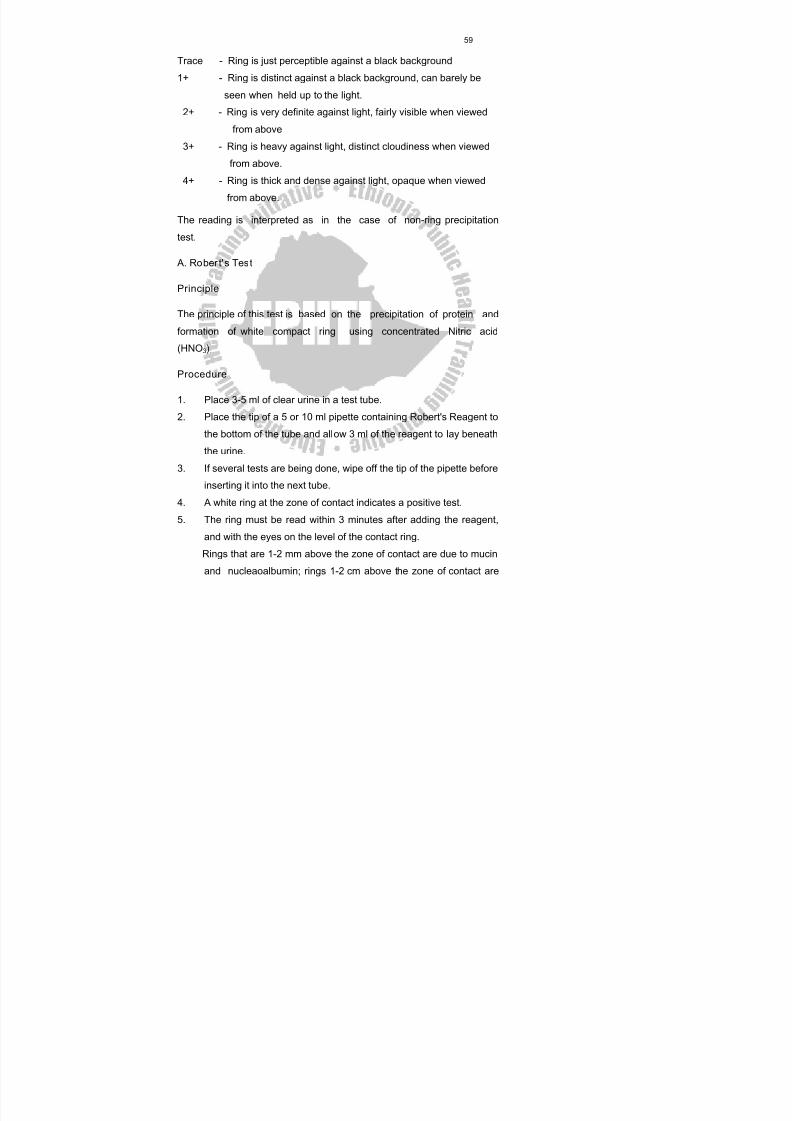
8/13/2019 LN Urinalysis Final
http://slidepdf.com/reader/full/ln-urinalysis-final 70/138
59
Trace - Ring is just perceptible against a black background
1+ - Ring is distinct against a black background, can barely be
seen when held up to the light.
2+ - Ring is very definite against light, fairly visible when viewed
from above
3+ - Ring is heavy against light, distinct cloudiness when viewedfrom above.
4+ - Ring is thick and dense against light, opaque when viewed
from above.
The reading is interpreted as in the case of non-ring precipitation
test.
A. Rober t' s Test
Principle
The principle of this test is based on the precipitation of protein and
formation of white compact ring using concentrated Nitric acid
(HNO 3).
Procedure
1. Place 3-5 ml of clear urine in a test tube.
2. Place the tip of a 5 or 10 ml pipette containing Robert's Reagent to
the bottom of the tube and allow 3 ml of the reagent to lay beneath
the urine.3. If several tests are being done, wipe off the tip of the pipette before
inserting it into the next tube.
4. A white ring at the zone of contact indicates a positive test.
5. The ring must be read within 3 minutes after adding the reagent,
and with the eyes on the level of the contact ring.
Rings that are 1-2 mm above the zone of contact are due to mucin
and nucleaoalbumin; rings 1-2 cm above the zone of contact are
8/13/2019 LN Urinalysis Final
http://slidepdf.com/reader/full/ln-urinalysis-final 71/138
60
due to urates, uric acid urea and bile, acids. These are not to be
reported positive for protein.
6. Report the result according to the chart given on the above for ring
tests.
7. The test may be performed by holding a test tube containing a few
ml of Robert's Reagent in an inclined position and allowing theclear urine to run slowly down the side of the tube from a pipette.
Preparation of Robert's Reagent (See Reagent Number 11 )
Note : If bile is present in the specimen, any colors ( red, violet,
blue, or green ) will be found at the line of contact.
B. Heller's Test
Principle : The same as Robert̀ s Test
Procedure
1. Perform the test as directed under Robert's test using concentrated
nitric acid instead of Rober's Reagent, and read the white ring at
the zone of contact in the same manner.
2. If bile is present, any colors (red, violet, blue or green) will be
found at the line of contact.
3. Interfering rings listed under Robert's Test also apply to Heller's
Test.
Note : Heller's Test is not suitable for routine analysis of proteinuria
because of the highly corrosive nature of concentrated nitric
acid.
Heller's Reagent : It is c oncentrated nitric acid.
C. Sulphosalicylic Acid Test
Principle
This test is based on the precipitation of protein (particularly
8/13/2019 LN Urinalysis Final
http://slidepdf.com/reader/full/ln-urinalysis-final 72/138
61
albumin ) by sulphosalicylic acid,
Procedure
1. Place 3ml centrifuged urine in a test tube.
2. Add 3 ml of 20 % sulphosalicylic acid.
3. Mix thoroughly and estimate the amount of turbidity 10
minutes later.
4. Read and record results according to the chart given for non-ring
precipition test.
Preparation of 20 % w/v Sulphosalicylic Acid Reagent (See Reagent
Number 14)
D. Heat and Acetic Acid Test
Principle
The test is based on the precipitation of protein by heat.
Procedure
1. Fill a test tube three-fourth full of clear urine, and gently heat the
upper portion of urine for 2 minutes to boil, being careful not to
shake the tube more than necessary. The lower portion of urine
is not heated so that it can be used as a control for
comparing.
Note: Rotate the tube to prevent cracking.
2. Now turbidity ( a white cloud ) can arise due either ofphosphates, carbonates, or protein.
3. Add 3 drops of 10% acetic acid drop by drop, boiling between
each drop.
4. A white cloud that disappeared is due to phosphates or carbonates.
Persistence or development of turbidity implies proteinuria.
5. Read the test and record results according to the chart for non-
ring precipition test.
8/13/2019 LN Urinalysis Final
http://slidepdf.com/reader/full/ln-urinalysis-final 73/138
62
Preparation of Acetic Acid Reagent (See Reagent Number 1)
Sensitivity
This method is the most sensitive for small amount of protein and can
reliably detect protein concentrations of 2 to 3 mg/dl.
II. Colorimetric Reagent Strip (Dipstick)Tests
The Colorimetric (dipstick) Protein Tests are more specific than
Turbidimetric Tests. They require only a drop of urine enough to
moisten the reagent area.The Colorimetric reagent strip test is based
on the ability of protein to alter the color of some cid-base
indicators without altering the pH. When an indicator, such as
tetrabromophenol blue is buffered at pH 3, it is yellow in solutions
with out protein but , in the presence of protein, the color will
change to green and then blue with increasing proteinconcentrations. In this case the pH of the urine is held constant by
means of a buffer so that any change of color of the indicator will
indicate the presence of protein.
The tests for urinary protein are all commercial ones, that are available
as reagent strip, tests (Dipsticks) either alone or in combination with
other tests. Example. albustix, Uristix, N-Multistix, Combur3 or
Combur9. Although the colorimetric tests are useful primarily as
screening tests for protein, these strip tests can be read
semiquantitatively as negative, trace, 1+, 2+, 3+, or 4+ to give a rough
estimate of the amount of protein present. To do this, the resulting color
must be matched closely with the color chart provided with the test
strips.The albustix and other multiple-reagent strips produced by ames
co. are plastic strips with protein test areas impregnated with citrate
buffer and tetrabromphenol blue. The citrate buffer maintains the pH at
3. At pH 3 tetrabromphenol blue is yellow in the absence of protein and
8/13/2019 LN Urinalysis Final
http://slidepdf.com/reader/full/ln-urinalysis-final 74/138
63
yellow - green, or blue in its presence. The shade of the color is
dependent on the amount of protein present. Falsely positive reactions
may occur when protein is absent, if the urine is exceptionally alkaline or
highly buffered.
Procedure
Observe the precautions and follow the instructions supplied by the
manufacturer.
1. Dip the reagent area of the strip briefly into the specimen.
2. Remove excess urine by tapping or drawing the edge of the strip
along the rim of the urine container.
3. Compare the color that develops with the color chart supplied by the
manufacturer and report as indicated on the chart.
Quantitative 24 hour Protein Determinations
Simple estimates of the protein content of urine are performed by
quantitating the amount of precipitation formed following the addition of a
specific chemical to the urine. The precipitate is measured either by
comparison with known standards (sulphosalicylic acid turbidity test) or
by recording the height of the column of precipitate in a specially-
designed tube (Esbach's test).
A. Sulpho salicyl ic Ac id Turbidi ty Test
Procedure
1. Pipette 2.5 ml of centrifuged urine into a test tube.
2. Add 7.5 ml of 3% sulphosalicylic acid.
3. Invert to mix
4. Let stand 30 minutes.
Compare the turbidity with known standards prepared from
solutions containing 10, 20, 30, 40, 75 and 100mg albumin/dl,
and estimate the concentration of the unknown. If the unknown
8/13/2019 LN Urinalysis Final
http://slidepdf.com/reader/full/ln-urinalysis-final 75/138
64
urine contains more than 100mg/dl protein, dilute the urine and
repeat the test.
B. Esbach’s Test
1. If a 24 hour urine collection is used, first, measure the total
volume; then filter some of the urine. The urine must be clear.
2. Do qualitative protein test, Robert’s or strip test.
- if the urine is +3, made 1:5 dilution.
- if the urine is +4 make 1:10 dilution.
- if the urine is trace, +1 or +2 non dilution is needed.
- if the urine is negative, a quantitative test is not done.
3. Test measure the pH of the urine. It should be acidic. If not, add
10 % acetic acid.
4. Add pumice powder to the 0.5 mark of the Esbach’s tube.
5. Add urine to the “U” mark.6. Add Esbach’s reagent to the “R” mark.
7. Mix slowly by inversion, 10 times.
8. Wait for 30 minutes. Read the highest of the column. Do not
subtract the amount of the pumice.
9. The result is now in gram per liter of protein in the urine. If the
urine has been diluted, multiply by the dilution factor, calculate,
and record the g % and the g / 24 hrs
Final report should include total volume.
The following formula is used to calculate the amount of urinary
protein excreted in 24 hrs.
g/24 hr = total volume x g/l
1000
8/13/2019 LN Urinalysis Final
http://slidepdf.com/reader/full/ln-urinalysis-final 76/138
65
4.4 Determination of Bilirubin
Introduction
Bilirubin is a waste product that must be eliminated from the body. It is
formed by the breakdown of hemoglobin in the reticuloendothelial cells
of the spleen and bone marrow, and then transported to the liver.
On its way to the liver it is not water-soluble, and is carried through the
blood stream linked to plasma albumin. This water insoluble form of
bilirubin is often referred to as free bilirubin or unconjugated bilirubin or
indirect bilirubin. Since this albumin - bound form is insoluble in water; it
does not appear in the urine. In the liver bilirubin is converted to a water-
soluble product by conjugation with glucuronic acid to form bilirubin
glucuronide. The water-soluble form is called cunjugated bilirubin. It is
also called direct bilirubin. The liver cells that form the conjugated
bilirubin excrete it into the bile and it is then excreted into the intestinaltract through the bile duct. In the small intestine this conjugated
bilirubin is converted by intestinal bacteria to urobilinogen or
stercobilinogen.
Even though normally the level of conjugated bilirubin in the blood is not
high enough to cause significant amounts to appear in the urine, this
water soluble and conjugated bilirubin can be excreted by the kidneys.
Normal Value: approximately up to 0.02 mg/dl (This amount is not
detected by routine qualitative or semi quantitative techniques).
Clinical Significance
Tests for urinary bilirubin and urobilingen were normally performed only
indicated by abnormal color of the urine or when liver disease or a
hemolytic condition was suspected from the patient's history. The
presence of bilirubin and urobilinogen in the urine is an early sign of liver
8/13/2019 LN Urinalysis Final
http://slidepdf.com/reader/full/ln-urinalysis-final 77/138
66
cell disease (hepatocellular disease) and obstruction of the bile flow from
the liver (Obstructive or post - hepatic jaundice).
Urine containing bilirubin will typically have been brown color and
produce a yellow foam when shaken. Bilirubin is not stable in solution,
but will be oxidized to biliverdin, which is a green pigment. Thus urine-
containing bilirubin will typically be red-brown when voided, and will turngreen on standing, especially if exposed to light. Tests for bilirubin will
not be positive in the presence of biliverdin; so the urine must be
examined when fresh.
Tests for Bilirub in
Tests for bilirubin are based on the oxidation of bilirubin to biliverdin.
Specimen : Freshly passed urine is required. Urine containing bilirubin
should be analyzed immediately after collection (with in 2 hrs of voiding).
If bilirubin exposed to sunlight, it will oxidize to biliverdin, which cannotbe detected by the reagents used in any of the tests. The following tests
are used to detect bilirubin in the urine.
A. Harr ison 's (Fouchet' s) Test
Principle
This test depends on precipitation of bilirubin with barium chloride, which
is then oxidized to biliverdin with Fouchet's reagent. The formation of
biliverdin gives a green color, which constitutes a positive reaction.
Procedure
1. Add 5 ml of a 10% solution of barium chloride to 10 ml of urine.
Mix, and let stand a Few minutes.
2. Filter through a small filter paper.
3. Spread the filter paper on a dry piece of filter paper and place one
or two drops of Fouchet's reagent.
8/13/2019 LN Urinalysis Final
http://slidepdf.com/reader/full/ln-urinalysis-final 78/138
67
4. A blue to green color indicates a positive reaction.
5. Report as positive or negative.
Prparation of Fouchet's Reagent (See Reagent Number 9 )
B. Barium chloride Filter Paper Method
It is the modification of Harrison's test. The barium chloride is suppliedon thick filter paper that has been soaked in a saturated solution of
barium chloride.
Principle
The principle of the test is the same as Harrison's (Fouchet's) Test.
Procedure
1. Hold a strip of barium chloride paper perpendicularly in the urine
for a few seconds2. Place one or two drops of Fouchet's Reagent on the saturated
area.
3. Look for the appearance of a green color, which constitutes a
positive reaction.
4. Report as positive (formation of a green color) or negative
(formation of any color except green, or no color formation).
Preparation of Barium Chloride Filter Paper( See Reagent Number 3)
Sensitivity
Harrison's Test is so sensitive that it detects as small as 0.1 - 0.2 mg of
bilirubin in 100ml of urine.
C. Diazotization Tests for Bilirubin
The tablet and reagent strip tests for bilirubin are based on the coupling
of bilirubin with a diazonium salt in an acid medium to form azobilirubin,
which gives a blue or purple color.
8/13/2019 LN Urinalysis Final
http://slidepdf.com/reader/full/ln-urinalysis-final 79/138
68
1. Icotest Tablet Test
The Ictotest tablet contains nitrobenzine diazonium, p-toluene sulfonate
(bilazo),
sulfosalicylic acid, and sodium bicarbonate. The mats are absorbent
asbestos
cellulose.
Procedure
1. Place five drops of urine on either side of the special test mat
supplied with the reagent tablets.
2. Place the tablet in the center of the moistened area.
3. Flow two drops of water on the tablet.
4. Observe the mat around the tablet for the appearance of a blue to
purple color within 30 seconds.
5. Report the results as positive or negative according to thefollowing criteria.
Negative: The mat shows no blue or purple within 30 seconds. Ignore
any color that forms after 30 seconds or a slight pink or red
that may appear.
Positive: The mat around the tablet turns blue or purple within 30
seconds. Ignore any color change on the tablet itself.
Sensitivity: Ictotest detects 0.1mg of bilirubin in 100ml of urine.
2. Reagent Strip Tests for Bili rub in (Ex. Multis tix)
Principle
These tests for bilirubin are available only on multiple-reagent strips in
conjugation with other tests. They are diazotization tests and are
analogous to the Icotest tablet test. The test area for bilirubin on
8/13/2019 LN Urinalysis Final
http://slidepdf.com/reader/full/ln-urinalysis-final 80/138
69
Multistix and other Ames Co. reagent strip products is impregnated with
2,4-dichloro-aniline diazonium salt.
The reagent strip tests for bilirubin are difficult to read and the color
formed after reaction with urine must be carefully compared with color
chart supplied by the manufacturer.
Procedure
1. Dip the reagent strip briefly into the urine specimen.
2. Remove excess urine by tapping the edge of the strip against the
rim of the urine Container. Hold the strip in a horizontal position to
prevent mixing of chemicals from adjacent reagent areas.
3. Compare the test areas for bilirubin closely with the color chart
supplied by the manufacturer. Multistix should be read 20
seconds after dipping.
4. Report the results as positive or negative for bilirubin.
Sensitivity
Multistix detects 0.2-0.4 mg of bilirubin in 100ml of urine.
4.5 Determination of Urobili nogen
Introduction
In the intestine, most of the bilirubin is converted to urobilinogen or
stercobilinogen by the action of certain bacteria that make up theintestinal flora. Approximately half of the urobilinogen formed in the
intestine is absorbed into the portal blood circulation and returned to the
liver. In the liver most of the urobilinogen is excreted into the bile once
again and returned to the intestine.
A very small amount of urobilinogen about 1 percent of the formed
urobilinogen is excreted from the body in the urine as urobilinogen or
can be also converted into urobilin, which gives the urine its
8/13/2019 LN Urinalysis Final
http://slidepdf.com/reader/full/ln-urinalysis-final 81/138
70
characteristic color with the other color pigments (urochroms).
Urobilinogen is also converted into urobilin when exposed to air .
Stercobilinogen in the intestine is either eliminated from the body
unchanged or oxidized to the colored compound stercobilin, which gives
the faces its characteristic color. Thus, urine normally contains only a
very small amount of urobilinogen and no bilirubin. Both are abnormalurinary constituents. However, there are several serious conditions in
which either one or both of these substances are found in the urine.
When testing for urobilinogen the urine specimen must be fresh, since it
is usually unstable and it is rapidly oxidized to urobilin. This oxidation
takes place so readily that most urine specimens that contain
urobilinogen will show an abnormal color caused by partial oxidation of
urobilin. The presence of urobilinogen and that of urobilin have the same
clinical significance, however, they take part in different chemical
reactions, and urine is more frequently tested for urobilinogen.
Normal value: Normally 1-4 mg of urobilinogen is excreted in the urine
each day.
Clinical Significance
Urine is often tested for increases in urobilinogen when investigating
hemolytic jaundice or liver disorder in which liver function is
impaired.
Test for Urobilinogen
A. Qual it ative Ehrl ich's Test for Urobi linogen
Principle
The test depends upon the reaction between urobilinogen and para-
dimethylaminobenzaldhyde to form a cherry (deep) red.
8/13/2019 LN Urinalysis Final
http://slidepdf.com/reader/full/ln-urinalysis-final 82/138
71
Procedure:
1. Place 10 ml urine in a test tube. Allow warming to room temperature.
2. Add 1 ml Ehrlich's reagent and mix.
3. Let stand 3 to 5 minutes
4. Normal amounts of urobilinogen present in the urine sample will
change the solution to pink. Abnormally high amounts of urobilinogenwill change the solution to a Cherry red color. This must be reported
positive for urobilinogen.
Disregard any pink or light red coloration. This test is of no value in
infections of the Urinary tract because some bacteria produce nitrites,
which give false positive reaction. Formaline interferes with the test
and should not be used as a preservative.
Preparation of Ehrlich’s Reagent- see reagent number 7
B. Reagent Strip Tests for Urobilinogen- Urobilis tix
The reagent strips have test areas for urobilinogen, which are based on
the Ehrlich's reaction in which p-dimethylaminobenzaldehyde reacts with
urobilinogen in a strongly acidic medium to form a colored aldehyde.
The reddish brown color that is formed varies with the amount of
urobilinogen present. After a timed interval, the color is compared with a
graded color chart. However, the test is not specific for urobilinogen and
reacts with substances know to react with Ehrlich's reagent.
Procedure:
1. Dip the reagent strip briefly into the specimen.
2. Remove excess urine by tapping the edge of the strip against the rim
of the urine Container.
3. Compare the color of the test area after 60 seconds with the color
chart supplied by the manufacturer.
8/13/2019 LN Urinalysis Final
http://slidepdf.com/reader/full/ln-urinalysis-final 83/138
72
Sensitivity
Urobilistix detects 0.1 mg in 100 ml of urine (0.1 Ehrlich units in 100 ml)
4.6 Test For Urobili n
Urobilin is an oxidation product of urobilinogen. Urobilin is colored and
urobilinogen is colorless. Both compounds have the same clinical
significance when present in urine; however, they undergo different
chemical reactions.
Schilesinger's Test for Urobilin
Principle
When zinc acetate reacts with urobilin it produces a green fluorescence.
Procedure1. Mix equal parts (10 ml each) of urine and alcoholic solution of zinc
acetate in a test tube. Mix and filter the mixture.
2. Take 10 ml of the filtrate and add 2 drops of Lugol's solution. Mix
by inversion.
3. Examine the solution for green fluorescence by viewing the tube
from above as it is passed through the direct rays of a fairly strong
light (sunlight or wood's light).
4. Report as positive or negative. Urobilin produce a green
fluorescence while
porphrins produce red fluorescence.
Preparation of Alcoholic Solution of Zinc Acetate (See Reagent
Number 13)
8/13/2019 LN Urinalysis Final
http://slidepdf.com/reader/full/ln-urinalysis-final 84/138
73
4.7 Determination of Hemogl obin
Introduction
Hemoglobin is a respiratory pigment in red blood cells composed of an
iron- containing group (heme) and a complex protein (globin). In
combination as hemoglobin it has the property of forming a reversible
combination with oxygen. So, it serves as a transporter of oxygen in the
blood from the lung to metabolically active tissues. It also transports
carbon dioxide and hydrogen ions to the lung from metabolically active
tissues.
Hemoglobin appears in the urine when there is extensive or rapid
destruction
(hemolysis) of circulating erythrocytes that the reticuloendothelial
system cannot metabolize or store the excessive amounts of free
hemoglobin.
Normal Value: The renal threshold for hemoglobin is 1.0 - 1.4 g/1.
Clinical significance
The presence of free Hemoglobin in the urine is referred to as
hemoglobinuria. Hemoglobinuria is usually related to hematuria- a
condition when intact red blood cells are present in the urine. Hematuria
is used to indicate bleeding somewhere in the urinary tract.Usually both
red blood cells and hemoglobin mark this disorder. Therefore, hematuria
can be distinguished from hemoglobinuria by a microscopic examination
of the sediment from a fresh urine specimen.The presence of
hemoglobin and the absence of red cells in the urine does not
necessarily mean that the hemoglobin was originally free urinary
hemoglobin. Red cells rapidly lysis in urine, especially when it has a
specific gravity of 1.006 or less or is alkaline. For this reason urine
should be absolutely fresh when examined for the presence of red cells.
8/13/2019 LN Urinalysis Final
http://slidepdf.com/reader/full/ln-urinalysis-final 85/138
74
Hemoglobinuria may occur with severe intravascular hemolysis when the
amount of hemoglobin being released into the plasma is more than can
be taken up by haptoglobin (the plasma protein that binds free
hemoglobin to prevent it being lost from the body). This results from a
variety of conditions and disease states. It may be the result of
hemolysis in the blood stream, in a particular organ, in the kidney oflower urinary tract or in the sample itself.
Causes of Intravascular Hemolysis
Intravascular Hemolysis is associated with:
- Hemolytic anemia.
- Severe infectious disease such as Falciparum Malaria, Yellow
Fever, Small Pox and Typhoid Fever.
- Glucose - 6- phosphate dehydrogenase (G6PD) deficiency.
- The ingestion of certain drugs.
- Escherichia coli septicaemic.
- Incompatible blood transfusion.
- Snake bites that cause acute hemolysis.
- Sickle cell disease- crisis with sever hemolysis.
- Severe viral hemorrhagic fever accompanied by intravascular
hemolysis.
- Poisonings with strong acids or mushrooms sever-burns and renal
infarction.- Significant amounts of free hemoglobin occur when ever excessive
numbers of red cells are present as a result of various renal
disorders.
8/13/2019 LN Urinalysis Final
http://slidepdf.com/reader/full/ln-urinalysis-final 86/138
75
Specimen
Urine containing hemoglobin appears brown or brown-gray on color and
is usually cloudy. It should be tested as soon as possible after it has
been passed.
Laboratory Tests for Hemoglobi n
Numerous Tests are available for the detection of hemoglobin in urine.
The tests commonly used are:
- Guaiac
- Benzidine
- Occultest
- Reagent Strip Tests (Hemastix)
Benzidine is no longer used, as it is a carcinogen.
General Principle of the Tests
These tests are based on the same general principles and reaction.
They all involve the presence of peroxidase activity of the hemoglobin
and hydrogen peroxide (H 2O2) or a suitable precursor which librates
oxygen. Peroxidase of the hemoglobin molecule liberates oxygen from
hydrogen peroxide and the librated oxygen reacts with an organic
reagent or chromogen (gum guaiac or 0-tolidine) to give a colored
compound, which is usually blue or green. The intensity of the color
depends on the amount of librated oxygen. The amount of librated
oxygen depends on the peroxidase ctivity of hemoglobin molecule,
which intern depends on the amount of hemoglobin found in the urine.
These reactions are summarized below:
Hemoglobin + H 2O2 Perosidase Oxygen
Oxygen + Chromogen Blue or green oxidation products
8/13/2019 LN Urinalysis Final
http://slidepdf.com/reader/full/ln-urinalysis-final 87/138
76
Since all these tests are based on the peroxidase activity of hemoglobin,
other substances with peroxidase activity also give positive reactions in
the tests.
Factors that Affect Hemoglobin Determination
False negative
- High specific gravity such as heavy proteinuria (over 5 g/1). This
prevent lysis of RBCs and may reduce the color reaction.
- Low to false negative results are obtained if the urine contains
large amounts of ascorbic acid.
- Nitrite delays test reaction.
- Formaline used as preservative, fix the cell and prevent hemolysis.
False po sitive
- Low specific gravity < 1.010 enhances lysis and produces color
reaction.
- Microbial peroxidase produces false the color reaction.
- False positive reactions can result from the presence of
contaminating oxidizing detergents on the urine such as bleach.
A. Guaiac Test
Procedure:
1. Pour into a test tube 4 drops of urine and a few drops of
concentrated acetic acid.
2. Pour the following into a second test tube
- A knife point of Guaiac
- 2 ml ethanol (95%)
- 2 ml fresh 3% H 2O2
Mix the above slowly and pour the same into the side of the urine
tube.
8/13/2019 LN Urinalysis Final
http://slidepdf.com/reader/full/ln-urinalysis-final 88/138
77
3. A green or blue color is a positive test. The chromogen in this test
gum guaiac.
B. Occult est (tablet method)
Principle
When the tablet is moistened with water tartaric acid and calciumacetate react with strontium peroxide to form hydrogen peroxide. The
hemoglobin in the urine catalytically decomposes hydrogen peroxide
liberating oxygen, which oxidizes orthotolidine to a blue derivative.
Procedure:
1. Place a piece of filter paper, which comes with the reagent on a
clean surface.
2. Place one drop of well-mixed urine in the middle of filter paper.
3. Place occultest tablet in middle of the moist area.4. Flow two drops of water over the tablet.
5. Observe color of filter paper around tablet exactly two minutes
later.
6. The test is positive, if a blue color appears on the filter paper
around the tablet.
7. Report as positive or negative.
Content of th e tablet
The tablet consists of orthotolidine, tartaric acid, strontium peroxide,sodium bicarbonate, calcium acetate, and red dye.
C. Reagent Strip Tests (Hemastix )
Principle:
When the strip is dipped in a urine containing hemoglobin, the action is
similar to that of occultest tablets in that the hemoglobin ( since it has a
8/13/2019 LN Urinalysis Final
http://slidepdf.com/reader/full/ln-urinalysis-final 89/138
78
peroxidase activity) catalyzes the oxidation of orthotolidine by the
peroxide. The oxidized orthotolidine is blue.
Procedure:
1. Follow the manufacturers directions and precautions.
2. Dip the test end of strip into a well-mixed specimen of urine and
remove immediately.3. After 30 seconds compare the test area with the color chart
provided.
4. Report as indicated on the color chart.
Composition of the strip
The test area is impregnated with orthotolidine, cumene hydroperoxide
and citrate buffer.
4.8 Determination of Urinary Calcium
Introduction
The bulk of calcium ions (Ca++) discharged by body is excreted in the
stool. However, there is small quantity of calcium that is normally
excreted urine. But it may increase depending up on the quantity of
dietary calcium ingested. The 24 hour test is most often ordered to
determine the function of the parathyroid gland, which maintains a
balance between calcium and phosphorous by means of parathyroid
hormone. Hyperparathyrpidism is a generalized disorder of calcium,
phosphate and bone that results from increased secretion of parathyroid
hormones and an increased excretion of urinary calcium. In
hypoparathyrodism the urinary calcium is decreased.
Interfering Factors
1. False positive
- High sodium and magnesium intake.
8/13/2019 LN Urinalysis Final
http://slidepdf.com/reader/full/ln-urinalysis-final 90/138
79
- High milk intake.
- Some drugs.
- If test done immediately after high calcium meal.
2. False negatives
- Increased dietary phosphates.
- Alkaline urine.- Some drugs.
Test fo r Calcium
The test for calcium in the urine is not included in the routine urinalysis.
The qualitative test of Sulkowitch is used for calcium determination.
Sulkowitch Test
Principle of Test
In this test a solution of ammonium oxalate ( about 4 % ) is added to
the urine. If calcium is present in excessive amounts, it drops out of
solution as a heavy white precipitate of calcium oxalate.
Procedure
1. Pour 5 ml of urine into a test tube.
2. Add 5 ml of Sulkowitch reagent ( see reagent No. 13 ).
3. Mix by inverting the tube several times.
4. Allow to stand 3 minutes.
5. If a fine white cloud appears, the calcium content is normal.
6. If no white cloud appears, the calcium content is decreased.
7. If a heavy white milky precipitate forms, the calcium content is
increased.
8. Report the calcium content as normal, decreased or increased.
Preparation of Sulkowitch Reagent (See Reagent Number 12).
8/13/2019 LN Urinalysis Final
http://slidepdf.com/reader/full/ln-urinalysis-final 91/138
80
4.9 Determination of Nitrit e
Introduction
Tests for nitrite was not a part of the routine urinalysis. It is included
recently on commercial multiple-reagent strips such as Combur 9 and N-
Multistix. The detection of nitrite in the urine can be used to indicate thepresence of bacteria such as Escherchi coli, proteius, klebsiella,
enterobacter, citrobacter, and salmonella will reduce urinary nitrate to
nitrite.The presence of urinary nitrite(in urine sample collected under
sterile condition) indicates the existence of a urinary tract infection.
Detection of such infections is particularly important in pregnant women
and young girls to prevent permanent renal damage. Since the detection
rate increases when the urine is held in the bladder for at least 4-6
hours, test for nitrite must be performed on the first morning urine
specimen. For proper detection of nitrite the patient diet must alsocontain enough reducible nitrate on which the bacteria can act.
Types of Method Used for Detection of Nitrite
There are two methods that are used to detect bacteria in the urine
during routine urinalysis.
1. Microscopic examination: urine sediment when examined
microscopically can reveal bacteria when present. If the sample is
collected under sterile condition.
2. Chemical dipstick method for nitrite can also give clue.
The nitrite area in the multiple reagent strip is calibrated so that any
shade of pink color that develops within 30 sec indicates an amount of
nitrite produced by 105 or more organisms per milliliter in the urine
specimen.
A positive result from the nitrite test is a reliable indication of significant
bacteriuria ( the presence of bacteria in the urine) and is an indication
8/13/2019 LN Urinalysis Final
http://slidepdf.com/reader/full/ln-urinalysis-final 92/138
81
for urine culture unless the specimen has been improperly collected or
stored after collection which allow bacterial growth.
A negative result should never be interpreted as indicating absence of
bacteriuria because:
a. If an overnight sample were not used, there may have been
insufficient time for the conversion of nitrate to nitrite to occur.Urine that has been left in the collection vessel for several hours
may be falsely positive.
b. Some amounts are caused by organisms that do not convert
nitrate to nitrite (such as enterococci, acinetobacter spp and some
pseudomonas species.).
c. The patient was in a vegetable free diet, which is the important
source for nitrate.
d. Administration chemotherapeutic agents should be discontinued
three days before the test, because antibiotic therapy may alter
bacterial metabolism so as to render nitrite detection invalid.
e. High doses of ascorbic acid.
f. Presence of urobilinogen
g. Low pH (< 6)
Even though the test depends upon the conversion of nitrate to nitrite by
certain bacterial action in the urine, not all bacteria in the bladder
convert nitrate to nitrite. Known nitrate reducing bacteria at significant
levels produce false negative results by reducing nitrate to ammonia
nitrite, and nitrous oxide, hydroxylamine, and nitrogen and will therefore
give a negative nitrite test.
Principle
The Reagent Strip ( Multistix with an acid pH) contains para-arsanilic
acid which reacts with nitrite, to give a diazonium salt, which by looping
with a benzoquinoline forms pink azo dye.
8/13/2019 LN Urinalysis Final
http://slidepdf.com/reader/full/ln-urinalysis-final 93/138
82
The pink color is therefore related to the presence of bacteria in the
urinary tract. However, the amounts of color produced cannot be related
to the number of bacteria present and the result should be reported only
as positive or negative.
Falsely positive reactions may be caused by bacterial growth in "old"
urine specimens or by medication such as phenazopyridine that colorsthe urine red or that turns red in an acidic medium.
Procedure
Follow the manufacturer's instruction and precautions:
1. Dip the test area of the strip briefly into the specimen.
2. Remove excess urine by tapping the edge of the strip along the
rim of the container.
3. Compare the color that develops with the color chart supplied by
the manufacturer.Report as positive or negative within the time specified by the
manufacturer.
Sensitivity
N-Multistix detect 0.075 mg of nitrite in 100 ml of urine and combur 9 test
detects 0.05 mg per 100 ml of urine.
4.10 Leukocytes Test
Introduction
Tests for leukocytes has become part of the routine urinalysis since
commercial multiple-reagent strips began to be marketed (N-Multistix or
Combur 9). The presence of leukocytes indicates inflammation at some
point along the urogenital tract.
8/13/2019 LN Urinalysis Final
http://slidepdf.com/reader/full/ln-urinalysis-final 94/138
83
Principle of Test
The reaction on the test strip reveals the presence of esterases that
occur in granulocytes. These esterases cleave an indoxyl ester, and the
indoxyl so liberated reacts with a diazonium salt to produce a violet dye.
Procedure
1. Insert the strip to the specimen. (no longer than 1 second).
2. Wipe off excess urine on the rim of the container.
3. After 60-120 seconds compare the test by matching with the color
scale on the label supplied by the manufacturer.
Most tests are done on a random specimen of freshly voided urine by
the patient. The specimen is collected in a clean dry container. If the
determination is not done immediately, the specimen should be
preserved at 5 0c in the refrigerator.
4.11 Determination of Indic an /Indoscyl e Sulphate
Introduction
Indican is driven from indole, which is the putrefaction product of protein,
by protolytic bacteria. Patients with bacterial over growth in the small
intestine excrete large amount of metabolites of amino acids such as
tryptophan or tyrosine. Indole is produced by bacterial action on
tryptophan in the intesintine, which mostly eliminated with feces, some
are absorbed and detoxified in the liver and excreted as indican in the
urine.
In normal urine the amount of indican excreted is small, it is
increased in
1. High protein diet
2. Pathological conditions such as
- Bacterial putrefactions.
- Enteritis.
8/13/2019 LN Urinalysis Final
http://slidepdf.com/reader/full/ln-urinalysis-final 95/138
84
- Pancreatic insufficiency.
- Intestinal Infection.
- Ulceration of intestinal mucosa.
To differentiate the pathological conditions from non-pathological, first
restrict the patient from protein intake and then do the test.
Test for Indican
Obemyares Test
Principle
HCL liberates indoxyle from indican and ferric chloride (FeCl 3) oxidizes
the indoxyle to indigo blue.
Procedure
1. Add 5 ml well mixed fresh urine and 5 ml obemyares reagent into
a test tube.
2. Add 4 drops of chloroform and mix several times by inverting until
all color dissolves out.
3. Add saturated ferric chloride drop by drop, mixing well after each
addition and record the number of drops you add to decolorize.
When indican is present, the chloroform layer shows a deep violent to
blue color. Normally when two drops are added, the color comes to light
sky blue, if it needs more. It shows the presence of increased amount of
indican.
Source of Errors
- Indican may be formed because of slow oxidation.
- Presence of iodide may case violent color removed by adding
crystals of sodium thiosulfate.
- Bile pigments – removed by shaking BaCl 2 and filtering.
8/13/2019 LN Urinalysis Final
http://slidepdf.com/reader/full/ln-urinalysis-final 96/138
85
4. 12 Determination of Melanin
Introduction
Melanin is pigment derived from tyrosine, which is normally present in
hair, skin and in the choroid layer of the eye.
There are two recognized metabolic pathways for the conversion oftyrosine to melanin:
1. The eumelanin pathway, which polymerize to brown or black
pigments.
2. The pheomelanin pathway – which polymerize to yellow or red
pigments.
Melanomas with pigments are normally transferred from melanocytes to
skin and mucus membrane cells.
In patients with tumors arising from the melanin producing cells, the
melanomas, the melanin may be excreted in the urine in large amount,
and its presence is indicative of metastasis of the tumor to the liver or
other organ.20% of patients with disseminated malignant melanoma
excrete a black urine due to melanin, or its precursor, the colorless
melanogen in the urine. The urine becomes black upon standing
(oxidation), where the chromogen /melanogen is changed into the
pigment called melanin which is a physical method to detect melanin.
Clinical Significance
Melanin occurs in metabolic tambours especially with metastasis of liver.
Chemical Tests for Melanin
There are two types of chemical test for melanin in urine depending up
on either by
a. Utilization of oxidizing agent (e.g. FeCl 3)
b Utilization of reducing action of melanogen
c. Oxidation by atmospheric air
8/13/2019 LN Urinalysis Final
http://slidepdf.com/reader/full/ln-urinalysis-final 97/138
86
A. Ferr ic Chlor ide Test
Principle: FeCl 3 oxidizes the melanogen to melanin.
Procedure
To 5 ml of freshly voided urine, add 1 ml 10% FeCl 3 drop by drop to
precipitate all the phosphates.
Add drop by drop 10% HCl to dissolve all the precipitate and it forms
different color.
Centrifuge and examine for black or gray precipitate of melanin.
If melanin is present, decant the supernatant fluid and add Na 2CO 3 until
alkaline to litmus, melanin precipitates will dissolve again, and then add
10% HCl until acid to litmus; melanin precipitates again as gray or black
sediment when centrifuged.
Preparation of 10 % Hydrochloric Acid Solution (See Reagent Number
10).
B. Thormahlel Test
Principle
Sodium Nitroprusside is reduced to ferocyanide (prussian blue) by
reducing action of melanogen.
8/13/2019 LN Urinalysis Final
http://slidepdf.com/reader/full/ln-urinalysis-final 98/138
87
Procedure
1. Prepare fresh solution of sodium nitroprusside by shaking few
crystals of sodium nitroprusside in 10 ml of distilled water.
2. Add 0.2 ml aqueous sodium nitroprusside to 5ml urine
3. Add 0.5 ml of 40% sodium hydroxide. During this time the urine
turns red due to the action of creatinine and other interferingsubstances.Add 3.5ml of 30% glacial acetic acid, and the color
may change to blue or dark if melanin present which is indication
for a positive reaction.
C. Oxidation by Atmospheric Air
Allow the urine sample to stand exposed to the air undisturbed for 24
hours. If melanogen presents, it will slightly oxidize to melanin by the air,
and turns dark brawn or black from the top downwards.
• Homogenstic acid gives the same effect, but the darkness ofmelanogen is not accelerated appreciably by alkali.
* Microscopic examination of sediment for melanin cast is also possible.
Exercises:
1. Discuss by comparison the Benedict's Qualitative and Glucose
oxidase Tests.
2. List down the possible substances, which give false positive
results in non-specific tests for glucose determination.
3. Mention the physiological effects of ketone accumulation in blood.
4. Write the principle of the test for determination of bilirubin and
hemoglobin.
5. Write the general principles for the two types of determination of
urinary protein.
8/13/2019 LN Urinalysis Final
http://slidepdf.com/reader/full/ln-urinalysis-final 99/138
88
CHAPTER FIVE
Microscopic Examination Of Urine
Objective :It is expected that using the information presented in this chapter the
students will be able to describe normal and abnormal urine sediments
with their diagnostic features.
Introduction
Microscopic examination of urine is one of the routine tests of urinalysis.
As mentioned in the introductory part of this lecture note, urine contains
many substances in addition to water. The amounts of solid substances,
which are found in the urine, may indicate an individual's health status,
i.e. whether one is healthy or sick.
Normally small amount of solid substances is found in the urine. But
when their concentration become high, it may indicate the existence of
abnormal physiological function of our body. Microscopic examination of
urine to some extent can be considered as “renal biopsy” because it
reveals more about the function of the kidneys.
Repeated evaluation of urine sediment is frequently valuable in following
the course and management of urinary tract disorders, because the
appearance of cellular elements, and casts in the urine is a reflection of
changes that take place in the kidney.
Urine sediments can grossly be categorized into organized and non-
organized sediments based on the substances they are composed of.
5.1. Procedure for Micros copic Examination of Urine
1. Assemble all necessary materials used for the collection,
8/13/2019 LN Urinalysis Final
http://slidepdf.com/reader/full/ln-urinalysis-final 100/138
89
centrifugation and examination. This include:
Clean dry plastic or Glass containers, which enable to collect
at least up to 15 ml of urine for routine urinalysis.
Hand (manual), or electrical centrifuge.
Conical centrifuge tubes, or regular test tubes.
Pasture pipette with rubber fit or automatic pipettes if possible. Slides and cover slides 20 x 20 mm.
Electrical or solar microscope, which has 10x and 40 x
objectives.
2. Preparation of patient
Explain the purpose of the test by using simple language. Do
not use medical terms or try to explain details of the
procedure.
Advise the patient how to collect the specimen. The first
morning urine or mid-stream urine specimen is more
preferable, because it is more concentrated.
If the patient is female, advice her to wash her genital organ
before giving the specimen. This is because bacteria that are
normally found on the genital tract may contaminate the
sample and affect the result.
Advise the patient to collect at least 15 ml of urine in to the
clean, sterilize and dry urine cup that is supplied from the
laboratory.
3. The collected urine sample should arrive at a diagnostic laboratory
as soon as possible.
If the urine sample is delayed by more than 2 hours, without
preservation, urine sediment appearance and constituent may
be changed and false results may be obtained and reported.
8/13/2019 LN Urinalysis Final
http://slidepdf.com/reader/full/ln-urinalysis-final 101/138
90
If it is difficult to deliver within 2 hrs, it is better to preserve
specimen in the refrigerator at the temperature between 2-6 0C
or use chemical preservatives.
4. Centrifugation of the urine specimen
a) Mix the urine specimen
b) Transfer about 10 ml of urine in the centrifuge tube.
Balance tubes in the centrifuge.
c) Centrifuge the specimen at a medium speed (from 1500 –
2000 rpm) for 3-5 minutes
d) Discard the supernatant by quick inversion of the tube
e) Re suspend the sediment that is at the bottom of the tube,
by tapping the tube by your fingers
f) Take the sediment by Pasteur pipette from the tube and
transfer a drop into the clean, sterilized and dry slide. If
Pasteur pipette is not available, gently incline the tube and
place drop of sediment into the clean, sterilized and dry
slide.
g) Apply cover slide on the urine sediment that is on the slide.
This will make specimen to be spread on the slide on one
cell thickness.
h) Put the slide on the stage of microscope and tie it by clips onthe stage.
i) Lower the condenser, close the diaphragm and look under
10x objective of the microscope. Casts tend to concentrate
near the edge of cover slide.
j) Then after looking through at least 20 fields of the low power
objective, change the objective in to 40x objective. Do not
8/13/2019 LN Urinalysis Final
http://slidepdf.com/reader/full/ln-urinalysis-final 102/138
91
forget to raise the condenser and opening of the diaphragm
when you change the objective in to the high power (40x).
Under high power objective also you should have to look for
a minimum of 10-15 fields).
k) Then report what you get under10 x (low power) and 40 x
(high power) on the laboratory request form of the patient.For determination of cellular elements, casts, etc, the
number of elements seen under at least 10 fields should be
counted and the average of this number is used for report
value. Other elements such as parasites are usually
reported as well.
5.2. Source of Errors in the Microscopic Examination of
Urine
Possible errors that may encounter during microscopical examination of
urine include:
Drying of the specimen on the slide.
During trial of observing 2 specimens in a single slide by putting at
each side of slide, (mix up of the specimens).
If the supernatant fluid after centrifugation is not poured off properly,
that is if some drop is left in the tube, it may decrease concentration
of urine sediments and false result may be reported
If the whole sediment with supernatant is discarded during inverting
down the tube for long period, the whole sediments will be discarded
and so again false negative result will be reported.
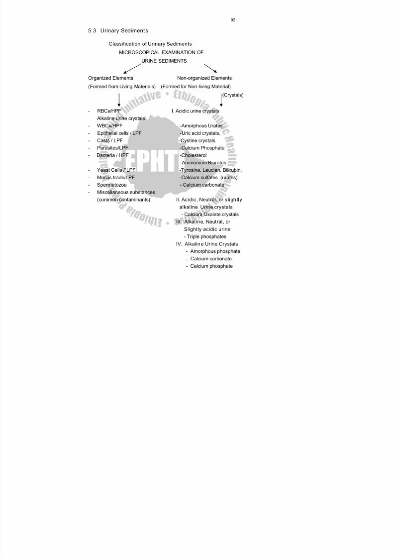
8/13/2019 LN Urinalysis Final
http://slidepdf.com/reader/full/ln-urinalysis-final 103/138
92
5.3 Urinary Sediment s
Classification of Urinary Sediments
MICROSCOPICAL EXAMINATION OF
URINE SEDIMENTS
Organized Elements Non-organized Elements
(Formed from Living Materials) (Formed for Non-living Material)
(Crystals)
- RBCs/HPF I. Acidic urine crystals Alkaline urine crystals
- WBCs/HPF -Amorphous Urates,- Epithelial cells / LPF -Uric acid crystals,- Casts / LPF -Cystine crystals- Parasites/LPF -Calcium Phosphate- Bacteria / HPF -Cholesterol
-Ammonium Biurates- Yeast Cells /`LPF -Tyrosine, Leucien, Bilirubin,- Mucus trade/LPF -Calcium sulfates (urates)- Spermatozoa - Calcium carbonate- Miscellaneous substances
(common contaminants ) II. Ac id ic , Neutral , or s ligh tlyalkaline Urine crystals - Calcium Oxalate crystals
III. Alkal ine, Neutral , orSlightly acidic urine - Triple phosphates
IV. Alkalin e Urine Crystals- Amorphous phosphate- Calcium carbonate- Calcium phosphate
8/13/2019 LN Urinalysis Final
http://slidepdf.com/reader/full/ln-urinalysis-final 104/138
93
5.4 Organized Urinary Sediments
RED BLOOD CELLS
Appearance : Normally RBCs appear in the fresh sample as intact,
small and faint yellowish discs, darker at the edges
- Measure 7-8 μm
- In concentrated urine may be crenated, and their size became
small (5-6 μm)
- In diluted urine, RBCs may be turgid and increase in size (9-10
μm)
- In alkaline urine, they may be small or entirely destroyed forming
massive of brownish granules
Clinical Implications : When the number of RBCs is found more than
their normal range, usually greater than 5 RBCs/HPF it may indicate:
Presence of disease conditions in the urinary tract, such as:
• Acute and chronic glomerulonephritis
• Renal stone
• Cystitis
• Prostates
• Trauma of the kidney
• Presence of parasites, such as: schistosoma.
• Presence of bacterial infection, such as: renal tuberculosis
• Other disease conditions, such as hemophilia, malignant
hypertension.
Temporarily (transient) increased RBC may be seen
- After strenuous exercise
- Exposure to cold temperature
Other substances confusing with RBCs
Yeast cells, and fat droplets may confuse with RBCs
morphologically. They may be differentiated by their morphology.
8/13/2019 LN Urinalysis Final
http://slidepdf.com/reader/full/ln-urinalysis-final 105/138
94
Red blood cells are some what round or disc shaped, and uniform in
size: while yeast cells are oval in shape, and have budding at the
surface. On the other hand fat droplets are irregular in size and they
are shiny.
Another means of differentiating RBCs from yeast and fat droplets is
that, when 5% of acetic acid is added under the cover slide, RBCswill hemolize, while yeast cell and fat droplets will not show any
change.
How to report result :
• After looking RBCs under the 40x objective, they can be reported by
mentioning the average number of RBCs/HPF.
Interfering factors:
Factors that may result falsly in high number of RBCs, i.e. without the
presence of actual renal or other normal physiological disturbancesincluded:
• Menstrual bleeding
• Vaginal bleeding
• Trauma to peranal area in female patients
• Following traumatic cateterization
• Due to some drugs, such as,
- Aspirin ingestion or over dose
- Anticoagulant therapy over dose
LEUKOCYTES (WBCs)
Normal range : 0-4 WBC/HPF.
Appearance: normally, clear granular disc shaped,
Measure 10-15 μm, the nuclei may be visible.
In alkaline urine, they may increase their size and become irregular.
Predominantly, polymorph nuclear neutrophils are seen.
8/13/2019 LN Urinalysis Final
http://slidepdf.com/reader/full/ln-urinalysis-final 106/138
95
Sometimes because of predominance of neutrophils and the
occurrence of bacterial cell together with polymorphonuclear cells,
WBCs are called pus cells.
WBCs (pus cells) may be seen in clumps.
It is also possible to see single irregular nuclei and small round lobed
nuclei in the WBCs, that are seen in the urine sedimentClinical implication : increased number of leukocyte urine are seen in
case of:
Urinary tract infection
All renal disease
Bladder tumor
Cystitis
Prostates
Acute or chronic bacterial infection such as renal tuberculosis,
temporarily increased number of leukocytes are also seen during:
- Fever, and
- After strenuous exercise
How to report the result :
- After observing the distribution of leukocytes under 40x objective, at
least 10 fields of microscope, it is possible to report as : 0-5
leukocytes / HPF, 20-39 leukocytes / HPF etc, that is by counting the
total leukocytes in 10 HPF and divide by 10.
Or, When 0-5 leukocytes / HPF are seen............. normal5-10 leukocytes / HPF are seen...................... few leukocytes / HPF
10-20 leukocytes/HPF are seen…...........moderate leukocytes/ HPF
20-30 leukocytes /HPF are seen …............... many leukocytes / HPF
Above 30 leukocytes / HPF / are seen ……… full/field
8/13/2019 LN Urinalysis Final
http://slidepdf.com/reader/full/ln-urinalysis-final 107/138
96
EPITHELIAL CELLS
• Normally few epithelial cells (0-2 / HPF) can be found
• Appearance
Their size differs depending on the site from which they
originated.
a. Those coming from renal cells- Size is small as compared to other epithelial cells
- It measures 10 μ to 18 μm in length, i.e., slightly larger than
leukocytes
- Very granular
- Have refractive and clearly visible nucleus
- Usually seen in association with proteins or casts ( in renal
disease).
b. Cells from pelvis and urethra of the kidney
- Size is larger than renal epithelia’s
- Those from pelvis area are granular with sort of tail, while those
from urethra are oval in shape
- Most of the time urethral epithelia is seen with together of
leukocytes and filaments (mucus trades and large in number)
- Pelvic epithelia’s seen usually with no leukocyte and mucus
trade, and are few in number
c. Bladder cells
- Are squamious epithelial cells?- Very large in size.
- Shape seems rectangular and often with irregular border.
- Have single nucleus.
* Here it is important to keep in mind that it is not expected from an
experienced Lab. technician after simply observing epithelial
cells, to say that these are urethral cells, and of pelvic origin and
reporting such a false result in the laboratory request form.
8/13/2019 LN Urinalysis Final
http://slidepdf.com/reader/full/ln-urinalysis-final 108/138
97
* Knowing the origin of the epithelial cells and reporting it, may have
more meaning when requested by the physician for special
purpose, especially by the urologists.
Clinical implication
Presence of epithelial cells in large number, mostly renal types mayindicate:
- Acute tubular damage
- Acute glomerulonephritis
- Silicate over dose
* The presence of large number of epithelial cells with large number
of Leukocytes and mucus trades (filaments) may indicate Urinary
Tract Infections (UTI).
Reporting o f the result:
• Epithelial cells distribution reported after looking under 10x (low
power objective) of the microscope.
• Usually they are reported semi quantitatively by saying
- Occasional epithelial cells /LPF ……1-3 epithelial cells seen in
the whole LPF
- Few epithelial cells / LPF...........……......... 2-4 epithelial / LPF
- Moderate epithelial cells / LPF..……........ 6-14 epithelial / LPF
- Many epithelial cells / LPF.............……... 15-25 epithelial/ LPF- Full of epithelial cells / LPF..............……...when the whole field
of 10 x objective covered by epithelial cells.
Interfering factors
• Squameous epithelial cells from female patients that shade from
vaginal area (together with vaginal discharge) may give false result
of high epithelial cells.

8/13/2019 LN Urinalysis Final
http://slidepdf.com/reader/full/ln-urinalysis-final 109/138
8/13/2019 LN Urinalysis Final
http://slidepdf.com/reader/full/ln-urinalysis-final 110/138
8/13/2019 LN Urinalysis Final
http://slidepdf.com/reader/full/ln-urinalysis-final 111/138
100
c. Waxy Casts (Renal Failure Casts)
Normal value
• Not seen in normal individuals.
Appearance
• Shorter and broader than hyaline casts.
• Composed of homogeneous, yellowish materials.
• Broad waxy casts are from two to six times the width of
ordinary
• casts and appear waxy and granular.
• Have high retractile index.
• May occur from cells (WBC, RBC, or Epithelial) casts, hyaline
casts.
Clinical significance
Waxy casts are found in
• Chronic renal disease.
• Tubular inflammation and degeneration.
• Localized nephron obstruction.
* The presence of waxy casts indicates severity of renal disease.
d. Fatty Casts
Normal range : normally not seen in health individuals.
Appearance:
• These are casts, which contain fat droplets inside them.
• Fat droplets are formed after accumulation of fat in the tubular
vessels, especially tubular epithelial and finally disintegrated.
Clinical Implication :
• The occurrence of fat droplets, oval, fat bodies, or fat casts is
very important sign of nephritic syndrome.
8/13/2019 LN Urinalysis Final
http://slidepdf.com/reader/full/ln-urinalysis-final 112/138
101
• Chronic renal disease.
• Inflammation and degeneration of renal tubules.
e. Cellular Casts
Cellular casts are casts, which contain
• Epithelial cells
• White blood cells
• Red blood cells
Normal range : normally not seen in normal individual
Appearance
• These are casts in which cellular elements are seen.
• Formed usually after accumulation of cellular element in the renal
tubules
Clinical Significance• Epithelial / renal / casts mostly seen in tubular degeneration.
• Red cell cast usually seen in acute glomerulonephritis cases.
• White blood cell casts seen mostly during pyelonephrites
conditions.
NOTE : Casts are very significant findings of urine microscopic
examination. This is because their presence indicates the existence of
renal disease. Sometimes it is possible to get a single cast having
course granules, fine granules and fat droplets, i.e. different substances
in a single cast, at the same time. At this time decision is made after
looking and evaluation of other fields and based on the majorities.
Reporting of Laboratory Result
• Casts are ex7amined under 10x objective of the microscope.
8/13/2019 LN Urinalysis Final
http://slidepdf.com/reader/full/ln-urinalysis-final 113/138
8/13/2019 LN Urinalysis Final
http://slidepdf.com/reader/full/ln-urinalysis-final 114/138
103
b. Schistosoma Haematobium
It is fluke that infect venules of the bladder.
Appearance of the egg
- It is found in the urine sediment.
- Has pale yellow brown color.
- Large and oval in shape.
- Has characteristic small spine at one end (terminal spine).
- Measure about 145 x 55 μm.
- The egg contains a full-developed miracedium. Sometimes the
miracedium hatch from the egg and can be seen swimming in the
urine. The miracedium swim in the urine by the help of ciliates that
are surrounding it.
High excretion of S. haematobium egg can be seen usual between 10.00
a.m. and 2 p.m. It is also important to remember that even when personsare highly infected, eggs may not be present in the urine. Therefore that
is important to examine several specimens collected on different days
and examine carefully, that is due to the irregular pattern of egg
excretion.
c. Wuchereria Bancroftie
• It is tissue nematode that invades lymph vessels. It is usually attack
lower limb.
• In chronic bancroftie filariasis, a condition called chyluria can occuri.e. passing of chyle in the urine. It occurs when the urogenital
lymphatic vessels, which are linked to those, that transport chyle
from the intestine became blocked and rupture.
• Chile consists of lymph and particles of digested fat (soluble in
ether).
• Urine containing chyle appears creamy white. When blood is also
present, the urine appears pinkish-white.
8/13/2019 LN Urinalysis Final
http://slidepdf.com/reader/full/ln-urinalysis-final 115/138
104
• Large, measuring 275-399 x 8-10 μm.
• Body curves are few, nuclei are distinct.
• Sheath stains pink with Giemsa and palely with haematoxylin.
• There is no nuclei in the tip of at the tail.
Other points that should be considered also
• The parasite usually found in high concentration during night from
10:00 p.m. – 4:00 a.m. and i.e. it has nocturnal periodicity.
• Differentiate from B. malai and L. loa by its tail feature.
• Differentiate from Mansonella species by its large size and sheath.
YEAST CELL
Yeast cells are fungi that are not normally seen in health individuals.
Appearance
- Variable in size
- Colorless.
- Oval in shape, and usually form budding.
- Have high refractive index.
- Usually confused with Red Blood Cells. The way in which one can
differentiate yeast cells from RBC is discussed in detail under Red
Blood Cells.
Clinical Significance
• They are usually of candida species (candida albicans) and are
common in patients with
- Urinary tract infection
- Vaginites
- Diabetic mellitus
- Intensive antibiotic or immunosuppressive therapy.
8/13/2019 LN Urinalysis Final
http://slidepdf.com/reader/full/ln-urinalysis-final 116/138
105
BACTERIA
Bacteria are the most common cause of UTI and aerobic gram-negative
bacilli, particularly, members of the enterobacteriacea, are the most
dominant agents. The Gram-positives account for proportionately large
number of infections in hospital inpatients. Normally, bacteria are not
seen in the healthy individual’s urine.
To check the presence or absence of bacteria a technician can either
check for Nitrate that was formed in the urine after breakdown of nitrite
into nitrate by the metabolic action of bacteria. Hence, dipstick test can
give indirect clue. Or one can use urine microscopy test to check the
presence of pus cells within the drop of urine or its sediment. Further
the observed bacterial cell can be identified by bacteriological culture.
Appearance
- Bacteria that are seen in the microscopic examination of the drop
of urine sample. Their shape varies with the type of bacteria
observed..
- Depending on the type of bacteria they can be either motile or non
motile organisms.
- They can be observed when examined under less than 40 x
(high power) objective of the microscope.
Clinical Significance
- Presence of bacteria may indicate the presence of UTI or
contamination by genital or intestinal microflora.
- To confirm what type of bacteria they are and whether or not they
are the causes of the disease, it is important to culture them in
appropriate media and perform biochemical tests for identification.

8/13/2019 LN Urinalysis Final
http://slidepdf.com/reader/full/ln-urinalysis-final 117/138
106
Report of the Result
The bacteria concentration before or without performing culture and
identification of the bacteria, can be reported as:
• Occasional bacteria HPF
• Few bacteria / HPF
• Moderate bacteria / HPF• Many bacteria / HPF
• Full of bacteria / HPF.
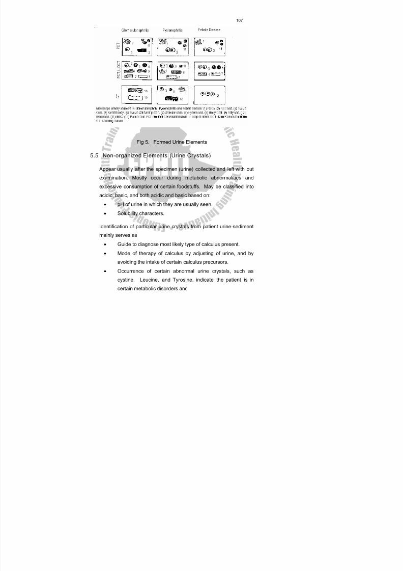
8/13/2019 LN Urinalysis Final
http://slidepdf.com/reader/full/ln-urinalysis-final 118/138
107
Fig 5. Formed Urine Elements
5.5 Non-organized Elements (Urine Crystals)
Appear usually after the specimen (urine) collected and left with out
examination. Mostly occur during metabolic abnormalities and
excessive consumption of certain foodstuffs. May be classified into
acidic, basic, and both acidic and basic based on:
• pH of urine in which they are usually seen.
• Solubility characters.
Identification of particular urine crystals from patient urine-sediment
mainly serves as
• Guide to diagnose most likely type of calculus present.
• Mode of therapy of calculus by adjusting of urine, and by
avoiding the intake of certain calculus precursors.
• Occurrence of certain abnormal urine crystals, such as
cystine. Leucine, and Tyrosine, indicate the patient is in
certain metabolic disorders and
8/13/2019 LN Urinalysis Final
http://slidepdf.com/reader/full/ln-urinalysis-final 119/138
108
Some drug crystals in the urine include, sulfonamides,
aspirin, caffeine, used to follow the treatment condition.
I. Acidic Urine Crystals
a. Amorphou s Urates (Anhydrous uric acid)
- Normally present in urine in different quantity.- Have pink to “brick red” color.
- From very small granules and seen in cluster.
- Dissolve in urine when the sample is gently heated.
- When urine is left in the refrigerator, it shows heavy
precipitation of urates.
b. Uric Acid Crystals
- Polymorphs (different in shape) i.e. square, prism,
hexagonal, rostelles etc.
- Yellow to yellow brown in color.
- Size is 30-150 μm
- Small quantity found in normal urine, but increases in
association with:
- Increased Purine metabolism in case of gout.
- Increased Nucleic Acid turn over, such as leukemia.
c. Cystine Crystals
- Rarely found.
- Flat, hexagonal plates with well defined edges.- Colorless, and highly retractile.
- Size is 30-60 μm.
- Found only in fresh urine, because if there is delay, they
are soluble and not seen.
- Appeared during cystinosis, which is a hereditary disease
(Wilson disease), or during transient acute phase of
8/13/2019 LN Urinalysis Final
http://slidepdf.com/reader/full/ln-urinalysis-final 120/138
109
pyelonephritis. Its appearance in the urine is called
cystinuria.
d. Cholesterol
- Rarely found.
- Colorless and retractile.
- Have “broken window” shape, with notches on one side.
- 50-100 μm in size.
- Soluble in ether.
- Seen in case of elevated cholesterol, chyluria.
e. Tyrosine
- Rarely found.
- Colorless or yellowish.
- Have fine silky needle in sheaves or rosettes shape.
- Indicate protein break down problem, or severe liverdisease.
f. Leucine
- Rarely found.
- Yellow to yellow brown in color.
- Spheroid in shape with striation.
- Seen in case of protein breakdown problem, or severe liver
disease.
- Lucien and Tyrosine crystals may occur together. Both are
amino acids usually; in case of severe liver disease, they willnot be metabolized, and excreted in urine.
g. Bilirubin
- Very rarely seen.
- Have reddish brown color.
- Seen in case of elevated Bilirubin.
8/13/2019 LN Urinalysis Final
http://slidepdf.com/reader/full/ln-urinalysis-final 121/138
110
- Have various tiny squarish, beads or amorphous needle
shape.
- Size is 5 μm (half RBC).
- Chemical test for bile pigments positive.
h. Calcium Sulfate Crystals
- Have large prism or flat bladder shaped.
- Seen separately or in bundles.
- Size 50-100 μm.
- Can be distinguished from calcium phosphate crystals by
measuring pH of urine.
II. Acid ic, Neutral, or Basic Urine Crystals
Calcium Oxalate Crystal
- Are colorless and refractive.
- Have octahedral, envelope, shape.
- Size 10-12 μm.
- Normally seen in small amount.
- After consumption of high calcium, or oxalate rich foods, such as
milk, tomatoes, asparagus, and orange, normally the crystals may
be seen.
- In dehydration condition, such as, in hot weather where there is
high perspiration and only small amount of water is consumed per
day Calcium oxalate crystals may be seen.
- Pathologically in large quantity may be seen in (severe chronic
renal disease, and urinary calculus).
III. Alkalin e, Neutral, or Sligh t Acid ic Urine Crys tals
Triple Phosphates
- Colorless and refractive.
- Have “coffin lids” 3 to 4 to 6 – sided prism.
8/13/2019 LN Urinalysis Final
http://slidepdf.com/reader/full/ln-urinalysis-final 122/138
111
- Shape, or fern leaf or star shape.
- Size 13 0- 150 μm.
- Seen in urine stasis (obstructive uropathy), or in urinary tract
infections.
- Their presence is frequently indicative of bacterial infection by
proteus marbles.
IV. Alkaline Urine Crystals
a. Amorphou s Phosphates
- Normally seen in alkaline urine.
- Small, whitish granules usually seen scattered.
- Soluble in 100g/1 acetic acid.
b. Calcium Carbonate
- Less commonly seen.
- Colorless.
- Have needle, spherical or dumbbells shape.
- Have very small crystals.
- If 100g/1, i.e. 10% acetic acid is added, they dissolve, give off
bubbles of gas.
c. Calcium Phosphates
- Seen in small amount in normal individual urine, and when they
are in large amount, may indicate chronic cystitis, or prosthetichypertrophy.
. Have star or needle shape.
- Colorless.
d. Ammonium Bruits (Urates )
- Normally seen in alkaline urine.
- Have bundle of needles or “thorn apple” sphere shape.
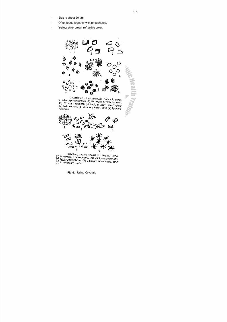
8/13/2019 LN Urinalysis Final
http://slidepdf.com/reader/full/ln-urinalysis-final 123/138
112
- Size is about 20 μm.
- Often found together with phosphates.
- Yellowish or brown refractive color.
Fig 6. Urine Crystals
8/13/2019 LN Urinalysis Final
http://slidepdf.com/reader/full/ln-urinalysis-final 124/138
113
MISCELLANEOUS
I. Spermatozoa
- Are small structures consisting of a head and tail, connected by a
short middle piece (neck).
- Easily recognized especially if they are motile.
- Frequently seen in the urine of males.
- They may see in the urine of females, when the urine collected
after coitus usually not reported, unless the physician has special
interest in it.
II Mucus Trades
- Formed by the precipitation of mucoprotein in cooled urine.
- Normally little mucus trades seen in normal individuals.
- Have fine, fiber like appearance.
- Wavy in shape and tapered at ends.- If not examined carefully may confuse with hyaline casts.
- Their presence in large amount with WBCs, may indicate UTI.
III. Other Contaminates and Art ifact Structure
- Muscle fibers- Vegetable cells - all are fairly seen and easily- Cotton fibers (wool fibers) recognizable.
- Structure from slide or cover slide - high retractile andnon-uniforminsize.
Fat droplets (other bubbles)- not evenly distributed.
- Oil droplets
- Pollen greens - are seasonal.- Starch granules - incomplete digestion of
starch
8/13/2019 LN Urinalysis Final
http://slidepdf.com/reader/full/ln-urinalysis-final 125/138
114
They can be confirmedby logos iodine.
* To minimize the above mentioned contaminants and artifacts
- Don't use dirty containers, slides and cover slides.
- Don't let urine specimen to open-air.
- Avoid contamination of urine with fats and oils.- Avoid the drying of sediments.
Methods for Examining Urine Sediments
A. Uns tained Urine Sediment
1. Bright field microsc opy of the unstained urine sediment
Traditionally, the urinary sediment has been examined microscopically
by placing a drop of urine sediment on a microscopic slide, cover with
cover slide and observing the preparation with the lower and high power,
objective of the bright field microscope.
When the sediment is examined under the bright field microscope,
correct light adjustment is essential, and the light must be sufficiently
reduced, by the correct positioning of the condenser and the iris
diaphragm to give contrast between the unstained structures and the
back ground liquid.
2. Phase Contrast s (PC)
P.C. illumination is useful in the examination of unstained urinary
sediment, particularly for translucent elements such as hyaline casts
and mucus threads, which have a refractive index similar to that of urine
in which they are suspended. Phase contrast has the advantage of
hardening the outlines even the most ephemeral formed elements.
B. Stained Preparation
Cellular detail is best seen with stained preparation.
8/13/2019 LN Urinalysis Final
http://slidepdf.com/reader/full/ln-urinalysis-final 126/138
115
The following stains are commonly used:
1. A crystal violet safranin stain (sternheimer and malbin) is useful in the
identification of cellular elements. It is commercially available as sedi-
stain.
Preparation of Reagents
Solution (1) Crystal violet ---------------------------- 3g
Ethanol (95%)--------------------------- 20 ml
Ammonium Oxalate-------------------- 0.8 g
Distilled water ------------------------- 80 ml
Solution (2) Safranin ----------------------------------1 g
Ethanol (95%)---------------------------40 ml
Distilled water--------------------------- 400 ml
The mixture should be filtered every 2 weeks!- Discard after 3 months
- Separately, solution (1) and solution (2) keep indefinitely
at room temperature.
In highly alkaline urine, the stains will precipitate.
Procedure
Add 1 or 2 drops of crystal violet safranin stain to approximately 1 ml of
concentrated urine sediment. Mix and place a drop of this suspension
on a slide and cover with cover slide.
Staining r eaction t o crystal – violet safranin stain:
RBC – Purple to dark purple.
WBC – Cytoplasm -violet to blue.
Nucleus – reddish purple.
Glitter cells – blue.
8/13/2019 LN Urinalysis Final
http://slidepdf.com/reader/full/ln-urinalysis-final 127/138
116
Nucleus Cytoplasm
Squameous epithelial purple pink to violet
Uroepithelial dark blue blue
Renal tubular cells dark purple orange purple
2. Methyl blue (Loeffler's stain)
3. Cyto Diachrome stains
When such stains are used, it is recommended that both the stained and
unstained sediment be mounted and observed, as the stain may cause
precipitation of some constituents. This is especially the problem with
alkaline urine specimens, because the precipitated materials may
obscure important pathological constituents.
Exercise:
Say True or False
1. The number of casts preserved decrease as the pH of the urine
decreases.
2. Presence of RBCs in the urine is always indicative of a renal
disease.
3. Waxy casts are the end stage in the degeneration of cellular casts.
4. Pyuria refers to elevated numbers of leucocytes in the urine.
5. The presence of Bacteria in the Urine is determined using only
Microscope.
8/13/2019 LN Urinalysis Final
http://slidepdf.com/reader/full/ln-urinalysis-final 128/138
117
APPENDIX
I. Reagent Preparation
Reagent No.1 Acetic Acid Reagent (10%)
Pipette 10 ml of glacial acetic acid into a 100ml volumetric flask that is
1/2 filled with distilled water. Dilute to 100-ml volume with distilled
water and mix.
Reagent No.2 Aceto ne Powder Reagent
Weigh each of the following with the rough balance. Grind each
separately with a mortar and pestle and put into a reagent bottle.
Ammonium Sulphate . . . . . . . . . . . . . . . . . . . . .. 20g
Sodium Carbonate . . . . . . . . . . . . . . . . . . .. …. . 20g
Sodium Nitroprusside . . . . . . . . . . . . . . . . . . . . . 1g
After all have been added to the reagent bottle, mix well.
Reagent No.3 Barium Chloride Filter Paper f or Bilirubin
Soak thick filter paper in saturated barium chloride
Dry & cut in to small strips (4 x 1/2 inch strips).
Reagent No.4 Benedict Reagent for Glucos e
To make 1 lit
A - 173 gm.---------------------------Trisodium citra
100 gm--------------------------- Sodium carbonate anhydrates
800 ml---------------------------- Distilled H 2O
Dissolve to make to 850 ml
B- 17,3 gm ------------------------- CuSO 4
100 ml ------------------------- Distilled H 2O
Stir slowly in to the first solution bring up to 1 lit. with distilled H 2O.
8/13/2019 LN Urinalysis Final
http://slidepdf.com/reader/full/ln-urinalysis-final 129/138
118
Reagent No.5 Bleach for Preservation of S.Hematobium egg in
urine
S. hematobium. egg preserved by adding 1ml (1-% v/v).
Domestic bleach in every 10 ml urine.
Reagent No.6 Bor ic Acid Preservative (1-%w/v) 10 gm/l
Boric acid --------------10gm.
Dissolve in 1000ml distilled water.
Reagent No.7 Ehrlich's Reagent for Uroblinogen
To make 200ml
para-dimethylaminobenzaldehyde --------------------- 4 gm
HCl concentrated ----------------------------------------- 40 ml
Distilled H 2O --------------------------------------------- 160ml
a. Weigh the para-dimethylaminobenzaldeyde andtransfer it to clean, leak proof bottle.
b. Measure the water and add to chemical and mix
c. Add conc. HCl and mix well.
d. Label the bottle and mark, as it is corrosive.
Store at room temperature the reagent is stable
for several weeks.
Reagent No. 8 10 % Ferric Chloride Reagent
Weigh 10 g of ferric chloride and transfer to a 100-ml volumetric flask.Dissolve and dilute to 100ml volume with water and mix.
Reagent No. 9 Fouchet’ s Reagent
Trichloroacetic acid ---------------------- 25gm
Distilled H 2O ------------------------------100ml
10% Ferric chloride (FeCl 3) -------------10ml
Mix well.
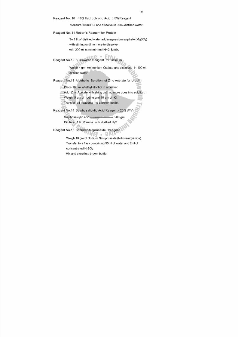
8/13/2019 LN Urinalysis Final
http://slidepdf.com/reader/full/ln-urinalysis-final 130/138
119
Reagent No. 10 10% Hydrochloric Acid (HCl) Reagent
Measure 10 ml HCl and dissolve in 90ml-distilled water.
Reagent No. 11 Robert's Reagent for Protein
To 1 lit of distilled water add magnesium sulphate (MgSO 4)
with stirring until no more to dissolve. Add 200-ml concentrated HN0 3 & mix.
Reagent No.12 Sulk owitch Reagent for Calcium
Weigh 4 gm Ammonium Oxalate and dissolved in 100 ml
distilled water.
Reagent No.13 Alcoholic Solution of Zinc Acetate for Urobilin
Place 100 ml of ethyl alcohol in a beaker.
Add Zinc Acetate with string until no more goes into solution.
Weigh 5 gm of Iodine and 10 gm of KI.
Transfer all reagents to a brown bottle.
Reagent No.14 Sulpho salicy lic Ac id Reagent ( 20% W/V)
Sulphosalcylic acid ----------------------- 200 gm
Dilute to 1 lit. Volume with distilled H 2O.
Reagent No.15 Sodiu mnit ropruss ide Rreagent
Weigh 10 gm of Sodium Nitroprusside (Nitroferricyanide).
Transfer to a flask containing 95ml of water and 2ml of
concentrated H 2SO 4.
Mix and store in a brown bottle.

8/13/2019 LN Urinalysis Final
http://slidepdf.com/reader/full/ln-urinalysis-final 131/138
120
Reagent No.16 Saturated Sodium Nitroprusside
Add 40 gm of Sodium Nitroprusside to 100 ml of water is a brown bottle.
Shake to dissolve as much as possible. Allow any undissolved salt to
remain in the bottom of the bottle.
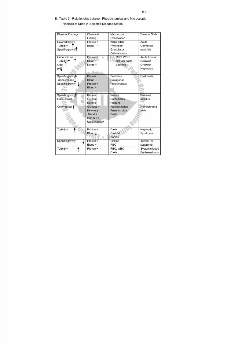
8/13/2019 LN Urinalysis Final
http://slidepdf.com/reader/full/ln-urinalysis-final 132/138
121
II. Table 3. Relationship between Physiochemical and Microscopic
Findings of Urine in Selected Disease States.
Physical Findings ChemicalFinding
MicroscopicObservation
Disease State
Colored brown
TurbiditySpecific gravity
Protein +
Blood +
WBC, RBC
Hyaline orGranular orCellular casts
Acute
Glomerulo-nephritis
Urine volumeTurbidityOdorpH
Protein +Blood +Nitrite +
- RBC, WBC- Cellular casts,- Bacteria
Acute tubularNecrosisOr lowerNephrosis
Specific gravityUrine volume
Specific gravity
ProteinBloodProtein +Blood +
ColorlessHexagonalPlate crystals
Cystinosis
Specific gravityOdor- sweet
ProteinGlucoseKetone
YeastsSome timesPresent
DiabetesMellitus
Color darker Glucose +Ketone +Blood +
Bilirubin +Urobilinogen+
Pigment ladenPrussian blueCasts
Hemochroma-tosis
Turbidity Portion +Blood +
CastsOval fatBodies
NephroticSyndrome
Specific gravity Protein +Blood +
SickledRBC
SickleCellsyndrome
Turbidity Protein + RBC, WBCCasts
Systemic lupusErythematosus

8/13/2019 LN Urinalysis Final
http://slidepdf.com/reader/full/ln-urinalysis-final 133/138
122
III. Table 4: Correct and Incorrect Approach in Urine Testing
Correct Approach Incorrect Approach
Use fresh urine Delay in the testing of urine without
preservation
Make quality control of reagents Using expired reagents
Be aware of normal as well as
abnormal results which are
significant
Believing urine results have little
significance in the overall diagnostic
picture of the patient
Follow the directions carefully Being careless
Accept only clear and proper
collection bottles
Using any container.
Be familiar with interfering
substances
Not giving due attention to cross
reaction and artifacts
Mix Urine properly Not mixing well
Record results accurately Not checking the results recorded
during the training of new personnel
Give proper training to
professionals
New personnel always jumping into
urinalysis because it is the easiest to do
and least significant
8/13/2019 LN Urinalysis Final
http://slidepdf.com/reader/full/ln-urinalysis-final 134/138
123
Glossary
Alkap tour ia : Genetically determined defect of metabolism in which
homogenestic acid is excreted in the urine, which turns dark on standing.
Alkal ine : Containing alkali, strictly a fluid with pH greater than 7.
Anur ia : Cessation of the production of urine.
Bile Pigments : Breakdown products of hemoglobin.
Cystine : Sulfur containing amino acid.
Cystinosis : A rare inborn error of metabolism of cystine and other
amino acids.
Cystinuria : Presence of abnormal amount of cystine in the urine.
Cystitis : Inflammation of the urinary bladder.
Diabetes Insipidu s : A syndrome caused by deficient secretion of anti-
diuretic hormone (ADH) by the pituitary gland, and characterized by
polyuria.
Diabetes Mellitus : A syndrome caused by a relative deficiency of
insulin.
Diuresis : An increased secretion of urine.
Diuretics : Drugs, which increase the volume of urine excreted.
Diurnal : Daily
Endocarditis : Inflammation of the endocardial lining of the heart.
Glomerulus’s : The filtration unit of a nephrone, consists of a coil of fine
capillaries opposed to an expansion of urinary epithelium.
Glomeurlar Filtrate : Ultra filtered blood through the glomerular
membrane.
8/13/2019 LN Urinalysis Final
http://slidepdf.com/reader/full/ln-urinalysis-final 135/138
8/13/2019 LN Urinalysis Final
http://slidepdf.com/reader/full/ln-urinalysis-final 136/138
8/13/2019 LN Urinalysis Final
http://slidepdf.com/reader/full/ln-urinalysis-final 137/138
126
Urinometer : A small glass instrument with a graduated stem used for
measuring the specific gravity of urine.
Urobiline : Pigmented derivative of urobilinogen
Urobilinogen : Derivative of bilirubine, which is made in the intestine by
the gut bacteria.
8/13/2019 LN Urinalysis Final
http://slidepdf.com/reader/full/ln-urinalysis-final 138/138
127
References:
Cheesbrough, Monica (1987)Medical Laboratory Manual for Tropical
Countries.Vol. 1. Cambridge : Butterworth-Heinemann Ltd.
Donna, R.C. and others (1995 )Human Anatomy and Physilogy. New
York: Mcgraw-H.I.L.L INC.
Free, A.H. et Free, H.M.(1975)Urinalysis in Clinical Laboratory Practice.
Cleve Land: CRC Press.
Harber, M.H.(1978) A Premier of Microscopic Urinalysis. Fountain valley:
Calif ICL scientific.
Lehninger, Albert (1982) Principles of Biochemistry. New York: Worth
Publisher INC.
Purvis, J.J.(1973) Laboratory Planning. Baltimore: Williams and Wilkins.
Ross, Doris L et Ann E. Neely(1983) Text Book of Urinalysis and Body
Fluids.
Rile, P.A. and Cunninghum P.J. (1985) Pocket Medical Dictionary, 3rd
ed. New Delhi-India : Jaypee Brothers , Medical Publisher.
Saxton, Byren (1986) Laboratory Tests : Implication for Nurses.2nd ed.
Sood, Ramnik (1987) Medical Laboratory Technology: Methods and
Interpretation. 4th ed. New Delhi-India : Jaypee Brothers.