Lmx1a Encodes a Rostral Set of Mesodiencephalic Dopaminergic ...
12
Lmx1a Encodes a Rostral Set of Mesodiencephalic Dopaminergic Neurons Marked by the Wnt/B-Catenin Signaling Activator R-spondin 2 Elisa J. Hoekstra 1,2 , Lars von Oerthel 1,2 , Lars P. van der Heide 1 , Willemieke M. Kouwenhoven 1 , Jesse V. Veenvliet 1 , Iris Wever 2 , Yong-Ri Jin 4 , Jeong K. Yoon 4 , Annemarie J. A. van der Linden 2 , Frank C. P. Holstege 3 , Marian J. Groot Koerkamp 3 , Marten P. Smidt 1,2 * 1 Swammerdam Institute for Life Sciences, University of Amsterdam, Amsterdam, The Netherlands, 2 Neuroscience and Pharmacology, Rudolf Magnus Institute of Neuroscience, University Medical Center Utrecht, Utrecht, The Netherlands, 3 Molecular Cancer Research, University Medical Center Utrecht, Utrecht, The Netherlands, 4 Center for Molecular Medicine, Maine Medical Center Research Institute, Maine, United States of America Abstract Recent developments in molecular programming of mesodiencephalic dopaminergic (mdDA) neurons have led to the identification of many transcription factors playing a role in mdDA specification. LIM homeodomain transcription factor Lmx1a is essential for chick mdDA development, and for the efficient differentiation of ES-cells towards a dopaminergic phenotype. In this study, we aimed towards a more detailed understanding of the subtle phenotype in Lmx1a-deficient (dreher) mice, by means of gene expression profiling. Transcriptome analysis was performed, to elucidate the exact molecular programming underlying the neuronal deficits after loss of Lmx1a. Subsequent expression analysis on brain sections, confirmed that Nurr1 is regulated by Lmx1a, and additional downstream targets were identified, like Pou4f1, Pbx1, Pitx2, C130021l20Rik, Calb2 and Rspo2. In line with a specific, rostral-lateral (prosomer 2/3) loss of expression of most of these genes during development, Nurr1 and C130021l20Rik were affected in the SNc of the mature mdDA system. Interestingly, this deficit was marked by the complete loss of the Wnt/b-catenin signaling activator Rspo2 in this domain. Subsequent analysis of Rspo22/2 embryos revealed affected mdDA neurons, partially phenocopying the Lmx1a mutant. To conclude, our study revealed that Lmx1a is essential for a rostral-lateral subset of the mdDA neuronal field, where it might serve a critical function in modulating proliferation and differentiation of mdDA progenitors through the regulation of the Wnt activator Rspo2. Citation: Hoekstra EJ, von Oerthel L, van der Heide LP, Kouwenhoven WM, Veenvliet JV, et al. (2013) Lmx1a Encodes a Rostral Set of Mesodiencephalic Dopaminergic Neurons Marked by the Wnt/B-Catenin Signaling Activator R-spondin 2. PLoS ONE 8(9): e74049. doi:10.1371/journal.pone.0074049 Editor: Judith Homberg, Radboud University, Netherlands Received May 2, 2013; Accepted July 25, 2013; Published September 16, 2013 Copyright: ß 2013 Hoekstra et al. This is an open-access article distributed under the terms of the Creative Commons Attribution License, which permits unrestricted use, distribution, and reproduction in any medium, provided the original author and source are credited. Funding: This work was funded by National Institute of Health grants (R01 AR055278 and P20 GM103465) to J.K.Y., a TIPharma grant (T5-207, Netherlands) and an NWO VICI grant (865.09.002, Netherlands) to M.P.S. The funders had no role in study design, data collection and analysis, decision to publish, or preparation of the manuscript. Competing Interests: The authors have declared that no competing interests exist. * E-mail: [email protected] Introduction Mesodiencephalic dopaminergic (mdDA) neurons function in regulating motor control and emotion related behavior, and loss of these neurons can cause Parkinson’s disease, and other neurolog- ical disorders as well. In order to understand the molecular mechanisms causing the selective degeneration of these neurons, insight in the pathways and factors involved in the development and maintenance of this subset of dopaminergic neurons is needed. The onset of mdDA development is induced by several extrinsic factors such as Shh, Fgf8 and Wnt1 [1–3]. In addition to this early signaling from the organizing centers in the developing CNS, several developmental factors are essential for the subdivision into different domains, and for the differentiation of newborn neurons towards mdDA neurons [3]. Among the factors implicated in mdDA neuron development, are Wnt1, Wnt5a, En1/2, Otx2, Foxa1, Foxa2, Ngn2, Nurr1 (Nr4a2), Pitx3, Msx1, and the LIM homeodo- main transcription factors Lmx1a and Lmx1b [4–14]. Several studies suggested a role for Lmx1a in establishing the mdDA neuronal phenotype [5,15]. Gain-and-loss-of-function studies in chick revealed that Lmx1a is needed for the specification of mdDA neurons. Moreover, Lmx1a can induce mouse embryonic stem cells (ESCs) into DA neurons [5]. In addition, a Wnt1-Lmx1a auto-regulatory loop was identified when differentiating ES cells into mdDA neurons [15]. Together, these experiments, in chick and ESCs, suggest an essential role for Lmx1a in the determination of mdDA neurons. Contradictory, studies performed in Lmx1a mutant mice, revealed a subtle phenotype only, with mild reduction of Th and Nurr1, and compromised Wnt-expression [13]. Other studies in Lmx1a mutants also revealed a moderate reduction in a number of (VTA) neurons [16,17]. Besides several studies indicating that Lmx1a functions in proliferation and neurogenesis, the precise role of Lmx1a in the mouse mdDA neuronal system is still not fully understood. Therefore, to understand the Lmx1a phenotype in depth, we studied the loss of function of Lmx1a in the Dreher mouse, which can be considered as a null mutant [17–19]. From this, we PLOS ONE | www.plosone.org 1 September 2013 | Volume 8 | Issue 9 | e74049
Transcript of Lmx1a Encodes a Rostral Set of Mesodiencephalic Dopaminergic ...
Lmx1a Encodes a Rostral Set of MesodiencephalicDopaminergic Neurons Marked by the Wnt/B-CateninSignaling Activator R-spondin 2Elisa J. Hoekstra1,2, Lars von Oerthel1,2, Lars P. van der Heide1, Willemieke M. Kouwenhoven1,
Jesse V. Veenvliet1, Iris Wever2, Yong-Ri Jin4, Jeong K. Yoon4, Annemarie J. A. van der Linden2,
Frank C. P. Holstege3, Marian J. Groot Koerkamp3, Marten P. Smidt1,2*
1 Swammerdam Institute for Life Sciences, University of Amsterdam, Amsterdam, The Netherlands, 2 Neuroscience and Pharmacology, Rudolf Magnus Institute of
Neuroscience, University Medical Center Utrecht, Utrecht, The Netherlands, 3 Molecular Cancer Research, University Medical Center Utrecht, Utrecht, The Netherlands,
4 Center for Molecular Medicine, Maine Medical Center Research Institute, Maine, United States of America
Abstract
Recent developments in molecular programming of mesodiencephalic dopaminergic (mdDA) neurons have led to theidentification of many transcription factors playing a role in mdDA specification. LIM homeodomain transcription factorLmx1a is essential for chick mdDA development, and for the efficient differentiation of ES-cells towards a dopaminergicphenotype. In this study, we aimed towards a more detailed understanding of the subtle phenotype in Lmx1a-deficient(dreher) mice, by means of gene expression profiling. Transcriptome analysis was performed, to elucidate the exactmolecular programming underlying the neuronal deficits after loss of Lmx1a. Subsequent expression analysis on brainsections, confirmed that Nurr1 is regulated by Lmx1a, and additional downstream targets were identified, like Pou4f1, Pbx1,Pitx2, C130021l20Rik, Calb2 and Rspo2. In line with a specific, rostral-lateral (prosomer 2/3) loss of expression of most of thesegenes during development, Nurr1 and C130021l20Rik were affected in the SNc of the mature mdDA system. Interestingly,this deficit was marked by the complete loss of the Wnt/b-catenin signaling activator Rspo2 in this domain. Subsequentanalysis of Rspo22/2 embryos revealed affected mdDA neurons, partially phenocopying the Lmx1a mutant. To conclude,our study revealed that Lmx1a is essential for a rostral-lateral subset of the mdDA neuronal field, where it might serve acritical function in modulating proliferation and differentiation of mdDA progenitors through the regulation of the Wntactivator Rspo2.
Citation: Hoekstra EJ, von Oerthel L, van der Heide LP, Kouwenhoven WM, Veenvliet JV, et al. (2013) Lmx1a Encodes a Rostral Set of MesodiencephalicDopaminergic Neurons Marked by the Wnt/B-Catenin Signaling Activator R-spondin 2. PLoS ONE 8(9): e74049. doi:10.1371/journal.pone.0074049
Editor: Judith Homberg, Radboud University, Netherlands
Received May 2, 2013; Accepted July 25, 2013; Published September 16, 2013
Copyright: � 2013 Hoekstra et al. This is an open-access article distributed under the terms of the Creative Commons Attribution License, which permitsunrestricted use, distribution, and reproduction in any medium, provided the original author and source are credited.
Funding: This work was funded by National Institute of Health grants (R01 AR055278 and P20 GM103465) to J.K.Y., a TIPharma grant (T5-207, Netherlands) andan NWO VICI grant (865.09.002, Netherlands) to M.P.S. The funders had no role in study design, data collection and analysis, decision to publish, or preparation ofthe manuscript.
Competing Interests: The authors have declared that no competing interests exist.
* E-mail: [email protected]
Introduction
Mesodiencephalic dopaminergic (mdDA) neurons function in
regulating motor control and emotion related behavior, and loss of
these neurons can cause Parkinson’s disease, and other neurolog-
ical disorders as well. In order to understand the molecular
mechanisms causing the selective degeneration of these neurons,
insight in the pathways and factors involved in the development
and maintenance of this subset of dopaminergic neurons is needed.
The onset of mdDA development is induced by several extrinsic
factors such as Shh, Fgf8 and Wnt1 [1–3]. In addition to this early
signaling from the organizing centers in the developing CNS,
several developmental factors are essential for the subdivision into
different domains, and for the differentiation of newborn neurons
towards mdDA neurons [3]. Among the factors implicated in
mdDA neuron development, are Wnt1, Wnt5a, En1/2, Otx2, Foxa1,
Foxa2, Ngn2, Nurr1 (Nr4a2), Pitx3, Msx1, and the LIM homeodo-
main transcription factors Lmx1a and Lmx1b [4–14].
Several studies suggested a role for Lmx1a in establishing the
mdDA neuronal phenotype [5,15]. Gain-and-loss-of-function
studies in chick revealed that Lmx1a is needed for the specification
of mdDA neurons. Moreover, Lmx1a can induce mouse embryonic
stem cells (ESCs) into DA neurons [5]. In addition, a Wnt1-Lmx1a
auto-regulatory loop was identified when differentiating ES cells
into mdDA neurons [15]. Together, these experiments, in chick
and ESCs, suggest an essential role for Lmx1a in the determination
of mdDA neurons. Contradictory, studies performed in Lmx1a
mutant mice, revealed a subtle phenotype only, with mild
reduction of Th and Nurr1, and compromised Wnt-expression
[13]. Other studies in Lmx1a mutants also revealed a moderate
reduction in a number of (VTA) neurons [16,17]. Besides several
studies indicating that Lmx1a functions in proliferation and
neurogenesis, the precise role of Lmx1a in the mouse mdDA
neuronal system is still not fully understood.
Therefore, to understand the Lmx1a phenotype in depth, we
studied the loss of function of Lmx1a in the Dreher mouse, which
can be considered as a null mutant [17–19]. From this, we
PLOS ONE | www.plosone.org 1 September 2013 | Volume 8 | Issue 9 | e74049
established that Lmx1a is essential for a rostral-lateral part of the
developing mdDA system. Furthermore, to elucidate the molec-
ular programming causing this phenotype, we performed micro-
array analysis on dissected brain material of Lmx1a-dr/dr embryos.
This revealed several Lmx1a target genes, such as Nurr1,
C130021L20Rik and Rspo2. The loss of these genes was region
specific, and occurred in a rostral group of mdDA neurons.
Subsequent analysis in Rspo22/2 embryos revealed a subtly
affected mdDA neuronal field, partially resembling the Lmx1a
phenotype. Altogether, Lmx1a is essential for the correct
programming of a rostral subset of developing mdDA neurons,
marked by the Wnt/b-catenin signaling activator Rspo2.
Materials and Methods
AnimalsThe Lmx1a mutant (strain name B6C3Fe a/a-Lmx1adr-J/J) was
obtained from Jackson Laboratory (Bar Harbor, ME), and
maintained on a C57Bl/6J background (Charles River). Geno-
typing was done by PCR using primers: forward 59-CAAA-
GAGCCCCTGGAG and reverse 59-GCATACGGATG-
GACTTCCC, resulting in a 236 bp fragment containing the drJ
mutation. Due to this drJ point mutation, a HpyCH4V-restriction
site disappears. Wild-type PCR product is restricted into fragments
of 52, 88 and 95 bp. Lmx1a-dr/dr product results in 140 and 95 bp
fragments. For embryo generation, heterozygous Lmx1a mice were
crossed, and the day of detection of a copulatory plug was
considered E0.5. Used animals were euthanized by CO2
asphyxiation, or decapitation. Rspo2-LacZ mutant mice were
generously provided by Jeong Kyo Yoon and Yong-Ri Jin [20].
Mice were maintained under standard conditions and all efforts
were made to minimize suffering. All procedures were according
to and fully approved by the Dutch Ethical Committee for animal
experimentation of the University Medical Center Utrecht (DEC
UMC-U, The Netherlands), and the University of Amsterdam
(UvA, The Netherlands).
In Situ HybridizationEmbryos and postnatal brains were frozen on dry ice. Sections
(16 um) were cut and collected on SuperFrost+ slides (Menzel-
Glaser). In situ hybridization (ISH) with digoxigenin-labeled RNA
probes was performed as described previously [21,22]. The
following probes were used: Th, a 1142 bp fragment of rat cDNA
[23]; Aadc, fragment containing bp 22–488 of the mouse coding
sequence [22]; NurrI, the 39-region of rat NurrI transcript; Ahd2,
fragment containing bp 568–1392 of the coding sequence; En1,
the 59-region of the transcript; Lmx1a, an 1150 bp fragment
containing bp 218–1366 of the coding sequence; Pitx3, a 285 bp
fragment of the 59-region of rat Pitx3 transcript [22]. For
microarray post ISH-analysis, cDNA from RNA originating from
E14.5 mouse midbrains was used for PCR with gene specific
primers (Table S1). The PCR fragments were cloned into pGEM-
t-easy and sequenced. Probes were generated by means of T7 or
SP6 RNA DIG-labeling according to manufacturer’s protocol
(Roche).
ImmunohistochemistryEmbryos were incubated in 4% para-formaldehyde in PBS at
4uC, followed by cryoprotection in 30% sucrose in PBS, before
freezing on dry ice. For immunohistochemistry, sections were
washed twice for 5 min in TBS, blocked in 4% fetal calf serum
(FCS) or 5% normal donkey serum in TBS for 30 min, and were
washed again. Slides were incubated with primary antibody in
THZT (50 mM Tris-HCl pH 7.6, 0.5 M NaCl, 0.5% Triton)
overnight, washed 36with TBS for 5 min and incubated 1 h with
secondary antibody. Slides were washed three times 10 min in
PBS and mounted using FluorSave (Calbiochem). Antibodies used:
rabbit anti-TH (Pel-Freez, 1:1000), sheep anti-TH (Millipore,
1:1000), rabbit anti-LMX1A (a kind gift of M. German, UCSF,
1:1000), rabbit anti-PITX3 [9](1:500) and AHD2 (Abcam, 1:100).
Secondary antibodies: goat anti-rabbit Alexa-Fluor-488 and -555,
donkey anti-sheep Alexa-Fluor-488, all 1:1000 (Invitrogen).
Combined In Situ Hybridization/ImmunohistochemistryISH on fresh frozen sections was performed as described
[21,22]. After this, slides were washed in TBS, incubated in 0.3%
H2O2 in TBS for 30 min, washed again, blocked with 4% FCS in
TBS for 30 min, washed again and incubated overnight with
rabbit anti-TH (Pel-Freez, 1:1000) in TBST (0.05 M Tris-HCl
pH 7.4, 0.9% NaCl, 0.5% Triton). Next day, sections were
washed in TBS, incubated for 1 h with avidin-biotin-peroxidase
reagent mix (ABC Elite kit, Vector Laboratories) in TBST. Slides
were washed again, and stained with 3,39-diamino-benzidine
(DAB) until TH regions showed a clear staining (with maximum of
10 min staining). The reaction was ended through washing with
water, slides were dehydrated with ethanol and mounted using
Entellan (Merck).
qPCRAnalysis was performed on a LightCycler 480 II (Roche) using a
One Step SYBR green kit (Qiagen) according to the manufactur-
er’s protocol. Dissected midbrain total RNA (10 ng) was used as a
template. Water was used as a non-template control. All samples
were normalized to 18 s reference. Table S2 lists primer sets used
for qPCR.
Microarray AnalysisRNA was isolated from E12.5 dissected midbrains of Lmx1a-
dr/dr and Lmx1a+/+ embryos, using Trizol according to
manufacturer’s protocol (Invitrogen). Each experimental sample
consisted of RNA derived from 5 midbrains, pooled and purified
on a column, according to protocol (Qiagen, RNeasy mini kit). All
RNA samples were analyzed using a 2100 BioAnalyzer (Agilent
Technologies) to ensure the quality of the RNA. Microarray
analysis was performed on 4 experimental samples, hybridized to a
reference pool of RNA derived from 20 Lmx1a+/+ midbrains.
Microarray analysis was performed as described with slight
modifications [24]. Agilent Mouse Whole Genome Gene Expres-
sion Microarrays V1 (Agilent Technologies, Belgium) sets were
used for all hybridization’s, in 4644 K lay-out, covering 41174
Mus musculus 60-mer oligonucleotide probes, representing genes
and transcripts. Hybridized slides were scanned on an Agilent
scanner (G2565BA) at 100% laser power, 30% PMT. After data
extraction using ImaGene 8.0 (BioDiscovery), print-tip Loess
normalization was performed on mean spot intensities [25]. Data
were analyzed using ANOVA [26] (R version 2.2.1/MAANOVA
version 0.98–7; http://www.r-project.org/). P-values were deter-
mined by a permutation F2 test, in which residuals were shuffled
5000 times globally. Genes with P,0.05 after family-wise error
correction (FWER) (or Benjamini-Hochberg correction/False
discovery rate control (FDR)) were considered significantly
changed. Details of the microarray data can be viewed at
http://www.ncbi.nlm.nih.gov/geo (Gene Expression Omnibus
accession number GSE45831).
Lmx1a Encodes Dopaminergic Subsets
PLOS ONE | www.plosone.org 2 September 2013 | Volume 8 | Issue 9 | e74049
Results
Lmx1a is Only Expressed during DevelopmentInitial studies showed that Lmx1a expression starts around E8.5–
E9 and remains expressed during life, although later studies
suggested a decrease in Lmx1a expression already at P12 [27].
In order to elucidate the temporal window of Lmx1a activity, we
performed in situ hybridization (ISH) experiments for Lmx1a and
Th on adjacent mouse brain sections, collected directly after birth
(P0), at P7 and at P14 (See Fig. S1). At P0, Lmx1a expression was
found at high levels in the posterior hypothalamus (the P3
tegmentum), retromammillary area, subthalamic nucleus, substan-
tia nigra pars compacta (SNc) and ventral tegmental area (VTA).
At P7, Lmx1a transcript levels were clearly decreased in these
regions, and at P14, Lmx1a expression was almost completely lost,
which was in clear contrast to the retained high level of Th present
in the mdDA region. When analyzing the expression in more
detail in the different expression areas, the posterior hypothalamic
area and retromammillary nucleus displayed a significantly lower
expression level, although still present. In the VTA, expression was
clearly lower and in the SNc only a few cells remained that express
Lmx1a (Fig. S1, arrowheads). To conclude, the drastically lowering
of Lmx1a transcript levels in mdDA neurons shortly after birth,
suggests that Lmx1a is not involved in adult mdDA neuronal
functions.
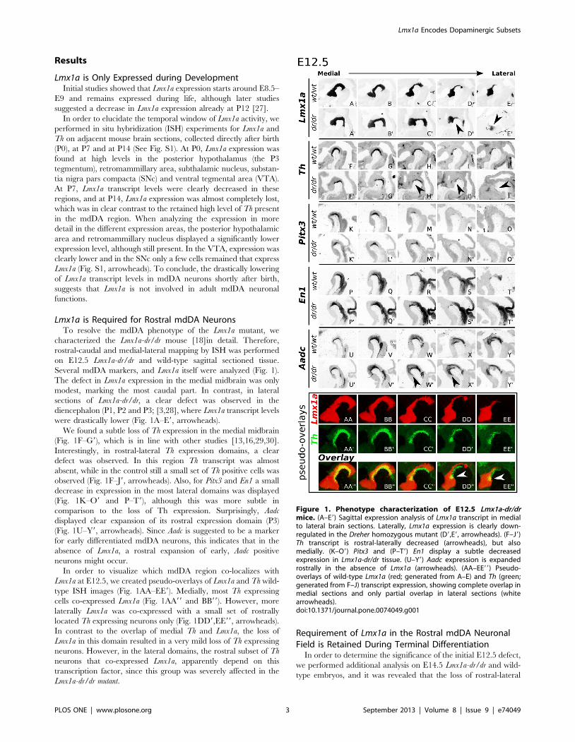
Lmx1a is Required for Rostral mdDA NeuronsTo resolve the mdDA phenotype of the Lmx1a mutant, we
characterized the Lmx1a-dr/dr mouse [18]in detail. Therefore,
rostral-caudal and medial-lateral mapping by ISH was performed
on E12.5 Lmx1a-dr/dr and wild-type sagittal sectioned tissue.
Several mdDA markers, and Lmx1a itself were analyzed (Fig. 1).
The defect in Lmx1a expression in the medial midbrain was only
modest, marking the most caudal part. In contrast, in lateral
sections of Lmx1a-dr/dr, a clear defect was observed in the
diencephalon (P1, P2 and P3; [3,28], where Lmx1a transcript levels
were drastically lower (Fig. 1A–E9, arrowheads).
We found a subtle loss of Th expression in the medial midbrain
(Fig. 1F–G9), which is in line with other studies [13,16,29,30].
Interestingly, in rostral-lateral Th expression domains, a clear
defect was observed. In this region Th transcript was almost
absent, while in the control still a small set of Th positive cells was
observed (Fig. 1F–J9, arrowheads). Also, for Pitx3 and En1 a small
decrease in expression in the most lateral domains was displayed
(Fig. 1K–O9 and P–T9), although this was more subtle in
comparison to the loss of Th expression. Surprisingly, Aadc
displayed clear expansion of its rostral expression domain (P3)
(Fig. 1U–Y9, arrowheads). Since Aadc is suggested to be a marker
for early differentiated mdDA neurons, this indicates that in the
absence of Lmx1a, a rostral expansion of early, Aadc positive
neurons might occur.
In order to visualize which mdDA region co-localizes with
Lmx1a at E12.5, we created pseudo-overlays of Lmx1a and Th wild-
type ISH images (Fig. 1AA–EE9). Medially, most Th expressing
cells co-expressed Lmx1a (Fig. 1AA99 and BB99). However, more
laterally Lmx1a was co-expressed with a small set of rostrally
located Th expressing neurons only (Fig. 1DD9,EE99, arrowheads).
In contrast to the overlap of medial Th and Lmx1a, the loss of
Lmx1a in this domain resulted in a very mild loss of Th expressing
neurons. However, in the lateral domains, the rostral subset of Th
neurons that co-expressed Lmx1a, apparently depend on this
transcription factor, since this group was severely affected in the
Lmx1a-dr/dr mutant.
Requirement of Lmx1a in the Rostral mdDA NeuronalField is Retained During Terminal Differentiation
In order to determine the significance of the initial E12.5 defect,
we performed additional analysis on E14.5 Lmx1a-dr/dr and wild-
type embryos, and it was revealed that the loss of rostral-lateral
Figure 1. Phenotype characterization of E12.5 Lmx1a-dr/drmice. (A–E9) Sagittal expression analysis of Lmx1a transcript in medialto lateral brain sections. Laterally, Lmx1a expression is clearly down-regulated in the Dreher homozygous mutant (D9,E9, arrowheads). (F–J9)Th transcript is rostral-laterally decreased (arrowheads), but alsomedially. (K–O9) Pitx3 and (P–T9) En1 display a subtle decreasedexpression in Lmx1a-dr/dr tissue. (U–Y9) Aadc expression is expandedrostrally in the absence of Lmx1a (arrowheads). (AA–EE99) Pseudo-overlays of wild-type Lmx1a (red; generated from A–E) and Th (green;generated from F–J) transcript expression, showing complete overlap inmedial sections and only partial overlap in lateral sections (whitearrowheads).doi:10.1371/journal.pone.0074049.g001
Lmx1a Encodes Dopaminergic Subsets
PLOS ONE | www.plosone.org 3 September 2013 | Volume 8 | Issue 9 | e74049
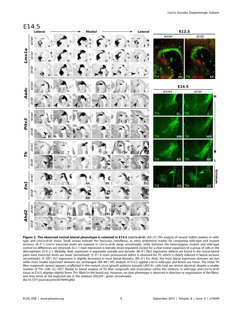
Figure 2. The observed rostral-lateral phenotype is retained in E14.5 Lmx1a-dr/dr. (AA–JJ9) ISH analysis of several mdDA markers in wild-type and Lmx1a-dr/dr tissue. Small arrows indicate the fasciculus retroflexus, as extra anatomical marker for comparing wild-type and mutantsections. (A–F99) Lmx1a transcript levels are lowered in Lmx1a-dr/dr (large arrowheads), while between the heterozygous mutant and wild-typecontrol no differences are observed. (G–L9) Aadc expression is laterally down-regulated, except for a clear rostral expansion of a group of cells in thediencephalon (G9K9,L9). Medially, Aadc expression is expanded rostrally and dorsally. (M–R9) Pitx3 expression defects are found in the rostral-lateralparts were transcript levels are lower (arrowhead). (S–X9) A more pronounced defect is observed for Th, which is clearly reduced in lateral sections(arrowheads). (Y–DD9) En1 expression is slightly decreased in most lateral domains. (EE–JJ9) For Ahd2, the most lateral expression domains are lostwhile more medial expression domains are unchanged. (KK–KK9) IHC analysis of E12.5 sagittal Lmx1a wild-type and knock-out tissue. The initial THfiber outgrowth (green) appears unaffected in the mutant, since growth patterns towards LMX1A+ cells (red) are almost identical, despite a smallernumber of TH+ cells. (LL–OO9) Medial to lateral analysis of TH fiber outgrowth and innervation within the striatum, in wild-type and Lmx1a-dr/drtissue at E14.5, displays slightly fewer TH+ fibers in the knock-out. However, no clear phenotype is observed in direction or organization of the fibers,and they arrive at the expected site in the striatum (OO,OO9, green arrowheads).doi:10.1371/journal.pone.0074049.g002
Lmx1a Encodes Dopaminergic Subsets
PLOS ONE | www.plosone.org 4 September 2013 | Volume 8 | Issue 9 | e74049
expression was retained in later developmental stages (Fig. 2A–
DD9).
Since the affected Th expression seemed to be restricted to a
rostral-lateral subset of mdDA neurons, we used Ahd2 as a marker
for this region [31]. Interestingly, and confirming the Th
expression data, the affected rostral-lateral mdDA cell-group
displayed a specific loss of Ahd2. At E14.5, the paramedian
expression domain of Ahd2 was unaffected (Fig. 2F–F9,H–H9),
whilst the most rostral-lateral Ahd2 domain was almost completely
devoid of transcript (Fig. 2EE–JJ9, arrowheads). These observa-
tions suggest a subset-specific loss of Th and Ahd2 in the rostral-
lateral mdDA region. Importantly, not all of these affected cells are
completely lost, since the mdDA markers Pitx3 and En1 are still
present in a part of this rostral-lateral sub-population (Fig. 2P–R9
and Y–DD9).
Most prominent defects were consequently observed in the
lateral-rostral part of the mdDA neuronal field. Therefore,
possible defects in the Th fiber outgrowth were analyzed. TH
immunohistochemistry was performed on E14.5 Lmx1a-dr/dr and
wild-type tissue to follow fiber outgrowth in the diencephalic
region (Fig. 2KK–OO9). The initial guidance direction of TH
fibers seemed unchanged between Lmx1a-dr/dr and control (Fig.
2KK,KK9 and LL–NN9). To confirm normal striatal innervation,
the arrival of TH bundles was analyzed at E14.5, and no obvious
defects were observed (Fig. 2OO,OO9).
Taken together, Lmx1a-dr/dr shows medially a subtle defect,
confirming previous reports [13,16,29]. Laterally, a rostral group
of mdDA neurons (SNc) is clearly affected, as was observed for
Lmx1a, Th, and Ahd2. Likely, the affected neurons are not
completely lost at this stage, since Pitx3 and En1 were less affected,
and Aadc expression was expanded, in this rostral group.
LMX1A Expression is Restricted to the Rostral mdDARegion during Terminal Differentiation
The detailed ISH mapping of Lmx1a-dr/dr suggests a subset-
specific requirement for Lmx1a, in the rostral-lateral domains of
the mdDA neuronal field. To determine into more detail this
rostral-lateral dependency, we analyzed the protein expression
patterns of LMX1A and TH, in several mdDA developmental
stages.
At E11.5, E12.0 and E12.5, medial expression of LMX1A
protein was broad and fully overlapped the mdDA area (Fig. S2).
Strikingly, when analyzing the expression pattern in the lateral TH
domain, LMX1A protein was co-expressed in a rostral subset of
TH neurons only (Fig. S2 D,G,H,I-J, arrowheads). In caudal-
lateral mdDA neurons, a set of TH+ neurons did not express
LMX1A (Fig. S2 I,J, asterisks), suggesting that there is a group of
mdDA neurons that does not depend on LMX1A, during these
developmental stages. Moreover, at E14.5 this restricted expres-
sion pattern was even more pronounced (Fig. S2 N–O). Medially,
LMX1A was co-expressed in a small and select set of rostral TH+neurons, whilst in the caudal mdDA area, no LMX1A expression
was detected. Importantly, in lateral domains at this stage, TH
expression largely co-localized with LMX1A protein (Fig. S2 P),
suggesting a functional relationship in this area and developmental
stage.
Taken together, during early mdDA differentiation, LMX1A is
broadly present in TH+ cells. During terminal differentiation,
LMX1A expression becomes restricted towards a subset of rostral
(P3) and lateral TH+ neurons. Notably, this restricted co-
localization underlines the observed phenotype in Lmx1a-dr/dr,
where Th, and Ahd2 are clearly affected in similar areas.
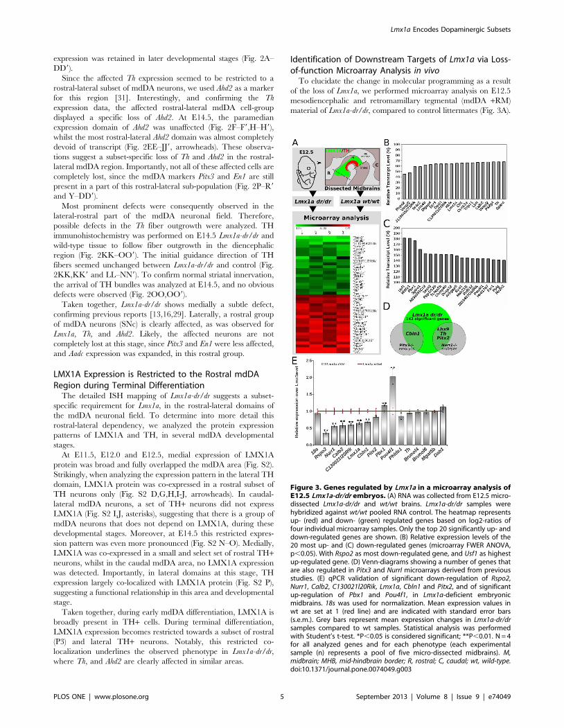
Identification of Downstream Targets of Lmx1a via Loss-of-function Microarray Analysis in vivo
To elucidate the change in molecular programming as a result
of the loss of Lmx1a, we performed microarray analysis on E12.5
mesodiencephalic and retromamillary tegmental (mdDA +RM)
material of Lmx1a-dr/dr, compared to control littermates (Fig. 3A).
Figure 3. Genes regulated by Lmx1a in a microarray analysis ofE12.5 Lmx1a-dr/dr embryos. (A) RNA was collected from E12.5 micro-dissected Lmx1a-dr/dr and wt/wt brains. Lmx1a-dr/dr samples werehybridized against wt/wt pooled RNA control. The heatmap representsup- (red) and down- (green) regulated genes based on log2-ratios offour individual microarray samples. Only the top 20 significantly up- anddown-regulated genes are shown. (B) Relative expression levels of the20 most up- and (C) down-regulated genes (microarray FWER ANOVA,p,0.05). With Rspo2 as most down-regulated gene, and Usf1 as highestup-regulated gene. (D) Venn-diagrams showing a number of genes thatare also regulated in Pitx3 and NurrI microarrays derived from previousstudies. (E) qPCR validation of significant down-regulation of Rspo2,Nurr1, Calb2, C130021l20Rik, Lmx1a, Cbln1 and Pitx2, and of significantup-regulation of Pbx1 and Pou4f1, in Lmx1a-deficient embryonicmidbrains. 18s was used for normalization. Mean expression values inwt are set at 1 (red line) and are indicated with standard error bars(s.e.m.). Grey bars represent mean expression changes in Lmx1a-dr/drsamples compared to wt samples. Statistical analysis was performedwith Student’s t-test. *P,0.05 is considered significant; **P,0.01. N = 4for all analyzed genes and for each phenotype (each experimentalsample (n) represents a pool of five micro-dissected midbrains). M,midbrain; MHB, mid-hindbrain border; R, rostral; C, caudal; wt, wild-type.doi:10.1371/journal.pone.0074049.g003
Lmx1a Encodes Dopaminergic Subsets
PLOS ONE | www.plosone.org 5 September 2013 | Volume 8 | Issue 9 | e74049
Microarray (ANOVA) analysis resulted in a total of 143
significantly regulated genes, of which 98 genes were down-
regulated and 45 were up-regulated. Importantly, Nurr1 (Nr4a2)
and Th are among the 20 most down-regulated genes (Fig. 3A,B),
confirming our phenotypic analysis. Interestingly, also the Lmx1a
transcript level was 35% reduced. Furthermore, the most down-
regulated gene was Rspo2, which transcript levels were reduced to
45% of wild-type levels. Among the 20 most up-regulated genes,
Usf1 was strongest up-regulated to 180% of wild-type levels
(Fig. 3C). In addition, Pbx1 was highly up-regulated, and
interestingly, the red nucleus (RN) neuronal marker Pou4f1 (Brn3a)
was up-regulated as well, suggesting a suppressive effect of Lmx1a
on this alternative RN fate. For subsequent analysis, a selection
was made among all significantly regulated genes based on
expression, and literature related to mdDA neurons (Phlda1;
Brunol4/6; Mgat5b; Nurr1; C130021l20Rik; Th; Calb2; Pbx1), high
fold-change (Rspo2) and migration (Dab1) (Table S3). In addition,
Pitx2 and Cbln1 were selected based on their regulation by Nurr1
and Pitx3 respectively (Fig. 3D) [32,33]. In order to validate our
microarray data first, we subjected the RNA samples to qPCR
analysis (Fig. 3E). Despite the previously observed subtle decrease
of Th expression in vivo and in the microarray analysis, we could
not confirm the down-regulation of this gene with the used qPCR
method, and neither of Phlda1, Brunol4, Brunol6, Mgat5b and Dab1.
Importantly, we confirmed clear down-regulation of Rspo2, Nurr1,
Calb2, C130021l20Rik, Lmx1a, Cbln1 and Pitx2. Furthermore, also
the up-regulation of Pbx1 and Pou4f1 was confirmed.
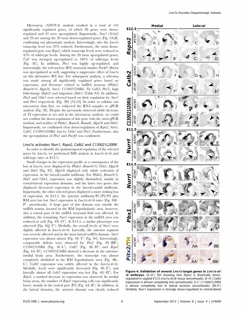
Lmx1a activates Nurr1, Rspo2, Calb2 and C130021L20RIKIn order to identify the spatiotemporal regulation of the selected
genes by Lmx1a, we performed ISH analysis in Lmx1a-dr/dr and
wild-type mice at E12.5.
Small changes in the expression profile as a consequence of the
loss of Lmx1a, were displayed for Phlda1, Brunol4/6, Cbln1, Mgat5b
and Dab1 (Fig. S3). Mgat5b displayed only subtle reduction of
expression, in the lateral-caudal midbrain. For Phlda1, Brunol4/6,
Dab1 and Cbln1, expression was slightly diminished, mainly in
rostral-lateral expression domains, and the latter two genes also
displayed decreased expression in the lateral-caudal midbrain.
Importantly, the other selected genes displayed a more striking loss
of expression. At E12.5, the anterior midbrain/P1/P2/P3 plus
RM area has lost Nurr1 expression in Lmx1a-dr/dr mice (Fig. 4M–
P9, arrowheads). A large part of this domain was outside the
mdDA system, located in the RM hypothalamic area, however,
also a rostral part of the mdDA neuronal field was affected. In
addition, the remaining Nurr1 expression in the mdDA area was
reduced as well (Fig. 4N–O9). At E14.5, a similar phenotype was
observed (Fig. 4Q–T9). Medially, the overall levels of Nurr1 were
slightly affected in Lmx1a-dr/dr. Laterally, the anterior segment
was severely affected and in the most lateral mdDA domain, Nurr1
expression was almost absent (Fig. 4S–T9; Fig. S4). Interestingly,
comparable deficits were observed for Pitx2 (Fig. 4Y–BB9),
C130021l20Rik (Fig. 4I–L9), Calb2 (Fig. 4E–H9) and Rspo2
(Fig. 4A–D9). C130021L20Rik showed a decrease in the anterior-
medial brain area. Furthermore, the transcript was almost
completely abolished in the RM hypothalamic area (Fig. 4K–
L9). Calb2 expression was widely affected in the Lmx1a-dr/dr.
Medially, levels were significantly decreased (Fig. 4E–F9), and
laterally almost all Calb2 expression was lost (Fig. 4G–H9). For
Rspo2, a marked decrease in expression was observed. In medial
brain areas, the number of Rspo2 expressing cells was significantly
lower, mainly in the rostral part (P3) (Fig. 4A–B9). In addition, in
the lateral domains, the anterior domain was clearly reduced
Figure 4. Validation of several Lmx1a target genes in Lmx1a-dr/dr embryos. (A–D9) ISH showing that Rspo2 is drastically down-regulated in sagittal E12.5 Lmx1a-dr/dr tissue (arrowheads). (E–H9) Calb2expression is almost completely lost (arrowheads). (I–L9) C130021L20Rikis almost completely lost in lateral sections (arrowheads). (M–P9)Similarly, Nurr1 expression is strongly down-regulated in rostral-lateral
Lmx1a Encodes Dopaminergic Subsets
PLOS ONE | www.plosone.org 6 September 2013 | Volume 8 | Issue 9 | e74049
(Fig. 4C–D9), and in the most lateral Rspo2 expression domains,
completely lost (Fig. S5).
Taken together, the genes discussed above are all influenced by
Lmx1a activity in vivo and the most marked deficits were observed
for Nurr1, C130021L20Rik, Calb2 and Rspo2. These findings
strongly suggest that these genes are direct or indirect down-
stream targets of Lmx1a. Moreover, the Lmx1a dependency is
region-specific and seems most severe for the rostral-lateral
expression domains (P3 and the RM hypothalamic area).
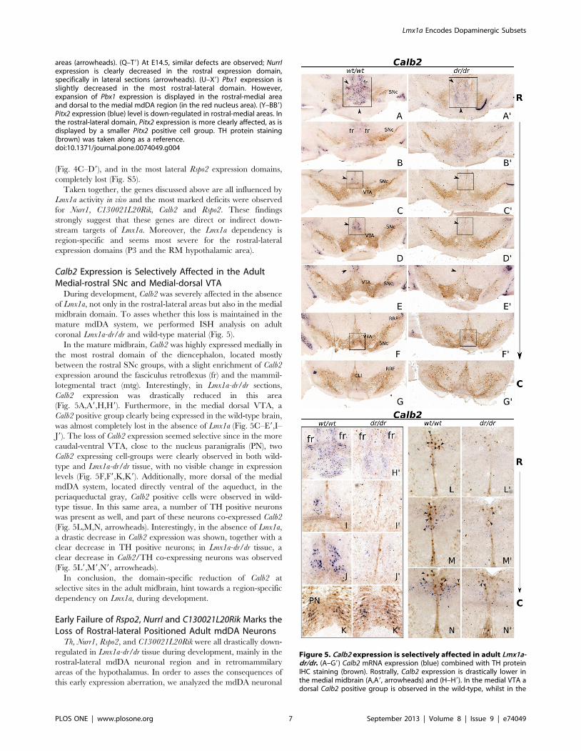
Calb2 Expression is Selectively Affected in the AdultMedial-rostral SNc and Medial-dorsal VTA
During development, Calb2 was severely affected in the absence
of Lmx1a, not only in the rostral-lateral areas but also in the medial
midbrain domain. To asses whether this loss is maintained in the
mature mdDA system, we performed ISH analysis on adult
coronal Lmx1a-dr/dr and wild-type material (Fig. 5).
In the mature midbrain, Calb2 was highly expressed medially in
the most rostral domain of the diencephalon, located mostly
between the rostral SNc groups, with a slight enrichment of Calb2
expression around the fasciculus retroflexus (fr) and the mammil-
lotegmental tract (mtg). Interestingly, in Lmx1a-dr/dr sections,
Calb2 expression was drastically reduced in this area
(Fig. 5A,A9,H,H9). Furthermore, in the medial dorsal VTA, a
Calb2 positive group clearly being expressed in the wild-type brain,
was almost completely lost in the absence of Lmx1a (Fig. 5C–E9,I–
J9). The loss of Calb2 expression seemed selective since in the more
caudal-ventral VTA, close to the nucleus paranigralis (PN), two
Calb2 expressing cell-groups were clearly observed in both wild-
type and Lmx1a-dr/dr tissue, with no visible change in expression
levels (Fig. 5F,F9,K,K9). Additionally, more dorsal of the medial
mdDA system, located directly ventral of the aqueduct, in the
periaqueductal gray, Calb2 positive cells were observed in wild-
type tissue. In this same area, a number of TH positive neurons
was present as well, and part of these neurons co-expressed Calb2
(Fig. 5L,M,N, arrowheads). Interestingly, in the absence of Lmx1a,
a drastic decrease in Calb2 expression was shown, together with a
clear decrease in TH positive neurons; in Lmx1a-dr/dr tissue, a
clear decrease in Calb2/TH co-expressing neurons was observed
(Fig. 5L9,M9,N9, arrowheads).
In conclusion, the domain-specific reduction of Calb2 at
selective sites in the adult midbrain, hint towards a region-specific
dependency on Lmx1a, during development.
Early Failure of Rspo2, NurrI and C130021L20Rik Marks theLoss of Rostral-lateral Positioned Adult mdDA Neurons
Th, Nurr1, Rspo2, and C130021L20Rik were all drastically down-
regulated in Lmx1a-dr/dr tissue during development, mainly in the
rostral-lateral mdDA neuronal region and in retromammilary
areas of the hypothalamus. In order to asses the consequences of
this early expression aberration, we analyzed the mdDA neuronal
areas (arrowheads). (Q–T9) At E14.5, similar defects are observed; NurrIexpression is clearly decreased in the rostral expression domain,specifically in lateral sections (arrowheads). (U–X9) Pbx1 expression isslightly decreased in the most rostral-lateral domain. However,expansion of Pbx1 expression is displayed in the rostral-medial areaand dorsal to the medial mdDA region (in the red nucleus area). (Y–BB9)Pitx2 expression (blue) level is down-regulated in rostral-medial areas. Inthe rostral-lateral domain, Pitx2 expression is more clearly affected, as isdisplayed by a smaller Pitx2 positive cell group. TH protein staining(brown) was taken along as a reference.doi:10.1371/journal.pone.0074049.g004
Figure 5. Calb2 expression is selectively affected in adult Lmx1a-dr/dr. (A–G9) Calb2 mRNA expression (blue) combined with TH proteinIHC staining (brown). Rostrally, Calb2 expression is drastically lower inthe medial midbrain (A,A9, arrowheads) and (H–H9). In the medial VTA adorsal Calb2 positive group is observed in the wild-type, whilst in the
Lmx1a Encodes Dopaminergic Subsets
PLOS ONE | www.plosone.org 7 September 2013 | Volume 8 | Issue 9 | e74049
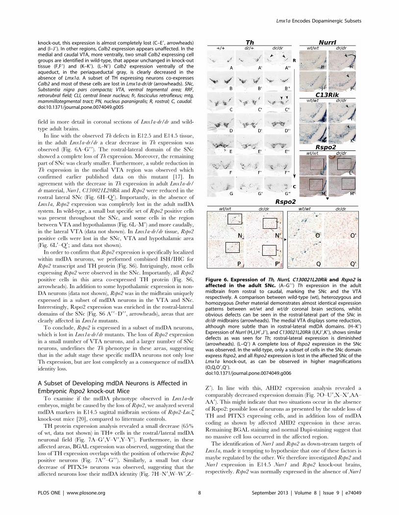
field in more detail in coronal sections of Lmx1a-dr/dr and wild-
type adult brains.
In line with the observed Th defects in E12.5 and E14.5 tissue,
in the adult Lmx1a-dr/dr a clear decrease in Th expression was
observed (Fig. 6A–G99). The rostral-lateral domain of the SNc
showed a complete loss of Th expression. Moreover, the remaining
part of SNc was clearly smaller. Furthermore, a subtle reduction in
Th expression in the medial VTA region was observed which
confirmed earlier published data on this mutant [17]. In
agreement with the decrease in Th expression in adult Lmx1a-dr/
dr material, Nurr1, C130021L20Rik and Rspo2 were reduced in the
rostral lateral SNc (Fig. 6H–Q9). Importantly, in the absence of
Lmx1a, Rspo2 expression was completely lost in the adult mdDA
system. In wild-type, a small but specific set of Rspo2 positive cells
was present throughout the SNc, and some cells in the region
between VTA and hypothalamus (Fig. 6L–M9) and more caudally,
in the lateral VTA (data not shown). In Lmx1a-dr/dr tissue, Rspo2
positive cells were lost in the SNc, VTA and hypothalamic area
(Fig. 6L9–Q9; and data not shown).
In order to confirm that Rspo2 expression is specifically localized
within mdDA neurons, we performed combined ISH/IHC for
Rspo2 transcript and TH protein (Fig. S6). Intriguingly, most cells
expressing Rspo2 were observed in the SNc. Importantly, all Rspo2
positive cells in this area co-expressed TH protein (Fig. S6,
arrowheads). In addition to some hypothalamic expression in non-
DA neurons (data not shown), Rspo2 was in the midbrain uniquely
expressed in a subset of mdDA neurons in the VTA and SNc.
Interestingly, Rspo2 expression was enriched in the rostral-lateral
domains of the SNc (Fig. S6 A99–D99, arrowheads), areas that are
clearly affected in Lmx1a mutants.
To conclude, Rspo2 is expressed in a subset of mdDA neurons,
which is lost in Lmx1a-dr/dr mutants. The loss of Rspo2 expression
in a small number of VTA neurons, and a larger number of SNc
neurons, underlines the Th phenotype in these areas, suggesting
that in the adult stage these specific mdDA neurons not only lose
Th expression, but are lost completely as a consequence of mdDA
identity loss.
A Subset of Developing mdDA Neurons is Affected inEmbryonic Rspo2 knock-out Mice
To examine if the mdDA phenotype observed in Lmx1a-dr
embryos, might be caused by the loss of Rspo2, we analyzed several
mdDA markers in E14.5 sagittal midbrain sections of Rspo2-LacZ
knock-out mice [20], compared to littermate controls.
TH protein expression analysis revealed a small decrease (65%
of wt, data not shown) in TH+ cells in the rostral/lateral mdDA
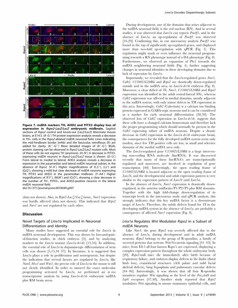
neuronal field (Fig. 7A–G9,V–V9,Y–Y9). Furthermore, in these
affected areas, BGAL expression was observed, suggesting that the
loss of TH expression overlaps with the position of otherwise Rspo2
positive neurons (Fig. 7A99–G99). Similarly, a small but clear
decrease of PITX3+ neurons was observed, suggesting that the
affected neurons lose their mdDA identity (Fig. 7H–N9,W–W9,Z–
Z9). In line with this, AHD2 expression analysis revealed a
comparably decreased expression domain (Fig. 7O–U9,X–X9,AA–
AA9). This might indicate that two situations occur in the absence
of Rspo2: possible loss of neurons as presented by the subtle loss of
TH and PITX3 expressing cells, and in addition loss of mdDA
coding as shown by affected AHD2 expression in these areas.
Remaining BGAL staining and normal Dapi-staining suggest that
no massive cell loss occurred in the affected region.
The identification of Nurr1 and Rspo2 as down-stream targets of
Lmx1a, made it tempting to hypothesize that one of these factors is
maybe regulated by the other. We therefore investigated Rspo2 and
Nurr1 expression in E14.5 Nurr1 and Rspo2 knock-out brains,
respectively. Rspo2 was normally expressed in the absence of Nurr1
knock-out, this expression is almost completely lost (C–E9, arrowheads)and (I–J9). In other regions, Calb2 expression appears unaffected. In themedial and caudal VTA, more ventrally, two small Calb2 expressing cellgroups are identified in wild-type, that appear unchanged in knock-outtissue (F,F9) and (K–K9). (L–N9) Calb2 expression ventrally of theaqueduct, in the periaqueductal gray, is clearly decreased in theabsence of Lmx1a. A subset of TH expressing neurons co-expressesCalb2 and most of these cells are lost in Lmx1a-dr/dr (arrowheads). SNc,Substantia nigra pars compacta; VTA, ventral tegmental area; RRF,retrorubral field; CLI, central linear nucleus; fr, fasciculus retroflexus; mtg,mammillotegmental tract; PN, nucleus paranigralis; R, rostral; C, caudal.doi:10.1371/journal.pone.0074049.g005
Figure 6. Expression of Th, NurrI, C130021L20Rik and Rspo2 isaffected in the adult SNc. (A–G99) Th expression in the adultmidbrain from rostral to caudal, marking the SNc and the VTArespectively. A comparison between wild-type (wt), heterozygous andhomozygous Dreher material demonstrates almost identical expressionpatterns between wt/wt and wt/dr coronal brain sections, whilstobvious defects can be seen in the rostral-lateral part of the SNc indr/dr midbrains (arrowheads). The medial VTA displays some reduction,although more subtle than in rostral-lateral mdDA domains. (H–K9)Expression of NurrI (H,J,H9,J9), and C130021L20Rik (I,K,I9,K9), shows similardefects as was seen for Th; rostral-lateral expression is diminished(arrowheads). (L–Q9) A complete loss of Rspo2 expression in the SNcwas observed. In the wild-type, only a subset of cells in the SNc domainexpress Rspo2, and all Rspo2 expression is lost in the affected SNc of theLmx1a knock-out, as can be observed in higher magnifications(O,Q,O9,Q9).doi:10.1371/journal.pone.0074049.g006
Lmx1a Encodes Dopaminergic Subsets
PLOS ONE | www.plosone.org 8 September 2013 | Volume 8 | Issue 9 | e74049
(data not shown). Also, in Rspo2-LacZ/LacZ tissue, Nurr1 expression
was hardly affected (data not shown). This indicated that Rspo2
and Nurr1 are not regulated by each other.
Discussion
Novel Targets of Lmx1a Implicated in NeuronalDifferentiation and Identity
Many studies have suggested an essential role for Lmx1a in
mdDA neuronal development. This was shown by loss-and-gain-
of-function studies, in chick embryos [5], and by analyzing
markers in the Lmx1a mutant (Lmx1a-dr/dr) [13,16]. In addition,
the essential role of Lmx1a in dopaminergic differentiation of stem
cells was shown [5,15,34–37]. In literature, it is suggested that
Lmx1a plays a role in proliferation and neurogenesis, but despite
the indication that several factors are regulated by Lmx1a, like
NurrI, Msx1 and Wnt1, the precise role of Lmx1a in the mouse is still
not clearly identified. In order to unravel the exact molecular
programming activated by Lmx1a, we performed an in vivo
transcriptome analysis by using Lmx1a-dr/dr embryonic mdDA
plus RM brain areas.
During development, one of the domains that arises adjacent to
the mdDA neuronal field, is the red nucleus (RN). And in several
studies, it was observed that Lmx1a can repress Pou4f1, and in the
absence of Lmx1a, an up-regulation of Pou4f1 was observed
[16,29]. Confirming this, in our microarray analysis Pou4f1 was
found in the top of significantly up-regulated genes, and displayed
more than two-fold up-regulation with qPCR (Fig. 3). This
regulation might mark or even influence the neuronal program-
ming towards a RN phenotype instead of a DA phenotype (Fig. 8).
Furthermore, we observed an expansion of Pbx1 towards the
mdDA neighboring neuronal fields (Fig. 4), further suggesting
changes in neuronal identities in these developing domains due to
lack of repression by Lmx1a.
Importantly, we revealed that the Lmx1a-regulated genes Nurr1,
Calb2, C130021L20Rik and Rspo2 are drastically down-regulated
outside and in the mdDA area, in Lmx1a-dr/dr embryos (Fig. 8).
Moreover, a clear deficit of Th, Nurr1, C130021L20Rik and Rspo2
expression was identified in the adult rostral-lateral SNc, whereas
Calb2 expression was affected in medial domains, mostly adjacent
to the mdDA system, with only minor defects in TH expression in
this area. Interestingly, Calb2 (Calretinin) is a calcium ion binding
protein expressed in GABA-ergic neurons and it can be considered
as a marker for early neuronal differentiation [38,39]. The
observed loss of Calb2 expression in Lmx1a-dr/dr, suggests that
these cells have a changed calcium homeostasis and therefore lack
the proper programming which should normally be present in the
Calb2 expressing subset of mdDA neurons. Despite a drastic
decrease in Calb2 expression in the Lmx1a-dr/dr embryonic brain,
the consequences for the fully developed mdDA system seem only
modest, since few TH positive cells are lost, in small and selective
domains of the medial mdDA area only.
The Lmx1a-regulated gene C130021L20Rik is a large interven-
ing non-coding RNA molecule (lincRNA). It was discovered
recently that many of these lincRNA’s are transcriptionally
regulated and moreover, are involved in regulation of gene
transcription [40]. Interestingly, the open reading frame of
C130021L20Rik is located adjacent to the open reading frame of
Lmx1b, and the developmental and adult expression pattern is very
similar to the expression pattern of Lmx1b (and Lmx1a).
In the absence of Lmx1a, Nurr1 expression is drastically down-
regulated, in the anterior midbrain/P1/P2/P3 plus RM domains.
Together with the high fold-change down-regulation of its
transcript levels in the microarray study and qPCR analysis, this
strongly indicates that this key mdDA factor is a downstream
target of Lmx1a. Therefore, the subtle defects found for Th in the
developing mdDA system in the absence of Lmx1a, are probably a
consequence of affected Nurr1 expression (Fig. 8).
Lmx1a Regulates Wnt Modulator Rspo2 in a Subset ofmdDA Neurons
Like Nurr1, the gene Rspo2 was severely affected due to the
absence of Lmx1a, during development and in adult mdDA
neurons. Rspo2 belongs to the group of R-spondins, a family of
secreted proteins that activate Wnt/b-catenin signaling [41–43]. In
mice, from E8.5 all four known Rspo’s are expressed, displaying a
complex expression pattern throughout the whole embryonic body
[20]. Rspo2-null mice die immediately after birth because of
respiratory failure, and embryos display defects in the limbs (distal
limb loss), craniofacial structures (cleft palate and mild facial
skeletal defects), lung hypoplasia and pulmonary vascular defects
[44–46]. Interestingly, it was shown that all four R-spondin
members regulate Wnt signaling at the level of the Frizzled8 and
Lrp6 receptors [47,48]. Another study reported that Rspo2
modulates Wnt signaling in mouse mammary epithelial cells, and
Figure 7. mdDA markers TH, AHD2 and PITX3 display loss ofexpression in Rspo2-LacZ/LacZ embryonic midbrain. Sagittalsections of Rspo2 control and knock-out (LacZ/LacZ) littermate mousebrains, at E14.5. (A–G9) TH protein expression analysis reveals a decreasein TH+ cells in the Rspo2-ablated mdDA neuronal field. Lines indicatingthe mid-hindbrain border (mhb) and the fasciculus retroflexus (fr) areadded for clarity. (A99–G99) More detailed images of (A9–G9). BGALprotein staining can be observed in Rspo2-LacZ/LacZ mutant cells. Mostof these cells do not express TH (anymore). (H–N9) A decrease in PITX3-expressing mdDA neurons in Rspo2-LacZ/LacZ tissue is shown. (O–U9)From lateral to medial to lateral, AHD2 analysis reveals a decrease inexpression in the paramedian and lateral mdDA neuronal subset, in theabsence of Rspo2. (V–X9) Higher magnifications of (C,C9), (J,J9) and(Q,Q9), showing a mild but clear decrease of mdDA neurons expressingTH, PITX3 and AHD2 in the paramedian midbrain. (Y–AA9) Highermagnifications of (F,F9), (M,M9) and (O,O9), showing a clear decrease inthe number of TH-, PITX3-, and AHD2-positive neurons in the lateralmdDA neuronal field.doi:10.1371/journal.pone.0074049.g007
Lmx1a Encodes Dopaminergic Subsets
PLOS ONE | www.plosone.org 9 September 2013 | Volume 8 | Issue 9 | e74049

that Rspo2 and Wnt1 act synergistically in the b-catenin pathway
[49]. Rspo’s can stabilize the level of cytosolic b-catenin and
synergize with Wnt-ligands in order to promote b-catenin
transcriptional activity [47,50,51]. Intriguingly, Wnt1 is important
for proliferation and patterning of the mdDA neuronal field [52],
and several papers suggested a role of Lmx1a together with Wnt1 in
mdDA differentiation. Recently, a novel Wnt1-Lmx1a auto-
regulatory loop was identified during mdDA differentiation of
mouse ES cells [15]. Moreover, it was described that the reduced
number of mdDA progenitors in Lmx1a-dr/dr and Lmx1a/Lmx1b
double mutants may be a consequence of proliferation defects and
an increase in cell cycle exit of the progenitors [16]. Also, their
data indicate that Wnt1 expression was slightly reduced in Lmx1a-
dr/dr, and specifically lost in the Lmx1a/Lmx1b double mutant,
suggesting that both genes specifically and redundantly regulate
Wnt1 expression in the mdDA domain [16]. Since Rspo2 acts in the
Wnt1/b-catenin signaling pathway, a reduction in Rspo2 expres-
sion might influences the end result of Wnt activity. We speculate
that the lack of Rspo2 protein in the affected neurons of the Lmx1a-
dr/dr mutant might induce the previously suggested early cell-cycle
exit and premature differentiation. In line with this, in the Lmx1a-
dr/dr mutant we observed in the current study a clear expansion of
Aadc expression, which might represent an expansion of early
mdDA neurons. However, it may also display an induction of
other monoaminergic neuronal phenotypes in this area.
Additionally, when comparing the loss of mdDA neurons (or a
loss of mdDA identity) between the Lmx1a and the Rspo2 mutant,
we found that the observed defects are partially identical between
both mutants, where the terminal differentiation markers Th and
Pitx3 showed the same subtle reduced expression. It is therefore
likely that part of the Lmx1a-phenotype is caused by the loss of
down-stream target Rspo2, affecting a number of mdDA neurons
positioned in the VTA and more prominently in the SNc.
ConclusionsWe have shown that Lmx1a is essential for a rostral-lateral subset
of developing mdDA neurons, and loss of Lmx1a results in loss of
the correct neuronal identity, leading to an affected SNc in the
adult mouse brain. Transcriptome analysis of Lmx1a-deficient
embryonic brains revealed several genes that are known for their
role in mdDA function and development, such as Th and Nurr1. In
addition, several novel genes were identified, leading to a better
understanding of the subset-specific phenotype of the Lmx1a-
deficient mdDA system, since these genes are mainly depending
on Lmx1a in the rostral-lateral mdDA neuronal field. Moreover,
loss of the highly regulated Lmx1a-dependent gene Rspo2, partially
phenocopied the Lmx1a-mutant. To conclude, in dept character-
ization and gene expression profiling of Lmx1a-deficient mdDA
neurons, provided a detailed map of the molecular pathway in
which Lmx1a is acting towards the correct development of mdDA
neuronal subsets.
Figure 8. A model integrating several identified targets of Lmx1a. Pou4f1 (Brn3a) was strongly up-regulated in the Lmx1a2/2 expressionarray, in line with a suggested role of Lmx1a in repressing an alternative, red nucleus, fate during development (dashed line). Lmx1a targetsC130021l20Rik and Calb2 require Lmx1a directly or indirectly for correct expression in subsets of the mdDA system. The Nurr1 transcriptional pathwayinduces differentiating mdDA neurons, and loss of Lmx1a resulted in affected Nurr1 expression, but also in affected expression of Nurr1 transcriptionaltargets, mainly in rostral-lateral mdDA neurons. Rspo2 is involved in the Wnt1/b-catenin signaling pathways, which is implicated in proliferation andcell-cycle exit. Loss of Lmx1a, resulted in decreased Rspo2 expression and loss of mdDA neuronal markers as Th, Ahd2 and Pitx3. Interestingly, loss ofRspo2 partially phenocopied the defects observed in the Lmx1a-dr/dr mdDA system.doi:10.1371/journal.pone.0074049.g008
Lmx1a Encodes Dopaminergic Subsets
PLOS ONE | www.plosone.org 10 September 2013 | Volume 8 | Issue 9 | e74049
Supporting Information
Figure S1 Lmx1a expression is down-regulated shortlyafter birth. Coronal sections of wild-type mouse tissue at P0, P7
and P14. In situ hybridization of Lmx1a and Th is shown from
rostral to caudal. Th was taken along to mark the SNc and VTA
and as a control for transcript levels. (A–F) A significant loss of
Lmx1a expression is observed in the SNc at P14, when compared
to P7 or P0, and when compared to Th. (A9,C9,E9) Lmx1a
expression in the SNc in more detail, showing that only few cells
remain that express Lmx1a, in low levels. (G–R) Both in caudal
SNc, and more caudally, the VTA, the expression of Lmx1a is
clearly diminished at P14 (arrowheads). P, postnatal day; SNc,
Substantia nigra pars compacta; VTA, ventral tegmental area.
(TIF)
Figure S2 LMX1A protein expression is restricted torostral-lateral TH expression at later developmentalstages. (A–H) IHC on E11.5 (A–D) and E12.0 (E–H) sagittal
wild-type mouse tissue, showing LMX1A protein (red) and TH
protein (green). Medially, full protein overlap is displayed, whereas
in lateral expression domains, only a subset of TH+ neurons
overlaps with LMX1A. (I–J) Higher magnifications of G and H,
showing the group of TH+ neurons that co-localize with LMX1A
(white arrowheads), and a group of TH expressing neurons that do
not express LMX1A (green asterisks). (K–M) The observed rostral-
lateral overlap is more clear at E12.5, where also in the medial
brain, a rostral specificity of LMX1A occurs. (N–P) At E14.5, the
rostral LMX1A/TH restriction is clearly observed in medial and
lateral midbrain areas.
(TIF)
Figure S3 Lmx1a regulates Phlda1, Brunol4, Brunol6,Cbln1, Mgat5b and Dab1 in selective areas of the Lmx1a-expression domain. Sagittal Lmx1a control and Lmx1a-dr/dr
sections, medial and lateral. On the right, higher magnifications of
the boxed areas are shown. (A–E9) Phlda1 expression is slightly
reduced in rostral parts of the brain (arrowheads). (F–O9) Brunol4
and Brunol6 both show a slight down-regulation in rostral-lateral
domains (G,G9, and arrowheads). (P–T9) Cbln1 expression is
diminished mainly in rostral areas. (U–Y9) Mgat5b transcript levels
are slightly reduced in the lateral-caudal midbrain (arrowheads).
(Z–DD9) Dab1 shows loss of expression in medial-rostral areas
(Z9,AA9,DD9, arrowheads). Also lateral-caudal, expression is
affected (CC9, arrowhead).
(TIF)
Figure S4 Lmx1a and NurrI expression throughout thesagittal brain at E14.5. Lmx1a expression (left columns) from
lateral (L) to medial (M) to lateral brain domains, in wild-type and
Lmx1a-dr/dr tissue, at E14.5. In lateral domains, Lmx1a expression
is down-regulated in the Lmx1a knock-out, and in all sections
throughout the brain, a rostral defect is shown. NurrI expression
(right columns) was analyzed in the same set-up. For NurrI a
drastically decrease in rostral and lateral expression can be
observed (arrowheads).
(TIF)
Figure S5 Lmx1a and Rspo2 expression throughout thesagittal brain at E12.5. Lmx1a expression (left columns) from
lateral (L) to medial (M) to lateral brain domains, in wild-type and
Lmx1a-dr/dr tissue, at E12.5. In lateral positions, Lmx1a expression
is clearly down-regulated in the Lmx1a knock-out, and in all
sections, a rostral defect can be seen. Rspo2 expression (right
columns) was analyzed in the same set-up. For Rspo2 an even more
drastic decrease in rostral and lateral expression can be observed
(arrowheads).
(TIF)
Figure S6 Rspo2 co-localizes with TH in adult mdDAneurons. (A–C) Combined ISH/IHC for Rspo2 (blue) and TH
(brown). Most Rspo2-positive cells are found in the rostral and
lateral mdDA system, in the SNc (A99,B99,C99, arrowheads) and
also some cells expressing Rspo2 are observed in the VTA
(A9,B9,C9). All Rspo2-positive cells also express TH, indicating
that Rspo2 is expressed in a subset of mdDA neurons (D).
(TIF)
Table S1 List of primers used for the generation of insitu hybridization probes.
(XLS)
Table S2 List of primers used for qPCR.
(XLS)
Table S3 A selection of genes regulated in E12.5 Lmx1a-dr/dr mouse embryos.
(PDF)
Acknowledgments
We gratefully thank Michael German for providing the LMX1A antibody,
Frank Jacobs for assistance with the microarray design, Raymond
Schellevis for genotyping and Roger Koot for assistance with additional
microarray data analysis.
Author Contributions
Conceived and designed the experiments: EJH MPS. Performed the
experiments: EJH LvO LvdH JVV WK IW AJAvdL. Analyzed the data:
EJH MPS. Wrote the paper: EJH MPS. Carried out microarray analysis
annotations and statistical analysis: FCPH MJG-K. Contributed Rspo2
mouse material: Y–RJ JKY.
References
1. Petros TJ, Tyson JA, Anderson SA (2011) Pluripotent stem cells for the study of
CNS development. Front Mol Neurosci 4: 30. doi:10.3389/fnmol.2011.00030.
2. Prakash N, Wurst W (2006) Genetic networks controlling the development of
midbrain dopaminergic neurons. J Physiol (Lond) 575: 403–410. doi:10.1113/
jphysiol.2006.113464.
3. Smits SM, Burbach JPH, Smidt MP (2006) Developmental origin and fate of
meso-diencephalic dopamine neurons. Prog Neurobiol 78: 1–16. doi:10.1016/
j.pneurobio.2005.12.003.
4. Andersson E, Jensen JB, Parmar M, Guillemot F, Bjorklund A (2006)
Development of the mesencephalic dopaminergic neuron system is compro-
mised in the absence of neurogenin 2. Development 133: 507–516. doi:10.1242/
dev.02224.
5. Andersson E, Tryggvason U, Deng Q, Friling S, Alekseenko Z, et al. (2006)
Identification of intrinsic determinants of midbrain dopamine neurons. Cell 124:
393–405. doi:10.1016/j.cell.2005.10.037.
6. Ferri ALM, Lin W, Mavromatakis YE, Wang JC, Sasaki H, et al. (2007) Foxa1
and Foxa2 regulate multiple phases of midbrain dopaminergic neuron
development in a dosage-dependent manner. Development 134: 2761–2769.
doi:10.1242/dev.000141.
7. Saucedo-Cardenas O, Quintana-Hau JD, Le WD, Smidt MP, Cox JJ, et al.
(1998) Nurr1 is essential for the induction of the dopaminergic phenotype and
the survival of ventral mesencephalic late dopaminergic precursor neurons. Proc
Natl Acad Sci USA 95: 4013–4018.
8. Smidt MP, Van Schaick HS, Lanctot C, Tremblay JJ, Cox JJ, et al. (1997) A
homeodomain gene Ptx3 has highly restricted brain expression in mesencephalic
dopaminergic neurons. Proc Natl Acad Sci USA 94: 13305–13310.
9. Smidt MP, Asbreuk CH, Cox JJ, Chen H, Johnson RL, et al. (2000) A second
independent pathway for development of mesencephalic dopaminergic neurons
requires Lmx1b. Nat Neurosci 3: 337–341. doi:10.1038/73902.
Lmx1a Encodes Dopaminergic Subsets
PLOS ONE | www.plosone.org 11 September 2013 | Volume 8 | Issue 9 | e74049
10. Smidt MP, Smits SM, Burbach JPH (2004) Homeobox gene Pitx3 and its role in
the development of dopamine neurons of the substantia nigra. Cell Tissue Res318: 35–43. doi:10.1007/s00441-004-0943-1.
11. Kele J, Simplicio N, Ferri ALM, Mira H, Guillemot F, et al. (2006) Neurogenin2 is required for the development of ventral midbrain dopaminergic neurons.
Development 133: 495–505. doi:10.1242/dev.02223.
12. Smits SM, Smidt MP (2006) The role of Pitx3 in survival of midbrain
dopaminergic neurons. J Neural Transm Suppl: 57–60.
13. Ono Y, Nakatani T, Sakamoto Y, Mizuhara E, Minaki Y, et al. (2007)
Differences in neurogenic potential in floor plate cells along an anteroposteriorlocation: midbrain dopaminergic neurons originate from mesencephalic floor
plate cells. Development 134: 3213–3225. doi:10.1242/dev.02879.
14. Puelles E, Annino A, Tuorto F, Usiello A, Acampora D, et al. (2004) Otx2
regulates the extent, identity and fate of neuronal progenitor domains in theventral midbrain. Development 131: 2037–2048. doi:10.1242/dev.01107.
15. Chung S, Leung A, Han B-S, Chang M-Y, Moon J-I, et al. (2009) Wnt1-lmx1a
forms a novel autoregulatory loop and controls midbrain dopaminergic
differentiation synergistically with the SHH-FoxA2 pathway. Cell Stem Cell 5:646–658. doi:10.1016/j.stem.2009.09.015.
16. Yan CH, Levesque M, Claxton S, Johnson RL, Ang S-L (2011) Lmx1a andlmx1b function cooperatively to regulate proliferation, specification, and
differentiation of midbrain dopaminergic progenitors. J Neurosci 31: 12413–12425. doi:10.1523/JNEUROSCI.1077-11.2011.
17. Deng Q, Andersson E, Hedlund E, Alekseenko Z, Coppola E, et al. (2011)Specific and integrated roles of Lmx1a, Lmx1b and Phox2a in ventral midbrain
development. Development 138: 3399–3408. doi:10.1242/dev.065482.
18. Millonig JH, Millen KJ, Hatten ME (2000) The mouse Dreher gene Lmx1a
controls formation of the roof plate in the vertebrate CNS. Nature 403: 764–769. doi:10.1038/35001573.
19. Chizhikov V, Steshina E, Roberts R, Ilkin Y, Washburn L, et al. (2006)
Molecular definition of an allelic series of mutations disrupting the mouse
Lmx1a (dreher) gene. Mamm Genome 17: 1025–1032. doi:10.1007/s00335-006-0033-7.
20. Nam J-S, Park E, Turcotte TJ, Palencia S, Zhan X, et al. (2007) Mouse R-
spondin2 is required for apical ectodermal ridge maintenance in the hindlimb.
Dev Biol 311: 124–135. doi:10.1016/j.ydbio.2007.08.023.
21. Smidt MP, Smits SM, Bouwmeester H, Hamers FPT, Van der Linden AJA, etal. (2004) Early developmental failure of substantia nigra dopamine neurons in
mice lacking the homeodomain gene Pitx3. Development 131: 1145–1155.
doi:10.1242/dev.01022.
22. Smits SM, Ponnio T, Conneely OM, Burbach JPH, Smidt MP (2003)
Involvement of Nurr1 in specifying the neurotransmitter identity of ventralmidbrain dopaminergic neurons. Eur J Neurosci 18: 1731–1738.
23. Grima B, Lamouroux A, Blanot F, Biguet NF, Mallet J (1985) Complete coding
sequence of rat tyrosine hydroxylase mRNA. Proc Natl Acad Sci USA 82: 617–
621.
24. Hamatani T, Falco G, Carter MG, Akutsu H, Stagg CA, et al. (2004) Age-associated alteration of gene expression patterns in mouse oocytes. Hum Mol
Genet 13: 2263–2278. doi:10.1093/hmg/ddh241.
25. Yang YH, Dudoit S, Luu P, Lin DM, Peng V, et al. (2002) Normalization for
cDNA microarray data: a robust composite method addressing single andmultiple slide systematic variation. Nucleic Acids Res 30: e15.
26. Wu TD (2002) Large-Scale Analysis of Gene Expression Profiles. BriefBioinform 3: 7–17. doi:10.1093/bib/3.1.7.
27. Zou H-L, Su C-J, Shi M, Zhao G-Y, Li Z-Y, et al. (2009) Expression of the LIM-homeodomain gene Lmx1a in the postnatal mouse central nervous system. Brain
Research Bulletin 78: 306–312. doi:10.1016/j.brainresbull.2008.12.001.
28. Puelles L, Rubenstein JLR (2003) Forebrain gene expression domains and the
evolving prosomeric model. Trends Neurosci 26: 469–476.
29. Nakatani T, Kumai M, Mizuhara E, Minaki Y, Ono Y (2010) Lmx1a andLmx1b cooperate with Foxa2 to coordinate the specification of dopaminergic
neurons and control of floor plate cell differentiation in the developing
mesencephalon. Dev Biol 339: 101–113. doi:10.1016/j.ydbio.2009.12.017.
30. Hoekstra EJ, Von Oerthel L, Van der Linden AJA, Schellevis RD, Scheppink G,et al. (2013) Lmx1a is an activator of Rgs4 and Grb10 and is responsible for the
correct specification of rostral and medial mdDA neurons. Eur J Neurosci 37:
23–32. doi:10.1111/ejn.12022.
31. Jacobs FMJ, Smits SM, Noorlander CW, Von Oerthel L, Van der Linden AJA,
et al. (2007) Retinoic acid counteracts developmental defects in the substantianigra caused by Pitx3 deficiency. Development 134: 2673–2684. doi:10.1242/
dev.02865.
32. Jacobs FMJ, Van der Linden AJA, Wang Y, Von Oerthel L, Sul HS, et al. (2009)
Identification of Dlk1, Ptpru and Klhl1 as novel Nurr1 target genes in meso-diencephalic dopamine neurons. Development 136: 2363–2373. doi:10.1242/
dev.037556.
33. Jacobs FMJ, Veenvliet JV, Almirza WH, Hoekstra EJ, Von Oerthel L, et al.(2011) Retinoic acid-dependent and -independent gene-regulatory pathways of
Pitx3 in meso-diencephalic dopaminergic neurons. Development 138: 5213–5222. doi:10.1242/dev.071704.
34. Sanchez-Danes A, Consiglio A, Richaud Y, Rodrıguez-Piza I, Dehay B, et al.
(2012) Efficient generation of A9 midbrain dopaminergic neurons by lentiviraldelivery of LMX1A in human embryonic stem cells and induced pluripotent
stem cells. Hum Gene Ther 23: 56–69. doi:10.1089/hum.2011.054.35. Friling S, Andersson E, Thompson LH, Jonsson ME, Hebsgaard JB, et al. (2009)
Efficient production of mesencephalic dopamine neurons by Lmx1a expressionin embryonic stem cells. Proc Natl Acad Sci USA 106: 7613–7618. doi:10.1073/
pnas.0902396106.
36. Barzilay R, Ben-Zur T, Bulvik S, Melamed E, Offen D (2009) Lentiviral deliveryof LMX1a enhances dopaminergic phenotype in differentiated human bone
marrow mesenchymal stem cells. Stem Cells Dev 18: 591–601. doi:10.1089/scd.2008.0138.
37. Roybon L, Hjalt T, Christophersen NS, Li J-Y, Brundin P (2008) Effects on
differentiation of embryonic ventral midbrain progenitors by Lmx1a, Msx1,Ngn2, and Pitx3. J Neurosci 28: 3644–3656. doi:10.1523/JNEUROSCI.0311-
08.2008.38. Brandt MD, Jessberger S, Steiner B, Kronenberg G, Reuter K, et al. (2003)
Transient calretinin expression defines early postmitotic step of neuronaldifferentiation in adult hippocampal neurogenesis of mice. Molecular and
Cellular Neuroscience 24: 603–613. doi:10.1016/S1044-7431(03)00207-0.
39. Niculescu MD, Craciunescu CN, Zeisel SH (2006) Dietary choline deficiencyalters global and gene-specific DNA methylation in the developing hippocampus
of mouse fetal brains. FASEB J 20: 43–49. doi:10.1096/fj.05-4707com.40. Khalil AM, Guttman M, Huarte M, Garber M, Raj A, et al. (2009) Many
human large intergenic noncoding RNAs associate with chromatin-modifying
complexes and affect gene expression. Proc Natl Acad Sci U S A 106: 11667–11672. doi:10.1073/pnas.0904715106.
41. Han XH, Jin Y-R, Seto M, Yoon JK (2011) A WNT/beta-catenin signalingactivator, R-spondin, plays positive regulatory roles during skeletal myogenesis.
J Biol Chem 286: 10649–10659. doi:10.1074/jbc.M110.169391.42. Jin Y-R, Turcotte TJ, Crocker AL, Han XH, Yoon JK (2011) The canonical
Wnt signaling activator, R-spondin2, regulates craniofacial patterning and
morphogenesis within the branchial arch through ectodermal-mesenchymalinteraction. Dev Biol 352: 1–13. doi:10.1016/j.ydbio.2011.01.004.
43. Yoon JK, Lee J-S (2012) Cellular signaling and biological functions of R-spondins. Cell Signal 24: 369–377. doi:10.1016/j.cellsig.2011.09.023.
44. Yamada W, Nagao K, Horikoshi K, Fujikura A, Ikeda E, et al. (2009)
Craniofacial malformation in R-spondin2 knockout mice. Biochem Biophys ResCommun 381: 453–458. doi:10.1016/j.bbrc.2009.02.066.
45. Bell SM, Schreiner CM, Wert SE, Mucenski ML, Scott WJ, et al. (2008) R-spondin 2 is required for normal laryngeal-tracheal, lung and limb morpho-
genesis. Development 135: 1049–1058. doi:10.1242/dev.013359.46. Nam J-S, Turcotte TJ, Yoon JK (2007) Dynamic expression of R-spondin family
genes in mouse development. Gene Expression Patterns 7: 306–312.
doi:10.1016/j.modgep.2006.08.006.47. Kim K-A, Wagle M, Tran K, Zhan X, Dixon MA, et al. (2008) R-Spondin
Family Members Regulate the Wnt Pathway by a Common Mechanism. MolBiol Cell 19: 2588–2596. doi:10.1091/mbc.E08-02-0187.
48. Nam J-S, Turcotte TJ, Smith PF, Choi S, Yoon JK (2006) Mouse cristin/R-
spondin family proteins are novel ligands for the Frizzled 8 and LRP6 receptorsand activate beta-catenin-dependent gene expression. J Biol Chem 281: 13247–
13257. doi:10.1074/jbc.M508324200.49. Klauzinska M, Baljinnyam B, Raafat A, Rodriguez-Canales J, Strizzi L, et al.
(2012) Rspo2/Int7 regulates invasiveness and tumorigenic properties of
mammary epithelial cells. Journal of Cellular Physiology 227: 1960–1971.doi:10.1002/jcp.22924.
50. Kazanskaya O, Glinka A, Del Barco Barrantes I, Stannek P, Niehrs C, et al.(2004) R-Spondin2 is a secreted activator of Wnt/beta-catenin signaling and is
required for Xenopus myogenesis. Dev Cell 7: 525–534. doi:10.1016/j.devcel.2004.07.019.
51. Kim K-A, Zhao J, Andarmani S, Kakitani M, Oshima T, et al. (2006) R-
Spondin proteins: a novel link to beta-catenin activation. Cell Cycle 5: 23–26.52. Megason SG, McMahon AP (2002) A mitogen gradient of dorsal midline Wnts
organizes growth in the CNS. Development 129: 2087–2098.
Lmx1a Encodes Dopaminergic Subsets
PLOS ONE | www.plosone.org 12 September 2013 | Volume 8 | Issue 9 | e74049


![[18F]Fluorodopa PETshows striatal dopaminergic dysfunction ...](https://static.fdocuments.us/doc/165x107/628e71a806be7c7a267428b6/18ffluorodopa-petshows-striatal-dopaminergic-dysfunction-.jpg)