LIZARDS FROM THE LATE MIOCENE OF POLGÁRDI...
14
NYMPHAEA Folia naturae Bihariae XXXIII 25 - 38 Oradea, 2006 LIZARDS FROM THE LATE MIOCENE OF POLGÁRDI (W-HUNGARY) MÁRTON VENCZEL Ţării Crişurilor Museum, B-dul Dacia 1-3, RO-410464 Oradea, România; e-mail: [email protected] Abstract. The Late Miocene (MN 13) localities of Polgárdi 4 Lower, Polgárdi 4 Upper, and Polgárdi 5 yielded at least five different saurian taxa: Lacerta cf. viridis, Lacertidae indet. (Lacertidae), Ophisaurus sp., Pseudopus pannonicus (Anguidae), and Varanus cf. hofmanni (Varanidae). In contrast to lacertid lizards which are reminiscent of some recent forms, the anguids and varanids belonged exclusively to extinct species. Anguis polgardiensis Bolkay, 1913 described from the locality Polgárdi 2 is not the synonym of Pseudopus (=Ophisaurus) pannonicus as it is indicated by the original description of this species. The composition of lizard fauna from Polgárdi 4 localities point to a mosaic of xeric or mesophilous vegetation, somewhat contrasting with those from Polgárdi 5 in which the occurrence of varanid lizards might indicate presence of aquatic habitats. Based on the available data the last occurrence date of Varanus cf. hofmanni might be from the Late Miocene of Polgárdi 5 locality. Introduction From the Upper Carboniferous limestone quarries of Somlyó Hill and Kőszár-Hill near the village of Polgárdi (W-Hungary) several vertebrate localities were discovered during the 20 th century (Kormos 1911, Kretzoi 1952, Freudenthal & Kordos 1989, Jánossy 1991). The best known locality is Polgárdi 2 (Polgárdi 1 yielded few bones only), found in a cave deposit from the NW part of the quarry in 1910; usually it is cited as ‘Polgárdi’ in the literature (Freudenthal & Kordos 1989). Polgárdi 3 was discovered in the early seventies of last century in a small karst opening, filled with strongly brecciated sediments. Polgárdi 4 was discovered in 1984 in the southern wall of the quarry in a karst fissure system (the eastern fissure colled as ‘Lower’, while the western one as ‘Upper’) yielding a rich microvertebrate
Transcript of LIZARDS FROM THE LATE MIOCENE OF POLGÁRDI...
NYMPHAEA Folia naturae Bihariae
XXXIII 25 - 38 Oradea, 2006
LIZARDS FROM THE LATE MIOCENE OF POLGÁRDI (W-HUNGARY)
MÁRTON VENCZEL
Ţării Crişurilor Museum, B-dul Dacia 1-3, RO-410464 Oradea, România; e-mail: [email protected]
Abstract. The Late Miocene (MN 13) localities of Polgárdi 4 Lower, Polgárdi 4 Upper, and Polgárdi 5 yielded at least fi ve different saurian taxa: Lacerta cf. viridis, Lacertidae indet. (Lacertidae), Ophisaurus sp., Pseudopus pannonicus (Anguidae), and Varanus cf. hofmanni (Varanidae). In contrast to lacertid lizards which are reminiscent of some recent forms, the anguids and varanids belonged exclusively to extinct species. Anguis polgardiensis Bolkay, 1913 described from the locality Polgárdi 2 is not the synonym of Pseudopus (=Ophisaurus) pannonicus as it is indicated by the original description of this species. The composition of lizard fauna from Polgárdi 4 localities point to a mosaic of xeric or mesophilous vegetation, somewhat contrasting with those from Polgárdi 5 in which the occurrence of varanid lizards might indicate presence of aquatic habitats. Based on the available data the last occurrence date of Varanus cf. hofmanni might be from the Late Miocene of Polgárdi 5 locality.
Introduction
From the Upper Carboniferous limestone quarries of Somlyó Hill and Kőszár-Hill near the village of Polgárdi (W-Hungary) several vertebrate localities were discovered during the 20th century (Kormos 1911, Kretzoi 1952, Freudenthal & Kordos 1989, Jánossy 1991). The best known locality is Polgárdi 2 (Polgárdi 1 yielded few bones only), found in a cave deposit from the NW part of the quarry in 1910; usually it is cited as ‘Polgárdi’ in the literature (Freudenthal & Kordos 1989). Polgárdi 3 was discovered in the early seventies of last century in a small karst opening, fi lled with strongly brecciated sediments. Polgárdi 4 was discovered in 1984 in the southern wall of the quarry in a karst fi ssure system (the eastern fi ssure colled as ‘Lower’, while the western one as ‘Upper’) yielding a rich microvertebrate

26
material (Freudenthal & Kordos 1989). Finally, Polgárdi 5 was discovered in 1988 in the NE part of the quarry (Jánossy 1991). The age of vertebrate assemblages may be defi ned as Pontian or Upper Turolian (mammalian biozone MN13). Studies on these vertebrate faunas focused on mammals (see Kretzoi 1952, Freudenthal & Kordos 1989, and references therein), birds (Jánossy 1991), or on other vertebrates (Bolkay 1913, Fejérváry-Lángh 1923, Szunyoghy 1932, Szyndlar 1991a, 1991b, Venczel 1994, 1997, 1998). In this paper I (1) provide a brief description of lizard material derived from the newly discovered Polgárdi 4 and 5 localities, (2) compare the described remains with those resulting from the classical localities, reevaluating the taxonomic status of Anguis polgardiensis Bolkay, 1913 and (3) discuss the palaeoenvironmental implications of the described remains. All the fossil remains described in this paper are housed in the paleontological collection of Hungarian Geological Institute, Budapest (MÁFI). The systematics follows Estes (1983), while the anatomical nomenclature is after Klembara (1979) and Roček (1984). Abbreviations used: MÁFI – Magyar Állami Földtani Intézet (Hungarian Geological Institute, Budapest); P4L – Polgárdi 4 Lower; P4U - Polgárdi 4 Upper; P5 - Polgárdi 5.
Systematic descriptions
Class Reptilia McCartney, 1802Order Squamata Merrem, 1820Suborder Lacertilia Owen, 1842
Lacertidae Bonaparte, 1831Lacerta cf. viridis
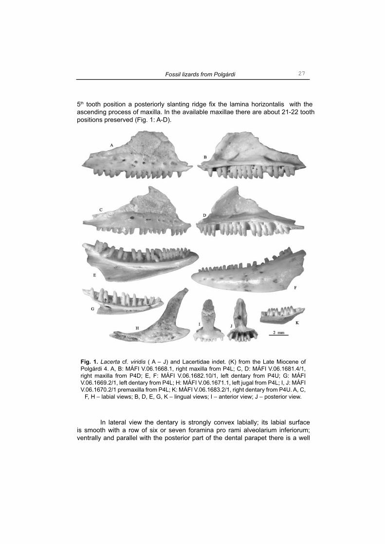
(Fig. 1: A-J)
Material examined: P4L: MÁFI V.06.1668.1, 1 maxilla; MÁFI V.06.1669.2, 2 dentaries; MÁFI V.06.1670.2, 2 premaxillae; MÁFI V.06.1671.1, 1 jugal; MÁFI V.06.1672.8, 8 maxillae; MÁFI V.06.1673.16, 16 dentaries; P4U: MÁFI V.06.1681.4, 4 maxillae; MÁFI V.06.1682.10, 10 dentaries.Description and comment. - In anterior or posterior views, the nasal (=dorsal) process of premaxilla is lanceolate with the distal portion distinctly sculptured; the palatine process is thin but prominent posteriorly (Fig. 1: I, J). In the two available specimens the number of the tooth positions is nine.
In lateral view, the external vertical wall (=ascending process) of maxilla is slightly convex labially and bear an external sculpture which consist of grooves and pits of various size. Below the sculptured surface there is a smooth area which usually is concave labially and bear a row of foramina pro rami nervorum alveolarium superiorum; regularly six or seven foramina are present, joined by few smaller ones situated dorsally to the main row. In medial view, the lamina horizontalis is prominent but relatively thin; the labial margin of the latter is slightly bent ventrally delimiting a relatively deep sulcus dentalis. From the level of the
Venczel M.
27
5th tooth position a posteriorly slanting ridge fi x the lamina horizontalis with the ascending process of maxilla. In the available maxillae there are about 21-22 tooth positions preserved (Fig. 1: A-D).
In lateral view the dentary is strongly convex labially; its labial surface is smooth with a row of six or seven foramina pro rami alveolarium inferiorum; ventrally and parallel with the posterior part of the dental parapet there is a well
Fig. 1. Lacerta cf. viridis ( A – J) and Lacertidae indet. (K) from the Late Miocene of Polgárdi 4. A, B: MÁFI V.06.1668.1, right maxilla from P4L; C, D: MÁFI V.06.1681.4/1, right maxilla from P4D; E, F: MÁFI V.06.1682.10/1, left dentary from P4U; G: MÁFI V.06.1669.2/1, left dentary from P4L; H: MÁFI V.06.1671.1, left jugal from P4L; I, J: MÁFI V.06.1670.2/1 premaxilla from P4L; K: MÁFI V.06.1683.2/1, right dentary from P4U. A, C,
F, H – labial views; B, D, E, G, K – lingual views; I – anterior view; J – posterior view.
Fossil lizards from Polgárdi
28
marked groove left by the coronoid. In medial view, the lamina horizontalis is prominent with its labial margin rounded and gradually tapering posteriorly. The subdental shelf is relatively wide and deep, while the Meckel’s groove is widely broadened posteriorly. In specimen MÁFI V.06.1682.10/1, 26 tooth positions are preserved (Fig. 1: E, F), and there are three replacement teeth, situated lingually to the tooth row; in a smaller individual from P4L, there are only 22 tooth positions.
The dentition is pleurodont; the teeth have monocuspid tips in the premaxilla and in the anterior section of dentary; except the posterior maxillary and dentary teeth which sometimes are tricuspid the remaining ones regularly bear bicuspid tips. The resorption pits are roughly circular, or sometimes oval in shape.
The processus zygomaticus of the jugal is well-marked, while the processus temporalis is relatively wide and slightly curved posterodorsally; the labial surface is sculptured (Fig. 1: H).
Except some variations of mainly ontogenic nature, all the above features are similar to recent Lacerta viridis. A closely related form, described as Lacerta cf. viridis was reported from the Late Miocene (MN 11) of Kohfi disch locality in Burgenland, Austria (Tempfer 2004).
Lacertidae indet. A(Fig. 1: K)
Material examined: P4L: MÁFI V.06.1674.5, 5 dentaries; P4U: MÁFI V.06.1683.2, 2 fr. dentaries; P5: MÁFI V.06.16790.1, 1 dentary.Description and comment. - All specimens belonged to small individuals. In medial view, the lamina horizontalis contrary to dentaries assigned to Lacerta cf. viridis has a nearly fl at lingual margin tapering posteriorly; the Meckel’s groove widens only moderately in posterior direction. Specimen MÁFI V.06.1690.1 preserves 24 tooth positions, and on the labial surface there is a clear imprint of the coronoid. The available material is reminiscent of several small-sized members of Lacertidae, including the genus Lacerta, Podarcis, or Zootoca. A somewhat similar form from the Late Miocene of Kohfi disch (MN 11) was assigned to Miolacerta tenuis by Tempfer (2004). However, the latter lacks for an imprint of the coronoid on the labial surface of the dentary (Roček 1984).
Lacertidae indet. B
Material examined: P4L: MÁFI V.06.1675.26, 26 vertebrae; P4U: MÁFI V.06.1684.7, 7 vertebrae.Description and comment. - The vertebrae are procoelous and of relatively small size, the majority of them belonging to the trunk region of the vertebral column. Because they lack for any relevant morphological features it is impossible to demonstrate if they belonged to the above described lacertid lizards.
Venczel M.
29
Anguidae Gray, 1825Genus Ophisaurus Daudin, 1803
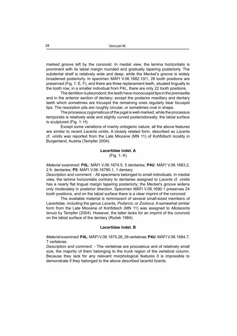
Ophisaurus sp.(Fig. 2)
Material examined: P4L: MÁFI V.06.1676.4, 1 dentary, 1 maxilla, 2 osteoderms; MÁFI V.06.1678.6, 5 osteoderms, 1 maxilla; MÁFI V.06.1679.25, 16 trunk vertebrae, 9 caudal vertebrae; P4U: MÁFI V.06.1687.1, 1 dentary; MÁFI V.06.1688.14, 14 vertebrae; MÁFI V.06.1689.6, 6 osteoderms; P5: MÁFI V.06.1691.2, 2 vertebrae.Description and comment. - The dentary is rather robust from medial or lateral view.
Fig. 2. Ophisaurus sp. from the Late Miocene of Polgárdi 4. A, B: MÁFI V.06.1676.4/2, left dentary from P4L; C, D: MÁFI V.06.1688.14/1, trunk vertebra from P4U; E: MÁFI
V.06.1676.4/3, osteoderm from P4L. A – lingual view, B – labial view, C – dorsal view, D – ventral view, E – lateral view.
Fossil lizards from Polgárdi

30
The margin of lamina horizontalis (= subdental shelf) is convex lingually producing a small angulations against the alveolar surface of the dental parapet. The Meckel’s groove faces ventrally and in lingual view its anterior section is visible only (Fig. 2: A). The dentition is of subpleurodont type while the number of tooth positions in the complete dentary is thirteen. The teeth are moderately elevated and pointed distally without any striations; the apical portion is compressed labiolingually and slightly recurved bearing a sharp mesiodistal crest; the base of teeth is slightly compressed mesiodistally. In lateral view the dentary is convex labially with relatively high coronoid process projected above the apices of posterior teeth; the supraangular process is relatively small and never reaches posteriorly the level of coronoid process; the incisura coronoideus is rather shallow (Fig. 2: B). About four or fi ve foramina pro rami nervi alveolaris inferioris is observed; the fi rst one is situated at the level of the third tooth position.
The maxillary bones are rather fragmentary; they preserve a similar type of dentition as those of dentaries.
The vertebrae assigned to this form are lightly built and of relatively small size (Fig. 2: C, D). The centrum is moderately elongated with the ventral surface slightly convex or fl attened; the subcentral ridges are well developed diverging just anterior to the condyle ending in the vicinity of synapophyses; the pre- and postzygapophyses are oval in shape; the interzygapophyseal ridges are slightly developed only.
The osteoderms are fl attened bearing a small anterior smooth surface and a posterior pit and ridge sculptured area provided with a prominent medial ridge (Fig. 2: E). From the locality Polgárdi 2, Bolkay (1913), based on a parietal, two maxillae and a dentary described a new species under the name Anguis polgardiensis (Plate XII: Fig. 1). In order to differentiate Anguis polgardiensis from A. fragilis the brief diagnosis given by the above author [scutum interparietale is widely triangular reaching the corners of the parietal (i.e. anterior margin of margo postfrontorbitalis), while the scutum occipitale is present; the marginal teeth are more robust, less recurved and blunt] in fact fi ts well to the European members of the genus Ophisaurus (sensu Klembara 1979), and also to the above described remains from Polgárdi 4. However, A. polgardiensis was enclosed in the synonymy of Pseudopus (=Ophisaurus) pannonicus without any comments by Estes (1983).
The dentary and vertebrae are reminiscent of Ophisaurus cf. spinari, and respectively of cf. Ophisaurus sp., described by Roček (1984) from the Lower Miocene (MN 4) of Dolnice, Czech Republic. Comparatively, the dentary in the Dolnice Ophisaurus is more elongated preserving a higher number of teeth with their stem comparatively taller than those from Polgárdi 4. In fact Ophisaurus spinari was described for the fi rst time by Klembara (1979), together with other two new taxa: O. fejfari and Anguis robustus. However, the latter author in his descriptions was based mainly on the morphology of parietal bones (e.g. see Klembara, 1979: Fig.1 and 2). Böhme (2002) demonstrated that typical for karst areas are the occurrence of parietals which are closely similar to Ophisaurus fejfari
Venczel M.

31
in combination with dentaries provided with teeth bearing cutting edges on their mesiodistal margins, whereas O. spinari is more frequent in sedimentary basins with fl uvio-lacustrine taphonomical context.
Several dentaries and vertebrae from the Late Miocene (MN 11) of Kohfi disch, closely resembling the Polgárdi Ophisaurus, were simply assigned to Anguis fragilis by Tempfer (2004). Thus the revision of all the above forms is strongly recommended.
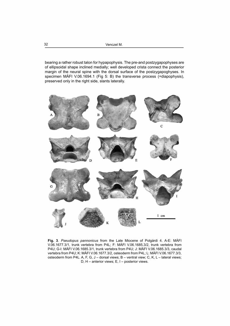
Genus Pseudopus Merrem, 1820Pseudopus pannonicus
(Fig. 3)
Material examined: P4L: MÁFI V.06.1677.3, 1 trunk vertebra, 2 osteoderms; MÁFI V.06.1680.26, 26 osteoderms; P4U: MÁFI V.06.1685.3, 2 trunk vertebrae, 1 caudal vertebra; MÁFI V.06.1686.2, 2 osteoderms; P5: MÁFI V.06.1692.3, 3 osteoderms.Description and comment. - The trunk vertebrae are heavily built and of large size (Fig. 3: A-I). The centrum length in three measured vertebrae ranges between 5.83-8.3 mm. In ventral view, the centrum is fl attened and of roughly triangular shape. The subcentral margin is slightly convex laterally, while the subcentral ridges are well developed diverging anteriorly from the faint precondylar constriction into the synapophyses. In dorsal view the interzygapophyseal ridge are well defi ned; the prezygapophyses are rounded. A much smaller specimen (Fig. 3: J) represent a caudal vertebra. The dorsal surface of the neural arch is strongly concave dorsally, provided with short but very prominent spinal process projected posterodorsally. The haemapophyses are broken off, but their remnants are preserved on the ventral surface of the centrum in the anterior proximity of the condyle.
The osteoderms assigned to this form are relatively large and thick. Their anterior margin is smooth, while their posterior section bears a pit and ridge sculpture without a medial crest (Fig. 3: K, L).
Family Varanidae Gray, 1827Genus Varanus Merrem, 1820
Varanus cf. hofmanni(Fig. 4-6)
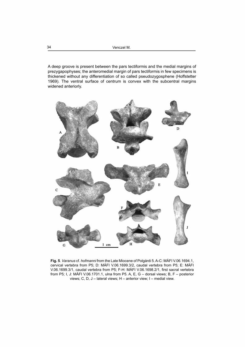
Material examined: P5: MÁFI V.06.1693.1, 1 trunk vertebra; MÁFI V.06.1694.1, 1 cervical vertebra; MÁFI V.06.1695.1, 1 trunk vertebra; MÁFI V.06.1696.1, 1 trunk vertebra, MÁFI V.06.1697.16, 10 trunk vertebrae, 1 cervical vertebra, 5 caudal vertebrae; MÁFI V.06.1698.2, 2 fi rst sacral vertebrae; MÁFI V.06.1699.3, 3 caudal vertebrae; MÁFI V.06.1700.3, 3 femurs; MÁFI V.06.1701.1, 1 ulna.Description and comment. - The neural arch of the cervical vertebrae is vaulted (Fig. 5: A-C) and provided with a relatively high and long neural spine; the latter arises at the level of the posterior margin of prezygapophyses. The centrum is relatively long
Fossil lizards from Polgárdi
32
bearing a rather robust talon for hypapophysis. The pre-and postzygapophyses are of ellipsoidal shape inclined medially; well developed crista connect the posterior margin of the neural spine with the dorsal surface of the postzygapoghyses. In specimen MÁFI V.06.1694.1 (Fig 5: B) the transverse process (=diapophysis), preserved only in the right side, slants laterally.
Fig. 3. Pseudopus pannonicus from the Late Miocene of Polgárdi 4. A-E: MÁFI V.06.1677.3/1, trunk vertebra from P4L; F: MÁFI V.06.1685.3/2, trunk vertebra from P4U; G-I: MÁFI V.06.1685.3/1, trunk vertebra from P4U; J: MÁFI V.06.1685.3/3, caudal vertebra from P4U; K: MÁFI V.06.1677.3/2, osteoderm from P4L; L: MÁFI V.06.1677.3/3, osteoderm from P4L. A, F, G, J – dorsal views; B – ventral view; C, K, L – lateral views;
D, H – anterior views; E, I – posterior views.
Venczel M.
33
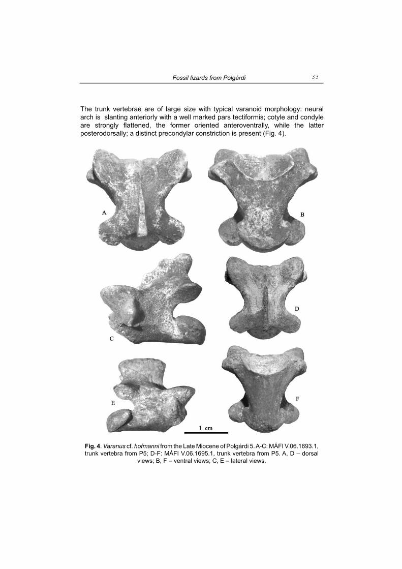
The trunk vertebrae are of large size with typical varanoid morphology: neural arch is slanting anteriorly with a well marked pars tectiformis; cotyle and condyle are strongly fl attened, the former oriented anteroventrally, while the latter posterodorsally; a distinct precondylar constriction is present (Fig. 4).
Fig. 4. Varanus cf. hofmanni from the Late Miocene of Polgárdi 5. A-C: MÁFI V.06.1693.1, trunk vertebra from P5; D-F: MÁFI V.06.1695.1, trunk vertebra from P5. A, D – dorsal
views; B, F – ventral views; C, E – lateral views.
Fossil lizards from Polgárdi
34
A deep groove is present between the pars tectiformis and the medial margins of prezygapophyses; the anteromedial margin of pars tectiformis in few specimens is thickened without any differentiation of so called pseudozygosphene (Hoffstetter 1969). The ventral surface of centrum is convex with the subcentral margins widened anteriorly.
Fig. 5. Varanus cf. hofmanni from the Late Miocene of Polgárdi 5. A-C: MÁFI V.06.1694.1, cervical vertebra from P5; D: MÁFI V.06.1699.3/2, caudal vertebra from P5; E: MÁFI V.06.1699.3/1, caudal vertebra from P5; F-H: MÁFI V.06.1698.2/1, fi rst sacral vertebra from P5; I, J: MÁFI V.06.1701.1, ulna from P5. A, E, G – dorsal views; B, F – posterior
views; C, D, J – lateral views; H – anterior view; I – medial view.
Venczel M.
35
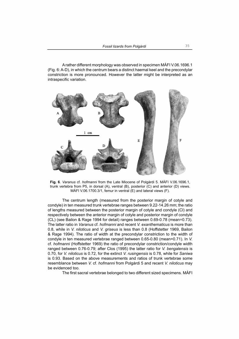
A rather different morphology was observed in specimen MÁFI V.06.1696.1 (Fig. 6: A-D), in which the centrum bears a distinct haemal keel and the precondylar constriction is more pronounced. However the latter might be interpreted as an intraspecifi c variation.
The centrum length (measured from the posterior margin of cotyle and condyle) in ten measured trunk vertebrae ranges between 9.22-14.26 mm; the ratio of lengths measured between the posterior margin of cotyle and condyle (Cl) and respectively between the anterior margin of cotyle and posterior margin of condyle (CL) (see Bailon & Rage 1994 for detail) ranges between 0.69-0.78 (mean=0.73). The latter ratio in Varanus cf. hofmanni and recent V. exanthematicus is more than 0.8, while in V. niloticus and V. griseus is less than 0.8 (Hoffstetter 1969, Bailon & Rage 1994). The ratio of width at the precondylar constriction to the width of condyle in ten measured vertebrae ranged between 0.65-0.80 (mean=0.71). In V. cf. hofmanni (Hoffstetter 1969) the ratio of precondylar constriction/condyle width ranged between 0.76-0.79; after Clos (1995) the latter ratio for V. bengalensis is 0.70, for V. niloticus is 0.72, for the extinct V. rusingensis is 0.78, while for Saniwa is 0.93. Based on the above measurements and ratios of trunk vertebrae some resemblance between V. cf. hofmanni from Polgárdi 5 and recent V. niloticus may be evidenced too.
The fi rst sacral vertebrae belonged to two different sized specimens. MÁFI
Fig. 6. Varanus cf. hofmanni from the Late Miocene of Polgárdi 5. MÁFI V.06.1696.1, trunk vertebra from P5, in dorsal (A), ventral (B), posterior (C) and anterior (D) views.
MÁFI V.06.1700.3/1, femur in ventral (E) and lateral views (F).
Fossil lizards from Polgárdi

36
V.06.1698.2/1 (Fig. 5: F-H) represent a nearly completely preserved specimen (except the anterior margin of its neural arch, the left distal end of transverse process, and neural spine); the centrum is relatively short with a peculiar transversal ridge on its condylar surface which in living animal restricted the shift between the fi rst and second sacral vertebrae; a bony ridge extend between the prezygapophysis and neural spine. While the fi rst character is present in the Nile monitor, the second one was reported from V. rusingensis too (Clos 1995).
The caudal vertebrae (Fig. 5 D, E) belonged to specimens of various size; all of them lacks for chevrons; the neural spine or remnants of it extends more than half the length of the centrum somewhat reminiscent of V. rusingensis (Clos 1995).
The ulna assigned to V. cf. hofmanni is rather robustly build, relatively short and mediolaterally fl attened (Fig. 5: I, J). The proximal shaft is distinctly wider than the distal and deeply concave medially. The sigmoid cavity is widely concave bearing a well-marked radial incisure; the olecranon was rather massive and of relatively low height. In living animal, the proximal end of the radius would have been placed in the incisure on the medial side of the ulnar proximal shaft. The distal shaft of the ulna is strongly dilated and convex. The femur is slightly S-shaped in dorsal or ventral view (Fig. 6: E, F). The proximal shaft exhibit a rather prominent trochanter major extending distally for about one-fourth of the bone length. The distal medial condyle is provided with a slightly prominent bony ridge running toward the proximal shaft for approximately one-fourth of the femoral length.
The above described fossils from Polgárdi 5, assigned with some doubts to Varanus hofmanni, display a combination of characters rather different from Iberovaranus and also from V. marathonensis. The former seems to have been restricted to the Iberian Peninsula, where as the latter is known from younger deposits including the early Ruscinian (MN 15) of Csarnóta 1, Hungary (Fejérváry 1918). After Hoffstetter (1969), the presence of a deep groove between the pars tectiformis and medial margin of prezygapophyses is typical for Varanus hofmanni; the latter structure is also present in the form reported from the Late Miocene of Kohfi disch, assigned to V. hofmanni by Tempfer (2004). Based on the available material from Csarnóta 1 and Çalta (see Fejérváry 1918, Rage & Sen 1976) is clear that in V. marathonensis the groove delimiting the pars tectiformis and prezygapophyses is less developed, and the cotyle and the condyle less enlarged laterally than in V. hofmanni. Since the latter form has been reported from the Upper Miocene (MN 11) of Kohfi disch (Tempfer 2004), it seems that it was continuously present in the Central Paratethys area extending its stratigraphical range to the end of the Miocene.
Concluding remarks
The composition of lizard faunas from the Polgárdi localities is in contrast with those of snakes which were more diversifi ed (Venczel 1994, 1998). Except
Venczel M.

37
Lacerta cf. viridis and perhaps Lacertidae indet., the other taxa described from these deposits (Ophisaurus, Pseudopus¸Varanus) belonged exclusively to extinct forms. The lacertids and anguids might have been distributed mainly in biotopes with xerothermic or perhaps mesophilous vegetation typical for karstic landscapes. However, the composition of snakes (Venczel 1994, 1998) and those of birds (Jánossy 1991) is suggestive of a rather diversifi ed paleoenvironment, with a slightly higher mean annual temperature (MAT) than nowadays. The presence of Varanus in Polgárdi 5 is also indicative of a distinctly higher MAT, as suggested by the worldwide distribution of extant Varanidae (Böhme 2003). However, the Varanus from Polgárdi 5 might have been linked to more humid biotopes as documented by the abundant aquatic bird remains derived from this locality (Jánossy 1991).
Acknowledgements
I would like to thank Professor László Kordos, head of Hungarian Geological Museum, Budapest for loan of fossil lizard material from Polgárdi 4 and 5, for helpful discussions related to the fossil localities of Polgárdi and for various supports during my visits in the Geological Institute. A partial fi nancial support has been provided by a grant from Domus Hungarica, Hungarian Academy of Science.
References
Bailon, S. & Rage, J-C. 1994. – Squamates néogènes et pléistocènes du Rift Occidental, Ouganda. in: Geology and Palaeobiology of the Albertine Rift Valley, Uganda-Zaire. Vol. II: Palaeobiology. CIFEG Occasional Publications, 1994/29, Orléans, pp.129-135.
Bolkay, J. 1913. – Additions to the fossil herpetology of Hungary from the Pannonian and Praeglacial periode. Mittheilungen aus dem Jahrbuche der königlichen Ungarischen geologischen Reichsanstalt, 21 (7): 217-230.
Böhme, M. 2002. – Lower Vertebrates (Teleostei, Amphibia, Sauria) from the Karpatian of the Korneuburg Basin – palaeoecological, environmental and palaeoclimatical implications. Beitraege zur Paläontologie, 27: 339-353.
Böhme, M. 2003. – The Miocene Climatic Optimum: evidence from ectothermic vertebrates of Central Europe. Palaeogeography Palaeoclimatology Palaeoecology, 195: 389-401.
Clos, L, M. 1995. – A new species of Varanus (Reptilia: Sauria) from the Miocene of Kenya. Journal of Vertebrate Paleontology, 15(2): 254-267.
Estes, R. 1983. – Sauria terrestria, Amphisbaenia. In: Handbuch der Paläoherpetologie, G. Fischer Verlag, Stuttgart/ New York.
Fejérváry, G. Gy. 1918. – Contributions to a monography on fossil Varanidae and on Megalanidae. Annales Musei Nationalis Hungarici, 16: 342-467.
Fejérváry-Lángh, A. M. 1923. – Beiträge zu einer Monographie der fossilen Ophisaurier. Palaeontologia Hungarica, 1 (7): 123-220.
Fossil lizards from Polgárdi

38
Freudenthal, M. & Kordos, L. 1989. – Cricetus polgardiensis sp. nov. and Cricetus kormosi SCHAUB, 1930 from the Late Miocene Polgárdi localities (Hungary). Scripta Geologica, 89: 71-100.
Hoffstetter, R. 1969. – Présence de Varanidae (Reptilia, Sauria) dans le Miocène de Catalugne. Considérations sur l’histoire de la famille. Bulletin du Muséum National D’Histoire Naturelle, 2. sér., 40(5): 1051-1064.Jánossy, D. 1991. – Late Miocene bird remains from Polgárdi (W-Hungary). Aquila,
98: 13-35.Klembara, J. 1979. – Neue Funde der Gattungen Ophisaurus and Anguis
(Squamata, Reptilia) aus dem Untermiozän Westböhmens (ČSSR). Věstník Ústředního ústavu geologického, 54(3): 163-169.
Kormos, T. 1911. – Der pliozäne Knochenfund bei Polgárdi. Földtani Közlöny, 41(1-2): 171-189.
Kretzoi, M. 1952. – Die Raubtiere der Hipparionfauna von Polgárdi. Annales Instituti Geologici Hungarici, 40(3): 5-42.
Rage, J. C. & Sen, S. 1976. – Les amphibians et les reptiles du Pliocène Supérieur de Çalta (Turquie). Géologie méditerranéenne, 3(2): 127-134.
Roček, Z. 1984. – Lizards (Reptilia: Sauria) from the Lower Miocene locality Dolnice (Bohemia, Czechoslovakia). Rozpravy Československé Akademie Věd, 94(1): 1-69.
Szunyoghy, J. von 1932. – Beiträge zur vergleichenden Formenlehre des Colubridenschädels, nebst einer kraniologischen Synopsis der fossilen Schlangen ungarns mit nomenklatorischen, systematischen und phyletischen Bemerkungen. Acta Zoologica, 13: 1-56.
Szyndlar, Z. 1991a. – A review of Neogene and Quaternary snakes of Central and Eastern Europe. Part I. Scolecophidia, Boidae, Colubrinae. Estudios Geologicos, 47(1-2): 103-126.
Szyndlar, Z. 1991b. – A review of Neogene and Quaternary snakes of Central and Eastern Europe. Part II. Natricinae, Elapidae, Viperidae. Estudios Geologicos, 47(3-4): 237-266.
Tempfer, M. P. 2004. – The herpetofauna (Amphibia: Caudata, Anura; Reptilia: Scleroglossa) of the Upper Miocene locality Kohfi disch, Burgenland, Austria. Unpublished Dissertation, Fakultät für Naturwissenschaften und Mathematik der Universität Wien, pp. 186.
Venczel, M. 1994. – Late Miocene snakes from Polgárdi (Hungary). Acta zoologica Cracoviensia, 37(1): 1-29.
Venczel, M. 1997. – Late Miocene anurans from Polgárdi (Hungary). Pp. 383-389, in: Böhme, W., Bischoff, W. & T. Ziegler (Eds.): Herpetologia Bonnensis, Bonn (SEH), 416 pp.
Venczel, M. 1998. – Late Miocene snakes (Reptilia: Serpentes) from Polgárdi (Hungary): a second contribution. Acta zoologica Cracoviensia, 41: 1-22.
Venczel M.