Sustaining Communities, Livestock and Wildlife: A Guide to Participatory Land-Use Planning
Upload
nguyentrucCategory
view
220download
3
Page 1 of 63
Livestock & Wildlife Disease Diagnosis at APHA
Guidance on sample and test selection
Version 3
February 2018
Published by the APHA Surveillance Intelligence Unit & Species Expert Groups
Surveillance Intelligence Unit Contacts
Richard Irvine, Head of APHA Surveillance Intelligence Unit - [email protected]
Veterinary Leads for Species Expert Groups:
Amanda Carson, Small Ruminants - [email protected]

Page 2 of 63
Susanna Williamson, Pigs - [email protected]
Gareth Hateley, Cattle - [email protected]
David Welchman, Poultry / Avian - [email protected]
Amanda Carson, Miscellaneous and Exotic Farmed Species – [email protected]
Paul Duff, Wildlife - [email protected]
Surveillance Intelligence Unit General Mailbox – [email protected]
Livestock Disease Diagnosis at APHA – Guidance on sample and test selection
Page 3 of 63
Contents
Topic Page
Introduction 3
Abbreviations 5
Sampling – by Discipline
Histology 8
Bacteriology 9
Serology 11
Parasitology 12
Virology 13
Sampling – by Species & Disease Condition
Birds 15
Cattle 27
Small Ruminants 33
Pigs 41
Miscellaneous and Exotic Farmed Species 54
Wildlife 61

Livestock Disease Diagnosis at APHA – Guidance on sample and test selection
Page 4 of 63
Introduction
Background
This booklet is intended to assist veterinary practitioners with sample and test selection for diagnosis of common clinical presentations in livestock and wildlife. It has been compiled by Animal and Plant Health Agency (APHA) Species Expert Group members and other veterinary colleagues.
The main remit of Government-funded scanning surveillance at APHA is the detection of new and (re)emerging livestock disease threats. The types of threat include novel pathogens, novel diseases, novel presentations of known diseases, occurrence of diseases in novel species, strains or serotypes which are new or exotic to Great Britain; marked changes in endemic disease trends; rare, newly emerging or concerning antimicrobial resistances and threats to food safety or public health.
APHA funded PME provision within the APHA scanning surveillance network in England and Wales includes the APHA Veterinary Investigation Centres and non-APHA partner postmortem providers (SAC CVS, University of Bristol, Royal Veterinary College, University of Surrey and Wales Veterinary Science Centre, Aberystwyth). Further details are available on this link http://apha.defra.gov.uk/postcode/pme.asp
In order to maximise the surveillance value of this service, for which Defra provides a significant level of financial support, APHA requires every submission to be accompanied by a fully completed submission form available from this link: http://apha.defra.gov.uk/vet-gateway/surveillance/forms.htm. Species-specific forms are available.
Please fully complete the submission form and send with the submitted samples or animals. Faxing, or scanning and emailing, the submission form when sending animals for postmortem examination is also acceptable, once there has been discussion with a Veterinary Investigation Officer (VIO), and the animals are accepted for postmortem examination. Similar information is needed by non-APHA partner post mortem providers.
Postal samples should be sent to APHA Penrith or APHA Starcross – addresses here.
For a list of available tests, their prices and other details, please refer to the current Disease Surveillance Price List
Veterinary staff at the APHA Veterinary Investigation Centres can give free advice where the details in this guidance do not provide sufficient information, or where assistance is needed to investigate complex, unusual or problematic outbreaks of disease, even if you do not send samples to APHA. VIOs at APHA Veterinary Investigation Centres should also be contacted directly to discuss submissions for postmortem examination: VIC Contact Details
Veterinary Investigation Diagnosis Analysis (VIDA)
The surveillance information you provide on the submission form is recorded in the VIDA database, together with any diagnosis reached. VIDA is a national database, providing analysis of all diagnostic submissions to APHA, SAC Consulting: Veterinary Services Disease Surveillance Centres and the network of non-APHA partner PME providers. Diagnoses follow strict criteria. VIDA allows monitoring of diagnoses, clinical syndromes and disease trends and epidemiological features associated with these. Submissions in which a diagnosis is not reached (DNR) after reasonable testing are also scrutinised to determine whether they provide any

Livestock Disease Diagnosis at APHA – Guidance on sample and test selection
Page 5 of 63
evidence of a new or (re)emerging threat. The Species Expert Groups analyse and report these VIDA data.
Submission of animals for postmortem examination
Whilst submission of animals for postmortem examination (PME) remains a key part of surveillance, these are costly and, as Government funding has reduced in recent years, it is all the more important that the types and numbers of animals submitted are representative, of suitable quality, and that PME is the most appropriate means of investigating the disease problem. It is therefore essential that, if considering submission of animals for diagnostic PME to APHA, you speak to the designated PME provider for the premises from which the animals will be submitted. This can be found using the on-line postcode search tool: http://apha.defra.gov.uk/postcode/pme.asp. You will need to know the clinical history of the case with the date and estimated time of death of animal(s) (if there is mortality) to be submitted.
Selection criteria for postmortem examination
Animals dead for more than 24 hours are likely to be of less diagnostic value and will usually only be accepted after careful consideration of factors likely to influence diagnostic value including storage temperature since death.
Animals dead for more than 48 hours will not be accepted.
Carcases which have been frozen may not be accepted since this limits their diagnostic value.
In outbreaks, a maximum of three mammalian and 5-10 bird carcases may be submitted together from a single disease incident on each farm.
Sometimes the clinical history indicates that diagnosis is best undertaken in the first instance by submission of samples rather than animals for PME.
Information in this guidance document provides general and species-specific guidelines on diagnosis of common disease presentations in livestock and selection of the most appropriate carcase or non-carcase material for different disease conditions.
Submission of live animals for postmortem examination
Occasionally it is advisable to submit live animals for clinical and postmortem examination; examples might be to investigate enteric or nervous disease. Submission of a combination of live affected and freshly dead birds is often appropriate for flock health investigations in poultry and game birds. However, submission of a live animal or bird must be discussed with veterinary staff at the relevant PME provider within the APHA network at the same time that the PME is agreed, and is not possible where an APHA-funded carcase collection service is being used as they are not able to transport live animals. The decision must take account of the current welfare in transport legislation in England, Scotland and Wales. To ensure the wellbeing of any live animals, the private veterinary surgeon should agree to its transport and ensure supervision for the journey in line with the current legislation which covers, inter alia, the transport of animals for non-commercial purposes. The legislation is here.
NOTIFIABLE DISEASES
It is important that all those involved with livestock health and production remain vigilant for signs of any notifiable disease. The Animal Health Act 1981 requires that anyone having in their charge an animal affected or suspected of having certain diseases must notify that fact to the veterinary

Livestock Disease Diagnosis at APHA – Guidance on sample and test selection
Page 6 of 63
authorities. The diseases covered by this legal requirement are known as notifiable diseases. If you suspect the presence of notifiable disease, you must immediately call APHA:
In England via the Defra Rural Services helpline: 03000 200 301.
In Wales on 0300 303 8268.
In Scotland via the local APHA field office - see https://www.gov.uk/government/organisations/animal-and-plant-health-agency/about/access-and-opening for further contact details.
There is information on notifiable diseases of farmed livestock available on the following links, including information on their clinical signs and pathology: APHA Notifiable Disease Pages OIE Information Sheets
Abbreviations
Ab Antibody
Ag Antigen
AGIDT Agar Gel Immunodiffusion Test
AT Agglutination Test
BAL Bronchoalveolar Lavage
BVD Bovine Viral Diarrhoea
CCN Cerebrocortical Necrosis
CEL Chicken Embryo Liver
CIT Citrate
CFT Complement Fixation Test
CIE Counter Immuno Electrophoresis
CNF Cytotoxic Necrotising Factor
CTM Charcoal Transport Medium
DAT Direct Agglutination Test
DEL Duck Embryo Liver
EDTA Ethylene Diamine Tetra-acetic Acid
ELISA Enzyme-Linked Immunosorbent Assay
EM Electron Microscopy
FAT Fluorescent Antibody Test
FAVN Fluorescent Antibody Virus Neutralisation
FPT Four Plate Test
HAT Haemagglutination Test
HAIT Haemagglutination Inhibition Test
Hb Haemoglobin
HEP Heparin
IBR Infectious Bovine Rhinotracheitis
ID Identification
IFAT Indirect Fluorescent Antibody Test
IHC Immunohistochemistry
IPMA Immunoperoxidase Monolayer Assay
IPX Immunoperoxidase Assay
LAT Latex Agglutination Test
LC Large Colony Variant
MAT Microscopic Agglutination Test
MIC Minimum Inhibitory Concentration
MRT Milk Ring Test
MZN Modified Ziehl-Neelsen Stain
NEFA Non-Esterified Fatty Acids
NPLA Neutralising Peroxide Linked Assay
OCD Osteochondritis Dissecans
PAGE Polyacrylamide Gel Electrophoresis
PBS Phosphate Buffered Saline
PCR Polymerase Chain Reaction
PCV Packed Cell Volume
PDNS Porcine Dermatitis and Nephropathy Syndrome
PED Porcine Epidemic Diarrhoea
PGE Parasitic Gastro-enteritis
PI3 Parainfluenza 3
PME Post Mortem Examination
PMWS Postweaning Multisystemic Wasting Syndrome
PNP Porcine Necrotising Pneumonia
PoA Price on Application
PRCV Porcine Respiratory Coronavirus
PRRS Porcine Reproductive and Respiratory Syndrome
RBC Red Blood Cells
RBT Rose Bengal Test
RIA Radio Immuno Assay
RSA Rapid Slide Agglutination
RSV Respiratory Syncytial Virus
SAF Scrapie Associated Fibrils
SAT Serum Agglutination Test
SC Small Colony Variant
SNT Serum Neutralisation Test
SPF Specific Pathogen Free
TGE Transmissible Gastro-enteritis
VMAT Vaginal Mucus Agglutination Test
VTEC VeroToxic Escherichia coli
VTM Virus Transport Medium
VI Virus Isolation
VIC Veterinary Investigation Centre/s (formerly
Regional Laboratories)
WBC White Blood Cells

Livestock Disease Diagnosis at APHA – Guidance on sample and test selection
Page 7 of 63
Colour Codes for Blood Tubes
Stopper Colour Anticoagulant Red None (for serum samples) Green HEP Purple EDTA Grey OXF Blue CIT

Page 8 of 63
Sampling by Discipline

Histology
Page 9 of 63
Please note autolysis can severely compromise the ability to assess disease processes within tissues and therefore the submission of material which is obviously autolysed at the time of collection and fixation may not yield any useful diagnostic information. Sampling
Tissue samples should not be more than 1 cm thick
Samples should be fully representative of the basic organ structure and include the junction between gross lesions and normal tissue
Samples should be immersed in 10-20 x their volume of fixative as soon as possible
Samples should be sent in an appropriately sized container with a wide opening
Brain is best fixed whole allowing the pathologist to select appropriate sites
Collect intestinal samples, as soon after death as possible (ideally within 30 minutes), from several sites of small and large intestine. Immersion fixation of gut tubes 1-2 cm in length is satisfactory, but avoid crushing with forceps. Gentle agitation of the sample in the fixative will help displace food material and allow fixative to enter the lumen
If the above guidelines are followed, primary fixation of most samples should take 24-48 hours – this time period will be extended if the fixative is cold (below 5˚C). However, whole brains will take longer - please discuss with your VIC. Packing and Sending Material must be properly packaged. Packaging must conform to the postal regulations for packaging of pathological material.
Urgent cases can be sent immediately if the container is filled with fixative so that primary fixation occurs in transit. If non urgent, tissue can be initially fixed for 48 hours then sent in a reduced volume of fixative. This method is particularly appropriate for brain.
The recommended fixative for most cases is 10% neutral buffered formalin.
Bacteriology
Page 10 of 63
Types of samples
Portions of fresh tissue in clean containers are suitable if they are not autolysed or contaminated, are submitted same day or by overnight post and are kept cool during transport
Purulent material is preferable to swabs
Faeces samples (not just swabs) are essential if tests other than basic bacteriology are required.
For anaerobic culture, fill container to brim or wrap tissues in cling film to exclude air
Charcoal swabs are suitable for aerobic and anaerobic culture but where anaerobes are the target organism, commercial transport media are available that are aimed specifically at anaerobe preservation. Please discuss with the VIC
Plain swabs required for fluorescent antibody test (FAT) e.g. for Streptococcus suis 2
Plain swabs with wire or plastic stems for PCR tests (not wooden stems) Sampling for Aerobic Bacteriology (request test code TC0101)
Tissues to be sampled should be as fresh as possible
Sear the surface of organs with a flame or heated scalpel blade prior to incision with a sterile scalpel and swab the incised surface
In cases of serositis (pleurisy, pericarditis, arthritis etc), rub the swab on the lesioned serosal surface and avoid just dipping the swab in fluid exudate
Sampling for Anaerobic Bacteriology
Follow similar guidelines to the above for aerobic bacteriology but, for anaerobic culture, request test code TC0528
For diagnosis of clostridial enterotoxaemia, send a minimum of 1 ml of small or large intestinal contents. Do not add any preservative. Submit for Clostridium perfringens toxin ELISA (TC0035)
For clostridial myositis or black disease in cattle or sheep, or Clostridium novyi infection (hepatitis) in pigs, take four impression smears from the cut surface of affected muscle or liver, air-dry and send in slide box for clostridial FAT (TC0032), or submit a portion of whole lesioned tissue in a sealed air tight container
Bacteriology
Initial isolation of most bacterial pathogens in cultures occurs after 24 hours incubation following initiation of cultures at the laboratory
Some exceptions are: - Haemophilus parasuis - minimum of 2 days - Salmonella spp. by enrichment - minimum 2 days - Campylobacter spp. - up to 5 days - Brucella abortus - minimum of 4 days - Mycobacterium avium paratuberculosis (Johnes) – up to 16 weeks - Mycobacterium species (TB) – 6 to 12 weeks
Full identification of bacterial pathogens can take from one day to a week, and occasionally longer depending on the nature of the particular pathogen and the degree of contamination.
Fastidious organisms exist such as Mycoplasma, Brachyspira and Campylobacter species and Leptospira serovars may require specialist techniques. Please contact your usual APHA VIC to discuss testing for these pathogens
Bacteriology
Page 11 of 63
Antimicrobial sensitivity
Antimicrobial sensitivity will be initiated, if requested, once the pathogen has been obtained in pure growth which may require subculture
Disc diffusion antimicrobial sensitivity testing (TC0401) takes 24 hours in most cases
Minimum inhibitory concentrations (MICs) can be undertaken for some pathogens for selected antimicrobials. Please contact your usual APHA VIC for more information
Mastitis examinations (TC0544)
Misleading results are obtained if milk samples are contaminated
Follow this procedure to avoid contamination:-
1. Wash and dry your hands thoroughly.
2. Wash teat to be sampled only if obviously dirty; dry immediately.
3. Discard first two draws of milk.
4. Clean end of teat:-
a) Use small piece of cotton wool, dampen with surgical spirit (80% spirit/20% water).
b) Rub end of teat with “swab” until visibly clean.
c) Repeat using a second swab, make sure swab appears clean after use. If not, repeat using another clean swab; last swab should be spotless after wiping.
5. To take sample:-
a) Open sterile sample bottle – Keep lid clean, never place open-side down and preferably hold it facing downwards in crook of little finger, do not allow lid to touch teat
b) Hold sample bottle at an angle to teat.
c) Discard a further draw of milk.
d) Collect 1-3 streams of milk to fill sample bottle at most half-full.
e) Immediately replace lid carefully.
6. Label sample bottle – include cow’s number, quarter, date, name of farm and farmer
Mycology
Request fungal culture (TC0101 Sabouraud’s medium) on the submission form
Fluids (e.g. foetal stomach contents) can also be examined for fungal hyphae by direct microscopy (TC0580)
For ringworm (dermatophyte) culture (TC0080), submit hair plucks
Dermatophyte cultures take up to three weeks
Serology
Page 12 of 63
General Serology is used to detect whether animals have been exposed to a particular pathogen. It is used in the diagnosis of disease and for monitoring the pathogen status of a group or herd/flock. If animals are vaccinated against the pathogen in question, serology is of debatable value as antibody produced to vaccine cannot usually be distinguished from that produced to field infection. Only a few DIVA (Differentiation of Infected from Vaccinated Animals) vaccines are available for veterinary use (e.g. gE-deleted IBR vaccine) which allow a distinction to be made. The possibility of maternally derived antibody being detected must be borne in mind in young animals; these may persist up to eight months of age (calves). Maternal antibodies interfere with interpretation and cannot be distinguished from antibody produced in response to active infection of the animal. Diagnostic serology
Sample several affected animals
Single serology
o presence of antibody only indicates exposure to infection
o does not indicate how recently the exposure occurred
o if negative, rules out involvement of some pathogens
o presence of antibody may be useful diagnostically if the animal(s) were supposed to be free from the pathogen
Paired serology
o sera tested from the same animals during acute and convalescent periods
o detects seroconversion (seronegative to seropositive) or a significant rise in titre
o establishes a temporal association of seroconversion and disease
o acute samples must be collected within the three to four days of clinical signs occurring or animals will already have seroconverted
o sampling interval can vary but should not be less than two weeks
o is not usually useful in reproductive disease investigation as maternal seroconversion has usually already occurred by the time disease manifests
Cohort serology
o used where conventional paired serology problematic e.g. in pigs or poultry with no identifiers
o sera collected from groups of pigs or poultry at different ages within one management system
o assists in assessing the timing of exposure

Serology
Page 13 of 63
Monitoring serology Healthy animals may be tested for antibody to a pathogen to establish the status of a group or herd/flock with respect to that pathogen, so long as the animals are not vaccinated (unless a DIVA vaccine is used). The aim of the monitoring needs to be clear and is usually either:
a) to detect presence of pathogen – here the detection of a single seropositive animal is sufficient b) to estimate prevalence of pathogen – here an estimate of the proportion of animals exposed to infection is needed and this usually involves testing a larger number of animals
In both situations, the numbers of animals tested from an epidemiological group depends on several factors including the suspected prevalence of infection, the degree of confidence needed in the results and the number of animals in the group. Epidemiological sample size calculation tables exist which assist in establishing the numbers of animals that should be sampled and tested for given group sizes, confidences and prevalences. The number of animals which are tested is also influenced by the logistics of sampling, cost of the tests and the sensitivity and specificity of serological tests available.
Parasitology Fresh Faeces
Submit in a wide mouthed, screw capped container sealed with insulation tape
Submit 3g minimum for individual faecal egg count (TC0060), 40g for fluke egg
examination (TC0061), and 50g for lungworm larvae examination (TC0062)
Monitoring faecal egg counts in sheep (the composite faecal egg count, TC0668): submit
10 x 3g (minimum) as separate faecal samples from each group; these will be pooled at
the laboratory
Monitoring fluke egg counts in cattle and sheep (TC0689): submit 10 x 5g (minimum) as
separate faecal samples from each group; these will be pooled at the laboratory
Blood
For blood parasites (TC0256) submit 2ml whole blood in EDTA tube, or two thinly spread
films air dried and fixed in methanol.
Skin
For skin parasites (TC0081) send multiple deep scrapings (i.e. firm enough to draw blood)
and scabs, with hair/feathers (for mange/feather mites) or plucked underlying hair (for
ringworm). Send fresh undamaged specimens of ticks, lice and fleas. All samples should
be submitted in screw-capped containers; please do not submit the scalpel blade
Virology
Page 14 of 63
Ruminant Respiratory Viruses Polymerase Chain Reaction (PCR) tests permit rapid identification of IBR, PI3 and RSV in both affected live animals and carcases. If collecting tissue samples only, submit both fresh and formalin-fixed samples that can be subject to microbiology testing and histopathology respectively. Animal selection
Select recently affected animals
Animals with mucopurulent nasal discharge are less likely to yield virus
Broncho-alveolar washings (BAL) and guarded intranasal brush swabs are the preferred samples
Nasal or ocular swabs are suitable for IBR, but are unlikely to detect PI3 or RSV
Plain swabs must be used, but do not use swabs with a wooden stem
Samples must be submitted as soon as possible after collection, certainly no longer than the day after collection
Carcases
Submit intact fresh carcases, pluck or portions of lung tissue
For the latter collect two or three blocks of lung tissue (2 cm cubes) from the junction between healthy and affected tissue
Tracheal and/or bronchial swabs may also be collected
Tissues or swabs should be forwarded APHA within 24 hours Isolation of viruses from field cases is not routinely undertaken, is time-consuming and often difficult as some mammalian respiratory viruses survive poorly in transport. When virus isolation is required, it may be necessary for the samples to be submitted in virus transport medium (VTM). Consult APHA before submission. Other Mammalian Viral Diseases
Sample as advised under the specific species sections
For enteric viruses send intestinal contents or faeces, without VTM, rather than swabs
For viral skin diseases send deep scrapings, fresh biopsies or aspirated fluid (if available) in screw-topped containers

Page 15 of 63
Sampling by Species and Disease Conditions

Avian
Page 16 of 63
Birds Poultry & Game Birds Please refer to the Sampling by discipline sections on pages 7 – 12 for general sampling information. Please feel free to discuss avian investigations with a VIO or avian pathologist prior to submission and supply a fully completed avian submission form that includes a good clinical history with affected bird age, morbidity and mortality patterns, information on medication and the vaccination programme and the type of husbandry system. For postmortem examination, consider submitting a batch of birds that comprises some that have recently died and live, affected birds (if they are fit to travel and their welfare is not compromised). Individual birds may be all that is available from small flocks.
Recommended batch sizes for postmortem submissions of birds are:
o Up to 10 birds if they are less than 2 weeks old.
o Up to 5 birds if they are older than 2 weeks old.
Acutely affected and untreated birds are ideal candidates for postmortem examination. Fresh carcases should be submitted as postmortem autolysis occurs rapidly, particularly with chicks. Carcases should not be frozen as this leads to tissue damage and renders histopathology useless. For brain histology, fix one half of a sagittal section of the head with the brain in situ and retain the other half of the brain fresh (unfixed) for other tests. For bacteriology, specialist avian pathogens may require a minimum of 2-3 days to achieve satisfactory growth, followed by identification. Definitive identification of some pathogens (such as Avibacterium paragallinarum) may require additional molecular testing such as 16S rRNA sequencing. Parasitic infections caused by motile protozoa (Spironucleus – formerly Hexamita, and Trichomonas/Tetratrichomonas) are particularly prevalent in game birds. It is essential that live birds are submitted for an accurate diagnosis to be made of intestinal motile protozoan infections. Serology can often be a useful diagnostic tool on a flock basis to help establish a diagnosis, particularly in some viral infections where virus isolation is difficult. This can include paired (cohort) serology. Information about available serology tests and packages, including test and sample types and costs is detailed under the ‘Avian’ section of the ‘Disease Surveillance Tests’ price list: http://science.vla.gov.uk/Tests/Default.aspx?SiteName=DST. Note that for some serology tests (Mycoplasma gallisepticum, M. meleagridis and M. synoviae RSA) only fresh serum samples can be used; frozen or haemolysed sera are not suitable. Provision of an accurate flock vaccination history is essential to aid interpretation of serological tests. Avian Virology Specific information about available tests, including test and sample types and costs is detailed under the ‘Avian’ section of the price list here. Please also feel free to discuss investigations with a VIO or avian pathologist prior to submission.

Avian
Page 17 of 63
Pools of tissue from each bird (brain and trachea in one pool; liver, lung, kidney and spleen in a second; intestinal tract in a separate pool) are useful for general virus isolation tests. Lymphoid tissue may also be required as a separate pool (thymus, spleen and/or bursa) depending on the investigation. If swabbing (for both virus isolation and/or PCR testing) plastic or wire-stemmed swabs are required. Do not use wooden stemmed swabs as these contain substances that can interfere with the tests. Some viruses target specific organs that may be useful to sample, for example: Trachea, caecal tonsils, kidney, reproductive tract for Infectious Bronchitis virus (IBV) infections. Further information about IBV can be found here.
Spleen is required for Haemorrhagic Enteritis Virus detection in turkeys and Marble
Spleen Disease in pheasants
Bursae are useful for the diagnosis of Infectious Bursal Disease (Gumboro)
Brain for avian encephalomyelitis virus and pigeon paramyxovirus type1 (PPMV-1)
Intestinal contents may be examined by electron microscopy (EM) when looking for enteric viruses
Skin/oropharyngeal samples for pox viruses

Avian
Page 18 of 63
Birds (Poultry, game birds and waterfowl)
In many cases, investigation requires the submission of birds for postmortem examination as described on page 15. The following is a brief guide to particular situations encountered in the field where the submission of samples is of diagnostic value. The list is not comprehensive; please contact a VIO or avian pathologist to discuss individual cases. NB: Please rule out notifiable disease. If suspected, contact your nearest AHO. NB: Please discuss all cases of suspected or confirmed poisoning in food animals with a VIO, as voluntary measures to control contamination of the food chain may be requested. In rare circumstances statutory controls imposed under the Food & Environmental Protection Act (FEPA) may be required. Diseases in Poultry and game birds
Category / Clinical signs / Description
Condition / cause Sample type Recommended tests Further
information
Increased Mortality:
NB rule out notifiable disease
Consider bacterial, viral, fungal, metabolic / toxic causes
Carcases Postmortem examination
Systemic bacterial infections / septicaemias
Carcases Fixed tissues Fresh tissues or swabs
Postmortem examination (>2 weeks :up to 5 birds <2 weeks: up to 10 birds) Histopathology Bacteriology Antibiotic Sensitivity tests
Postmortem examination farmed poultry and game birds over two weeks (TC0001) Postmortem examination farmed poultry and game birds up to two weeks (TC0021) TC0008/TC0010 Primary bacterial culture (TC0101). Anaerobic bacterial culture (TC0528) Antibiotic sensitivity test - aerobe (TC0401)
For systemic infections, aseptic cultures from spleen, liver and heart valves (if abnormal) are often useful.

Avian
Page 19 of 63
Category / Clinical signs / Description
Condition / cause Sample type Recommended tests Further
information
Metabolic toxic causes
Blood
Example: glucose
Glucose requires OxF anticoagulant, Consult lab of choice for advice on individual lab test and sample requirements
Tissues Example: lead Lead testing requires kidney (preferably). Consult lab of choice for advice on individual lab test and sample requirements
Increased mortality in pheasants
Consider coronavirus nephritis
Carcases, fixed kidney tissue
Postmortem examination
Histopathology
Fresh caecal tonsil or swab
Infectious bronchitis virus/coronavirus
IBV RT PCR (single swab or sample, TC0787) or (pooled swabs, TC0887)
Kidney tissue can also be used
Consider Marble Spleen Disease
Carcases, fixed spleen
Postmortem examination
Histopathology
Fresh spleen Virus detection HEV/MSD AGIDT (TC0910)
Wet litter, abnormal faeces & caecal cores
Consider bacteria, parasitic, viral, toxic / nutritional causes; trauma
Carcases Postmortem examination
Fixed intestine Histopathology TC0008
TC0010
Please fix the material as soon as possible to reduce autolysis
Faeces, caecal contents
Samples for Brachyspira culture should be sent if possible so that anaerobic conditions are maintained (for example in a full, screw cap sample pot. The pot should be thoroughly sealed with waterproof
Avian
Page 20 of 63
Category / Clinical signs / Description
Condition / cause Sample type Recommended tests Further
information
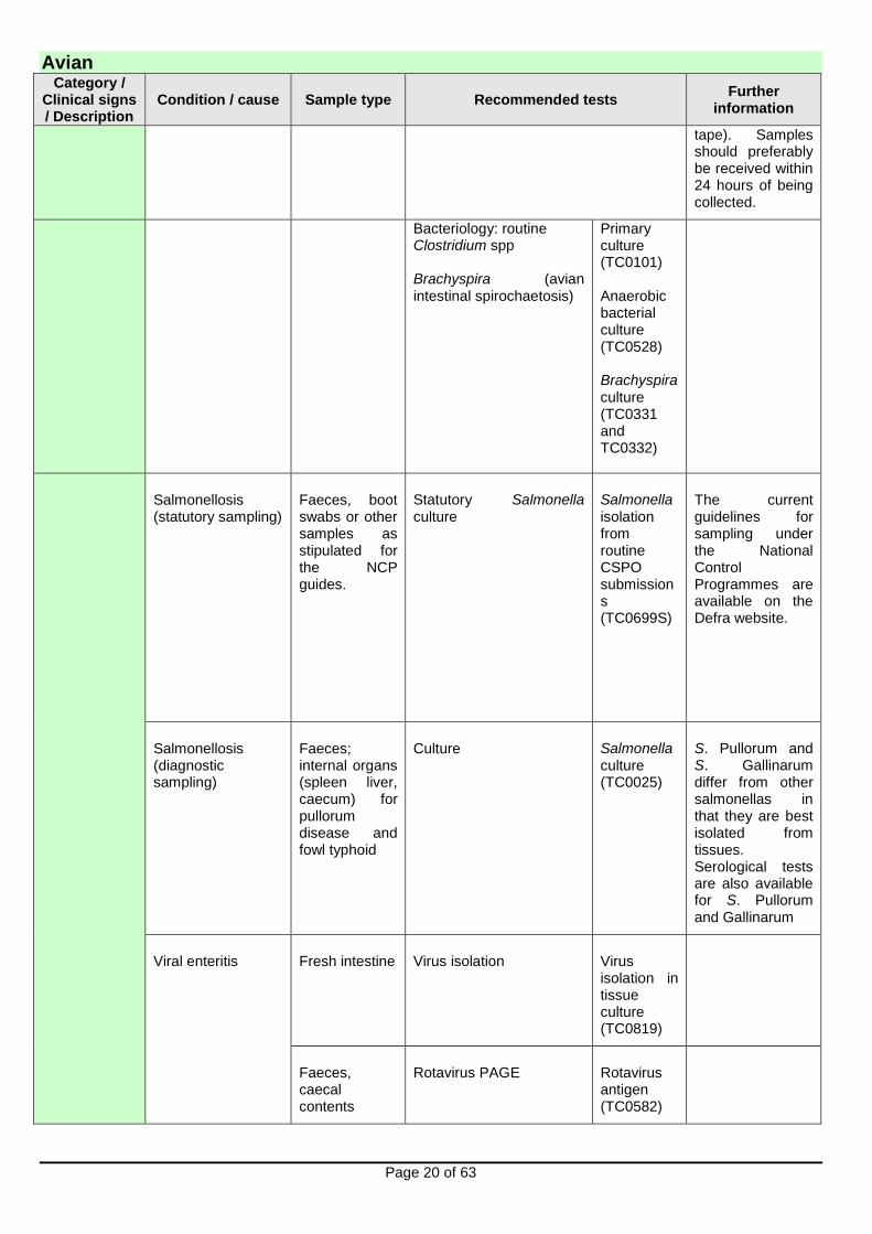
tape). Samples should preferably be received within 24 hours of being collected.
Bacteriology: routine Clostridium spp Brachyspira (avian intestinal spirochaetosis)
Primary culture (TC0101) Anaerobic bacterial culture (TC0528) Brachyspira culture (TC0331 and TC0332)
Salmonellosis (statutory sampling)
Faeces, boot swabs or other samples as stipulated for the NCP guides.
Statutory Salmonella culture
Salmonella isolation from routine CSPO submissions (TC0699S)
The current guidelines for sampling under the National Control Programmes are available on the Defra website.
Salmonellosis (diagnostic sampling)
Faeces; internal organs (spleen liver, caecum) for pullorum disease and fowl typhoid
Culture Salmonella culture (TC0025)
S. Pullorum and S. Gallinarum differ from other salmonellas in that they are best isolated from tissues. Serological tests are also available for S. Pullorum and Gallinarum
Viral enteritis Fresh intestine Virus isolation Virus isolation in tissue culture (TC0819)
Faeces, caecal contents
Rotavirus PAGE Rotavirus antigen (TC0582)

Avian
Page 21 of 63
Category / Clinical signs / Description
Condition / cause Sample type Recommended tests Further
information
EM for virus detection Electron microscopy (TC0317)
Haemorrhagic Enteritis Virus of turkeys
Fixed spleen
Fresh spleen
Histopathology
Virus detection
HEV/MSD AGIDT (TC0910)
Intestinal parasitism Faeces or caecal contents
Egg/oocyst counts Worm egg/coccidial oocyst count (TC0060)
Intestinal contents
Worm presence; worm identification
Microscopy (TC0580), parasite identification (TC0616)
Motile protozoan infection in game birds
Intestinal contents from freshly dead bird
Microscopy for protozoa Microscopy (TC0580)
Material must be examined very fresh. Live or freshly dead birds should be submitted. Fixed intestine can also be of value for histopathology.
Lameness / recumbency
Consider viral, bacterial, deficiencies, toxic/nutritional, trauma
Tissue (tendon, muscle, tibiotarsus, femoral head, skin); swabs of lesioned areas for bacteriology
Postmortem examination
Histopathology
TC0008 / TC0010
Sample should be immersed in 10-20 x volume in 10% formalin using a container with a wide opening

Avian
Page 22 of 63
Category / Clinical signs / Description
Condition / cause Sample type Recommended tests Further
information
See also under ‘wet litter,..’ and ‘nervous disease’
Bacteriology: Primary bacterial culture (TC0101)
Antibiotic sensitivity – aerobe (TC0401)
Tissues and swabs must be taken aseptically
Virology: Reovirus isolation
Virus isolation in tissue culture (TC0819)
Mycoplasma spp DGGE/PCR (TC0672)
Samples for Mycoplasma detection should preferably be sent in Mycoplasma transport broth
Rickets and other skeletal disorders
Affected bones Histopathology TC0008 / TC0010
For rickets, histopathology of growth plates required (such as proximal tibia)
Clotted blood (serum)
Mycoplasma serology M. gallisepticum and M synoviae Rapid Slide Agglutination (RSA, TC0306, TC0308); RSA flock screen (PC0932); or Western Immunoblotting (TC0749)
Minimum of 10 birds for flock screen. For RSA the serum must be freshly taken and not haemolysed or frozen.
The RSA is not recommended for use in game birds.
Avian
Page 23 of 63
Category / Clinical signs / Description
Condition / cause Sample type Recommended tests Further
information
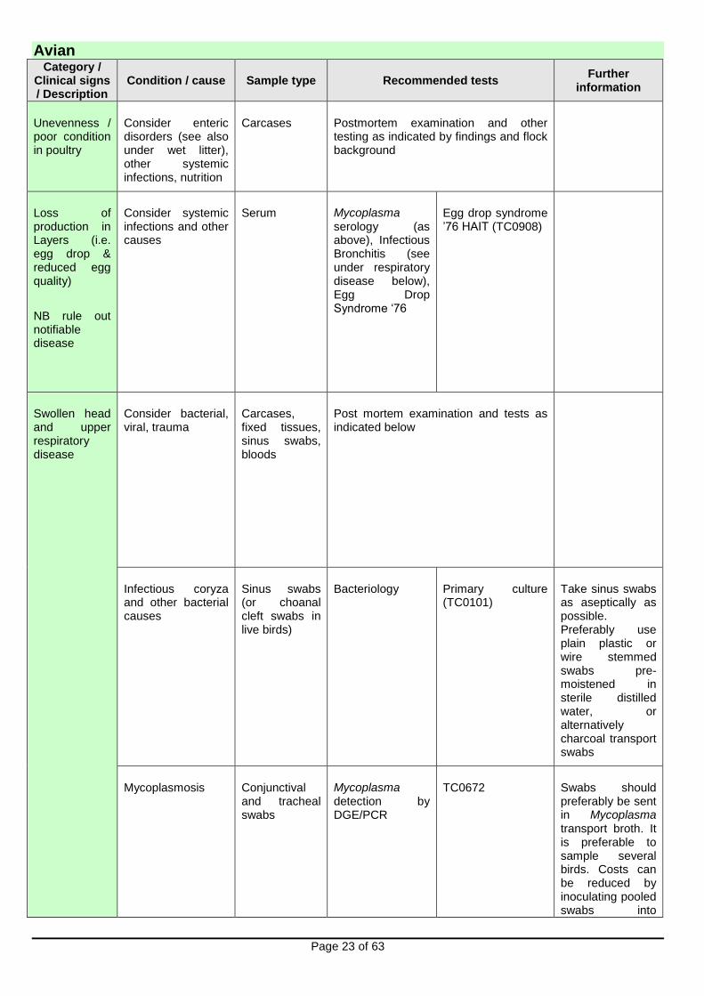
Unevenness / poor condition in poultry
Consider enteric disorders (see also under wet litter), other systemic infections, nutrition
Carcases Postmortem examination and other testing as indicated by findings and flock background
Loss of production in Layers (i.e. egg drop & reduced egg quality)
NB rule out notifiable disease
Consider systemic infections and other causes
Serum Mycoplasma serology (as above), Infectious Bronchitis (see under respiratory disease below), Egg Drop Syndrome ‘76
Egg drop syndrome ’76 HAIT (TC0908)
Swollen head and upper respiratory disease
Consider bacterial, viral, trauma
Carcases, fixed tissues, sinus swabs, bloods
Post mortem examination and tests as indicated below
Infectious coryza and other bacterial causes
Sinus swabs (or choanal cleft swabs in live birds)
Bacteriology Primary culture (TC0101)
Take sinus swabs as aseptically as possible. Preferably use plain plastic or wire stemmed swabs pre-moistened in sterile distilled water, or alternatively charcoal transport swabs
Mycoplasmosis Conjunctival and tracheal swabs
Mycoplasma detection by DGE/PCR
TC0672 Swabs should preferably be sent in Mycoplasma transport broth. It is preferable to sample several birds. Costs can be reduced by inoculating pooled swabs into

Avian
Page 24 of 63
Category / Clinical signs / Description
Condition / cause Sample type Recommended tests Further
information
Clotted blood or serum
Mycoplasma serology (as above)
transport broth.
The RSA is not recommended in game birds.
Avian meta pneumovirus (aMPV, ART, TRT)
Oropharyngeal swab Clotted blood or serum
PCR ELISA
aMPV PCR (TC0786) ELISA (TC0940)
Plastic or wire stemmed swabs must be used
Infectious bronchitis and IBV-like gamma coronaviruses
Oropharyngeal or cloacal swabs Clotted blood or serum
RT PCR HAIT
RT PCR for single swabs (TC0787), or for a pool of up to 10 swabs (TC0887) HAIT for a specified single strain (TC0912), or for 3 specified strains (TC0640)
Plastic or wire stemmed swabs must be used. Positive results are followed up by sequencing of S1 gene to identify strain Interpretation of IBV serology requires knowledge of the IBV vaccination history. Paired serology is recommended
Respiratory cryptosporidiosis
Carcases or fresh or fixed heads
Histopathology TC0008 / TC0010 C. parvum recognised in red grouse and occasionally other species
Respiratory disease
Consider bacterial, viral, fungal, parasitic and non-infectious causes. There is often a mixed aetiology.
See under swollen head and upper respiratory infections
Carcases, fixed tissues, swabs, bloods
Postmortem examination, histopathology and tests as indicated below and under swollen head/upper respiratory disease as above
Avian
Page 25 of 63
Category / Clinical signs / Description
Condition / cause Sample type Recommended tests Further
information
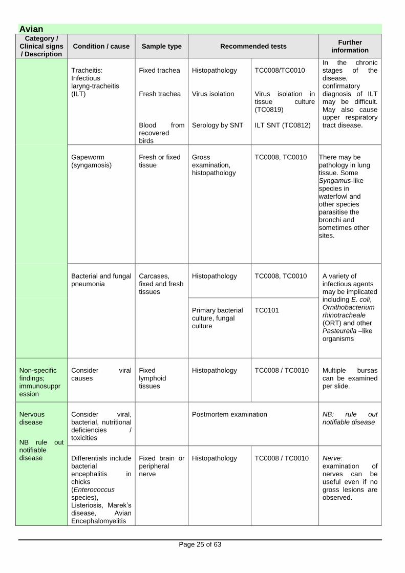
Tracheitis: Infectious laryng-tracheitis (ILT)
Fixed trachea Fresh trachea Blood from recovered birds
Histopathology Virus isolation Serology by SNT
TC0008/TC0010 Virus isolation in tissue culture (TC0819) ILT SNT (TC0812)
In the chronic stages of the disease, confirmatory diagnosis of ILT may be difficult. May also cause upper respiratory tract disease.
Gapeworm (syngamosis)
Fresh or fixed tissue
Gross examination, histopathology
TC0008, TC0010 There may be pathology in lung tissue. Some Syngamus-like species in waterfowl and other species parasitise the bronchi and sometimes other sites.
Bacterial and fungal pneumonia
Carcases, fixed and fresh tissues
Histopathology TC0008, TC0010 A variety of infectious agents may be implicated including E. coli, Ornithobacterium rhinotracheale (ORT) and other Pasteurella –like organisms
Primary bacterial culture, fungal culture
TC0101
Non-specific findings; immunosuppression
Consider viral causes
Fixed lymphoid tissues
Histopathology TC0008 / TC0010 Multiple bursas can be examined per slide.
Nervous disease
NB rule out notifiable disease
Consider viral, bacterial, nutritional deficiencies / toxicities
Postmortem examination NB: rule out notifiable disease
Differentials include bacterial encephalitis in chicks (Enterococcus species), Listeriosis, Marek’s disease, Avian Encephalomyelitis
Fixed brain or peripheral nerve
Histopathology TC0008 / TC0010 Nerve: examination of nerves can be useful even if no gross lesions are observed.

Avian
Page 26 of 63
Category / Clinical signs / Description
Condition / cause Sample type Recommended tests Further
information
crazy chick disease (Vitamin E deficiency)
Brain: fix one half of a sagittal section of the head with the brain in situ and retain the other half of the brain fresh (unfixed) for other tests.
Bacterial encephalitis
Swab of fresh brain
Bacterial culture Primary culture (TC0101), Listeria culture (TC0663)
Aseptic sampling is essential
Neoplasia Consider viral aetiology (Marek’s disease; rarely Avian Leukosis Virus or other oncogenic viruses)
Fixed tumour, liver, spleen and nerve
Fresh tumour tissue
Post mortem examination
Histopathology TC0008 / TC0010
Retain fresh tissues frozen in case required for molecular testing, especially if unusual tumour distribution
Skin and feathers
Consider ectoparasites (mites, lice)
Collect parasites in water for identification
Microscopy Ectoparasites- microscopic examination (TC0081)
Correct identification of mite species is important in planning control measures
Poxvirus (also sometimes see lesions in oral cavity)
Fixed lesion tissue
Fresh tissue
Histopathology
Electron microscopy or virus isolation
TC0008 / TC0010
Electron microscopy-avian (TC0317); virus isolation for pox virus (TC0817)
‘Dry’ pox form refers to lesions on skin, ‘wet’ pox to lesions in oral cavity.
Unusual conditions
Any variations of the above – cause unknown
Carcases Histopathology on fixed affected tissue
Postmortem examination Histopathology (TC0008, TC0010)
Please contact one of our specialist avian pathologists for advice: Also keep fresh frozen material at -70oC for possible future analysis
Avian
Page 27 of 63
Selected diseases of waterfowl (otherwise see above)
Category Condition / cause
Sample type Recommended tests Further information
Water fowl
Duck Viral Enteritis, Duck Viral Hepatitis, Goose Parvovirus
Carcases Fixed tissues Fresh tissues Clotted blood (serum)
Postmortem examination Histopathology Virus isolation
TC0008 / TC0010 Virus isolation in duck/goose eggs (TC0820) DVE SNT (TC0906), DVH SNT (TC0907) GPV AGIDT (TC0302)
Please contact the laboratory to discuss tissues of choice
Duck septicaemia (Riemerella anatipestifer)
Carcases Swabs
Bacteriology
Primary culture (TC0101)
Aseptic swabs of brain tissue are particularly useful

Cattle
Page 28 of 63
Abortion and Stillbirth Whole fetus, placenta and maternal serum are the submission of choice. There is a statutory requirement to report all bovine abortion cases to the local APHA Field Service.
Category Condition/ cause
Sample type Recommended test Further information
Adult Most bacterial causes and mycotic abortion
Fetal stomach contents and placenta
Culture, stained smears and wet preparation
(TC0101 for routine culture, TC0026 for Campylobacter, TC0580 for wet preparation for fungi)
Can be combined with PCRs for Neospora caninum and BVD virus (see below for sample requirements) under TC0015. If a full range of samples is submitted, testing will be carried out in a stepwise fashion. If there are no significant findings from step 1 (bacteriology/mycology), then the PCRs will be carried out as step 2. Tests additional to steps 1 and 2 (e.g. PCR for Leptospira sp.) can be commissioned, but will require additional samples and will attract an additional charge. Please discuss with a VIO.
Neospora caninum
Fetal brain
PCR on fresh brain (TC0852)
A positive PCR result confirms Neospora infection of the fetus, but does not confirm that neosporosis was the cause of abortion. Confirmation of Neospora abortion can be achieved through histopathology on fixed tissue.
Fixed fetal brain; Fixed fetal heart
Histopathology (PC0006)
BVD virus Fetal spleen or thymus
PCR (TC0655)
IBR / BVD / L.hardjo / N.caninum (Maternal Serology)
Blood - clotted
Bovine abortion/stillbirth serology package A (PC0387) (L.hardjo/ N.caninum) Bovine abortion/stillbirth serology package B (PC0405) (IBR /BVD/ L.hardjo/N.caninum)
Paired samples are of limited diagnostic value. Single samples are useful in maintaining disease surveillance and can rule out neosporosis.

Cattle
Page 29 of 63
Iodine deficiency
Fetal thyroid Iodine assay Consider in stillbirths and fetal death in last week of gestation.
Fixed fetal thyroid
Histopathology (PC0006)
Investigation of cattle herd infertility requires a systematic approach. Laboratory investigations can be an important component of this approach. Please discuss individual herd problems and the potential for laboratory testing to inform an investigation with a VIO.
When investigating suspected cases of bovine venereal campylobacteriosis, please note that Campylobacter culture and identification from sheath washings and vaginal mucus samples requires a specific sampling kit and submission form. Samples for this test (TC0098) should be sent to APHA Starcross, ensuring arrival within 24 hours of sampling. Please obtain further details from a VIO before submitting these samples.
Enteric Disorders
Category Condition / cause
Sample type
Recommended test Further information
Calves 1 – 5 days
E. coli (K99 +ve), Salmonella, cryptosporidia, rotavirus, coronavirus
Faeces (5g)
Enteric package for 1 – 5 day old calves (PC0069)
Individual components may be selected.
Calves 6 - 21 days
Salmonella, cryptosporidia, rotavirus, coronavirus
Faeces (5g)
Enteric package for 6 – 21 day old calves (PC0070)
Individual components may be selected.
Calves from 22 days
Salmonella, coccidiosis, PGE
Faeces (10g)
Enteric package for young ruminants (PC0071)
Individual components may be selected: Salmonella culture (TC0025) and worm egg/coccidial oocyst count (TC0060).
Adult
Salmonella, fasciolosis, Johne’s disease
Faeces (40g) and Blood - clotted
Enteric package for adult cattle (PC0073)
Johne’s disease testing is carried out by serology (ELISA). Additional tests such as PCR on faeces may be carried out for an additional charge but require additional faeces. Individual components may be selected: Salmonella culture (TC0025), fluke egg examination (TC0061) and Johne’s disease antibody ELISA (TC0366).
All ages Persistent BVD infection and Mucosal Disease
Blood – heparin or clotted
ELISA antigen (TC0772) and antibody tests (TC0390).
In calves ≤ 30 days old, consider use of BVD PCR (TC0655).

Cattle
Page 30 of 63
Category Condition / cause
Sample type
Recommended test Further information
All ages Acute BVD infection
Blood – clotted or heparin
Paired ELISA antibody test (TC0390)
PCR (heparin blood) (TC0655) on acute sample
Diagnosis of acute infection by antibody ELISA requires paired acute and convalescent sera with an interval of 3 weeks
Adult Winter dysentery (Coronavirus-associated diarrhoea)
Blood – paired clotted
Paired ELISA antibody test (TC0176)
Usually seen in housed adult dairy cattle. Usually characterised by high morbidity but low mortality, with spontaneous recovery in a few days.
Ill thrift
Category Condition / cause
Sample type
Recommended test
Further information
Adult, pre- and post-weaned
Endoparasitism Faeces (50g)
Worm egg count and examination for fluke eggs (PC0064)
Individual components may be selected: Worm egg count (TC0060) and fluke egg examination (TC0061). A composite worm egg count (TC0688) is available, but is usually used for monitoring rather than diagnostic purposes. A composite fluke egg examination (TC0689) is available.
All ages Trace element deficiency
–Consult testing laboratory
) - Copper and GSH-Px (for selenium)
Sample at least 6 animals. Individual components may be selected. Liver copper assay may also provide useful information
All ages Persistent BVD infection and Mucosal Disease
Blood - heparin
ELISA antigen (TC0772) and antibody tests (TC0390).
In calves ≤ 30 days old, consider use of BVD PCR (TC0655).
Adult Adults: Johne’s disease
Blood - clotted
ELISA antibody test (TC0366)
Additional tests such as PCR on faeces may be carried out for an additional charge.
Cattle
Page 31 of 63
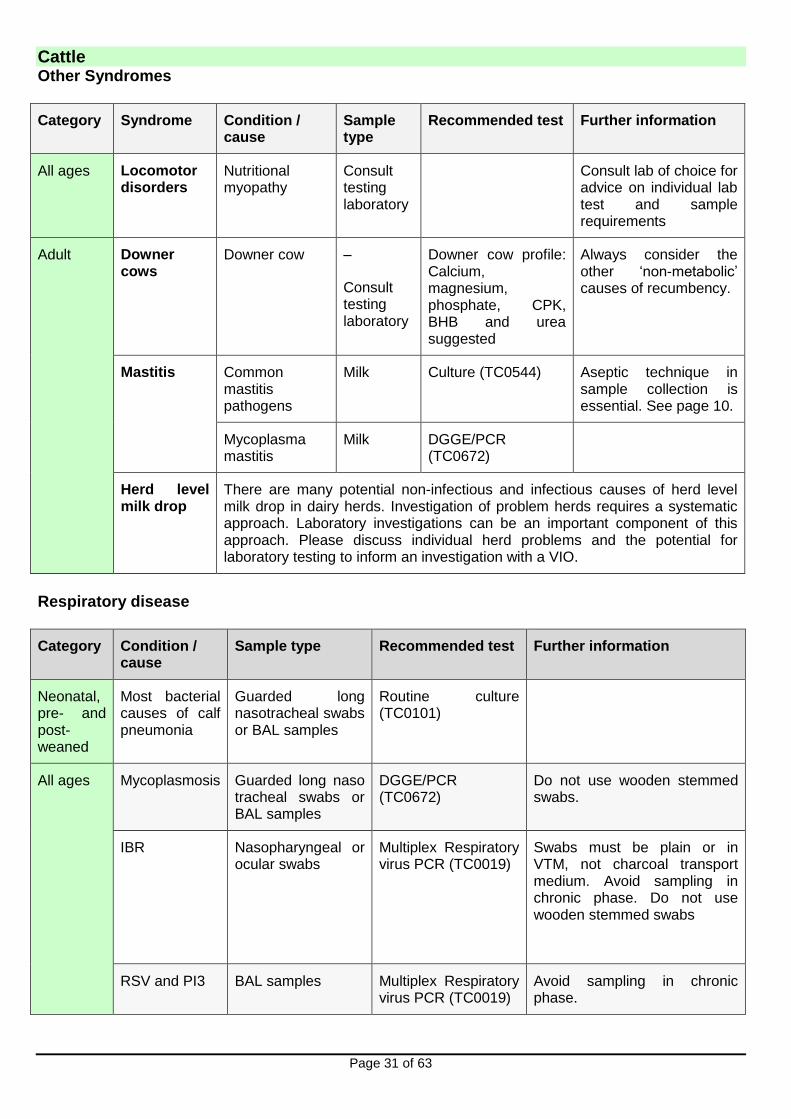
Other Syndromes
Category Syndrome Condition / cause
Sample type
Recommended test Further information
All ages Locomotor disorders
Nutritional myopathy
Consult testing laboratory
Consult lab of choice for advice on individual lab test and sample requirements
Adult
Downer cows
Downer cow –
Consult testing laboratory
Downer cow profile: Calcium, magnesium, phosphate, CPK, BHB and urea suggested
Always consider the other ‘non-metabolic’ causes of recumbency.
Mastitis Common mastitis pathogens
Milk Culture (TC0544) Aseptic technique in sample collection is essential. See page 10.
Mycoplasma mastitis
Milk DGGE/PCR (TC0672)
Herd level milk drop
There are many potential non-infectious and infectious causes of herd level milk drop in dairy herds. Investigation of problem herds requires a systematic approach. Laboratory investigations can be an important component of this approach. Please discuss individual herd problems and the potential for laboratory testing to inform an investigation with a VIO.
Respiratory disease
Category Condition / cause
Sample type Recommended test Further information
Neonatal, pre- and post-weaned
Most bacterial causes of calf pneumonia
Guarded long nasotracheal swabs or BAL samples
Routine culture (TC0101)
All ages
Mycoplasmosis Guarded long naso tracheal swabs or BAL samples
DGGE/PCR (TC0672)
Do not use wooden stemmed swabs.
IBR Nasopharyngeal or ocular swabs
Multiplex Respiratory virus PCR (TC0019)
Swabs must be plain or in VTM, not charcoal transport medium. Avoid sampling in chronic phase. Do not use wooden stemmed swabs
RSV and PI3 BAL samples Multiplex Respiratory virus PCR (TC0019)
Avoid sampling in chronic phase.

Cattle
Page 32 of 63
Category Condition / cause
Sample type Recommended test Further information
IBR, RSV, P13, BVD, Mycoplasma bovis and Histophilus somni
(Serology)
Blood - paired clotted
Bovine respiratory disease serology package A (PC0384) (IBR, RSV, P13, BVD)
Bovine respiratory disease serology package B (PC0385)
(IBR, RSV, P13, BVD, Mycoplasma bovis and Histophilus somni)
Paired acute and convalescent sera collected with an interval of 2 -3 weeks.
Dictyocaulosis (lungworm)
Faeces (50g)
Blood - clotted
Baermann examination (TC0062)
ELISA antibody test (TC0507)
Positive Baermann result indicates patent infestation. Baermann examination will be negative in the pre-patent phase. Positive antibody ELISA result indicates exposure in current/recent grazing season, but not necessarily current patent infestation. Haematology (EDTA blood) can provide useful supportive evidence of relative or absolute eosinophilia.
Malignant Catarrhal Fever
Blood - heparin PCR for OvHV-2 (TC0747)
Sudden death - Always consider anthrax. Any suspicion of disease call APHA:
In England via the Defra Rural Services helpline: 03000 200 301.
In Wales on 0300 303 8268.
In Scotland via the local APHA field office - see https://www.gov.uk/government/organisations/animal-and-plant-health-agency/about/access-and-opening for further contact details.
Category Condition / cause Sample type Recommended test
Further information
Adult
Hypomagnesaemia (cows)
Eye fluid (preferably vitreous humour)
Consult lab of choice for advice on individual lab test and sample requirements
Blood samples from at least six cows in same cohort are useful to screen for blood magnesium concentrations.

Cattle
Page 33 of 63
Category Condition / cause Sample type Recommended test
Further information
Under two years
Blackleg (Clostridium chauvoei)
Four air-dried impression smears of cut surface of muscle lesion (or muscle lesion in a full sealed container to exclude air)
FAT (TC0032)
Submission of a carcase for postmortem examination is probably the preferred diagnostic approach.
Fixed muscle lesion
Histopathology (PC0006)
All ages Lead poisoning Kidney Tissue lead (TC0246)
Please discuss all cases of suspected lead poisoning with a VIO. Please see footnote below.
Fixed = Tissue has been immersed in a suitable fixative such as 10% neutral buffered formalin.
Nervous disease Investigation of fatal cases of nervous disease often requires examination of the whole brain. Submission of a carcase for postmortem examination is probably the preferred diagnostic approach in fatal cases.
Category Condition / cause Sample type Recommended test
Further information
All ages Hypomagnesaemia Consult lab of choice for advice on individual lab test and sample requirements
Nervous acetonaemia
Consult lab of choice for advice on individual lab test and sample requirements
Lead poisoning ) Please discuss all cases of suspected lead poisoning with a VIO. Please see footnote below.
Kidney (from carcase)
)
Skin disease Skin scrape/scab material
Microscopic examination (TC0081)
Histological examination of a fixed skin punch biopsy can be a useful diagnostic approach in more complex skin disease cases
NB: Please discuss all cases of suspected or confirmed poisoning in food animals with a VIO, as voluntary measures to control contamination of the food chain may be requested. In rare circumstances statutory controls imposed under the Food & Environmental Protection Act (FEPA) may be required.
Small Ruminants (Sheep, Goats)
Page 34 of 63
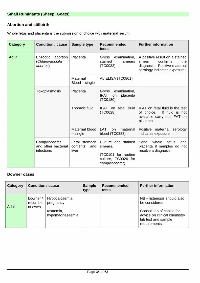
Abortion and stillbirth Whole fetus and placenta is the submission of choice with maternal serum
Category Condition / cause Sample type Recommended tests
Further information
Adult Enzootic abortion (Chlamydophila abortus)
Placenta
Gross examination, stained smears (TC0033)
A positive result on a stained smear confirms the diagnosis. Positive maternal serology indicates exposure
Maternal Blood – single
Ab ELISA (TC0801)
Toxoplasmosis Placenta Gross examination, IFAT on placenta (TC0180)
Thoracic fluid IFAT on fetal fluid (TC0628)
IFAT on fetal fluid is the test of choice. If fluid is not available carry out IFAT on placenta
Maternal blood – single
LAT on maternal blood (TC0383)
Positive maternal serology indicates exposure
Campylobacter and other bacterial infections
Fetal stomach contents and liver
Culture and stained smears.
(TC0101 for routine culture, TC0026 for campylobacter)
Send whole fetus and placenta if samples do not resolve a diagnosis
Downer cases
Category Condition / cause Sample type
Recommended tests
Further information
Adult
Downer / recumbent ewes
Hypocalcaemia, pregnancy
toxaemia, hypomagnesaemia
NB – listeriosis should also be considered
Consult lab of choice for advice on clinical chemistry lab test and sample requirements

Small Ruminants (Sheep, Goats)
Page 35 of 63
Enteric disorders
Category Condition / cause
Sample type Recommended tests
Further information
Small ruminants 1-5 days
Bacteria (E.coli, Salmonella), rotavirus,
Cryptosporidia
Faeces Enteric package 1-5 day old small ruminants including goats (PC0059):
5g faeces required
Individual components;
Bacterial culture with identification of isolates (TC101) and sentest as appropriate,
Salmonella culture (TC0025),
rotavirus PAGE,
Cryptosporidia smear (TC0033)
Small ruminants 6-21 days
Salmonella, rotavirus,
Cryptosporidia
Faeces Enteric package 6-21 day old small ruminants including goats (PC0066).
5g of faeces required
Small ruminants over 3 weeks
Coccidia & PGE, Salmonella
Faeces Enteric package young ruminants (PC0071)
10g of faeces required
Individual components may be selected: Salmonella culture (TC0025),
Worm egg and coccidial oocyst counts (TC0060)
Adult Johne’s disease, Salmonella, fluke and PGE
Faeces
Enteric package adult sheep and goats (PC0075): (40g faeces).
Note that fasciolosis and Johne’s disease do not usually present with diarrhoea and individual components of the enteric package may be selected:
Johne’s disease smear (TC0776),
Fluke eggs (TC0061),
Worm egg count (TC0060), Salmonella culture (TC0025)
Adult sheep usually acquire immunity to enteric parasites

Small Ruminants (Sheep, Goats)
Page 36 of 63
Category Condition / cause
Sample type Recommended tests
Further information
Blood Blood (Johne’s disease): ELISA (TC0366)
Diagnosis of Johne’s disease in sheep may require postmortem examination as ELISA and faecal microscopy are less useful in this species compared to in cattle.

Small Ruminants (Sheep, Goats)
Page 37 of 63
Ill thrift
Category Condition / cause
Sample type Recommended tests
Further information
Young animals
Parasitism Faeces Worm egg count (TC0060)
Further information on investigation of anthelmintic resistance can be found at www.scops.org.uk
Border disease
Blood – Heparin and Clotted
ELISA for antibodies (TC0292)
PCR for virus (TC0755)
Often there is a history of abortions/hairy shaker lambs earlier in the year.
Copper deficiency
Suggest sampling 5-6 animals not receiving concentrates. Consult lab of choice for advice on test and sample requirements
Vitamin B12 (cobalt) deficiency
Suggest sampling 5-6 animals; animals should not be yarded for more than 6 hours prior to sampling as this may falsely elevate serum B12 levels. Consult lab of choice for advice on test and sample requirements.
Adult
Parasitism Faeces Worm egg count (TC0060),
Fluke egg count (TC0061)
Further information on investigation of anthelmintic resistance can be found at www.scops.org.uk
Johne’s disease
Faeces
Blood - clotted
Johne’s disease smear (TC0776)
Ab ELISA (TC0366)
Diagnosis of Johne’s disease in sheep may require postmortem examination as ELISA and faecal microscopy are less useful in this species compared to in cattle.
Copper deficiency
Suggest sampling 5-6 animals not receiving concentrates. Consult lab of choice for advice on test and sample requirements.
Small Ruminants (Sheep, Goats)
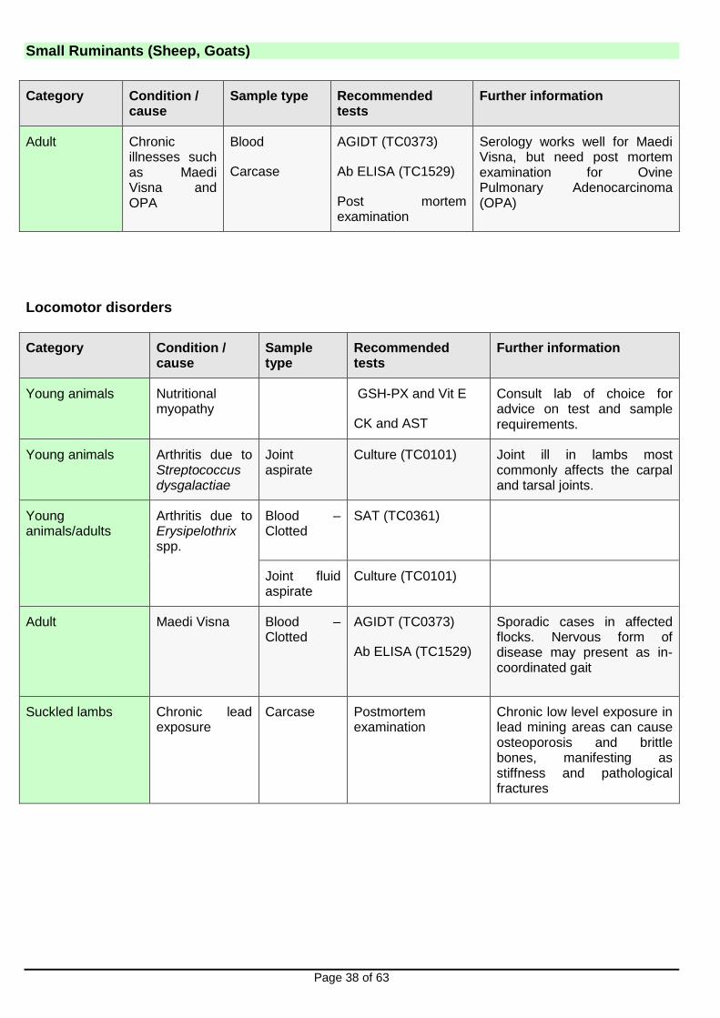
Page 38 of 63
Category Condition / cause
Sample type Recommended tests
Further information
Adult Chronic illnesses such as Maedi Visna and OPA
Blood
Carcase
AGIDT (TC0373)
Ab ELISA (TC1529)
Post mortem examination
Serology works well for Maedi Visna, but need post mortem examination for Ovine Pulmonary Adenocarcinoma (OPA)
Locomotor disorders
Category Condition / cause
Sample type
Recommended tests
Further information
Young animals Nutritional myopathy
GSH-PX and Vit E
CK and AST
Consult lab of choice for advice on test and sample requirements.
Young animals Arthritis due to Streptococcus dysgalactiae
Joint aspirate
Culture (TC0101) Joint ill in lambs most commonly affects the carpal and tarsal joints.
Young animals/adults
Arthritis due to Erysipelothrix spp.
Blood – Clotted
SAT (TC0361)
Joint fluid aspirate
Culture (TC0101)
Adult Maedi Visna Blood – Clotted
AGIDT (TC0373)
Ab ELISA (TC1529)
Sporadic cases in affected flocks. Nervous form of disease may present as in-coordinated gait
Suckled lambs Chronic lead exposure
Carcase Postmortem examination
Chronic low level exposure in lead mining areas can cause osteoporosis and brittle bones, manifesting as stiffness and pathological fractures
Small Ruminants (Sheep, Goats)
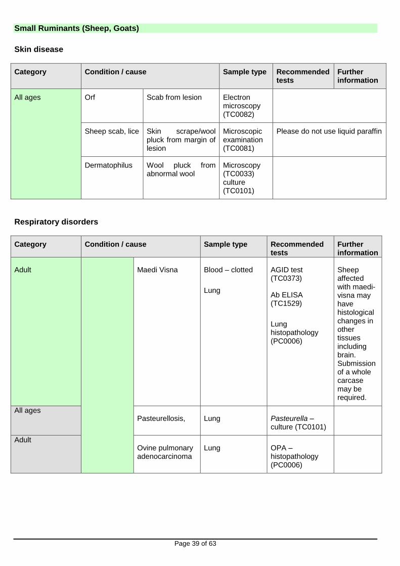
Page 39 of 63
Skin disease
Category Condition / cause Sample type Recommended tests
Further information
All ages Orf Scab from lesion Electron microscopy (TC0082)
Sheep scab, lice Skin scrape/wool pluck from margin of lesion
Microscopic examination (TC0081)
Please do not use liquid paraffin
Dermatophilus Wool pluck from abnormal wool
Microscopy (TC0033) culture (TC0101)
Respiratory disorders
Category Condition / cause Sample type Recommended tests
Further information
Adult
Maedi Visna Blood – clotted
Lung
AGID test (TC0373)
Ab ELISA (TC1529)
Lung histopathology (PC0006)
Sheep affected with maedi-visna may have histological changes in other tissues including brain. Submission of a whole carcase may be required.
All ages Pasteurellosis, Lung Pasteurella –
culture (TC0101)
Adult Ovine pulmonary adenocarcinoma
Lung OPA – histopathology (PC0006)
Small Ruminants (Sheep, Goats)
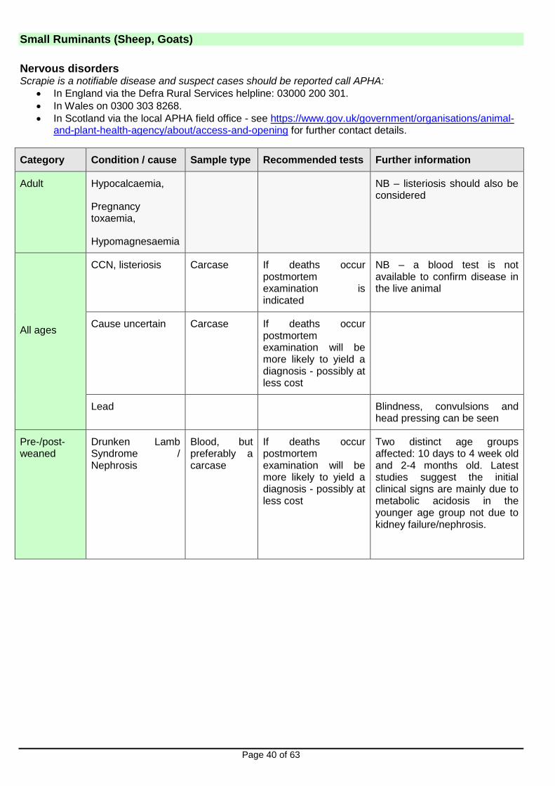
Page 40 of 63
Nervous disorders Scrapie is a notifiable disease and suspect cases should be reported call APHA:
In England via the Defra Rural Services helpline: 03000 200 301.
In Wales on 0300 303 8268.
In Scotland via the local APHA field office - see https://www.gov.uk/government/organisations/animal-and-plant-health-agency/about/access-and-opening for further contact details.
Category Condition / cause Sample type Recommended tests Further information
Adult
Hypocalcaemia,
Pregnancy toxaemia,
Hypomagnesaemia
NB – listeriosis should also be considered
All ages
CCN, listeriosis Carcase If deaths occur postmortem examination is indicated
NB – a blood test is not available to confirm disease in the live animal
Cause uncertain Carcase If deaths occur postmortem examination will be more likely to yield a diagnosis - possibly at less cost
Lead Blindness, convulsions and head pressing can be seen
Pre-/post-weaned
Drunken Lamb Syndrome / Nephrosis
Blood, but preferably a carcase
If deaths occur postmortem examination will be more likely to yield a diagnosis - possibly at less cost
Two distinct age groups affected: 10 days to 4 week old and 2-4 months old. Latest studies suggest the initial clinical signs are mainly due to metabolic acidosis in the younger age group not due to kidney failure/nephrosis.
Small Ruminants (Sheep, Goats)
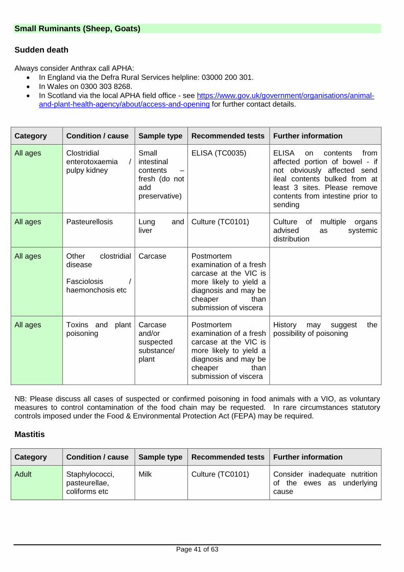
Page 41 of 63
Sudden death Always consider Anthrax call APHA:
In England via the Defra Rural Services helpline: 03000 200 301.
In Wales on 0300 303 8268.
In Scotland via the local APHA field office - see https://www.gov.uk/government/organisations/animal-and-plant-health-agency/about/access-and-opening for further contact details.
Category Condition / cause Sample type Recommended tests Further information
All ages Clostridial enterotoxaemia / pulpy kidney
Small intestinal contents – fresh (do not add preservative)
ELISA (TC0035) ELISA on contents from affected portion of bowel - if not obviously affected send ileal contents bulked from at least 3 sites. Please remove contents from intestine prior to sending
All ages Pasteurellosis Lung and liver
Culture (TC0101) Culture of multiple organs advised as systemic distribution
All ages Other clostridial disease
Fasciolosis / haemonchosis etc
Carcase Postmortem examination of a fresh carcase at the VIC is more likely to yield a diagnosis and may be cheaper than submission of viscera
All ages Toxins and plant poisoning
Carcase and/or suspected substance/ plant
Postmortem examination of a fresh carcase at the VIC is more likely to yield a diagnosis and may be cheaper than submission of viscera
History may suggest the possibility of poisoning
NB: Please discuss all cases of suspected or confirmed poisoning in food animals with a VIO, as voluntary measures to control contamination of the food chain may be requested. In rare circumstances statutory controls imposed under the Food & Environmental Protection Act (FEPA) may be required. Mastitis
Category Condition / cause Sample type Recommended tests Further information
Adult Staphylococci, pasteurellae, coliforms etc
Milk Culture (TC0101)
Consider inadequate nutrition of the ewes as underlying cause

Pigs
Page 42 of 63
General Please feel free to discuss diagnostic investigations with a VIO prior to submission of samples and you must speak to a VIO or vet at non-APHA partner PME provider site before submitting pigs. Please provide a fully completed submission form that includes a full clinical history including information on medication and vaccination. For post mortem examination, consider submitting a batch of up to three pigs, typical of the clinical problem being investigated. It is sometimes appropriate to submit pigs which have been euthanased or which are live (if they are fit to travel and their welfare is not compromised). This should be discussed with the VIO or vet at non-APHA partner PME provider site before submission. Individual pigs or samples may be all that it is possible to submit from small herds.
NB: Please discuss all cases of suspected or confirmed poisoning in food animals with a VIO, as voluntary measures to control contamination of the food chain may be requested. In rare circumstances statutory controls imposed under the Food & Environmental Protection Act (FEPA) may be required. REPRODUCTIVE DISEASE: with fetopathy e.g. abortion, stillbirth, weak piglets at birth
Category Condition / cause Sample type Recommended tests Further information
Adult Suspected infectious cause
Whole litter including placentas
Abortion/stillbirth investigation (TC0011 one sow), (TC0012 two sows)
Diagnostic postmortem examination and stepwise diagnostic testing at discretion of VIO to include PRRS, leptospirosis and bacterial/fungal causes from the outset where material submitted is suitable
Porcine Reproductive and Respiratory Syndrome (PRRS)
Fetal thymus, spleen or lung
PRRSv PCR (TC0718)
Fetal tissue preferable but sometimes virus only detected in serum from aborting sow.
Detection of PRRS in abortions in vaccinated sows may need multiple samples and sows should be sampled at the time of abortion.
PRRSv serology useful in sows if not vaccinated but only diagnostic if paired
Maternal serum
PRRSv PCR (TC0718)
(pooled PRRSv PCR not suitable for adult pigs)

Pigs
Page 43 of 63
Category Condition / cause Sample type Recommended tests Further information
Porcine parvovirus PPV
Fetal heart PPV PCR Mummified fetuses can also be tested by PCR. Fetal serology by HAIT only useful if fetuses at ≥ 70 days gestation
Fetal fluid (e.g. pleural)
PPV HAIT TC0375
Leptospirosis Fetal kidney Pathogenic Leptospira PCR (TC0856)
Autolysis interferes with the PCR test.
Positive PCR results should be followed up with serology to identify the infecting serovar.
Histopathology (liver, kidney) can provide supporting evidence.
Maternal sera as follow-up to positive PCR
Leptospira MAT 6 pools, 19 serovars (TC0399)
Swine influenza Nasal swabs (plain) from sows
Swine influenza PCR (TC0771)
Consider if sows are showing transient pyrexia, malaise and/or respiratory signs. Pigs sampled for virus must be in the first few days of infection. Testing for virus is free of charge, see http://apha.defra.gov.uk/documents/surveillance/diseases/swine-influenza.pdf
Maternal paired sera
Swine influenza HAIT serology (TC0160 four strains)
Erysipelas (and other bacterial causes including fungi)
Fetal stomach contents (liver is second choice)
Bacterial culture including fungal
(TC0101)
Collect from stomach using a plain vacutainer to limit contamination. Do not pool from different fetuses.
Histopathology on placenta also useful for confirming fungal placentitis
PCV2-associated disease
Fetal heart – fresh and fixed
Histopathology (PC0006 and PCV-2 IHC if necessary)
If myocarditis detected by histopathology, PCV-2 IHC will be progressed
Pigs
Page 44 of 63
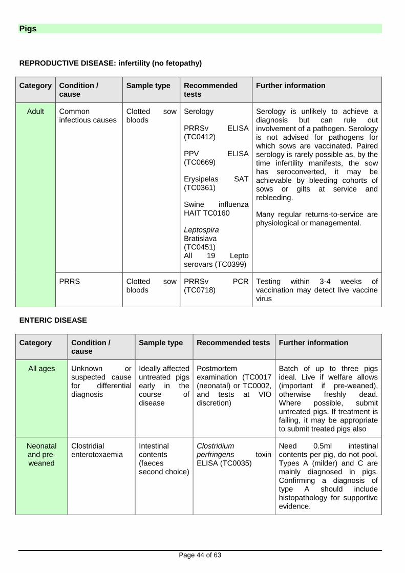
REPRODUCTIVE DISEASE: infertility (no fetopathy)
Category Condition / cause
Sample type Recommended tests
Further information
Adult Common infectious causes
Clotted sow bloods
Serology
PRRSv ELISA (TC0412)
PPV ELISA (TC0669)
Erysipelas SAT (TC0361)
Swine influenza HAIT TC0160
Leptospira Bratislava (TC0451) All 19 Lepto serovars (TC0399)
Serology is unlikely to achieve a diagnosis but can rule out involvement of a pathogen. Serology is not advised for pathogens for which sows are vaccinated. Paired serology is rarely possible as, by the time infertility manifests, the sow has seroconverted, it may be achievable by bleeding cohorts of sows or gilts at service and rebleeding.
Many regular returns-to-service are physiological or managemental.
PRRS Clotted sow bloods
PRRSv PCR (TC0718)
Testing within 3-4 weeks of vaccination may detect live vaccine virus
ENTERIC DISEASE
Category Condition / cause
Sample type Recommended tests Further information
All ages Unknown or suspected cause for differential diagnosis
Ideally affected untreated pigs early in the course of disease
Postmortem examination (TC0017 (neonatal) or TC0002, and tests at VIO discretion)
Batch of up to three pigs ideal. Live if welfare allows (important if pre-weaned), otherwise freshly dead. Where possible, submit untreated pigs. If treatment is failing, it may be appropriate to submit treated pigs also
Neonatal and pre-weaned
Clostridial enterotoxaemia
Intestinal contents (faeces second choice)
Clostridium perfringens toxin ELISA (TC0035)
Need 0.5ml intestinal contents per pig, do not pool. Types A (milder) and C are mainly diagnosed in pigs. Confirming a diagnosis of type A should include histopathology for supportive evidence.

Pigs
Page 45 of 63
Category Condition / cause
Sample type Recommended tests Further information
Neonatal and pre- weaned (cont)
Colibacillosis
(Enterotoxigenic E.coli)
Intestinal contents or faeces (swabs second choice)
Bacterial culture (TC0101) and (TC0829) E.coli fimbrex (K88) test typing
Further E. coli typing possible at extra charge (TC0040)
Non-ETEC E.coli e.g. attaching and effacing E.coli (AEEC), Enterohaemorrhagic E.coli (EHEC)
Live pigs Post mortem examination (TC0017 (neonatal) or TC0002)
Culture results with histopathology essential for diagnosis and intestines must be fixed within minutes of death
Further E. coli serotyping possible at extra charge (TC0040)
Intestinal contents, faeces or swabs and fixed intestines
Bacterial culture (TC0101 and TC0829 E.coli fimbrex test typing and histopathology PC0006)
Rotaviral enteritis Intestinal contents or faeces
Rotavirus PAGE (TC0582)
Cryptosporidiosis Intestinal contents or faeces
Cryptosporidium smear and ID (TC0492)
Rarely diagnosed in pigs, zoonotic
Coccidiosis Intestinal contents or faeces (min. 3g)
Coccidial oocyst count (TC0702). Speciation possible at extra cost (TC0648)
Absence of oocysts does not rule out a diagnosis of coccidiosis. Intestinal histopathology may be required to achieve a diagnosis and needs live affected pigs to be submitted
Salmonellosis Intestinal contents or faeces (swabs second choice)
Salmonella culture (TC0025)
Rare in neonates, uncommon preweaning. Zoonotic
Transmissible gastro-enteritis
Intestinal contents or faeces
TGE/PED PCR (TC0198)
PCR is method of choice for diagnosis
Last VIDA diagnosis in GB in 1999
Paired sera TGEv ELISA (TC0488)
Porcine epidemic diarrhoea
Intestinal contents or faeces
PEDv PCR (TC0398) PCR is method of choice for diagnosis
Last VIDA diagnosis in GB in 2002, virulent strain not detected in UK
Paired sera TGE/PED ELISA (TC0377)

Pigs
Page 46 of 63
Category Condition / cause
Sample type Recommended tests Further information
Hypogammaglobulinaemia (poor colostral antibody uptake)
Clotted bloods from piglets up to one-week-old
Gammaglobulin estimation (ZST) Consult lab of choice for advice on test and sample requirements.
Useful to determine if poor colostral antibody transfer is predisposing to enteric disease in neonatal piglets
Weaners to about 6 weeks old (two weeks post-weaning)
E.coli, salmonellosis, rotaviral enteritis, TGE and PED
As above As above As above
Bowel oedema (verocytotoxic E.coli)
Intestine, intestinal contents or faeces
Bacterial culture (TC0101), E.coli serotyping (TC0040)
Isolation of causative E. coli more likely from untreated severe cases. Brain histopathology can provide supportive evidence
Growers to adults
Salmonellosis, bowel oedema, TGE and PED
As above As above As above
Brachyspira hyodysenteriae (swine dysentery)
Intestine, intestinal contents or faeces
Brachyspira hyodysenteriae FAT (TC0032)
Brachyspira culture (TC0031)
Brachyspira PCR (TC0495)
Where B. hyodysenteriae isolated, tiamulin MIC testing should be considered – discuss with a VIO at your usual APHA VIC
Fill container to below brim (anaerobic organism).
Brachyspira pilosicoli colitis
Intestine, intestinal contents or faeces
Brachyspira culture (TC0031)
Brachyspira PCR (TC0495)
Fill container to below brim (anaerobic organism).
Lawsonia intracellularis
Lesioned intestine fresh and fixed
MZN smear (TC0033)
Histopathology (PC0006) and silver stains
Several forms exist (necrotic ileitis, proliferative enteropathy, haemorrhagic enteropathy). MZN smears needs very fresh material and can be insensitive.
PCR alone is not diagnostic Intestinal contents or faeces
Lawsonia PCR (TC0657)
Trichuris colitis or other nematodes (Hyostrongylus, Ascaris spp.)
Faeces Worm egg count (TC0060)
Egg output can be low. Hyostrongylus species associated with anaemia and illthrift

Pigs
Page 47 of 63
Category Condition / cause
Sample type Recommended tests Further information
Gastric ulceration, intestinal torsion
Dead pigs Post mortem examination (TC0017 (neonatal) or TC0002)
RESPIRATORY DISEASE
Category Condition / cause Sample type Recommended tests Further information
All ages Unknown or suspected cause for differential diagnosis
Ideally affected untreated pigs or plucks
Post mortem examination (TC0017 (neonatal) or TC0002, and tests at VIO discretion)
Batch of up to three pigs/plucks ideal. Where possible, submit untreated pigs. If treatment is failing, it may be appropriate to submit treated pigs also. Severe cases early in the course of disease are ideal. Porcine respiratory disease commonly involves multiple pathogens.
All ages, especially post-weaning
PRRS Fresh lung, spleen, serum or lymph node
PRRSv PCR (TC0718)
Pooled PRRSv PCR on serum (TC0918)
Vaccinated pigs may become viraemic when undergoing challenge and positive PCR results require further investigation. PRRS vaccination history with timing of vaccination should be stated.
Serology useful if not vaccinated.
Paired sera Paired serology PRRSv ELISA (TC0412)
PCV2-associated respiratory disease
Lymph node and fixed lung
Histopathology (PC0006 and PCV-2 IHC if necessary)
If lymphoid lesions seen with viral inclusions, PCV-2 IHC is not progressed by VIO as not necessary
Swine influenza Pooled tonsil, trachea and lung (max 3 pigs) or plain nasal swabs (max 12 pigs)
Paired sera
Swine influenza PCR (TC0771)
Swine influenza HAIT (TC0160)
Do not pool tissues from different pigs. Pigs sampled for virus must be in the first few days of infection. Testing for virus is free of charge, see http://apha.defra.gov.uk/documents/surveillance/diseases/swine-influenza.pdf
Enzootic pneumonia (Mycoplasma hyopneumoniae)
Fresh lung Mycoplasma DGGE/PCR (TC0672)
Sample lung from cranioventral region at interface between consolidated and non-consolidated lung
Pigs
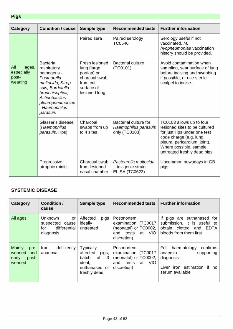
Page 48 of 63
Category Condition / cause Sample type Recommended tests Further information
All ages, especially post-weaning
Paired sera Paired serology TC0546
Serology useful if not vaccinated. M. hyopneumoniae vaccination history should be provided.
Bacterial respiratory pathogens - Pasteurella multocida, Strep suis, Bordetella bronchiseptica, Actinobacillus pleuropneumoniae, Haemophilus parasuis
Fresh lesioned lung (large portion) or charcoal swab from cut surface of lesioned lung
Bacterial culture (TC0101)
Avoid contamination when sampling, sear surface of lung before incising and swabbing if possible, or use sterile scalpel to incise.
Glässer’s disease (Haemophilus parasuis, Hps)
Charcoal swabs from up to 4 sites
Bacterial culture for Haemophilus parasuis only (TC0103)
TC0103 allows up to four lesioned sites to be cultured for just Hps under one test code charge (e.g. lung, pleura, pericardium, joint). Where possible, sample untreated freshly dead pigs.
Progressive atrophic rhinitis
Charcoal swab from lesioned nasal chamber
Pasteurella multocida – toxigenic strain ELISA (TC0623)
Uncommon nowadays in GB pigs
SYSTEMIC DISEASE
Category Condition / cause
Sample type Recommended tests Further information
All ages Unknown or suspected cause for differential diagnosis
Affected pigs ideally untreated
Postmortem examination (TC0017 (neonatal) or TC0002, and tests at VIO discretion)
If pigs are euthanased for submission, it is useful to obtain clotted and EDTA bloods from them first
Mainly pre-weaned and early post-weaned
Iron deficiency anaemia
Typically affected pigs, batch of 3 ideal, euthanased or freshly dead
Postmortem examination (TC0017 (neonatal) or TC0002, and tests at VIO discretion)
Full haematology confirms anaemia supporting diagnosis
Liver iron estimation if no serum available
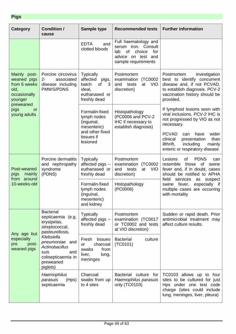
Pigs
Page 49 of 63
Category Condition / cause
Sample type Recommended tests Further information
EDTA and clotted bloods
Full haematology and serum iron. Consult lab of choice for advice on test and sample requirements
Mainly post-weaned pigs from 6 weeks old, occasionally younger preweaned pigs or young adults
Porcine circovirus 2- associated disease including PMWS/PDNS
Typically affected pigs, batch of 3 ideal, euthanased or freshly dead
Postmortem examination (TC0002 and tests at VIO discretion)
Postmortem investigation best to identify concurrent disease and, if not PCVAD, to establish diagnosis. PCV-2 vaccination history should be provided.
If lymphoid lesions seen with viral inclusions, PCV-2 IHC is not progressed by VIO as not necessary.
PCVAD can have wider clinical presentation than illthrift, including mainly enteric or respiratory disease
Formalin-fixed lymph nodes (inguinal, mesenteric) and other fixed tissues if lesioned
Histopathology (PC0006 and PCV-2 IHC if necessary to establish diagnosis)
Post-weaned pigs mainly from around 10-weeks-old
Porcine dermatitis and nephropathy syndrome (PDNS)
Typically affected pigs – euthanased or freshly dead
Postmortem examination (TC0002 and tests at VIO discretion)
Lesions of PDNS can resemble those of swine fever and, if in doubt, cases should be notified to APHA field services as suspect swine fever, especially if multiple cases are occurring with mortality
Formalin-fixed lymph nodes (inguinal, mesenteric) and kidney
Histopathology (PC0006)
Any age but especially pre post-weaned pigs
Bacterial septicaemia (e.g. erysipelas, streptococcal, pasteurellosis, Klebsiella pneumoniae and Actinobacillus suis and colisepticaemia in preweaned piglets)
Typically affected pigs – freshly dead
Postmortem examination (TC0017 or TC0002 and tests at VIO discretion)
Sudden or rapid death. Prior antimicrobial treatment may affect culture results.
Fresh tissues or charcoal swabs from liver, lung, meninges
Bacterial culture (TC0101)
Haemophilus parasuis (Hps) septicaemia
Charcoal swabs from up to 4 sites
Bacterial culture for Haemophilus parasuis only (TC0103)
TC0103 allows up to four sites to be cultured for just Hps under one test code charge (sites could include lung, meninges, liver, pleura)

Pigs
Page 50 of 63
Category Condition / cause
Sample type Recommended tests Further information
Any age
Toxicity Affected pigs Postmortem examination (TC0017 (neonatal) or TC0002, and tests at VIO discretion)
The two toxicities most frequently diagnosed in pigs at APHA are coal tar (liver necrosis) and bracken (cardiomyopathy and acute heart failure). Information on coal tar: http://apha.defra.gov.uk/documents/surveillance/diseases/coal-tar-toxicity.pdf
MUSCULOSKELETAL DISEASE
Category Condition / cause
Sample type Recommended tests Further information
Any age but mainly post-weaned
Infectious arthritis including erysipelas, Streptococcus suis and Mycoplasma hyosynoviae
Typically affected pigs – freshly dead
Postmortem examination (TC0017 or TC0002 and tests at VIO discretion)
PME allows diagnosis of osteochondrosis (OCD), an important differential in older pigs.
It can be problematic isolating Erysipelothrix species from joints
Joint swabs or fluid aspirates
Bacterial culture (TC0101)
Paired sera Erysipelothrix sp. SAT (TC0361)
Infectious arthritis due to Haemophilus parasuis
Joint swabs or fluid aspirates
Bacterial culture for Haemophilus parasuis only (TC0103)
Fibrinous polyarthritis may be part of Glässers disease. TC0103 allows up to four lesioned sites to be cultured for just Hps under one test code charge
Infectious synovitis due to Mycoplasma hyosynoviae
Joint swabs or fluid aspirates
M. hyosynoviae DGGE/PCR TC0672
Mainly older postweaned pigs or young breeding replacements. Histopathology on synovium provides supportive evidence
Post-weaned and young breeding pigs
Osteochondrosis dissecans, femoral epiphyseolysis, fractures
Typically affected pigs – freshly dead
Postmortem examination (TC0002 and tests at VIO discretion)
Gross pathology alone is often diagnostic. Bone analysis and histopathology would be considered if findings suggest an osteodystrophy

Pigs
Page 51 of 63
NERVOUS DISEASE
Category Condition / cause
Sample type Recommended tests Further information
Any age
Unknown or suspected cause for differential diagnosis
Typically affected pigs – live if welfare allows or freshly dead
Postmortem examination (TC0017 or TC0002 and tests at VIO discretion)
Where pigs are euthanased for submission, trauma to the brain due to the method of euthanasia limits full diagnostic investigation. Veterinary administration of barbiturate is ideal.
Salt poisoning (water deprivation)
Typically affected pigs – live if welfare allows or freshly dead
Postmortem examination (TC0017 or TC0002 and brain histopathology)
Pigs are particularly sensitive to water deprivation-induced salt poisoning. Submission of heads of affected pigs would allow diagnosis of salt poisoning
Neonatal Congenital tremor (CT)
Typically affected pigs – live if welfare allows or freshly dead
Postmortem examination (TC0017 and histopathology on brain and spinal cord)
Clinical history and histopathology identify the type of CT. Classical swine fever is a cause of congenital tremor type A1 and, in CT outbreaks, the submitting vet must consider whether there are grounds to report as suspect swine fever to APHA field services
Pre and post-weaned mainly
Bacterial meningitis
Typically affected pigs – freshly dead
Postmortem examination (TC0017 or TC0002)
Streptococcus suis and Haemophilus parasuis are the main bacterial causes. Cultures must include chocolate agar plates for H. parasuis isolation. Submission of heads of affected pigs would also allow diagnosis.
Meningeal swabs (charcoal)
Bacterial culture (TC0101)
Meningeal swabs (plain)
Streptococcus suis 2 FAT (TC0032)
Post-weaned pigs
Bowel oedema Typically affected pigs – live if welfare allows or freshly dead
Postmortem examination (TC0017 or TC0002)
Verocytotoxigenic E. coli types involved. Histopathology on brain provide supportive evidence and may be indicated where clinical signs/gross lesions not typical. Isolation of causative E. coli more likely from untreated severe cases.
Intestinal contents or faeces
Bacterial culture (TC0101)

Pigs
Page 52 of 63
Category Condition / cause
Sample type Recommended tests Further information
Any age Viral polioencephalo-myelitis (e.g. porcine sapelovirus)
Typically affected pigs – live if welfare allows or freshly dead
Postmortem examination (TC0017 or TC0002)
Postmortem examination best option as uncommon and need to rule out differentials. Aujeszky’s disease and Teschen disease are causes of viral encephalitis and are notifiable diseases. Submitting vet must consider whether there are grounds to notify severe outbreaks of nervous disease to APHA field services, especially if unresponsive to antimicrobial treatment.
Sapelovirus PCR can be done at discretion of VIO if histopathology and other findings suggestive of involvement
Fixed brain and spinal cord
Histopathology (PC0006 suspect lesions confirmed by IHC)
Fresh spinal cord
Sapelovirus PCR possible but not available as commercial test
UROGENITAL DISEASE
Category Condition / cause
Sample type Recommended tests
Further information
Breeding females most likely
Cystitis and pyelonephritis
Typically affected pigs –freshly dead
Postmortem examination (TC0002)
Anaerobic culture essential to isolate Actinobaculum suis
Histopathology allows diagnosis but not identification of causative organism
Fresh kidney Bacterial culture aerobic (TC0101) and anaerobic (TC0528)
Fixed kidney Histopathology (PC0006)

Pigs
Page 53 of 63
SKIN DISEASE
Category Condition / cause
Sample type Recommended tests
Further information
Any age
Many conditions, e.g. exudative epidermitis, mange, ringworm, swine pox, parakeratosis, congenital conditions
Affected pigs Discuss with APHA VIC
It is often helpful for images of the skin condition to be sent to the APHA VIC
Charcoal swabs
Bacterial culture (TC0101) for Staph. hyicus
Hair plucks Ringworm culture (TC0080)
Skin biopsies Electron microscopy for pox virus (TC0082)
Skin/ear wax scrapings
Ectoparasite examination (TC0081)
CIRCULATORY DISEASE
Category Condition / cause
Sample type Recommended tests
Further information
Any age Unknown or suspected cause for differential diagnosis
Typically affected pigs – live if welfare allows or freshly dead
Postmortem examination (TC0017 or TC0002 and tests at VIO discretion)
Signs often sudden death or non-specific meriting submission of pigs.
Pre-weaned pigs
Thrombocytopaenia purpura
(isoimmune disease – colostral antibodies to foetal thrombocyte antigens)
Typically affected pigs – live if welfare allows or freshly dead
Postmortem examination (TC0017 and tests at VIO discretion)
Signs include weakness, skin haemorrhages and death of pigs up to about 30 days old. Gilt litters not affected. Lesions can resemble the swine fevers; a good clinical history and examination is usually sufficient to rule out concern, if in doubt, report cases to APHA field services
Pre and post-weaned pigs
Mycoplasma suis
(eperythrozoonosis)
Blood (EDTA)
Mycoplasma DGGE/PCR (TC0672)
Blood parasite detection (TC0826)
Full haematology detects anaemia and assists interpretation. Bone marrow and other histopathology assists interpretation
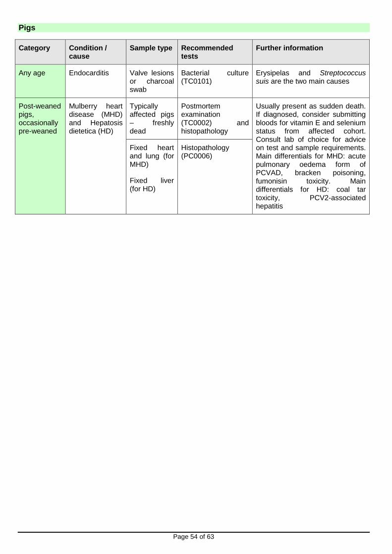
Pigs
Page 54 of 63
Category Condition / cause
Sample type Recommended tests
Further information
Any age Endocarditis Valve lesions or charcoal swab
Bacterial culture (TC0101)
Erysipelas and Streptococcus suis are the two main causes
Post-weaned pigs, occasionally pre-weaned
Mulberry heart disease (MHD) and Hepatosis dietetica (HD)
Typically affected pigs – freshly dead
Postmortem examination (TC0002) and histopathology
Usually present as sudden death. If diagnosed, consider submitting bloods for vitamin E and selenium status from affected cohort. Consult lab of choice for advice on test and sample requirements. Main differentials for MHD: acute pulmonary oedema form of PCVAD, bracken poisoning, fumonisin toxicity. Main differentials for HD: coal tar toxicity, PCV2-associated hepatitis
Fixed heart and lung (for MHD)
Fixed liver (for HD)
Histopathology (PC0006)
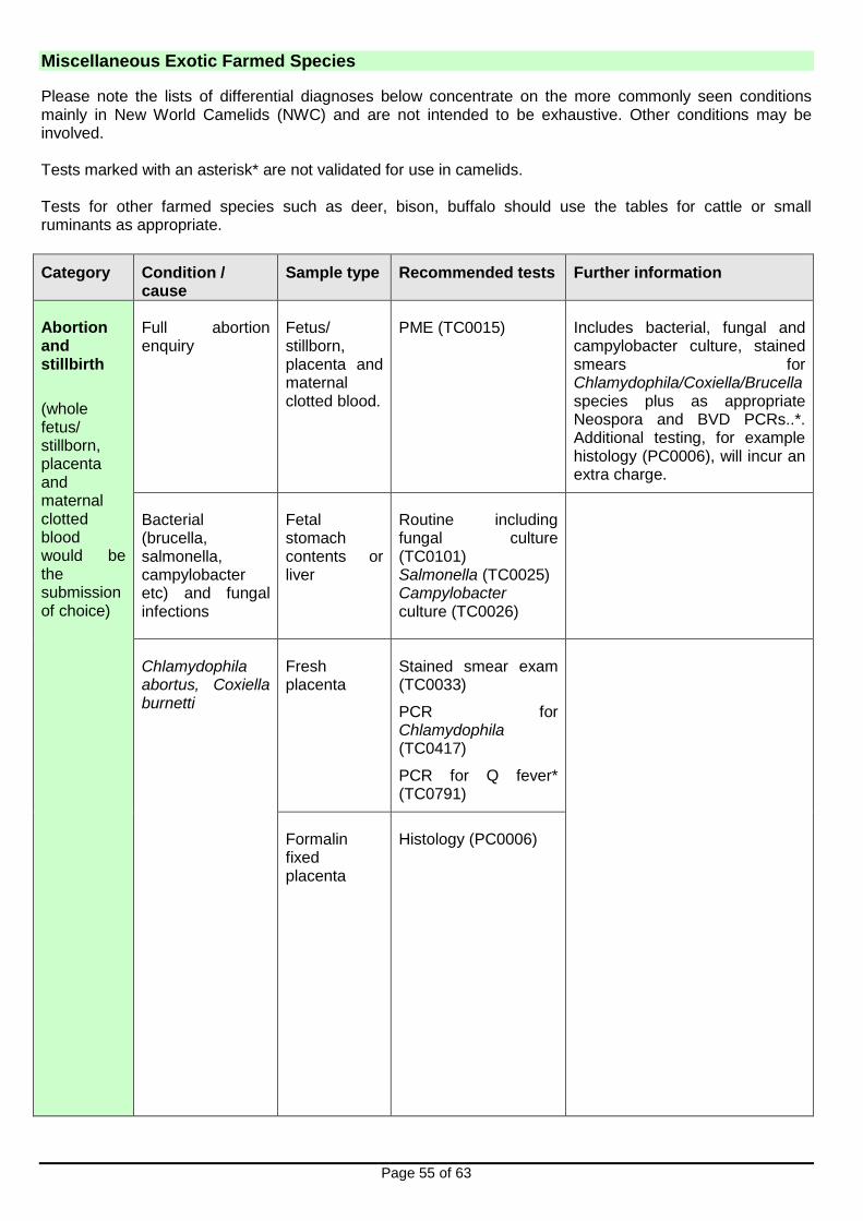
Miscellaneous Exotic Farmed Species
Page 55 of 63
Please note the lists of differential diagnoses below concentrate on the more commonly seen conditions mainly in New World Camelids (NWC) and are not intended to be exhaustive. Other conditions may be involved. Tests marked with an asterisk* are not validated for use in camelids. Tests for other farmed species such as deer, bison, buffalo should use the tables for cattle or small ruminants as appropriate.
Category Condition / cause
Sample type Recommended tests Further information
Abortion and stillbirth
(whole fetus/ stillborn, placenta and maternal clotted blood would be the submission of choice)
Full abortion enquiry
Fetus/ stillborn, placenta and maternal clotted blood.
PME (TC0015)
Includes bacterial, fungal and campylobacter culture, stained smears for Chlamydophila/Coxiella/Brucella species plus as appropriate Neospora and BVD PCRs..*. Additional testing, for example histology (PC0006), will incur an extra charge.
Bacterial (brucella, salmonella, campylobacter etc) and fungal infections
Fetal stomach contents or liver
Routine including fungal culture (TC0101) Salmonella (TC0025) Campylobacter culture (TC0026)
Chlamydophila abortus, Coxiella burnetti
Fresh placenta
Stained smear exam (TC0033)
PCR for Chlamydophila (TC0417)
PCR for Q fever* (TC0791)
Formalin fixed placenta
Histology (PC0006)

Miscellaneous Exotic Farmed Species
Page 56 of 63
Category Condition / cause
Sample type Recommended tests Further information
Neospora caninum
Formalin fixed heart and whole brain.
Histology (PC0006)
Fresh brain stem
Neospora PCR* (TC0852)
A positive PCR result confirms Neospora infection of the fetus, but does not confirm that neosporosis was the cause of abortion. Confirmation of Neospora abortion can be achieved through histopathology on fixed tissue (usually brain and heart).
Leptospirosis Fresh kidney PCR for pathogenic leptospira (TC0856)
BVD Fresh foetal spleen or thymus.
PCR for BVD antigen* (TC0655)
Enteric disorders
Category Condition / cause
Sample type Recommended tests
Further information
< 7 days of age
Bacteria (E.coli, salmonella), rotavirus, cryptosporidia
Faeces (5g) Enteric package (PC0059)
Individual components – routine culture and identification of isolates (TC0101) and sentest as appropriate, salmonella (TC0025), rotavirus PAGE (TC0582), cryptosporidia (TC0492).
7-21 days of age
Salmonella, rotavirus, cryptosporidia
Faeces (5g) Enteric package (PC0066)
As above but no routine culture or identification of isolates.
Note cryptosporidiosis has been recorded as the cause of diarrhoea and death in animals up to 8 months of age.
> 7days of age
Giardia Faeces (5g) Flotation (TC0491)

Miscellaneous Exotic Farmed Species
Page 57 of 63
Category Condition / cause
Sample type Recommended tests
Further information
> 3 weeks of age
Coccidia, parasitic gastroenteritis
Faeces (10g) Worm and coccidia count (TC0860)
Camelids only. Coccidiosis is more common between 3 weeks and 3 months of age.
Nematodirosis can be a problem in young animals from 2 months of age.
Haemonchonsis often presents as severe anaemia.
Adults Liver fluke, coccidia, parasitic gastroenteritis
Faeces (40g + 10g)
Parasitology package (PC0864): Fluke egg count (TC0061) plus worm and coccidia count (TC0860)
Camelids only
Ill thrift/ weight loss (Consider TB which is zoonotic in all age groups)
Category Condition/ cause
Sample type Recommended tests
Further information
Young animals
Coccidia, parasitic gastroenteritis
Faeces (10g) Worm and coccidia count (TC0860)
Camelids only. Coccidiosis is more common between 3 weeks and 3 months of age. Nematodirosis can be a problem in young animals from 2 months of age. Haemonchonsis often presents as severe anaemia.
BVD Heparin blood or fresh lymphoid tissue (eg.spleen, thymus,lymph node).
PCR for BVD antigen* (TC0655)
Clotted blood SNT for BVD antibodies* (TC1165)
Any age Mycoplasma haemolamae
EDTA blood Mycoplasma DGGE/PCR (TC0672)
Can cause anaemia, vague signs or no overt disease.
Note the causative organism can sometimes be detected on examination of a fresh blood smear (TC0256).
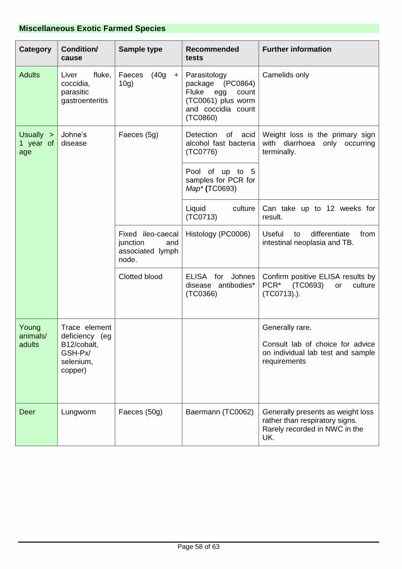
Miscellaneous Exotic Farmed Species
Page 58 of 63
Category Condition/ cause
Sample type Recommended tests
Further information
Adults Liver fluke, coccidia, parasitic gastroenteritis
Faeces (40g + 10g)
Parasitology package (PC0864) Fluke egg count (TC0061) plus worm and coccidia count (TC0860)
Camelids only
Usually > 1 year of age
Johne’s disease
Faeces (5g)
Detection of acid alcohol fast bacteria (TC0776)
Weight loss is the primary sign with diarrhoea only occurring terminally.
Pool of up to 5 samples for PCR for Map* (TC0693)
Liquid culture (TC0713)
Can take up to 12 weeks for result.
Fixed ileo-caecal junction and associated lymph node.
Histology (PC0006) Useful to differentiate from intestinal neoplasia and TB.
Clotted blood ELISA for Johnes disease antibodies* (TC0366)
Confirm positive ELISA results by PCR* (TC0693) or culture (TC0713).).
Young animals/ adults
Trace element deficiency (eg B12/cobalt, GSH-Px/ selenium, copper)
Generally rare.
Consult lab of choice for advice on individual lab test and sample requirements
Deer Lungworm Faeces (50g) Baermann (TC0062) Generally presents as weight loss rather than respiratory signs. Rarely recorded in NWC in the UK.

Miscellaneous Exotic Farmed Species
Page 59 of 63
Other Conditions
Category Condition / cause
Sample type Recommended tests Further information
Nervous disorders
CCN/ encephalopathy
Carcase PME
Meningitis/ encephalitis eg listeriosis
Carcase
PME Often bacterial cause in young crias.
Lead poisoning Carcase PME Consult lab of choice for advice on individual lab test and sample requirements
May present as a sudden death.
Please discuss all cases of suspected lead poisoning in food producing animals with a VIO. Please see footnote below.
Unknown cause Carcase (plus clotted, EDTA and heparin bloods if available)
PME
Blood samples can be useful particularly when an assessment of hepatic or renal function is required.
Respiratory disease
Pneumonia BAL Culture (TC0101) Viral infections of camelid respiratory system are rare.
Consider TB which is zoonotic in all age groups Carcase PME
NB: Please discuss all cases of suspected or confirmed poisoning in food animals with a VIO, as voluntary measures to control contamination of the food chain may be requested. In rare circumstances statutory controls imposed under the Food & Environmental Protection Act (FEPA) may be required.
Category Condition / cause
Sample type Recommended tests Further information
Skin disease
Parasites (Chorioptes, Psoroptes, Sarcoptes, Demodex, harvest mites, lice)
Superficial and deep skin scrapes and hair plucks.
Microscopic exam (TC0081)
Suggest sampling of interdigital and margins of affected areas for mites.
Sarcoptic mange represents a significant zoonotic risk.
Demodex – rare.Prevalence of lice in NWC in the UK is

Miscellaneous Exotic Farmed Species
Page 60 of 63
Category Condition / cause
Sample type Recommended tests Further information
unknown – likely to be low.
Bacteria including CLA
Deep swab or tissue sample by biopsy.
Culture (TC0101) CLA and S.aureus are rare in NWC – more common in Old World Camelids.
Dermatophilus Fresh moist scale and crust with hair pluck.
Stained smear exam (TC0033)
Culture (TC0101)
Rare in NWC – more common in Old World Camelids.
Dermatophytosis / ringworm
Hair pluck. Wet preparation (TC0580)
Rare to uncommon and tends to be self limiting in NWC.
Skin scale and scrape
Selective culture (TC0080)
Orf Fresh skin tissue.
EM (TC0082) Uncommon to rare.
Formalin fixed tissue
Histology (PC0006)
Neoplasia Formalin fixed skin tissue
Histology (PC0006) Papillomas, fibropapillomas, squamous cell carcinoma, melanocytoma and mast cell tumours.
Unknown skin disease
Formalin fixed tissue/ biopsy
Histology (PC0006) Useful in assessing skin disease if other tests have failed to establish a diagnosis.
Category Condition / cause
Sample type Recommended tests Further information
Sudden death (consider anthrax)
Clostridial enterotoxaemia
Small intestinal contents.
Clostridial toxin ELISA (TC0035)
Carcase PME
Other clostridial diseases
Carcase PME

Miscellaneous Exotic Farmed Species
Page 61 of 63
Category Condition / cause
Sample type Recommended tests Further information
Toxicities, poisoning
Carcase PME Copper and lead are the most common heavy metals involved in toxicities. See also lead poisoning in nervous disease section above. Please discuss all cases of suspected toxicities/poisoning in food producing animals with a VIO.
(Nutritional) myopathy
Carcase PME Consult lab of choice for advice on individual lab test and sample requirements
Can be associated with cardiac myonecrosis.
Unknown cause Carcase PME Hepatic lipidosis and abdominal disorders such as C3 ulceration and peritonitis can present as sudden death.
A fresh carcase is more likely to yield a definitive diagnosis in these cases.
Musculoskeletal disease
(Note recumbency can result from nervous, parasitic, digestive, urinary, cardiovascular, metabolic or septic/toxic conditions)
(Nutritional) myopathy
Carcase PME Consult lab of choice for advice on individual lab test and sample requirements

Wildlife
Page 62 of 63
Unusual and mass mortality of wildlife (all terrestrial vertebrate species) can be examined under the Diseases of Wildlife Scheme (APHA DoWS) and the AIV In Wild Birds Project. Carcases for postmortem can be submitted by veterinary surgeons, wildlife charities and members of the public. We will provide the person submitting the carcase with a written report. We would ask for prior consultation before submission of carcases please. Cases accepted are generally examined free of charge with the submitter receiving copies of PM reports. New and emerging disease, zoonoses (in particular, mass mortalities of wild birds that may be caused by Avian Influenza Virus) and disease threats to livestock and biodiversity are the priorities in this Defra supported surveillance. Any suspect malicious or accidental poisoning or misuse of pesticides or agrochemicals affecting wildlife should be reported to the Defra Wildlife Incident Investigation Scheme (WIIS) in the first instance (Freephone 0800 321 600). Wild Bird Mortalities APHA has a surveillance programme for wild bird mortalities, including testing for avian influenza viruses and the investigation of mass mortality incidents. Such incidents are defined as five or more wild birds of any species found dead at any location in England, Scotland and Wales. Members of the public are asked to remain vigilant for mass mortality incidents and report these to the Defra Helpline: 03459 33 55 77 or 08459 33 55 77. Cases can also be discussed with vets at your usual APHA VIC in England or Wales, or your local SAC Disease Surveillance Centre in Scotland. Personal hygiene precautions should also be taken when handling dead garden and wild birds. The table below provides basic guidance for frequently occurring incidents involving wildlife; it is not intended to be exhaustive.
Category Condition / cause Sample type Recommended tests
Further information
Nervous disease in wild birds
Consider Avian Influenza Virus, West Nile Virus, Paramyxovirus, Avian botulism and poisoning
Carcases and live affected birds
PME (TC0004)
Clinical history essential. Virology may take several weeks
Paramyxovirus, usually in wild pigeons and doves
Carcases and live affected birds
PME (TC0004)
Pigeons with PMV often show nervous signs e.g. torticollis
Avian Botulism
Live affected birds are best. Carcases
PME (TC0004)
AB causes flaccid, floppy, paralysis. No sensitive laboratory tests – diagnosis based on clinical history
Avian Influenza Virus
Carcases
PME (TC0004)
AIV may cause mass mortality In wild birds.
Mortality in garden
Garden bird salmonellosis
Carcases
Refer to the Garden Wildlife Health
Birds will be screened by APHA for West Nile Virus

Wildlife
Page 63 of 63
Category Condition / cause Sample type Recommended tests
Further information
birds
Scheme. Phone +44-20-7449-6685
Garden bird trichomonosis
Carcases Refer to the Garden Wildlife Health Scheme. Phone +44-20-7449-6685
Birds will be screened by APHA for West Nile Virus
Wild Mammals
Category Condition / cause Sample type
Recommended tests
Further information
Nervous disease
Listeriosis, yersinosis, consider rabies
Carcases PME (TC0004) Laboratory testing and histopathology may take several days
Bats Consider EBL (Bat rabies). Bacterial disease, parasites, trauma, cat predation, posioning
Carcases PME (TC0004) All bats submitted will be screened for rabies by APHA prior to further testing
Respiratory disease
Consider parasites, bacterial and viral infections
Carcases PME (TC0004) Lab testing including histopathology
Enteric disease
Consider parasites, bacteria (salmonella, yersiniasis and listeriosis) and viral disease
Carcases PME (TC0004) Lab testing