Liver Damage cure at Genetic Level
52
Pre-clinical & Molecular Evaluation of Hepatoprotective Activity in Combination with Gallic Acid Presented by: Dose Rajdeep S. M.Pharm, 4 th semester (Pharmacology) Roll no.-750 Guided By- Prof. Dr. Anupama A. Suralkar (Sarda) Head, Department of Pharmacology Co-Guided By- Dr. Anuja Bapat Wobble Base Bio Research. Padm. Dr. D. Y. Patil Institute of Pharmaceutical Sciences and Research, Pimpri, Pune – 18. 1
-
Upload
rajdeep-dose -
Category
Documents
-
view
148 -
download
1
Transcript of Liver Damage cure at Genetic Level

1
Pre-clinical & Molecular Evaluation of Hepatoprotective Activity in Combination with Gallic Acid
Presented by:
Dose Rajdeep S.
M.Pharm, 4th semester (Pharmacology)
Roll no.-750
Guided By-Prof. Dr. Anupama A. Suralkar (Sarda)
Head, Department of Pharmacology
Co-Guided By-Dr. Anuja Bapat
Wobble Base Bio Research.
Padm. Dr. D. Y. Patil Institute of Pharmaceutical Sciences and Research,Pimpri, Pune – 18.

Introduction

Liver is a vital part of the digestive system. All nutrients and toxins that enter the body are eventually processed
through it. It produces and regulates bile, a greenish-yellow fluid necessary for digestion, and breaks down substances into
products that the body can either use or eliminate. [45]
Located mainly on the upper right side of the abdomen just under the rib cage,
About the size of a football and weighs 1.3 to 1.8 kgs. It has two large parts, called lobes, each
made up of many small units called lobules, which contain tiny blood vessels[45].
The majority of cells in the liver are hepatocytes, which constitute two-third mass of the liver.[45]
The remaining cell types are Kupffer’s cells, stellate cells, endothelial cells and blood vessels,
bile ductular cells and supporting structures [53].
Cirrhosis refers to scarring of liver which results in abnormal liver function as a consequence of chronic (long-term)
liver injury. It is a leading cause of illness and death in the World.[52, 64]
Status of Cirrhosis: [64]
About 3.3 million deaths in 2014 are estimated to have been caused by liver cirrhosis. One in every twenty deaths in the
world (7.6% for men, 4.0% for women). Out of the total death occurring in India 2.31 % are due to Cirrhosis.

LiteratureSurvey

Cirrhosis: [18,57,50]
It is an irreversible and progressive disease that ultimately causes death. Cirrhosis of liver is a pathologic entity characterized by: 1) Necrosis of liver cells, causing liver failure and death. 2) Fibrosis, which involve both central vein and portal areas.3) Regenerative nodules, a result of hyperplasia of surviving liver cells.4) Distortion of normal hepatic lobular architecture. 5) Diffuse involvement of the whole liver.
Cirrhosis is classified according to its causes:• Cryptogenic cirrhosis may include cirrhosis following immune mediated chronic active hepatitis or injury due to drugs or
chemicals because there is no way to identify these causes.
• Alcoholic Cirrhosis is associated with evidence of fatty change or acute alcoholic’s hepatitis. It is typically fatty micro nodular cirrhosis.
• Virus Induced Cirrhosis: Cirrhosis may fallow chronic active hepatitis resulting from infection with hepatitis B and C viruses. Typically virus
induced cirrhosis is macro nodular.
• Biliary cirrhosis:Primary Biliary cirrhosis: It causes portal fibrosis & obstructive jaundice. Secondary Biliary cirrhosis: Biliary cirrhosis causes fine nodularity.

Causes and Symptoms of Cirrhosis: [30, 50, 57]
Causes of Cirrhosis Symptoms of Cirrhosis

Mechanism of Liver Damage: [24]
Due to its unique metabolism and close relationship with its gastrointestinal tract, the liver is susceptible to injury from drugs and other substances. Many chemicals damage mitochondria. Its dysfunction releases excessive amount of oxidants which in turns injures hepatic cells. Activation of some enzymes in the cytochrome P-450 system such as CYP2E1 lead to oxidative stress. Injury to hepatocyte and bile duct cells lead to accumulation of bile acid inside liver. This promotes liver damage.
Assessment of Liver Damage: [4, 23]
In the experimental models of liver injury, it is important to assess the severity of damage produced by the insult and the ability of a given therapeutic agent to protect or reverse it.Quantitative liver function tests are based on a chemical principle. A known amount of an exogenous compound which is normally processed by liver and whose fate in the body is sufficiently characterized is administered. Any change in its disposition can be quantified, which reflects a specific functional defect in perfusion, metabolism and or excretion.Liver weight: It is in constant proportion with the body weight. Hence, changes in liver weight reflect changes in the body weight, e.g. it decreases in catabolic states. A shrunken liver is also indicative of an insult of long duration, for example, in cirrhosis. An increase in the liver weight may be due to an excessive regenerative activity.Liver volume: Congestion of the liver due to internal hemorrhage or in conditions associated with obstruction to venous out flow, thereby increasing backpressure in the venous system may lead to increased liver volume. Purplish brown discoloration of the liver may indicate hemorrhage, while yellow discoloration is associated with hyperbilirubinaemia. In cirrhotic condition the liver becomes nodular.Histopathological examination:Various stains can be employed to detect the changes in liver structure, e.g. haemotoxylin-eosin stain gives idea about any change in parenchyma. The reticulin stain gives an idea about the liver architecture by staining the reticulin fibers, Masson-trichome stain is a specific stain for collagen fibers; similarly serin red can also be used to detect the deposition of collagen.

Gallic Acid: [79]
Gallic acid, 3, 4, 5-trihydroxybenzoic acid can be found in gallnuts, sumac, witch hazel, tea leaves, oak bark, etc. It possesses antioxidant, antifungal, antiviral, anticancer activities. It is a phenolic plant secondary metabolite which provides desirable health benefits beyond basic nutrition. Epidemiological evidence suggests that consumption of a diet rich in GA is beneficial to human health, including anti-inflammatory, antimicrobial, antiallergic, and antitumor activity, but the most well-known action of GA is its antioxidant activity. GA exhibits protective effects against hepatic damage in rats. Furthermore, GA possesses other properties such as hydrogen peroxide production in the presence of certain metals, the ability to selectively induce apoptosis in tumor cells but not normal cells and promotes apoptotic cell death in lung fibroblasts.
Mechanism of Action: [79]
The activation of hepatic stellate cells (HSCs) is believed to play an important role in the development of liver fibrosis. In healthy liver, HSCs function as vitamin A storage. However, during liver injury, HSCs become active and produce myofibroblasts capable of secreting extracellular matrix (ECM) proteins.GA causes selective HSC cell death through a Ca2+/calpain-1 mediated necrosis cascade. This suggests that GA represents a potential therapeutic agent to combat liver cirrhosis.
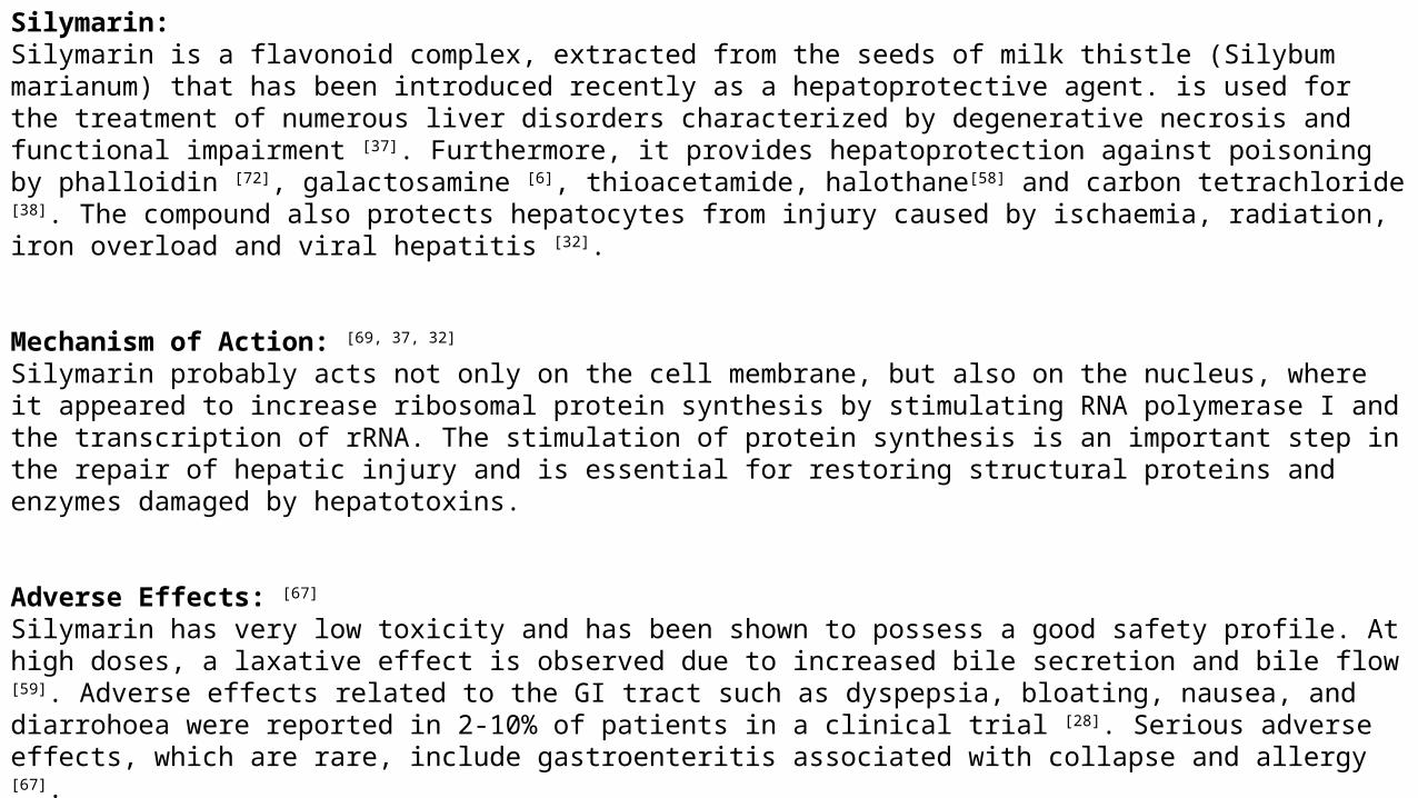
Silymarin:Silymarin is a flavonoid complex, extracted from the seeds of milk thistle (Silybum marianum) that has been introduced recently as a hepatoprotective agent. is used for the treatment of numerous liver disorders characterized by degenerative necrosis and functional impairment [37]. Furthermore, it provides hepatoprotection against poisoning by phalloidin [72], galactosamine [6], thioacetamide, halothane[58] and carbon tetrachloride [38]. The compound also protects hepatocytes from injury caused by ischaemia, radiation, iron overload and viral hepatitis [32].
Mechanism of Action: [69, 37, 32]
Silymarin probably acts not only on the cell membrane, but also on the nucleus, where it appeared to increase ribosomal protein synthesis by stimulating RNA polymerase I and the transcription of rRNA. The stimulation of protein synthesis is an important step in the repair of hepatic injury and is essential for restoring structural proteins and enzymes damaged by hepatotoxins.
Adverse Effects: [67]
Silymarin has very low toxicity and has been shown to possess a good safety profile. At high doses, a laxative effect is observed due to increased bile secretion and bile flow [59]. Adverse effects related to the GI tract such as dyspepsia, bloating, nausea, and diarrohoea were reported in 2-10% of patients in a clinical trial [28]. Serious adverse effects, which are rare, include gastroenteritis associated with collapse and allergy [67].

Molecular Studies: [29, 40, 60,]
A gene is a region of DNA that encodes a functional RNA or protein product, and is a molecular unit of heredity.Gene expression: Gene expression is the process by which information from a gene is used in the synthesis of a functional gene product. These products are often proteins, but in non-protein coding genes such as rRNA genes or tRNA genes, the product is a structural or housekeeping RNA. In addition, small non-coding RNAs (miRNA, piRNA) and various classes of long non coding RNAs are involved in a variety of regulatory functions. When studying gene expression with real-time polymerase chain reaction (PCR), scientists usually investigate changes – increases or decreases – in the expression of a particular gene or set of genes by measuring the abundance of the gene-specific transcript. The investigation monitors the response of a gene to treatment with a compound or drug of interest, under a defined set of conditions. Gene expression studies can also involve looking at profiles or patterns of expression of several genes. Whether quantitating changes in expression levels or looking at overall patterns of expression, real-time PCR is used by most scientists performing gene expression.
Real Time Polymerase Chain Reaction (RT-PCR):- [8, 62, 60]
Real-time PCR, also known as quantitative reverse transcription PCR (RT-qPCR) and quantitative PCR (qPCR)—is one of the most powerful and sensitive gene analysis techniques available. It is used for a broad range of applications including quantitative gene expression analysis, genotyping, copy number, drug target validation, biomarker discovery, pathogen detection, and measuring RNA interference. Real-time PCR measures PCR amplification as it occurs, so that it is possible to determine the starting concentration of nucleic acid. In traditional PCR, which is based on end-point detection, results are collected after the reaction is complete, making it impossible to determine the starting concentration of nucleic acid. Every real-time PCR contains a fluorescent reporter molecule, a TaqMan® probe or SYBR® Green dye, for exaple—to monitor the accumulation of PCR product. As the quantity of target amplicon increases, so does the amount of fluorescence emitted from the fluorophore.

Advantages of real-time PCR include: [8, 62, 60]
• Generation of accurate quantitative data,
• Increased dynamic range of detection,
• Increased precision to detect smaller fold changes,
• Increased throughput.
Genes responsible for Liver Disease: [8, 62, 60]
Some of the hepatic genes are enlisted below:
1) Proliferation and cell death:
1. c-myc proto-oncogene protein
2. Wee1/p87
2) Metabolism:
3. alcohol sulfotransferase 1
4. hepatic lipase
3) DNA damage/Stress:
1. GADD153 (growth arrest and DNA-damage-inducible protein)
2. GADD45
4) Extra cellular Material/cellular skeleton:
3. Integrin beta 2
4. Vimentin

There are three phases in a basic PCR run: [8, 62, 60]
• Exponential – Exact doubling of product occurs at every cycle (assuming 100% reaction efficiency). Exponential amplification
occurs because all of the reagents are fresh and available, the kinetics of the reaction push the reaction to favor doubling of
amplicon.
• Linear (High Variability) – As the reaction progresses, some of the reagents are consumed as a result of amplification. The
reactions start to slow down and the PCR product is no longer doubled at each cycle.
• Plateau (End-Point: Gel detection for traditional methods) – The reaction has stopped, no more products are made, and if left
long enough, the PCR products begin to degrade. Each tube or reaction plateaus at a different point, due to the different reaction
kinetics for each sample. These differences can be seen in the plateau phase. The plateau phase is the end point, where traditional
PCR takes its measurement.

Need ofWork

Gallic acid (GA) is a phenolic plant secondary metabolite which provides desirable health benefits beyond basic nutrition.
Epidemiological evidence suggests that consumption of a diet rich in GA is beneficial to human health, including anti-
inflammatory, antimicrobial, anti-allergic, and antitumor activity and antioxidant activity. GA possesses significant antioxidant
activity and protect the liver from the harmful effects of free radicals that are formed as a result of various metabolic
processes in the body.
Studies suggest that GADD153 gene is related to Apoptosis. Regular consumption of alcohol and taking higher
concentration of regular paracetamol causes damage to liver. Hence the present study was designed for pre-clinical &
molecular evaluation of Hepatoprotective Activity in Combination with Gallic Acid.

Aims &Objectives
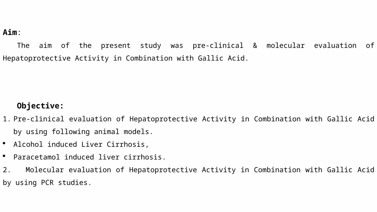
Aim:
The aim of the present study was pre-clinical & molecular evaluation of Hepatoprotective Activity in Combination with
Gallic Acid.
Objective:
1. Pre-clinical evaluation of Hepatoprotective Activity in Combination with Gallic Acid by using following animal models.
Alcohol induced Liver Cirrhosis,
Paracetamol induced liver cirrhosis.
2. Molecular evaluation of Hepatoprotective Activity in Combination with Gallic Acid by using PCR studies.

Plan ofWork

1. Collection of Drugs and Chemicals
2. Procurement of animals & Permission to carryout animal experiments
3. Dose selection
4. Pharmacological evaluation for hepatoprotective activity
1. Alcohol Induced Liver Cirrhosis,
2. Paracetamol Induced Liver Cirrhosis.
5. Statistical analysis of data and interpretation of results
6. Discussion
7. Summary and Conclusion
8. Future Scope
Materials& Methods
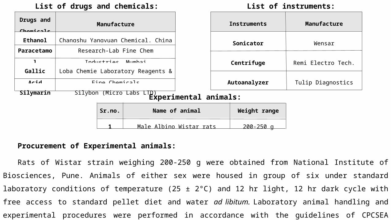
List of drugs and chemicals:
Drugs and
ChemicalsManufacture
Ethanol Changshu Yangyuan Chemical, China
Paracetamol Research-Lab Fine Chem Industries, Mumbai
Gallic AcidLoba Chemie Laboratory Reagents & Fine
Chemicals
Silymarin Silybon (Micro Labs LTD)
List of instruments:
Instruments Manufacture
Sonicator Wensar
Centrifuge Remi Electro Tech.
Autoanalyzer Tulip Diagnostics
Experimental animals:Sr.no. Name of animal Weight range
1 Male Albino Wistar rats 200-250 g
Procurement of Experimental animals:
Rats of Wistar strain weighing 200-250 g were obtained from National Institute of Biosciences, Pune. Animals of either sex
were housed in group of six under standard laboratory conditions of temperature (25 ± 2°C) and 12 hr light, 12 hr dark cycle with
free access to standard pellet diet and water ad libitum. Laboratory animal handling and experimental procedures were performed
in accordance with the guidelines of CPCSEA (198/99/CPCSEA) and experimental protocol was approved by Institutional Animal
Ethics Committee (DYIPSR/IAEC/14-15/P-11).

6.2 Pharmacological evaluation for hepatoprotective activity:
6.2.1 To evaluate the hepatoprotective activity of Gallic Acid using alcohol induced liver cirrhosis: [39, 49]
Groups Treatment Observations
I- Normal Sterile Water Body Weight, Liver Weight, SGPT, SGOT, GT Total Protein, Albumin, Bilirubin, Alkaline Phosphatase, Histopathology, Molecular Study(GADD153, c-myc, Weel/p87)
II- Alcohol 30% 30% Ethanol (2ml)
III-Alcohol30%+Silymarin 30% Ethanol (2 ml) + Silymarin (50 mg/kg p.o.)
IV- Alcohol 30% +Low Gallic Acid
30% Ethanol (2ml) + Gallic Acid (25 mg/kg p.o.)
V-Alcohol 30% +High Gallic Acid
30% Ethanol (2ml) + Gallic Acid (50 mg/kg p.o.)
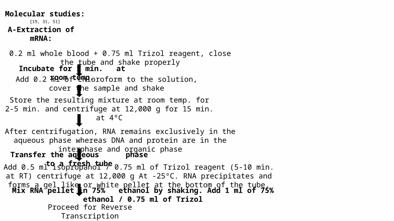
Molecular studies: [19, 31, 51]
A-Extraction of mRNA:
0.2 ml whole blood + 0.75 ml Trizol reagent, close the tube and shake properly
Incubate for 5 min. at room temp.Add 0.2 ml of chloroform to the solution, cover the sample and shake
Store the resulting mixture at room temp. for 2-5 min. and centrifuge at 12,000 g for 15 min. at 4ºC
After centrifugation, RNA remains exclusively in the aqueous phase whereas DNA and protein are in the interphase and organic phase
Transfer the aqueous phase to a fresh tube
Add 0.5 ml isopropanol / 0.75 ml of Trizol reagent (5-10 min. at RT) centrifuge at 12,000 g At -25ºC. RNA precipitates and forms a gel like or white pellet at the bottom of the tube.
Mix RNA pellet in 75% ethanol by shaking. Add 1 ml of 75% ethanol / 0.75 ml of Trizol
Proceed for Reverse Transcription

B- DNA treatment:1) Set up the DNAse digestion reaction as follows:RNA in water or TE buffer 1-8 µl
RQ1 RNAse-Free DNAse 10x Reaction buffer 1 µl
RQ1 RNAse-free DNAse 1 U/ µg RNA
Nuclease-free water to a final volume 10 µl
2) Incubate at 37ºC for 30 min.3) Add 1 µl of RQ1 DNAse Stop Solution to terminate the reaction.4) Incubate at 65ºC for 10 minutes to inactivate the DNAse.5) Add all, or a portion of, the treated RNA to the RT-PCR.
C-Synthesis of cDNA:
1) Place reverse transcriptase enzyme on ice.2) Thaw 10x buffer, random decamers, dNTP and place it on ice.3) Mix all reagents step by step as mentioned in the table below-4) Mix gently, spin briefly.5) Incubate in a thermal cycle at-• 44ºC for 1 hrs.• 92ºC for 10 min to inactivate the reverse transcriptase.6) Proceed to PCR.

6.2.2: To evaluate the hepatoprotective activity of Gallic Acid in paracetamol induced liver cirrhosis: [42, 49]
Groups Treatment Observations
I- Normal Sterile Water Body Weight, Liver Weight, SGPT, SGOT, GT Total Protein, Albumin, Bilirubin, Alkaline Phosphatase, Histopathology, Molecular Study
(GADD153, c-myc, Weel/p87)
II- Alcohol 30% Paracetamol (2 g/kg)
III-Alcohol30%+
Silymarin Paracetamol (2 g/kg) + Silymarin (25 mg/kg p.o.)
IV-Alcohol 30%+
Low Gallic AcidParacetamol (2 g/kg) + Gallic Acid (25 mg/kg p.o.)
V-Alcohol 30%+
High Gallic AcidParacetamol (2 g/kg) + Gallic Acid (50 mg/kg p.o.)

Statistical Analysis Data Analysis:
Arithmetic means of the values of readings were calculated for each experiments. The results obtained were used
for statistical analysis using INTA Software. The data obtained from various models of hepatotoxicity in rat
experiments were subject to Analysis of Variance (ANOVA) followed by Dunnett’s t-test using INTA software.
Values of p ˂ 0.01 was considered statistically significant.

Results

1st day
7th day
14th day
21st day
28th day
35th day
42nd day
49th day
56th day
0
50
100
150
200
250
300
350
ns ns#
###
#
##
##
##
* ** *** **
***** * * * **
** **
* * * * **** **
Alcohol Induced Liver Cirrhosis-Body Weight
Normal Alcohol (30%) Alc + Sily Alc + Low GA Alc + Low GA
Days
Bod
y W
eigh
t (M
ean
± SE
M)
Graph 7.1.1 Effect of Alcohol induced Liver Cirrhosis on body weight of animals:

Graph 7.1.2 Effect of Alcohol induced Liver Cirrhosis on liver weight:
Normal Alc(30%) Alc(30%+Sily
Alc(30%)+Low GA
Alc(30%)+High GA
0
2
4
6
8
10
12
14
16 ##
** ****
Alcohol Induced Liver Cirrhosis-Liver Weight
Groups
Wt.
of L
iver
(Mea
n ±
SEM
)

Fig 7.1.1 Histopathological representation of Liver in effect of Alcohol induced Liver Cirrhosis(H & E stain, 40X):
Normal Liver Alcohol (30%) Alcohol (30%) + Silymarin
Alcohol (30%) + Low Gallic Acid Alcohol (30%) + High Gallic Acid
vascular changescellular infiltrationdegeneration and necrosis of cell
fatty changes

Graph 7.1.3 Effect of Alcohol induced Liver Cirrhosis on different Biochemical Estimation:
Normal Alc 30% Alc30% +Sily Alc30% +Low GA
Alc30% +High GA
0
50
100
150
200
250
300
##
**
**
ns
##
**
**
**
SGPT
SGOT
Normal Alc 30% Alc30% +Sily Alc30% +Low GA
Alc30% +High GA
0
50
100
150
200
250
##
*
**
**
GGT
Normal Alc 30% Alc30% +Sily Alc30% +Low GA
Alc30% +High GA
0
50
100
150
200
250
300
350
400
450
500
##**
****
Total Proteins
Normal Alc 30% Alc30% +Sily Alc30% +Low GA
Alc30% +High GA
0
50
100
150
200
250
300
350
##
**
**
**
Alkaline Phosphatase
Normal Alc 30% Alc30% +Sily Alc30% +Low GA
Alc30% +High GA
0
2
4
6
8
10
12
14
16
18
20
##
**
**
ns
Albumin
Normal Alc 30% Alc30% +Sily Alc30% +Low GA
Alc30% +High GA
0
10
20
30
40
50
60
70
80
90
100
## **
**
**
Bilirubin
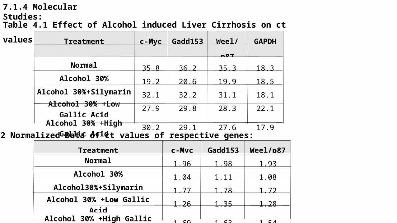
7.1.4 Molecular Studies:
Table 4.1 Effect of Alcohol induced Liver Cirrhosis on ct values of respective genes:
Treatment c-Myc Gadd153 Weel/p87 GAPDH
Normal 35.8 36.2 35.3 18.3
Alcohol 30% 19.2 20.6 19.9 18.5
Alcohol 30%+Silymarin 32.1 32.2 31.1 18.1Alcohol 30% +Low Gallic
Acid27.9 29.8 28.3 22.1
Alcohol 30% +High Gallic Acid
30.2 29.1 27.6 17.9
Table 4.2 Normalized Data of ct values of respective genes:Treatment c-Myc Gadd153 Weel/p87
Normal 1.96 1.98 1.93
Alcohol 30% 1.04 1.11 1.08
Alcohol30%+Silymarin 1.77 1.78 1.72
Alcohol 30% +Low Gallic Acid 1.26 1.35 1.28
Alcohol 30% +High Gallic Acid 1.69 1.63 1.54

Graph 7.2.1 Effect of Paracetamol induced Liver Cirrhosis on body weight of animals:
1st day 7th day 14thday0
20
40
60
80
100
120
140
ns*
**#
##
ns **ns
*
Paracetamol Induced Liver Cirrhosis-Body Weight
Normal Para Para+Sily Para+LowGA Para+High GA
Days
Bod
y W
eigh
t (M
ean
± SE
M)

Graph 7.2.2 Effect of Paracetamol induced Liver Cirrhosis on liver weight of
animals:
Normal Para Par+Sily Para+Low GA Para+High GA0
2
4
6
8
10
12
14
11.27
##
**** **
Paracetamol Induced Liver Cirrhosis-Liver Wight
Groups
Ave
rage
Wt.
of L
iver
(Mea
n ±
SEM
)

Fig 7.2.1 Histopathological representation of Liver in effect of Paracetamol induced Cirrhosis (H & E stain, 40X):
vascular changescellular infiltrationdegeneration and necrosis of cell
fatty changes
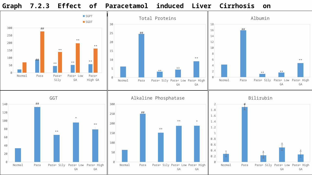
Graph 7.2.3 Effect of Paracetamol induced Liver Cirrhosis on different Biochemical Estimation:
Normal Para Para+ Sily Para+ Low GA
Para+ High GA
0
50
100
150
200
250
300
##** ** **
##
**
**
**
SGPT
SGOT
Normal Para Para+ Sily Para+ Low GA Para+ High GA0
20
40
60
80
100
120
140 ##
**
*
**
GGT
Normal Para Para+ Sily Para+ Low GA
Para+ High GA
0
5
10
15
20
25
30
##
****
**
Total Proteins
Normal Para Para+ Sily Para+ Low GA
Para+ High GA
0
50
100
150
200
250
300
##
**
** *
Alkaline Phosphatase
Normal Para Para+ Sily Para+ Low GA Para+ High GA
0
2
4
6
8
10
12
14
16
18
##
** **
**
Albumin
Normal Para Para+ Sily Para+ Low GA
Para+ High GA
0
0.2
0.4
0.6
0.8
1
1.2
1.4
1.6
1.8
2 #
*
*
*
Bilirubin

Discussion

Alcohol induced Liver damage:
• The increased level of SGPT, SGOT & ALP after 56 days of continual feeding with high concentration 30% of ethanol were indications for alcohol intoxication to the liver. In the group of animals treated with Alcohol (30%) in combination with Gallic Acid, there was significant decrease in SGPT, SGOT & ALP as compared to ethanol treated animals.
• Results from histological images showed that accumulations of fatty droplets in the hepatocytes provided clear evidence that the pre-induction with 30% of ethanol induced liver damage, including loss of cell membrane integrity, accumulation of fatty acids, and necrotic cell death in the mice. While in the groups of animals treated with Alcohol (30%) in combination with Gallic Acid lesser liver damage was observed along minimal necrosis which might be supportive for hepatoprotective activity.
• While in case of other biochemical parameters like GGT, Total Proteins, Albumin & Bilirubin there was increase in the level of this parameters in the group of Alcohol (30%) in combination with Gallic Acid in comparison to that of plain Alcohol (30%).
• The increase in the body weight & liver weight after 56 days of continual feeding with high concentration of ethanol (30%) were indications for alcohol intoxication to the liver. In the group of animals treated with Alcohol (30%) in combination with Gallic Acid, there was significant decrease in body weight & liver weight as compared to ethanol treated animals.

Molecular Studies:• We studied the expression profile of 1 stress related gene, viz., GADD153 and 2 genes associated with expression of heat shock
proteins, cell proliferation and cell death, i.e., c-myc and Wee1/p87 respectively. They are over expressed during events of liver injury making them potential markers for monitoring liver recovery process at the molecular level. These genes are compared with the corresponding value obtained from the GAPDH gene which was used as the normalizer or house-keeping gene.
• In real time PCR, Ct (threshold cycle) is the intersection between an amplification curve and a threshold line and is relative value of the concentration of target in the reaction mixture. The Ct value is inversely proportional to the target concentration and therefore higher value for Ct is associated with lower amount of target complimentary DNA in case of gene expression studies.
• As expected, the Ct reduced in cases of liver injury caused due to administration of alcohol (30%), while in the case of Gallic acid along with alcohol (30%), there was reduced expression of all the 3 genes as compared to untreated animals that might be either due to reduced liver injury caused by way of slower progression towards liver tissue damage or simultaneous recovery process that occurred due to treatment with Gallic acid.
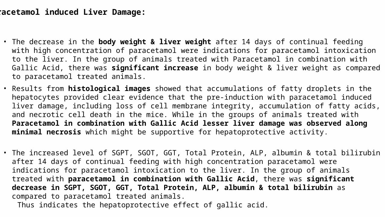
Paracetamol induced Liver Damage:
• The decrease in the body weight & liver weight after 14 days of continual feeding with high concentration of paracetamol were indications for paracetamol intoxication to the liver. In the group of animals treated with Paracetamol in combination with Gallic Acid, there was significant increase in body weight & liver weight as compared to paracetamol treated animals.
• Results from histological images showed that accumulations of fatty droplets in the hepatocytes provided clear evidence that the pre-induction with paracetamol induced liver damage, including loss of cell membrane integrity, accumulation of fatty acids, and necrotic cell death in the mice. While in the groups of animals treated with Paracetamol in combination with Gallic Acid lesser liver damage was observed along minimal necrosis which might be supportive for hepatoprotective activity.
• The increased level of SGPT, SGOT, GGT, Total Protein, ALP, albumin & total bilirubin after 14 days of continual feeding with high concentration paracetamol were indications for paracetamol intoxication to the liver. In the group of animals treated with paracetamol in combination with Gallic Acid, there was significant decrease in SGPT, SGOT, GGT, Total Protein, ALP, albumin & total bilirubin as compared to paracetamol treated animals.
Thus indicates the hepatoprotective effect of gallic acid.
Hence gallic acid in combination with alcohol and paracetamol has shown hepatoprotective activity by restoring the levels of all the biochemical parameters in alcohol and paracetamol liver damage.
This study is also supported by molecular studies, where there was reduction in expression of 3 genes i.e. c-MYC, GADD153 and Weel/p87 upon administration of Gallic acid along with alcohol in animals as compared to untreated animals .
Therefore this study supports our hypothesis that gallic acid when given in combination with liver damaging alcohol and paracetamol, shown hepatoprotective effect and thus may prolong the damage to liver.

Summary &conclusion

Summary:
The present study was designed for pre-clinical & molecular evaluation of Hepatoprotective Activity in Combination with Gallic Acid.
In alcohol & paracetamol induced liver damage, gallic acid in combination with both in showed significant restoration of serum marker enzymes SGOT, SGPT, GGT, ALP along with Total Protein, albumin and total bilirubin which increased in plain alcohol & paracetamol liver damage.
Conclusion:
Gallic acid in combination with alcohol and paracetamol has shown hepatoprotective activity by restoring the levels of all the biochemical parameters in alcohol and paracetamol induced liver damage.
This study is also supported by molecular studies, where there was reduction in expression of 3 genes i.e. c-MYC, GADD153 and Weel/p87 upon administration of Gallic acid along with alcohol in animals as compared to untreated animals.

FutureScope

As results have shown that gallic acid reduces the deleterious effects of alcohol and paracetamol.
In regard to future perspective we would like to suggest to make a formulation containing alcohol or paracetamol with different doses of gallic acid and conduct clinical trial on larger scale and also work for a better formulation of the two.

References
1. Armbrust T, Batusic D, Xia L, Ramadori G. Early gene expression of hepatocyte growth factor in mononuclear phagocytes of rat liver after administration of carbon tetrachloride. Liver 2002; 22, 486–494.
2. Ávila DS, et al. Hepatoprotective activity of a vinylic telluride against acute exposure to acetaminophen. European Journal of Pharmacology 2011; 661(90), 92–101.
3. AydIn AF, Küskü-Kiraz Z, Dogru-Abbasoglu S, Güllüoglu M, Uysal M, Koçak-Toker N. Effect of carnosine against thioacetamide induced liver cirrhosis in rat. Peptides 2010; 31(1), 67–71.
4. Balister WF, Shaw LM. Fundamentals of Clinical Chemistry, 3rd edition, W.B. Saunders Co, Philadelphia 1982; 729-761.5. Bansal AK, Bansal M, Soni G, Bhatnagar D. Protective role of Vitamin E pre-treatment on nitrosodiethylamine induced
oxidative stress in rat liver. Chem Biol Interact, 2005; 156(2), 101–111.6. Barbarino F, Neumann E, Deaciuc J, et al., Effect of silymarin on experimental liver lesions. Rev Roum Med Intern, 1981; 19,
347-57.7. Barouki, R., Chobert MN, Finidori J, Aggerbeck M, Nalpas B, Hanoune J. Ethanol effects in a rat hepatoma cell line:
induction of gamma-lutamyltransferase. Hepatology 1983; 3(3), 323-339.8. Bartosiewicz MJ, Jenkins D, Penn S, Emery J, Buckpitt A. Unique gene expression patterns in liver and kidney associated with
exposure to chemical toxicant. J. Pharmacol. Exp. Ther. 2001; 297, 895–905. 9. Bhattacharjee R, Sil PC. The protein fraction of Phyllanthus niruri plays a protective role against acetaminophen induced
hepatic disorder via its antioxidant properties. Phytotherapy Research 2006; 20 (7), 595–601.10. Bowman WC, Rand M J. Textbook of Pharmacology. Blackwell scientic publication, Oxford London 1982; 2nd edition, 34-39.11. Cederbaum AI, Lu Y, Wu D. Role of oxidative stress in alcohol-induced liver injury. Arch Toxicol 2009; 83(6); 519–548.12. Chen YH, Lin FY, Liu PL, et al. Antioxidative and hepatoprotective effects of magnolol on acetaminophen-induced liver
damage in rats. Archives of Pharmacal Research 2009, 32 (2), 221–228.13. Craig JR, Alcoholic Hepatitis. Anderson’s Pathology, 9th edition, The C.V. Mosby Company, St. Louis 1990; 1232-36.14. Dancygier H, Seitz HK, Mueller S. Alcoholic Liver Disease. Springer, Berlin, Germany, 2010.15. Dancygier H, Strassburg CP. Hepatic Drug Metabolism and Drug Toxicity. Springer, Berlin, Germany, 2010.

16. Das SK, Vasudevan DM. Alcohol-induced oxidative stress. Life Sciences 2007; 81 (3), 177–187.17. Davila JC, Lenherr A, Acosta D. Protective effect of flavonoids on drug-induced hepatotoxicity in vitro. Toxicology 1989;
267-86.18. Dienstag JL, Wands JR, Isselbacher KJ. Toxic and induced hepatitis, Harrison’s Principles of Internal Medicine, Twelfth
edition, MCGraw-Hill, New York; 1333-37.19. Farr S, Dunn R T. Concise review: Gene expression applied to toxicology. Toxicol. Sci. 1999; 50, 1–9. 20. Friedman SL, McQuaid KR, Grendell JH. Current diagnosis & treatment in gastroenterology. Lang Medical Books/McGraw-
Hill; New York, 2002.21. Fromenty B, Pessayre D. Impaired mitochondrial function in microvesicular steatotis. Effect of drugs, ethanol, hormones and
cytokines 1997; 26, 43-53.22. Gilani AH, Janbaz KH. Preventive and curative effects of Artemisia absinthium on acetaminophen and CCl4 induced
hepatotoxicity. General Pharmacology 1995; 26 (2), 309–315.23. Godkar PB, Godkar PD. Textbook of medicinal Laboratory technology. 2nd edition, Bhalani Publication house, Mumbai; 331-
351.24. Guyton AC, Hall JE. Textbook of Medicinal Physiology, 10th edition, A Harcourt Publishers International Company, Singapore.25. Hall PM. Alcoholic Liver Disease. Elsevier Health Sciences 2007; Philadelphia, USA, 6.26. Hinson JA, Roberts DW, James LP. Mechanisms of acetaminophen-induced liver necrosis in Adverse Drug Reactions,
Handbook of Experimental Pharmacology 2010; 369–405.27. Hong RW, Rounds JD, Helton WS, Robinson MK, Wilmore DW. Glutamine preserves liver glutathione after lethal hepatic
injury. Annals of Surgery 1992; 215 (20), 114–119.28. Jacobs PB, Dennehy C, Ramirez G, Sapp J, Lawrence VA. Milk thistle for the treatment of liver disease. A systematic review
and meta-analysis. Am J Med 2002; 113,506-15.29. Jiang Y, Liu J, Waalkes M, Kang YJ. Changes in the gene expression associated with carbon tetrachloride-induced liver
fibrosis persist after cessation of dosing in mice. Toxicol Sci 2004; 79(2), 404-10.

30. Kaplowitz N. Drug Induced Hepatotoxicity. Ann Intern Med 1986; 104,826.31. Liu J, Li C, Waalkes MP, Clark J, Myers P, Saavedra JE, Keefer, L K. The nitric oxide donor, V-PYRRO/NO, protects against
acetaminophen-induced hepatotoxicity in mice. Hepatology 2003; 37, 324–333. 32. Luper S. A review of plants used in the treatment of liver disease. Altern Med Rev 1998; 3, 410-21.33. Luster MI, Simeonova PP, Gallucci RM, Matheson JM, Yucesoy B. Immunotoxicology: role of inflammation in chemical-
induced hepatotoxicity. International Journal of Immunopharmacology 2000; 22(12), 1143–1147.34. Mathurin P, et al. Corticosteroids improve short-term survival in patients with severe alcoholic hepatitis: meta-analysis of
individual patient data. Gut 60 2011; 255–260.35. McClain CJ, Kromhount JP. Protestation of acetaminophen hepatotoxicity by alcohol. JAMA 1980; 244, 251-3.36. Monga SPS, Apte U, Krishnamurthy P. Detoxification Functions of the Liver. Springer.37. Morazzoni P, Bombardelli E, Silybum marianum (Carduus marianus). Fitoterapia 1995; 3(42).38. Mourelle M, Muriel P, Favari L, et al. Prevention of CCl4-induced liver cirrhosis by silymarin. Fundam Clin Pharmacol 1989;
3, 183-91.39. Muhammad Habib-ur-Rehman, et al. Ethanol Induced Hepatotoxicity in Albino Rats. Journal of the College of Physicians and
Surgeons Pakistan 2011; 21 (10), 642-643.40. Mukai T, Mera K, Nishida K, Nakashima M, Sasaki H, Sakaeda T, Nakamura J. A novel method for preparation of animal
models of liver damage: Liver targeting of carbon tetrachloride in rats. Biol Pharm Bull 2002; 25, 1494–1497. 41. Muriel P, Mourelle M. Prevention by silymarin of membrane alterations in acute CCl4 liver damage. J Appl Toxicol 1990; 10,
275-9.42. Nayeema Akther, et al. Hepatoprotective activity of Marrubium vulgare against paracetamol induced toxicity. Department of
Medicinal and Elemental Toxicology; Faculty of Science, Jamia Hamdard, New Delhi 110062, India.43. Nkosi CZ, Opoku AR, Terblanche SE. Effect of pumpkinseed (Cucurbita pepo) protein isolate on the activity levels of certain
plasma enzymes in CCl4-induced liver injury in lowprotein fed rats. Phy the Res 2005; 19, 341–345.44. Nordman R. Alcoholic and antioxidant systems. Alcohol 1994; 29, 513-552.
45. O’Shea RS, Dasarathy S, McCullough AJ, Alcoholic liver disease. Gastroenterol 2010; 105, 14–32. 46. Parakrama C, Clieve R. Consise Pathology, 3rd edition, MCGraw-Hill,Medical Publishing Division.47. Prajapati ND, Purohit SS, Sharma AK, Kumar T. A Handbook of Medicinal Plant. Jodhpur Agrobios 2003; 203-15.48. Ramachandran R, Kakar S. Histological patterns in drug-induced liver disease. Journal of Clinical Pathology 2009; 62 (6),
481–492.49. Ravichandran Senthilkumar et.al. Hepatoprotective effect of Rhodiola imbricata rhizome against paracetamol-induced liver
toxicity in rats. Saudi Journal of Biological Sciences 2014; 21 (5), 409–416.50. Robins and Cotran et al. Pathologic basis of Disease, 7th edition, Elsevier India Pvt. Ltd., New Delhi.51. Ruepp S, Tonge RP, Wallis NT, Davison MD, Orton TC, Pognan F. Genomic and proteomic investigations of acetaminophen
(APAP) toxicity in mouse in vivo. Toxicol. Sci. 2000; 54, 384 (abstract).52. Runyon BA. AASLD Practice Guidelines Committee: Management of adult patients with ascites due to cirrhosis: an update.
Hepatology 2011; 49 (6), 2087-2107.53. Said Y, Salem M, Mouelhi L, et al. Correlation between liver biopsy and fibro test in the evaluation of hepatic fibrosis in
patients with chronic hepatitis. C. Tunis Med 2010; 88(8), 573-578. 54. Saroj BK, Mani D, Mishra SK. Scientific validation of polyherbal hepatoprotective formulation against paracetamol induced
toxicity. Asian Pacific Journal of Tropical Biomedicine, 2012; 2 (3), S1742–S1746.55. Seeff LB, Adler E. Acetaminophen hepatotoxicity in alcoholics: A therapeutic misadventure. Ann Int. Med 1986; 104, 399-
404.56. Shah M et al. Evaluation of the effect of aqueous extract from powders of root, stem, leaves and whole plant of
phyllanthusdebilis against CCL4 induced rat liver dysfunction. Indian Drugs 2002; 39, 333-337.57. Sherlock S. Diseases of the Liver and Biliary System, 7th edition, Blackwell scientific publication, Oxford London, 1985, 304-
329.58. Siegers CP, Frühling A, Younes M. Influence of dithiocarb, (+) catechin and silybine on hepatotoxicity in the hypoxic rat
model. Acta. Pharmacol Toxicol 1983; 53, 125-959. Silybum marianum (Milk Thistle). Altern Med Rev 1999; 4, 272-4.
60. Simeonova PP, Gallucci R M, Hulderman T, Wilson R, Kommineni C, Rao M, Luster M I. The role of tumor necrosis factor-alpha in liver toxicity, inflammation, and fibrosis induced by carbon tetrachloride. Toxicol Appl Pharmacol 2001; 177, 112–120.
61. Sonnenbichler J, Zetl I. Biochemical effects of the flavonolignane silibinin on RNA, protein and DNA synthesis in rat livers. Cody V, Middleton E, Harborne JB. Plant flavonoids in biology and medicine: biochemical, pharmacological and structure-activity relationship. New York: Alan R Liss Inc., 1986, 319-31.
62. Stoyanovsky DA, Cederbaum AI. Metabolism of carbon tetrachloride to trichloromethyl radical: An ESR and HPLC-study. Chem Res Toxicol 1999; 12, 730–736.
63. Subramanian M, Balakrishnan S, Chinnaiyan SK, Sekar VK, Chandu AN. Hepatoprotective effect of leaves of Morinda tinctoria Roxb. against paracetamol induced liver damage in rats. Drug Invention Today 2013;5 (3),223–228.
64. Thomas MB, Jaffe D, Choti MM, et al. Hepatocellular carcinoma: con sensus recommendations of the National Cancer Institute Clinical Tri als Planning Meeting. J Clin Oncol 2010; 28(25); 3994-4005.
65. Tietz NW. In: Fundamentals of Clinical Chemistry, 3rd edition, W.B. Saunders Co, 1987; 391.66. Tortora GJ, Grabowski SR. Principles of Anatomy and Physiology, 7th edition, Harper Collins Collage publishers, New York,
792-795.67. Tyutyulkova N, Gorantcheva U, Tuneva S, et al. Effect of silymarin on the microsomal glycoprotein and protein biosynthesis
in liver of rats with experimental galactosamine hepatitis. Methods Exp Clin Pharmacol 1983; 5, 181-4.68. Valenzuela A, Garrido A. Biochemical bases of the pharmacological action of the flavonoid silymarin and of its structural
isomer silibinin. Biol Res 1994; 27, 105-12.69. Vogel G, Trost W, Braatz R, et al. Untersuchungen zu pharmakodynamik, angriffspunkt und wirkungsmechanismsus von
silymarin, dem antihepatotoxischen prinzip aus Silybum mar. (L.) gaertn. Arzneimittelforschung 1975; 25, 82-9.70. Vogel G, Trost W. Zur anti-phalloidinaktivität der silymarine silybin und disilybin. Arzneimittelforschung 1975; 25, 392-3.71. Wang S, Nagrath D. Liver Tissue Engineering.-Biomaterials for Tissue Engineering Applications: A Review of the Past and
Future Trends 2010, 14,389.

72. Williams AJ, Barry RE. Free radical generation by neutrophils: A potential mechanisms of cellular injury in acute alcoholic hepatitis. GUT, 1987; 28, 1157-1161.
73. World Health Organization/ Global Health Observatory/ Information System on Alcohol and Health.74. Yahya F, Mamat SS, Kamarolzaman FF. Hepatoprotective activity of methanolic extract of Bauhinia purpurea leaves against
paracetamol-induced hepatic damage in rats. Evidance-Based and Complimentary Alternative Medicine 2013.75. Yanpallewar SU, Sen S, Tapas S,Kumar M, Raju SS, Acharya SB. Effect of Azadirachta indica on paracetamol-induced
hepatic damage in albino rats. Phytomedicine 2003; 10 (5), 391–396.76. Yen FL, Wu TH, Lin LT, Lin CC. Hepatoprotective and antioxidant effects of Cuscuta chinensis against acetaminophen-
induced hepatotoxicity in rats. Journal of Ethnopharmacology 2007; 111 (1), 123–128. 77. Yin M, et al. Essential role of tumor necrosis factor alpha in alcohol-induced liver injury in mice. Gastroenterology 1999; 117,
942–952.78. Zou Y, Lu Y, Wei D. Antioxidant activity of a flavonoid rich extract of Hypericum perforatum L. J Agr Food Chem 2004; 52,
5032-5039.

52