LISA DU TOIT - SAAPI€¦ · LISA DU TOIT ASSOCIATE PROFESSOR OF PHARMACEUTICAL CHEMISTRY SENIOR...
28
LISA DU TOIT ASSOCIATE PROFESSOR OF PHARMACEUTICAL CHEMISTRY SENIOR SCIENTIST: WITS ADVANCED DRUG DELIVERY PLATFORM RESEARCH UNIT DEPARTMENT OF PHARMACY AND PHARMACOLOGY, WITS UNIVERSITY
Transcript of LISA DU TOIT - SAAPI€¦ · LISA DU TOIT ASSOCIATE PROFESSOR OF PHARMACEUTICAL CHEMISTRY SENIOR...
LISA DU TOIT
ASSOCIATE PROFESSOR OF PHARMACEUTICAL CHEMISTRYSENIOR SCIENTIST: WITS ADVANCED DRUG DELIVERY PLATFORM RESEARCH UNIT
DEPARTMENT OF PHARMACY AND PHARMACOLOGY, WITS UNIVERSITY

• 3D-Printing = a highly futuristic technology until recently.
• Was predominantly used in manufacturing as stereolithography - create almost any object by fusing different materials - layer by layer → physical version of a digital 3D image.
• Over the past 15 years - 3D printing expanded into healthcare industry.
• Fast becoming established as an advanced and transformative manufacturing technique: – creating a solid 3D object from a digital model– on demand– at a relatively low cost– customisable, complex shapes, surfaces and
architectures – diverse materials
• The convenient cost-effective manufacture of personalised pharmaceutical, medical and dental products = ↑↑ advantage over traditional manufacturing methods.
• Opportunity to use it for personalized healthcare.
INTRODUCTION

• Pharmaceutical drug research and development could be improved drastically by 3D printing – cheaper and safer drug testing
• Application of 3D printing to medicine - revolutionary implications - enhancing the quality of patients’ lives.
• Actual and potential applications include:– Customizable 3D-Printed Drugs, Drug Delivery Devices and Implants– Medical Prostheses– Anatomical Models for Surgical Preparation and Surgical Practice– Bioprinting of Tissues and Organs
APPLICATIONS

THE MARKET
• The world demand for 3D printers, materials and software is projected to increase by 21%percent per year and increased to $5 billion in 2017.
• Past → compared to other sectors, 3D printing technology played a minor role in healthcare.Healthcare only accounted for 1.6 percent of all investments made into the $700 million 3Dprinting industry. But - expected to grow to 21% over the next 10 years.
• Future → latest research shows more drastic development for health and medicine. Using 3Dprinting for medical applications could amount to a market value of $2.13 billion by 2020(MarketsandMarkets.com).

3D PRINTING BASICS
• 3D printing is a process that creates a three-dimensional object by building successive layers of raw material.
• Each new layer is attached to the previous one until the object is complete.
• Objects are produced from a digital 3D file, such as a computer-aided design (CAD) drawing or a Magnetic Resonance Image (MRI).
• The flexibility of 3D printing allows researchers to implement changes easily without the need to set up additional equipment or tools.
Example procedure for creating customisable tablets

PERSONALIZED DRUG DOSING
• Personalized 3D printed oral tablets – especially where patients respond to the same drugs indifferent ways.
• Use each patient’s individual information e.g. mass, metabolism, age, race and gender - toproduce their optimal medication dose and drug release rate.
• 3D printing enables printing of tablets as complex construct of layers/combination ofpolymers printed in different shapes - combination of drugs to treat multiple ailmentssimultaneously e.g. a multidrug polypill for cardiac ailments - enhances patient compliance.
• Personalized implants allows for printing implants that match patients' anatomical features -this technique is gaining traction for medical devices e.g. bone grafts.
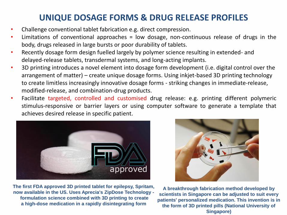
UNIQUE DOSAGE FORMS & DRUG RELEASE PROFILES• Challenge conventional tablet fabrication e.g. direct compression.• Limitations of conventional approaches = low dosage, non-continuous release of drugs in the
body, drugs released in large bursts or poor durability of tablets.• Recently dosage form design fuelled largely by polymer science resulting in extended- and
delayed-release tablets, transdermal systems, and long-acting implants.• 3D printing introduces a novel element into dosage form development (i.e. digital control over the
arrangement of matter) – create unique dosage forms. Using inkjet-based 3D printing technology to create limitless increasingly innovative dosage forms - striking changes in immediate-release, modified-release, and combination-drug products.
• Facilitate targeted, controlled and customised drug release: e.g. printing different polymericstimulus-responsive or barrier layers or using computer software to generate a template thatachieves desired release in specific patient.
The first FDA approved 3D printed tablet for epilepsy, Spritam,
now available in the US. Uses Aprecia's ZipDose Technology -
formulation science combined with 3D printing to create
a high-dose medication in a rapidly disintegrating form
A breakthrough fabrication method developed by
scientists in Singapore can be adjusted to suit every
patients' personalized medication. This invention is in
the form of 3D printed pills (National University of
Singapore)

3D BIOPLOTTING FOR BIOINSPIRED TISSUE ENGINEERING
• 3D Bioplotting is a specialised arm of 3D printing that employs biological materials in computer-aided tissue engineering to create structures for enhanced biomimicry.
• Goal = achieve an effective combination of cell and bioactive components for restoration and/orimprovement of biological functionality.

Bioprinting
Biological componentsMatrix +
‘Bioink’
Layer-by-layer
Applications
Tissue engineering
Organ PrintingOrgan-on-a-chip
• Cells• Growth factors• Drugs
• Biomaterials• Nanostructures• Synthetic or
natural
3D-BIOPLOTTING AT THE WADDP
Design (CAD)

Tissue Engineering of Biomaterials
Fabrication Approaches for Nanostructured Material Design
Biorelevant Nanosized Additives
Implantation/injection at Tissue Defect Site
Natural/ Synthetic Polymers
Ceramics
Bioactive Glass
Hybrid/Composite
Electrospinning3D Printing
Cells
Bioactive Nanoparticles (e.g. CaP, HA, MNPs, nanodiamonds), growth factors
Selective laser sintering
Thermally-induced phase separation Stereolithography
Sol-gel approach
Rapid Prototyping Techniques
3D-BIOPLOTTING AT THE WADDP

• A fixed three drug dose combination of the anti-HIV drugs is required that provides controlledlevels of drugs within the therapeutic window with reduced side effects.
Solution:• Designed a novel 3D-printed (3DP) Fixed-Dose Combination tablet
Key Features:• Humic acid-polyquaternium 10 complex (HA-PQ10) as the bio-ink to achieve controlled release
of three drugs: efavirenz (EFV), tenofovir disoproxil fumarate (TDF) and emtricitabine (FTC) asthe model drug regimen.
EFV, TDF and FTC mixed in one sludge (preferred
configuration)
Separate layers of EFV, TDF and FTC
Conventional anti-HIV FDC
Example of 3DP anti-HIV FDCs
3D-PRINTED CONTROLLED RELEASE TRITHERAPEUTIC TABLET

3DP tablet manufacture by sludge extrusion from cartridge onto the build
platformCompressed air
Polyethylene cartridge
Injection nozzle
3DP tablet
Printing ‘ink’
0 hour 2 hours 4 hours 6 hours
24 hours
Gel layer
Free polymer chains
Interface layer
Dry tablet core
18 hours12 hours8 hours
Time (hours)
0 2 4 6 8 10 12 14 16 18 20 22 24
Concentr
ation (
g/m
L)
0.0
0.2
0.4
0.6
0.8
1.0
1.2
1.4
1.6
1.8
TDF 3DP FDC
TDF Atripla
Time (hours)
0 2 4 6 8 10 12 14 16 18 20 22 24
Co
nce
ntr
atio
n (
g/m
L)
0.0
0.5
1.0
1.5
2.0EFV 3DP FDC
EFV Atripla
Time (hours)
0 2 4 6 8 10 12 14 16 18 20 22 24
Co
nce
ntr
atio
n (
g/m
L)
0
5
10
15
20
25FTC 3DP FDC
FTC Atripla
(a)
(b)
(c)
In vivo drug levels highlighting enhanced relative bioavailability of all 3 anti-HIV drugs
from the 3DP FDC vs. Atripla® Controlled hydration of the 3D-Printed tablet architecture

A 3D BIOPLOTTED PVA-PAA HYDROGEL LOADED-POLYCAPROLACTONE SCAFFOLD FOR THE DELIVERY OF
HYDROPHILIC IN-SITU FORMED SODIUM INDOMETHACIN
• The addition of anti-inflammatory agents to 3D bioplotted structural support scaffolds → acutetreatment of site-specific inflammation + long-term structural support to allow for adequate tissuerepair and rehabilitation.
Solution:• NSAID incorporated into the architecture of the PCL scaffold via a nanostructured semi-IPN
composed of PVA and PAA.
Key Features:• Nano-enhancement: Nanostructured semi-IPNs allow for the production of a relatively dense
hydrogel matrix + strong mechanical properties + more efficient drug loading.• The prepared PCL-PVA-PAA (PPP) scaffold is proposed as a structural support system for load-
bearing tissue damage where inflammation is prevalent.
Nanostructured IPN with sodium indomethacin for incorporation in PCL scaffold

Microscopic images of (a) the 3D-printed PCL scaffold and (b) the PPP scaffold highlighting uniform adhesion of the PVA-PAA hydrogel within the octagonal
structure
2.4
5.6
0.0
4.5
Time (mins)
0 100 200 300 400 500
Dru
g R
ele
ase (
%)
0
20
40
60
80
100
Heig
ht
Incre
ase (
%)
100
120
140
160
180
200
Sodium Indomethacin Release
PVA-PAA Gel Height
Inflammation-sensitive scaffold hydration and release of anti-inflammatory agent
Uniaxial strain testing and tracking of the hydrated PPP scaffold depicting biomimetic physicomechanical attributes
Time (min)
0 100 200 300 400
Shear
Com
ple
x m
odulu
s G
* (P
a)
0
5000
10000
15000
20000
tan d
0.0
0.2
0.4
0.6
0.8
1.0
1.2Shear Complex modulus G*
tan d
Tan δ & the shear complex modulus (G*) approached 1.00 - the hydrogel was fully hydrated with appreciable resistance to
deformation
(a) (b)
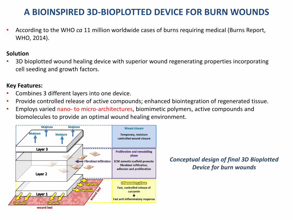
A BIOINSPIRED 3D-BIOPLOTTED DEVICE FOR BURN WOUNDS
Conceptual design of final 3D BioplottedDevice for burn wounds
• According to the WHO ca 11 million worldwide cases of burns requiring medical (Burns Report, WHO, 2014).
Solution• 3D bioplotted wound healing device with superior wound regenerating properties incorporating
cell seeding and growth factors.
Key Features:• Combines 3 different layers into one device.• Provide controlled release of active compounds; enhanced biointegration of regenerated tissue. • Employs varied nano- to micro-architectures, biomimetic polymers, active compounds and
biomolecules to provide an optimal wound healing environment.

Precursor Hydrogel(Partially crosslinked)
• Biocompatible(Bioinert)
• Lys:Ala 60:40• Biocompatible
(Cell adhesive)• Antimicrobial
4-Arm PEG-SGPoly(Lys)n(Ala)m
3D Structure(Partially crosslinked)
3D Scaffold(Fully crosslinked)
Design of the 3D Bioplotted Device for Burn Wounds

Chunky + solution
Too Soft
Too Soft
Chunky + Solution
Chunky + solution

• Traditional treatments for orthopedic defects due = orthopedic implants, allografts, and autografts → challenges of infection, inadequate healing following invasive surgeries, inadequate donors, and morbidity at the donor site.
Solution: • Design a 3D bioplotted pseudo-bone drug delivery system – inserted at the site of bone
fracture (model = clavicle).
Key Features:• The implantable system comprised of a copolymeric biomatrix based on a thermoresponsive
gel (thermogel) - loaded with a statin drug.• Nano-enhancement: Nano-functionalized with growth factors and cells.
Application of the 3D Bioplotter for
fabrication of a jaw bone scaffold

Mechanistic Overview of the 3D Bioplotted Scaffold in Bone Fractures

• 39 thermo-gel designs were loaded with a statin drug and optimized for 3D bioprinting –using MATLAB®.
• Variables of PPF and PF127 were studied in response to duration of release of simvastatinand thermo-gelation temperature.
Rheological evaluation of the 3D bioink in relation to change in temperature = thermogelling
3D representation of the designed formulations using a cubic function surface plot: highest point = optimum polymer
concentrations
Optimization and Analysis

• Ex vivo evaluation of the 3D bioprinted pseudo-bone scaffold via ultrasound and X-ray
• Evaluation on a fracture-induced human clavicle bone → cell proliferation, fracture filling andbone repair
X-ray and ultrasound images on a human clavicle osteoporotic female bone showing bone regeneration
Before fracture After fracture After 3D scaffold treatment
Ex Vivo Evaluation in a Bone Model
SEM visualization of osteoblast-like cell attachment to the
bioplotted scaffold
SEM analysis of scaffold layers → nano-
architecture and inner porous nature = nutrient
diffusion + cell attachment and
proliferation
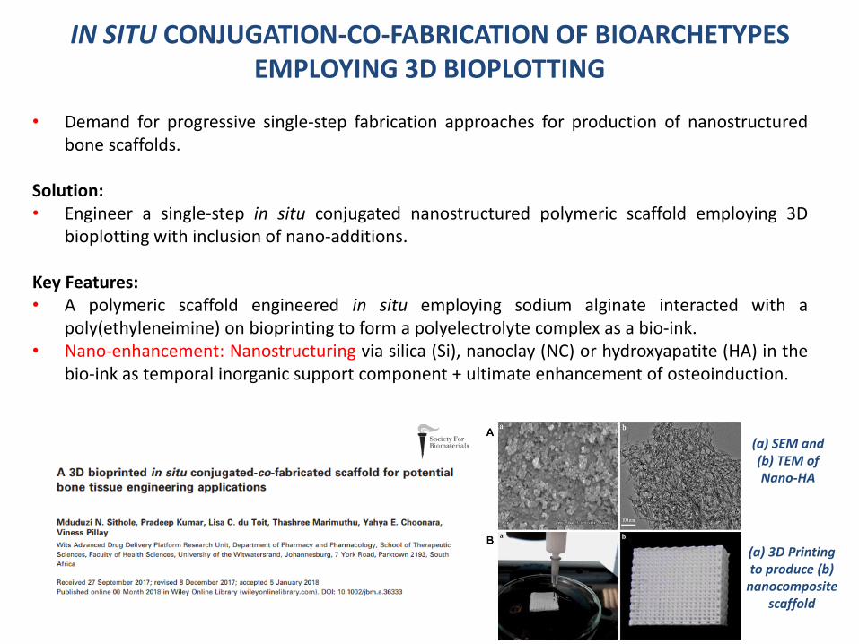
IN SITU CONJUGATION-CO-FABRICATION OF BIOARCHETYPES EMPLOYING 3D BIOPLOTTING
• Demand for progressive single-step fabrication approaches for production of nanostructuredbone scaffolds.
Solution:• Engineer a single-step in situ conjugated nanostructured polymeric scaffold employing 3D
bioplotting with inclusion of nano-additions.
Key Features:• A polymeric scaffold engineered in situ employing sodium alginate interacted with a
poly(ethyleneimine) on bioprinting to form a polyelectrolyte complex as a bio-ink.• Nano-enhancement: Nanostructuring via silica (Si), nanoclay (NC) or hydroxyapatite (HA) in the
bio-ink as temporal inorganic support component + ultimate enhancement of osteoinduction.
(a) SEM and (b) TEM of Nano-HA
(a) 3D Printing to produce (b)
nanocomposite scaffold
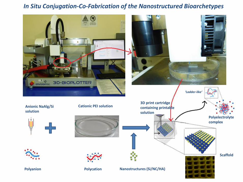
Anionic NaAlg/Sisolution
Cationic PEI solution3D print cartridge containing printable solution
Polyanion Polycation
Scaffold
Polyelectrolytecomplex
‘Ladder-like’
Nanostructures (Si/NC/HA)
In Situ Conjugation-Co-Fabrication of the Nanostructured Bioarchetypes

Scaffold roughness modulates the biological response through enhancement of cellular adhesion
A. Surface and B. Cross-sectional schematic of the proposed design of the bioplotted scaffold. C. SEM image of the
surface section of the bioplotted Alg-PEI/Si or NC or HA scaffold
Magnetic Resonance Imaging of the bioplotted scaffold
BioTester images and corresponding real-time image analysis and force-displacement graphs representing scaffold strength a) before
application of force and b) during maximum stretch
Architectural Visualization and Physicomechanical Analysis

Assessment of osteoblast-like MG63 cell differentiation: (a) Day 0 (b) Day 2 (c) Day 3 - The osteoblast-like MG63 cells were successfully seeded in the bioplotted scaffolds at Day 3
(a) Scaffold without cells. (b-d) Day 1-7 showing increasing confluence of
osteoblast-like MG63 cells on the scaffold. After Day 1 cells demonstrated adherence and growth to the printed scaffold. From
Day 3 - cells formed colonies which increased density (Day 7)

APPLICATION OF CHEMICAL MODIFICATION FOR ENHANCED PHYSICOMECHANICAL PROPERTIES OF 3D BIOPLOTTED SCAFFOLDS
• Progressive approaches required for fine-tuningphysicomechanical properties of bioplotted scaffolds.
Solution:• Surface chemical modifications and subsequent
functionalization of 3D printed scaffolds (e.g.aminolysis/hydrolysis with chitosan attachment;alkynylated and grafted with zwitterionic PCBMA-N₃).
• Identified a plasticizer - improved the printability ofthe bioink and mechanical properties of the printedconstruct.
• Nano-enhancement: Incorporation of nano-strengthening agents for enhanced physicomechanics.
Image and SEM of 3D bioplotted scaffold

3D PRINTED, ARTIFICIAL EXTRACELLULAR MATRIX FOR POTENTIAL NEUROREGENERATION
• Nerve regeneration capabilities of the human nervous system are insufficient– Incomplete repair and regain of function– No successful therapeutic scaffold for regeneration of damaged
nerves
Solution: develop a 3D printed, self-assembled biomaterial artificialextracellular matrix (aECM) for nerve regeneration
Key Features:1) Polymer (GelMA)• Semi-synthetic hydrogel: Provides RDG groups for cellular adhesion • Thermo-responsive physical crosslinking behaviour • UV crosslinkable hydrogel 2) Peptide-Hydrogel• Successful incorporation of native peptides• Maintenance of original polymer behaviour• 3D printable system
3D bioplotted peptide hydrogel
Gelation of Peptide-Hydrogel combinations at a 10°C temperature hold over 4 hours (n=3)
Control: Thermo-responsive behaviour, gelation at low
temperatures
Peptide complex: Increase in G’ values, stiffer gel. Native protein network complex mimicking?

• The largest challenge in this field - ability to balance all performance objectives for successfulscaffold integration within areas of biological and physical complexity.
• A large volume of current research has shown marked advancement in one direction, e.g.biocompatibility BUT with inadequate results in mechanical strength.
• Integration of appropriate porosity into scaffolds to improve cell viability and promote healthyvasculature throughout the repaired tissue.
• Materials that can closely mimic the properties of the natural tissue are essential (biocompatibility+ mechanical loading requirements).
• Further future research into novel nanostructured bio-inks.
• Further advancement to 3D-bioprinting of organs - experts project that science is less than 20 yearsaway from a fully functioning 3D printed heart. Currently - 3D printed heart is still challenged by theintricate nature of vasculature.