Lipoprotein(a) Induces Human Aortic Valve Interstitial...
14
MINI-FOCUS: AORTIC VALVE DISEASE Lipoprotein(a) Induces Human Aortic Valve Interstitial Cell Calcification Bin Yu, PHD, a Anouar Hafiane, PHD, a George Thanassoulis, MD, a Leah Ott, BS, a Nial Filwood, MD, a Marta Cerruti, PHD, b Ophélie Gourgas, MS, b Dominique Shum-Tim, MD, a Hamood Al Kindi, MD, a Benoit de Varennes, MD, a Alawi Alsheikh-Ali, MD, PHD, c Jacques Genest, MD, a Adel Schwertani, DM, PHD a VISUAL ABSTRACT Yu, B. et al. J Am Coll Cardiol Basic Trans Science. 2017;2(4):358–71. HIGHLIGHTS Lp(a) significantly increased alkaline phosphatase activity, phosphate and calcium content, and matrix vesicle formation and induced apoptosis and calcification of normal human aortic valve interstitial cells. The type of minerals induced by Lp(a) resembles that seen in calcified human aortic valves as shown by Raman spectroscopy. Lp(a)-induced calcification of human aortic valve interstitial cells is mediated by activation of MAPK38, GSK3b, and Wnt signaling. Inhibition of GSK3b and MAPK38 significantly reduced lipoprotein(a)- induced aortic valve interstitial cell calcification. Lp(a)is abundant in calcified aortic valves, and lipoprotein(a) immunoreactivity colocalized with that of oxidized phospholipids. From the a Divisions of Cardiology and Cardiac Surgery, Department of Medicine, Surgery and Pathology, McGill University, Montreal, Quebec, Canada; b Department of Materials Engineering, McGill University, Montreal, Quebec, Canada; and the c College of Medicine, Mohammed Bin Rashid University of Medical and Health Sciences, Dubai, United Arab Emirates. This work was supported by the Canadian Institutes of Health Research and the Heart and Stroke Foundation of Quebec, Canada. Dr. Thanassoulis is a consultant for and has received a research grant from Ionis; and is on the advisory board for Amgen. All other authors have reported that they have no relationships relevant to the contents of this paper to disclose. All authors attest they are in compliance with human studies committees and animal welfare regulations of the authors’ institutions and Food and Drug Administration guidelines, including patient consent where appropriate. For more infor- mation, visit the JACC: Basic to Translational Science author instructions page. Manuscript received December 21, 2016; revised manuscript received March 30, 2017, accepted March 30, 2017. JACC: BASIC TO TRANSLATIONAL SCIENCE VOL. 2, NO. 4, 2017 ª 2017 THE AUTHORS. PUBLISHED BY ELSEVIER ON BEHALF OF THE AMERICAN COLLEGE OF CARDIOLOGY FOUNDATION. THIS IS AN OPEN ACCESS ARTICLE UNDER THE CC BY-NC-ND LICENSE ( http://creativecommons.org/licenses/by-nc-nd/4.0/ ). ISSN 2452-302X http://dx.doi.org/10.1016/j.jacbts.2017.03.015
Transcript of Lipoprotein(a) Induces Human Aortic Valve Interstitial...

J A C C : B A S I C T O T R A N S L A T I O N A L S C I E N C E V O L . 2 , N O . 4 , 2 0 1 7
ª 2 0 1 7 T H E A U T H O R S . P U B L I S H E D B Y E L S E V I E R O N B E H A L F O F T H E AM E R I C A N
C O L L E G E O F C A R D I O L O G Y F O UN DA T I O N . T H I S I S A N O P E N A C C E S S A R T I C L E U N D E R
T H E C C B Y - N C - N D L I C E N S E ( h t t p : / / c r e a t i v e c o mm o n s . o r g / l i c e n s e s / b y - n c - n d / 4 . 0 / ) .
I S S N 2 4 5 2 - 3 0 2 X
h t t p : / / d x . d o i . o r g / 1 0 . 1 0 1 6 / j . j a c b t s . 2 0 1 7 . 0 3 . 0 1 5
MINI-FOCUS: AORTIC VALVE DISEASE
Lipoprotein(a) Induces Human AorticValve Interstitial Cell Calcification
Bin Yu, PHD,a Anouar Hafiane, PHD,a George Thanassoulis, MD,a Leah Ott, BS,a Nial Filwood, MD,aMarta Cerruti, PHD,b Ophélie Gourgas, MS,b Dominique Shum-Tim, MD,a Hamood Al Kindi, MD,a
Benoit de Varennes, MD,a Alawi Alsheikh-Ali, MD, PHD,c Jacques Genest, MD,a Adel Schwertani, DM, PHDa
VISUAL ABSTRACT
F
M
o
s
D
o
A
in
m
M
Yu, B. et al. J Am Coll Cardiol Basic Trans Science. 2017;2(4):358–71.
rom the aDivisions of Cardiology and Cardiac Surgery, Department of Medicine, Surgery
ontreal, Quebec, Canada; bDepartment of Materials Engineering, McGill University, Montreal
f Medicine, Mohammed Bin Rashid University of Medical and Health Sciences, Dubai, Unit
upported by the Canadian Institutes of Health Research and the Heart and Stroke
r. Thanassoulis is a consultant for and has received a research grant from Ionis; and is on
ther authors have reported that they have no relationships relevant to the contents of t
ll authors attest they are in compliance with human studies committees and animal w
stitutions and Food and Drug Administration guidelines, including patient consent wh
ation, visit the JACC: Basic to Translational Science author instructions page.
anuscript received December 21, 2016; revised manuscript received March 30, 2017, accepte
HIGHLIGHTS
� Lp(a) significantly increased alkaline
phosphatase activity, phosphate and
calcium content, and matrix vesicle
formation and induced apoptosis and
calcification of normal human aortic valve
interstitial cells.
� The type of minerals induced by Lp(a)
resembles that seen in calcified human
aortic valves as shown by Raman
spectroscopy.
� Lp(a)-induced calcification of human
aortic valve interstitial cells is mediated
by activation of MAPK38, GSK3b, and Wnt
signaling.
� Inhibition of GSK3b and MAPK38
significantly reduced lipoprotein(a)-
induced aortic valve interstitial cell
calcification.
� Lp(a)is abundant in calcified aortic valves,
and lipoprotein(a) immunoreactivity
colocalized with that of oxidized
phospholipids.
and Pathology, McGill University,
, Quebec, Canada; and the cCollege
ed Arab Emirates. This work was
Foundation of Quebec, Canada.
the advisory board for Amgen. All
his paper to disclose.
elfare regulations of the authors’
ere appropriate. For more infor-
d March 30, 2017.

R E V I A T I O N S
J A C C : B A S I C T O T R A N S L A T I O N A L S C I E N C E V O L . 2 , N O . 4 , 2 0 1 7 Yu et al.A U G U S T 2 0 1 7 : 3 5 8 – 7 1 Lp(a) in Aortic Valve Disease
359
SUMMARYAB B
AND ACRONYM S
ALP = alkaline phosphatase
apo(a) = apolipoprotein(a)
BMP = bone morphogenetic
protein
FWHM = full width half
maximum
HAVIC = human aortic valve
interstitial cell
LDL = low-density lipoprotein
Lp(a) = lipoprotein(a)
Lipoprotein(a), or Lp(a), significantly increased alkaline phosphatase activity, release of phosphate, calcium
deposition, hydroxyapatite, cell apoptosis, matrix vesicle formation, and phosphorylation of signal transduction
proteins; increased expression of chondro-osteogenic mediators; and decreased SOX9 and matrix Gla protein
(p < 0.001). Inhibition of MAPK38 and GSK3b significantly reduced Lp(a)-induced calcification of human
aortic valve interstitial cells (p < 0.001). There was abundant presence of Lp(a) and E06 immunoreactivity in
diseased human aortic valves. The present study demonstrates a causal effect for Lp(a) in aortic valve calcification
and suggests that interfering with the Lp(a)pathway could provide a novel therapeutic approach in the man-
agement of this debilitating disease. (J Am Coll Cardiol Basic Trans Science 2017;2:358–71) © 2017 The Authors.
Published by Elsevier on behalf of the American College of Cardiology Foundation. This is an open access
article under the CC BY-NC-ND license (http://creativecommons.org/licenses/by-nc-nd/4.0/).
1 = oxidized LDL receptor 1 LOX-MAPK = mitogen-activated
protein kinase
MGP = matrix Gla protein
mRNA = messenger
ribonucleic acid
= oxidized phospholipid
A ortic valve stenosis is a debilitating and rela-tively common condition for which no medi-cal therapy exists. Classically thought to
result from degeneration of the aortic valve, a num-ber of novel contributing mechanisms have beenidentified, including the role of mechanical stress,age-related changes in valvular cells and extracellularmatrix, the regulation of calcium flux in both physio-logical and cardiovascular calcification (1), and therole played by the renin-angiotensin system in pro-moting valve remodeling (2,3). Despite the similar-ities between aortic valve stenosis and coronaryartery disease, both in terms of clinical risk factorsand histopathological manifestations, statins do nothave any effect on the progression of aortic valve ste-nosis (4,5), and it is increasingly evident that thepathogenesis of the 2 conditions is different.
Recently, a genome-wide association study in 3large cohorts revealed that a single-nucleotide poly-morphism in the apolipoprotein(a) (LPA) locus wassignificantly associated with the presence of aorticvalve calcification (6,7). This variant was also shownto associate with clinical aortic valve stenosis andvalve replacement in several longitudinal cohorts,and current genetic evidence using the concept ofMendelian randomization suggests a probable causalrole for circulating lipoprotein(a) [Lp(a)]. LPA en-codes apolipoprotein(a) [apo(a)], an evolutionarilynovel protein expressed only in primates, which ishighly homologous with plasminogen, suggesting apossible origin via gene duplication. LPA is predom-inately expressed in hepatocytes (8–10); oncesecreted, apo(a) binds low-density lipoprotein (LDL)via disulfide bridging and stearic interaction (11),forming Lp(a). The latter has long been recognized asa risk factor for cardiac disease, but its exact rolein vivo has remained contentious (12).
Nonetheless, several lines of evidencesuggest that Lp(a) could play a fundamentalrole in the pathogenesis of aortic valve ste-nosis and its precursor lesion, aortic valve
sclerosis. High levels of circulating Lp(a) have beenassociated with an increased incidence of aortic valvestenosis (13) and aortic valve sclerosis (14) in diversehuman populations. Lp(a) scavenges oxidized phos-pholipid (OxPL) in human plasma (15), binding itcovalently at a lysine binding site in kringle domainIV type 10 (KIV10) of Lp(a) that is thought tocontribute to the pathological activity of Lp(a) (16).The association between high levels of Lp(a) andcalcific aortic valve disease, in contrast, could resultfrom either direct effects of Lp(a) or the higher serumOxPL content seen in patients with high Lp(a) levels.Although a previous study showed that Lp(a)enhanced atherosclerosis and vascular calcification inLDL receptor–defective Watanabe heritable hyper-lipidemic transgenic rabbits (17), the effects of Lp(a)on human aortic valve interstitial cells (HAVICs)remain to be established.Here, we hypothesized that exposure of culturedHAVICs isolated from noncalcified valves to Lp(a)would induce their phenotypic differentiation viaactivation of pro-osteogenic pathways. Therefore,we tested the effect of Lp(a) on HAVIC proliferation,apoptosis, differentiation, and gene and proteinexpression profile. We also determined whattypes of phosphate and calcium minerals areinduced by Lp(a) in HAVICs using Raman spectros-copy. Furthermore, we sought to determine theeffect of Lp(a) on phosphorylation of mitogen-activated protein kinases (MAPKs). Finally, wedocumented the tissue presence of Lp(a) and OxPLsin a large number of calcified and normal aorticvalves.
OxPL

FIGURE 1 Effects of Lp(a) Treatment on HAVICs
HAVICs were incubated in OSM in the presence or absence of 50 mg/ml purified Lp(a) (Athens Research & Technology, Inc., Athens, Georgia)
for 48 h. (A) Cell proliferation measured using an MTT protocol and presented as relative cell proliferation rate. *p ¼ 0.013 vs. OSM control.
(B)Medium and cellular ALP activity measured with an ALP activity colorimetric assay kit and normalized to total protein concentration. Lp(a)-
treated cells showed higher ALP activity in medium (**p ¼ 0.0001) and cell lysate (*p ¼ 0.003) than OSM control. (C) Medium phosphate
release measured using a phosphate colorimetric assay kit (BioVision, Inc., Milpitas, California). *p ¼ 0.0001 vs. OSM (control). (D) No
detectable OxPLs were seen on purified Lp(a) using anti-OxPL E06 antibody. Purified human Lp(a) (1 mg) was loaded on 6% sodium dodecyl
sulfate polyacrylamide gel electrophoresis. Lane 1: Lp(a) was detected using anti-Lp(a) antibody (diluted at 1:10,000; Novus Biologicals, LLC,
Littleton, Colorado); lane 2: no OxPLs were detected in purified Lp(a) using E06 antibody (diluted at 1:1,000; Avanti Polar Lipids, Inc.,
Alabaster, Alabama). ALP ¼ alkaline phosphatase; HAVICS ¼ human aortic valve interstitial cells; Lp(a) ¼ lipoprotein(a); OSM ¼ osteogenic
medium; OxPL ¼ oxidized phospholipid.
Yu et al. J A C C : B A S I C T O T R A N S L A T I O N A L S C I E N C E V O L . 2 , N O . 4 , 2 0 1 7
Lp(a) in Aortic Valve Disease A U G U S T 2 0 1 7 : 3 5 8 – 7 1
360
METHODS
A detailed Methods section is provided in theSupplemental Appendix. Briefly, primary humanHAVIC cell lines were generated as described previ-ously (18,19), with cultured HAVICs showing positivestaining of a-smooth muscle actin, indicating myofi-broblast phenotype, after 2 passages. HAVICs at pas-sages 3 to 5 were used for all experiments. To inducecalcification, HAVICs were cultured in osteogenicmedium (full Dulbecco’s modified Eagle’s mediumplus 2 mmol/l buffered phosphate, pH 7.4). The studywas approved by the McGill University Health Centre
Ethics Committee, and written consents wereobtained from all participating patients.
RESULTS
EFFECTS OF Lp(a) ON HAVICs. Although previousstudies have delineated the effect of Lp(a) onvascular cells (10), its effect on valve interstitial cellsremains to be elucidated. Here, we found that short-term (up to 48 h) incubation of HAVICs isolatedfrom noncalcified human aortic valves with 50 mg/mlLp(a) significantly increased cell proliferation(p ¼ 0.013) (Figure 1A), cellular (p ¼ 0.003) and

FIGURE 2 Effects of Lp(a) Treatment on HAVIC Calcification and Apoptosis
(A) Prolonged Lp(a) treatment for 3 weeks at 50 mg/ml in OSM increased calcification nodule formation in HAVICs (arrows). (B) Calcium
deposition measured after 2 weeks of Lp(a) incubation. *p ¼ 0.03, **p ¼ 0.001 vs. control; #p ¼ 0.04 vs. Lp(a)-treated cells. (C) Lp(a)
demonstrated a greater effect on calcium deposition than LDL (Alfa Aesar, Tewksbury, Massachusetts) after 2 weeks of incubation in OSM.
*p < 0.00001 vs. OSM control; #p ¼ 0.006, Lp(a) treated vs. LDL treated. (D) Lp(a) effects on HAVIC apoptosis assessed using caspase-3/7
activity. *p ¼ 0.0003, Lp(a)-treated vs. DMEM; #p ¼ 0.007 Lp(a)-treated vs. OSM. DMEM ¼ Dulbecco’s modified Eagle’s medium;
LDL ¼ low-density lipoprotein; other abbreviations as in Figure 1.
J A C C : B A S I C T O T R A N S L A T I O N A L S C I E N C E V O L . 2 , N O . 4 , 2 0 1 7 Yu et al.A U G U S T 2 0 1 7 : 3 5 8 – 7 1 Lp(a) in Aortic Valve Disease
361
medium (p ¼ 0.0001) alkaline phosphatase (ALP)activity (Figure 1B), and release of phosphate(p ¼ 0.0001) (Figure 1C). OxPLs were not detectedusing mouse anti-OxPL antibody EO6 in the purifiedhuman Lp(a) (Figure 1D). Prolonged incubation (3weeks) of HAVICs with Lp(a) significantly increasedcalcification nodules (Figure 2A), resulted in highercalcium deposition (Figures 2B and 2C) and caspase-3and caspase-7 activity (Figure 2D), and decreasedcellular proliferation as demonstrated by MTT assay(19.63 � 4.12%; p < 0.01) (Supplemental Figure 1).Incubation of HAVICs with Lp(a) plus OxPLs signifi-cantly augmented the effects of Lp(a) on calciumdeposition (Figure 2B). The effect of Lp(a) on HAVICmineralization was significantly higher than that ofLDL (p ¼ 0.006) (Figure 2C). To characterize the typeof minerals induced by Lp(a) treatment, we usedRaman spectroscopy, which revealed that the spectracollected on HAVICs incubated in osteogenic mediumin the presence of Lp(a) for 3 weeks showed a n1PO4
peak centered at 960 cm�1 (Figure 3A), which is
indicative of hydroxyapatite. A similar peak wasfound on the spectra of synthetic hydroxyapatite,bone, and enamel, as well as HAVICs collected fromcalcified heart valves (Figure 3A). HAVICs incubatedin osteogenic medium for the same period butwithout addition of Lp(a) did not show a significantpresence of phosphate-containing minerals. A com-plete list of peak assignments for these spectra ispresented in Supplemental Appendix.
By integrating the area of the n1 phosphate peakand dividing this value by the area of the peak at1,440 cm�1, which is relative to the stretching ofCH2/CH3 groups, we estimated the amount of inor-ganic deposits relative to the amount of organic ma-terial found in HAVICs isolated from calcified heartvalves and HAVICs incubated in osteogenic mediumfor 3 weeks, with or without the presence of Lp(a)(Figure 3B). HAVICs treated with Lp(a) showed moreabundant mineral deposition than nontreated cells(p ¼ 0.002) or HAVICs collected from calcified aorticvalves (p ¼ 0.03).

FIGURE 3 Effects of Lp(a) on Inorganic Mineralization in HAVICs
(A) Representative Raman spectra. Noncalcified HAVICs were incubated in OSM in the absence (a) or presence (b) of Lp(a) for 3 weeks; calcified HAVICs were cultured in
DMEM medium (c) and bone (d), enamel (e), and standard HA (f). n ¼ 3 for each sample. (B) Inorganic to organic ratio measured by dividing the area of the n1PO4 peak
(inorganic marker) at 960 cm�1 and the area of the CH2/CH3 (organic compounds containing -CH2/-CH3 groups) stretching at 1,440 cm�1 (n ¼ 3 for each sample).
NoncalcifiedHAVICswere incubated inOSM for 3weeks in the absence or presence of Lp(a), and stenoticHAVICswere cultured inDMEMfor 3weeks. Lp(a)-treatedHAVICs
and calcified HAVICs showed significantly higher inorganic-organic ratios than nontreated OSM control, with *p ¼ 0.002 and p ¼ 0.0001, respectively. Lp(a)-treated
HAVICs showed a higher ratio against calcified HAVICs (#p ¼ 0.03). (C) FWHM measured on the n1PO4 peak at 960 cm�1 for the spectra of HAVICs from calcified heart
valve, noncalcified HAVICs incubated with Lp(a) in OSM for 3 weeks, enamel, bone, and HA. *Significant differences: Lp(a)-treated HAVICs vs. calcified AV, p< 0.001; vs.
enamel, bone, and HA, p < 0.0001. AV ¼ aortic valve; FWHM ¼ full width at half maximum; HA ¼ hydroxyapatite; other abbreviations as in Figures 1 and 2.
Yu et al. J A C C : B A S I C T O T R A N S L A T I O N A L S C I E N C E V O L . 2 , N O . 4 , 2 0 1 7
Lp(a) in Aortic Valve Disease A U G U S T 2 0 1 7 : 3 5 8 – 7 1
362
To further characterize the type of calcific nodulesinduced by Lp(a) compared with diseased aorticvalves, we estimated the degree of crystallinity of thehydroxyapatite deposits by measuring the full widthat half maximum (FWHM) of the n1 phosphate peak.Wider peaks indicate a less crystalline material,whereas sharper peaks show a higher crystallinity.Clearly, the peak of synthetic hydroxyapatite ismuch sharper than any of those found in biologicalsamples [bone, enamel, calcified heart valves, andLp(a)-treated HAVICs] (Figure 3A). We comparedthe FWHM measured on all the spectra (Figure 3C).There was no difference in the FWHM measuredfor deposits found on HAVICs treated with Lp(a) (24.4� 0.6) compared with HAVICs from calcified heartvalves (22.5 � 0.5), which indicates somewhat similarcrystalline deposits on Lp(a)-treated cells (Figure 3C).Significantly lower FWHMs (p < 0.0001) were foundon synthetic hydroxyapatite (5.3 � 0.1) and natural
samples such as bone (18.1 � 0.2) and enamel (11.7 �0.3) (Figure 3C), which indicates that less crystallinehydroxyapatite is deposited in ectopic calcification inheart valves than in physiological hard tissues.
To assess the morphological characteristics ofLp(a)-induced calcification in HAVICs, we usedfluorescence microscopy, which revealed the pres-ence of numerous calcium-containing membranebudding vesicles (Figure 4A). Scanning electron mi-croscopy confirmed the presence of various-sizedmicrovesicles on HAVICs (Figure 4B). Extracellularvesicles released in the culture medium were char-acterized with quantification techniques such asnanoparticle tracking analysis. Medium vesicle con-centrations of Lp(a)-treated HAVICs were found to besignificantly higher than those of nontreated cells inserum-free osteogenic medium at 7 days, and therewas different size profiling between 50 and 500 nm inresponse to Lp(a) treatment (Figures 4C and 4D).

FIGURE 4 Presence of Extracellular Vesicles in HAVICs
(A) HAVICs exhibited calcium-containing cellular vesicles. 1: Phase-contrast image; 2: membrane lipophilic Vybrant DiO dye stains (Thermo
Fisher Scientific, Carlsbad, California) of vesicles of various sizes (green); 3: high-calcium-containing vesicles (arrows) stained with Calcium
Orange AM (Thermo Fisher Scientific) (red); 4: superimposed image of 1, 3, and nuclear (blue) stained with NucBlue reagent (Thermo Fisher
Scientific). (B) Cluster of extracellular vesicles on HAVICs observed using scanning electron microscopy (arrows). (C) HAVICs were treated
with 50 mg/ml purified Lp(a) for 7 days in serum-free DMEM. The medium was freshly changed at days 3 and 5. The harvested media (days
3 and 7) were subjected to extracellular vesicle profiling using NanoSight NS500 (Malvern Instruments, Malvern, United Kingdom). Lp(a)-
treated cells show higher particle concentration than nontreated, with *p ¼ 0.04 at day 3 in DMEM, and **p ¼ 0.0001 at day 7 in OSM; n ¼ 3
for each group. (D) Extracellular vesicle size profiling shows different particle concentration vs. particle size profiling of Lp(a)-treated HAVICs
at 7 days. Each condition was sampled 5 times for size profiling using NanoSight NS500. Data were combined and presented as average
particle concentration vs. particle size (mean � SEM). Abbreviations as in Figures 1 and 2.
J A C C : B A S I C T O T R A N S L A T I O N A L S C I E N C E V O L . 2 , N O . 4 , 2 0 1 7 Yu et al.A U G U S T 2 0 1 7 : 3 5 8 – 7 1 Lp(a) in Aortic Valve Disease
363
To examine the effects of Lp(a) on the inflammatorystate of HAVICs, profiling of cytokine releasewas testedwith a Proteome Profiler of human cytokine array,panel A (R&D Systems Inc., Minneapolis, Minnesota).Incubation of HAVICs with 50 mg/ml Lp(a) for 48 h didnot affect the release of inflammatory cytokines orchemokines, including interleukin-6, interleukin-8,monocyte chemoattractant protein-1, plasminogenactivator inhibitor-1, macrophage migration inhibitory
factor, and GROa (Figure 5A). Conversely, phospho-MAPK profiling assayed with a Proteome Profiler ofhuman phospho-MAPK array found that Lp(a) signifi-cantly increased phosphorylation of MAPK38, MAPKkinase 3/6 (MKK3/6), MSK2, and GSK3a/b (Figures 5Band 5C). Inhibition of MAPK38 with SB203580 orGSK3a/b with CHIR99021 significantly reduced ALPactivity and calcium deposition (Figure 5D). PD184352significantly increased calcium deposition (Figure 5D).
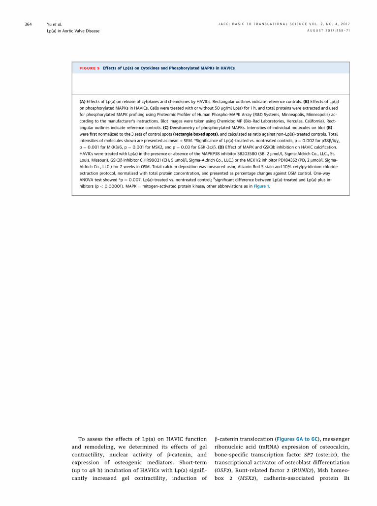
FIGURE 5 Effects of Lp(a) on Cytokines and Phosphorylated MAPKs in HAVICs
(A) Effects of Lp(a) on release of cytokines and chemokines by HAVICs. Rectangular outlines indicate reference controls. (B) Effects of Lp(a)
on phosphorylated MAPKs in HAVICs. Cells were treated with or without 50 mg/ml Lp(a) for 1 h, and total proteins were extracted and used
for phosphorylated MAPK profiling using Proteomic Profiler of Human Phospho-MAPK Array (R&D Systems, Minneapolis, Minneapolis) ac-
cording to the manufacturer’s instructions. Blot images were taken using Chemidoc MP (Bio-Rad Laboratories, Hercules, California). Rect-
angular outlines indicate reference controls. (C) Densitometry of phosphorylated MAPKs. Intensities of individual molecules on blot (B)
were first normalized to the 3 sets of control spots (rectangle boxed spots), and calculated as ratio against non–Lp(a)-treated controls. Total
intensities of molecules shown are presented as mean � SEM. *Significance of Lp(a)-treated vs. nontreated controls, p ¼ 0.002 for p38b/d/y,
p ¼ 0.001 for MKK3/6, p ¼ 0.001 for MSK2, and p ¼ 0.03 for GSK-3a/b. (D) Effect of MAPK and GSK3b inhibition on HAVIC calcification.
HAVICs were treated with Lp(a) in the presence or absence of the MAPKP38 inhibitor SB203580 (SB; 2 mmol/l, Sigma-Aldrich Co., LLC., St.
Louis, Missouri), GSK3b inhibitor CHIR99021 (CH; 5 mmol/l, Sigma-Aldrich Co., LLC.) or the MEK1/2 inhibitor PD184352 (PD; 2 mmol/l, Sigma-
Aldrich Co., LLC.) for 2 weeks in OSM. Total calcium deposition was measured using Alizarin Red S stain and 10% cetylpyridinium chloride
extraction protocol, normalized with total protein concentration, and presented as percentage changes against OSM control. One-way
ANOVA test showed *p ¼ 0.007, Lp(a)-treated vs. nontreated control; #significant difference between Lp(a)-treated and Lp(a) plus in-
hibitors (p < 0.00001). MAPK ¼ mitogen-activated protein kinase; other abbreviations as in Figure 1.
Yu et al. J A C C : B A S I C T O T R A N S L A T I O N A L S C I E N C E V O L . 2 , N O . 4 , 2 0 1 7
Lp(a) in Aortic Valve Disease A U G U S T 2 0 1 7 : 3 5 8 – 7 1
364
To assess the effects of Lp(a) on HAVIC functionand remodeling, we determined its effects of gelcontractility, nuclear activity of b-catenin, andexpression of osteogenic mediators. Short-term(up to 48 h) incubation of HAVICs with Lp(a) signifi-cantly increased gel contractility, induction of
b-catenin translocation (Figures 6A to 6C), messengerribonucleic acid (mRNA) expression of osteocalcin,bone-specific transcription factor SP7 (osterix), thetranscriptional activator of osteoblast differentiation(OSF2), Runt-related factor 2 (RUNX2), Msh homeo-box 2 (MSX2), cadherin-associated protein B1
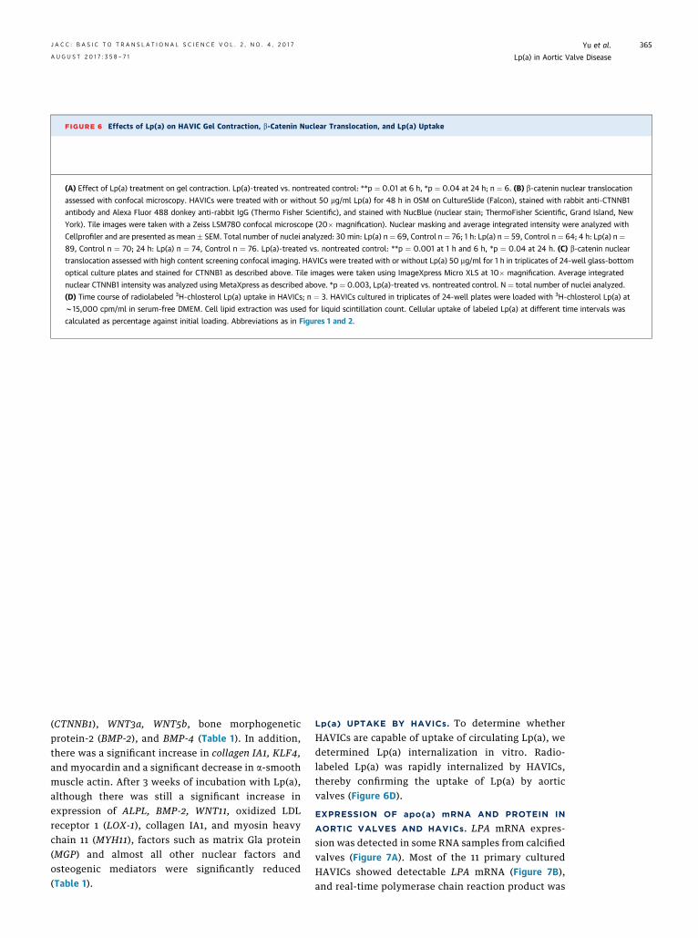
FIGURE 6 Effects of Lp(a) on HAVIC Gel Contraction, b-Catenin Nuclear Translocation, and Lp(a) Uptake
(A) Effect of Lp(a) treatment on gel contraction. Lp(a)-treated vs. nontreated control: **p ¼ 0.01 at 6 h, *p ¼ 0.04 at 24 h; n ¼ 6. (B) b-catenin nuclear translocation
assessed with confocal microscopy. HAVICs were treated with or without 50 mg/ml Lp(a) for 48 h in OSM on CultureSlide (Falcon), stained with rabbit anti-CTNNB1
antibody and Alexa Fluor 488 donkey anti-rabbit IgG (Thermo Fisher Scientific), and stained with NucBlue (nuclear stain; ThermoFisher Scientific, Grand Island, New
York). Tile images were taken with a Zeiss LSM780 confocal microscope (20� magnification). Nuclear masking and average integrated intensity were analyzed with
Cellprofiler and are presented as mean � SEM. Total number of nuclei analyzed: 30 min: Lp(a) n ¼ 69, Control n ¼ 76; 1 h: Lp(a) n ¼ 59, Control n ¼ 64; 4 h: Lp(a) n ¼89, Control n ¼ 70; 24 h: Lp(a) n ¼ 74, Control n ¼ 76. Lp(a)-treated vs. nontreated control: **p ¼ 0.001 at 1 h and 6 h, *p ¼ 0.04 at 24 h. (C) b-catenin nuclear
translocation assessed with high content screening confocal imaging. HAVICs were treated with or without Lp(a) 50 mg/ml for 1 h in triplicates of 24-well glass-bottom
optical culture plates and stained for CTNNB1 as described above. Tile images were taken using ImageXpress Micro XLS at 10� magnification. Average integrated
nuclear CTNNB1 intensity was analyzed using MetaXpress as described above. *p ¼ 0.003, Lp(a)-treated vs. nontreated control. N ¼ total number of nuclei analyzed.
(D) Time course of radiolabeled 3H-chlosterol Lp(a) uptake in HAVICs; n ¼ 3. HAVICs cultured in triplicates of 24-well plates were loaded with 3H-chlosterol Lp(a) at
w15,000 cpm/ml in serum-free DMEM. Cell lipid extraction was used for liquid scintillation count. Cellular uptake of labeled Lp(a) at different time intervals was
calculated as percentage against initial loading. Abbreviations as in Figures 1 and 2.
J A C C : B A S I C T O T R A N S L A T I O N A L S C I E N C E V O L . 2 , N O . 4 , 2 0 1 7 Yu et al.A U G U S T 2 0 1 7 : 3 5 8 – 7 1 Lp(a) in Aortic Valve Disease
365
(CTNNB1), WNT3a, WNT5b, bone morphogeneticprotein-2 (BMP-2), and BMP-4 (Table 1). In addition,there was a significant increase in collagen IA1, KLF4,and myocardin and a significant decrease in a-smoothmuscle actin. After 3 weeks of incubation with Lp(a),although there was still a significant increase inexpression of ALPL, BMP-2, WNT11, oxidized LDLreceptor 1 (LOX-1), collagen IA1, and myosin heavychain 11 (MYH11), factors such as matrix Gla protein(MGP) and almost all other nuclear factors andosteogenic mediators were significantly reduced(Table 1).
Lp(a) UPTAKE BY HAVICs. To determine whetherHAVICs are capable of uptake of circulating Lp(a), wedetermined Lp(a) internalization in vitro. Radio-labeled Lp(a) was rapidly internalized by HAVICs,thereby confirming the uptake of Lp(a) by aorticvalves (Figure 6D).
EXPRESSION OF apo(a) mRNA AND PROTEIN IN
AORTIC VALVES AND HAVICs. LPA mRNA expres-sion was detected in some RNA samples from calcifiedvalves (Figure 7A). Most of the 11 primary culturedHAVICs showed detectable LPA mRNA (Figure 7B),and real-time polymerase chain reaction product was

TABLE 1 Osteogenic-Related Genes Differentially Regulated in
HAVICs Treated With Lp(a) in Osteogenic Medium
Gene NameRelative NormalizedExpression (Fold)
Day 2
BGLAP 4.99
SP7 4.44
WNT3a 1.92
RUNX2 1.72
BMP2 1.69
MYOCD 1.66
CTNNB1 1.62
COL1A1 1.52
WNT5b 1.49
OSF2 1.46
MSX2 1.45
BMP4 1.36
KLF4 1.33
SOX9 0.66
MGP 0.64
aSMA 0.54
OPN 0.51
LOX1 0.27
3 Weeks
MYH11 2.70
WNT11 2.60
LOX1 1.87
COL1A1 1.80
BMP2 1.50
ALPL 1.42
OSF2 0.70
MYOCD 0.64
MGP 0.60
CTNNB1 0.56
RUNX2 0.52
aSMA 0.49
WNT3a 0.37
SP7 0.26
OPN 0.24
BGLAP 0.05
HAVIC ¼ human aortic valve interstitial cell; Lp(a) ¼ lipoprotein(a).
Yu et al. J A C C : B A S I C T O T R A N S L A T I O N A L S C I E N C E V O L . 2 , N O . 4 , 2 0 1 7
Lp(a) in Aortic Valve Disease A U G U S T 2 0 1 7 : 3 5 8 – 7 1
366
sequenced to confirm the correct identity of LPAmRNA (Figure 7C). Western blot showed weak tostrong apo(a) protein in calcified human aortic valves(Figure 7D, lanes 4 to 11). Apo(a) protein was also seenin cultured HAVICs (Figure 7D). Endogenous LPAexpression was detected in cellular vesicles of varioussizes and in endoplasmic reticulum in culturedHAVICs using mouse anti-LPA antibody (Figure 7E).
Lp(a) AND OxPL IMMUNOREACTIVITY IN CALCIFIED
HUMAN AORTIC VALVES. Only weak and infrequentapo(a) immunoreactivity was seen in normal aorticvalves or normal leaflets of stenotic aortic valves(Figure 8A). In contrast, strong immunoreactivity
for apo(a) was seen in calcified aortic valves. Theimmunostaining was evident in areas of sub-endothelial, thickened fibrosa; calcification; activatedmyofibroblasts; and mononuclear inflammatory cells(Figures 8B, 8C, 8D, 8F, 8J, and 8M). Inflammatorycells were identified using anti-CD68 antibody (Figure8E), and myofibroblast cells were identified usinganti-aSMA antibody (Figure 8G).
Deposition of OxPLs in aortic valve tissues wasdetected with E06 monoclonal antibody. The latter isa murine monoclonal immunoglobulin M naturalantibody that recognizes the phosphocholine headgroup of OxPLs and was developed using apolipo-protein E knockout mice fed a high-fat diet (20).Immunoreactivity for E06 was mainly seen in endo-thelial cells and subendothelium of normal leaflets ofthe human aortic valve. Strong immunostaining forE06 was seen in subendothelial, thickened fibrosa;calcification; lipid core; and mononuclear inflamma-tory cells in calcified leaflets of the human aorticvalve (Figures 8H, 8I, 8K, and 8L). E06 immuno-reaction was seen in similar regions to that of Lp(a) indiseased segments, but they exhibited differentialimmunostaining patterns, with some cases showingvery strong E06 staining but minimal Lp(a) stainor vice versa (Figures 8I, 8J, 8L, and 8M). E06 exhibi-ted a wider distribution and more cellular stainingthan Lp(a). Colocalization studies revealed the occa-sional presence of Lp(a) and E06 in the same cells.Negative control sections immunostained withnonimmune serum or pre-absorbed with the respec-tive antigens did not reveal any staining (datanot shown).
Mean ranks of Lp(a) and E06 immunostaining weresignificantly higher in calcified than in normal leaflets(p < 0.0002). There was a significant correlationbetween the immunostaining for Lp(a) and E06(r ¼ 0.284, p ¼ 0.001). Also, there was a significantcorrelation between the immunostaining for Lp(a)and E06, respectively, and the presence of lipids(Lp(a): r ¼ 0.243, p < 0.005; E06: r ¼ 0.527, p < 0.0001)and calcification (Lp(a): r ¼ 0.343, p < 0.05; E06: r ¼0.548, p < 0.0001). Only E06, not Lp(a), immunore-activity correlated with the presence of inflammation(r ¼ 0.30, p < 0.001). Moreover, there was a significantcorrelation between E06 immunostaining and peakpressures (Pmean r ¼ 0.32; Pmax r ¼ 0.326, p < 0.01).
DISCUSSION
Although several studies have suggested a potentialrole for Lp(a) in the pathogenesis of cardiovasculardiseases, it remained unclear whether the LPA gene islocally expressed and whether Lp(a) could directly

FIGURE 7 LPA mRNA and Protein Expression Were Detected in Primarily Cultured HAVICs and Aortic Valve Tissues
(A) RT-PCR detection of LPA mRNA expression in primarily cultured HAVICs. Lane 0: DNA marker; lanes 2, 5, and 8 show positive signal at
products 136 bp. (B) RT-PCR detection of LPA mRNA expression in stenotic aortic valves. Lane 0: DNA marker; lanes 1, 2, 5, 6, 7, 9, and 10
show positive signal at 136 bp. (C) Sequencing of gel purified RT-PCR product confirms the correct identity of LPA mRNA. (D) LPA protein
expression in stenotic aortic valve tissues and HAVICs. (Top) Lanes 1 through 3, nonstenotic valves; lanes 4 through 11, stenotic valves; lane
12, human serum sample serving as loading marker. Lanes 8 and 9 show double bands indicating possible dual isoforms. (Bottom) LPA
protein expression in 7 cultured HAVICs. Lane 1 show double bands indicating possible dual isoforms. (E) Immunofluorescence cytochemistry
detection of LPA in HAVICs using mouse anti-LPA antibody and Alexa Fluor 488 donkey anti-mouse immunoglobulin G (green); solid arrows
show various-sized cellular vesicles. Red indicates pseudocolored F-actin stained with ActinRed reagent; blue indicates nuclei stained with
NucBlue reagent. LPA ¼ apolipoprotein(a) gene; mRNA ¼ messenger ribonucleic acid; RT-PCR ¼ real-time polymerase chain reaction; other
abbreviations as in Figure 1.
J A C C : B A S I C T O T R A N S L A T I O N A L S C I E N C E V O L . 2 , N O . 4 , 2 0 1 7 Yu et al.A U G U S T 2 0 1 7 : 3 5 8 – 7 1 Lp(a) in Aortic Valve Disease
367
induce calcification and remodeling of the humanaortic valve. The present study demonstrates for thefirst time a causal relationship between Lp(a) and theinduction of aortic valve calcification. Previousstudies have shown increased plasma levels of Lp(a)in patients with aortic valve disease, and downstreamsignaling from Lp(a) such as autotaxin producedsimilar effects (13,14,21). A recent study showed thatelevated levels of Lp(a) and OxPLs could predict theprogression of aortic valve stenosis (22). Here, wesystematically studied the distribution of Lp(a) andE06 in relation to disease severity in 112 cases ofdiseased aortic valves and demonstrated the presenceof both molecules and their correlation with thepresence of tissue lipid deposition and calcific nod-ules. We also observed a significant correlation be-tween Lp(a) and OxPL immunoreactivities. E06
immunoreactivity, but not Lp(a), correlated with tis-sue inflammation and peak pressures. In 2 recentlypublished articles, Nordestgaard and Langsted et al.(23,24) reported that increased plasma Lp(a) was notassociated with increased inflammatory markers andeluded to the possibility that E06 merely recognizesapoB-100 on the Lp(a) particle. They also suggestedthat Lp(a) could cause aortic valve stenosis through amechanism other than OxPL-dependent inflamma-tion. Here, we demonstrate that Lp(a) along, withoutthe presence of OxPLs induced an osteogenic differ-entiation of HAVICs.
Our data show that LPA is locally expressed in thestenotic aortic valve and can also be taken up by valvecells. We also demonstrated a significant increase inthe number of matrix vesicles in response to Lp(a)treatment. Moreover, we have shown that Lp(a)

FIGURE 8 Immunostaining of LPA and OxPLs in Human Aortic Valve Tissues
(A) Normal valve shows no obvious LPA immunoreactivity; aorta side on top (dashed arrow). High LPA immunoreactivity in thickened fibrosa
(B), calcified lesion (C, J), monocytes in inflammatory region (D), myofibroblast cells (F), and lipid core (M). (E) CD68 immunoreactivity in the
same region of LPA staining (D). (G) a-Smooth muscle actin staining shows the myofibroblast cells in the same region of LPA staining (F). High
E06 immunoreactivity in subendothelial, thickened fibrosa, and ventricularis (H), calcified lesion (I), angiogenic vessels (K), and lipid core (L).
(J) LPA immunoreactivity in the calcified region of E06 staining (I). (M) LPA immunoreactivity in the lipid core of E06 staining (L).
Abbreviations as in Figures 1 and 7.
Yu et al. J A C C : B A S I C T O T R A N S L A T I O N A L S C I E N C E V O L . 2 , N O . 4 , 2 0 1 7
Lp(a) in Aortic Valve Disease A U G U S T 2 0 1 7 : 3 5 8 – 7 1
368
induces osteogenic differentiation of HAVICs throughinduction of ALPL and pro-osteogenic mediatorssuch as osteocalcin, osterix, Runx2, BMP-2, and Wnts.Furthermore, the signaling pathway of Lp(a)-mediated
calcification involves activation of several MAPKsknown to induce BMP-2 mediated osteoblast differ-entiation (25). These findings confirm a causal rela-tionship between Lp(a) and aortic valve calcification.

J A C C : B A S I C T O T R A N S L A T I O N A L S C I E N C E V O L . 2 , N O . 4 , 2 0 1 7 Yu et al.A U G U S T 2 0 1 7 : 3 5 8 – 7 1 Lp(a) in Aortic Valve Disease
369
Raman spectroscopy can be used to identify thecalcium phosphate phases present in biological min-eral deposits by analyzing the position of the peakthat corresponds to the n1 stretching of PO4 groups.The mineral deposits found on Lp(a)-treated HAVICswere composed of hydroxyapatite, the same mineralfound both in hard physiological tissues (bone andteeth) and in ectopic mineralization. Although almostno minerals were deposited by HAVICs incubated inosteogenic medium for as long as 3 weeks, a largeamount was found upon treatment with Lp(a). Therelative amount of inorganic to organic material inLp(a)-treated HAVICs was higher than that measuredin HAVICs collected from calcified heart valves. Thiswas somewhat expected, because cells isolated fromcalcified valves lose most of their calcium duringisolation and subculturing. The crystallinity of thehydroxyapatite deposits found in Lp(a)-treatedHAVICs was somewhat lower than that found inHAVICs collected from calcified heart valves, whichcould imply an earlier deposition stage in Lp(a)-treated samples. Ectopic calcifications are known tobe less crystalline than physiological ones (26).
Our data showed that Lp(a)-induced HAVIC osteo-genic differentiation and apoptosis were associatedwith increased phosphorylation of several kinasesknown to be involved in cellular remodeling andapoptosis, such as MAPK38 and GSK3b. Moreover,inhibition of MAPK38 or GSK3b resulted in a signifi-cant reduction of Lp(a)-induced HAVIC calciumdeposition. Our findings are consistent with previousstudies that demonstrated an important role forMAPK inhibition in the reduction of aortic valveinterstitial cell calcification (1,27,28). On the otherhand, inhibition of MEK1/2 with PD184352 resulted ina significant increase in calcium deposition. PD184352is known to increase cell apoptosis, a process thoughtto play a pivotal role in valve calcification, and thisexplains the augmented effects on cell calcification.Importantly, the most profound decrease in calciumdeposition was observed with the GSK3b inhibitorCHIR99021. Inactivation of GSK3b by phosphorylationattenuates degradation of b-catenin, and nucleartranslocation of b-catenin is favored. The latter isknown to drive the process of osteogenic differenti-ation through activation of Wnt and BMP-2 signalingpathway. However, CHIR99021 appears to be blockingthe non-canonical Wnt-GSK3MT pathway, therebyoffering an alternative way to mitigate Lp(a)-inducedeffects in aortic valve calcification.
HAVICs are thought to undergo a phenotypicswitch in aortic valve disease that results in increasedmatrix deposition and calcification. Here, we haveshown that Lp(a) increased gel contractility and
expression of myocardin, KLF4, MYH11, and collagen,factors known to be involved in myofibroblast dif-ferentiation and matrix deposition. Moreover, inagreement with previous studies demonstrating thepresence of a number of osteogenic mediators incalcified valves (29), we have demonstrated increasedALP activity in response to Lp(a) treatment. The in-crease in ALP activity was associated with increasedcalcific nodules, calcium, and phosphate deposition.In addition, incubation of HAVICs with Lp(a) inducedthe expression of a number of nuclear transcriptionfactors such as CTNNB1 (b-catenin) and osterix,known to play an important role in valve calcification(30). It is well established that in the presence of Wntligand, CTNNB1 is not ubiquitinated and accumulatesin the nucleus, where it acts as a coactivator fortranscription factors of the TCF/LEF (T-cell factor/lymphoid enhancer-binding factor) family, therebyinducing Wnt responsive genes (27). Moreover,BMP-2, which has been detected in valve interstitialcells isolated from the aortic valve of aged rats, isthought to stimulate calcification by activatingWnt/b-catenin signaling, thereby increasing theexpression of alkaline phosphatase and upregulatingexpression of the osteochondrogenic transcriptionfactor MSX2 (27,31). We found that both BMP-2 andMSX2 were increased by Lp(a). Indeed, MSX2 hasbeen identified in valves from experimental modelsof aortic valve calcification, where it was localized toareas of calcification (32). These signaling pathwaysconverge to induce expression of the master osteo-blast transcription factor RUNX2 (33), another factorthat was induced by Lp(a). Once RUNX2 is expressed,cells are committed to an osteoblast lineage, upre-gulate expression of calcification-related proteins(including osteocalcin), and undergo calcification(34). Indeed, our study showed a significant increasein osteocalcin. Taken together, these findingsindicate that Lp(a) is capable of driving HAVICs to anosteoblast-like phenotype in which these cells expressall of the markers of functional osteoblasts, elaboratebone matrix proteins, and mineralize to form calcificnodules typical of aortic valve calcification.
Downstream of the BMP-Smad and BMP-Wnt/b-catenin signaling pathways is the transcriptionfactor osterix. This transcription factor has also beenshown to be necessary for bone formation and hasbeen detected in activated valve interstitial cells andinflammatory cells in calcified human aortic valves(33,35). Here, we showed that incubation of HAVICswith Lp(a) significantly increased the expression ofosterix and Wnts such as WNT3a and WNT11. Wntproteins belong to a family of secreted lipid-modifiedpolypeptide ligands that bind to receptor complexes

PERSPECTIVES
COMPETENCY IN MEDICAL KNOWLEDGE: This
study demonstrated that Lp(a) induced HAVIC
remodeling and calcification, as well as the expression
of extrahepatic LPA, in the diseased human aortic
valve.
TRANSLATIONAL OUTLOOK: These findings may
help to pave the way for new therapeutic possibilities
to prevent or reverse Lp(a)-induced aortic valve
calcification by targeting LPA or Lp(a)-lowering drugs
such as the PCSK9 inhibitor.
Yu et al. J A C C : B A S I C T O T R A N S L A T I O N A L S C I E N C E V O L . 2 , N O . 4 , 2 0 1 7
Lp(a) in Aortic Valve Disease A U G U S T 2 0 1 7 : 3 5 8 – 7 1
370
of frizzled protein/lipoprotein receptor–related pro-tein 5 or 6, which leads to an accumulation ofb-catenin in the nucleus. Activation of this signalingpathway in experimental models of aortic valvecalcification and explanted human valves has beenconfirmed by demonstrating the expression of Wnt3a,lipoprotein receptor–related protein 5, and nuclearb-catenin in calcified valve tissue (28,36).
MGP and osteocalcin are both calcium-bindingproteins that are thought to participate in the orga-nization of bone tissue. Both have glutamate residuesthat are post-translationally carboxylated by theenzyme gamma-glutamyl carboxylase in a reactionthat requires vitamin K hydroquinone. MGP preventscalcification by inhibiting BMP signaling (37). Inter-estingly, our data show that Lp(a) has reciprocaleffects on MGP and BMP-2 expression in HAVICs.MGP levels have been shown to be significantly lowerin patients with aortic valve calcification than inindividuals without valve disease (38). Experimentalmodels of MGP deficiency develop early valve calci-fication, whereas transgenic mouse models thatoverexpress MGP are protected, even in the setting ofhypercholesterolemia (37,39). These findings suggestthat the Lp(a)-induced repression of MGP expressionobserved in our study might also contribute to theprogression of aortic valve calcification. Another genethat showed bidirectional expression in responseto Lp(a) was LOX-1. Lp(a) treatment significantlyincreased LOX-1 expression by day 21. LOX-1 isthought to be a receptor for Lp(a) and could beinvolved in the regulation of Fas-induced apoptosis.Of note, 10 and 21 days of treatment of HAVICs withLp(a) significantly increased apoptosis and decreasedOPN. In light of the above findings, effects of Lp(a) onMGP, OPN, and LOX-1 warrant further investigation.
CONCLUSIONS
We used a variety of techniques to demonstrate forthe first time that Lp(a) without the presence ofOxPLs causes HAVIC remodeling and calcification. Wealso demonstrated the expression of the LPA gene inthe diseased aortic valve. The development of PCSK9inhibitors, known to decrease serum Lp(a), or Lp(a)-targeted drugs could therefore pave the way for newtherapeutic possibilities to prevent or reverse Lp(a)-induced aortic valve calcification. Further studiesare needed to address the in vivo effects of kinase andWnt inhibitors in the prevention of aortic valvecalcification.
ADDRESS FOR CORRESPONDENCE: Dr. Adel Schwertani,Divisions of Cardiology and Cardiac Surgery, Departmentof Medicine, Surgery and Pathology, McGill University,1001 Decarie Boulevard, Montreal, Quebec H4A 3J1,Canada. E-mail: [email protected].
RE F E RENCE S
1. Rajamannan NM, Evans FJ, Aikawa E, et al.Calcific aortic valve disease: not simply adegenerative process: a review and agenda forresearch from the National Heart and Lungand Blood Institute Aortic Stenosis WorkingGroup: executive summary: calcific aortic valvedisease 2011 update. Circulation 2011;124:1783–91.
2. Fujisaka T, Hoshiga M, Hotchi J, et al. Angio-tensin II promotes aortic valve thickening inde-pendent of elevated blood pressure inapolipoprotein-E deficient mice. Atherosclerosis2013;226:82–7.
3. Wakabayashi K, Tsujino T, Naito Y, et al.Administration of angiotensin-converting enzymeinhibitors is associated with slow progression ofmild aortic stenosis in Japanese patients. HeartVessels 2011;26:252–7.
4. Chan KL, Teo K, Dumesnil JG, Ni A, Tam J,ASTRONOMER Investigators. Effect of lipidlowering with rosuvastatin on progression ofaortic stenosis: results of the Aortic Stenosis Pro-gression Observation: Measuring Effects of Rosu-vastatin (ASTRONOMER) trial. Circulation 2010;121:306–14.
5. Rossebo AB, Pedersen TR, Boman K, et al., forthe SEAS Investigators. Intensive lipid loweringwith simvastatin and ezetimibe in aortic stenosis.N Engl J Med 2008;359:1343–56.
6. Thanassoulis G, Campbell CY, Owens DS, et al.,for the CHARGE Extracoronary Calcium WorkingGroup. Genetic associations with valvular calcifi-cation and aortic stenosis. N Engl J Med 2013;368:503–12.
7. Kamstrup PR, Tybjaerg-Hansen A,Nordestgaard BG. Elevated lipoprotein(a) and risk
of aortic valve stenosis in the general population.J Am Coll Cardiol 2014;63:470–7.
8. Ramharack R, Spahr MA, Kreick JS, Sekerke CS.Expression of apolipoprotein[a] and plasminogenmRNAs in cynomolgus monkey liver and extrahe-patic tissues. J Lipid Res 1996;37:2029–40.
9. van den Ende A, van der Hoek YY, Kastelein JJ,KoschinskyML,LabeurC,RosseneuM.Lipoprotein [a].Adv Clin Chem 1996;32:73–134.
10. Hobbs HH, White AL. Lipoprotein(a): intriguesand insights. Curr Opin Lipidol 1999;10:225–36.
11. Fless GM, ZumMallen ME, Scanu AM. Isolationof apolipoprotein(a) from lipoprotein(a). J LipidRes 1985;26:1224–9.
12. Tsimikas S, Witztum JL. The role of oxidizedphospholipids in mediating lipoprotein(a) athero-genicity. Curr Opin Lipidol 2008;19:369–77.

J A C C : B A S I C T O T R A N S L A T I O N A L S C I E N C E V O L . 2 , N O . 4 , 2 0 1 7 Yu et al.A U G U S T 2 0 1 7 : 3 5 8 – 7 1 Lp(a) in Aortic Valve Disease
371
13. Glader CA, Birgander LS, Soderberg S, et al.Lipoprotein(a), Chlamydia pneumoniae, leptin andtissue plasminogen activator as risk markers forvalvular aortic stenosis. Eur Heart J 2003;24:198–208.
14. Gotoh T, Kuroda T, Yamasawa M, et al. Cor-relation between lipoprotein(a) and aortic valvesclerosis assessed by echocardiography (the JMSCardiac Echo and Cohort Study). Am J Cardiol1995;76:928–32.
15. Bergmark C, Dewan A, Orsoni A, et al. A novelfunction of lipoprotein [a] as a preferential carrierof oxidized phospholipids in human plasma. J LipidRes 2008;49:2230–9.
16. Leibundgut G, Scipione C, Yin H, et al. De-terminants of binding of oxidized phospholipids onapolipoprotein (a) and lipoprotein (a). J Lipid Res2013;54:2815–30.
17. Sun H, Unoki H, Wang X, et al. Lipoprotein(a)enhances advanced atherosclerosis and vascularcalcification in WHHL transgenic rabbits express-ing human apolipoprotein(a). J Biol Chem 2002;277:47486–92.
18. Albanese I, Yu B, Al-Kindi H, et al. Role ofnoncanonical Wnts signaling pathway in humanaortic valve calcification. Arterioscler Thromb VascBiol 2017;37:543–52.
19. Gould RA, Butcher JT. Isolation of valvularendothelial cells. J Vis Exp 2010:e2158.
20. Palinski W, Hörkkö S, Miller E, et al. Cloning ofmonoclonal autoantibodies to epitopes of oxidizedlipoproteins from apolipoprotein E-deficient mice:demonstration of epitopes of oxidized low densitylipoprotein in human plasma. J Clin Invest 1996;98:800–14.
21. Bozbas H, Yildirir A, Atar I, et al. Effects ofserum levels of novel atherosclerotic risk factorson aortic valve calcification. J Heart Valve Dis2007;16:387–93.
22. Capoulade R, Chan KL, Yeang C, et al. Oxidizedphospholipids, lipoprotein(a), and progression of
calcific aortic valve stenosis. J Am Coll Cardiol2015;66:1236–46.
23. Nordestgaard BG, Langsted A. How doeselevated lipoprotein(a) cause aortic valve steno-sis? J Am Coll Cardiol 2015;66:1247–9.
24. Langsted A, Varbo A, Kamstrup PR,Nordestgaard BG. Elevated lipoprotein (a) doesnot cause low-grade inflammation, despite causalassociation with aortic valve stenosis andmyocardial infarction: a study of 100,578 in-dividuals from the general population. J ClinEndocrinol Metab 2015;100:2690–9.
25. Chester AH, Taylor PM. Molecular and func-tional characteristics of heart-valve interstitialcells. Philos Trans R Soc Lond B Biol Sci 2007;362:1437–43.
26. Mangialardo S, Cottignoli V, Cavarretta E,Salvador L, Postorino P, Maras A. Pathologicalbiominerals: Raman and infrared studies of bio-apatite deposits in human heart valves. ApplSpectrosc 2012;66:1121–7.
27. Leopold JA. Cellular mechanisms of aorticvalve calcification. Circ Cardiovasc Interv 2012;5:605–14.
28. Rajamannan NM. Mechanisms of aortic valvecalcification: the LDL-density-radius theory: atranslation from cell signaling to physiology. Am JPhysiol Heart Circ Physiol 2010;298:H5–15.
29. Wirrig EE, Yutzey KE. Conserved transcrip-tional regulatory mechanisms in aortic valvedevelopment and disease. Arterioscler ThrombVasc Biol 2014;34:737–41.
30. Rajamannan NM. Bicuspid aortic valve disease:the role of oxidative stress in Lrp5 bone formation.Cardiovasc Pathol 2011;20:168–76.
31. Seya K, Yu Z, Kanemaru K, et al. Contributionof bone morphogenetic protein-2 to aortic valvecalcification in aged rat. J Pharmacol Sci 2011;115:8–14.
32. Sider KL, Zhu C, Kwong AV, Mirzaei Z, deLange CF, Simmons CA. Evaluation of a porcine
model of early aortic valve sclerosis. CardiovascPathol 2014;23:289–97.
33. Bostrom KI, Rajamannan NM, Towler DA. Theregulation of valvular and vascular sclerosis byosteogenic morphogens. Circ Res 2011;109:564–77.
34. Johnson RC, Leopold JA, Loscalzo J. Vascularcalcification: pathobiological mechanisms andclinical implications [published correction appearsin Circ Res 2009;105:e8]. Circ Res 2006;99:1044–59.
35. Alexopoulos A, Bravou V, Peroukides S, et al.Bone regulatory factors NFATc1 and Osterix inhuman calcific aortic valves. Int J Cardiol 2010;139:142–9.
36. Caira FC, Stock SR, Gleason TG, et al. Humandegenerative valve disease is associated withup-regulation of low-density lipoprotein receptor-related protein 5 receptor-mediated bone forma-tion. J Am Coll Cardiol 2006;47:1707–12.
37. Yao Y, Bennett BJ, Wang X, et al. Inhibition ofbone morphogenetic proteins protects againstatherosclerosis and vascular calcification. Circ Res2010;107:485–94.
38. Koos R, Krueger T, Westenfeld R, et al.Relation of circulating Matrix Gla-protein andanticoagulation status in patients with aorticvalve calcification. Thromb Haemost 2009;101:706–13.
39. Luo G, Ducy P, McKee MD, et al. Spontaneouscalcification of arteries and cartilage in micelacking matrix GLA protein. Nature 1997;386:78–81.
KEY WORDS oxidized phospholipids,Raman spectroscopy, real-time PCR, stenosis
APPENDIX For an expanded Materials andMethods section as well as supplemental tablesand a figure, please see the online version ofthis paper.


![Case Report Lipoprotein Apheresis in the Treatment of · threefold increase of the risk for aortic valve stenosis [20]. Lp(a) concentration has also been associated with the risk](https://static.fdocuments.us/doc/165x107/5b030b197f8b9a8c688b86ce/case-report-lipoprotein-apheresis-in-the-treatment-of-increase-of-the-risk-for-aortic.jpg)