Lipopolysaccharide Binding Protein Enhancesthe ... · gene expression in alveolar macrophages by a...
11
Lipopolysaccharide Binding Protein Enhances the Responsiveness of Alveolar Macrophages to Bacterial Lipopolysaccharide Implications for Cytokine Production in Normal and Injured Lungs Thomas R. Martin, John C. Mathison,* Peter S. Tobias,* Didier J. Leturcq,t Ann M. Monarty,t Richard J. Maunder, and Richard J. Ulevitch* Medical Research Service, Seattle Veterans Affairs Medical Center, and Division of Pulmonary and Critical Care Medicine, Department ofMedicine, University of Washington School ofMedicine, Seattle, Washington 98108; *Department ofImmunology, Research Institute of the Scripps Clinic, La Jolla, California 92037; and tR. W Johnson Pharmaceutical Research Institute, San Diego, California 92121 Abstract Introduction A plasma lipopolysaccharide (LPS)-binding protein (LBP) has been shown to regulate the response of rabbit peritoneal macrophages and human blood monocytes to endotoxin (LPS). We investigated whether LBP is present in lung fluids and the effects of LBP on the response of lung macrophages to LPS. Immunoreactive LBP was detectable in the lavage fluids of pa- tients with the adult respiratory distress syndrome by immuno- precipitation followed by Western blotting, and also by specific immunoassay. In rabbits, the LBP appeared to originate out- side of the lungs, inasmuch as mRNA transcripts for LBP were identified in total cellular RNA from liver, but not from lung homogenates or alveolar macrophages. Purified LBP enhanced the response of human and rabbit alveolar macrophages to both smooth form LPS (Escherichia coli 0 1 1B:4) and rough form LPS (Salmonella minnesota Re595). In the presence of LBP and LPS, the onset of tumor necrosis factor-a (TNFa) produc- tion occurred earlier and at an LPS threshold dose that was as much as 1,000-fold lower for both types of LPS. In rabbit al- veolar macrophages treated with LBP and LPS, TNFa mRNA appeared earlier, reached higher levels, and had a prolonged half-life as compared with LPS treatment alone. Neither LPS nor LPS and LBP affected pH, or ICa"]I in alveolar macro- phages. Specific monoclonal antibodies to CD14, a receptor that binds LPS/LBP complexes, inhibited TNFa production by human alveolar macrophages stimulated with LPS alone or with LPS/LBP complexes, indicating the importance of CD14 in mediating the effects of LPS on alveolar macrophages. Thus, immunoreactive LBP accumulates in lung lavage fluids in pa- tients with lung injury and enhances LPS-stimulated TNFa gene expression in alveolar macrophages by a pathway that depends on the CD14 receptor. LBP may play an important role in augmenting TNFa expression by alveolar macrophages within the lungs. (J. Clin. Invest. 1992. 90:2209-2219.) Key words: alveolar macrophages * CD14 receptor * endotoxin * en- dotoxin binding protein * tumor necrosis factor This work was presented in part at the annual meeting of the American Thoracic Society, Anaheim, CA, May 1991. Address reprint requests to Dr. Martin, Pulmonary and Critical Care Medicine, 1 1 IB, Seattle VAMC, 1660 S. Columbian Way, Seat- tle, WA 98108. Receivedfor publication 22 November 1991 and in revisedform 13 July 1992. The Journal of Clinical Investigation, Inc. Volume 90, December 1992, 2209-2219 Lipopolysaccharide (LPS) binding protein (LBP)' is a glyco- protein that is present in the plasma of normal animals and humans ( 1, 2). During acute-phase reactions, the plasma con- centration of LBP rises by > 10-fold. In rabbits, LBP is pro- duced in hepatocytes as a 50-kD protein that is glycosylated before secretion and circulates as a 60-kD glycoprotein (3). Previous studies have indicated that the LBP molecule con- tains a high-affinity binding site that binds the lipid A portion of LPS with 1: 1 stoichiometry and Kd's in the nanomolar range (4). LBP opsonizes LPS-bearing particles, enhancing their asso- ciation with the surface of monocyte-derived macrophages (5). Binding of LPS/LBP complexes to monocytes occurs via the CD 14 antigen, which is present on monocytes, macrophages, and stimulated PMN (6). Several lines of evidence suggest that LBP may play an im- portant role in the host response to endotoxin. LBP lowers the threshold concentration at which LPS from smooth and rough forms of bacteria initiate the secretion oftumor necrosis factor- a (TNFa) by cells of the monocyte/macrophage lineage (7). Immunodepletion of LBP from normal rabbit plasma mark- edly attenuates LPS-induced TNFa production in rabbit whole blood (7). Antibodies to the CD14 receptor on monocytes block the TNFa production in human whole blood in response to LPS (6). Transfection of CD14 into the murine pre-B cell line 70Z/ 3 enhances IgM surface expression in response to LPS and LBP by several orders of magnitude (8). Thus, the LBP/CD 14-dependent pathway appears to play an important role in mediating LPS-induced cytokine production by mono- cytic cells. Our goal in the present study was to determine the potential relevance of this LBP/CD 14 pathway for inflammatory reac- tions in the lungs. Normally, the lung airspace is isolated from the plasma compartment by a relatively impermeable epithe- lial barrier. During acute lung injury, such as that which occurs in patients with the adult respiratory distress syndrome (ARDS), the epithelial barrier is damaged and permeability increases markedly, allowing the movement of plasma proteins into the alveolar spaces (9). In these patients, production of cytokines such as TNFa in the lungs during lung injury may contribute to local and systemic manifestations of disease. Therefore, we investigated two questions: first, whether LBP is 1. Abbreviations used in this paper: ARDS, adult respiratory distress syndrome; BCECF/AM, 2',7'-bis-(2-carboxyethyl)-5-(and-6 )car- boxyfluorescein acetoxymethyl ester; LBP, lipopolysaccharide binding protein; SPF, specific pathogen free; TNFa, tumor necrosis factor-a. Effects of Lipopolysaccharide Binding Protein on Alveolar Macrophages 2209
-
Upload
phungtuong -
Category
Documents
-
view
220 -
download
0
Transcript of Lipopolysaccharide Binding Protein Enhancesthe ... · gene expression in alveolar macrophages by a...

Lipopolysaccharide Binding Protein Enhances the Responsivenessof Alveolar Macrophages to Bacterial LipopolysaccharideImplications for Cytokine Production in Normal and Injured Lungs
Thomas R. Martin, John C. Mathison,* Peter S. Tobias,* Didier J. Leturcq,tAnn M. Monarty,t Richard J. Maunder, and Richard J. Ulevitch*Medical Research Service, Seattle Veterans Affairs Medical Center, and Division of Pulmonary and Critical Care Medicine, Departmentof Medicine, University of Washington School of Medicine, Seattle, Washington 98108; *Department of Immunology, Research Instituteof the Scripps Clinic, La Jolla, California 92037; and tR. WJohnson Pharmaceutical Research Institute, San Diego, California 92121
Abstract Introduction
A plasma lipopolysaccharide (LPS)-binding protein (LBP)has been shown to regulate the response of rabbit peritonealmacrophages and human blood monocytes to endotoxin (LPS).Weinvestigated whether LBP is present in lung fluids and theeffects of LBP on the response of lung macrophages to LPS.Immunoreactive LBP was detectable in the lavage fluids of pa-tients with the adult respiratory distress syndrome by immuno-precipitation followed by Western blotting, and also by specificimmunoassay. In rabbits, the LBP appeared to originate out-side of the lungs, inasmuch as mRNAtranscripts for LBPwereidentified in total cellular RNAfrom liver, but not from lunghomogenates or alveolar macrophages. Purified LBPenhancedthe response of human and rabbit alveolar macrophages to bothsmooth form LPS (Escherichia coli 0 1 1B:4) and rough formLPS (Salmonella minnesota Re595). In the presence of LBPand LPS, the onset of tumor necrosis factor-a (TNFa) produc-tion occurred earlier and at an LPS threshold dose that was asmuch as 1,000-fold lower for both types of LPS. In rabbit al-veolar macrophages treated with LBPand LPS, TNFa mRNAappeared earlier, reached higher levels, and had a prolongedhalf-life as compared with LPS treatment alone. Neither LPSnor LPS and LBP affected pH, or ICa"]I in alveolar macro-phages. Specific monoclonal antibodies to CD14, a receptorthat binds LPS/LBP complexes, inhibited TNFa productionby human alveolar macrophages stimulated with LPS alone orwith LPS/LBP complexes, indicating the importance of CD14in mediating the effects of LPSon alveolar macrophages. Thus,immunoreactive LBP accumulates in lung lavage fluids in pa-tients with lung injury and enhances LPS-stimulated TNFagene expression in alveolar macrophages by a pathway thatdepends on the CD14 receptor. LBP may play an importantrole in augmenting TNFa expression by alveolar macrophageswithin the lungs. (J. Clin. Invest. 1992. 90:2209-2219.) Keywords: alveolar macrophages * CD14receptor * endotoxin * en-dotoxin binding protein * tumor necrosis factor
This work was presented in part at the annual meeting of the AmericanThoracic Society, Anaheim, CA, May 1991.
Address reprint requests to Dr. Martin, Pulmonary and CriticalCare Medicine, 1 1 IB, Seattle VAMC, 1660 S. Columbian Way, Seat-tle, WA98108.
Receivedfor publication 22 November 1991 and in revisedform 13July 1992.
The Journal of Clinical Investigation, Inc.Volume 90, December 1992, 2209-2219
Lipopolysaccharide (LPS) binding protein (LBP)' is a glyco-protein that is present in the plasma of normal animals andhumans ( 1, 2). During acute-phase reactions, the plasma con-centration of LBP rises by > 10-fold. In rabbits, LBP is pro-duced in hepatocytes as a 50-kD protein that is glycosylatedbefore secretion and circulates as a 60-kD glycoprotein (3).Previous studies have indicated that the LBP molecule con-tains a high-affinity binding site that binds the lipid A portionof LPS with 1: 1 stoichiometry and Kd's in the nanomolar range(4). LBPopsonizes LPS-bearing particles, enhancing their asso-ciation with the surface of monocyte-derived macrophages (5).Binding of LPS/LBP complexes to monocytes occurs via theCD14 antigen, which is present on monocytes, macrophages,and stimulated PMN(6).
Several lines of evidence suggest that LBP may play an im-portant role in the host response to endotoxin. LBP lowers thethreshold concentration at which LPS from smooth and roughforms of bacteria initiate the secretion of tumor necrosis factor-a (TNFa) by cells of the monocyte/macrophage lineage (7).Immunodepletion of LBP from normal rabbit plasma mark-edly attenuates LPS-induced TNFa production in rabbit wholeblood (7). Antibodies to the CD14 receptor on monocytesblock the TNFa production in human whole blood in responseto LPS (6). Transfection of CD14 into the murine pre-B cellline 70Z/ 3 enhances IgM surface expression in response toLPS and LBP by several orders of magnitude (8). Thus, theLBP/CD14-dependent pathway appears to play an importantrole in mediating LPS-induced cytokine production by mono-cytic cells.
Our goal in the present study was to determine the potentialrelevance of this LBP/CD 14 pathway for inflammatory reac-tions in the lungs. Normally, the lung airspace is isolated fromthe plasma compartment by a relatively impermeable epithe-lial barrier. During acute lung injury, such as that which occursin patients with the adult respiratory distress syndrome(ARDS), the epithelial barrier is damaged and permeabilityincreases markedly, allowing the movement of plasma proteinsinto the alveolar spaces (9). In these patients, production ofcytokines such as TNFa in the lungs during lung injury maycontribute to local and systemic manifestations of disease.Therefore, we investigated two questions: first, whether LBP is
1. Abbreviations used in this paper: ARDS, adult respiratory distresssyndrome; BCECF/AM, 2',7'-bis-(2-carboxyethyl)-5-(and-6 )car-boxyfluorescein acetoxymethyl ester; LBP, lipopolysaccharide bindingprotein; SPF, specific pathogen free; TNFa, tumor necrosis factor-a.
Effects of Lipopolysaccharide Binding Protein on Alveolar Macrophages 2209

present in the alveolar spaces normally or when epithelial per-meability is altered by acute lung injury; and second, whetherLBP augments LPS-induced TNFa production by rabbit andhuman alveolar macrophages.
Methods
Reagents. Rabbit LBP was isolated from acute-phase rabbit serum aspreviously described ( 1). The final LBP preparation was sterile by cul-ture on blood agar and gave a negative reaction for endotoxin contami-nation in a chromogenic limulus assay (Whittaker M.A. Bioproducts,Boston, MA). The LBP was aliquotted and stored frozen at -70'Cbefore use. Endotoxin preparations (LPS) from Salmonella minnesotaRe595 (List Biologicals, Campbell, CA) and Escherichia coli OJJll:B4(Sigma Chemical Co., St. Louis, MO)were solubilized at 5 mg/ml in10 mMEDTAby sonication, aliquotted, and stored frozen at -70'C.For each assay, an aliquot of stock LPS was thawed, sonicated for lO s,and then diluted to the appropriate concentrations in 0.9% NaCl con-taining 10 mMHepes buffer. Murine monoclonal antibodies 3c10(anti-CD14) and 60.3 (anti-CD18) were the respective gifts of WesVan Voorhis, Seattle, WAand Pat Beatty, Seattle WA. Murine mono-clonal antibodies 28C5 (anti-CD 14), 1E8 (anti-LBP), and 18G4(anti-LBP) were generated by Didier LeTurcq from recombinant hu-man soluble CD14 and recombinant human LBP.
Measurement of LBP in lung lavagefluids. Bronchoalveolar lavagefluid was obtained as described from two healthy volunteers who werefree of lung disease (10) and from six patients with ARDSwho weremechanically ventilated ( 11). ARDSwas defined as the presence ofcritical hypoxemia on supplemental oxygen, diffuse pulmonary infil-trates, normal pulmonary capillary wedge pressure, and no other obvi-ous cause for these findings. Lavage fluids from normal volunteers andpatients with ARDSwere stored frozen (-70°C) until analyzed.
Immunoreactive LBP was measured in bronchoalveolar lavagefluid from normal volunteers and patients with ARDSusing two differ-ent methods: immunoprecipitation and enzyme immunoassay. LBPwas immunoprecipitated using polyclonal anti-rabbit LBP antibodyprepared by immunizing goats or rats with purified rabbit LBP as de-scribed (1). Aliquots (850 ,l) of the six ARDSand the two normallavage fluids were mixed with undiluted goat anti-rabbit LBP anti-serum ( 150 ,ul) ( 15% vol/vol) and incubated overnight at 4°C. Thisantiserum concentration had previously been shown to give optimalimmunoprecipitation results ( 1 ). To test the reproducibility of the re-sults, the two ARDSlavage fluids with the highest protein concentra-tions (nos. 4 and 6) were also incubated with a rat anti-rabbit LBPantibody. After the overnight incubation, the fluids were spun at12,000 g in a microcentrifuge at 4°C for 30 min to pellet the precipi-tates. The precipitates were washed once in buffer containing 150 mMNaCl, 50 mMHepes, 0.2% Tween-20 (Bio-Rad Laboratories, Rich-mond, CA), then sonicated for 60 s, and respun at 12,000 gfor 60 min.The resulting precipitates were washed again in H20 and respun for anadditional 60 min. The final precipitates were resuspended in Laemmlireducing buffer containing 5% f,-mercaptoethanol and heated at 72°Cfor 30 min, then at 100°C for 2 min. The proteins were resolved byelectrophoresis through 10% SDS-polyacrylamide gels and transferredto nitrocellulose membranes by electroblotting for 12 h at 4°C. Thenitrocellulose membranes were incubated overnight in blocking buffercontaining 1% gelatin, 3% bovine serum albumin, 0.05% Tween-20and then reacted with purified goat anti-rabbit LBP ( 1:400 dilution)for 2 h at 21 'C. The membranes were washed, incubated with recombi-nant '251-protein G(New England Nuclear, Boston, MA) for 30 min,washed thoroughly, and analyzed by autoradiography overnight at-70°C using XOMAT-ARfilm (Eastman Kodak Co., Rochester, NY).
LBP also was measured quantitatively in the same bronchoalveolarlavage fluids by enzyme immunoassay using murine monoclonal anti-bodies raised to purified human LBP. Microtiter plate wells werecoated overnight with a murine monoclonal anti-LBP antibody( 1 E8), then washed three times and blocked with casein. Lavage fluids(200 Ml) were added to each well and incubated for 1 h at 371C. The
wells were washed twice and incubated with a different biotinylatedmurine monoclonal anti-LBP antibody (18G4). The wells werewashed again and the signal was developed by incubating with strepta-vidin-biotin-horseradish peroxidase complex for 30 min at 370C, fol-lowed by o-phenylenediamine for 30 min and then 4.0 N H2SO4to stopthe reaction. The LBP concentration was determined by measuring theOD490 nm in each well and comparing the results with a standardcurve using purified human LPB. The assay is linear over the range of30-250 ng LBP per well.
Cell recovery. Human alveolar macrophages were recovered bybronchoalveolar lavage from normal human volunteers who gave writ-ten informed consent as previously described ( 10). Briefly, the oro-pharynx was anesthetized with topical lidocaine and a flexible fiberop-tic bronchoscope (model FBI 5A; Pentax Precision Instrument Co.,Orangeburg, NJ) was passed into the lower airway. Single subsegmentsof both the right middle lobe and the lingula were lavaged with fiveseparate 30-ml aliquots of sterile pyrogen-free 0.9% NaCl and eachaliquot was recovered by gentle suction. The cells were recovered bycentrifugation and the lavage fluids were cultured quantitatively onblood agar to assess the degree of bacterial contamination. The cellswere washed twice in LPS-free RPMI-1640 media (Whittaker M.A.Bioproducts) containing 10 mMHepes buffer, 2mML-glutamine, 100U/ml penicillin, 100 Mg/ml streptomycin, and 50 ug/ml gentamicin(complete RPMI-1640), counted, and suspended in complete RPMI-1640 media at 1 X 10⁶ alveolar macrophages/ml before use in theassays.
Rabbit alveolar macrophages were recovered from specific patho-gen-free (SPF) rabbits by bronchoalveolar lavage. Female rabbitsweighing 1.5-2.0 kg were obtained from Western Oregon Rabbitry,Eugene, ORand monitored in the vivarium of the Seattle VA MedicalCenter for 1 wk before use. The rabbits were anesthetized deeply withintravenous pentobarbital and then exsanguinated by closed cardiacpuncture. The thorax was opened and the lungs were lavaged in situ ata transpulmonary pressure of - 20 cm H20 with five separate 50-mlaliquots of sterile, pyrogen-free 0.9% NaCl containing 0.6 mMEDTA.The lavage aliquots from each animal were pooled and the cells werepelleted by centrifugation at 200 g for 15 min. The cells were washedtwice in complete RPMI-1640. Total cell counts were performed in ahemacytometer. Differential counts were performed on cytospin prepa-rations stained with a modified Wright Giemsa stain (Diff-Quik, Scien-tific Products Co., McGaw Park, IN). Aliquots of the fresh lavagefluids ( 10 and 100 ul) were cultured quantitatively on blood agar toassess sterility. We used SPF rabbits because preliminary studiesshowed that the bronchoalveolar lavage fluids of non-SPF rabbits oftenwere contaminated with low numbers of Gram-negative bacteria. Thelavage fluids of the SPF animals were almost always sterile. Only alveo-lar macrophages from SPF animals with sterile lavage fluids were usedin these studies.
Recovery of total cellular RNAand Northern blotting analysis. To-tal cellular RNAwas recovered from rabbit alveolar macrophages byCsCl density gradient centrifugation using modifications of previouslydescribed methods ( 12, 13). Briefly, alveolar macrophages were lysedin a buffer containing 4.0 Mguanidine thiocyanate, 25 mMtrisodiumcitrate, 0.5% sodium laroyl sarcosine, 10 mMEDTA, 0.7% 2-mercap-toethanol, and 0.33% antifoam A (Sigma Chemical Co.). The lysateswere underlayered with 5.7 MCsCl and centrifuged for 12 h at 1 10,000g in an ultracentrifuge using either a TiSO or a Ti75 rotor (BeckmanInstruments Inc., Palo Alto, CA). The RNApellets were solubilized in10 mMTris/0. 1 mMEDTAand purified by phenol-cloroform extrac-tion followed by precipitation in 70% ethanol/0.3 Msodium acetate.The RNAconcentration was measured by spectrophotometry at 260nM. The recovery of total RNAaveraged - 3-5 ug of RNA/ 106 alveo-lar macrophages. The total cellular RNA( 15 ,g per lane) was dena-tured in 1 Mglyoxal containing 50%DMSOand 10 mMsodium phos-phate and electrophoresed through 1.2% agarose for 12 h at 1.0 V/cm.The quality of the RNAwas assessed routinely by staining the gels in0.0 1%acridine orange and examining the 28 S and 18 S RNAbands byultraviolet fluorescence. The RNAwas transferred to nylon mem-
2210 Martin et al.

branes (Biotrans, ICN Biomedicals, Inc., Irvine, CA) by overnight cap-illary transfer using 3 MNaCl containing 0.3 Mtrisodium acetate andthen cross-linked to the membranes by ultraviolet irradiation. Themembranes were hybridized at 60'C for 12 h using antisense oligonu-cleotide probes for rabbit TNFaand glyderaldehyde-3-phosphate dehy-drogenase that were labeled with [a32P]ATP (New England Nuclear)using Klenow's fragment of DNApolymerase exactly as described( 12 ). The resulting hybridized membranes were developed by autoradi-ography at -70'C for up to 24 h using an intensification screen andKodak X-OMATAR film. The Northern analyses were quantified ei-ther directly using an automated phosphor-imaging system (Phosphor-Imager 400A Molecular Dynamics, Sunnyvale, CA) to count the radio-activity of each individual lane ( 14), or indirectly by performing video-densitometry of the images on the exposed xray film (Visage 2000, BioImage Products, Millipore Corp., Ann Arbor, MI).
Localization of LBP mRNAin tissue specimens using polymerasechain reaction (PCR) . To determine the cellular localization of mRNAfor LBP, we used the polymerase chain reaction to amplify mRNAtranscripts for LBP in total cellular RNAfrom rabbit liver, lung, andalveolar macrophages. A localized inflammatory reaction was pro-duced in a normal SPF rabbit by injecting 1.0 ml of 3%AgNO3(wt/wt)in sterile pyrogen-free water subcutaneously between the shoulderblades as described ( 1). The animal was then killed 20 h later. The liverwas removed aseptically and a portion was minced and immersed im-mediately into ice cold lysis buffer. The lungs were lavaged as de-scribed, and then slices of the left lower lobe were placed in ice cold lysisbuffer. The lavage cell pellets were washed twice and then lysed in coldlysis buffer. The liver and lung slices were homogenized in cold lysisbuffer, and then the total cellular RNAwas recovered from the liver,lung and alveolar macrophage specimens as described above. The pres-ence of LBP mRNAtranscripts was analyzed with the PCRusing theGeneAmpmethod (Perkin Elmer Cetus, Norwalk CT). The RNAwasreverse transcribed using M-MLVreverse transcriptase, then amplifiedusing AmpliTaq DNApolymerase in the presence of a sense/antisenseprimer pair specific for the coding region of the rabbit LBP cDNAsequence (predicted size 299 bases). For comparison, we used a sense/antisense primer pair specific for the coding sequence of the rabbit IL8cDNA (predicted size 306 bases) because IL-8 mRNAis known to beproduced in liver ( 15) and alveolar macrophages ( 16). The rabbit IL8cDNA was isolated from a cDNA library that we had prepared fromLPS-stimulated rabbit alveolar macrophages and had a coding se-quence identical to that reported for rabbit IL-8 cloned from rabbitsplenocytes ( 17). The cycling protocol consisted of melting at 94°C for1 min, annealing at 55°C for 2 min, and extending at 720 for 3 min.After 30 amplification cycles, the reaction products were subjected toelectrophoresis in 6%acrylamide and visualized by staining with ethid-ium bromide. The PCRreactions were performed simultaneously us-ing RNAfrom the acute-phase rabbit liver, lungs, and alveolar macro-phages. Additional tubes were included using RNArecovered fromnormal rabbit alveolar macrophages incubated for 4 h in the presenceor absence of 1 ng/ml Re595 LPS. This concentration of LPS waschosen because it was shown to produce marked accumulation of cyto-kine mRNAat 4 h (TNFa).
The identities of the LBP and IL8 PCRproducts were verified inthree ways. First, the PCRproducts gave bands of the predicted size onpolyacrylamide gel electrophoresis. Second, restriction digestion of thePCRproducts using restriction enzymes with sites within the predictedtarget sequences gave digestion products of exactly the predicted size.Third, the PCR products were cloned into the plasmid vectorPCR1000 (In Vitrogen Co, San Diego, CA), which was then insertedinto E. coli by electroporation. The plasmid was then harvested and theinsert was sequenced using the Sequenase reaction system (U.S. Bio-chemical Corp., Cleveland, OH). The observed sequences of the rabbitLBP and IL-8 inserts matched the known base sequences of the pre-dicted target sequences.
When the PCRprimers were used to amplify the rabbit liver RNA
preparations without first conducting the reverse-transcription step, no
signal for LBP was detected, making it unlikely that trace amounts of
genomic DNAthat might have been present in the samples could havebeen the target of the amplification reaction.
Measurement of TNFa. TNFa was measured in a cytotoxicity as-say using the L929 cell line as described ( 12, 18). L929 cells weresuspended in RPMI media containing 10 mMHepes, 2 mML-gluta-mine, penicillin ( 100 U/ml), streptomycin ( 100 Mg/ml), and actino-mycin D ( 1.0 Ag/ml) and aliquotted in 96-well microtiter plates at 2x 10' per well. The cells were allowed to adhere to the plates for 2 h at370C in 5% Co2. Then aliquots of each sample (5 ,ul) were added toduplicate wells and titered by serial threefold dilutions. After incuba-tion for 18 h at 370C/5% C02, the residual cells were fixed and stainedwith 0.2% crystal violet/3.7% formalin, and the absorbance in eachwell was measured spectrophotometrically (model MR5000, DynatechLabs, Inc., Chantilly, VA). The cytolytic activity in each sample wasquantitated by comparing the half-maximal lytic concentration in thesamples with that in wells containing serial threefold dilutions of astandard of known TNFa activity prepared from LPS-treated RAW264.7 cells (5 x I04 U/ml). The TNFa activity in the RAWcell stan-dard was calibrated using a human recombinant TNFa standard ob-tained from the National Institute for Biological Standards and Con-trol, Hertfordshire, UK.
Measurement of CD14 expression on alveolar macrophages andblood monocytes. Wemeasured the expression of the CD14 receptor onthe surface of human alveolar macrophages (1 X 10⁶ cells/ml in com-plete RPMI-1640 media) stimulated with LPS with or without addedLBP. For comparison, peripheral blood mononuclear cells were recov-ered on the same day by density gradient centrifugation (Mono-polyResolving Medium, Flow Laboratories, Inc., Costa Mesa, CA) fromthe same volunteers who underwent bronchoalveolar lavage andtreated in the same manner as the alveolar macrophages. The alveolarmacrophages and blood mononuclear cells were incubated in 1.5 mlpolypropylene tubes (to minimize adherence) with 011l:B4 LPS (10ng/ml) with or without LBP (100 ng/ml) at 37°C in 5%C02/air for 4h. CD14 was measured by indirect immunofluorescence using the mu-rine antibody My4 (Coulter Laboratories, Hialeah, FL) as the primaryantibody and goat anti-mouse IgG as the detecting antibody (phycoer-ythrin conjugate, Accurate Chemical & Scientific Corp., Westbury,NY). For comparison, the cells were labeled with murine 60.3, whichlabels CD18 (19). The alveolar macrophages and blood mononuclearcells were washed twice, suspended at 5 X 10⁶/ ml in HBSScontaining0.1 %bovine serum albumin and 0.1 %NaN3, and incubated on ice for30 min with the primary antibody at a dilution of 1:100, followed by a30-min incubation with the secondary antibody at a dilution of 1:500.After the labeling procedure, the cells were washed and then analyzedusing a FACSScan instrument (Becton Dickinson & Co., MountainView, CA) with the gatings set to identify monocytes and macrophages.
Measurement of intracellular pH and calcium. Intracellular pH inrabbit alveolar macrophages was measured using the fluorescent intra-cellular probe, 2',7'-bis-(2-carboxyethyl)-5-(and-6)carboxyfluores-cein (BCECF; Molecular Probes, Inc., Eugene, OR) (20). Alveolarmacrophages were suspended at 1.5 X 10⁶/ ml in complete RPMI 1640and loaded with 0.5 MMBCECF/acetoxymethyl (AM) ester for 1 h at37°C, washed twice, and resuspended in buffer containing 140 mMNaCl, 5 mMKCl, 1.8 mMCaCl2, 0.8 mMMgSO4, 5 mMglucose, 20mMHepes, and then placed in a quartz cuvette in a model LS5Bfluorescence spectrometer (Perkin Elmer Corp., Norwalk, CT)equipped with a thermostatically controlled water-jacketed cell holder(37°C). The excitation wavelength was set at 505 nmand emission wasscanned between 510 and 600 nm at 3-min intervals before and afterthe addition of LPS or LBP. The height of the emission peak at 530 nmwas recorded at each time. The height of the 530 nmpeak decreases aspH falls. The system was calibrated over the pH range 6.5-8.0 by incu-bating alveolar macrophages in calibrating buffers of known pH con-taining 25 mMNaCl, 120 mMKCl, 1.8 mMCaCl2, 0.8 mMMgSO4, 5mMglucose, 20 mMHepes, and nigericin 10 Mg/ml (Sigma ChemicalCo.) to allow equilibration of intracellular and extracellular pH. Usingrabbit alveolar macrophages, the plot of pH vs. BCECFemission inten-sity at 530 nm was linear over the pH range 6.5-7.5. A change in
Effects of Lipopolysaccharide Binding Protein on Alveolar Macrophages 2211
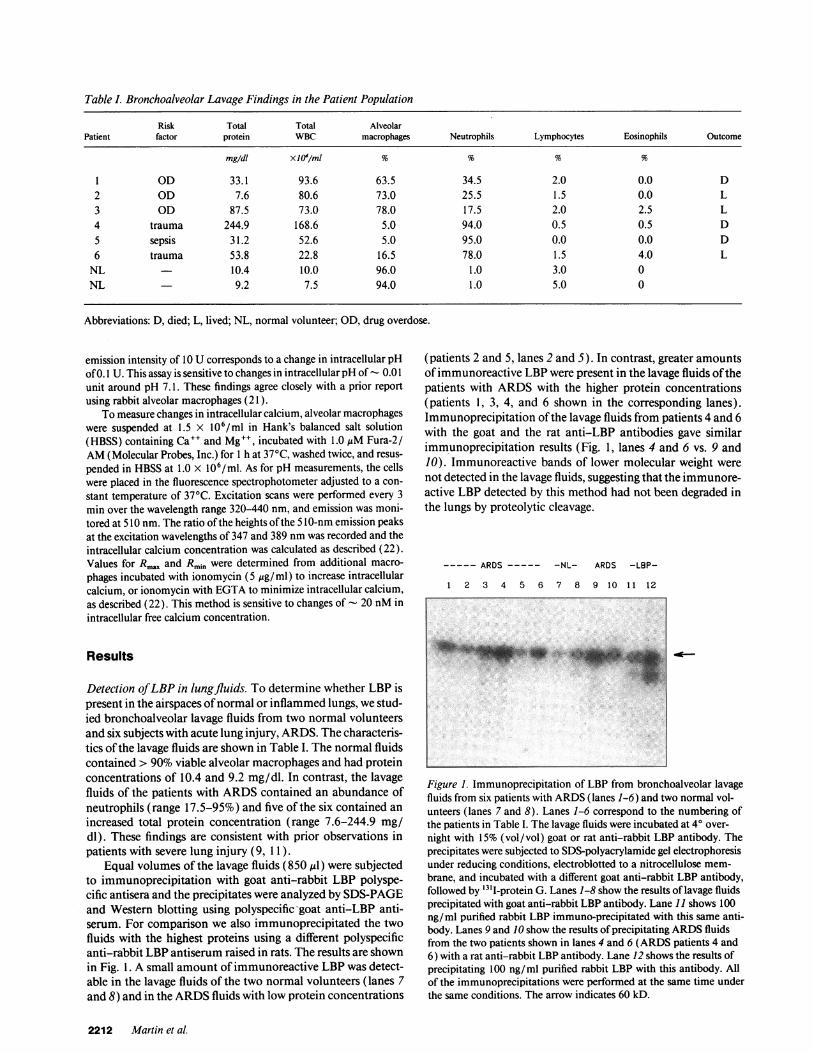
Table I. Bronchoalveolar Lavage Findings in the Patient Population
Risk Total Total AlveolarPatient factor protein WBC macrophages Neutrophils Lymphocytes Eosinophils Outcome
mg/dl XJ0'/ml % % % %
1 OD 33.1 93.6 63.5 34.5 2.0 0.0 D2 OD 7.6 80.6 73.0 25.5 1.5 0.0 L3 OD 87.5 73.0 78.0 17.5 2.0 2.5 L4 trauma 244.9 168.6 5.0 94.0 0.5 0.5 D5 sepsis 31.2 52.6 5.0 95.0 0.0 0.0 D6 trauma 53.8 22.8 16.5 78.0 1.5 4.0 L
NL 10.4 10.0 96.0 1.0 3.0 0NL 9.2 7.5 94.0 1.0 5.0 0
Abbreviations: D, died; L, lived; NL, normal volunteer; OD, drug overdose.
emission intensity of 10 U corresponds to a change in intracellular pHof 0.1 U. This assay is sensitive to changes in intracellular pH of - 0.01unit around pH 7.1. These findings agree closely with a prior reportusing rabbit alveolar macrophages (21 ).
To measure changes in intracellular calcium, alveolar macrophageswere suspended at 1.5 x 10⁶/ml in Hank's balanced salt solution(HBSS) containing Ca"+ and Mg", incubated with 1.0 MMFura-2/AM(Molecular Probes, Inc.) for 1 h at 370C, washed twice, and resus-pended in HBSSat 1.0 X 10⁶/ml. As for pH measurements, the cellswere placed in the fluorescence spectrophotometer adjusted to a con-stant temperature of 370C. Excitation scans were performed every 3min over the wavelength range 320-440 nm, and emission was moni-tored at 510 nm. The ratio of the heights of the 510-nm emission peaksat the excitation wavelengths of 347 and 389 nmwas recorded and theintracellular calcium concentration was calculated as described (22).Values for Rma, and Rmin were determined from additional macro-phages incubated with ionomycin (5 gg/ml) to increase intracellularcalcium, or ionomycin with EGTAto minimize intracellular calcium,as described (22). This method is sensitive to changes of - 20 nMinintracellular free calcium concentration.
Results
Detection of LBP in lungfluids. To determine whether LBP ispresent in the airspaces of normal or inflammed lungs, we stud-ied bronchoalveolar lavage fluids from two normal volunteersand six subjects with acute lung injury, ARDS. The characteris-tics of the lavage fluids are shown in Table I. The normal fluidscontained > 90% viable alveolar macrophages and had proteinconcentrations of 10.4 and 9.2 mg/dl. In contrast, the lavagefluids of the patients with ARDScontained an abundance ofneutrophils (range 17.5-95%) and five of the six contained anincreased total protein concentration (range 7.6-244.9 mg/dl). These findings are consistent with prior observations inpatients with severe lung injury (9, 11).
Equal volumes of the lavage fluids (850 MlI) were subjectedto immunoprecipitation with goat anti-rabbit LBP polyspe-cific antisera and the precipitates were analyzed by SDS-PAGEand Western blotting using polyspecific -goat anti-LBP anti-serum. For comparison we also immunoprecipitated the twofluids with the highest proteins using a different polyspecificanti-rabbit LBP antiserum raised in rats. The results are shownin Fig. 1. A small amount of immunoreactive LBP was detect-able in the lavage fluids of the two normal volunteers (lanes 7and 8) and in the ARDSfluids with low protein concentrations
(patients 2 and 5, lanes 2 and 5). In contrast, greater amountsof immunoreactive LBP were present in the lavage fluids of thepatients with ARDSwith the higher protein concentrations(patients 1, 3, 4, and 6 shown in the corresponding lanes).Immunoprecipitation of the lavage fluids from patients 4 and 6with the goat and the rat anti-LBP antibodies gave similarimmunoprecipitation results (Fig. 1, lanes 4 and 6 vs. 9 and10). Immunoreactive bands of lower molecular weight werenot detected in the lavage fluids, suggesting that the immunore-active LBP detected by this method had not been degraded inthe lungs by proteolytic cleavage.
--.---ARDS-.--- -NL- ARDS -LBP-
1 2 3 4 5 6 7 8 9 10 11 12
Figure 1. Immunoprecipitation of LBP from bronchoalveolar lavagefluids from six patients with ARDS(lanes 1-6) and two normal vol-unteers (lanes 7 and 8). Lanes 1-6 correspond to the numbering ofthe patients in Table I. The lavage fluids were incubated at 40 over-night with 15% (vol/vol) goat or rat anti-rabbit LBP antibody. Theprecipitates were subjected to SDS-polyacrylamide gel electrophoresisunder reducing conditions, electroblotted to a nitrocellulose mem-brane, and incubated with a different goat anti-rabbit LBP antibody,followed by "3'I-protein G. Lanes 1-8 show the results of lavage fluidsprecipitated with goat anti-rabbit LBP antibody. Lane 11 shows 100ng/ml purified rabbit LBP immuno-precipitated with this same anti-body. Lanes 9 and 10 show the results of precipitating ARDSfluidsfrom the two patients shown in lanes 4 and 6 (ARDS patients 4 and6) with a rat anti-rabbit LBP antibody. Lane 12 shows the results ofprecipitating 100 ng/ml purified rabbit LBP with this antibody. Allof the immunoprecipitations were performed at the same time underthe same conditions. The arrow indicates 60 kD.
2212 Martin et al.
'a il I" .: 11111h'iw
-ip- a
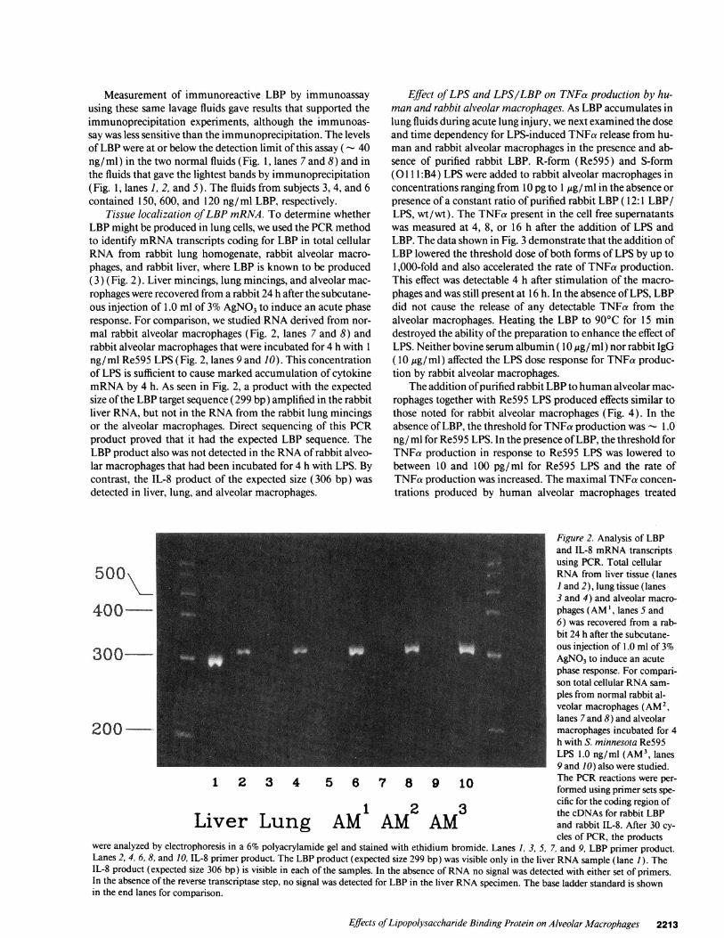
Measurement of immunoreactive LBP by immunoassayusing these same lavage fluids gave results that supported theimmunoprecipitation experiments, although the immunoas-say was less sensitive than the immunoprecipitation. The levelsof LBP were at or below the detection limit of this assay ( - 40ng/ml) in the two normal fluids (Fig. 1, lanes 7 and 8) and inthe fluids that gave the lightest bands by immunoprecipitation(Fig. 1, lanes 1, 2, and 5). The fluids from subjects 3, 4, and 6contained 150, 600, and 120 ng/ml LBP, respectively.
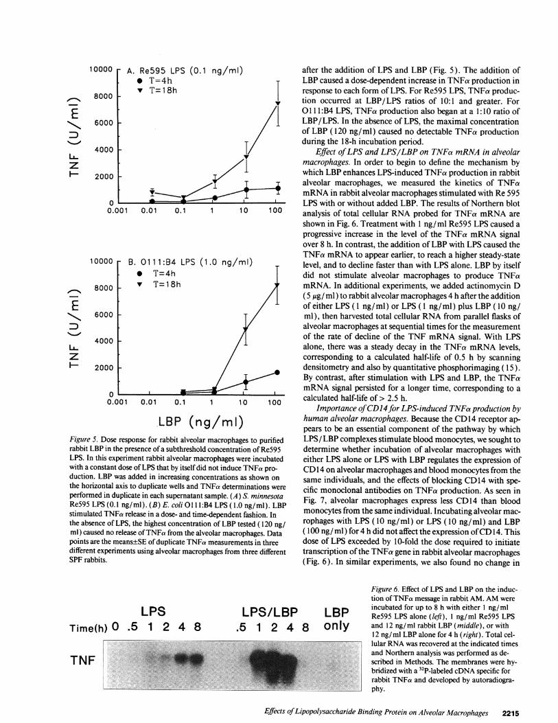
Tissue localization of LBP mRNA. To determine whetherLBP might be produced in lung cells, we used the PCRmethodto identify mRNAtranscripts coding for LBP in total cellularRNA from rabbit lung homogenate, rabbit alveolar macro-phages, and rabbit liver, where LBP is known to be produced(3) (Fig. 2). Liver mincings, lung mincings, and alveolar mac-rophages were recovered from a rabbit 24 h after the subcutane-ous injection of 1.0 ml of 3%AgNO3to induce an acute phaseresponse. For comparison, we studied RNAderived from nor-mal rabbit alveolar macrophages (Fig. 2, lanes 7 and 8) andrabbit alveolar macrophages that were incubated for 4 h with 1ng/ml Re595 LPS (Fig. 2, lanes 9 and 10). This concentrationof LPS is sufficient to cause marked accumulation of cytokinemRNAby 4 h. As seen in Fig. 2, a product with the expectedsize of the LBP target sequence (299 bp) amplified in the rabbitliver RNA, but not in the RNAfrom the rabbit lung mincingsor the alveolar macrophages. Direct sequencing of this PCRproduct proved that it had the expected LBP sequence. TheLBP product also was not detected in the RNAof rabbit alveo-lar macrophages that had been incubated for 4 h with LPS. Bycontrast, the IL-8 product of the expected size (306 bp) wasdetected in liver, lung, and alveolar macrophages.
Effect of LPS and LPS/LBP on TNFa production by hu-man and rabbit alveolar macrophages. As LBP accumulates inlung fluids during acute lung injury, we next examined the doseand time dependency for LPS-induced TNFa release from hu-man and rabbit alveolar macrophages in the presence and ab-sence of purified rabbit LBP. R-form (Re595) and S-form(O1 1:B4) LPS were added to rabbit alveolar macrophages inconcentrations ranging from 10 pg to 1 ,ug/ ml in the absence orpresence of a constant ratio of purified rabbit LBP ( 12:1 LBP/LPS, wt/wt). The TNFa present in the cell free supernatantswas measured at 4, 8, or 16 h after the addition of LPS andLBP. The data shown in Fig. 3 demonstrate that the addition ofLBP lowered the threshold dose of both forms of LPS by up to1,000-fold and also accelerated the rate of TNFa production.This effect was detectable 4 h after stimulation of the macro-phages and was still present at 16 h. In the absence of LPS, LBPdid not cause the release of any detectable TNFa from thealveolar macrophages. Heating the LBP to 90°C for 15 mindestroyed the ability of the preparation to enhance the effect ofLPS. Neither bovine serum albumin (10 utg/ml) nor rabbit IgG(10 ,ig/ml) affected the LPS dose response for TNFa produc-tion by rabbit alveolar macrophages.
The addition of purified rabbit LBP to human alveolar mac-rophages together with Re595 LPS produced effects similar tothose noted for rabbit alveolar macrophages (Fig. 4). In theabsence of LBP, the threshold for TNFa production was - 1.0ng/ ml for Re595 LPS. In the presence of LBP, the threshold forTNFa production in response to Re595 LPS was lowered tobetween 10 and 100 pg/ml for Re595 LPS and the rate ofTNFa production was increased. The maximal TNFa concen-trations produced by human alveolar macrophages treated
Figure 2. Analysis of LBPand IL-8 mRNAtranscriptsusing PCR. Total cellular
500 RNAfrom liver tissue (lanes1 and 2), lung tissue (lanes3 and 4) and alveolar macro-
400 phages (AM', lanes S and6) was recovered from a rab-bit 24 h after the subcutane-ous injection of 1.0 ml of 3%
300 AgNO3to induce an acutephase response. For compari-son total cellular RNAsam-ples from normal rabbit al-veolar macrophages (AM2,lanes 7 and 8) and alveolar
200 _ macrophages incubated for 4h with S. minnesota Re595LPS 1.0 ng/ml (AM3, lanes9 and 10) also were studied.
1 2 3 4 5 6 7 8 9 10 The PCRreactions were per-formed using primer sets spe-cific for the coding region of
1 2 3 the cDNAs for rabbit LBPLiver Lung AM AM AM and rabbit IL-8. After 30 cy-
cles of PCR, the productswere analyzed by electrophoresis in a 6% polyacrylamide gel and stained with ethidium bromide. Lanes 1, 3, 5, 7, and 9, LBP primer product.Lanes 2, 4, 6, 8, and 10, IL-8 primer product. The LBP product (expected size 299 bp) was visible only in the liver RNAsample (lane 1). TheIL-8 product (expected size 306 bp) is visible in each of the samples. In the absence of RNAno signal was detected with either set of primers.In the absence of the reverse transcriptase step, no signal was detected for LBP in the liver RNAspecimen. The base ladder standard is shownin the end lanes for comparison.
Effects of Lipopolysaccharide Binding Protein on Alveolar Macrophages 2213

A. 4ho Re595 LPS
- * LPS/LBP
6
5
4
3
2
f0, fr.t::,::&
-1 1 -10 -9 -8 -7 -6
D. 4ho 01 11B:4 LPS* LPS/LBP
,--,
46--1,0 -1 1 -10 -9 -8 -7 -6
0
6
5
4
3
2
B. 8h 6
5
4
3
2K.
0 - 1 1 -10 -9 -8 -70
-6
_ E. 8h 6
5
4
3
2
0o ' I0 -11 -10 -9 -8 -7 -6
C. 16h
0 1/ 1 I I I0 -1 1 -10 -9 -8 -7 -6
F. 16F
0 -11 -10 -9 -8 -7 -6
LPS (log g/ml)
0 -10 -9 -8 -7
LBP (log g/ml)
LPS (log g/ml)
0 -10 -9 -8 -7
LBP (log g/ml)
LPS (log g/ml)
0 -10 -9 -8 -7
LBP (log g/ml)Figure 3. The production of TNFa by rabbit alveolar macrophages stimulated with LPS and purified rabbit LBP. Rabbit alveolar macrophageswere incubated for 4 h (A and D), 8 h (B and E), or 16 h (C and F) with increasing concentrations of LPS in the absence or presence of a12-fold higher concentration (wt/wt) of purified rabbit LBP as indicated in the legend. LBP was not added at the highest concentrations of addedLPS. (A-C) S. Minnesota Re595 LPS (rough form) is shown. (D-F) E. coli 01 ll:B4 LPS (smooth form) is shown. The addition of LBP withLPS caused TNFa production to begin sooner and at a lower LPS concentration than with LPS alone. LBP did not stimulate TNFa productionin the absence of LPS. Data points are the means of duplicate TNFa measurements. This experiment was repeated four times with similarresults using alveolar macrophages from different animals.
with LPS and LBP approached those seen with human alveolarmacrophages incubated with a 100-fold higher concentrationof the Re595 LPS.
Wenext sought to define the LBP dose response for en-hancing LPS-induced TNFa release by alveolar macrophages.
Concentrations of Re595 LPS (0.1 ng/ml) or 01 1lB:4 LPS( 1.0 ng/ml) that induced little or no TNFa release from rabbitalveolar macrophages during 18 h of incubation were mixedwith varying amounts of LBP (0.012-120 ng/ml). TNFa re-lease in the macrophage supernatants was measured 4 or 18 h
A. 4h- 0 Re595 LPS- . LPS/LBP
0 -11 -10 -9 -8 -7 -6
6
5
4
3
2
0
B. 8h
I'LI0 -11 -10 -9 -8 -7 -6
LPS dose (g/ml)
/ I ' '
0 -10 -9 -8 -7
LBP dose (log g/ml)
LPS dose (log g/ml)
X , /
0 - 10 -9 -8 -7
LBP dose (log g/ml)
LPS dose (log g/ml)
0 -10 -9 -8 -7
LBP dose (log g/ml)
Figure 4. The production of TNFa by human alveolar macrophages stimulated with LPS and purified rabbit LBP. The format is the same as thatin Fig. 3. Human alveolar macrophages were incubated with rough form S. Minnesota Re595 LPS for 4, 8, or 16 h in the absence or presenceof a 12-fold excess of LBP as indicated. As with rabbit alveolar macrophages, LBP caused TNFa production to begin sooner and at lower con-
centrations of LPS than with LPS alone. Data points are the means of duplicate TNFa measurements. This experiment was performed four timesusing alveolar macrophages from four different normal human volunteers with similar results.
2214 Martin et al.
6
5
4
3
2
0
NE
x
LEz
6
E 5"Nqo 4
z0
-= 6EN 5
pi0 4
F-cx 3
22U- 1
z O C. 16h6
5
4
3
2
0
0
__e'1-0..
I I
10000
8000 -
L_z
6000
4000
A. Re595 LPS (0.1_ T-A Invv
1 -=±11
T=18h
2000 -
0 Lo.0C 0.01 0.1
10000 B. 01 1 1:B4 LPS* T=4h
onnfl v T=18h
ED
z
ouuu
6000
4000
2000
0 -0.001
(1
0.01 0.1
LBP (ncFigure 5. Dose response for rabbit alveolarrabbit LBP in the presence of a subthresholdLPS. In this experiment rabbit alveolar maciwith a constant dose of LPS that by itself didduction. LBP was added in increasing conccthe horizontal axis to duplicate wells and TPperformed in duplicate in each supernatant sRe595 LPS (0.1 ng/ml). (B) E. coli 011 l:Bstimulated TNFa release in a dose- and time-the absence of LPS, the highest concentrationml) caused no release of TNFa from the alvepoints are the means±SE of duplicate TNFadifferent experiments using alveolar macroplSPF rabbits.
ng/mi) after the addition of LPS and LBP (Fig. 5). The addition ofT- LBP caused a dose-dependent increase in TNFa production in
response to each form of LPS. For Re595 LPS, TNFa produc-I tion occurred at LBP/LPS ratios of 10:1 and greater. For
0 1 1 1:B4 LPS, TNFa production also began at a 1:10 ratio ofLBP/LPS. In the absence of LPS, the maximal concentrationof LBP (120 ng/ml) caused no detectable TNFa productionduring the 18-h incubation period.
Effect of LPS and LPS/LBP on TNFa mRNAin alveolarmacrophages. In order to begin to define the mechanism by
T which LBP enhances LPS-induced TNFa production in rabbitI1 alveolar macrophages, we measured the kinetics of TNFa
mRNAin rabbit alveolar macrophages stimulated with Re 595LPS with or without added LBP. The results of Northern blot
1 1 0 100 analysis of total cellular RNAprobed for TNFa mRNAareshown in Fig. 6. Treatment with 1 ng/ml Re595 LPS caused aprogressive increase in the level of the TNFa mRNAsignalover 8 h. In contrast, the addition of LBP with LPS caused theTNFa mRNAto appear earlier, to reach a higher steady-state
.0 ng/mlI) level, and to decline faster than with LPS alone. LBP by itselfdid not stimulate alveolar macrophages to produce TNFamRNA. In additional experiments, we added actinomycin D( 5 ,ug/ ml) to rabbit alveolar macrophages 4 h after the additionof either LPS ( 1 ng/ml) or LPS ( 1 ng/ml) plus LBP ( 10 ng/ml), then harvested total cellular RNAfrom parallel flasks ofalveolar macrophages at sequential times for the measurementof the rate of decline of the TNF mRNAsignal. With LPSalone, there was a steady decay in the TNFa mRNAlevels,corresponding to a calculated half-life of 0.5 h by scanningdensitometry and also by quantitative phosphorimaging ( 15).By contrast, after stimulation with LPS and LBP, the TNFamRNAsignal persisted for a longer time, corresponding to a
1 1 0 100 calculated half-life of > 2.5 h.Importance of CD14for LPS-induced TNFaproduction by
/ mI) human alveolar macrophages. Because the CD14 receptor ap-pears to be an essential component of the pathway by which
macrophages to purified LPS/LBP complexes stimulate blood monocytes, we sought toconcentration of Re595 determine whether incubation of alveolar macrophages withrophages were incubated either LPS alone or LPS with LBP regulates the expression ofnot induce TNFa pro- CD14 on alveolar macrophages and blood monocytes from theentrations as shown on same individuals, and the effects of blocking CD14 with spe-,4Fa determinations were cific monoclonal antibodies on TNFa production. As seen inample. (A) S. minnesota Fig. 7, alveolar macrophages express less CD14 than blood
de pendent fashion. InP monocytes from the same individual. Incubating alveolar mac-
n of LBP tested (120 ng/ rophages with LPS (10 ng/ml) or LPS (10 ng/ml) and LBPolar macrophages. Data ( 100 ng/ml) for 4 h did not affect the expression of CDl 4. Thismeasurements in three dose of LPS exceeded by 10-fold the dose required to initiatehages from three different transcription of the TNFa gene in rabbit alveolar macrophages
(Fig. 6). In similar experiments, we also found no change in
LPSTime(h) O .5 1 2 4 8
LPS/LBP.5 1 2 4 8
LBPonly
TNF
L-
Figure 6. Effect of LPS and LBP on the induc-tion of TNFa message in rabbit AM. AMwereincubated for up to 8 h with either 1 ng/mlRe595 LPS alone (left), 1 ng/ml Re595 LPSand 12 ng/ml rabbit LBP (middle), or with12 ng/ml LBP alone for 4 h (right). Total cel-lular RNAwas recovered at the indicated timesand Northern analysis was performed as de-scribed in Methods. The membranes were hy-bridized with a 32P-labeled cDNAspecific forrabbit TNFa and developed by autoradiogra-phy.
Effects of Lipopolysaccharide Binding Protein on Alveolar Macrophages 2215
-400b-
)1
1. ,''. ...d .A-"I
W. -Vf

io 162FL2
104
veolar macrophages, as these events might contribute to signaltransduction pathways that mediate cytokine production afterligation of CD14. As seen in Fig. 9, neither LPS nor LPS/LBPcomplexes affected either [Ca t ] or pHi in rabbit alveolar mac-rophages within the first 30 min of incubation. These experi-ments were conducted at the concentrations of LPS (1 -10 ng/ml) and LBP ( 10-100 ng/ml) that were shown by Northern
A
E
ILzI--
Figure 7. The effect of LPS and LPS/LBP on CD14 expression byhuman monocytes (A) and alveolar macrophages (B). The cells werelabeled with MY4by indirect immunofluorescence and analyzed byflow cytometry. The vertical axis shows cell number in each fluores-cence channel; the horizontal axis is a log1o scale of red fluorescenceintensity. In each panel the labels refer to cells treated as follows: 1,untreated cells, 4 h, labeled with the second antibody only (goatanti-mouse IgG); 2, untreated cells, 4 h; 3, LPS 10 ng/ml, 4 h; 4, LPS10 ng/ml + LBP 100 ng/ml, 4 h.
the expression of CD14 on rabbit alveolar macrophages (notshown). In comparison, the CD14 fluorescence profile ofblood monocytes from the same individual shifted slightly tothe left after incubation with LPS and LPS/LBP, suggestingdown-regulation of the CD14 receptor on some of the cells.
To test the importance of CDl 4 on mediating the responsesof human alveolar macrophages to LPS and LPS/LBP com-plexes, we incubated alveolar macrophages with LPS or LPS/LBP in the presence of saturating concentrations of 28C5 (5Atg/ml), a specific murine monoclonal antibody raised againstpurified recombinant human CD14 that was expressed inCHOcells. This antibody labels human alveolar macrophagesand blood monocytes, immunoprecipitates a single band con-sistent with CD14 from HL60 cells differentiated with1,25 (OH )2-vitamin D, and competes with radiolabeled 3CI0,another anti-CD 14 antibody (6), in labeling HL60 cells. Asshown in Fig. 8, the antibody 28C5 inhibited TNFa productionby human alveolar macrophages incubated with LPS alone,and completely blocked the enhancement of TNFaproductioncaused by LBP. We obtained similar results in this systemwhen we used the 3C10 antibody. The 28C5 antibody had noeffect on the production of TNFa by alveolar macrophages inresponse to heat-killed Staphylococcus aureus (Fig. 8).
Effect ofLPS and LBPon intracellular calcium andpH. Wenext asked whether the addition of LPS or LPS/LBP com-plexes might cause changes in intracellular Ca"+ or pH in al-
B
EN
z
1000
800
600 1-
400 k200
0
1000
800
600
0o
0F
V
400 _
2C
C
EN
z
4000 -
3000 _
2000 -
1000 -
0
* LPS0 LPS+28C5V LPS+aHCG
- Jo
I I I I I I
.01 0.1 1 10 100 1000
LPS (ng/ml)
LPS/LBPLPS/LBP+28C5LPS/LBP+aHCG
V
I II
1I
I,'-0o0
I
& AM only* AM+HKSA0 AM+HKSA+28C5
o0
AI I I I I
0 10:1 20:1 100:1
Bacteriac:AM ratioFigure 8. The effect of anti-CD 14 antibody on the response of humanalveolar macrophages to LPS and LPS/LBP. (A) Humanalveolarmacrophages were incubated for 8 h with increasing concentrationsof LPS with or without the monoclonal antibodies 28C5 (5 ,4g/ml),which labels CD14, or an irrelevant antibody (anti-human chorionicgonadotropin) of the same isotype. (B) Alveolar macrophages fromthe same volunteer incubated with LPS and 10-fold higher concen-
trations of LPB (wt/wt) with or without 28C5 or anti-human chori-onic gonadotropin. (C) Alveolar macrophages from the same volun-teer incubated for 18 h with increasing concentrations of heat-killedS. aureus with or without 28C5. The inhibitory effects of 28C5 were
tested in three experiments with alveolar macrophages from differentvolunteers with similar results.
a)
Ez
0a)
250
0
L
0.01 0.1 1 10 100 1000
LPS (ng/ml)
2216 Martin et al.
DO -
0 L-I

1000 A 3 B Figure 9. The effect of LPS and LBP on
900 [ B intracellular free calcium (A) and pH(B) in rabbit AM. (A) To measure in-
800 7.2 * tracellular free calcium, alveolar mac-
j 700 *LP/LBP lano rophages were loaded with 1.0 AM_ 600 Fura2/AM then stimulated with Re595
, 500s 7.1 LPS ( 1 ng/ml) and purified rabbit LBP+ LPS/LBP (10 ng/ml). The excitation wavelength+ 4300 was set at 510 nm. The ratio of the3_ 7.0 emission intensity at 347/389 nm was
200 used to calculate the intracellular cal-
100 cium concentration, shown on the ver-
0 6.9 t I tical axis. The open circles show the0 5 10 15 20 25 30 0 5 10 15 20 25 30 35 40 response of cells incubated without LPS
Time (min ) Time (min ) or LBP. The response in cells treatedwith LPS alone ( 1 ng/ml) was identicalto the data shown for untreated cells.
When ionomycin was added (1lAg/ml), the intracellular calcium concentration increased abruptly to over 1 ,M. (B) To measure intracellularpH, AMwere loaded with 0.5 tm BCECF/AMand stimulated with Re595 LPS ( 1 ng/ml) and purified rabbit LBP ( 10 ng/ml). The excitationwavelength was set at 505 nm and the intensity of the emission peak at 530 was recorded. Intracellular pH, (vertical axis) was calculated froma standard curve derived by incubating additional cells in high potassium buffers of known pH. Iono denotes the addition of ionomycin (1OAg/ ml) as a positive stimulus. The open circles show the response of cells in the absence of LPS and LBP. The response in cells treated with LPSalone (1 ng/ ml) was identical to the data shown for untreated cells. The experiments in A and B were performed three different times usingalveolar macrophages from different rabbits with similar results.
analysis to result in substantial TNFa message production byRe595 LPS alone and enhanced TNFa message productionwith added LBP (Fig. 3).
Discussion
The major goals of this study were first, to investigate whetherimmunoreactive LBP accumulates in fluid recovered fromnormal or injured human lungs, and second to investigate theeffect of LBP on LPS-induced TNFa production by rabbit andhuman alveolar macrophages. The results indicate that immu-noreactive LBP is normally present in very low concentrationsin the alveolar fluid, and that its concentration rises substan-tially in patients with lung injury. In rabbits, LBP in lung fluidsappears to originate from an extrapulmonary source, as we didnot detect LBP mRNAin RNAextracted either from rabbitlung tissue or from normal or LPS-stimulated rabbit alveolarmacrophages. LBP enhances the response of human and rabbitalveolar macrophages to LPS. In the presence of LBPand LPS,the maximal TNFa response by alveolar macrophages occursearlier than with LPS alone and it also occurs at lower concen-trations of both smooth and rough form LPS. The CD14 recep-tor plays an important role in mediating the effects of LPS andLBP on alveolar macrophages, as the effects of LPS and LBPcan be inhibited by anti-CD 14 monoclonal antibodies. Thus,LBP that accumulates in the lungs during lung injury may havean important regulatory effect on the response of alveolar mac-rophages to endotoxin that enters the airspaces.
The origin of the LBP in the lungs and the mechanism bywhich it enters the lungs is of considerable interest. The resultsof the experiments using the sensitive PCRmethod indicatethat in rabbits the LBP present in lung lavage fluid probablyderives from the liver and not from the lungs. Although alveo-lar macrophages are capable of producing a variety of proteins,mRNAtranscripts for LBP were not detected in normal rabbitalveolar macrophages, alveolar macrophages obtained during asystemic acute-phase response, or alveolar macrophages stimu-lated with LPS. In addition, we have not been able to detect
LBP activity by immunoassay in the supernatants of LPS-stim-ulated human or rabbit alveolar macrophages (not shown).The data confirms an earlier study in which LPB message wasdetected in rabbit liver RNAby Northern analysis (3). LBP(60 kD) is similar in molecular mass to albumin (67 kD),which constitutes - 50% of the protein in bronchoalveolar la-vage fluid (23). Whenthe permeability of the epithelial barrierin the lungs is altered, much higher molecular mass proteins,e.g., IgM (900 kD), that normally are excluded from the alveo-lar spaces are readily detectable in lavage fluid (9, 23, 24).Under these conditions, proteins of the size of LBP would beexpected to pass rapidly from the plasma compartment into thealveolar spaces. These lines of evidence support the interpreta-tion that LBP is synthesized in the liver and that it accumulatesin the lungs as lung epithelial permeability increases in re-sponse to injury.
Our findings have important implications for the mecha-nisms that regulate the production of cytokines such as TNFawithin the lungs during inflammatory reactions. TNFa isthought to be an important mediator of the sepsis syndrome inanimal models (25-27), and sepsis syndrome both precedesand complicates ARDSin many patients (28). The alveolarmacrophage plays a prominent role in the response of the lungto endotoxins because it responds to LPS by producing anarray of cytokines that modify the inflammatory response. Inaddition to TNFa, these include IL-6 (29), IL-8 (30) and to alesser extent IL- 1I3 (31, 32). Although other cells of the alveolarenvironment, including epithelial cells (33), fibroblasts (34),endothelial cells ( 16), and mesothelial cells ( 35 ) have the ca-pacity to produce some cytokines, particularly IL-8, the alveo-lar macrophage appears to be the primary cell in the airspacethat recognizes and responds to LPS (reviewed in reference 35).
Our results suggest that LBP may play a major role in regu-lating the sensitivity of alveolar macrophages to LPS by bothlowering the threshold and accelerating the time course of cyto-kine production. A previous study showed that LBP augmentsthe response of elicited rabbit peritoneal exudate macrophagesto smooth and rough form LPS (7). The present studies show
Effects of Lipopolysaccharide Binding Protein on Alveolar Macrophages 2217

that LBP enhances the effect of LPS on a nonelicited residentmacrophage population in the lungs of both rabbits, fromwhich the LBPwas derived, and normal humans. As lung mac-rophages may be an important local source of TNFa duringacute lung injury, the accumulation of LBP in the inflammedairspaces may provide an endogenous mechanism for amplify-ing or regulating the response of lung macrophages to localconcentrations of LPS within the lungs.
The intracellular pathways by which LBP accentuates theeffect of LPS on alveolar macrophage warrant further study.The binding of LPS/ LBPcomplexes to surface CD14 on mono-cytes appears to be the first step in the recognition of LPS/ LBPcomplexes by monocytic cells (6). The present data confirmprior reports that CD14 is present on alveolar macrophages(36, 37), although the expression of CD14 on alveolar macro-phages is considerably less than on peripheral blood mono-cytes. Wefound that LPS and LPS/LBP induce TNFa geneexpression without affecting the density of CD14 on alveolarmacrophages. Wealso found that specific monoclonal antibod-ies directed at epitopes on CD14 blocked the effect of LPS andLPS/LBP on inducing TNFa expression. Although other cellsurface proteins have been identified that maycontribute to theeffects of endotoxin in other systems (38-40), our data supportthe primary importance of the CD14pathway in mediating theresponse of alveolar macrophages to LPS. Previous studiesshowed that antibodies to CD14 prevent the binding of LPS/LBP bearing particles to the surface of monocyte-derived mac-rophages (5) and block the production of TNFa in wholeblood in response to LPS (6). Our observation that an anti-body directed at CD14 inhibits the response to LPS as well asLPS/LBP strengthens the interpretation that CD14 functionsas an important part of a membrane complex that mediates theeffects of LPS as well as LPS/LBP complexes.
The signals that follow binding of LPS/LBP complexes toCD14 remain uncertain. CD14 is anchored in the plasmamembrane by a phosphatidylinositol tail (41). Phosphatidyl-inositol-anchored proteins are not typical Gprotein-linked re-ceptors and it remains uncertain whether receptors such asCD14 transmit intracellular signals directly (42). An alterna-tive possibility for CD14-mediated triggering of macrophages isthat the binding of LPS/LBP complexes to CD14 does nottransmit a signal directly, but regulates the density or affinity ofa nearby second receptor that in turn transmits a signal intra-cellularly. Recent evidence provides indirect support for thispossibility, as ligation of CD14 upregulates adherence recep-tors on monocytes causing aggregation (43). Wefound thatneither LPS nor LPS/ LBP complexes affect the initial concen-trations of calcium of pH in rabbit alveolar macrophages, sug-gesting that these pathways are unlikely to contribute to intra-cellular signaling initiated by ligation of CD14 by LPS/LBPcomplexes. The effects of LPS/LBP complexes and CD14 onother signaling pathways associated with activation in macro-phages, such as protein kinase C activation (44, 45) and tyro-sine phosphorylation (46, 47), warrant further study.
Wefound that one important intracellular consequence ofthe signaling events initiated by LPS/LBP complexes is an in-crease in the kinetics of TNFa mRNAmessage expression. Inalveolar macrophages treated with LPS and LBP, TNFamRNAappeared earlier, accumulated to higher levels, andthen disappeared earlier than in macrophages treated with LPSalone. The regulation of TNFa expression is complex, and de-pends on promoter sequences in the TNFa gene, AU rich se-
quences in the 3'-untranslated region of the TNFa mRNAthatboth destabilize the mRNAand repress its translation (48, 49),and ribonuclease activity that destroys the mRNA(50). Theearlier appearance of TNFa mRNAthat we observed suggeststhat LBP/LPS complexes increase the rate of transcription ofthe TNFa gene above that seen with LPS alone, although fur-ther direct analysis of gene transcription is necessary to provethis. The observation that TNFa mRNAhalf-life is prolongedby LPS/LBP complexes is supported by recent experimentswith elicited peritoneal macrophages (51 ). The data do notestablish whether the apparent increase in TNFa mRNAt112 isa unique effect of LBP or a consequence of increased intracel-lular delivery of LPS in the presence of LBP.
Although these studies indicate that LBP enhances LPS-driven TNFa production by normal alveolar macrophages, itremains to be determined whether the effect of LBP is similarfor other cytokines produced by alveolar macrophages. Inother studies, we have found that LBP also accelerates the pro-duction of IL-8 by rabbit alveolar macrophages and the humanmonocytic cell line, THP- 1. Furthermore, the effects of LBPonimmunologically activated alveolar macrophages need to bestudied in detail. Because TNFa is an important mediator ofthe sepsis syndrome that both causes and complicates lung in-jury, the finding that LBP accumulates in the lung during in-flammation and regulates the production of TNFa by alveolarmacrophages is likely to have clinical importance. Strategies toblock the effects of LBP on alveolar macrophages may be use-ful in limiting some types of inflammatory reactions in thelungs.
Acknowledgments
Wethank Venus Wong, Steve Mongovin, John Ruzinski, Nguyet Ky,Loren Hatlen, and Nora Wolfson for expert technical assistance.
This study was supported in part by the Medical Research Serviceof the Department of Veterans Affairs and by grants AI-29103, HL-30542, AI-15 136, AI-25563, GM-28485, and GM-37696 from the Na-tional Institutes of Health.
References
1. Tobias, P. S., K. Soldau, and R. J. Ulevitch. 1986. Isolation ofalipopolysac-charide-binding acute phase reactant from rabbit serum. J. Exp. Med. 164:777-793.
2. Tobias, P. S., J. C. Mathison, and R. J. Ulevitch. 1988. A family of lipopoly-saccharide binding proteins involved in responses to Gram-negative sepsis. J.Biol. Chem. 263:13479-13481.
3. Ramadori, G., K.-H. Meyer zum Buschenfelde, P. S. Tobias, J. C. Mathi-son, and R. J. Ulevitch. 1990. Biosynthesis of lipopolysaccharide-binding proteinin rabbit hepatocytes. Pathobiology. 58:89-94.
4. Tobias, P. S., K. Soldau, and R. J. Ulevitch. 1989. Identification of a lipid Abinding site in the acute phase reactant lipopolysaccharide binding protein. J.Bio. Chem. 264:10867-10871.
5. Wright, S. D., P. S. Tobias, R. J. Ulevitch, and R. A. Ramos. 1989. Lipo-polysaccharide (LPS) binding protein opsonizes LPS-bearing particles for recog-nition by a novel receptor on macrophages. J. Exp. Med. 170:1231-1241.
6. Wright, S. D., R. A. Ramos, P. S. Tobias, R. J. Ulevitch, and J. C. Mathi-son. 1990. CD14, a receptor for complexes of lipopolysaccharide (LPS) and LPSbinding protein. Science (Wash. DC). 249:1431-1433.
7. Schumann, R. R., S. R. Leong, G. W. Flaggs, P. W. Gray, S. D. Wright, J. C.Mathison, P. S. Tobias, and R. J. Ulevitch. 1990. Structure and function oflipopolysaccharide binding protein. Science (Wash. DC). 249:1429-1431.
8. Lee, J.-D., K. Kato, P. S. Tobias, T. N. Kirkland, and R. J. Ulevitch. 1992.Transfection of CD14 into 70Z/3 cells dramatically enhances the sensitivity tocomplexes of lipopolysaccharide (LPS) and LPS binding protein. J. Exp. Med.175:1697-1705.
9. Holter, J. F., J. E. Weiland, E. R. Pacht, G. E. Gadek, and W. B. Davis.1986. Protein permeability in the adult respiratory distress syndrome: loss of sizeselectivity of the alveolar epithelium. J. Clin. Invest. 78:1513-1522.
2218 Martin et al.

10. Martin, T. R., G. Raugi, T. L. Merritt, and W. R. Henderson. 1987. Therelative contribution of leukotriene B4 to the neutrophil chemotactic activityproduced by the human alveolar macrophage. J. Clin. Invest. 80:1114-1124.
1 1. Martin, T. R., B. P. Pistorese, L. D. Hudson, and R. J. Maunder. 1991.Function of lung and blood neutrophils in patients with the adult respiratorydistress syndrome. Implications for the pathogenesis of lung infections. Am. Rev.Respir. Dis. 144:254-262.
12. Mathison, J. C., G. D. Virca, N. E. Wolfson, P. S. Tobias, K. Glaser, andR. J. Ulevitch. 1990. Adaptation to bacterial lipopolysaccharide controls lipopoly-saccharide induced tumor necrosis factor production in rabbit macrophages. J.Clin. Invest. 85:1108-1118.
13. Chirgwin, J. M., A. E. Preybyla, R. J. MacDonald, and W. J. Rutter. 1979.Isolation of biologically active ribonucleic acid from sources enriched in ribonu-clease. Biochemistry. 18:5294-5299.
14. Johnston, R. F., S. C. Pickett, and D. L. Barker. 1990. Autoradiographyusing storage phosphor technology. Electrophoresis. 1 1:355-360.
15. Thornton, A. J., R. M. Strieter, I. Lindley, M. Baggiolini, and S. L. Kun-kel. 1990. Cytokine-induced gene expression of a neutrophil chemotactic factor/IL8 in human hepatocytes. J. Immunol. 144:2609-2613.
16. Strieter, R. M., S. L. Kunkel, H. J. Showell, D. G. Remick, S. H. Phan,P. A. Ward, and R. M. Marks. 1989. Endothelial cell gene expression of a neutro-phil chemotactic factor by TNFa, LPS, and ILI-#. Science (Wash. DC).243:1467-1469.
17. Yoshimura, T., and N. Yuhki. 1991. Neutrophil attractant/activationprotein- I and monocyte chemoattractant protein- I in rabbit. cDNAcloning andtheir expression in spleen cells. J. Immunol. 146:3483-3488.
18. Ruff, M. R., and G. F. Gifford. 1981. Tumor necrosis factor. In Lympho-kines. Volume 2. E. Pick and M. Landy, editors. Academic Press, Inc., NewYork.235-272.
19. Beatty, P. G., J. A. Ledbetter, P. J. Martin, T. H. Price, and J. A. Hansen.1983. Definition of a common leukocyte cell-surface antigen (Lp95-150) asso-ciated with diverse cell mediated immune functions. J. Immunol. 131:2913-2918.
20. Wiener, I. D., and L. L. Hamm. 1989. Use of fluorescent dye BCECFtomeasure intracellular pH in cortical collecting tubule. Am. J. Physiol. 256:F957-964.
21. Bidani, A., S. E. S. Brown, T. A. Heming, R. Gurich, and T. D. Dubose, Jr.1989. Cytoplasmic pH in pulmonary macrophages: recovery from acid load isNa+ independent and NEMsensitive. Am. J. Physiol. 257:C65-C76.
22. Grynkiewicz, G., M. Peonie, and R. Y. Tsien. 1985. A new generation ofCa++ indicators with greatly improved fluorescence properties. J. Biol. Chem.260:3440-3450.
23. Martin, T. R., B. P. Pistorese, E. Y. Chi, R. B. Goodman, and M. A.Matthay. 1989. The effects of leukotriene B4 in the human lung: recruitment ofneutrophils into the alveolar spaces without a change in protein permeability. J.Clin. Invest. 84:1609-1619.
24. Schoene, R. B., E. R. Swenson, C. J. Pizzo, P. H. Hackett, R. C. Roach,W. J. Mills, W. R. Henderson, and T. R. Martin. 1988. The lung at high altitude:bronchoalveolar lavage in acute mountain sickness and pulmonary edema. J.Appl. Physiol. 64:2605-2613.
25. Beutler, B., I. W. Milsark, and A. C. Cerami. 1985. Passive immunizationagainst cachectin/ tumor necrosis factor protects mice from lethal effect of endo-toxin. Science (Wash. DC). 229:869-87 1.
26. Tracey, K. J., Y. Fong, D. G. Hesse, K. R. Manogue, A. T. Lee, G. C. Kuo,S. F. Lowry, and A. Cerami. 1987. Anti-cachectin/TNF monoclonal antibodiesprevent septic shock during lethal bacteraemia. Nature (Lond.). 330:662-664.
27. Mathison, J. C., N. E. Wolfson, and R. J. Ulevitch. 1988. Participation oftumor necrosis factor in the mediation of Gram negative bacterial lipopolysaccha-ride-induced injury in rabbits. J. Clin. Invest. 81:1925-1937.
28. Montgomery, A. B., M. A. Stager, C. J. Carrico, and L. D. Hudson. 1985.Causes of mortality in patients with the adult respiratory distress syndrome. Am.Rev. Respir. Dis. 132:485-489.
29. Kotloff, R. M., J. Little, and J. A. Elias. 1990. Human alveolar macro-phage and blood monocyte interleukin-6 production. Am. J. Respir. Cell. Mol.Biol. 3:497-505.
30. Rankin, J. A., I. Sylvester, S. Smith, T. Yoshimura, and E. J. Leonard.1990. Macrophages cultured in vitro release leukotriene B4 and neutrophil attrac-tant/activation protein (interleukin 8) sequentially in response to stimulationwith lipopolysaccharide and zymosan. J. Clin. Invest. 86:1556-1564.
31. Wewers, M. D., S. I. Rennard, A. J. Hance, P. B. Bitterman, and R. G.Crystal. 1984. Normal human alveolar macrophages obtained by bronchoalveo-lar lavage have a limited capacity to release interleukin-1. J. Clin. Invest.74:2208-22 18.
32. Bernaudin, J.-F., K. Yamauchi, M. D. Wewers, M. J. Tocci, V. J. Ferrans,and R. G. Crystal. 1988. Demonstration by in situ hybridization of dissimilarIL- I beta gene expression in human alveolar macrophages and blood monocytesin response to lipopolysaccharide. J. Immunol. 141:3822-3828.
33. Standiford, T. J., S. L. Kunkel, M. A. Basha, S. W. Chensue, J. P. LynchIII, G. B. Toews, J. Westwick, and R. M. Strieter. 1990. Interleukin-8 gene expres-sion by a pulmonary epithelial cell line. A model for cytokine networks in thelung. J. Clin. Invest. 86:1945-1953.
34. Strieter, R. M., S. H. Phan, H. J. Showell, D. G. Remick, J. P. Lynch, M.Genord, C. Raiford, M. Eskandari, R. M. Mards, and S. L. Kunkel. 1989. Mono-kine-induced neutrophil chemotactic factor gene expression in human fibro-blasts. J. Biol. Chem. 264:10621-10626.
35. Goodman, R. B., R. G. Wood, T. R. Martin, and G. T. Kinasewitz. 1992.Cytokine-stimulated human mesothelial cells produce chemotactic activity forneutrophils, including NAP1 /IL8. J. Immunol. 148:457-465.
36. Hoogsteden, H. C., J. J. M. van Dongen, P. T. W. van Hal, M. Delahaye,W. Hop, and C. Hilvering. 1989. Phenotype of blood monocytes and alveolarmacrophages in interstitial lung disease. Chest. 95:574-577.
37. Hance, A. J., S. Douches, R. J. Winchester, V. Ferrans, and R. G. Crystal.1985. Characterization of mononuclear phagocyte subpopulations in the humanlung by using monoclonal antibodies: changes in alveolar macrophage phenotypeassociated with pulmonary sarcoidosis. J. Immunol. 134:284-292.
38. Hailing, J. L., D. R. Hamill, M.-G. Lei, and D. C. Morrison. 1992. Identi-fication and characterization of lipopolysaccharide-binding proteins on humanperipheral blood cell populations. Infect. Immun. 60:845-852.
39. Golenbock, D. T., R. Y. Hampton, C. R. H. Raetz, and S. D. Wright.1990. Human phagocytes have multiple lipid A-binding sites. Infect. Immun.58:4069-4075.
40. Couturier, C., N. Haeffner-Cavaillon, M. Caroff, and M. D. Kazatchkine.1991. Binding sites for endotoxins (lipopolysaccharides) on human monocytes.J. Immunol. 147:1899-1904.
41. Haziot, A., S. Chen, E. Ferraro, M. G. Low, R. Silber, and S. M. Goyert.1988. The monocyte differentiation antigen, CD14, is anchored to the cell mem-brane by a phosphatidylinositol linkage. J. Immunol. 141:547-552.
42. Low, M. G., and A. R. Saltiel. 1988. Structural and functional roles ofglycosyl-phosphatidylinositol in membranes. Science (Wash. DC). 239:268-275.
43. Launer, R. P., Geha, R. S., and D. Vercelli. 1990. Engagement of themonocyte surface antigen CD14 induces lymphocyte function-associated anti-gen- I /intercellular adhesion molecule- I -dependent homotypic adhesion. J. Im-munol. 145:1390-1394.
44. Wightman, P. D., and C. R. H. Raetz. 1984. The activation of proteinkinase C by biologically active lipid moieties of lipopolysaccharide. J. Biol.Chem. 259:10048-10052.
45. Rosen, A., A. C. Nairn, P. Greengard, Z. A. Cohn, and A. Aderem. 1989.Bacterial lipopolysaccharide regulates the phosphorylation of the 68K proteinkinase C substrate in macrophages. J. Bio. Chem. 264:9118-9121.
46. Weinstein, S. L., M. R. Gold, and A. L. DeFranco. 1991. Bacterial lipo-polysaccharide stimulates protein tyrosine phosphorylation in macrophages.Proc. Natl. Acad. Sci. USA. 88:4148-4152.
47. Stefanova, I., V. Horejsi, J. Ansotegui, W. Knapp, and H. Stockinger.1991. GPI-anchored cell-surface molecules complexed to protein tyrosine ki-nases. Science (Wash. DC). 254:1016-1018.
48. Han, J., T. Brown, and B. Beutler. 1990. Endotoxin-responsive sequencescontrol cachectin/tumor necrosis factor biosynthesis at the translational level. J.Exp. Med. 171:465-475.
49. Han, J., J. G. Huez, and B. Beutler. 1991. Interactive effects of the tumornecrosis factor promoter and 3'-untranslated regions. J. Immunol. 146:1843-1848.
50. Beutler, B., P. Thompson, J. Keyes, K. Hagerty, and D. Crawford. 1988.Assay of a ribonuclease that preferentially hydrolyses mRNAscontaining cyto-kine-derived UA-rich instability sequences. Biochem. Biophys. Res. Commun.152:973-980.
51. Mathison, J. C., P. S. Tobias, E. Wolfson, and R. J. Ulevitch. 1992. Plasmalipopolysaccharide (LPS)-binding protein: a key component in macrophage rec-ognition of Gram-negative LPS. J. Immunol. 149:200-206.
Effects of Lipopolysaccharide Binding Protein on Alveolar Macrophages 2219