Lipid transfer from plants to arbuscular mycorrhiza fungi
33
*For correspondence: caroline. [email protected] † These authors contributed equally to this work Present address: ‡ Mass Spectrometry Metabolomics Facility, Cluster of Excellence on Plant Sciences, University of Cologne Biocenter, Cologne, Germany; § Analytics Facility, Center for Plant Molecular Biology, University of Tu ¨ bingen, Tu ¨ bingen, Germany Competing interests: The authors declare that no competing interests exist. Funding: See page 28 Received: 31 May 2017 Accepted: 13 July 2017 Published: 20 July 2017 Reviewing editor: Gary Stacey, University of Missouri, United States Copyright Keymer et al. This article is distributed under the terms of the Creative Commons Attribution License, which permits unrestricted use and redistribution provided that the original author and source are credited. Lipid transfer from plants to arbuscular mycorrhiza fungi Andreas Keymer 1† , Priya Pimprikar 1† , Vera Wewer 2‡ , Claudia Huber 3 , Mathias Brands 2 , Simone L Bucerius 1 , Pierre-Marc Delaux 4 , Verena Klingl 1 , Edda von Ro ¨ penack-Lahaye 5§ , Trevor L Wang 6 , Wolfgang Eisenreich 3 , Peter Do ¨ rmann 2 , Martin Parniske 1 , Caroline Gutjahr 1 * 1 Faculty of Biology, Genetics, LMU Munich, Biocenter Martinsried, Munich, Germany; 2 Institute of Molecular Physiology and Biotechnology of Plants, University of Bonn, Bonn, Germany; 3 Biochemistry, Technical University Munich, Garching, Germany; 4 Laboratoire de Recherche en Sciences Ve ´ ge ´ tale, Centre National de la Recherche Scientifique, Toulouse, France; 5 Faculty of Biology, Plant Sciences, LMU Munich, Biocenter Martinsried, Munich, Germany; 6 John Innes Centre, Norwich Research Park, Norwich, United Kingdom Abstract Arbuscular mycorrhiza (AM) symbioses contribute to global carbon cycles as plant hosts divert up to 20% of photosynthate to the obligate biotrophic fungi. Previous studies suggested carbohydrates as the only form of carbon transferred to the fungi. However, de novo fatty acid (FA) synthesis has not been observed in AM fungi in absence of the plant. In a forward genetic approach, we identified two Lotus japonicus mutants defective in AM-specific paralogs of lipid biosynthesis genes (KASI and GPAT6). These mutants perturb fungal development and accumulation of emblematic fungal 16:1w5 FAs. Using isotopolog profiling we demonstrate that 13 C patterns of fungal FAs recapitulate those of wild-type hosts, indicating cross-kingdom lipid transfer from plants to fungi. This transfer of labelled FAs was not observed for the AM-specific lipid biosynthesis mutants. Thus, growth and development of beneficial AM fungi is not only fueled by sugars but depends on lipid transfer from plant hosts. DOI: 10.7554/eLife.29107.001 Introduction Arbuscular mycorrhiza (AM) is a widespread symbiosis between most land plants and fungi of the Glomeromycota (Smith and Read, 2008). The fungi provide mineral nutrients to the plant. These nutrients are taken up from the soil and released inside root cortex cells at highly branched hyphal structures, the arbuscules (Javot et al., 2007). For efficient soil exploration, arbuscular mycorrhiza fungi (AMF) develop extended extraradical hyphal networks. Their growth requires a large amount of energy and carbon building blocks, which are transported mostly as lipid droplets and glycogen to the growing hyphal tips (Bago et al., 2002, 2003). AMF are obligate biotrophs, as they depend on carbon supply by their host (Smith and Read, 2008). In the past, detailed 13 C-labeled tracer- based NMR studies demonstrated that hexose sugars are a major vehicle for carbon transfer from plants to fungi (Shachar-Hill et al., 1995). In addition, a fungal hexose transporter, with high trans- port activity for glucose is required for arbuscule development and quantitative root colonization as shown by host induced gene silencing (Helber et al., 2011), indicating the importance of hexose transfer for intra-radical fungal development. AMF store carbon mainly in the form of lipids (Tre ´panier et al., 2005). The predominant storage form is triacylglycerol (TAG) and the major proportion of FAs found in AMF is composed of 16:0 (palmitic acid), and of 16:1w5 (palmitvaccenic acid). The latter is specific to AM fungi and certain Keymer et al. eLife 2017;6:e29107. DOI: 10.7554/eLife.29107 1 of 33 RESEARCH ARTICLE
Transcript of Lipid transfer from plants to arbuscular mycorrhiza fungi
*For correspondence: caroline.
†These authors contributed
equally to this work
Present address: ‡Mass
Spectrometry Metabolomics
Facility, Cluster of Excellence on
Plant Sciences, University of
Cologne Biocenter, Cologne,
Germany; §Analytics Facility,
Center for Plant Molecular
Biology, University of Tubingen,
Tubingen, Germany
Competing interests: The
authors declare that no
competing interests exist.
Funding: See page 28
Received: 31 May 2017
Accepted: 13 July 2017
Published: 20 July 2017
Reviewing editor: Gary Stacey,
University of Missouri, United
States
Copyright Keymer et al. This
article is distributed under the
terms of the Creative Commons
Attribution License, which
permits unrestricted use and
redistribution provided that the
original author and source are
credited.
Lipid transfer from plants to arbuscularmycorrhiza fungiAndreas Keymer1†, Priya Pimprikar1†, Vera Wewer2‡, Claudia Huber3,Mathias Brands2, Simone L Bucerius1, Pierre-Marc Delaux4, Verena Klingl1,Edda von Ropenack-Lahaye5§, Trevor L Wang6, Wolfgang Eisenreich3,Peter Dormann2, Martin Parniske1, Caroline Gutjahr1*
1Faculty of Biology, Genetics, LMU Munich, Biocenter Martinsried, Munich,Germany; 2Institute of Molecular Physiology and Biotechnology of Plants, Universityof Bonn, Bonn, Germany; 3Biochemistry, Technical University Munich, Garching,Germany; 4Laboratoire de Recherche en Sciences Vegetale, Centre National de laRecherche Scientifique, Toulouse, France; 5Faculty of Biology, Plant Sciences, LMUMunich, Biocenter Martinsried, Munich, Germany; 6John Innes Centre, NorwichResearch Park, Norwich, United Kingdom
Abstract Arbuscular mycorrhiza (AM) symbioses contribute to global carbon cycles as plant
hosts divert up to 20% of photosynthate to the obligate biotrophic fungi. Previous studies
suggested carbohydrates as the only form of carbon transferred to the fungi. However, de novo
fatty acid (FA) synthesis has not been observed in AM fungi in absence of the plant. In a forward
genetic approach, we identified two Lotus japonicus mutants defective in AM-specific paralogs of
lipid biosynthesis genes (KASI and GPAT6). These mutants perturb fungal development and
accumulation of emblematic fungal 16:1w5 FAs. Using isotopolog profiling we demonstrate that13C patterns of fungal FAs recapitulate those of wild-type hosts, indicating cross-kingdom lipid
transfer from plants to fungi. This transfer of labelled FAs was not observed for the AM-specific
lipid biosynthesis mutants. Thus, growth and development of beneficial AM fungi is not only fueled
by sugars but depends on lipid transfer from plant hosts.
DOI: 10.7554/eLife.29107.001
IntroductionArbuscular mycorrhiza (AM) is a widespread symbiosis between most land plants and fungi of the
Glomeromycota (Smith and Read, 2008). The fungi provide mineral nutrients to the plant. These
nutrients are taken up from the soil and released inside root cortex cells at highly branched hyphal
structures, the arbuscules (Javot et al., 2007). For efficient soil exploration, arbuscular mycorrhiza
fungi (AMF) develop extended extraradical hyphal networks. Their growth requires a large amount
of energy and carbon building blocks, which are transported mostly as lipid droplets and glycogen
to the growing hyphal tips (Bago et al., 2002, 2003). AMF are obligate biotrophs, as they depend
on carbon supply by their host (Smith and Read, 2008). In the past, detailed 13C-labeled tracer-
based NMR studies demonstrated that hexose sugars are a major vehicle for carbon transfer from
plants to fungi (Shachar-Hill et al., 1995). In addition, a fungal hexose transporter, with high trans-
port activity for glucose is required for arbuscule development and quantitative root colonization as
shown by host induced gene silencing (Helber et al., 2011), indicating the importance of hexose
transfer for intra-radical fungal development.
AMF store carbon mainly in the form of lipids (Trepanier et al., 2005). The predominant storage
form is triacylglycerol (TAG) and the major proportion of FAs found in AMF is composed of 16:0
(palmitic acid), and of 16:1w5 (palmitvaccenic acid). The latter is specific to AM fungi and certain
Keymer et al. eLife 2017;6:e29107. DOI: 10.7554/eLife.29107 1 of 33
RESEARCH ARTICLE
bacteria and is frequently used as marker for the detection of AM fungi in soil (Graham et al., 1995;
Bentivenga and Morton, 1996; Madan et al., 2002; Trepanier et al., 2005). Fungus-specific
16:1w5 FAs are not exclusive to glycerolipids but also incorporated into membrane phospholipids
(van Aarle and Olsson, 2003). Furthermore, 18:1w7 and 20:1q11 are considered specific for AMF
but do not occur in all AMF species (Madan et al., 2002; Stumpe et al., 2005).
It has long been assumed that AMF use sugars as precursors for lipid biosynthesis (Pfeffer et al.,
1999). However, de novo biosynthesis of fungal fatty acids (FAs) was only observed inside colonized
roots and not in extraradical mycelia or spores (Pfeffer et al., 1999; Trepanier et al., 2005). The
authors concluded that AM fungi can produce FAs only inside the host. The hypothesis that plants
directly provide lipids to the fungus could not be supported at that time (Trepanier et al., 2005),
due to experimental limitations and the lack of appropriate plant mutants. However, recently avail-
able whole genome sequences of AMF have revealed that genes encoding multi-domain cytosolic
FA synthase subunits, typically responsible for most of the de novo 16:0 FA synthesis in animals and
fungi, are absent from the genomes of the model fungi Rhizophagus irregularis, Gigaspora margarita
and Gigaspora rosea (Wewer et al., 2014; Ropars et al., 2016; Salvioli et al., 2016; Tang et al.,
2016). Hence, AMF appear to be unable to synthesize sufficient amounts of 16:0 FAs, but their
genomes do encode the enzymatic machinery for 16:0 FA elongation to higher chain length and for
FA desaturation (Trepanier et al., 2005; Wewer et al., 2014).
Development of fungal arbuscules is accompanied by activation of a cohort of lipid biosynthesis
genes in arbuscocytes (arbuscule-containing plant cells) (Gaude et al., 2012a, 2012b). Furthermore,
lipid producing plastids increase in numbers and together with other organelles such as the endo-
plasmic reticulum change their position and gather in the vicinity of the arbuscule (Lohse et al.,
2005; Ivanov and Harrison, 2014), symptomatic of high metabolic activity to satisfy the high
eLife digest Most land plants are able to form partnerships with certain fungi – known as
arbuscular mycorrhiza fungi – that live in the soil. These fungi supply the plant with mineral nutrients,
especially phosphate and nitrogen, in return for receiving carbon-based food from the plant. To
exchange nutrients, the fungi grow into the roots of the plant and form highly branched structures
known as arbuscules inside plant cells.
Due to the difficulties of studying this partnership, it has long been believed that plants only
provide sugars to the fungus. However, it has recently been discovered that these fungi lack
important genes required to make molecules known as fatty acids. Fatty acids are needed to make
larger fat molecules that, among other things, store energy for the organism and form the
membranes that surround each of its cells. Therefore, these results raise the possibility that the plant
may provide the fungus with some of the fatty acids the fungus needs to grow.
Keymer, Pimprikar et al. studied how arbuscules form in a plant known as Lotus japonicus, a close
relative of peas and beans. The experiments identified a set of mutant L. japonicus plants that had
problems forming arbuscules. These plants had mutations in several genes involved in fat production
that are only active in plant cells containing arbuscules.
Further experiments revealed that certain fat molecules that are found in fungi, but not plants,
were present at much lower levels in samples from mutant plants colonized with the fungus,
compared to samples from normal plants colonized with the fungus. This suggests that the fungi
colonizing the mutant plants may be starved of fat molecules. Using a technique called stable
isotope labelling it was possible to show that fatty acids made in normal plants can move into the
colonizing fungus.
The findings of Keymer, Pimprikar et al. provide evidence that the plant feeds the fungus not only
with sugars but also with fat molecules. The next challenge will be to find out exactly how the fat
molecules are transferred from the plant cell to the fungus. Many crop plants are able to form
partnerships with arbuscular mycorrhizal fungi. Therefore, a better understanding of the role of fat
molecules in these relationships may help to breed crop plants that, by providing more support to
their fungal partner, may grow better in the field.
DOI: 10.7554/eLife.29107.002
Keymer et al. eLife 2017;6:e29107. DOI: 10.7554/eLife.29107 2 of 33
Research article Plant Biology
demands of arbscocytes for metabolites including lipids. The importance of plant lipid biosynthesis
for arbuscule development has been demonstrated by Medicago truncatula mutants in AM-specific
paralogs of two lipid biosynthesis genes FatM and REDUCED ARBUSCULAR MYCORRHIZA2
(RAM2) (Wang et al., 2012; Bravo et al., 2017). FatM encodes an ACP-thioesterase, which termi-
nates fatty acid chain elongation in the plastid by cleaving the ACP off the acyl group releasing free
FAs and soluble ACP (Jones et al., 1995). RAM2 encodes a glycerol 3-phosphate acyl transferase
(GPAT) and is most similar to Arabidopsis GPAT6. In Arabidopsis, GPAT6 acetylates the sn-2 posi-
tion of glycerol-3-phosphate with an FA and cleaves the phosphate from lysophosphatidic acid,
thereby producing sn-2-monoacylglycerol (ßMAG, Yang et al., 2010). Mutations in both FatM and
RAM2 impair arbuscule branching (Wang et al., 2012; Bravo et al., 2017). In addition, arbuscule
branching requires a complex of two half ABC transporters STR and STR2 (Zhang et al., 2010;
Gutjahr et al., 2012). The substrate of STR/STR2 is unknown but other members of the ABCG trans-
porter family are implicated in lipid transport (Wittenburg and Carey, 2002; Wang et al., 2011;
Fabre et al., 2016; Hwang et al., 2016; Lee et al., 2016). Therefore, and due to its localization in
the peri-arbuscular membrane (Zhang et al., 2010) it was speculated that the STR/STR2 complex
may transport lipids towards arbuscules (Gutjahr et al., 2012; Bravo et al., 2017). Transcriptional
activation of RAM2 and STR is controlled by the GRAS transcription factor REDUCED ARBUSCULAR
MYCORRHIZA1 (RAM1) (Gobbato et al., 2012; Park et al., 2015; Pimprikar et al., 2016) and also
in ram1 mutants, arbuscule branching is impaired (Park et al., 2015; Xue et al., 2015;
Pimprikar et al., 2016). Thus, RAM1, FatM, RAM2 and STR/STR2 appear to form an AM-specific
operational unit for lipid biosynthesis and transport in arbuscocytes. Consistently, they were found
to be absent from genomes of plants that have lost the ability to form AM (Delaux et al., 2014;
Favre et al., 2014; Bravo et al., 2016).
Here, we analyzed two Lotus japonicus mutants identified in a forward genetic screen, which are
impaired in arbuscule branching (Groth et al., 2013). Positional cloning combined with genome
resequencing revealed mutations in a novel AM-specific b-keto-acyl ACP synthase I (KASI) gene and
in the L. japonicus ortholog of M. truncatula RAM2. KASI likely acts upstream of RAM2 in producing
16:0 FAs. The identity of the genes and the phenotypes led us to hypothesize that AMF may depend
on delivery of 16:0 FAs from the plant host. Using a combination of microscopic mutant characteriza-
tion, lipidomics and isotopolog profiling of 16:0 and 16:1w5 FAs in roots and extraradical fungal
mycelium, we provide strong evidence for requirement of both genes for AM-specific lipid biosyn-
thesis and cross-kingdom lipid transfer from plants to AMF.
Results
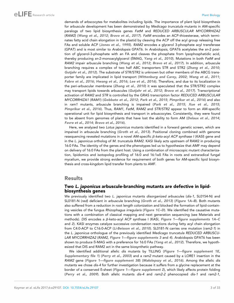
Two L. japonicus arbuscule-branching mutants are defective in lipid-biosynthesis genesWe previously identified two L. japonicus mutants disorganized arbuscules (dis-1, SL0154-N) and
SL0181-N (red) deficient in arbuscule branching (Groth et al., 2013) (Figure 1A–B). Both mutants
also suffered from a reduction in root length colonization and blocked the formation of lipid-contain-
ing vesicles of the fungus Rhizophagus irregularis (Figure 1C–D). We identified the causative muta-
tions with a combination of classical mapping and next generation sequencing (see Materials and
methods). DIS encodes a b-keto-acyl ACP synthase I (KASI, Figure 1—figure supplements 1A–C
and 2). KASI enzymes catalyze successive condensation reactions during fatty acyl chain elongation
from C4:0-ACP to C16:0-ACP (Li-Beisson et al., 2010). SL0181-N carries one mutation (ram2-1) in
the L. japonicus orthologue of the previously identified Medicago truncatula REDUCED ARBUSCU-
LAR MYCORRHIZA2 (RAM2, Figure 1—figure supplements 3 and 4). Arabidopsis GPAT6 has been
shown to produce ß-MAG with a preference for 16:0 FAs (Yang et al., 2012). Therefore, we hypoth-
esized that DIS and RAM2 act in the same biosynthetic pathway.
We identified additional allelic dis mutants by TILLING (Figure 1—figure supplement 1E,
Supplementary file 1) (Perry et al., 2003) and a ram2 mutant caused by a LORE1 insertion in the
RAM2 gene (Figure 1—figure supplement 3B) (Małolepszy et al., 2016). Among the allelic dis
mutants we chose dis-4 for further investigation because it suffers from a glycine replacement at the
border of a conserved ß-sheet (Figure 1—figure supplement 2), which likely affects protein folding
(Perry et al., 2009). Both allelic mutants dis-4 and ram2-2 phenocopied dis-1 and ram2-1,
Keymer et al. eLife 2017;6:e29107. DOI: 10.7554/eLife.29107 3 of 33
Research article Plant Biology
respectively. Furthermore, transgenic complementation of both dis-1 and ram2-1 with the wild-type
versions of the mutated genes restored arbuscule-branching and wild-type-like levels of root length
colonization and vesicle formation (Figure 1A-B). Taken together this confirmed identification of
both causal mutations.
ram2-1
ram2-2
WT
ram2-1; EV
25µm 25µm
ram2-1;
pRAM2:gRAM2
WT; EV
25µm
18/20
dis-1; EV
WT; EV
dis-4
dis-1
WT
dis-1; pDIS:gDIS
25µm
20/20
18/20
20/20
A B
100
90
80
70
60
50
40
30
20
10
0
total
hyphopodia
int. hyphae
arbuscules
vesicles
root le
ngth
colo
niz
ed [%
] a
ab
aa a
a
ab
a
a
b
b
a
a
ram2-2 WTram2-1
root le
ngth
colo
niz
ed [%
]
0
10
20
30
40
50
60
70
80
90
100
total
hyphopodia
int. hyphae
arbuscules
vesicles
dis-4 WTdis-1
a
a
b
a a a
a
a
b
a
a
b
aa
b
C D
18/18
22/24
18/18
Figure 1. DIS and RAM2 are required for arbuscule branching and vesicle formation. Arbuscule phenotype and complementation of dis (A) and ram2
(B) mutants. The fungus was stained with wheat-germ agglutinin (WGA)-AlexaFluor488. (C-D) Percent root length colonization of dis (C) and ram2 (D)
mutants as compared to wild-type. Different letters indicate significant differences among treatments (ANOVA; posthoc Tukey). (C): n = 13; p�0.1, F2,10= 8.068 (total & int. hyphae); p�0.001 F2,10 = 124.5 (arbuscules); p�0.001, F2,10 = 299.1 (vesicles) (D): n = 15; p�0.1, F2,12 = 10.18 (total & int. hyphae);
p�0.001 F2,12 = 57.86 (arbuscules); p�0.001, F2,12 = 72.37 (vesicles). (A-D) Plants were inoculated with R. irregularis and harvested at 5 weeks post
inoculation (wpi).
DOI: 10.7554/eLife.29107.003
The following figure supplements are available for figure 1:
Figure supplement 1. Identification of the dis mutation.
DOI: 10.7554/eLife.29107.004
Figure supplement 2. Protein sequence alignment of L. japonicus DIS with other KASI proteins.
DOI: 10.7554/eLife.29107.005
Figure supplement 3. Identification of mutation in the RAM2 gene.
DOI: 10.7554/eLife.29107.006
Figure supplement 4. Protein sequence alignment of L.
DOI: 10.7554/eLife.29107.007
Keymer et al. eLife 2017;6:e29107. DOI: 10.7554/eLife.29107 4 of 33
Research article Plant Biology
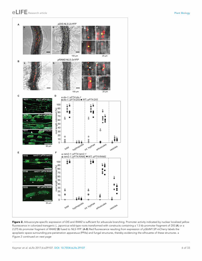
DIS and RAM2 expression in arbuscocytes is sufficient for arbusculedevelopmentTranscript levels of both DIS and RAM2 increased in colonized roots (Figure 3—figure supplement
1A). To analyze the spatial activity pattern of the DIS and RAM2 promoters during colonization we
fused 1.5 kb for DIS and 2.275 kb for RAM2 upstream of the translational start site to the uidA gene.
Consistent with a role of both genes in arbuscule development GUS activity was predominantly
detected in arbuscocytes (arbuscule-containing cells) in both wild-type and the corresponding
mutant roots (Figure 2—figure supplement 1A–B).
To correlate promoter activity with the precise stage of arbuscule development we used nuclear
localized YFP as a reporter. To visualize the fungus, the promoter:reporter cassette was co-trans-
formed with a second expression cassette containing secreted mCherry fused to the SbtM1 pro-
moter. This promoter drives expression in colonized cells, in cells neighboring apoplastically growing
hyphae and in cells forming pre-penetration apparatuus (PPAs, cytoplasmic aggregations that
assemble in cortex cells prior to arbuscule development) (Genre et al., 2008; Takeda et al., 2009,
2012). When expressed under the control of the SbtM1 promoter, secreted mCherry accumulates in
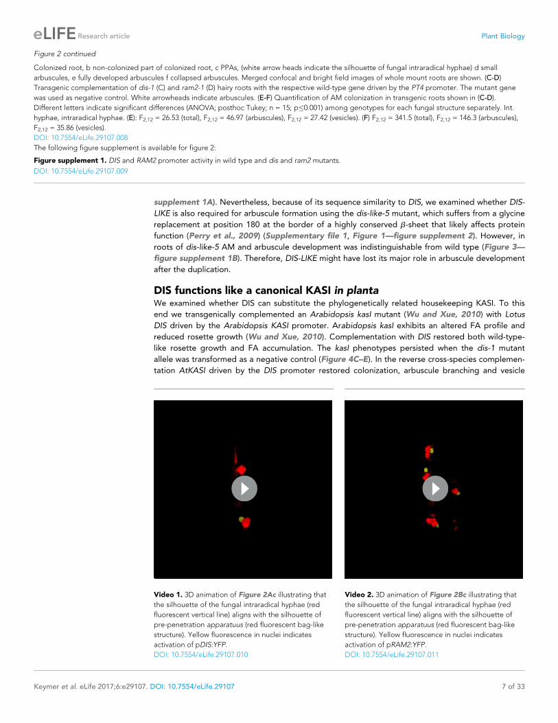
the apoplast surrounding fungal structures and PPAs, thereby revealing the silhouette of these struc-
tures (Figure 2A–B, Videos 1–2). Nuclear localized YFP fluorescence indicated activity of both pro-
moters in cells containing PPAs (c, Videos 1–2) and containing sparsely branched (d) or mature (e)
arbuscules. Furthermore, we rarely detected YFP fluorescence in non-colonized cells in direct neigh-
borhood of arbuscocytes, which were possibly preparing for PPA formation (a). However, YFP signal
was absent from cells containing collapsed arbuscules (f), indicating that the promoters were active
during arbuscule development and growth but inactive during arbuscule degeneration (Figure 2A–
B). RAM2 promoter activity was strictly correlated with arbuscocytes, while the DIS promoter
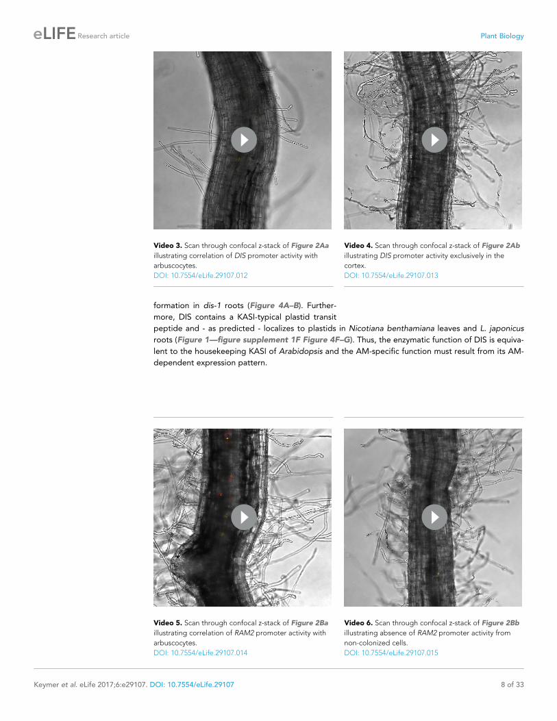
showed additional activity in cortical cells of non-colonized root segments (Figure 2A–B, Figure 2—
figure supplement 1C–D, Videos 3–6).
To examine, whether arbuscocyte-specific expression of DIS and RAM2 is sufficient for fungal
development we complemented the dis-1 and ram2-1 mutants with the corresponding wild-type
genes fused to the arbuscocyte-specific PT4 promoter (Volpe et al., 2013). This restored arbuscule-
branching, vesicle formation as well as root length colonization in the mutants (Figure 2C–F), show-
ing that arbuscocyte-specific expression of DIS and RAM2 suffices to support AM development.
Thus, expression of lipid biosynthesis genes in arbuscocytes is not only important for arbuscule
branching but also for vesicle formation and quantitative colonization.
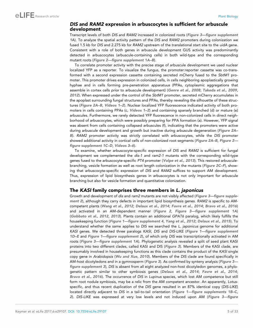
The KASI family comprises three members in L. japonicusGrowth and development of dis and ram2 mutants are not visibly affected (Figure 3—figure supple-
ment 2), although they carry defects in important lipid biosynthesis genes. RAM2 is specific to AM-
competent plants (Wang et al., 2012; Delaux et al., 2014; Favre et al., 2014; Bravo et al., 2016)
and activated in an AM-dependent manner (Figure 2, Figure 3—figure supplement 1A)
(Gobbato et al., 2012, 2013). Plants contain an additional GPAT6 paralog, which likely fulfills the
housekeeping function (Figure 1—figure supplement 4, Yang et al., 2012; Delaux et al., 2015). To
understand whether the same applies to DIS we searched the L. japonicus genome for additional
KASI genes. We detected three paralogs KASI, DIS and DIS-LIKE (Figure 1—figure supplement
1D–E and Figure 1—figure supplement 2), of which only DIS was transcriptionally activated in AM
roots (Figure 3—figure supplement 1A). Phylogenetic analysis revealed a split of seed plant KASI
proteins into two different clades, called KASI and DIS (Figure 3). Members of the KASI clade, are
presumably involved in housekeeping functions as this clade contains the product of the KASI single
copy gene in Arabidopsis (Wu and Xue, 2010). Members of the DIS clade are found specifically in
AM-host dicotyledons and in a gymnosperm (Figure 3). As confirmed by synteny analysis (Figure 3—
figure supplement 3), DIS is absent from all eight analyzed non-host dicotyledon genomes, a phylo-
genetic pattern similar to other symbiosis genes (Delaux et al., 2014; Favre et al., 2014;
Bravo et al., 2016). The occurrence of DIS in Lupinus species, which lost AM competence but still
form root nodule symbiosis, may be a relic from the AM competent ancestor. An apparently, Lotus-
specific, and thus recent duplication of the DIS gene resulted in an 87% identical copy (DIS-LIKE)
located directly adjacent to DIS in a tail-to-tail orientation (Figure 1—figure supplements 1B–C,
2). DIS-LIKE was expressed at very low levels and not induced upon AM (Figure 3—figure
Keymer et al. eLife 2017;6:e29107. DOI: 10.7554/eLife.29107 5 of 33
Research article Plant Biology
roo
t le
ng
th c
olo
niz
ed
[%
]
0
10
20
30
40
50
60
70
80
90
100
dis-1; pPT4:dis-1dis-1; pPT4:DIS WT; pPT4:DIS
total
hyphopodia
arbuscules
vesicles60 µm
19/19
16/17
15/17
dis-1; pPT4:dis-1
dis-1; pPT4:DIS
WT; pPT4:DIS
60 µm
33/33
37/38
11/12 ram2-1; pPT4:ram2-1
WT; pPT4:RAM2
ram2-1; pPT4:RAM2
E
roo
t le
ng
th c
olo
niz
ed
[%
]
ram2-1; pPT4:ram2-1ram2-1; pPT4:RAM2 WT; pPT4:RAM2
C D
F
A
a
b b
a a a
a
b b
b
b
a
a
bb
int. hyphae
0
10
20
30
40
50
60
70
80
90
100
a a
a
aaaa
b b b b
b b
b
b
total
hyphopodia
arbuscules
vesicles
int. hyphae
Ba b c d
e f
100 µm 25 µm
a b c d
e f
100 µm 25 µm
pDIS:NLS-2xYFP
pRAM2:NLS-2xYFP
Figure 2. Arbuscocyte-specific expression of DIS and RAM2 is sufficient for arbuscule branching. Promoter activity indicated by nuclear localized yellow
fluorescence in colonized transgenic L. japonicus wild-type roots transformed with constructs containing a 1.5 kb promoter fragment of DIS (A) or a
2.275 kb promoter fragment of RAM2 (B) fused to NLS-YFP. (A-B) Red fluorescence resulting from expression of pSbtM1:SP-mCherry labels the
apoplastic space surrounding pre-penetration apparatuus (PPAs) and fungal structures, thereby evidencing the silhouette of these structures. a
Figure 2 continued on next page
Keymer et al. eLife 2017;6:e29107. DOI: 10.7554/eLife.29107 6 of 33
Research article Plant Biology
supplement 1A). Nevertheless, because of its sequence similarity to DIS, we examined whether DIS-
LIKE is also required for arbuscule formation using the dis-like-5 mutant, which suffers from a glycine
replacement at position 180 at the border of a highly conserved b-sheet that likely affects protein
function (Perry et al., 2009) (Supplementary file 1, Figure 1—figure supplement 2). However, in
roots of dis-like-5 AM and arbuscule development was indistinguishable from wild type (Figure 3—
figure supplement 1B). Therefore, DIS-LIKE might have lost its major role in arbuscule development
after the duplication.
DIS functions like a canonical KASI in plantaWe examined whether DIS can substitute the phylogenetically related housekeeping KASI. To this
end we transgenically complemented an Arabidopsis kasI mutant (Wu and Xue, 2010) with Lotus
DIS driven by the Arabidopsis KASI promoter. Arabidopsis kasI exhibits an altered FA profile and
reduced rosette growth (Wu and Xue, 2010). Complementation with DIS restored both wild-type-
like rosette growth and FA accumulation. The kasI phenotypes persisted when the dis-1 mutant
allele was transformed as a negative control (Figure 4C–E). In the reverse cross-species complemen-
tation AtKASI driven by the DIS promoter restored colonization, arbuscule branching and vesicle
Figure 2 continued
Colonized root, b non-colonized part of colonized root, c PPAs, (white arrow heads indicate the silhouette of fungal intraradical hyphae) d small
arbuscules, e fully developed arbuscules f collapsed arbuscules. Merged confocal and bright field images of whole mount roots are shown. (C-D)
Transgenic complementation of dis-1 (C) and ram2-1 (D) hairy roots with the respective wild-type gene driven by the PT4 promoter. The mutant gene
was used as negative control. White arrowheads indicate arbuscules. (E-F) Quantification of AM colonization in transgenic roots shown in (C-D).
Different letters indicate significant differences (ANOVA; posthoc Tukey; n = 15; p�0.001) among genotypes for each fungal structure separately. Int.
hyphae, intraradical hyphae. (E): F2,12 = 26.53 (total), F2,12 = 46.97 (arbuscules), F2,12 = 27.42 (vesicles). (F) F2,12 = 341.5 (total), F2,12 = 146.3 (arbuscules),
F2,12 = 35.86 (vesicles).
DOI: 10.7554/eLife.29107.008
The following figure supplement is available for figure 2:
Figure supplement 1. DIS and RAM2 promoter activity in wild type and dis and ram2 mutants.
DOI: 10.7554/eLife.29107.009
Video 1. 3D animation of Figure 2Ac illustrating that
the silhouette of the fungal intraradical hyphae (red
fluorescent vertical line) aligns with the silhouette of
pre-penetration apparatuus (red fluorescent bag-like
structure). Yellow fluorescence in nuclei indicates
activation of pDIS:YFP.
DOI: 10.7554/eLife.29107.010
Video 2. 3D animation of Figure 2Bc illustrating that
the silhouette of the fungal intraradical hyphae (red
fluorescent vertical line) aligns with the silhouette of
pre-penetration apparatuus (red fluorescent bag-like
structure). Yellow fluorescence in nuclei indicates
activation of pRAM2:YFP.
DOI: 10.7554/eLife.29107.011
Keymer et al. eLife 2017;6:e29107. DOI: 10.7554/eLife.29107 7 of 33
Research article Plant Biology
formation in dis-1 roots (Figure 4A–B). Further-
more, DIS contains a KASI-typical plastid transit
peptide and - as predicted - localizes to plastids in Nicotiana benthamiana leaves and L. japonicus
roots (Figure 1—figure supplement 1F Figure 4F–G). Thus, the enzymatic function of DIS is equiva-
lent to the housekeeping KASI of Arabidopsis and the AM-specific function must result from its AM-
dependent expression pattern.
Video 3. Scan through confocal z-stack of Figure 2Aa
illustrating correlation of DIS promoter activity with
arbuscocytes.
DOI: 10.7554/eLife.29107.012
Video 4. Scan through confocal z-stack of Figure 2Ab
illustrating DIS promoter activity exclusively in the
cortex.
DOI: 10.7554/eLife.29107.013
Video 5. Scan through confocal z-stack of Figure 2Ba
illustrating correlation of RAM2 promoter activity with
arbuscocytes.
DOI: 10.7554/eLife.29107.014
Video 6. Scan through confocal z-stack of Figure 2Bb
illustrating absence of RAM2 promoter activity from
non-colonized cells.
DOI: 10.7554/eLife.29107.015
Keymer et al. eLife 2017;6:e29107. DOI: 10.7554/eLife.29107 8 of 33
Research article Plant Biology

Esa Thhalv10000874m Bra Bra025025 Bra Bra017565 Bra Bra022029 Aly 494344 Cru Carubv10026344m AT5G46290.3 KASI Cpa evm.model.supercontig 166.28 Gra Gorai.006G093400.1 Pt XP 002305334.1 Pt XP 002316735.1 Gra Gorai.009G156400.1 Gra Gorai.010G165600.1 Tca Thecc1EG029513t1 Sly Solyc08g016170.2.1 Cuscuta GAQC01004821.1 Vv XP 002272874.2 Stu PGSC0003DMP400014579 Sly Solyc02g070790.2.1 Utricularia Scf00389.g16797.t1 Mtr Medtr4g096690.1 Lalb 25301 LjKASI Pvu Phvul.002G194300.1 Gma Glyma08g08910.1 Gma Glyma05g25970.1 Mgu mgv1a005817m Bv 21760 ecdj.t1 Sit Si033275m Sit Si039166m Pvi Pavirv00002168m Sbi Sb02g041620.1 Bdi Bradi1g19190.1 Clanceolata JU991156.1 Pvi Pavirv00070739m Bdi Bradi1g46610.1 Osa Os06g09630.1 Pvi Pavirv00049911m Sit Si006383m Zma GRMZM2G012863 Sbi Sb10g006430.1 Zma GRMZM2G135498 Clanceolata JU992615.1 Gma Glyma08g02850.1 Gma Glyma18g10220.1 Gma Glyma05g36690.2 Pvu Phvul.002G300400.1 Lang AOCW01160110.1 Lalb 32493 Medtr8g099695.1 LjDIS LjDIS-like Mgu mgv1a006855m Mgu mgv1a024999m Mgu mgv1a005480m Stu PGSC0003DMP400021710 Sly Solyc08g082620.2.1 Gra Gorai.004G131800.1 Tca Thecc1EG016498t1 Cpa evm.model.supercontig 79.6 Vv XP 002265207.1 Pt XP 002303661.2 Pt XP 002299456.1 Sm XP 002960175.1 Sm XP 002964698.1 Sm XP 002961627.1 Pnutans GACA01011151.1 Pp XP 001762687.1 Pnutans GACA01018941.1 Pp XP 001762048.1 Pp XP 001776124.1 Lcruciata-3372
100
100
100
100 100
100 100
100 100
99 99
95
92
88
82
82 79
90
76 99
75
68
67
65 72
63 65
62
61
57
64
46
45
76
100
45
43
43
42
42
41
40
36 100
87
31 51
30
30 89
88 92
52
27
25
25
22
17
15
15
27
100
99
74
83
14
79 6
6
Dicots
Gymnosperms Lycophytes
Mosses
Liverworts
Non-host dicots
Monocots
DIS
KASI
Figure 3. Phylogenetic tree of KASI proteins in land plants. Protein sequences were aligned using MAFFT.
Phylogenetic trees were generated by neighbor-joining implemented in MEGA5 (Tamura et al., 2011). Partial gap
Figure 3 continued on next page
Keymer et al. eLife 2017;6:e29107. DOI: 10.7554/eLife.29107 9 of 33
Research article Plant Biology
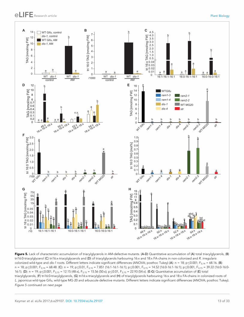
The AM-specific increase in 16:0 and 16:1v5 FA containing lipids isabolished in the dis mutantTo characterize the role of DIS in determining the lipid composition of non-colonized and colonized
roots we quantified triacylglycerols (TAGs), diacylglycerols (DAGs), galactolipids and phospholipids
in wild-type and dis-1. The lipid profile of colonized roots contains both plant and fungal lipids, how-
ever using the fungal marker FA 16:1w5 and previous data on fungus-specific lipids (Wewer et al.,
2014), many fungal lipids can be clearly distinguished from plant lipids. The lipid profile of non-colo-
nized roots was not affected by the dis-1 mutation. However, the strong and significant increase of
16:0 and 16:1 (most probably fungus-specific 16:1w5) containing TAGs, which is characteristic for
colonization of wild-type roots (Wewer et al., 2014) was abolished in dis-1 (Figure 5A–D, Figure 5—
figure supplement 1B). Also, AM- and fungus-specific DAG and phospholipid molecular species
were enhanced in colonized wild-type roots but not in colonized dis-1 roots (Figure 5—figure sup-
plements 1A and 2). In contrast, galactolipids were not affected by root colonization or genotype
(Figure 5—figure supplement 3). In summary, DIS affects the glycerolipid and phospholipid profile
of colonized L. japonicus roots and does not interfere with lipid accumulation in the non-colonized
state. Most lipids affected by the DIS mutation are fungus-specific and therefore reflect the amount
of root colonization and of fungal lipid-containing vesicles. However, since the root lipid profile is
hardly affected, absence of FA elongation by DIS was the cause of reduced lipid accumulation and
root colonization.
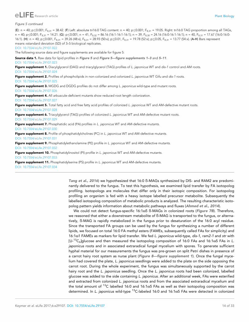
RAM1, DIS, RAM2 and STR are required for accumulation of AMsignature lipidsSimilar to dis and ram2 L. japonicus mutants in the ABCG half-transporter STR and the GRAS protein
RAM1 are affected in arbuscule branching (Kojima et al., 2014; Pimprikar et al., 2016; Xue et al.,
2015), quantitative root colonization and formation of lipid-containing fungal vesicles (Figure 5—fig-
ure supplement 4). Moreover, the AM-dependent transcriptional activation of DIS and KASIII, the
latter of which is a single copy gene in L. japonicus and produces precursors for DIS-activity by cata-
lyzing FA chain elongation from C2 to C4, was absent from ram1 mutants (Figure 6). In contrast,
induction of the single copy gene KASII, which elongates fatty acyl chains from C16 to C18 was not
hampered by RAM1 deficiency. Thus, RAM1 may play an important role in the regulation of lipid bio-
synthesis in arbuscocytes, since it also mediates expression of RAM2 and STR (Gobbato et al.,
2012; Park et al., 2015; Pimprikar et al., 2016; Luginbuehl et al., 2017).
We hypothesized that RAM1, DIS, RAM2 and STR form a specific operational unit for lipid biosyn-
thesis and transport in arbuscocytes. Therefore, we directly compared their impact on the AM-spe-
cific root lipid profile and measured galactolipids, phospholipids, TAGs and also total and free fatty
acids in colonized roots of ram1, dis, ram2, str mutants and wild-type in parallel. Consistent with our
previous observation in dis-1, galactolipid accumulation was similar in colonized roots of wild-type
and all mutants (Figure 5—figure supplement 3C–D). In contrast, total 16:0 FAs (FAMEs) as well as
16:1 and 18:1 (likely 18:1w7 FA of fungal origin) FAs were strongly reduced in all colonized mutants
compared to the corresponding wild-type. Free FAs showed a similar pattern except for 18:1 FAs
Figure 3 continued
deletion (95%) was used together with the JTT substitution model. Bootstrap values were calculated using 500
replicates. DIS likely originated before the angiosperm divergence (red star).
DOI: 10.7554/eLife.29107.016
The following source data and figure supplements are available for figure 3:
Source data 1. Accession numbers for protein sequences used in the phyologenic tree.
DOI: 10.7554/eLife.29107.017
Figure supplement 1. Transcript accumulation of KASI and RAM2 genes.
DOI: 10.7554/eLife.29107.018
Figure supplement 2. Shoot phenotypes of dis and ram2 mutants.
DOI: 10.7554/eLife.29107.019
Figure supplement 3. Genomic comparison of the DIS locus in host and non-host species.
DOI: 10.7554/eLife.29107.020
Keymer et al. eLife 2017;6:e29107. DOI: 10.7554/eLife.29107 10 of 33
Research article Plant Biology
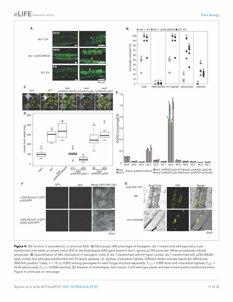
A B dis-1; EV dis-1; pDIS:AtKASI WT; EV
0
10
20
30
40
50
60
70
80
90
100
total hyphopodia int. hyphae arbuscules vesicles
root le
ngth
colo
niz
ed [%
]
a
bb bb
a
a a a
a
b
b
b
b
a
24/24
60 µm
22/24
8/8
dis-1; EV
dis-1; pDIS:AtKASI
WT; EV
kasI WTkasI;
pAtKASI:AtKASI
kasI;
pAtKASI:Ljdis-1
kasI;
pAtKASI:LjDIS
C
rosette fre
sh w
eig
ht [m
g]
100
200
300
400
500
0
kasI
WT
kasI
pAtK
ASI:A
tKASI #12 #29
kasIpAtKASI:Ljdis-1
#8 #20
kasIpAtKASI:LjDIS
a
b
b
a
a
b
b
E
GFP RFP Merge (GFP, RFP, BF)
p35S:AtLhcb1.3-GFP
p35S:RFP
p35S:AtLhcb1.3-GFP
p35S:LjDIS-RFP
25µm
Bright Field YFP Merge
20µm
pUbi:DIS-YFP
AM
non-colonized
D
F G
Col-0kasI
kasI; pAtKASI:AtKASIkasI; pAtKASI:Ljdis #12kasI; pAtKASI:Ljdis #29
kasI; pAtKASI:LjDIS #8kasI; pAtKASI:LjDIS #20
34:6 34:5 34:4 34:3 34:2 34:1 36:6 36:436:5 36:3 36:2 36:10
0.2
0.4
0.6
0.8
1.0
1.2
DG
DG
[n
mo
l/m
g]F
Wa
b
b
b
b
aa
Figure 4. DIS function is equivalent to a canonical KASI. (A) Microscopic AM phenotype of transgenic dis-1 mutant and wild-type hairy roots
transformed with either an empty vector (EV) or the Arabidopsis KASI gene fused to the L. japonicus DIS promoter. White arrowheads indicate
arbuscules. (B) Quantification of AM colonization in transgenic roots of dis-1 transformed with EV (open circles), dis-1 transformed with pDIS-AtKASI
(grey circles) and wild-type transformed with EV (black squares). int. hyphae, intraradical hyphae. Different letters indicate significant differences
(ANOVA; posthoc Tukey; n = 15; p�0.001) among genotypes for each fungal structure separately. F2,12 = 0.809 (total and intraradical hyphae), F2,12 =
43.65 (arbuscules), F2,12 = 0.0568 (vesicles). (C) Rosettes of Arabidopsis, kasI mutant, Col-0 wild-type plants and kasI mutant plants transformed either
Figure 4 continued on next page
Keymer et al. eLife 2017;6:e29107. DOI: 10.7554/eLife.29107 11 of 33
Research article Plant Biology
(Figure 5—figure supplement 5). Also for TAGs and phospholipids, AMF-specific molecular species
and 16:0 FA containing molecular species were strongly reduced in all mutants (Figure 5E–H, Fig-
ure 5—figure supplements 6–11). However, the two allelic ram2 mutants formed an exception.
They specifically over-accumulated 16:0-16:0 FA-containing phospholipids in particular 32:0 PA and
32:0-PC but also to a smaller extend 32:0-PE and 32:0-PI (Figure 5—figure supplements 6–10). A
similar pattern was observed for tri-16:0 TAGs (Figure 5F). This suggests that RAM2 acts down-
stream of DIS in a biosynthetic pathway and uses the 16:0 FAs synthesized by DIS in arbuscocytes as
substrates. In the absence of functional RAM2 the FA products of DIS, are probably redirected into
phospholipid biosynthesis and storage lipid biosynthesis via PA and PC (Li-Beisson et al., 2010)
leading to the observed higher accumulation of 16:0 FA containing lipid species in ram2 mutants.
This higher accumulation of specific lipids did not correlate with colonization levels in ram2 mutants
(Figure 5—figure supplement 4) confirming that reduced colonization levels are not the primary
cause for altered lipid profiles in the colonized mutant roots. Instead, defective AM-specific lipid bio-
synthesis in the mutants more likely impairs fungal development.
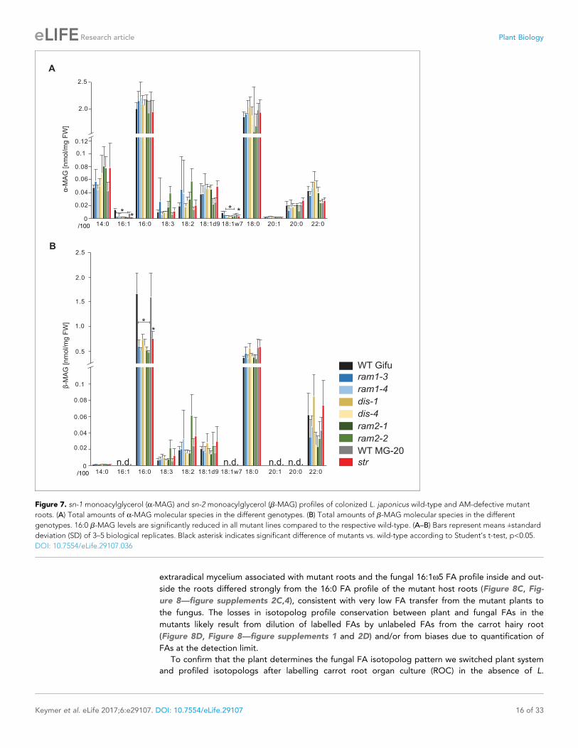
The abundance of 16:0 ß-monoacyl-glycerol is reduced in all mutantsThe first step in TAG and phospholipid production after FA biosynthesis is the esterification of FAs
with glycerol by GPATs in the plastid or endoplasmic reticulum to produce a-MAGs (sn1/3-MAGs,
Li-Beisson et al., 2010). RAM2 is predicted to produce a different type of glycerolipid ß-MAG (sn2-
MAG) with a preference towards 16:0 and 18:1 FAs (Yang et al., 2010; Wang et al.,
2012; Yang et al., 2012). To examine the role of RAM2 in MAG biosynthesis, we quantified a-MAG
and ß-MAG species in colonized roots of wild-type and all mutants. The abundance of ß-MAGs was
generally lower than that of a-MAGs (Figure 7). The amount of most a-MAG species did not differ
among the genotypes. Only the fungus-specific 16:1 and 18:1w7 a-MAGs were reduced in all
mutants reflecting the lower fungal biomass (Figure 7A). Fungus-specific ß-MAGs with 16:1 and
18:1w7 acyl groups were not detected and most ß-MAG molecular species accumulated to similar
levels in all genotypes. Exclusively the levels of 16:0 ß-MAGs were significantly lower in all mutants
as compared to the corresponding wild-type roots (Figure 7B). This supports a role of RAM2 in 16:0
ß-MAG synthesis during AM and a role of DIS in providing 16:0 FA precursors for RAM2 activity. A
low accumulation, of 16:0 ß-MAGs in ram1 mutants is consistent with RAM1’s role in regulating the
FA and lipid biosynthesis genes (Figure 6) (Gobbato et al., 2012; Pimprikar et al., 2016). In str
16:0 ß-MAGs likely did not accumulate because of reduced RAM2 expression in str roots due to low
root length colonization and/or a regulatory feedback loop (Bravo et al., 2017).
DIS, RAM2 and STR are required for transfer of 13C label from plant tofungusIn plants, ß-MAGs serve as precursors for cutin polymers at the surface of aerial organs (Yang et al.,
2012; Yeats et al., 2012). For their use in membrane or storage lipid biosynthesis they first need to
be isomerized to a-MAGs (Li-Beisson et al., 2010). The recruitment of a GPAT6 (RAM2) instead of a
a-MAG-producing GPAT for AM-specific lipid synthesis supports the idea that RAM2-products are
destined for something else than membrane biosynthesis of the host cell. Since AM fungal genomes
lack genes encoding cytosolic FA synthase subunits (Wewer et al., 2014; Ropars et al., 2016;
Figure 4 continued
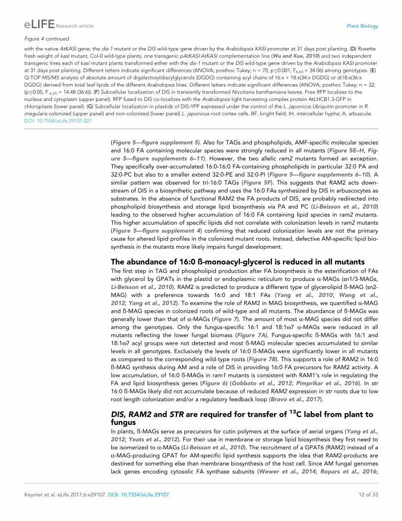
with the native AtKASI gene, the dis-1 mutant or the DIS wild-type gene driven by the Arabidopsis KASI promoter at 31 days post planting. (D) Rosette
fresh weight of kasI mutant, Col-0 wild-type plants, one transgenic pAtKASI:AtKASI complementation line (Wu and Xue, 2010) and two independent
transgenic lines each of kasI mutant plants transformed either with the dis-1 mutant or the DIS wild-type gene driven by the Arabidopsis KASI promoter
at 31 days post planting. Different letters indicate significant differences (ANOVA; posthoc Tukey; n = 70; p�0.001; F6,63 = 34.06) among genotypes. (E)
Q-TOF MS/MS analysis of absolute amount of digalactosyldiacylglycerols (DGDG) containing acyl chains of 16:x + 18:x(34:x DGDG) or di18:x(36:x
DGDG) derived from total leaf lipids of the different Arabidopsis lines. Different letters indicate significant differences (ANOVA; posthoc Tukey; n = 32;
(p�0.05, F 6,25 = 14.48 (36:6)). (F) Subcellular localization of DIS in transiently transformed Nicotiana benthamiana leaves. Free RFP localizes to the
nucleus and cytoplasm (upper panel). RFP fused to DIS co-localizes with the Arabidopsis light harvesting complex protein AtLHCB1.3-GFP in
chloroplasts (lower panel). (G) Subcellular localization in plastids of DIS-YFP expressed under the control of the L. japonicus Ubiquitin promoter in R.
irregularis colonized (upper panel) and non-colonized (lower panel) L. japonicus root cortex cells. BF, bright field; IH, intercellular hypha; A, arbuscule.
DOI: 10.7554/eLife.29107.021
Keymer et al. eLife 2017;6:e29107. DOI: 10.7554/eLife.29107 12 of 33
Research article Plant Biology
A B
E
WTGifu
str
WT MG20
ram2-2
ram2-1
dis-4
dis-1
ram1-3
ram1-4
tri 16:0
TA
G [nm
ol/m
g F
W]
0
0.5
1.0
1.5
2.0
2.5
3.0
/100
a
a
b b bbbbb
WT G
ifu
dis-
1
ram
1-3
ram
1-4
dis-
4
ram
2-1
ram
2-2
WT M
G20 st
r0
0.1
0.2
0.3
0.5
0.6
0.7
0.8
0.9
1.0
tri 16:0
TA
G [m
ol%
]
WT G
ifu
dis-
1
ram
1-3
ram
1-4
dis-
4
ram
2-1
ram
2-2
WT M
G20 st
r
bb
a
a a a a
a
a
0
2
4
6
8
10
12
14
16
WT G
ifu
dis-
1
ram
1-3
ram
1-4
dis-
4
ram
2-1
ram
2-2
WT-
MG20 st
r
TA
G [
nm
ol/m
g F
W]
a
a
b b b b b b b
0
2
4
6
8
10
12
14
TA
G [
nm
ol/m
g F
W]
a aa tri 1
6:0
TA
G [
nm
ol/m
g F
W]
WT dis-1WTdis-1
control AM
b
WT dis-1WTdis-1
control AM
F
a a
a
aa
a
a aa
bb
b
0
0.01
0.02
0.03
0.04
0.5
1.0
1.5
2.0
2.5
3.0
3.5
4.04.5
tri 1
6:x
TA
G [
nm
ol/m
g F
W]
WT Gifu, control
WT Gifu, AM
dis-1, AM
dis-1, control
16:1-16:1-16:1 16:0-16:1-16:1 16:0-16:0-16:1
C
a
a ac
a a
b
b
b
b
bb
b
b
bb
b bbb
bb
bb
b b b
0
0.01
0.02
0.03
0.04
0.05
15
35
55
75
tri 1
6:x
TA
G [
nm
ol/m
g F
W]
/10 16:1-16:1-16:1 16:0-16:1-16:1 16:0-16:0-16:1
0
0.1
0.2
0.3
0.4
0.7
0.6
6
8
10
12
TA
G [nm
ol/m
g F
W]
D
G
48:x50:x
52:x54:x
16:x-16:x-16:x
16:x-16:x-18:x
16:x-18:x-18:x
18:x-18:x-18:x
0
0.1
0.2
0.3
0.4
0.5
0.69111315
TA
G [nm
ol/m
g F
W]
48:x
16:x-16:x-16:x50:x
16:x-16:x-18:x52:x
16:x-18:x-18:x54:x
18:x-18:x-18:x
H
aa
a
b
a a a
b
n.s.
b
a
a
a
aa
bbbbbbb bbbbbbb
aa
a a
bbbbbbb
ababd
bdac
c
ddbd
bd
a aa
b
0
1
2
3
4
5
6
7
/1000
Figure 5. Lack of characteristic accumulation of triacylglycerols in AM-defective mutants. (A-D) Quantitative accumulation of (A) total triacylglycerols, (B)
tri16:0-triacylglycerol (C) tri16:x-triacylglycerols and (D) of triacylglycerols harbouring 16:x and 18:x FA-chains in non-colonized and R. irregularis
colonized wild-type and dis-1 roots. Different letters indicate significant differences (ANOVA; posthoc Tukey) (A): n = 18; p�0.001; F3,14 = 68.16. (B):
n = 18; p�0.001; F3,14 = 68.48. (C): n = 19; p�0.01, F3,15 = 7.851 (16:1-16:1-16:1); p�0.001, F3,15 = 14.52 (16:0-16:1-16:1); p�0.001, F3,15 = 39.22 (16:0-16:0-
16:1). (D): n = 19; p�0.001, F3,15 = 12.15 (48:x), F3,15 = 15.56 (50:x); p�0.01, F3,15 = 22.93 (54:x). (E-G) Quantitative accumulation of (E) total
triacylglycerols, (F) tri16:0-triacylglycerols, (G) tri16:x-triacylglycerols and (H) of triacylglycerols harbouring 16:x and 18:x FA-chains in colonized roots of
L. japonicus wild-type Gifu, wild-type MG-20 and arbuscule-defective mutants. Different letters indicate significant differences (ANOVA; posthoc Tukey).
Figure 5 continued on next page
Keymer et al. eLife 2017;6:e29107. DOI: 10.7554/eLife.29107 13 of 33
Research article Plant Biology
Tang et al., 2016) we hypothesized that 16:0 ß-MAGs synthesized by DIS- and RAM2 are predomi-
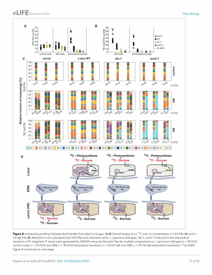
nantly delivered to the fungus. To test this hypothesis, we examined lipid transfer by FA isotopolog
profiling. Isotopologs are molecules that differ only in their isotopic composition. For isotopolog
profiling an organism is fed with a heavy isotope labelled precursor metabolite. Subsequently the
labelled isotopolog composition of metabolic products is analyzed. The resulting characteristic isoto-
polog pattern yields information about metabolic pathways and fluxes (Ahmed et al., 2014).
We could not detect fungus-specific 16:1w5 ß-MAGs in colonized roots (Figure 7B). Therefore,
we reasoned that either a downstream metabolite of ß-MAG is transported to the fungus, or alterna-
tively, ß-MAG is rapidly metabolized in the fungus prior to desaturation of the 16:0 acyl residue.
Since the transported FA groups can be used by the fungus for synthesizing a number of different
lipids, we focused on total 16:0 FA methyl esters (FAMEs, subsequently called FAs for simplicity) and
16:1w1 FAMEs as markers for lipid transfer. We fed L. japonicus wild-type, dis-1, ram2-1 and str with
[U-13C6]glucose and then measured the isotopolog composition of 16:0 FAs and 16:1w5 FAs in L.
japonicus roots and in associated extraradical fungal mycelium with spores. To generate sufficient
hyphal material for our measurements the fungus was pre-grown on split Petri dishes in presence of
a carrot hairy root system as nurse plant (Figure 8—figure supplement 1). Once the fungal myce-
lium had covered the plate, L. japonicus seedlings were added to the plate on the side opposing the
carrot root. During the whole experiment, the fungus was simultaneously supported by the carrot
hairy root and the L. japonicus seedling. Once the L. japonicus roots had been colonized, labelled
glucose was added to the side containing L. japonicus. After an additional week, FAs were esterified
and extracted from colonized L. japonicus roots and from the associated extraradical mycelium and
the total amount of 13C labelled 16:0 and 16:1w5 FAs as well as their isotopolog composition was
determined. In L. japonicus wild-type 13C-labeled 16:0 and 16:1w5 FAs were detected in colonized
Figure 5 continued
(E): n = 40; p�0.001; F8,31 = 38.42. (F) Left: absolute tri16:0 TAG content: n = 40; p�0.001; F8,31 = 19.05. Right: tri16:0 TAG proportion among all TAGs,
n = 40; p�0.001; F8,31 = 14.21. (G): p�0.001; n = 41, F8,32 = 86.16 (16:1-16:1-16:1); n = 39, F8,30 = 24.16 (16:0-16:1-16:1); n = 40, F8,31 = 17.67 (16:0-16:0-
16:1). (H): n = 40; p�0.001, F8,31 = 39.26 (48:x), F8,31 = 28.93 (50:x); p�0.01, F8,31 = 19.78 (52:x); p�0.05, F8,31 = 13.77 (54:x). (A-H) Bars represent
means ±standard deviation (SD) of 3–5 biological replicates.
DOI: 10.7554/eLife.29107.022
The following source data and figure supplements are available for figure 5:
Source data 1. Raw data for lipid profiles in Figure 5 and Figure 5—figure supplements 1–3 and 5–11.
DOI: 10.7554/eLife.29107.023
Figure supplement 1. Diacylglycerol (DAG) and triacylglycerol (TAG) profiles of L. japonicus WT and dis-1 control and AM roots.
DOI: 10.7554/eLife.29107.024
Figure supplement 2. Profiles of phospholipids in non-colonized and colonized L. japonicus WT Gifu and dis-1 roots.
DOI: 10.7554/eLife.29107.025
Figure supplement 3. MGDG and DGDG profiles do not differ among L. japonicus wild-type and mutant roots.
DOI: 10.7554/eLife.29107.026
Figure supplement 4. All arbuscule-deficient mutants show reduced root length colonization.
DOI: 10.7554/eLife.29107.027
Figure supplement 5. Total fatty acid and free fatty acid profiles of colonized L. japonicus WT and AM-defective mutant roots.
DOI: 10.7554/eLife.29107.028
Figure supplement 6. Triacylglycerol (TAG) profiles of colonized L. japonicus WT and AM-defective mutant roots.
DOI: 10.7554/eLife.29107.029
Figure supplement 7. Phosphatidic acid (PA) profiles in L. japonicus WT and AM-defective mutants.
DOI: 10.7554/eLife.29107.030
Figure supplement 8. Profile of phosphatidylcholines (PC) in L. japonicus WT and AM-defective mutants.
DOI: 10.7554/eLife.29107.031
Figure supplement 9. Phosphatidylethanolamine (PE) profile in L. japonicus WT and AM-defective mutants.
DOI: 10.7554/eLife.29107.032
Figure supplement 10. Phosphatidylinositol (PI) profile in L. japonicus WT and AM-defective mutants.
DOI: 10.7554/eLife.29107.033
Figure supplement 11. Phosphatidylserine (PS) profile in L. japonicus WT and AM-defective mutants.
DOI: 10.7554/eLife.29107.034
Keymer et al. eLife 2017;6:e29107. DOI: 10.7554/eLife.29107 14 of 33
Research article Plant Biology
roots as well as in the extraradical fungal mycelium (Figure 8A–B, Figure 8—figure supplement
2A–B), indicating that 13C-labelled organic compounds were transferred from the root to the fungus.
No labelled FAs were detected in the fungal mycelium when the fungus was supplied with [U-13C6]
glucose in absence of a plant host (Figure 8A–B, Figure 8—figure supplements 2A–B,3), indicating
that the fungus itself could not metabolize labelled glucose to synthesize FAs. The three mutants
incorporated 13C into 16:0 FAs at similar amounts as the wild-type but hardly any 13C was trans-
ferred to the fungus (Figure 8A–B, Figure 8—figure supplement 2A–B).
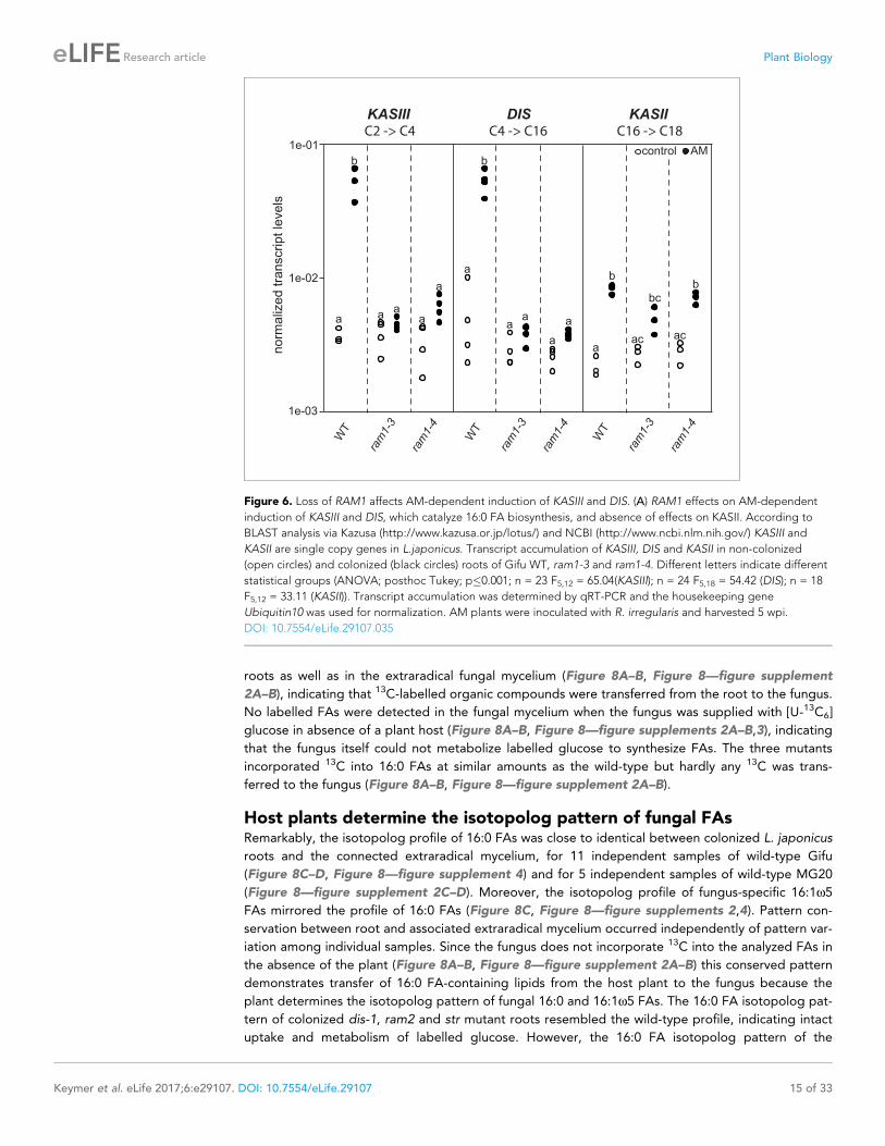
Host plants determine the isotopolog pattern of fungal FAsRemarkably, the isotopolog profile of 16:0 FAs was close to identical between colonized L. japonicus
roots and the connected extraradical mycelium, for 11 independent samples of wild-type Gifu
(Figure 8C–D, Figure 8—figure supplement 4) and for 5 independent samples of wild-type MG20
(Figure 8—figure supplement 2C–D). Moreover, the isotopolog profile of fungus-specific 16:1w5
FAs mirrored the profile of 16:0 FAs (Figure 8C, Figure 8—figure supplements 2,4). Pattern con-
servation between root and associated extraradical mycelium occurred independently of pattern var-
iation among individual samples. Since the fungus does not incorporate 13C into the analyzed FAs in
the absence of the plant (Figure 8A–B, Figure 8—figure supplement 2A–B) this conserved pattern
demonstrates transfer of 16:0 FA-containing lipids from the host plant to the fungus because the
plant determines the isotopolog pattern of fungal 16:0 and 16:1w5 FAs. The 16:0 FA isotopolog pat-
tern of colonized dis-1, ram2 and str mutant roots resembled the wild-type profile, indicating intact
uptake and metabolism of labelled glucose. However, the 16:0 FA isotopolog pattern of the
norm
aliz
ed t
ranscript
levels
1e-03
1e-02
1e-01
KASIII DIS KASII
WT
ram
1-3
ram
1-4
WT
ram
1-3
ram
1-4
WT
ram
1-3
ram
1-4
control AM
a
b
ac
bc
ac
b
b
a
aa a
a
aa
aa
b
a
C2 -> C4 C4 -> C16 C16 -> C18
Figure 6. Loss of RAM1 affects AM-dependent induction of KASIII and DIS. (A) RAM1 effects on AM-dependent
induction of KASIII and DIS, which catalyze 16:0 FA biosynthesis, and absence of effects on KASII. According to
BLAST analysis via Kazusa (http://www.kazusa.or.jp/lotus/) and NCBI (http://www.ncbi.nlm.nih.gov/) KASIII and
KASII are single copy genes in L.japonicus. Transcript accumulation of KASIII, DIS and KASII in non-colonized
(open circles) and colonized (black circles) roots of Gifu WT, ram1-3 and ram1-4. Different letters indicate different
statistical groups (ANOVA; posthoc Tukey; p�0.001; n = 23 F5,12 = 65.04(KASIII); n = 24 F5,18 = 54.42 (DIS); n = 18
F5,12 = 33.11 (KASII)). Transcript accumulation was determined by qRT-PCR and the housekeeping gene
Ubiquitin10 was used for normalization. AM plants were inoculated with R. irregularis and harvested 5 wpi.
DOI: 10.7554/eLife.29107.035
Keymer et al. eLife 2017;6:e29107. DOI: 10.7554/eLife.29107 15 of 33
Research article Plant Biology
extraradical mycelium associated with mutant roots and the fungal 16:1w5 FA profile inside and out-
side the roots differed strongly from the 16:0 FA profile of the mutant host roots (Figure 8C, Fig-
ure 8—figure supplements 2C,4), consistent with very low FA transfer from the mutant plants to
the fungus. The losses in isotopolog profile conservation between plant and fungal FAs in the
mutants likely result from dilution of labelled FAs by unlabeled FAs from the carrot hairy root
(Figure 8D, Figure 8—figure supplements 1 and 2D) and/or from biases due to quantification of
FAs at the detection limit.
To confirm that the plant determines the fungal FA isotopolog pattern we switched plant system
and profiled isotopologs after labelling carrot root organ culture (ROC) in the absence of L.
*
WT Gifu
str
WT MG-20
ram2-2
ram2-1
dis-4
dis-1
ram1-3
ram1-4
B
A
0
2.0
2.5
0.02
0.04
0.06
0.08
**
**
0.5
2.0
2.5
0
0.02
0.04
0.06
0.08
*
Figure 7. sn-1 monoacylglycerol (a-MAG) and sn-2 monoacylglycerol (b-MAG) profiles of colonized L. japonicus wild-type and AM-defective mutant
roots. (A) Total amounts of a-MAG molecular species in the different genotypes. (B) Total amounts of b-MAG molecular species in the different
genotypes. 16:0 b-MAG levels are significantly reduced in all mutant lines compared to the respective wild-type. (A–B) Bars represent means ±standard
deviation (SD) of 3–5 biological replicates. Black asterisk indicates significant difference of mutants vs. wild-type according to Student’s t-test, p<0.05.
DOI: 10.7554/eLife.29107.036
Keymer et al. eLife 2017;6:e29107. DOI: 10.7554/eLife.29107 16 of 33
Research article Plant Biology
carrot
WT
dis-1
ram2-1
no plant
A B
0
10
20
30
40
50
60
FA
o.e
[%
]
0
10
20
30
40
50
60
70
16
:0 F
A o
.e [
%]
control roots AM roots extraradical mycelia AM roots extraradical mycelia
*
***
*** *
***
***
***
*** *
70
M+1 M+2 M+3 M+4 M+5 M+6 M+7 M+8 M+9 M+10 M+16M+11 M+12 M+13 M+14 M+15
C carrot
0
20
40
60
80
100
0
20
40
60
80
100
0
20
40
60
80
100
16
:0 F
A
FA
Re
lati
ve
fra
cti
on
of
iso
top
olo
gs
[%
]
13.0212.90
16.59
20.8516.67
27.56
52.6831.40
29.63
59.8637.43
35.28
45.8224.38
21.92
MR MRMR
R RR
MR MRMR
Lotus WT
20.0718.83
16.86
8.814.34
19.15
12.7719.63
22.29.30
14.5614.06
3.876.06
5.55
MR MRMR
R RR
MR MRMR
dis-1
26.7515.37
16.79
19.915.66
7.610.48
0.133.02
2.872.42
5.160.88
0.140.62
MR MRMR
R RR
MR MRMR
ram2-1
co
ntr
ol
A
M
o.e [%]20.0916.62
15.53
10.949.20
13.130.03
0.040.24
0.10.12
0.90.080.06
0.13
o.e [%]
o.e [%]
AM
R RR
R MRMR
MR MRMR
M
D
ca
rro
t (H
R)
ER
ML
otu
s
12C
13C
FA
13C - Glucose+
12C - Sucrose
+ z
FA
DIS
RAM2
STR
12C - Sucrose
12C - Photosynthesis+
13C - Glucose
12C
13C
+ z
12C
13CFA
DIS
RAM2
STR
12C - Photosynthesis+
13C - Glucose
12C - Sucrose
+ z
DIS
RAM2
STR
FA
12C - Photosynthesis+
13C - Glucose
12C - Sucrose
12C
13C
Figure 8. Isotopolog profiling indicates lipid transfer from plant to fungus. (A–B) Overall excess (o.e.) 13C over air concentration in 16:0 FAs (A) and in
16:1w5 FAs (B) detected in non-colonized (only 16:0 FAs) and colonized carrot, L. japonicus wild-type, dis-1, ram2-1 roots and in the extraradical
mycelium of R. irregularis. P values were generated by ANOVA using the Dunnett Test for multiple comparisons to L. japonicus wild-type (n = 29 (16:0
control roots); n = 33 (16:0 root AM); n = 39 (16:0 extraradical mycelium); n = 33 (16:1w5 root AM); n = 39 (16:1w5 extraradical mycelium), ***p<0.001,
Figure 8 continued on next page
Keymer et al. eLife 2017;6:e29107. DOI: 10.7554/eLife.29107 17 of 33
Research article Plant Biology
japonicus seedlings (Figure 8D, Figure 8—figure supplement 1). In these root organ cultures, sugar
uptake from the medium does not compete with photosynthesis, as in whole seedlings. Additionally,
the carrot roots explore a larger surface of the Petri dish, increasing access to substances in the
nutrient medium. Consequently, and likely because of increased uptake of labelled glucose from the
medium, the isotopolog pattern of carrot ROCs differed from Lotus and was shifted towards more
highly labeled 16:0 FA isotopologs. This fingerprint was again recapitulated in the extraradical fungal
mycelium as well as in fungus-specific 16:1w5 FAs inside and outside the root for 10 independent
samples (Figure 8C, Figure 8—figure supplement 4). These data provide strong support for direct
transfer of a 16:0 FA containing lipid from plants to AMF (Figure 9).
DiscussionHere we identified DIS and RAM2, two AM-specific paralogs of the lipid biosynthesis genes KASI
and GPAT6 using forward genetics in Lotus japonicus. The dis and ram2 mutants enabled us to dem-
onstrate lipid transfer from plants to AMF using isotopolog profiling.
During AM symbiosis, an array of lipid biosynthesis genes is induced in arbuscocytes
(Gaude et al., 2012a, 2012b), indicating a large demand for lipids in these cells. Indeed, two genes
encoding lipid biosynthesis enzymes, the thioesterase FatM and the GPAT6 RAM2, have previously
been shown to be required for arbuscule branching in M. truncatula (Wang et al., 2012;
Bravo et al., 2017; Jiang et al., 2017). Both enzymes have a substrate preference for 16:0 FAs
(Salas and Ohlrogge, 2002; Yang et al., 2012; Bravo et al., 2017) and, consistent with this, we and
others observed that colonized ram2 mutant roots over-accumulate 16:0 FA containing phospholi-
pids and TAGs (Figure 7, [Bravo et al., 2017]), indicating re-channeling of superfluous 16:0 FAs in
the absence of RAM2 function and placing RAM2 downstream of FatM (Figure 9).
Our discovery of DIS, a novel and AM-specific KASI gene, now provides evidence for the enzyme
which synthesizes these 16:0 FAs in arbuscocytes. The arbuscule phenotype, as well as the lipid pro-
file of colonized dis mutants is very similar to fatm and ram2 mutants except for the accumulation of
16:0 FA-containing lipids in ram2 (Figure 1, Figure 5 and all figure supplements), consistent with
the predicted function. Together, this strongly suggests that DIS, FatM and RAM2 act in the same
lipid biosynthesis pathway, which is specifically and cell-autonomously induced when a resting root
cortex cell differentiates into an arbuscocyte (Figure 2A–B, Figure 9, [Bravo et al., 2017]). Interest-
ingly, DIS was exclusively found in genomes of AM-competent dicotyledons and a gymnosperm (Fig-
ure 3). This implies that DIS has been lost at the split of the mono- from dicotyledons. Despite the
Figure 8 continued
**p<0.01, *p<0.05). (C) Relative fraction of 13C isotopologs for 16:0 FAs of three replicates of carrot, L. japonicus WT Gifu, dis-1, ram2-1 in control roots
(upper panel) and AM roots and each of the associated R. irregularis extraradical mycelia with spores (middle panel) and 16:1w5 FAs in AM roots and
extraradical mycelia with spores (lower panel). Individual bars and double bars indicate individual samples. Values from roots are indicated by ‘R’ and
from fungal extraradical mycelia with spores by ‘M’. For carrot and L. japonicus WT the 13C labelling pattern of 16:0 and 16:1w5 FAs in the plant is
recapitulated in the fungal extraradical mycelium. Extraradical mycelium associated with dis-1 and ram2-1 does not mirror these patterns. Compare
bars for AM roots and extraradical mycelium side by side. Black numbers indicate 13C o. e. for individual samples. Colors indicate 13C-isotopologs
carrying one, two, three, etc. 13C-atoms (M + 1, M + 2, M + 3, etc.). (D) Schematic and simplified illustration of carbon flow and 12C vs.13C-carbon
contribution to plant lipid metabolism and transport to the fungus in the two-compartment cultivation setup used for isotope labelling. Carbohydrate
metabolism and transport is omitted for simplicity. ERM, extraradical mycelium.
DOI: 10.7554/eLife.29107.037
The following source data and figure supplements are available for figure 8:
Source data 1. Raw data for isotopolog profiles in Figure 8 and Figure 8—figure supplements 2,4.
DOI: 10.7554/eLife.29107.038
Figure supplement 1. Two-compartment cultivation setup used for labelling experiments.
DOI: 10.7554/eLife.29107.039
Figure supplement 2. Isotopolog profiles of wild-type MG20 and str.
DOI: 10.7554/eLife.29107.040
Figure supplement 3. Proportion of 16:0 and 16:1w5 FA containing only non-labelled 12C in plant and fungal tissue.
DOI: 10.7554/eLife.29107.041
Figure supplement 4. Isotopolog profiles of additional samples.
DOI: 10.7554/eLife.29107.042
Keymer et al. eLife 2017;6:e29107. DOI: 10.7554/eLife.29107 18 of 33
Research article Plant Biology
GPAT
plastid
endoplasmic reticulum
fatty acids
ACPACP
ACP
CoA + ATP
AMP + PPAcyl-CoA
(16:0/18:1/18:0)
Glycerol-3-Phosphat
LPAPA DAG TAG
PC PE
PS
CDP-DAGPG,PI
GPAT
plastid
endoplasmic reticulum
fatty acids
ACPACP
ACP
CoA + ATP
AMP + PPAcyl-CoA
(16:0/18:1/18:0)
Glycerol-3-Phosphat
LPAPA DAG TAG
PC PE
PS
CDP-DAGPG,PI
FatBFatA FatA
FatB
FatAFatB
desaturation
FA
-> 16:1 5
synthesis
?
arbuscule
co
lon
ize
d c
ell
no
n-c
olo
niz
ed
cell
FatBFatA
FatAFatB
FatAFatB
?
?
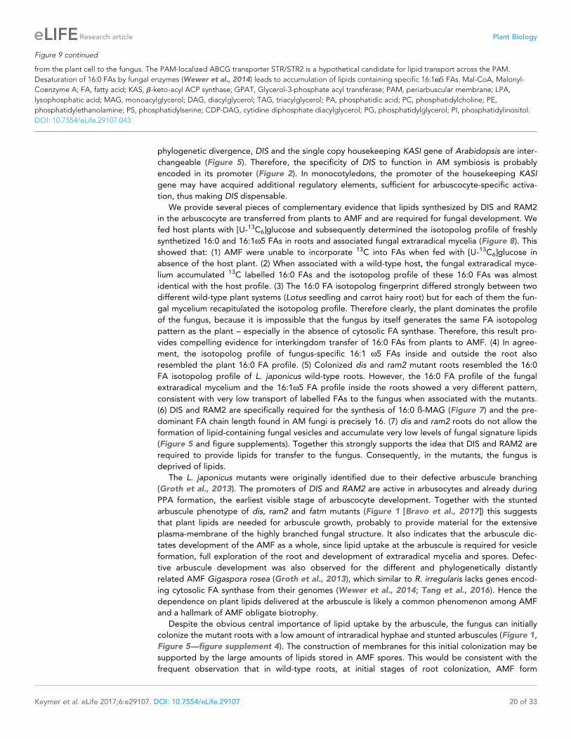
Figure 9. Schematic representation of plant fatty acid and lipid biosynthesis in a non-colonized root cell and a root cell colonized by an arbuscule. In
non-colonized cells FAs are synthesized in the plastid, bound via esterification to glycerol to produce LPA in the ER, where further lipid synthesis and
modification take place. Upon arbuscule formation AM-specific FA and lipid biosynthesis genes encoding DIS, FatM and RAM2 are activated to
synthesize specifically high amounts of 16:0 FAs and 16:0-ß-MAGs or further modified lipids (this work and Bravo et al., 2017). These are transported
Figure 9 continued on next page
Keymer et al. eLife 2017;6:e29107. DOI: 10.7554/eLife.29107 19 of 33
Research article Plant Biology
phylogenetic divergence, DIS and the single copy housekeeping KASI gene of Arabidopsis are inter-
changeable (Figure 5). Therefore, the specificity of DIS to function in AM symbiosis is probably
encoded in its promoter (Figure 2). In monocotyledons, the promoter of the housekeeping KASI
gene may have acquired additional regulatory elements, sufficient for arbuscocyte-specific activa-
tion, thus making DIS dispensable.
We provide several pieces of complementary evidence that lipids synthesized by DIS and RAM2
in the arbuscocyte are transferred from plants to AMF and are required for fungal development. We
fed host plants with [U-13C6]glucose and subsequently determined the isotopolog profile of freshly
synthetized 16:0 and 16:1w5 FAs in roots and associated fungal extraradical mycelia (Figure 8). This
showed that: (1) AMF were unable to incorporate 13C into FAs when fed with [U-13C6]glucose in
absence of the host plant. (2) When associated with a wild-type host, the fungal extraradical myce-
lium accumulated 13C labelled 16:0 FAs and the isotopolog profile of these 16:0 FAs was almost
identical with the host profile. (3) The 16:0 FA isotopolog fingerprint differed strongly between two
different wild-type plant systems (Lotus seedling and carrot hairy root) but for each of them the fun-
gal mycelium recapitulated the isotopolog profile. Therefore clearly, the plant dominates the profile
of the fungus, because it is impossible that the fungus by itself generates the same FA isotopolog
pattern as the plant – especially in the absence of cytosolic FA synthase. Therefore, this result pro-
vides compelling evidence for interkingdom transfer of 16:0 FAs from plants to AMF. (4) In agree-
ment, the isotopolog profile of fungus-specific 16:1 w5 FAs inside and outside the root also
resembled the plant 16:0 FA profile. (5) Colonized dis and ram2 mutant roots resembled the 16:0
FA isotopolog profile of L. japonicus wild-type roots. However, the 16:0 FA profile of the fungal
extraradical mycelium and the 16:1w5 FA profile inside the roots showed a very different pattern,
consistent with very low transport of labelled FAs to the fungus when associated with the mutants.
(6) DIS and RAM2 are specifically required for the synthesis of 16:0 ß-MAG (Figure 7) and the pre-
dominant FA chain length found in AM fungi is precisely 16. (7) dis and ram2 roots do not allow the
formation of lipid-containing fungal vesicles and accumulate very low levels of fungal signature lipids
(Figure 5 and figure supplements). Together this strongly supports the idea that DIS and RAM2 are
required to provide lipids for transfer to the fungus. Consequently, in the mutants, the fungus is
deprived of lipids.
The L. japonicus mutants were originally identified due to their defective arbuscule branching
(Groth et al., 2013). The promoters of DIS and RAM2 are active in arbusocytes and already during
PPA formation, the earliest visible stage of arbuscocyte development. Together with the stunted
arbuscule phenotype of dis, ram2 and fatm mutants (Figure 1 [Bravo et al., 2017]) this suggests
that plant lipids are needed for arbuscule growth, probably to provide material for the extensive
plasma-membrane of the highly branched fungal structure. It also indicates that the arbuscule dic-
tates development of the AMF as a whole, since lipid uptake at the arbuscule is required for vesicle
formation, full exploration of the root and development of extraradical mycelia and spores. Defec-
tive arbuscule development was also observed for the different and phylogenetically distantly
related AMF Gigaspora rosea (Groth et al., 2013), which similar to R. irregularis lacks genes encod-
ing cytosolic FA synthase from their genomes (Wewer et al., 2014; Tang et al., 2016). Hence the
dependence on plant lipids delivered at the arbuscule is likely a common phenomenon among AMF
and a hallmark of AMF obligate biotrophy.
Despite the obvious central importance of lipid uptake by the arbuscule, the fungus can initially
colonize the mutant roots with a low amount of intraradical hyphae and stunted arbuscules (Figure 1,
Figure 5—figure supplement 4). The construction of membranes for this initial colonization may be
supported by the large amounts of lipids stored in AMF spores. This would be consistent with the
frequent observation that in wild-type roots, at initial stages of root colonization, AMF form
Figure 9 continued
from the plant cell to the fungus. The PAM-localized ABCG transporter STR/STR2 is a hypothetical candidate for lipid transport across the PAM.
Desaturation of 16:0 FAs by fungal enzymes (Wewer et al., 2014) leads to accumulation of lipids containing specific 16:1w5 FAs. Mal-CoA, Malonyl-
Coenzyme A; FA, fatty acid; KAS, b-keto-acyl ACP synthase; GPAT, Glycerol-3-phosphate acyl transferase; PAM, periarbuscular membrane; LPA,
lysophosphatic acid; MAG, monoacylglycerol; DAG, diacylglycerol; TAG, triacylglycerol; PA, phosphatidic acid; PC, phosphatidylcholine; PE,
phosphatidylethanolamine; PS, phosphatidylserine; CDP-DAG, cytidine diphosphate diacylglycerol; PG, phosphatidylglycerol; PI, phosphatidylinositol.
DOI: 10.7554/eLife.29107.043
Keymer et al. eLife 2017;6:e29107. DOI: 10.7554/eLife.29107 20 of 33
Research article Plant Biology

arbuscules immediately after reaching the inner cortex and before colonizing longer distances, possi-
bly as a strategy to accquire lipids quickly after the reserves in the spore have been depleted. Alter-
natively, it is possible that plant housekeeping enzymes provide lipids to intraradical hyphae before
arbuscule formation. Activity of the housekeeping KASI may also be responsible for slightly higher
colonization levels observed for dis in some experiments as compared to other mutants.
It has recently been reported that photosynthetic wild-type nurse plants can restore arbuscule-
branching in Medicago ram2 and str mutants (Jiang et al., 2017; Luginbuehl et al., 2017), suggest-
ing that lipids can be supplied to arbuscules via the extraradical hyphal network and intraradical
hyphae through this route support arbuscule fine-branching. Based on four observations, we favor
an alternative szenario, in which lipids need to be provided cell-autonomously by the arbuscocyte to
support arbuscule fine-branching. However, we cannot exclude that our observations differ from the
reported observations due to growth conditions or plant species. (1) Presence of nurse carrot hairy
roots did not restore arbuscule branching in dis, ram2 and str (Figure 8—figure supplement 1C–F).
(2) dis and ram2 were found in a forward genetics screen based on their stunted arbuscule pheno-
type. In this screen, the fungal inoculum was provided via chive nurse plants (Groth et al., 2013). (3)
Map-based cloning of Lotus dis, ram2 and str (Kojima et al., 2014) was performed with segregating
mutant populations grown in the same pot, in which the wild-type and heterozygeous siblings acted
as nurse plants on the homozygeous mutants. In this system, the stunted arbuscule phenotype was
easily observable. (4) Arbuscule branching in a rice str mutant was not restored by wild type nurse
plants (Gutjahr et al., 2012).
It still remains to be shown, which types of lipids are transported from the plant arbuscocyte to
the fungal arbuscule and how. RAM2 is the most downstream acting enzyme in arbuscocyte-specific
lipid biosynthesis known to date (Figure 9). It is predicted to synthesize ß-MAG and we and others
have shown that 16:0 ß-MAGs are indeed reduced in colonized roots of dis, fatm and ram2 mutants,
providing evidence that this is likely the case (Figure 7, [Bravo et al., 2017]). Although, we cannot
exclude that a downstream metabolite of 16:0 ß-MAG is transported to the fungus, 16:0 ß-MAG as
transport vehicle for 16:0 FAs to the fungus is a good candidate because conceptually this molecule
may bear certain advantages. It has been shown in Arabidopsis that ß-MAGs are not used for plant
storage or membrane lipid biosynthesis but rather as pre-cursors for cuticle formation (Li et al.,
2007). The production of ß-MAGs could therefore, be a way, to withdraw FAs from the plants own
metabolism to make them available to the fungus. In addition, ß-MAGs are small and amphiphilic
and could diffuse across the short distance of the hydrophilic apoplastic space between plant and
fungal membrane. At the newly growing arbuscule branches the distance between the plant and fun-
gal membrane is indeed very small and has been measured to be 80–100 nm on TEM images of
high-pressure freeze-substituted samples (Bonfante, 2001). However, we could not detect fungus-
specific 16:1w5 ß-MAGs in colonized roots. This could mean that the fungus metabolizes them
before desaturation of the 16:0 FAs to synthesize membrane and storage lipids. Alternatively, ß-
MAGs may not be taken up by the fungus. ß-MAGs are known to isomerize to a-MAGs in acid or
basic conditions (Iqbal and Hussain, 2009). It is therefore, possible that they isomerize in the acidic
periarbuscular space (Guttenberger, 2000) before being taken up by the arbuscule.
How are MAGs transported across the peri-arbuscular membrane? Good candidates for MAG
transporters are the ABCG half transporters STR and STR2. Similar ABCG transporters have been
implicated genetically in cuticle formation, which also requires ß-MAGs (Pighin et al., 2004;
Panikashvili et al., 2011; Yeats et al., 2012). The half ABCG transporters STR and STR2 are both
independently required for arbuscule branching and they need to interact to form a full transporter
(Zhang et al., 2010). We found that colonized roots of a L. japonicus str mutant, did not allow the
formation of fungal vesicles and had the same lipid profile as dis and ram2 (Figure 5 and figure sup-
plements). Furthermore, our 13C labelling experiment demonstrated that str mutants do not transfer
lipids to the fungus (Figure 8—figure supplement 2). Although these are encouraging indications,
strong evidence for the role of STR in lipid transport across the periarbuscular membrane is still lack-
ing and the substrate of STR remains to be determined. Therefore, currently, it cannot be excluded
that mutation of str has an indirect effect on lipid transport and alternative mechanisms for example
lipid translocation via vesicle fission and fusion are possible. Nevertheless, also in AMF, several ABC
transporter genes are expressed in planta (Tisserant et al., 2012; Tang et al., 2016). They are not
characterized, but if lipid transport via ABC transporters instead of other mechanisms would play a
role, some of them could be involved in uptake of lipids into the fungal cytoplasm.
Keymer et al. eLife 2017;6:e29107. DOI: 10.7554/eLife.29107 21 of 33
Research article Plant Biology
We found that mutants in the GRAS gene RAM1 are impaired in AM-specific lipid accumulation
in colonized roots and in AM-mediated activation of DIS and the single copy gene KASIII (Figure 6),
in addition to FatM, RAM2 and STR (Wang et al., 2012; Park et al., 2015; Pimprikar et al., 2016;
Luginbuehl et al., 2017). This suggests that plants have evolved an AM-specific regulatory module
for lipid production in arbuscocytes and delivery to the fungus. It remains to be shown, whether
RAM1 regulates lipid biosynthesis genes directly and how this occurs mechanistically.
Our finding that plants transfer lipids to AMF completely changes the previous view that the fun-
gus receives only sugars from the plant (Pfeffer et al., 1999; Trepanier et al., 2005). It will now be
interesting to determine the relative contributions of sugar and lipid transfer to AMF, and whether
this may be a determinant of variation in root length colonization and extraradical mycelium forma-
tion depending on the plant-fungal genotype combination (Sawers et al., 2017). An interesting
question refers to why AMF have lost the genes encoding cytosolic FA synthase to depend on the
lipid biosynthesis machinery of the host. FA biosynthesis consumes more energy than biosynthesis of
carbohydrates and organic carbon provided by the plant needs to be transported in fungal hyphae
over long distances from the inside of the root to the extremities of the extraradical mycelium.
Therefore, it is conceivable that supply of plant lipids to the fungus plus fungal lipid transport is
more energy efficient for the symbiosis as a whole than fungal carbohydrate transport plus fungal
lipid biosynthesis. Hence, inter-organismic lipid transfer followed by loss of fungal FA biosynthesis
genes may have been selected for during evolution because it likely optimized the symbiosis for
most rapid proliferation of extraradical mycelium, thus ensuring efficient mineral nutrient acquisition
from the soil for supporting the plant host. Lipid transfer across kingdoms has also been observed in
human parasites or symbiotic bacteria of insects (Caffaro and Boothroyd, 2011; Elwell et al., 2011;
Herren et al., 2014). It will be interesting to learn whether this is a more widespread phenomenon
among biotrophic inter-organismic interactions.
Materials and methods
Plant growth and inoculation with AM fungiLotus japonicus ecotype Gifu wild-type, ram1-3, ram1-4, dis-1, dis-4, dis-like-5, ram2-1, ram2-2 and
ecotype MG-20 wild-type and str mutant (kindly provided by Tomoko Kojima (NARO, Tochigi,
Japan) seeds were scarified and surface sterilized with 1% NaClO. Imbibed seeds were germinated
on 0.8% Bacto Agar (Difco) at 24˚C for 10–14 days. Seedlings were cultivated in pots containing
sand/vermiculite (2/1 vol.) as substrate. For colonization with Rhizophagus irregularis roots were
inoculated with 500 spores (SYMPLANTA, Munich, Germany or Agronutrition, Toulouse, France) per
plant. Plants were harvested 5 weeks post inoculation (wpi); except for dis-1 complementation in
Figure 1A, which was harvested at 4 wpi. Arabidopsis thaliana seeds of Col-0 wild-type, kasI mutant
in the Col-0 background and the transgenically complemented kasI mutant were surface sterilized
with 70% EtOH +0.05% Tween 20% and 100% EtOH, germinated on MS-Medium for 48 hr at 4˚C in
the dark followed by 5–6 days at 22˚C (8 hr light/dark).
Identification of DIS by map-based cloning and next generationsequencingThe L. japonicus dis mutant (line SL0154, [Groth et al., 2013]) resulting from an EMS mutagenesis
program (Perry et al., 2003, 2009) was backcrossed to ecotype Gifu wild-type and outcrossed to
the polymorphic mapping parent ecotype MG-20. The dis locus segregated as a recessive mono-
genic trait and was previously found to be linked to marker TM2249 on chromosome 4 (Groth et al.,
2013). We confirmed the monogenic segregation as 26 of 110 individuals originating from the cross
to MG-20 (c2: P(3:1)=0.74) and 32 of 119 individuals originating from the cross to Gifu (c2: P(3:1)
=0.63) exhibited the mutant phenotype. To identify SL0154-specific mutations linked to the dis
locus, we employed a genome re-sequencing strategy. Nuclear DNA of Gifu wild-type and the
SL0154 mutant was subjected to paired end sequencing (2 � 100 bp) of a 300–500 bp insert library,
on an Illumina Hi-Seq 2000 instrument resulting in between 16.7 and 19.5 Gigabases per sample,
equivalent to roughly 35–41 fold coverage assuming a genome size of 470 Megabases. Reads were
mapped to the reference genome of MG-20 v2.5 (Sato et al., 2008) and single nucleotide polymor-
phisms identified using CLC genomics workbench (CLC bio, Aarhus, Denmark). SL0154-specific
Keymer et al. eLife 2017;6:e29107. DOI: 10.7554/eLife.29107 22 of 33
Research article Plant Biology
SNPs were identified by subtracting Gifu/MG-20 from SL0154/MG-20 polymorphisms. 19 potentially
EMS induced (11x G->A, 8x C->T) SNPs called consistently in all mapped reads from SL0514 but not
in Gifu were identified between the markers TM0046/TM1545, the initial dis target region (Fig-
ure 1—figure supplement 1A. In a screen for recombination events flanking the dis locus, 63
mutants out of 254 total F2 individuals of a cross MG-20 x SL0154 were genotyped with markers
flanking the dis locus (Figure 1—figure supplement 1B). Interrogating recombinant individuals with
additional markers in the region narrowed down the target interval between TM2249 and BM2170
(2 cM according to markers; ca. 650 kb). In this interval, 3 SL0154-specific SNPs with typical EMS sig-
nature (G to A transition) remained, of which one was predicted to be located in exon 3 of
CM0004.1640.r2 (reference position 40381558 in L. japonicus genome version 2.5; http://www.
kazusa.or.jp/lotus/), a gene annotated as ketoacyl-(acyl carrier protein) synthase. This co-segregation
together with phenotyping of one additional mutant allele obtained through TILLING
(Supplementary file 1, [Perry et al., 2003, 2009]) as well as transgenic complementation
(Figure 1A)) confirmed the identification of the mutation causing the dis phenotype of the SL0514
line. The two remaining mutations in the target region were located in a predicted intron of chr4.
CM0004.1570.r2.a, a cyclin-like F-box protein (reference position: 40356684) and in a predicted
intergenic region (reference position: 40364479). Untranslated regions of DIS and DIS-LIKE were
determined using the Ambion FirstChoice RLM RACE kit according to manufacturer‘s instructions
(http://www.ambion.de/). DIS sequence information can be found under the NCBI accession number
KX880396.
Identification of RAM2 by map-based cloning and Sanger sequencingThe L. japonicus Gifu mutant reduced and degenerate arbuscules (red, line SL0181-N) resulting from
an EMS mutagenesis (Perry et al., 2003, 2009) was outcrossed to the ecotype MG-20 and previ-
ously reported to segregate for two mutations, one on chromosome 1 and one on chromosome 6
(Groth et al., 2013). They were separated by segregation and the mutation on chromosome 1 was
previously found in the GRAS transcription factor gene REDUCED ARBUSCULAR MYCORRHIZA 1
(RAM1) (Pimprikar et al., 2016). A plant from the F2 population, which showed wild-type phenotype
but was heterozygous for the candidate interval on chromosome 6 and homozygous Gifu for the
candidate interval on chromosome 1 was selfed for producing an F3. The F3 generation segregated
for only one mutation as 38 out of 132 individuals exhibited the mutant phenotype (c2: P(3:1)=0.68).
A plant from the F3 population, which displayed wild-type phenotype but was heterozygous for the
candidate interval on chromosome 6 was selfed for producing an F4. The F4 generation also segre-
gated for only one mutation as 17 out of 87 individuals exhibited the mutant phenotype (c2: P(3:1)
=0.76). To identify the mutation on chromosome 6 linked to the previously identified interval
(Groth et al., 2013), we employed additional markers for fine mapping in F3 segregating and F4
mutant populations. This positioned the causative mutation between TM0082 and TM0302 (Fig-
ure 1—figure supplement 3A). Due to a suppression of recombination in this interval we could not
get closer to the mutation and also next generation sequencing (see [Pimprikar et al., 2016] for the
methodology) failed to identify a causative mutation. The Medicago truncatula ram2 mutant displays
stunted arbuscules similar to our mutant (Wang et al., 2012). L. japonicus RAM2 had not been
linked to any chromosome but was placed on chromosome 0, which prevented identification of a
RAM2 mutation in the target interval on chromosome 6. Therefore, we sequenced the RAM2 gene
by Sanger sequencing. Indeed, mutants with stunted arbuscule phenotype in the F3 and F4 genera-
tion carried an EMS mutation at base 1663 from G to A leading to amino acid change from Glycine
to Glutamic acid, which co-segregated with the mutant phenotype (Figure 1—figure supplement
3B-C). An additional allelic mutant ram2-2 (Figure 1—figure supplement 3B) caused by a LORE1
retrotransposon insertion (Małolepszy et al., 2016) and transgenic complementation with the wild-
type RAM2 gene confirmed that the causative mutation affects RAM2 (Figure 1B). Untranslated
regions of RAM2 were determined using the Ambion FirstChoice(R) RLM RACE kit according to
manufacturer’s instructions (http://www.ambion.de/). A 1345 bp long sequence upstream of ATG
was available from the http://www.kazusa.or.jp/lotus/blast.html. To enable cloning a 2275 bp pro-
moter fragment upstream of ATG of RAM2 the remaining upstream sequence of 1047 bp was deter-
mined by primer walking on TAC Lj T46c08. L. japonicus RAM2 sequence information can be found
under the NCBI accession number KX823334 and the promoter sequence under the number
KX823335.
Keymer et al. eLife 2017;6:e29107. DOI: 10.7554/eLife.29107 23 of 33
Research article Plant Biology
Plasmid generationGenes and promoter regions were amplified using Phusion PCR according to standard protocols
and using primers indicated in Supplementary file 2. Plasmids were constructed as indicated in
Supplementary file 3. For localization of DIS in L. japonicus hairy roots the LIII tricolor plasmid
(Binder et al., 2014) was used. The plasmid containing 35S:RFP for localization of free RFP in Nicoti-
ana benthamiana leaves was taken from Yano et al. (2008).
Induction of transgenic hairy roots in L. japonicusHypocotyls of L. japonicus were transformed with plasmids shown in Supplementary file 3 for hairy
root induction using transgenic Agrobacterium rhizogenes AR1193 as described (Takeda et al.,
2009).
Floral dipping and rosette growth assay of Arabidopsis thalianaFive plants per pot were sown. One week before transformation the primary bolt was cut off to
induce growth of secondary floral bolts. 5 ml LB culture of A. tumefaciens transformed with a binary
vector was incubated at 28˚C, 300 rpm over night. 500 ml of the preculture was added to 250 ml LB
medium with appropriate antibiotics. This culture was incubated again at 28˚C, 300 rpm over night
until an OD600 of 1.5 was reached. Plants were watered and covered by plastic bags the day before
the dipping to ensure high humidity. The cells were harvested by centrifugation (10 min, 5000 rpm)
and resuspended in infiltration medium (0.5 x MS medium, 5% sucrose). The resuspended cell cul-
ture was transferred to a box and Silwet L-77 was added (75 ml to 250 ml medium). The floral bolts
of the plants were dipped into the medium for 5 s and put back into plastic bags and left in horizon-
tal position for one night. After that, plants were turned upright, bags were opened and mature sili-
ques were harvested. For rosette growth assays T3 plants were used. 31 days post sowing the
rosettes were photographed and then cut and dried in an oven at 65˚C for the determination of
rosette dry weight.
Spatial analysis of promoter activityFor promoter:GUS analysis L. japonicus hairy roots transformed with plasmids containing the DIS
and RAM2 promoter fused to the uidA gene and colonized by R. irregularis were subjected to GUS
staining as described (Takeda et al., 2009). To correlate DIS and RAM2 promoter activity precisely
with the stage of arbuscule development two expression cassettes were combined in the same
golden gate plasmid for simultaneous visualization of arbuscule stages and promoter activity. The
fungal silhouette including all stages of arbuscule development and pre-penetration apparatuus
were made visible by expressing secretion peptide coupled mCherry under the control of the SbtM1
promoter region comprising 704 bp upstream of the SbtM1 gene (Takeda et al., 2009). Promoter
activity was visualized using a YFP reporter fused to a nuclear localization signal (NLS).
Transient transformation of N. benthamiana leavesN. benthamiana leaves were transiently transformed by infiltration of transgenic A. tumefaciens
AGL1 as described (Yano et al., 2008).
Real time qRT-PCRFor analysis of transcript levels, plant tissues were rapidly shock frozen in liquid nitrogen. RNA was
extracted using the Spectrum Plant Total RNA Kit (www.sigmaaldrich.com). The RNA was treated
with Invitrogen DNAse I amp. grade (www.invitrogen.com) and tested for purity by PCR. cDNA syn-
thesis was performed with 500 ng RNA using the Superscript III kit (www.invitrogen.com). qRT-PCR
was performed with GoTaq G2 DNA polymerase (Promega), 5 x colorless GoTaq Buffer (Promega)
and SYBR Green I (Invitrogen S7563, 10.000x concentrated, 500 ml) - diluted to 100x in DMSO. Pri-
mers (Supplementary file 2) were designed with primer3 (58). The qPCR reaction was run on an iCy-
cler (Biorad, www.bio-rad.com/) according to manufacturer’s instructions. Thermal cycler conditions
were: 95˚C 2 min, 45 cycles of 95˚C 30 s, 60˚C/62˚C 30 s and 72˚C 20 s followed by dissociation
curve analysis. Expression levels were calculated according to the DDCt method (Rozen and Skalet-
sky, 2000). For each genotype and treatment three to four biological replicates were tested and
each sample was represented by two to three technical replicates.
Keymer et al. eLife 2017;6:e29107. DOI: 10.7554/eLife.29107 24 of 33
Research article Plant Biology
Sequence alignement and phylogenyL. japonicus KASI, DIS, DIS-LIKE, RAM2, Lj1g3v2301880.1 (GPAT6) protein sequences were retrieved
from Lotus genome V2.5 and V3.0 respectively (http://www.kazusa.or.jp/lotus/) and A. thaliana KASI,
E. coli KASI, E. coli KASII, M. truncatula RAM2 and Medtr7g067380 (GPAT6) were obtained from
NCBI (http://www.ncbi.nlm.nih.gov). The sequences from L. japonicus were confirmed with a
genome generated by next generation sequencing in house. Protein alignment for DIS was per-
formed by CLC Main Workbench (CLC bio, Aarhus, Denmark). The Target Peptide was predicted
using TargetP 1.0 Server (www.cbs.dtu.dk/services/TargetP-1.0/). RAM2 Protein alignment was per-
formed by MEGA7 using ClustalW. The percentage identity matrix was obtained by Clustal Omega
(http://www.ebi.ac.uk/Tools/msa/clustalo/).
To collect sequences for phylogeny construction corresponding to potential DIS orthologs, Lotus
DIS and KASI (outgroup) protein sequences were searched in genome and transcriptome datasets
using BLASTp and tBLASTn respectively. The list of species and the databases used are indicated in
Figure 3—source data 1. Hits with an e-value >10�50 were selected for the phylogenetic analysis.
Collected sequences were aligned using MAFFT (http://mafft.cbrc.jp/alignment/server/) and the
alignment manually checked with Bioedit. Phylogenetic trees were generated by Neighbor-joining
implemented in MEGA5 (Tamura et al., 2011). Partial gap deletion (95%) was used together with
the JTT substitution model. Bootstrap values were calculated using 500 replicates.
Synteny analysisA ~200 kb sized region in the L. japonicus genome containing the DIS locus (CM00041640.r2.a) was
compared to the syntenic region in A. thaliana (Col-0) using CoGe Gevo (https://genomevolution.
org/CoGe/GEvo.pl - (Lyons et al., 2008) as described in Delaux et al. (2014). Loci encompassing
DIS orthologs from Medicago truncatula, Populus trichocarpa, Carica papaya, Phaseolus vulgaris and
Solanum lycopersicum were added as controls.
AM staining and quantificationRhizophagus irregularis in colonized L. japonicus roots was stained with acid ink (Vierheilig et al.,
1998). Root length colonization was quantified using a modified gridline intersect method
(McGonigle et al., 1990). For confocal laser scanning microscopy (CLSM) fungal structures were
stained with 1 mg WGA Alexa Fluor 488 (Molecular Probes, http://www.lifetechnologies.com/) (Pan-
chuk-Voloshina et al., 1999).
MicroscopyFor quantification of AM colonization in L. japonicus roots a light microscope (Leica) with a 20x mag-
nification was used. For observation of GUS-staining in L. japonicus hairy roots an inverted micro-
scope (Leica DMI6000 B) was used with 10x and 20x magnification. Transformed roots were
screened by stereomicroscope (Leica MZ16 FA) using an mCherry fluorescent transformation marker
or the pSbtM1:mCherry marker for fungal colonization (for Figure 2A and B). Confocal microscopy
(Leica SP5) for WGA-AlexaFluor488 detection using 20x and 63x magnification was performed as
described (Groth et al., 2010). Transgenic roots showing mCherry fluorescence signal due to SbtM1
promoter activity linked with fungal colonization were cut into pieces immediately after harvesting.
The living root pieces were placed on a glass slide with a drop of water, covered by a cover slip and
immediately subjected to imaging. Sequential scanning for the YFP and RFP signal was carried out
simultaneously with bright field image acquisition. YFP was excited with the argon ion laser 514 nm
and the emitted fluorescence was detected from 525 to 575 nm; RFP was excited with the Diode-
Pumped Solid State laser at 561 nm and the emitted fluorescence was detected from 580 to 623
nm. Images were acquired using LAS AF software. Several z-optical sections were made per area of
interest and assembled to a z-stack using Fiji. The z-stack movies and 3D projections were produced
using the 3D viewer function in Fiji (Schindelin et al., 2012).
Extraction and purification of phospho- and glycoglycerolipids andtriacylglycerolsApproximately 50–100 mg of root or leaf material was harvested, weighed and immediately frozen
in liquid nitrogen to avoid lipid degradation. The frozen samples were ground to a fine powder
Keymer et al. eLife 2017;6:e29107. DOI: 10.7554/eLife.29107 25 of 33
Research article Plant Biology
before extraction with organic solvents. Total lipids were extracted as described previously
(Wewer et al., 2011, 2014). Briefly, 1 mL chloroform/methanol/formic acid (1:1:0.1, v/v/v) was
added and the sample was shaken vigorously. At this point the internal standards for TAG and fatty
acid analysis were added. Phase separation was achieved after addition of 0.5 mL 1M KCl/0.2 M
H3PO4 and subsequent centrifugation at 4000 rpm for 5 min. The lipid-containing chloroform phase
was transferred to a fresh glass tube and the sample was re-extracted twice with chloroform. The
combined chloroform phases were dried under a stream of air and lipids were re-dissolved in 1 mL
chloroform to yield the total lipid extract.
For phospho- and glycerolipid analysis 20 ml of the total lipid extract were mixed with 20 ml of the
internal standard mix and 160 ml of methanol/chloroform/300 mM ammonium acetate (665:300:35,
v/v/v) (Welti et al., 2002). For triacylglycerol analysis 500 ml of the total lipid extract were purified
by solid phase extraction on Strata silica columns (1 ml bed volume; Phenomenex) as described
(Wewer et al., 2011). TAGs were eluted from the silica material with chloroform, dried under a
stream of air and re-dissolved in 1 mL methanol/chloroform/300 mM ammonium acetate
(665:300:35, v/v/v).
Extraction and purification of free fatty acids and monoacylglycerol(MAG)Total lipids were extracted into chloroform and dried as described above. 15–0 FA and a mixture of
15–0 a-MAG and b-MAG were added as internal standard before the extraction. Dried extracts
were resuspended in 1 ml n-hexane and applied to silica columns for solid-phase extraction with a
n-hexane:diethylether gradient. Free fatty acids were eluted with a mixture of 92:8 (v/v) n-hexane:
diethylether as described bevore (Gasulla et al., 2013) and pure diethylether were used for elution
of MAG.
Analysis of total fatty acids and free fatty acids by GC-FIDFor measurement of total fatty acids, 100 ml of the total lipid extract were used. For measurement of
free fatty acids, the SPE-fraction containing free fatty acids was used. Fatty acid methyl esters
(FAMEs) were generated from acyl groups of total lipids and free fatty acids by addition of 1 mL 1N
methanolic HCL (Sigma) to dried extracts and incubation at 80˚C for 30 min (Browse et al., 1986).
Subsequently, FAMEs were extracted by addition of 1 mL n-hexane and 1 mL of 0.9% (w/v) NaCl
and analyzed on a gas chromatograph with flame-ionization detector (GC-FID, Agilent 7890A
PlusGC). FAMEs were separated on an SP 2380 fused silica GC column (Supelco, 30 mx 0.53 mm,
0.20 mm film) as described (Wewer et al., 2013), with a temperature -gradient starting at 100˚C,increased to 160˚C with 25˚C/min, then to 220˚C with10˚C/min and reduced to 100˚C with 25 ˚C/min. FAMEs were quantified in relation to the internal standard pentadecanoic acid (15:0).
For MAG measurement, dried diethylether fractions were resuspended in 4:1 (v/v %) pyridine:N-
Methyl-N-(trimethylsilyl)trifluoroacetamide (MSTFA), incubated at 80˚C for 30 min, dried and re-sus-
pended in hexane prior to application on an Agilent 7890A Plus gas chromatography-mass spec-
trometer. MAGs were quantified by extracted ion monitoring, using [M+ - 103] for a-MAGs and [M+
- 161] for b-MAGs as previously reported for 16:0 MAG (Destaillats et al., 2010) and 24:0 MAG
(Li et al., 2007).
Quantification of glycerolipids by Q-TOF MS/MSPhosphoglycerolipids (PC, PE, PG, PI, PS), glycoglycerolipids (MGDG, DGDG, SQDG) and triacylgly-
cerol (TAG) were analyzed in positive mode by direct infusion nanospray Q-TOF MS/MS on an Agi-
lent 6530 Q-TOF instrument as described previously (Lippold et al., 2012; Gasulla et al., 2013). A
continuous flow of 1 ml/min methanol/chloroform/300 mM ammonium acetate (665:300:35, v/v/v)
(Welti et al., 2002) was achieved using a nanospray infusion ion source (HPLC/chip MS 1200 with
infusion chip). Data are displayed as X:Y, where X gives the number of C atoms of the fatty acid
chain and Y the amount of desaturated carbo-carbon bonds inside that fatty acid chain.
Internal standardsInternal standards for phospho- and glycoglycerolipid analysis were prepared as described previ-
ously (Gasulla et al., 2013; Wewer et al., 2014). The following standards were dissolved in 20 ml of
Keymer et al. eLife 2017;6:e29107. DOI: 10.7554/eLife.29107 26 of 33
Research article Plant Biology
chloroform/methanol (2:1, v/v): 0.2 nmol of each di14:0-PC, di20:0-PC, di14:0-PE, di20:0-PE, di14:0
PG, di20:0 PG, di14:0 PA and di20:0 PA; 0.03 nmol of di14:0-PS and di20:0-PS; 0.3 nmol of 34:0-PI;
0.15 nmol of 34:0-MGDG, 0.10 nmol of 36:0-MGDG; 0.2 nmol of 34:0-DGDG, 0.39 nmol of 36:0
DGDG and 0.4 nmol of 34:0 SQDG. 1 nmol each of tridecanoin (tri-10:0) and triundecenoin (tri-11:1),
and 2 nmol each of triarachidin (tri-20:0) and trierucin (tri22:1) were used as internal standards for
TAG quantification (Lippold et al., 2012). For quantification of total fatty acids and free fatty acids 5
mg of pentadecanoic acid (FA 15:0) was added to the samples (Wewer et al., 2013).
Cultivation and 13C-Labeling of L. japonicus and Daucus carota hairyrootsThe method for cultivation and stable isotope labelling of Lotus japonicus and Daucus carota hairy
roots as well as for isotopolog profiling are described in more detail at Bio-protocol (Keymer et al.,
2018). To determine lipid transfer from L. japonicus to the fungus we used the carrot root organ cul-
ture system (Becard et al., 1988) to obtain sufficient amounts of fungal material for isotopolog pro-
filing. (On petri dishes this was not possible with L. japonicus and in particular the lipid mutants
alone). One compartment (carrot compartment) of the 2- compartmented petri dish system
(Trepanier et al., 2005) was filled with MSR-medium (3% gelrite) containing 10% sucrose to support
the shoot-less carrot root, and the other compartment (Lotus compartment) was filled with MSR-
medium (3% gelrite) without sucrose. Ri T-DNA transformed Daucus carota hairy roots were placed
in the carrot compartment. 1 week later, roots were inoculated with R. irregularis. Petri dishes were
incubated at constant darkness and 30˚C. Within 5 weeks R. irregularis colonized the carrot roots
and its extraradical mycelium spread over both compartments of the petri dish and formed spores.
At this stage two 2 week old L. japonicus seedlings (WT, dis-1, ram2-1) were placed into the Lotus
compartment (Figure 8—figure supplement 1).
The plates were incubated at 24˚C (16 hr light/8 hr dark). To keep the fungus and root in the dark
the petri dishes were covered with black paper. 3 weeks after Lotus seedlings were placed into the
petri dish [U-13C6]glucose (100 mg diluted in 2 ml MSR-medium) (Sigma-Aldrich) was added to the
Lotus compartment. Therefore, only Lotus roots but not the carrot roots took up label. For transfer
experiments with carrot roots no Lotus plant was placed into the Lotus compartment and the
[U-13C6]glucose was added to the carrot compartment. 1 week after addition of [U-13C6]glucose the
roots were harvested. The extraradical mycelium was extracted from the agar using citrate buffer pH
6 and subsequent filtration, after which it was immediately shock-frozen in liquid nitrogen.
Isotopolog profiling of 13C-labelled 16:0 and 16:1v5 fatty acidsRoot and fungal samples were freeze dried and subsequently derivatised with 500 ml MeOH contain-
ing 3 M HCl (Sigma-Aldrich) at 80˚C for 20 hr. MeOH/HCL was removed under a gentle stream of
nitrogen and the methyl esters of the fatty acids were solved in 100 ml dry hexane.
Gas chromatography mass spectrometry was performed on a GC-QP 2010 plus (Shimadzu, Duis-
burg, Germany) equipped with a fused silica capillary column (equity TM-5; 30 m by 0.25 mm,0.25-m
m film thickness; Supelco, Bellafonte, PA). The mass detector worked in electron ionization (EI)
mode at 70 eV. An aliquot of the solution was injected in split mode (1:5) at an injector and interface
temperature of 260˚C. The column was held at 170˚C for 3 min and then developed with a tempera-
ture gradient of 2 ˚C/min to a temperature of 192˚C followed by a temperature gradient of 30˚C/min to a final temperature of 300˚C. Samples were analyzed in SIM mode (m/z values 267 to 288) at
least three times. Retention times for fatty acids 16:1w5 (unlabeled m/z 268) and 16:0 (unlabeled m/
z 270) are 12.87 min and 13.20 min, respectively. Data were collected with LabSolution software (Shi-
madzu, Duisburg, Germany). The overall 13C enrichment and the isotopolog compositions were cal-
culated according to (Lee et al., 1991) and (Ahmed et al., 2014). The software package is open
source and can be downloaded by the following link: http://www.tr34.uni-wuerzburg.de/software_
developments/isotopo/.
Four independent labeling experiments were performed. Overall excess (o.e.) is an average value
of 13C atoms incorporated into 16:0/16:1w5 fatty acids.
Keymer et al. eLife 2017;6:e29107. DOI: 10.7554/eLife.29107 27 of 33
Research article Plant Biology
Data availabilityLunularia cruciata: For this species, the raw RNAseq reads have been previously deposited to NCBI
under the accession number SRR1027885. It is annotated with Rhizophagus irregularis (10% of
sequences) as the transcriptome was partly prepared from Lunularia plant tissue colonized by the
fungus Rhizophagus irregularis. The corresponding Lunularia transcriptomic assembly is available at
www.polebio.lrsv.ups-tlse.fr/Luc_v1/
StatisticsAll statistical analyses (Source code 1) were performed and all boxplots were generated in R (www.
r-project.org).
AcknowledgementsWe are grateful to Tomoko Kojima (NARO, Tochigi, Japan) for providing Lotus japonicus str mutant
seeds and to LotusBase for providing LORE1 insertion lines. We thank Julia Mayer for help with set-
ting up carrot hairy root systems, Samy Carbonnel, David Chiasson, Michael Paries and Jose Antonio
Villaecija-Aguilar for contributing a golden gate module.
Additional information
Funding
Funder Grant reference number Author
Deutsche Forschungsge-meinschaft
SFB924 B03 Function of theGRAS protein RAM1 inarbuscule development
Caroline Gutjahr
Deutsche Forschungsge-meinschaft
Emmy Noether programGU1423/1-1
Caroline Gutjahr
Deutsche Forschungsge-meinschaft
PA493/7-1 Plant genesrequired for arbuscularmycorrhiza symbiosis
Martin Parniske
Deutsche Forschungsge-meinschaft
SFB924 B03 Geneticdissection of arbuscularmycorrhiza development
Martin Parniske
Deutsche Forschungsge-meinschaft
SPP1212 Peter Dormann
Deutsche Forschungsge-meinschaft
Research Training GroupGRK 2064 Do520/15-1
Peter Dormann
Hans Fischer Gesellschaft e. V. Wolfgang Eisenreich
Biotechnology and BiologicalSciences Research Council
Trevor L Wang
The funders had no role in study design, data collection and interpretation, or the decision tosubmit the work for publication.
Author contributions
AK, Conceptualization, Data curation, Formal analysis, Investigation, Visualization, Writing - contrib-
uted method description and figure legends; PP, Conceptualization, Data curation, Formal analysis,
Investigation, Visualization; VW, Conceptualization, Investigation, Methodology; CH, Data curation,
Investigation, Writing - contributed method description; MB, Data curation, Investigation, Methodol-
ogy, Writing - contributed method description; SLB, VK, Investigation; P-MD, Formal analysis, Inves-
tigation; EvR-L, Investigation, generation of important preliminary data; TLW, Resources, Funding
acquisition, Methodology; WE, Resources, Supervision, Funding acquisition; PD, Conceptualization,
Supervision, Funding acquisition; MP, Conceptualization, Supervision, Funding acquisition, Investiga-
tion, Project administration, Writing—review and editing; CG, Conceptualization, Formal analysis,
Keymer et al. eLife 2017;6:e29107. DOI: 10.7554/eLife.29107 28 of 33
Research article Plant Biology
Supervision, Funding acquisition, Investigation, Writing—original draft, Project administration, Writ-
ing—review and editing
Author ORCIDs
Andreas Keymer, http://orcid.org/0000-0001-9933-3662
Mathias Brands, http://orcid.org/0000-0001-6548-1448
Pierre-Marc Delaux, http://orcid.org/0000-0002-6211-157X
Peter Dormann, http://orcid.org/0000-0002-5845-9370
Martin Parniske, http://orcid.org/0000-0001-8561-747X
Caroline Gutjahr, http://orcid.org/0000-0001-6163-745X
Additional filesSupplementary files. Source code 1. Source code for ANOVA statistical test in R
DOI: 10.7554/eLife.29107.044
. Supplementary file 1. Mutations in DIS and DIS-LIKE identified by TILLING or in a LORE1 insertion
collection.
DOI: 10.7554/eLife.29107.045
. Supplementary file 2. Primers used in this study.
DOI: 10.7554/eLife.29107.046
. Supplementary file 3. Plasmids used in this study were produced by classical cloning, Gateway clon-
ing (Entry plasmids and Destination plasmids) and Golden Gate cloning (Level I, II and III). The
Golden Gate toolbox is described in Binder et al. (2014). EV, empty vector; HR, hairy root; trafo,
transformation.
DOI: 10.7554/eLife.29107.047
. Supplementary file 4. Accession numbers for protein sequences used in the phyologenic tree
(Figure 3).
DOI: 10.7554/eLife.29107.048
ReferencesAhmed Z, Zeeshan S, Huber C, Hensel M, Schomburg D, Munch R, Eylert E, Eisenreich W, Dandekar T. 2014.’Isotopo’ a database application for facile analysis and management of mass isotopomer data. Database 2014:bau077. doi: 10.1093/database/bau077, PMID: 25204646
Bago B, Pfeffer PE, Abubaker J, Jun J, Allen JW, Brouillette J, Douds DD, Lammers PJ, Shachar-Hill Y. 2003.Carbon export from arbuscular mycorrhizal roots involves the translocation of carbohydrate as well as lipid.Plant Physiology 131:1496–1507. doi: 10.1104/pp.102.007765, PMID: 12644699
Bago B, Zipfel W, Williams RM, Jun J, Arreola R, Lammers PJ, Pfeffer PE, Shachar-Hill Y. 2002. Translocation andutilization of fungal storage lipid in the arbuscular mycorrhizal symbiosis. Plant Physiology 128:108–124.doi: 10.1104/pp.010466, PMID: 11788757
Bentivenga SP, Morton JB. 1996. Congruence of fatty acid methyl ester profiles and morphological characters ofarbuscular mycorrhizal fungi in Gigasporaceae. PNAS 93:5659–5662. doi: 10.1073/pnas.93.11.5659,PMID: 11607684
Binder A, Lambert J, Morbitzer R, Popp C, Ott T, Lahaye T, Parniske M. 2014. A modular plasmid assembly kitfor multigene expression, gene silencing and silencing rescue in plants. PLoS One 9:e88218. doi: 10.1371/journal.pone.0088218, PMID: 24551083
Bonfante P. 2001. At the interface between mycorrhizal fungi and plants: the structural organization of cell wall,plasma membrane and cytoskeleton. In: Hock B (Ed). The Mycota IX. Heidelberg: Springer Verlag. doi: 10.1007/978-3-662-07334-6_4
Bravo A, Brands M, Wewer V, Dormann P, Harrison MJ. 2017. Arbuscular mycorrhiza-specific enzymes FatM andRAM2 fine-tune lipid biosynthesis to promote development of arbuscular mycorrhiza. New Phytologist 214:1631–1645. doi: 10.1111/nph.14533, PMID: 28380681
Bravo A, York T, Pumplin N, Mueller LA, Harrison MJ. 2016. Genes conserved for arbuscular mycorrhizalsymbiosis identified through phylogenomics. Nature Plants 2:15208. doi: 10.1038/nplants.2015.208,PMID: 27249190
Browse J, McCourt PJ, Somerville CR. 1986. Fatty acid composition of leaf lipids determined after combineddigestion and fatty acid methyl ester formation from fresh tissue. Analytical Biochemistry 152:141–145. doi: 10.1016/0003-2697(86)90132-6, PMID: 3954036
Keymer et al. eLife 2017;6:e29107. DOI: 10.7554/eLife.29107 29 of 33
Research article Plant Biology
Becard G, Fortin JA, Ja F. 1988. Early events of vesicular-arbuscular mycorrhiza formation on Ri T-DNAtransformed roots. New Phytologist 108:211–218. doi: 10.1111/j.1469-8137.1988.tb03698.x
Caffaro CE, Boothroyd JC. 2011. Evidence for host cells as the Major contributor of lipids in the intravacuolarnetwork of Toxoplasma-infected cells. Eukaryotic Cell 10:1095–1099. doi: 10.1128/EC.00002-11, PMID: 21685319
Delaux PM, Radhakrishnan GV, Jayaraman D, Cheema J, Malbreil M, Volkening JD, Sekimoto H, Nishiyama T,Melkonian M, Pokorny L, Rothfels CJ, Sederoff HW, Stevenson DW, Surek B, Zhang Y, Sussman MR, Dunand C,Morris RJ, Roux C, Wong GK, et al. 2015. Algal ancestor of land plants was preadapted for symbiosis. PNAS112:13390–13395. doi: 10.1073/pnas.1515426112, PMID: 26438870
Delaux PM, Varala K, Edger PP, Coruzzi GM, Pires JC, Ane JM. 2014. Comparative phylogenomics uncovers theimpact of symbiotic associations on host genome evolution. PLoS Genetics 10:e1004487. doi: 10.1371/journal.pgen.1004487, PMID: 25032823
Destaillats F, Cruz-Hernandez C, Nagy K, Dionisi F. 2010. Identification of monoacylglycerol regio-isomers bygas chromatography-mass spectrometry. Journal of Chromatography A 1217:1543–1548. doi: 10.1016/j.chroma.2010.01.016, PMID: 20097347
Elwell CA, Jiang S, Kim JH, Lee A, Wittmann T, Hanada K, Melancon P, Engel JN. 2011. Chlamydia trachomatisco-opts GBF1 and CERT to acquire host sphingomyelin for distinct roles during intracellular development. PLoSPathogens 7:e1002198. doi: 10.1371/journal.ppat.1002198, PMID: 21909260
Fabre G, Garroum I, Mazurek S, Daraspe J, Mucciolo A, Sankar M, Humbel BM, Nawrath C. 2016. The ABCGtransporter PEC1/ABCG32 is required for the formation of the developing leaf cuticle in Arabidopsis. NewPhytologist 209:192–201. doi: 10.1111/nph.13608, PMID: 26406899
Favre P, Bapaume L, Bossolini E, Delorenzi M, Falquet L, Reinhardt D. 2014. A novel bioinformatics pipeline todiscover genes related to arbuscular mycorrhizal symbiosis based on their evolutionary conservation patternamong higher plants. BMC Plant Biology 14:333. doi: 10.1186/s12870-014-0333-0, PMID: 25465219
Gasulla F, Vom Dorp K, Dombrink I, Zahringer U, Gisch N, Dormann P, Bartels D. 2013. The role of lipidmetabolism in the acquisition of desiccation tolerance in Craterostigma plantagineum: a comparative approach.The Plant Journal 75:726–741. doi: 10.1111/tpj.12241, PMID: 23672245
Gaude N, Bortfeld S, Duensing N, Lohse M, Krajinski F. 2012a. Arbuscule-containing and non-colonized corticalcells of mycorrhizal roots undergo extensive and specific reprogramming during arbuscular mycorrhizaldevelopment. The Plant Journal 69:510–528. doi: 10.1111/j.1365-313X.2011.04810.x, PMID: 21978245
Gaude N, Schulze WX, Franken P, Krajinski F. 2012b. Cell type-specific protein and transcription profilesimplicate periarbuscular membrane synthesis as an important carbon sink in the mycorrhizal symbiosis. PlantSignaling & Behavior 7:461–464. doi: 10.4161/psb.19650, PMID: 22499167
Genre A, Chabaud M, Faccio A, Barker DG, Bonfante P. 2008. Prepenetration apparatus assembly precedes andpredicts the colonization patterns of arbuscular mycorrhizal fungi within the root cortex of both Medicagotruncatula and Daucus carota. The Plant Cell Online 20:1407–1420. doi: 10.1105/tpc.108.059014, PMID: 18515499
Gobbato E, Marsh JF, Vernie T, Wang E, Maillet F, Kim J, Miller JB, Sun J, Bano SA, Ratet P, Mysore KS, DenarieJ, Schultze M, Oldroyd GE. 2012. A GRAS-type transcription factor with a specific function in mycorrhizalsignaling. Current Biology 22:2236–2241. doi: 10.1016/j.cub.2012.09.044, PMID: 23122845
Gobbato E, Wang E, Higgins G, Bano SA, Henry C, Schultze M, Oldroyd GE. 2013. RAM1 and RAM2 functionand expression during arbuscular mycorrhizal symbiosis and Aphanomyces euteiches colonization. PlantSignaling & Behavior 8:e26049. doi: 10.4161/psb.26049, PMID: 24270627
Graham JH, Hodge NC, Morton JB. 1995. Fatty Acid methyl ester profiles for characterization of glomaleanfungi and their endomycorrhizae. Applied and Environmental Microbiology 61:58–64. PMID: 16534923
Groth M, Kosuta S, Gutjahr C, Haage K, Hardel SL, Schaub M, Brachmann A, Sato S, Tabata S, Findlay K, WangTL, Parniske M. 2013. Two Lotus japonicus symbiosis mutants impaired at distinct steps of arbusculedevelopment. The Plant Journal 75:117–129. doi: 10.1111/tpj.12220, PMID: 23627596
Groth M, Takeda N, Perry J, Uchida H, Draxl S, Brachmann A, Sato S, Tabata S, Kawaguchi M, Wang TL, ParniskeM. 2010. NENA, a Lotus japonicus homolog of Sec13, is required for rhizodermal infection by arbuscularmycorrhiza fungi and rhizobia but dispensable for cortical endosymbiotic development. The Plant Cell Online22:2509–2526. doi: 10.1105/tpc.109.069807, PMID: 20675572
Gutjahr C, Radovanovic D, Geoffroy J, Zhang Q, Siegler H, Chiapello M, Casieri L, An K, An G, Guiderdoni E,Kumar CS, Sundaresan V, Harrison MJ, Paszkowski U. 2012. The half-size ABC transporters STR1 and STR2 areindispensable for mycorrhizal arbuscule formation in rice. The Plant Journal 69:906–920. doi: 10.1111/j.1365-313X.2011.04842.x, PMID: 22077667
Guttenberger M. 2000. Arbuscules of vesicular-arbuscular mycorrhizal fungi inhabit an acidic compartmentwithin plant roots. Planta 211:299–304. doi: 10.1007/s004250000324, PMID: 10987547
Helber N, Wippel K, Sauer N, Schaarschmidt S, Hause B, Requena N. 2011. A versatile monosaccharidetransporter that operates in the arbuscular mycorrhizal fungus Glomus sp is crucial for the symbioticrelationship with plants. The Plant Cell 23:3812–3823. doi: 10.1105/tpc.111.089813, PMID: 21972259
Herren JK, Paredes JC, Schupfer F, Arafah K, Bulet P, Lemaitre B. 2014. Insect endosymbiont proliferation islimited by lipid availability. eLife 3:e02964. doi: 10.7554/eLife.02964, PMID: 25027439
Hwang JU, Song WY, Hong D, Ko D, Yamaoka Y, Jang S, Yim S, Lee E, Khare D, Kim K, Palmgren M, Yoon HS,Martinoia E, Lee Y. 2016. Plant ABC transporters enable many unique aspects of a terrestrial plant’s lifestyle.Molecular Plant 9:338–355. doi: 10.1016/j.molp.2016.02.003, PMID: 26902186
Keymer et al. eLife 2017;6:e29107. DOI: 10.7554/eLife.29107 30 of 33
Research article Plant Biology
Iqbal J, Hussain MM. 2009. Intestinal lipid absorption. AJP: Endocrinology and Metabolism 296:E1183–E1194.doi: 10.1152/ajpendo.90899.2008, PMID: 19158321
Ivanov S, Harrison MJ. 2014. A set of fluorescent protein-based markers expressed from constitutive andarbuscular mycorrhiza-inducible promoters to label organelles, membranes and cytoskeletal elements inMedicago truncatula. The Plant Journal 80:1151–1163. doi: 10.1111/tpj.12706, PMID: 25329881
Javot H, Penmetsa RV, Terzaghi N, Cook DR, Harrison MJ. 2007. A Medicago truncatula phosphate transporterindispensable for the arbuscular mycorrhizal symbiosis. PNAS 104:1720–1725. doi: 10.1073/pnas.0608136104,PMID: 17242358
Jiang Y, Wang W, Xie Q, Liu N, Liu L, Wang D, Zhang X, Yang C, Chen X, Tang D, Wang E. 2017. Plants transferlipids to sustain colonization by mutualistic mycorrhizal and parasitic fungi. Science 356:1172–1175. doi: 10.1126/science.aam9970, PMID: 28596307
Jones A, Davies HM, Voelker TA. 1995. Palmitoyl-acyl carrier protein (ACP) thioesterase and the evolutionaryorigin of plant acyl-ACP thioesterases. The Plant Cell Online 7:359–371. doi: 10.1105/tpc.7.3.359, PMID: 7734968
Keymer A, Huber C, Eisenreich W, Gutjahr C. 2018. Tracking Lipid Transfer by Fatty Acid Isotopolog Profilingfrom Host Plants to Arbuscular Mycorrhiza Fungi. BIO-PROTOCOL 8:e2786. doi: 10.21769/BioProtoc.2786
Kojima T, Saito K, Oba H, Yoshida Y, Terasawa J, Umehara Y, Suganuma N, Kawaguchi M, Ohtomo R. 2014.Isolation and phenotypic characterization of Lotus japonicus mutants specifically defective in arbuscularmycorrhizal formation. Plant and Cell Physiology 55:928–941. doi: 10.1093/pcp/pcu024, PMID: 24492255
Lee JY, Kinch LN, Borek DM, Wang J, Wang J, Urbatsch IL, Xie XS, Grishin NV, Cohen JC, Otwinowski Z, HobbsHH, Rosenbaum DM. 2016. Crystal structure of the human sterol transporter ABCG5/ABCG8. Nature 533:561–564. doi: 10.1038/nature17666, PMID: 27144356
Lee WN, Byerley LO, Bergner EA, Edmond J. 1991. Mass isotopomer analysis: theoretical and practicalconsiderations. Biological Mass Spectrometry 20:451–458. doi: 10.1002/bms.1200200804, PMID: 1768701
Li Y, Beisson F, Ohlrogge J, Pollard M. 2007. Monoacylglycerols are components of root waxes and can beproduced in the aerial cuticle by ectopic expression of a suberin-associated acyltransferase. Plant Physiology144:1267–1277. doi: 10.1104/pp.107.099432, PMID: 17496107
Li-Beisson Y, Shorrosh B, Beisson F, Andersson MX, Arondel V, Bates PD, Baud S, Bird D, Debono A, Durrett TP,Franke RB, Graham IA, Katayama K, Kelly AA, Larson T, Markham JE, Miquel M, Molina I, Nishida I, Rowland O,et al. 2010. Acyl-lipid metabolism. The Arabidopsis Book 8:e0133. doi: 10.1199/tab.0133, PMID: 22303259
Lippold F, vom Dorp K, Abraham M, Holzl G, Wewer V, Yilmaz JL, Lager I, Montandon C, Besagni C, Kessler F,Stymne S, Dormann P. 2012. Fatty acid phytyl ester synthesis in chloroplasts of Arabidopsis. The Plant Cell 24:2001–2014. doi: 10.1105/tpc.112.095588, PMID: 22623494
Lohse S, Schliemann W, Ammer C, Kopka J, Strack D, Fester T. 2005. Organization and metabolism of plastidsand mitochondria in arbuscular mycorrhizal roots of Medicago truncatula. Plant Physiology 139:329–340.doi: 10.1104/pp.105.061457, PMID: 16126866
Luginbuehl LH, Menard GN, Kurup S, Van Erp H, Radhakrishnan GV, Breakspear A, Oldroyd GED, Eastmond PJ.2017. Fatty acids in arbuscular mycorrhizal fungi are synthesized by the host plant. Science 356:1175–1178.doi: 10.1126/science.aan0081, PMID: 28596311
Lyons E, Pedersen B, Kane J, Alam M, Ming R, Tang H, Wang X, Bowers J, Paterson A, Lisch D, Freeling M.2008. Finding and comparing syntenic regions among Arabidopsis and the outgroups papaya, poplar, andgrape: coge with rosids. Plant Physiology 148:1772–1781. doi: 10.1104/pp.108.124867, PMID: 18952863
Madan R, Pankhurst C, Hawke B, Smith S. 2002. Use of fatty acids for identification of AM fungi and estimationof the biomass of AM spores in soil. Soil Biology and Biochemistry 34:125–128. doi: 10.1016/S0038-0717(01)00151-1
Małolepszy A, Mun T, Sandal N, Gupta V, Dubin M, Urbanski D, Shah N, Bachmann A, Fukai E, Hirakawa H,Tabata S, Nadzieja M, Markmann K, Su J, Umehara Y, Soyano T, Miyahara A, Sato S, Hayashi M, Stougaard J,et al. 2016. The LORE1 insertion mutant resource. The Plant Journal 88:306–317. doi: 10.1111/tpj.13243,PMID: 27322352
McGonigle TP, Miller MH, Evans DG, Fairchild GL, Swan JA. 1990. A new method which gives an objectivemeasure of colonization of roots by vesicular-arbuscular mycorrhizal fungi. New Phytologist 115:495–501.doi: 10.1111/j.1469-8137.1990.tb00476.x
Panchuk-Voloshina N, Haugland RP, Bishop-Stewart J, Bhalgat MK, Millard PJ, Mao F, Leung WY, Haugland RP.1999. Alexa dyes, a series of new fluorescent dyes that yield exceptionally bright, photostable conjugates.Journal of Histochemistry & Cytochemistry 47:1179–1188. doi: 10.1177/002215549904700910, PMID: 10449539
Panikashvili D, Shi JX, Schreiber L, Aharoni A. 2011. The Arabidopsis ABCG13 transporter is required for flowercuticle secretion and patterning of the petal epidermis. New Phytologist 190:113–124. doi: 10.1111/j.1469-8137.2010.03608.x, PMID: 21232060
Park HJ, Floss DS, Levesque-Tremblay V, Bravo A, Harrison MJ. 2015. Hyphal Branching during ArbusculeDevelopment Requires Reduced Arbuscular Mycorrhiza1. Plant Physiology 169:2774–2788. doi: 10.1104/pp.15.01155, PMID: 26511916
Perry J, Brachmann A, Welham T, Binder A, Charpentier M, Groth M, Haage K, Markmann K, Wang TL, ParniskeM. 2009. TILLING in Lotus japonicus identified large allelic series for symbiosis genes and revealed a Bias infunctionally defective ethyl methanesulfonate alleles toward glycine replacements. Plant Physiology 151:1281–1291. doi: 10.1104/pp.109.142190, PMID: 19641028
Keymer et al. eLife 2017;6:e29107. DOI: 10.7554/eLife.29107 31 of 33
Research article Plant Biology
Perry JA, Wang TL, Welham TJ, Gardner S, Pike JM, Yoshida S, Parniske M. 2003. A TILLING reverse geneticstool and a web-accessible collection of mutants of the legume Lotus japonicus. Plant Physiology 131:866–871.doi: 10.1104/pp.102.017384, PMID: 12644638
Pfeffer PE, Douds DD, Becard G, Shachar-Hill Y. 1999. Carbon uptake and the metabolism and transport oflipids in an arbuscular mycorrhiza. Plant Physiology 120:587–598. doi: 10.1104/pp.120.2.587, PMID: 10364411
Pighin JA, Zheng H, Balakshin LJ, Goodman IP, Western TL, Jetter R, Kunst L, Samuels AL. 2004. Plant cuticularlipid export requires an ABC transporter. Science 306:702–704. doi: 10.1126/science.1102331, PMID: 15499022
Pimprikar P, Carbonnel S, Paries M, Katzer K, Klingl V, Bohmer MJ, Karl L, Floss DS, Harrison MJ, Parniske M,Gutjahr C. 2016. A CCaMK-CYCLOPS-DELLA complex activates transcription of RAM1 to regulate Arbusculebranching. Current Biology 26:987–998. doi: 10.1016/j.cub.2016.01.069, PMID: 27020747
Ropars J, Toro KS, Noel J, Pelin A, Charron P, Farinelli L, Marton T, Kruger M, Fuchs J, Brachmann A, Corradi N.2016. Evidence for the sexual origin of heterokaryosis in arbuscular mycorrhizal fungi. Nature Microbiology 1:16033. doi: 10.1038/nmicrobiol.2016.33, PMID: 27572831
Rozen S, Skaletsky H. 2000. Primer3 on the WWW for general users and for biologist programmers. In: KrawetsS, Misener S (Eds). Bioinformatics Methods and Protocols: Methods in Molecular Biology. Totwana: HumanaPress. p. 365–386.
Salas JJ, Ohlrogge JB. 2002. Characterization of substrate specificity of plant FatA and FatB acyl-ACPthioesterases. Archives of Biochemistry and Biophysics 403:25–34. doi: 10.1016/S0003-9861(02)00017-6,PMID: 12061798
Salvioli A, Ghignone S, Novero M, Navazio L, Venice F, Bagnaresi P, Bonfante P. 2016. Symbiosis with anendobacterium increases the fitness of a mycorrhizal fungus, raising its bioenergetic potential. The ISMEJournal 10:130–144. doi: 10.1038/ismej.2015.91, PMID: 26046255
Sato S, Nakamura Y, Kaneko T, Asamizu E, Kato T, Nakao M, Sasamoto S, Watanabe A, Ono A, Kawashima K,Fujishiro T, Katoh M, Kohara M, Kishida Y, Minami C, Nakayama S, Nakazaki N, Shimizu Y, Shinpo S, TakahashiC, et al. 2008. Genome structure of the legume, Lotus japonicus. DNA Research 15:227–239. doi: 10.1093/dnares/dsn008, PMID: 18511435
Sawers RJ, Svane SF, Quan C, Grønlund M, Wozniak B, Gebreselassie MN, Gonzalez-Munoz E, Chavez MontesRA, Baxter I, Goudet J, Jakobsen I, Paszkowski U. 2017. Phosphorus acquisition efficiency in arbuscularmycorrhizal maize is correlated with the abundance of root-external hyphae and the accumulation of transcriptsencoding PHT1 phosphate transporters. New Phytologist 214:632–643. doi: 10.1111/nph.14403, PMID: 28098948
Schindelin J, Arganda-Carreras I, Frise E, Kaynig V, Longair M, Pietzsch T, Preibisch S, Rueden C, Saalfeld S,Schmid B, Tinevez JY, White DJ, Hartenstein V, Eliceiri K, Tomancak P, Cardona A. 2012. Fiji: an open-sourceplatform for biological-image analysis. Nature Methods 9:676–682. doi: 10.1038/nmeth.2019, PMID: 22743772
Shachar-Hill Y, Pfeffer PE, Douds D, Osman SF, Doner LW, Ratcliffe RG. 1995. Partitioning of intermediarycarbon metabolism in vesicular-arbuscular mycorrhizal leek. Plant Physiology 108:7–15. doi: 10.1104/pp.108.1.7, PMID: 12228450
Smith S, Read D. 2008. Mycorrhizal Symbiosis. Academic Press London.Stumpe M, Carsjens JG, Stenzel I, Gobel C, Lang I, Pawlowski K, Hause B, Feussner I. 2005. Lipid metabolism inarbuscular mycorrhizal roots of Medicago truncatula. Phytochemistry 66:781–791 https://doi.org/. doi: 10.1016/j.phytochem.2005.01.020, PMID: 15797604
Takeda N, Maekawa T, Hayashi M. 2012. Nuclear-localized and deregulated calcium- and calmodulin-dependentprotein kinase activates rhizobial and mycorrhizal responses in Lotus japonicus. The Plant Cell Online 24:810–822. doi: 10.1105/tpc.111.091827, PMID: 22337918
Takeda N, Sato S, Asamizu E, Tabata S, Parniske M. 2009. Apoplastic plant subtilases support arbuscularmycorrhiza development in Lotus japonicus. The Plant Journal 58:766–777. doi: 10.1111/j.1365-313X.2009.03824.x, PMID: 19220794
Tamura K, Peterson D, Peterson N, Stecher G, Nei M, Kumar S. 2011. MEGA5: molecular evolutionary geneticsanalysis using maximum likelihood, evolutionary distance, and maximum parsimony methods. MolecularBiology and Evolution 28:2731–2739. doi: 10.1093/molbev/msr121, PMID: 21546353
Tang N, San Clemente H, Roy S, Becard G, Zhao B, Roux C. 2016. A survey of the Gene repertoire of Gigasporarosea unravels conserved features among Glomeromycota for Obligate Biotrophy. Frontiers in Microbiology 7:233. doi: 10.3389/fmicb.2016.00233, PMID: 26973612
Tisserant E, Kohler A, Dozolme-Seddas P, Balestrini R, Benabdellah K, Colard A, Croll D, Da Silva C, Gomez SK,Koul R, Ferrol N, Fiorilli V, Formey D, Franken P, Helber N, Hijri M, Lanfranco L, Lindquist E, Liu Y, Malbreil M,et al. 2012. The transcriptome of the arbuscular mycorrhizal fungus Glomus intraradices (DAOM 197198)reveals functional tradeoffs in an obligate symbiont. New Phytologist 193:755–769. doi: 10.1111/j.1469-8137.2011.03948.x, PMID: 22092242
Trepanier M, Becard G, Moutoglis P, Willemot C, Gagne S, Avis TJ, Rioux JA. 2005. Dependence of arbuscular-mycorrhizal fungi on their plant host for palmitic acid synthesis. Applied and Environmental Microbiology 71:5341–5347. doi: 10.1128/AEM.71.9.5341-5347.2005, PMID: 16151123
van Aarle IM, Olsson PA. 2003. Fungal lipid accumulation and development of mycelial structures by twoarbuscular mycorrhizal fungi. Applied and Environmental Microbiology 69:6762–6767. doi: 10.1128/AEM.69.11.6762-6767.2003, PMID: 14602638
Vierheilig H, Coughlan AP, Wyss U, Piche Y. 1998. Ink and vinegar, a simple staining technique for arbuscular-mycorrhizal fungi. Applied and Environmental Microbiology 64:5004–5007. PMID: 9835596
Keymer et al. eLife 2017;6:e29107. DOI: 10.7554/eLife.29107 32 of 33
Research article Plant Biology
Volpe V, Dell’Aglio E, Giovannetti M, Ruberti C, Costa A, Genre A, Guether M, Bonfante P. 2013. An AM-induced, MYB-family gene of Lotus japonicus (LjMAMI) affects root growth in an AM-independent manner. ThePlant Journal 73:442–455. doi: 10.1111/tpj.12045, PMID: 23051146
Wang E, Schornack S, Marsh JF, Gobbato E, Schwessinger B, Eastmond P, Schultze M, Kamoun S, Oldroyd GE.2012. A common signaling process that promotes mycorrhizal and oomycete colonization of plants. CurrentBiology 22:2242–2246. doi: 10.1016/j.cub.2012.09.043, PMID: 23122843
Wang J, Grishin N, Kinch L, Cohen JC, Hobbs HH, Xie XS. 2011. Sequences in the nonconsensus nucleotide-binding domain of ABCG5/ABCG8 required for sterol transport. Journal of Biological Chemistry 286:7308–7314.doi: 10.1074/jbc.M110.210880, PMID: 21209088
Welti R, Li W, Li M, Sang Y, Biesiada H, Zhou HE, Rajashekar CB, Williams TD, Wang X. 2002. Profilingmembrane lipids in plant stress responses. role of phospholipase D alpha in freezing-induced lipid changes inArabidopsis. The Journal of Biological Chemistry 277:31994–32002. doi: 10.1074/jbc.M205375200,PMID: 12077151
Wewer V, Brands M, Dormann P. 2014. Fatty acid synthesis and lipid metabolism in the obligate biotrophicfungus rhizophagus irregularis during mycorrhization of Lotus japonicus. The Plant Journal 79:398–412. doi: 10.1111/tpj.12566, PMID: 24888347
Wewer V, Dombrink I, vom Dorp K, Dormann P. 2011. Quantification of sterol lipids in plants by quadrupoletime-of-flight mass spectrometry. Journal of Lipid Research 52:1039–1054. doi: 10.1194/jlr.D013987,PMID: 21382968
Wewer V, Dormann P, Holzl G. 2013. Analysis and quantification of plant membrane lipids by thin-layerchromatography and gas chromatography. In: Munnik T, Heilmann I (Eds). Plant Lipid Signaling Protocols.Totowa: Humana Press. p. 69–78. doi: 10.1007/978-1-62703-401-2_8
Wittenburg H, Carey MC. 2002. Biliary cholesterol secretion by the twinned sterol half-transporters ABCG5 andABCG8. Journal of Clinical Investigation 110:605–609. doi: 10.1172/JCI0216548, PMID: 12208859
Wu GZ, Xue HW. 2010. Arabidopsis b-ketoacyl-[acyl carrier protein] synthase i is crucial for fatty acid synthesisand plays a role in chloroplast division and embryo development. The Plant Cell 22:3726–3744. doi: 10.1105/tpc.110.075564, PMID: 21081696
Xue L, Cui H, Buer B, Vijayakumar V, Delaux PM, Junkermann S, Bucher M. 2015. Network of GRAS transcriptionfactors involved in the control of arbuscule development in Lotus japonicus. Plant Physiology 167:854–871.doi: 10.1104/pp.114.255430, PMID: 25560877
Yang W, Pollard M, Li-Beisson Y, Beisson F, Feig M, Ohlrogge J. 2010. A distinct type of glycerol-3-phosphateacyltransferase with sn-2 preference and phosphatase activity producing 2-monoacylglycerol. PNASUSA 107:12040–12045. doi: 10.1073/pnas.0914149107, PMID: 20551224
Yang W, Simpson JP, Li-Beisson Y, Beisson F, Pollard M, Ohlrogge JB. 2012. A land-plant-specific glycerol-3-phosphate acyltransferase family in Arabidopsis: substrate specificity, sn-2 preference, and evolution. PlantPhysiology 160:638–652. doi: 10.1104/pp.112.201996, PMID: 22864585
Yano K, Yoshida S, Muller J, Singh S, Banba M, Vickers K, Markmann K, White C, Schuller B, Sato S, Asamizu E,Tabata S, Murooka Y, Perry J, Wang TL, Kawaguchi M, Imaizumi-Anraku H, Hayashi M, Parniske M. 2008.CYCLOPS, a mediator of symbiotic intracellular accommodation. PNAS 105:20540–20545. doi: 10.1073/pnas.0806858105, PMID: 19074278
Yeats TH, Martin LB, Viart HM, Isaacson T, He Y, Zhao L, Matas AJ, Buda GJ, Domozych DS, Clausen MH, RoseJK. 2012. The identification of cutin synthase: formation of the plant polyester cutin. Nature Chemical Biology8:609–611. doi: 10.1038/nchembio.960, PMID: 22610035
Zhang Q, Blaylock LA, Harrison MJ. 2010. Two Medicago truncatula half-ABC transporters are essential forarbuscule development in arbuscular mycorrhizal symbiosis. The Plant Cell Online 22:1483–1497. doi: 10.1105/tpc.110.074955, PMID: 20453115
Keymer et al. eLife 2017;6:e29107. DOI: 10.7554/eLife.29107 33 of 33
Research article Plant Biology