LIPID MATRIX MICROENCAPSULATION FOR EFFECTIVE …
192
LIPID MATRIX MICROENCAPSULATION FOR EFFECTIVE DELIVERY OF ESSENTIAL OILS AND ORGANIC ACIDS TO IMPROVE GUT HEALTH IN WEANED PIGLETS By Janghan Choi A thesis submitted to The Faculty of Graduate Studies of The University of Manitoba In partial fulfillment of the requirements of the degree of MASTER OF SCIENCE Department of Animal Science University of Manitoba Winnipeg, Manitoba, Canada Copyright © 2019 by Janghan Choi
Transcript of LIPID MATRIX MICROENCAPSULATION FOR EFFECTIVE …

LIPID MATRIX MICROENCAPSULATION FOR
EFFECTIVE DELIVERY OF ESSENTIAL OILS
AND ORGANIC ACIDS TO IMPROVE GUT
HEALTH IN WEANED PIGLETS
By
Janghan Choi
A thesis submitted to The Faculty of Graduate Studies of
The University of Manitoba
In partial fulfillment of the requirements of the degree of
MASTER OF SCIENCE
Department of Animal Science University of Manitoba
Winnipeg, Manitoba, Canada
Copyright © 2019 by Janghan Choi

i
ABSTRACT
Essential oils (EO) are considered as one of the most promising antibiotic alternatives
in the swine industry due to their gut health-promoting effects. However, EO are very volatile,
evaporate quickly during feed processing and storage, and are rapidly absorbed in the upper
gastrointestinal tract in pigs. Micro-encapsulation (e.g., lipid matrix micro-encapsulation) has
been popularly used to deliver bioactive compounds (e.g., EO and vitamins) to the animal’s
gut. However, there is a lack of information on the stability of EO during feed processing and
storage, and the intestinal release of EO from the lipid matrix microparticles in weaned piglets.
More studies are still needed to comprehensively understand the mechanisms behind the
protection of micro-encapsulated EO against pathogens in weaned piglets. Therefore, the
purposes of the thesis were to 1) evaluate the stability of thymol microencapsulated in
combination with organic acids (OA) in commercially available lipid matrix microparticles
during feed pelleting process and storage; 2) determine the intestinal release of thymol from
the lipid matrix microparticles with in vitro and in vivo approaches; and 3) investigate the
effects microencapsulated OA and EO on growth performance, immune system, gut barrier
function, nutrient absorption, and microbiota in weaned piglets challenged with
enterotoxigenic Escherichia coli (ETEC) F4. The lipid matrix microparticles were able to
maintain the stability of thymol during a feed pelleting process and storage (12 weeks) and
allow a slow and progressive intestinal release of thymol in the weaned piglets. Moreover, the
supplementation of micro-encapsulated OA and EO alleviated diarrhea and inflammation
response, and improved gut barrier integrity, intestinal morphology, enzyme activities, and
nutrient transport in the weaned piglets experimentally infected with ETEC F4. In conclusion,
micro-encapsulated OA and EO can improve gut health in weaned piglets with physiological
challenges and can be used as an alternative to antibiotics for swine production.

ii
ACKNOWLEGMENTS
First and foremost, I sincerely appreciate my supervisor, Dr. Chengbo Yang, for
providing me an opportunity to work on this project and for his hard-working to help me
complete the experiments and the program. I also appreciate his kind and generous attitude to
listen to my academic concerns as well as my personal concerns. His tremendous knowledge
and his kind attitude helped me to set a role model in my academic life and in my career in the
future. I also appreciate my co-supervisor, Dr. Martin Nyachoti, for his valuable suggestions
and supports. My appreciation goes to committee members, Dr. Song Liu and Dr. Karmin O
for their comments and willingness to review my thesis.
I am also grateful for the tremendous support from Dr. Shangxi Liu, the research
associate, for his help on sample analysis and for academic comments on the studies. I also
want to acknowledge my laboratory colleagues and staff including Xiaoya Zhao, Faith
Omonijo, Qianru Hui, Marion Mogire, Bingqi Dong, Yanhong Chen, Chongwu Yang, Fernando
Esposito, and Dr. Peng Lu for helping my animal experiments and sample analysis. My special
appreciation goes to Lucy Wang in the Department of Biosystems Engineering for preparing E.
coli and numerous discussions for my studies. I also thank Atanas Karamanov, the technician
in Dr. Nyachoti’s lab for supporting my animal experiments. I appreciate Dennis Joseph, Shari
Rey and the late Dennis Labossiere, at Food and Human Nutritional Sciences, for their help on
the use of a gas chromatography-flame ionization detector. Special thanks go to Robert Stuski
and Pezas Condori for their assistance with animal care. I want to say thank you to my friends
in the Animal Science including Bonjin Koo, Jinyoung Lee, and Dr. Jongwoong Kim for
countless discussions on my studies and for encouraging me to study hard. I also thank Dr.
Jinyoung Jeong, a previous visiting scholar in the Dr. Nyachoti group for providing a lot of
advice on studies and life. I also appreciate professors from the Department of Animal Science

iii
and Biotechnology at Chungnam National University including Dr. Jungmin Heo, Dr. Minho
Song, Dr. Seunghwan Lee, and Dr. Junheon Lee for supporting me to apply for this program
and for valuable advice being a good researcher.
The financial supports, as research grants awarded to Dr. Chengbo Yang, from Natural
Sciences and Engineering Council of Canada (NSERC) CRD Grant, Manitoba Pork Council,
Jefo Nutrition Inc., and the Start-Up Grant from the University of Manitoba. I also
acknowledge the Manitoba Graduate Scholarship (MGS) for providing me with the financial
support and travel awards in 2018 and 2019 from the Canadian Society of Animal Science for
giving me opportunities to present my studies at the conferences.
Finally, my sincere appreciation goes to my parents, Seongwook Choi and Yoosook
Rho, for their unconditional and endless love which made me possible to study abroad. I also
appreciate my brother, Sooyeol Choi, for taking the responsibility of looking after our parents.

iv
FOREWORD
Part of this thesis has been presented as an oral presentation at the ASAS-CSAS Annual
Meeting & Trade Show in Austin, USA on July 6-12, 2019. This thesis was written in
manuscript format, and it is made up of two manuscripts published or prepared for publication.
All manuscripts published or prepared for publication during my M.Sc. program have been
listed as follows:
1. Choi, J., Li W., Schindell, B., Ni, L., Liu, S., Zhao, X., Gong, J., Nyachoti, M.,
and Yang, C. 2019. Molecular cloning, tissue distribution and expression of
cystine/glutamate exchanger in different tissues during development in broiler
chickens. Anim. Nutri., In Press. https://doi.org/10.1016/j.aninu.2019.10.001
2. Choi, J., Wang, L., Lahaye, L., Liu, S., Nyachoti, M., Yang, C. 2019. Evaluation
of lipid matrix microparticles for intestinal delivery of essential oils in weaned
piglets. Transl. Anim. Sci., In Press. https://doi.org/10.1093/tas/txz176
3. Choi, J., Wang, L., Liu, S., Lu, P., Zhao, X., Liu, H., Lahaye, L., Liu, S., Nyachoti,
M., Yang, C. 2019. Effects of micro-encapsulated formula of organic acids and
essential oils on the nutrient absorption, immunity, microbiota and gut barrier
function of weaned piglets challenged with enterotoxigenic Escherichia coli F4.
J. Anim. Sci., Under Preparation. (Chapter 5)
4. Yang, C., Choi, J., Rodas-Gonzalez, A., Diarra, M.S., Wang, Q., Gong, J., Yang,
C. 2019. Effects of encapsulated citral and cinnamon as alternatives to in-feed
antibiotics on growth performance, intestinal morphology and meat quality in
broiler chickens. Poult. Sci., Under Preparation.
5. Mogire, M., Choi, J., Adewole, D., Liu, S., Yang, C., Lu, P., Rodas-Gonzalez, A.,
Yang, C. 2019. Effect of red osier dogwood extracts as an alternative to in-feed

v
antibiotics on growth performance, gut health and meat quality in broiler chickens.
Poult. Sci., Under Preparation.

vi
TABLE OF CONTENTS
ABSTRACT ............................................................................................................................... i
FOREWORD........................................................................................................................... iv
TABLE OF CONTENTS ........................................................................................................ vi
LIST OF TABLES .................................................................................................................... x
LIST OF FIGURES ...............................................................................................................xii
LIST OF ABBREVIATIONS .............................................................................................. xiii
LIST OF APPENDICE…………………………………………………………………… xvii
1.0 CHAPTER 1 GENERAL INTRODUCTION .......................................................... 1
2.0 CHAPTER 2 LITERATURE REVIEW ................................................................... 6
2.1 Gut ecosystem and its alteration during the weaning phase ................................. 6
2.1.1 Gut morphology ............................................................................................... 6
2.1.2 Digestive enzymes and pH of gut .................................................................... 7
2.1.3 Nutrient transporters and sensors ................................................................... 10
2.1.4 Gut barrier integrity and tight junction proteins ............................................ 12
2.1.5 Immune system .............................................................................................. 12
2.2 Gut microbiota .................................................................................................... 16
2.2.1 Understanding gut microbiota and its development ...................................... 16
2.3 Assessment methods of gut health and gut barrier integrity in pigs ................... 18
2.3.1 Considerations for in vitro and in vivo evaluation methods........................... 18
2.3.2 C. elegans model ............................................................................................ 18
2.3.3 In vitro porcine intestinal cell model ............................................................. 19
2.3.4 Ussing chamber system.................................................................................. 20
2.3.5 Experimental infection animal diseases models ............................................ 21
2.3.6 “Omics” and molecular techniques for studying gut microbiota ................... 22
2.4 Effects of dietary ingredients on gut microbiota, barrier integrity, and digestive physiology in pigs ............................................................................................................. 25
2.4.1 Carbohydrates (Dietary fiber) ........................................................................ 25
2.4.2 Proteins and functional amino acids .............................................................. 26

vii
2.4.3 Lipids (Fatty acids) ........................................................................................ 27
2.4.4 Minerals ......................................................................................................... 30
2.4.5 Vitamins ......................................................................................................... 33
2.5 Antibiotics........................................................................................................... 34
2.6 Antibiotic alternatives ......................................................................................... 37
2.6.1 Probiotics ....................................................................................................... 37
2.6.2 Prebiotics........................................................................................................ 38
2.6.3 Bacteriophages ............................................................................................... 39
2.6.4 Antimicrobial peptides ................................................................................... 42
2.6.5 Medium chain fatty acids (MCFA) ................................................................ 42
2.6.6 Exogenous enzymes ....................................................................................... 43
2.6.7 Phytochemicals (EO and plant extracts) ........................................................ 45
2.7 Conclusion .......................................................................................................... 51
3.0 CHAPTER 3 HYPOTHESES AND OBJECTIVES .............................................. 52
3.1 Hypotheses .......................................................................................................... 52
3.2 Objectives ........................................................................................................... 52
4.0 CHAPTER 4 MANUSCRIPT I ............................................................................... 53
4.1 Abstract ............................................................................................................... 53
4.2 Introduction......................................................................................................... 55
4.3 Materials and Methods ....................................................................................... 56
4.3.1 Materials ........................................................................................................ 57
4.3.2 Thymol stability in the lipid matrix microparticles during feed pelleting process and storage ..................................................................................................... 57
4.3.3 In vitro release of thymol in simulated gastric and intestinal fluids .............. 60
4.3.4 In vivo recovery rate along the gut of weaned piglets ................................... 63
4.3.5 Gas chromatographic determination of thymol ............................................. 67
4.3.6 Calculation of thymol concentrations and recovery rates .............................. 68
4.3.7 Statistical analyses ......................................................................................... 69
4.4 Results ................................................................................................................ 69
viii
4.5 Discussion ........................................................................................................... 76
4.6 Conclusion .......................................................................................................... 82
5.0 CHAPTER 5 MANUSCRIPT II ............................................................................. 87
5.1 Abstract ............................................................................................................... 87
5.2 Introduction......................................................................................................... 89
5.3 Materials and Methods ....................................................................................... 90
5.3.1 Virulence factors of enterotoxigenic Escherichia coli (ETEC) F4 ................ 91
5.3.2 Genetic susceptibility screening and piglet selection .................................... 93
5.3.3 Preparation of enterotoxigenic Escherichia coli F4....................................... 93
5.3.4 Animals and experimental design .................................................................. 94
5.3.5 In vivo gut permeability ................................................................................. 98
5.3.6 Sample collection ........................................................................................... 98
5.3.7 Ussing chamber .............................................................................................. 99
5.3.8 Intestinal morphology analysis .................................................................... 100
5.3.9 Total antioxidant capacity, total GSH and GSH/GSSG assays .................... 100
5.3.10 Digestive enzyme activity assays................................................................. 101
5.3.11 RNA extraction and Real-time PCR analysis .............................................. 102
5.3.12 Western blotting ........................................................................................... 106
5.3.13 Measuring ETEC F4 abundance by droplet digital PCR (ddPCR) .............. 107
5.3.14 Statistical analyses ....................................................................................... 107
5.4 Results .............................................................................................................. 108
5.4.1 Growth performance, rectal temperature and diarrhea score ....................... 108
5.4.2 Gut permeability and glucose transport ....................................................... 115
5.4.3 Intestinal morphology and goblet cells ........................................................ 118
5.4.4 Digestive enzyme maximal activities .......................................................... 120
5.4.5 Total antioxidant capacity (TAC), total GSH and GSH/GSSG ................... 122
5.4.6 Relative mRNA abundance in jejunum........................................................ 124
5.4.7 Relative protein abundance of tight junction proteins and nutrient transporter ……………………………………………………………………………..127

ix
5.4.8 ETEC F4 abundance in the colon digesta .................................................... 129
5.5 Discussion ......................................................................................................... 131
6.0 CHAPTER 6 GENERAL DISCUSSION AND CONCLUSION ........................ 139
6.1 General discussion ............................................................................................ 139
6.2 General conclusion ........................................................................................... 143
7.0 CHAPTER 7 FUTURE DIRECTIONS ................................................................ 144
8.0 REFERENCES ....................................................................................................... 145

x
LIST OF TABLES
Table 2.1 Endogenous enzymes and their reaction in pigs. .......................................... 8
Table 2.2 Parameters for evaluating the immune system of pigs. .............................. 14
Table 2.3 Effects of functional amino acids on pigs. .................................................. 28
Table 2.4 Beneficial effects and shortcomings of each antibiotic alternative and feasible solutions. ............................................................................................... 35
Table 2.5 Effects of essential oils on piglets............................................................... 47
Table 4.1 The composition of a wheat-soybean meal basal diet for the feed pelleting experiment .......................................................................................................... 84
Table 4.2 The composition of diets used for the in vivo release experiment .............. 85
Table 5.2 The ingredient composition of the basal diet (kg, as-fed basis). ................ 96
Table 5.1 Primer sequences for gene expression of virulence factors of Escherichia coli F4, Escherichia coli F4 receptor, tight junction proteins, nutrient transporters, inflammatory cytokines and digestive enzymes of pigs. .................................. 104
Table 5.3 Effects of micro-encapsulated organic acids and essential oils on the growth performance of weaned piglets during the pre-challenge period, post-challenge period and whole period. .................................................................................. 109
Table 5.4 Effects of micro-encapsulated organic acids and essential oils on electrophysiological properties including transepithelial electrical resistance and SGLT1 dependent short-circuit current and flux of fluorescein isothiocyanate–dextran 4 kDa of weaned piglets jejunum mounted in Ussing chambers and flux of fluorescein isothiocyanate–dextran 70 kDa in weaned piglets................................................................................................................ 116
Table 5.5 Effects of micro-encapsulated organic acids and essential oils on morphology including villus height (VH), crypt depth (CD), VH:CD and the number of goblet cells per 100 μm VH and 100 μm CD in the mid-jejunum of weaned piglets . 119
Table 5.6 Effects of micro-encapsulated organic acids and essential oils on the activities of brush border digestive enzymes in the mid-jejunum of weaned piglets .......................................................................................................................... 121
Table 5.7 Effects of micro-encapsulated organic acids and essential oils on the total antioxidant capacity (TAC), total glutathione (GSH), oxidized glutathione (GSSG), and reduced GSH:GSSG in the mid-jejunum of weaned piglets ....... 123
Table 5.8 Effects of micro-encapsulated organic acids and essential oils on the relative mRNA abundance of genes associated with gut barrier integrity, nutrient transporters, immune system, and digestive enzymes in the mid-jejunum of

xi
weaned piglets. ................................................................................................. 125
xii
LIST OF FIGURES
Figure 1.1 Schematic illustration of the gut ecosystem of pigs .................................... 2
Figure 2.2 Advantages and disadvantages of gut microbiota in piglets.. ................... 17
Figure 2.3 Mechanism of bacteriophage therapy.. ...................................................... 41
Figure 2.4 Schematic diagram illustrating the four different potential mechanisms by which essential oils improve the gut ecosystem and growth performance of piglets ............................................................................................................................ 49
Figure 4.1 The flow diagram of the in vitro release profile study .............................. 62
Figure 4.2 Effect of feed pelleting process on total thymol content in a diet either non-supplemented or supplemented with thymol microencapsulated in the lipid matrix microparticles ..................................................................................................... 71
Figure 4.3 The stability of thymol microencapsulated in the lipid matrix microparticles in the mash feed (A) and pelleted feed (B) during storage ................................. 72
Figure 4.4 In vitro release profile of thymol from the lipid matrix microparticles in simulated pig gastric fluid (SGF) and simulated pig intestinal fluid (SIF) ........ 74
Figure 4.5 The recovery rate of thymol along the gut of weaned piglets fed a diet either non-supplemented or supplemented with thymol microencapsulated in the lipid matrix microparticles. ......................................................................................... 75
Figure 5.1 Agarose gel electrophoresis of the amplification products of virulence genes (Genomic DNA = A and RNA expression = B) in enterotoxigenic Escherichia coli F4 ................................................................................................................. 92
Figure 5.2 Effects of micro-encapsulated organic acids and essential oils on anal temperature in weaned piglets. ......................................................................... 113
Figure 5.3 Effects of micro-encapsulated organic acids and essential oils on diarrhea score in weaned piglets. .................................................................................... 114
Figure 5.4 Effects of micro-encapsulated organic acids and essential oils on the relative abundance of protein associated with gut barrier integrity and nutrient transporters in weaned piglets. ......................................................................... 128
Figure 5.5 Effects of micro-encapsulated organic acids and essential oils on DNA abundance of faeG (F4 fimbriae) in the colon digesta in weaned piglets ........ 130
xiii
LIST OF ABBREVIATIONS
AB/PAS Alcian blue/The periodic acid–Schiff
ADFI Average daily feed intake
ADG Average daily gain
AGP Antibiotic growth promoters
AMP Antimicrobial peptides
ANOVA Analysis of variance
APN Aminopeptidase N
ASCT2 Neutral amino acid transporter 2
B0AT1 Neutral amino acid transporter 1
BW Body weight
Ca Calcium
CaSR Calcium sensing receptors
CD Crypt depth
CD 4+ Cluster of differentiation 4+
CLDN1 Claudin 1
CLDN3 Claudin 3
Ct Threshold cycle
Cu Copper
CycA Cyclophilin-A
ddPCR Droplet digital PCR
DEPC Diethylpyrocarbonate
DF Dietary fiber
DGGE Denaturing gradient gel electrophoresis
DHA Docosahexaenoic acid
dpi Day post-inoculum
EAAC1 Excitatory amino-acid carrier 1

xiv
EO Essential oils
EPA Eicosapentaenoic acid
ETEC Enterotoxigenic Escherichia coli
FCR Feed conversion ratio
FISH Fluorescent in situ hybridization
FITC-D4 Fluorescein isothiocyanate-dextran 4 kDa
FITC-D70 Fluorescein isothiocyanate-dextran 70 kDa
FUT1 Fucosyltransferase 1
GC-FID Gas chromatography – flame ionization detector
GIP Glucose dependent insulinotropic peptide
GLP1 Glucagon like peptide 1
GLP2 Glucagon like peptide 2
GSH Glutathione
GSSG Oxidized glutathione
hpi Hour post-inoculum
IAP Intestinal alkaline phosphatase
IL10 Interleukin 10
IL1β Interleukin 1β
IL6 Interleukin 6
IL8 Interleukin 8
IPEC-J2 Porcine intestinal epithelial cells
KRB Krebs ringer buffers
LCFA Long chain fatty acids
LPS Lipopolysaccharides
LTB4 Leukotriene B4
MCFA Medium chain fatty acids
MGA Maltase-glucoamylase
xv
MHC Major histocompatibility complex
MLCK Myosin light chain kinase
MUC2 Mucin 2
MUC4 Mucin 4
MUPP1 Multi-PDZ domain protein 1
NMR Nuclear magnetic resonance
NSP Non-starch polysaccharides
OA Organic acids
OCLN Occludin
P Phosphorous
P53 Tumor protein 53
PBS Phosphate-buffered saline
PCR Polymerase chain reaction
PepT1 Peptide transporter 1
PGC1α Peroxisome proliferator-activated receptor gamma coactivator 1α
PUFA Polyunsaturated fatty acids
PVDF Polyvinylidene difluoride
PWD Post-weaning diarrhea
Q-PCR Quantitative PCR
RIPA Radioimmunoprecipitation assay
ROS Oxygen reactive species
SBM Soybean meal
SCFA Short chain fatty acids
SGF Simulated gastric fluid
SGLT1 Na+-glucose cotransporter 1
SI Sucrase-isomaltase
SIF Simulated intestinal fluid

xvi
SSCP Single strand conformation polymorphism
T1R2 Type 1 taste receptors 2
T1R3 Type 1 taste receptors 3
TAC Total antioxidant capacity
TBST Tris-buffered saline with 0.1% Tween 20
TEER Transepithelial electrical resistance
TGGE Temperature gradient gel electrophoresis
TLR2 Toll-like receptor 2
TLR4 Toll-like receptor 4
TLR5 Toll-like receptor 5
TLR7 Toll-like receptor 7
TNF-α Tumor necrosis factor-α
T-RFLP Terminal-restriction fragment length polymorphism
TSA Tryptic soy agar
VH Villus height
Vmax Maximal enzyme activity
ZnO Zinc oxide
ZO1 Zonula occludens 1
ZO2 Zonula occludens 2
ZO3 Zonula occludens 3

xvii
LIST OF APPENDICE
Appendix 1. Partial gas chromatography-flame ionization detector (GC-FID) chromatogram of thymol (compounds of interested) in the feed and α-methyl-trans-cinnamaldehyde (internal standard) ........................................................ 174

1
1.0 CHAPTER 1 GENERAL INTRODUCTION
Weaning is one of the most demanding and complex phases during which piglets
confront diverse stressors such as a sudden separation from their dam, sharing space with new
litters, and a diet change from liquid milk to a solid feed (Vente-Spreeuwenberg et al., 2003).
During the weaning phase, piglets frequently have diarrheic syndromes and other intestinal
disturbances because piglets have an anatomically and functionally immature “gut ecosystem”
(Domeneghini et al., 2006). The correct and timely functional development of the “gut
ecosystem” is essential for piglets to remain protected from the dramatic changes that occur in
the weaning phase (Domeneghini et al., 2006). The term “gut ecosystem” is based on the idea
that various components of the gut such as gut morphology, digestive enzymes, nutrient
transporters and sensors, immune system, and gut barrier integrity are interconnected and
interact with each other (Fig. 1.1).

2
Figure 1.1 Schematic illustration of the gut ecosystem of pigs. The components of the gut
ecosystem including intestinal morphology, digestive enzymes, gut barrier function, nutrient
transporters and sensors, and gut microbiota are complexly interconnected and interact with
each other. Because digestive enzymes, immunoglobulins, and mucus are secreted from villus
and crypts, gut morphology affects digestive enzymes, the immune system and gut barrier
function (Kong et al., 2018). In addition, gut morphology affects nutrient transporting because
mucus secreted from goblet cells has functions of lubricating nutrients to be transported (Kim
and Ho, 2010). Digestive enzymes can affect gut microbiota and the immune system by
modulating gut pH and releasing more beneficial nutrients from substrates. Gut barrier function
can be modulated by gut microbiota because pathogens and toxins damage tight junction
proteins and by the immune system since cytokines modulate the expression of the tight
junction proteins (Al-Sadi et al., 2009). Gut microbiota influences the pH of the gut because
beneficial bacteria produce lactic acid and short-chain fatty acids, and it affects the immune

3
system because pathogens and toxins can damage the immune system (Flint et al., 2012).
Because the expression of nutrient transporters and sensors are affected by the available
nutrients, nutrient transporter and sensors can be affected by digestive enzymes and gut
microbiota which compete for nutrients with the host (Zhang et al., 2013). Nutrients sensors
can affect the development of the gut ecosystem and secretion of digestive enzymes by
releasing diverse hormones (Janssen and Depoortere, 2013). The components of the gut
ecosystem are closely interconnected.

4
Antibiotic growth promoters (AGP) have been supplemented to piglet’s diet because of
their effectivity in augmenting growth rate, controlling diarrhea and reducing mortality due to
diseases (Cromwell, 2002). However, the concerns of drug residues in meat products and
producing drug-resistant bacteria which can be delivered to both livestock and humans have
led to the ban or restriction of AGP use in the swine industry (Thacker, 2013). European Union
has banned the use of AGP in animal production and many authorities and countries are
expected to follow (Bengtsson and Wierup, 2006). Canada have also restricted the use of AGP
in livestock production since December 2018 (Omonijo, 2018). However, according to World
bank (2017), global antibiotic consumption in livestock was approximated to range from
63,000 to over 240,000 metric tons yearly, and these quantities may have increased due to
increased population and developed economy (Murphy et al., 2017; Vieco-Saiz et al., 2019).
While the ban of antibiotics was essential to prevent the transmission of antibiotic-resistant
bacteria from the livestock, the prohibition on AGP in animal feed induced a reduction in the
efficiency of animal production because of higher frequency of infection in the animals (Cheng
et al., 2014). Therefore, there is an urgent need to find appropriate alternatives for antibiotics.
Alternatives for AGP should have antimicrobial and growth-promoting effects without causing
bacterial resistance and side effects to livestock and humans (Yang et al., 2015). Diverse AGP
alternatives such as EO (Dong et al., 2019), organic acids (OA) (Rasschaert et al., 2016;
Upadhaya et al., 2016), medium chain fatty acids (MCFA) (Kuang et al., 2015), probiotics
(Zhou et al., 2015), prebiotics (Liu et al., 2018), bacteriophages (Kim et al., 2017a; Lee et al.,
2017) and their synergistic effects have been studied and some of these are practically applied
in the swine industry.
Essential oils (EO), which are synthesized via secondary metabolic pathways of plants
and play an important role in defending the plant against pathogens, have gained a lot of

5
attention in several fields due to their diverse and relevant biological activities (Li et al., 2019).
Among the benefits of EO, antimicrobial effects have made EO to be used in the medical and
food industry (Swamy et al., 2016). For instance, thymol and carvacrol effectively controlled
the oral pathogens and food-borne pathogens in meat products (Ramos et al., 2013). In the
swine industry, EO were also applied as AGP alternatives due to their antimicrobial,
antioxidative and anti-inflammatory properties (Omonijo et al., 2018b). However, EO have
lipophilic and volatile properties, which could be obstacles in the delivery of EO within the pig
gut (Zhang et al., 2016). Most of the EO, without proper protection, are evaporated or oxidized
during feed processing and delivery to the pig gut, and thus little amount of EO may be able to
reach the lower gut of pigs where most pathogens reside and propagate (Zhang et al., 2014).
Encapsulation has become one of the most popular methods to deliver EO into the lower
gut (Yang et al., 2016b). An ideal encapsulation should not only present the stability of EO but
also release EO specifically in the target regions of the intestine (Chen et al., 2016). Many
materials including polysaccharides (alginate and xanthan gum), proteins (whey protein and
gelatin) and lipids (milk fat and hydrogenated oil) have been used to encapsulate EO for
effective delivery in the gut (El Asbahani et al., 2015). Hydrogenated oil has been considered
one of the most cost-effective materials for encapsulating EO in the feed because hydrogenated
oil has low cytotoxicity (Müller et al., 2000) and higher stability (Souto and Müller, 2010).
Therefore, lipid matrix micro-encapsulation) has been popularly used to deliver bioactive
compounds (e.g., EO and vitamins) to the animal’s gut. However, there is a lack of information
on the stability of EO during feed processing and storage, and the intestinal release of EO from
the lipid matrix microparticles in weaned piglets. More studies are still needed to
comprehensively understand the mechanisms behind the protection of micro-encapsulated EO
against pathogens in weaned piglets.

6
The following literature review summarizes the properties of gut ecosystem
components during weaning, evaluation methods of AGP alternatives, and possible nutrients
and feed additives to replace AGP.
2.0 CHAPTER 2 LITERATURE REVIEW
2.1 Gut ecosystem and its alteration during weaning phase
2.1.1 Gut morphology
Gut morphology is predominately related to the area, height, and density of the villus
and crypts. Villus bulges into the lumen covered mainly with mature enterocytes and
accompanied by occasional mucus-secreting goblet cells have functions of nutrient digestion
and absorption, along with defending against pathogens and toxins (Hooper, 2015). Enterocytes
are the major cell type in the intestinal epithelium in villus and play crucial roles in nutrient
absorption and secretion of digestive enzymes and immunoglobulins (Kong et al., 2018).
Goblet cells, which account for around 10% of intestinal epithelial cells, secrete mucus to
protect the intestinal wall from pathogenic bacteria and toxins and to lubricate the passage of
nutrients through the intestinal wall (Kim and Ho, 2010). Crypts are moat-like invaginations
of the epithelium around the villus and are lined with largely younger epithelial cells that
migrate to the villus tip as they mature (Brown et al., 2006). Increased villus height (VH) and
decreased crypt depth (CD) represent a development in the digestion and absorption of
nutrients (Hou et al., 2010). Increased VH means more surface for the absorption process and
more epithelial cells in the small intestine, which have important roles in digestion and
immunity; a decreased depth of crypts indicates that the epithelial cells in the small intestinal
villus are growing rapidly (Zhang and Xu, 2006). The ratio of the VH/CD is a useful tool for
estimating the digestive capacity in the small intestine because the decreased VH is less
7
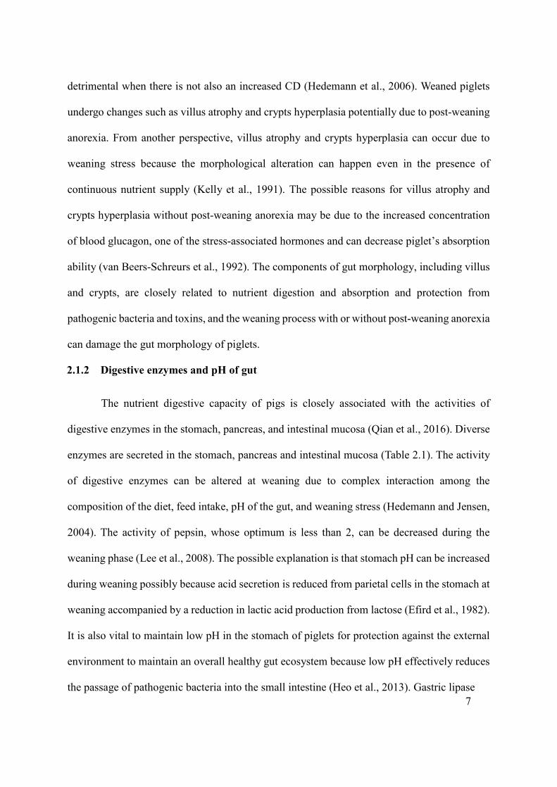
detrimental when there is not also an increased CD (Hedemann et al., 2006). Weaned piglets
undergo changes such as villus atrophy and crypts hyperplasia potentially due to post-weaning
anorexia. From another perspective, villus atrophy and crypts hyperplasia can occur due to
weaning stress because the morphological alteration can happen even in the presence of
continuous nutrient supply (Kelly et al., 1991). The possible reasons for villus atrophy and
crypts hyperplasia without post-weaning anorexia may be due to the increased concentration
of blood glucagon, one of the stress-associated hormones and can decrease piglet’s absorption
ability (van Beers‐Schreurs et al., 1992). The components of gut morphology, including villus
and crypts, are closely related to nutrient digestion and absorption and protection from
pathogenic bacteria and toxins, and the weaning process with or without post-weaning anorexia
can damage the gut morphology of piglets.
2.1.2 Digestive enzymes and pH of gut
The nutrient digestive capacity of pigs is closely associated with the activities of
digestive enzymes in the stomach, pancreas, and intestinal mucosa (Qian et al., 2016). Diverse
enzymes are secreted in the stomach, pancreas and intestinal mucosa (Table 2.1). The activity
of digestive enzymes can be altered at weaning due to complex interaction among the
composition of the diet, feed intake, pH of the gut, and weaning stress (Hedemann and Jensen,
2004). The activity of pepsin, whose optimum is less than 2, can be decreased during the
weaning phase (Lee et al., 2008). The possible explanation is that stomach pH can be increased
during weaning possibly because acid secretion is reduced from parietal cells in the stomach at
weaning accompanied by a reduction in lactic acid production from lactose (Efird et al., 1982).
It is also vital to maintain low pH in the stomach of piglets for protection against the external
environment to maintain an overall healthy gut ecosystem because low pH effectively reduces
the passage of pathogenic bacteria into the small intestine (Heo et al., 2013). Gastric lipase

8
Table 2.1 Endogenous enzymes and their reaction in pigs.
Origin Enzymes Major enzyme reaction References
Stomach Pepsin Polypeptides -> Polypeptide fragments
(Campos and Sancho, 2003)
Gastric lipase Triacylglycerol -> Diacylglycerol + Carboxylate
(Gargouri et al., 1986)
Pancreas Lipase Triacylglycerol r -> Diacylglycerol + Carboxylate
(Cera et al., 1990)
Trypsin Polypeptides ->
Polypeptide fragments
(Makkink et al., 1994)
Chymotrypsin Polypeptides ->
Polypeptide fragments
(Makkink et al., 1994)
Amylase Amylose -> Maltose and glucose (Pandol et al., 1985)
Intestinal mucosa Maltase-glucoamylase
Maltotriose and Maltose -> Glucose
(Van Beers et al., 1995)
Sucrase-isomaltase
Sucrose -> Glucose and fructose;
α-limit dextrin -> Glucose
(Van Beers et al., 1995)
Lactase Lactose -> Glucose + galactose (Van Beers et al., 1995)
Aminopeptidase Polypeptides ->
Amino acid + Polypeptide
(Maroux et al., 2018)
Intestinal alkaline
phosphate
Phosphate monoester -> Alcohol + Phosphate
(López-Canut et al., 2009)
9
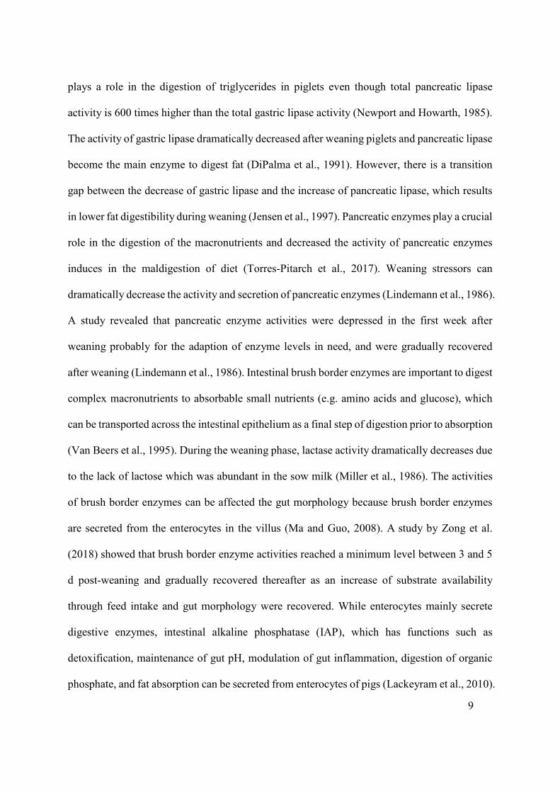
plays a role in the digestion of triglycerides in piglets even though total pancreatic lipase
activity is 600 times higher than the total gastric lipase activity (Newport and Howarth, 1985).
The activity of gastric lipase dramatically decreased after weaning piglets and pancreatic lipase
become the main enzyme to digest fat (DiPalma et al., 1991). However, there is a transition
gap between the decrease of gastric lipase and the increase of pancreatic lipase, which results
in lower fat digestibility during weaning (Jensen et al., 1997). Pancreatic enzymes play a crucial
role in the digestion of the macronutrients and decreased the activity of pancreatic enzymes
induces in the maldigestion of diet (Torres-Pitarch et al., 2017). Weaning stressors can
dramatically decrease the activity and secretion of pancreatic enzymes (Lindemann et al., 1986).
A study revealed that pancreatic enzyme activities were depressed in the first week after
weaning probably for the adaption of enzyme levels in need, and were gradually recovered
after weaning (Lindemann et al., 1986). Intestinal brush border enzymes are important to digest
complex macronutrients to absorbable small nutrients (e.g. amino acids and glucose), which
can be transported across the intestinal epithelium as a final step of digestion prior to absorption
(Van Beers et al., 1995). During the weaning phase, lactase activity dramatically decreases due
to the lack of lactose which was abundant in the sow milk (Miller et al., 1986). The activities
of brush border enzymes can be affected the gut morphology because brush border enzymes
are secreted from the enterocytes in the villus (Ma and Guo, 2008). A study by Zong et al.
(2018) showed that brush border enzyme activities reached a minimum level between 3 and 5
d post-weaning and gradually recovered thereafter as an increase of substrate availability
through feed intake and gut morphology were recovered. While enterocytes mainly secrete
digestive enzymes, intestinal alkaline phosphatase (IAP), which has functions such as
detoxification, maintenance of gut pH, modulation of gut inflammation, digestion of organic
phosphate, and fat absorption can be secreted from enterocytes of pigs (Lackeyram et al., 2010).

10
In addition, because IAP is an intrinsic enzyme, which is more subtle to alterations in the brush
border, the IAP activity may represent the gut maturation of pigs (Ghafoorunissa, 2001).
Optimal digestive enzyme activities and pH are important for nutrient digestion and gut
ecosystem of piglets and low enzyme activities can be accompanied by the alteration of gut pH
and impaired gut morphology during weaning.
2.1.3 Nutrient transporters and sensors
Nutrient absorption can be divided into paracellular and transcellular pathways
(Karasov, 2017). Nutrient transporters belong to transcellular absorption which represent either
the uptake of small molecules by active (carrier-mediated) or passive (carrier-unmediated)
transport (Wijtten et al., 2011). The expression of nutrient transporters is an important indicator
for nutrient utilization capacity of animals (Moran et al., 2010b). Early weaning decreases the
function of the Na+-glucose cotransporter 1 (SGLT1) and amino acid transport activities in the
jejunum and ileum of piglets (Li et al., 2018). When pigs were weaned after 4 weeks, active
absorption between 1 and 15 d after weaning was either similar to or higher than absorption
before weaning, which showed that when pigs were weaned after 4 weeks, active absorption is
not negatively affected by the weaning process (Lodemann et al., 2008). The activities of
nutrient transporters can be recovered with the repair of intestinal architecture after the weaning
phase (Lin et al., 2014). Regarding passive absorption, a study showed that 1 week after
weaning, the absorption of D-xylose decreased to approximately 50% of the pre-weaning level
(Kelly et al., 1990). Also, even 14 d after weaning, the absorption of D-xylose only reached
65% of the absorption level measured before weaning, which may imply that weaning can have
a permanent effect on passive absorption (Miller et al., 1984). Wijtten et al. (2011) supposed
that the decreased passive transcellular absorption after weaning is a defense mechanism to

11
protect the uncontrolled transport of potentially harmful agents from entering the body.
Nutrient transporters are closely associated with the utilization of nutrients in pigs, and the
weaning process can decrease the activities of nutrient transporters.
Nutrient sensors have been studied mainly to understand the dietary requirements and
preferences of animals. However, a few studies found that nutrient sensors are also closely
associated with the gut ecosystem of pigs (Lee et al., 2012a; Janssen and Depoortere, 2013).
Not only do the porcine nutrient sensors exist in the oral cavity, but they also exist in different
organs and act as a chemosensory system. The heterodimeric sweet taste receptors comprising
Type 1 taste receptors 2 (T1R2) and Type 1 taste receptors 3 (T1R3) are expressed in intestinal
enteroendocrine cells in pigs (Moran et al., 2010a). According to Daly et al. (2012), T1R2 and
T1R3 has functions of intestinal glucose sensing, inducing GLP1 (glucagon like peptide 1),
GLP-2 and GIP (glucose dependent insulinotropic peptide) release, which have been proved
using endocrine cell lines, native intestinal tissue explants and knock out mice deficient in
alpha-gustducin or T1R3. Shirazi-Beechey et al. (2014) showed that GLP1 and GIP improved
insulin secretion and GLP2 improved intestinal growth and glucose absorption. Calcium
sensing receptors (CaSR) also presented in the gastro-intestinal tract of pigs (Zhao et al., 2019).
Fatty acid sensors 40 (GPR40), GPR 43, and GPR 120 were found in the gastrointestinal tract
(Song et al., 2015). Nutrient sensory cells in the gut are known to be involved in the secretion
of gut hormones and also other physiological functions (Roura et al., 2016). For instance, CaSR
have functions of sensing nutrients, maintaining ion homeostasis, regulating the digestive
process, controlling colonic fluid balance and inducing the growth of epithelial cells (Zhao et
al., 2019). Most importantly, the activation of CaSR decreased the intestinal inflammatory
response in weaned piglets (Huang et al., 2015). Not only do nutrient sensors have functions
of nutrient sensing, but they are also involved in regulating the gut ecosystem of pigs.

12
2.1.4 Gut barrier integrity and tight junction proteins
Gut barrier integrity is maintained by a single layer of epithelium, mainly epithelial
cells and tight junctions on the gastrointestinal tract of pigs (Wang et al., 2014). Tight junctions,
multiprotein complexes located on the apical side of epithelial cells, play an important role in
maintaining cell polarity and regulating barrier integrity that prevents pathogens and toxins
from crossing the epithelial sheet between adjacent cells (Zhao et al., 2011). Tight junctions
are constituted of transmembrane proteins including occludin (OCLN) and claudin (CLDN),
junctional adhesion molecules, and peripheral membrane proteins such as zonula occludens 1
(ZO1), ZO2, ZO3, and the multi-PDZ domain protein 1 (MUPP1) (Moeser et al., 2017). The
efficiency of cell-cell adhesion (e.g. gut barrier integrity) is determined by the quantity and
distribution of the tight junctions (Li et al., 2012d). The weaning process impairs tight junction
integrity and increases intestinal permeability which can induce the pathogenesis of numerous
gastrointestinal diseases, such as inflammatory bowel disease, irritable bowel syndrome, celiac
disease, and infectious enterocolitis (Odenwald and Turner, 2013). Compared with the
preweaning stage (0 d), on 3 d, 7 d, and 14 d after weaning, jejunal transepithelial electrical
resistance (TEER), which represents the intestinal mucosal barrier, was decreased, mRNA
expression of OCLN, CLDN1 and ZO1 were reduced, and gut permeability (paracellular flux
of dextran) was increased (Zhao et al., 2011). However, damaged gut barrier function and
intestinal permeability in piglets recovered after 2 weeks of weaning (Peace et al., 2011). Gut
barrier integrity is mainly maintained by tight junction proteins and can be damaged due to
weaning stressors, resulting in diverse gastrointestinal diseases.
2.1.5 Immune system
Swine have a complex immune system which has functions of recognizing and attacking

13
pathogens and toxins. The immune system can be divided into two categories: innate immunity,
which is general and non-specific and includes macrophages and cytokines; and adaptive
immunity, which is specific and characterized by immunological memory, dendritic cells, and
lymphocytes. Immune parameters to assess the activities of the immune system of pigs can be
chosen based on the purpose of the experiment (Table 2.2). During the weaning period, the
protective immunity shifts from passive maternal immunity to the active immunity of the piglet
(Weiner et al., 2015). Due to the absence of necessary immune function during the weaning
phase, piglets can have symptoms of diarrhea, inflammation, or even mortality (Han et al.,
2016). Many studies documented that the immune system can be damaged, and an intestinal
inflammatory response can be activated during the weaning phase. After 1 and 2 days of
weaning, a decrease in jejunal expression of major histocompatibility complex (MHC) class 1
mRNA and an increase in the cluster of differentiation 4+ (CD 4+) T cells in jejunal villus were
found in piglets weaned at 21 d of age (Heo et al., 2013). Moreover, compared to normal
weaned piglets (20 d), early-weaned piglets (at 16 d and 18 d) challenged with Enterotoxigenic
Escherichia coli (ETEC), had a lower number of mast cells and higher pro-inflammatory
cytokines such as interleukin 6 (IL6) and IL8 in ileal mucosa (McLamb et al., 2013). Intestinal
mast cells play an important role in the innate immune response to bacterial, parasitic and viral
infections by releasing pro-inflammatory cytokines including tumor necrosis factor-α (TNF-α),
IL6 and leukotriene B4 (LTB4) that mediate neutrophil recruitment into infected sites
(Abraham and John, 2010). A study by Hu et al. (2013b) showed that after weaning, the
expression of pro-inflammatory cytokines increased and returned to pre-weaning values within
two weeks. The immune system, essential to piglets in protecting against pathogens and toxins,
can be damaged during the weaning phase because of weaning stressors and a lack of protection
derived from maternal milk.

14
Table 2.2 Parameters for evaluating the immune system of pigs.
Parameters Category Properties References
CD3+ T cells
(cluster of differentiation)
Adaptive immunity
Activated when antigens present
(Xiong et al., 2015b, a)
CD4+ T cells Adaptive immunity
Activated when antigens present
(Shen et al., 2009; Xiong et al., 2015b, a)
CD8+ T cells Adaptive immunity
Activated when antigens present
(Shen et al., 2009; Xiong et al., 2015b, a)
T-helper cell 17 Adaptive immunity
Effector memory T cells (Luo et al., 2015)
Interferon-γ Adaptive immunity
Pro-inflammatory cytokine
Interleukin-1 Adaptive immunity
Pro-inflammatory cytokine (Cavaillon, 2001; Lessard et al., 2015; Xiong et al., 2015b)
Interleukin-2 Adaptive immunity
Pro-inflammatory cytokine (Sugiharto et al., 2015; Xiong et al., 2015b)
Interleukin-4 Adaptive immunity
Anti-inflammatory cytokine
(Cavaillon, 2001; Lessard et al., 2015)
Interleukin-6 Adaptive immunity
Pro-inflammatory cytokine (Scheller et al., 2011; McLamb et al., 2013; Lessard et al., 2015; Wang et al., 2018a)
Interleukin-8 Adaptive immunity
Pro-inflammatory cytokine (McLamb et al., 2013)
Interleukin-10 Adaptive immunity
Anti-inflammatory
cytokine
(Cavaillon, 2001; Lessard et al., 2015; Sugiharto et al., 2015; Xiong et al., 2015b)
Pro-inflammatory cytokine (Shen et al., 2009)
Interleukin-12 Adaptive immunity
Pro-inflammatory cytokine (Cavaillon, 2001; Lessard et al., 2015)
Interleukin-17A Adaptive immunity
Pro-inflammatory cytokine (Luo et al., 2015)

15
Interleukin-17F Adaptive immunity
Pro-inflammatory cytokine (Luo et al., 2015)
Interleukin-21 Adaptive immunity
Pro-inflammatory cytokine (Luo et al., 2015)
Interleukin-22 Adaptive immunity
Pro-inflammatory cytokine (Luo et al., 2015)
Tumor necrosis factor -α Adaptive immunity
Pro-inflammatory cytokine (Lessard et al., 2015; Wang et al., 2018a)
PGE2 (Prostaglandin) Natural immunosuppressive molecule which reduces inflammatory responses.
(Kim et al., 2016)
Immunoglobulin G Adaptive immunity
Antibody (Takeyama et al., 2015)
Immunoglobulin A Adaptive immunity
Antibody (Sugiharto et al., 2015; Takeyama et al., 2015; Xiong et al., 2015b)
Immunoglobulin M Adaptive immunity
Antibody (Sugiharto et al., 2015; Rieckmann et al., 2018)
Natural killer cells Innate immunity
Leukocyte (Denyer et al., 2006; Annamalai et al., 2015)
Mast cells Innate immunity
Leukocyte (Pohl et al., 2017; Wang et al., 2018a)
Eosinophils Innate immunity
Leukocyte (Li et al., 2014)
Neutrophils Innate immunity
Leukocyte (Li et al., 2014)
Monocytes Innate immunity
Leukocyte (Li et al., 2014)

16
2.2 Gut microbiota
2.2.1 Understanding gut microbiota and its development
The lower gastrointestinal tract of swine is a natural shelter for diverse microbiota
including bacteria, archaea, fungi, protozoans, and viruses, which have a symbiotic relationship
with the animal (Barko et al., 2018). The microbiota has important roles in energy homeostasis,
normal digestive functions, metabolism for vitamin synthesis, defense against pathogens,
immunological activities such as catabolism of toxins, and neurodevelopment of pigs (Stanley
et al., 2016). However, gut microbial populations such as E. coli, Salmonella spp., and
Clostridia spp. can also induce diseases including post-weaning diarrhea (PWD) in the pig.
There are both pros and cons of microbiota that can influence the animal’s development and
gut ecosystem (Fig. 2.1) (Pieper et al., 2006). During the weaning phase, weaning stress can
result in a microbial imbalance because of the increased pathogenic bacteria including E. coli
and Salmonella spp. and reduced beneficial bacteria such as lactic acid-producing bacteria
including Lactobacillus spp. and Bifidobacterium spp. (Thu et al., 2011). While increased
beneficial bacteria (e.g., lactic acid-producing bacteria) have a function of preventing diseases,
an increase in pathogenic E. coli can account for PWD.

17
Figure 2.1 Advantages and disadvantages of gut microbiota in piglets. Gut microbiota can
positively or negatively affect the host.

18
2.3 Assessment methods of gut health and gut barrier integrity in pigs
2.3.1 Considerations for in vitro and in vivo evaluation methods
The growing interest in identifying new antimicrobials has been accompanied by an
equal interest in developing fast and reliable screening and evaluating methods. Traditionally,
bioassays such as well diffusion, disk-diffusion, and agar or broth dilution have been among
the commonly used techniques (Balouiri et al., 2016). Other novel and/or high-throughput
assays such as the use of enterocyte cultures, Caenorhabditis elegans (C. elegans), and
experimental animal models have started taking the lead in the past few years because these
methods can provide a better understanding of the screened substance’s impact on cellular
viability.
2.3.2 C. elegans model
In the past four decades, many researchers have been extensively using the C. elegans
model within the fields of biological research, including innate immunity and microbial
pathogenesis studies. This system relies solely on the elicited innate immune defenses to cope
with pathogen attacks as C. elegans lacks an adaptive immune system (Ewbank and Zugasti,
2011). The promise of this system is that many pathogenic microbes trigger specific
mechanisms of innate immunity and lead to the overexpression of certain polypeptides (some
have antibacterial activity) that only manifest during the course of pathogenesis. The use of the
C. elegans system can aid in capturing such peptides and enhance the possibility for
understanding the underlying mechanism(s) in large high-throughput in vivo screens of newly
developed antimicrobials (Kong et al., 2016). This nematode system, if used correctly, offers
inexpensive and robust screening platforms of antibiotics relying on a vast body of knowledge
accumulated in regard to worm physiology and its bacterial/fungal pathogen interactions (Kong
19
et al., 2016). One of the drawbacks that pertain to this model is that this system does not denote
the mode of action of bioactive compounds, hence generating the necessity for secondary
studies that involve target-based screens such as the genetic knockdown of host or bacterial
genes (Kim et al., 2017b). For innate immunity and microbial pathogenesis studies, the
C. elegans model is a cost-effective and informative method that can be used with secondary
studies to learn the mechanisms underlying the obtained results.
2.3.3 In vitro porcine intestinal cell model
Initially, in vitro experiments with porcine intestinal cell lines were conducted to study
a number of bacterial infections (Schierack et al., 2006). However, porcine cell lines nowadays
are used for pathogen studies and also for studying bioactive compounds to illustrate the
interaction between the host cells and pathogens or bioactive compounds (Burt et al., 2016;
Omonijo et al., 2018b). One of the most commonly used cell lines for study in pigs is porcine
IPEC-J2, which are porcine intestinal columnar epithelial cells, isolated from the mid-jejunum
of a neonatal piglet (Brosnahan and Brown, 2012). The factors that make this cell line unique
include the fact that it is obtained from small intestinal tissue (compared to the common human
colon-derived lines HT-29, T84, and Caco-2) and is not transformed (compared to the porcine
small intestinal line, IPI-2I) (Brosnahan and Brown, 2012). There are diverse response criteria
need in the experiments of in vitro porcine intestinal cell model including viability, TEER,
verification of tight junction proteins by immunofluorescence, mucin production, proliferation
assays, invasion assays, adhesion assays, and gene expression analysis of target genes including
pro- and anti-inflammatory cytokines (Schierack et al., 2006). The cell line model provides
diverse data for understanding the interactions between host cells and pathogens or bioactive
compounds.

20
2.3.4 Ussing chamber system
Ussing chamber, an ex vivo method, is used to study the gut barrier function and the
transport of ions, nutrients, and drugs across various epithelial tissues, especially intestine
tissues of animals (He et al., 2013). An intestinal tissue sample can be mounted between the
two chambers with Krebs Ringer buffers (KRB) and gas (5% CO2 and 95% O2) in the chamber
keeping the tissue alive during the incubation period (Clarke, 2009). Electrophysiological
properties including TEER, short circuit current (Isc), potential difference (mV) and epithelial
conductance (G) (the inverse of resistance) can be measured with the Ussing chamber. Gut
tightness, generated by tight junctions and mucus, can be expressed as TEER (Mardones et al.,
2004). Increased TEER shows increased gut barrier function, and a reduced TEER represents
decreased gut barrier function. The potential difference (mV) reflects the transmural potential
difference that is generated by ion transport across the epithelium while short-circuit current
(Isc) represents the net transmural ion transport (Woyengo, 2011). Increased
electrophysiological response (ΔIsc) reflects either increased electrogenic anion secretion (e.g.
Cl- and HCO3-) or increased electrogenic cation absorption (Na+). The transport activities of
some nutrients (glucose and some amino acids) that are transported by sodium- or chloride-
dependent transporters can be estimated by calculating ΔIsc after adding the nutrient in the
mucosa side chamber. Also, the absorption of materials such as glucose, heparin,
oligonucleotides, antibiotics, and amino acids can be determined by adding the nutrient to the
mucosal area and calculating its concentration at the serosal side after a specific time (Tang et
al., 2012). To study gastrointestinal epithelium permeability, the proportion of fluorescence-
labeled substances such as fluorescein isothiocyanate-dextran and different isotope substances
that can pass through the gastrointestinal epithelium can be calculated with exposed sample
area and time (Cao et al., 2018). The Ussing chamber method is more accurate than cell culture-

21
based models, because of the presence of adequate paracellular permeability, an apical mucous
layer, active transport proteins and drug-metabolizing enzymes (Castella et al., 2006).
Advantages of employing Ussing chamber include the ability to study the regional differences
along the intestine and bidirectional drug transport, and only a small amount of analytes are
required for analysis (Balimane et al., 2000). Thus, Ussing chamber can provide the
understanding of gut barrier integrity and transport of ions, nutrients, and drugs across
epithelial tissues of a pig’s intestine.
2.3.5 Experimental infection animal diseases models
The screening strategies to find AGP alternatives usually start with conventional
bacteria-centered assays but in a later stage, it is pivotal to use experimental animal models to
simulate the infection and treatment courses in order to investigate the effects of AGP
alternatives on the gut ecosystem and growth performance of pigs. Experimental models can
provide greater knowledge regarding different doses (concentrations in the feed) and
mechanisms for new AGP alternatives to determine the role of this AGP alternative in the
treatment of distinct infections.
The PWD induced by ETEC is an important worldwide disease in swine production
because PWD usually results in weight loss, slow growth, more treatment costs, body weight
heterogeneity, and even mortality (Lyutskanov, 2011). ETEC that expresses F4 (also designated
K88) or F18 adhesive fimbriae facilitating the colonization of intestinal mucosa, produce toxins
once they colonize in the intestine (Adewole et al., 2016). The experimental ETEC-challenged
pig model has been well-established and used by many researchers. The oral gavage of ETEC
to pigs is one of the most common methods to induce PWD in piglets (Bhandari et al., 2008).
The dosage of oral administration can range approximately from 3 mL of 1 × 10⁹ CFU·mL-1 to

22
5 mL of 1 × 10¹² CFU·mL-1 and the experimental period differs depending on the experiment’s
purpose and conditions (Adewole et al., 2016). While some studies showed that growth
performance was not influenced by the experimental ETEC challenge (Nyachoti et al., 2012),
a number of other studies reported that growth performance was reduced due to the
experimental ETEC challenge (Trevisi et al., 2009; Lee et al., 2012b). One of the possible
reasons for this difference is that there is variance in terms of the responses in piglets to ETEC.
Whether pigs are or are not susceptible to ETEC infection depends on the existence of a gene
(Mucin 4 (MUC4) for ETEC F4; Fucosyltransferase 1 (FUT1) for ETEC F18) which is
inherited as a simple Mendelian trait (there are two genotypes: resistant or susceptible) (Jensen
et al., 2006). Once the pigs are gene screened to select resistant or susceptible genes towards
ETEC F4 or F18-diarrhea in the ETEC challenge experiment, the experiment will be more
accurate by decreasing the initial variance towards ETEC F4-diarrhea (Ren et al., 2012). In
addition, it would be advantageous to select susceptible pigs to obtain diarrheic piglets to
decrease the variation of the clinical symptoms from ETEC (Jensen et al., 2006). Besides ETEC,
Salmonella spp. (a diarrhea and inflammation model), lipopolysaccharides (LPS) (an
inflammation model), and also diquat (an oxidative stress model) can be used to investigate the
effects of nutrients and AGP alternatives on challenged pigs (Boyen et al., 2008; Liu et al.,
2012a; Cao et al., 2018).
2.3.6 “Omics” and molecular techniques for studying gut microbiota
The main purpose of “omics” technologies is the non-targeted identification of all gene
products (transcripts, proteins, and metabolites) existing in a specific biological sample. Omics
technologies include genomics, transcriptomics (gene expression profiling), proteomics, and
metabolomics (Deusch et al., 2015). These advanced omics technologies serve to investigate

23
microbial communities as a whole and to explore more comprehensive studies of the
composition and functionality of gut microbiota (Gong et al., 2018). With metagenomics, a
collective way to study both the structure and function of microbiota, total community DNA is
obtained from fecal or digesta samples and the microbiome is analyzed by whole-genome
shotgun (Gong and Yang, 2012). In metatranscriptomics, total RNA extracted from complex
microbial populations is used; and this method provides information about how the host and
diet affect the microbiota (Sekirov et al., 2010). Metabolomics can be used to determine the
function of gut microbiota through the investigation of microbiota and host metabolite profiles
with nuclear magnetic resonance (NMR), mass spectroscopy and other methods (Gong and
Yang, 2012). Metaproteomics investigates the protein stock of a specific sample at a specific
time point, which permits the identification of the active microbial fraction and their expressed
metabolic pathways (Wilmes and Bond, 2006). Metagenomics (Looft et al., 2014; Mann et al.,
2014), metabolomics (Hanhineva et al., 2013; Pieper et al., 2014) and metaproteomics (Tilocca
et al., 2017) have all been used to study pig gut microbiota. There are no published
metatranscriptomics studies on pig’s gut microbiota.
In addition to omics techniques, there are still many other culture-independent
techniques including the PCR-based DNA profiling, quantitative PCR (Q-PCR), fluorescent in
situ hybridization (FISH), flow cytometry, DNA sequencing, and DNA microarray in the field.
The PCR-based DNA profiling method including denaturing gradient gel electrophoresis
(DGGE), temperature gradient gel electrophoresis (TGGE), single strand conformation
polymorphism (SSCP), and terminal-restriction fragment length polymorphism (T-RFLP) have
been widely used to characterize the gut microbiota (Gong et al., 2002; Li et al., 2003). The
principle of all PCR-based DNA profiling techniques including DGGE, TGGE, SSCP and T-
RFLP needs to employ PCR primers targeting 16S rRNA genes (or cpn60 genes) to amplify

24
16S rRNA (or cpn60) sequences from target bacteria (Gong and Yang, 2012). Q-PCR is a
technique to study bacterial population size of gut microbiota by quantification based on
primers with fluorescence-labeled group- or strain-specific probes or with a non-sequence
specific DNA-binding dye (SYBR® green) during the PCR amplification procedure of a target
gene (Feng et al., 2010). FISH can be utilized to investigate the microbial population and
oligonucleotides labeled with fluorescence substances targeting 16S rRNA genes and are
commonly used for the FISH analysis. The application of fluorescence in situ hybridization
can be used for visualization and quantification of the human gastrointestinal microbiota. Flow
cytometry is a technique used to count and assess mammalian cells and to study bacterial
populations (Festin et al., 1987). In the analysis with flow cytometry, bacterial cells in a
collected sample are fixed, and hybridized by using fluorescein-labeled antibodies (Festin et
al., 1987) and then automatic analysis with flow cytometry to investigate microbial
communities (Wallner et al., 1995). DNA sequencing can provide collective data on bacterial
diversity and community structures by using full-length 16s rRNA gene sequences through the
Sanger sequencing method (Xia et al., 2013). The DNA microarray has enabled researchers to
analyze many genes in a single experiment (Schena et al., 1995). With this method,
oligonucleotide probes obtained based on whole genomic DNA or 16S rRNA genes or cpn60
are produced and shown onto the array. Different fluorescence-labeled samples and a reference
are merged, fragmented, and hybridized with the microarray and finally, the difference of
fluorescence density can be determined to indicate the prevalence of target bacteria (Gong and
Yang, 2012). Each omics and molecular technique have pros and cons, and several techniques
should be utilized in a single experiment to supplement the results of the others.
25
2.4 Effects of dietary ingredients on gut microbiota, barrier integrity, and digestive
physiology in pigs
2.4.1 Carbohydrates (Dietary fiber)
The carbohydrate fraction can be classified on the basis of the number of glycosidic
linkages, into monosaccharides (sugar), oligosaccharides, and two broad classes of
polysaccharides: starch and non-starch polysaccharides (NSP) (Lindberg, 2014). According to
the cell wall structure, NSP and lignin have been defined as dietary fiber (DF) in the feed
(Theander et al., 1994; Lindberg, 2014). Nonetheless, because non-digestible oligosaccharides
such as raffinose, stachyose, and fructo-oligosaccharides, as well as resistant starch, are
degraded by microbial enzymes and are further processed via similar metabolic pathways to
produce short-chain fatty acids (SCFA), DF should include non-digestible oligosaccharides
and resistant starch (Scott et al., 2013). In the swine industry, DF has been thought to be a
feasible alternative to AGP because DF can improve growth performance, alleviate PWD, and
modulate gut microbiota of pigs (Jha and Berrocoso, 2016). To be specific, DF has a great
impact on both the mucosa and the microbiota and consequently has an important role in the
regulation of the gut ecosystem (Heinritz et al., 2016). The DF resists host digestion and
absorption, and is fermented by the microbiota, and selectively improves the abundance and
activity of beneficial bacteria in the gut (Montagne et al., 2003). The classification was
conducted on DF based on physicochemical properties because DF has different metabolic and
physiological functions depends on viscosity, hydration, and fermentability (Agyekum and
Nyachoti, 2017). Once beneficial bacteria colonize the gut, the abundance of pathogenic
bacteria decreases because the colonizable areas of microbes are limited in the gut ecosystem
of pigs (Cilieborg et al., 2016). In addition, through fermentation, OA including lactic acid and
SCFA including formate, acetate, propionate, and butyrate, can be produced and have diverse

26
beneficial effects on the gut ecosystem. These OA and SCFA can constrain the growth of enteric
bacterial pathogens including Salmonella spp., E. coli, and Clostridium spp. in the small and
large intestine by providing an acidic environment (Wang et al., 2018b; He et al., 2019).
Furthermore, some SCFA (e.g. acetate, butyrate, and propionate) influences the development
of the gut ecosystem of piglets by activating epithelium cell proliferation (Montagne et al.,
2003). However, a high concentration of DF in the feed can decrease the weight gain and feed
intake of piglets because DF is indigestible by the host and DF can give a satiety, possibly
decreasing feed intake, when bulky fibrous diets were provided; more studies are needed to
find the appropriate concentration of DF in the piglet’s diet (Wu et al., 2018). According to
Agyekum and Nyachoti (2017), the feed containing a high concentration of DF can be possibly
provided to the piglets by reducing particle size, pelleting, and using exogenous enzymes,
which may increase nutrient utilization in the piglets. Hence, the appropriate amount of the DF
with some treatments that increase nutrient utilization can improve the gut ecosystem of pigs
without affecting growth performance by decreasing the abundance of pathogenic bacteria and
developing the gut ecosystem of piglets by producing SCFA.
2.4.2 Proteins and functional amino acids
Spray-dried plasma, a protein source, can be used as an AGP alternative due to its
immunoglobulin-rich property for piglets who may lack the ability to produce
immunoglobulins themselves (Pérez-Bosque et al., 2016). Bosi et al. (2004) showed that a diet
containing spray-dried plasma enhanced growth performance and improved gut barrier
integrity, specific antibody defense and decreased inflammatory cytokine expression in piglets
challenged with ETEC F4. Amino acids, the building blocks of proteins, are known to have
diverse functions in the gut ecosystem of pigs (Wang et al., 2007). Every amino acid has

27
different gut health-promoting effects in piglets (Table 2.3). However, a high protein diet can
increase the risk of inducing intestinal disorders because feeding a higher protein diet can
increase the abundance of proteolytic microbes, including E. coli and C. perfringens, which
damage intestinal integrity and increase protein fermentation (Lin and Visek, 1991; Nousiainen,
1991). The products of protein fermentation include toxic metabolites such as ammonia and
amines as well as malodorous compounds including skatole and indole (Cone et al., 2005; Le
et al., 2008). Furthermore, nitrogen excretion, which causes environmental pollution, can be
induced by a high protein diet (Tous et al., 2016). Thus, while functional protein sources and
amino acids can benefit the gut ecosystem of piglets, high protein diets should be avoided in
the swine industry and exact levels of amino acids should be provided to meet the requirements.
2.4.3 Lipids (Fatty acids)
Lipids and fatty acids have a number of crucial biological functions such as serving as
energy sources, acting as structural components of cell membranes, participating in signaling
pathways, and modulating the immune system (Fahy et al., 2011). SCFA, fatty acids with a
chain of less than six carbon atoms, include acetate, propionate, and butyrate (Liu, 2015).
SCFA are the fermentation products of DF and probiotics in the lower gastrointestinal tract and
play an important role in improving gut health and restricting inflammation in the small and
large intestine of pigs (Rossi et al., 2010). Supplemental sodium butyrate improved jejunal gut
morphology and intestinal barrier function in the jejunum which has shown to increase TEER,
decrease paracellular flux of dextran (4kDa) and reduce the portion of degranulated mast cells
and their inflammatory mediators content including histamine, tryptase, and mRNA expression
of TNF-α and IL6 (Wang et al., 2018a). In addition, OCLN, one of the tight junction proteins,
was increased in the duodenum and tended to be increased in the jejunum and colon when

28
Table 2.3 Effects of functional amino acids on pigs.
Functional Amino acids Observations References
Arginine Improved the immune system including serum immunoglobulin G and immunoglobulin M in early weaned piglets
(Li et al., 2012b)
Attenuated the effect of oxidative stress by improving gut morphology and relieving the expression of pro-inflammatory cytokines in piglets
(Zheng et al., 2017)
Improved growth performance, gut morphology, barrier function (claudin1 mRNA expression), and antioxidant capacity (glutathione peroxidase mRNA expression) of low birth weight piglets.
(Zheng et al., 2018)
Improved intestinal development and increased expression of vascular endothelial growth factor (Yao et al., 2011)
Aspartate Attenuated the lipopolysaccharide challenge effect by improving intestinal morphology and barrier function and reduced toll like receptor 4, tumor necrosis factor-α and ileal caspase-3 protein expression.
(Wang et al., 2017)
Alleviated growth suppression and effects of oxidative stress when challenged by hydrogen peroxide.
(Duan et al., 2016)
Glutamate Prevented intestinal epithelial damage (gut atrophy) after weaning (Wu et al., 1996)
Enhanced the expression tight junction protein and regulate corticotropin-releasing factor signaling in the jejunum.
(Wang et al., 2014)
Stimulated the mTOR signaling and increases protein synthesis in enterocytes (Wang et al., 2007)
Enhanced the activities of lactase, maltase and sucrase in jejunum mucosa in weaned piglets (He et al., 2016)
Leucine Increased mucin production in the jejunal mucosa when challenged by Porcine Rotavirus (Mao et al., 2015)
Lysine Increased the population of Lactobacillus and Bifidobacterium in caecum and colon and raised the (Zhou et al., 2018)

29
expression of amino acid transporters
Threonine Increased intestinal mucin synthesis and immune status of intrauterine growth-retarded weanling piglets.
(Zhang et al., 2018a)

30
weaned piglets were fed with supplemental butyrate (Grilli et al., 2016). Sodium butyrate
decreased the incidence of diarrhea in weaned piglets (Feng et al., 2018). MCFA, which are
abundant in milk fat and various feed materials including coconut, palm, and cuphea seed oils,
are saturated 6-12 carbon fatty acids. MCFA supplementation enhanced gut development
through increased higher VH and improved growth performance in weaned piglets
(Hanczakowska et al., 2011). In addition, the antimicrobial properties and gut health
promoting-effects in pigs show that MCFA can be considered as AGP alternatives (Zentek et
al., 2012). Long-chain fatty acids (LCFA) are fatty acids with a chain length of 14 or more
carbons including omega-3 polyunsaturated fatty acids (PUFA), eicosapentaenoic acid (EPA)
[20:5(n-3)], and docosahexaenoic acid (DHA) [22:6(n-3)], are rich in fish oil. Fish oils have
been shown to modulate the immune system by inhibiting the over-release of intestinal pro-
inflammatory cytokines in piglets (Arnardottir et al., 2012). According to Liu et al. (2012b),
supplementation of fish oil could improve intestinal morphology and gut integrity and reduce
intestinal inflammation by decreasing TNF-α and toll-like receptors 4 (TLR4) expression in
piglets challenged with LPS. Lipids and fatty acids have the ability to enhance gut health and
modulate gut inflammation, whereas MCFA are known to have an antimicrobial effect.
2.4.4 Minerals
An appropriate amount of minerals must be supplied in swine rations to meet or exceed
the requirement since minerals have functions in the digestive processes such as; metabolizing
proteins, fats, and carbohydrates; and as the structure of chromosomes, enzymes, nerves, blood,
skeleton, hair and milk. Minerals are an important factor in reproduction, growth, production,
and resistance to parasites and diseases (Carlson and Boren, 2001). Calcium (Ca) and
phosphorous (P) are essential minerals for pigs, particularly for the bone formulation, and are

31
also closely linked in the digestive processes and metabolism (Suttle, 2010). Many studies have
demonstrated that Ca and P are related to the gut ecosystem and gut microbiota in weaned
piglets. According to Metzler-Zebeli et al. (2012), a high Ca-P diet decreased the expression of
IL1β, a proinflammatory cytokine, in the duodenum and reduced cecal CD by 14% compared
with low Ca-P diets. Furthermore, reduced dietary P altered transcription of phospholipase C,
Ca signaling, and nuclear factor of activated T-cells signaling, which may indicate that P has
an immunomodulatory function (Just et al., 2018). Since Ca and P are vital nutrients required
for bacteria for a number of metabolic processes in the bacterial cells (Durand and
Komisarczuk, 1988), differences in the intestinal availability and amount of Ca and P may alter
the growth of certain bacterial species and genera. Furthermore, another study revealed that a
high Ca-P diet increased Lactobacillus spp. by 1.4-fold in the stomach of pigs (Mann et al.,
2014), however, a high concentration of P can increase the abundance of pathogens (Heyer et
al., 2015).
Copper (Cu) and zinc oxide (ZnO) have an antimicrobial property, therefore they can
inhibit pathogens and reduce the loss of nutrients due to fermentation. Pharmacological
concentrations (above 3,000 mg/kg) of Cu and ZnO have been considered as effective growth
promoters in the diets of weaned piglets (Højberg et al., 2005). Cu and ZnO could have an
impact on reducing the population of Clostridia spp. and E. coli in weaned piglets (Song et al.,
2013; Dębski, 2016). In addition to antimicrobial effects, ZnO is also known to have a role in
anti-inflammation, antidiarrhea and restoration of mucosal barrier integrity (Jensen, 2016).
This is because, zinc is essential in the maintenance of the gut barrier function by regulating
the OCLN proteolysis and CLDN3 transcription and by upregulating protein kinase C zeta type
(PKCζ) via GPR39 (Miyoshi et al., 2016; Shao et al., 2017). Feeding diosmectite-ZnO
composite alleviated PWD symptoms, improved gut morphology, improved gut integrity by
32
upregulating tight junction proteins (OCLN, CLDN1 and ZO1 in jejunal mucosa), and
decreased mRNA expression level of pro-inflammatory cytokines (TNF-α, IL6, and interferon-
γ) in weaned piglets (Hu et al., 2013a). Weaned piglets fed with dietary ZnO showed a higher
abundance of transporters in the ileum (Yu et al., 2017). Supplementation of coated ZnO
reduced the microbiota species richness and Shannon diversity index in the jejunal digesta and
feces, indicating that ZnO has an impact on intestinal microbiota diversity (Shen et al., 2014).
In contrast, Yu et al. (2017) have shown that both ZnO and antibiotics increased the microbiota
diversity of ileal digesta, while they reduced the microbiota diversity of the colonic digesta.
Since dietary Cu and/or ZnO can accumulate in pig feces and eventually increase the mineral
content in the soil which negatively impacts the environment, there is now a sense that
pharmacological concentrations of Cu and ZnO should be replaced with other bioactive
compounds (Poulsen, 1998). This issue can probably be resolved with different encapsulating
techniques decreasing the amount of ZnO supplementation and allowing a slower release of
ZnO throughout the gut of piglets (Kim et al., 2015). Ciesinski et al. (2018) showed that a
pharmacological concentration of ZnO can induce the presence of antimicrobial resistant E.
coli in the swine gut. Therefore, a high concentration of ZnO can show beneficial effects to gut
health of piglets, but the supplementation of high levels of ZnO can cause environmental issues
and generation of resistant bacteria, which may decrease the potency of ZnO as an AGP
alternative.
Enough dietary iron should be supplemented in the diet of pigs to prevent iron
deficiency which can result in infection-related mortality and morbidity from diarrhea (Wayhs
et al., 2004). However, a high concentration of iron should be avoided in pig nutrition because
iron is the first limiting nutrient for some pathogens in mammals, therefore a high concentration
of iron can induce the growth of some pathogens (Klasing, 2007). A high concentration of iron
33
has increased the growth and virulence of Salmonella enterica in in vitro model (Tan et al.,
2019). Pigs fed with low and high dietary iron in the feed had increased gut permeability, and
pigs fed high dietary iron had increased the expression of pro-inflammatory cytokines such as
TNF-α, IL1β and IL6 (Li et al., 2016). In summary, appropriate levels of Cu, ZnO and iron
benefit piglets through antimicrobial and antidiarrheal effects, however, a high concentration
of these minerals can cause a malfunction in piglets and result in environmental pollution.
2.4.5 Vitamins
An appropriate amount of vitamins should be supplemented in a piglet’s diet to meet
the requirement. However, to overcome oxidative stress, which is an imbalance between
reactive oxygen species (ROS) and antioxidants which causes cellular damage and is related to
weaning stress, higher concentrations of vitamins can be supplemented in the diets of piglets
to relieve oxidative stress (Debier, 2007). Adding vitamins such as provitamin A carotenoids
and vitamins C and E, which are natural antioxidants, can enhance the gut ecosystem of weaned
piglets protecting it against oxidative stress-induced diseases (Deng et al., 2010). Alpha, beta,
and gamma carotene are referred to as provitamins because they can be converted to vitamin A
(Clemens et al., 1992). The benefits of provitamin A carotenoids are mainly related to their
antioxidant properties (Britton, 2008). According to Kang et al. (2018), supplementation of the
spontaneous oxidation of beta-carotene decreased necrotic enteritis in broiler chickens
potentially because of its strong antioxidant property. In addition, fully oxidized beta-carotene
functions to enhance the innate immune system and decrease inflammatory processes
(Duquette et al., 2014). Rey et al. (2017) reported that supplementation of a high dose of
vitamin E (250 mg·kg-1) decreased muscle oxidation for piglets, and supplementation of a high
dose of vitamin C (500 mg·kg-1) increased the serum antioxidant power and serum IgG
34
concentration. Antioxidant blend including vitamins C and E could compensate for some of the
harmful changes caused by weaned, by upregulating the gene expression of tumor protein 53
(p53) and peroxisome proliferator-activated receptor gamma coactivator 1α (PGC1α), which
have roles in cell cycle arrest, DNA repair, apoptosis and senescence, and activate
mitochondrial biogenesis and mitochondrial respiration (Zhu et al., 2012). However, a study
showed that high concentrations of vitamin E (10 times of the dose recommended by NRC
(2012) could also decrease gut morphology and function by inhibiting jejunal epithelial cell
proliferation (Chen et al., 2019). An appropriate amount of vitamins (e.g. vitamins C and E)
should be supplemented in the diets of piglets to attenuate oxidative stress, but an exceedingly
high concentration of vitamins can cause toxicity to the host.
2.5 Antibiotics
Antibiotics have been used for 60 years in the swine industry due to their efficiency in
promoting the growth rate, their ability to augment feed utilization, their cost-effectiveness,
and their effectiveness in decreasing mortality from clinical diseases (Allen et al., 2013). There
are four mechanisms by which are beneficial for swine production: (1) reducing sub-clinical
infections, (2) decreasing growth-depressing microbial metabolites (3) suppressing microbial
use of nutrients, and (4) increasing absorption and use of nutrients (Gaskins et al., 2002).
However, because of the concerns of drug residues in meat products and the delivery of drug-
resistant bacteria to humans, many countries have prohibited the use of in-feed antibiotics for
swine as a regular means of growth promotion (Van der Fels-Klerx et al., 2011). In the past 20
years, a great deal of research has been conducted to find alternatives to AGP to sustain swine
gut health and performance (Thacker, 2013). Each of the investigated replacements has
different advantages and disadvantages (Table 2.4).

35
Table 2.4 Beneficial effects and shortcomings of each antibiotic alternative and feasible solutions.
Antibiotic alternatives Beneficial effects Shortcomings Feasible solutions
Probiotics
Improve the intestinal development, host’s immune regulation, gut microbiota modulation and alleviation of toxins effects (Barba-Vidal et al., 2018)
Storage and delivering to target site issues: survival of probiotics bacteria (Shah et al., 1995)
Microencapsulation of probiotics to increase stability in storage and to increase survivability in stomach (Martín et al., 2015)
Prebiotics
Modulate the balance of intestinal microbial population by increasing beneficial bacteria while decreasing pathogenic bacteria (Steed and Macfarlane, 2009; Herfel et al., 2011)
Whether prebiotics are beneficial to the immune system is still controversial (Pandey et al., 2015)
Stimulate body’s immune system (Drulis-Kawa et al., 2012)
Needs more research (Pandey et al., 2015)
Bacteriophages Modulate the growth of specific harmful bacteria in pigs and do not harm useful bacteria unlike antibiotics (Kim et al., 2017a).
Possibility of producing resistant bacteria (León and Bastías, 2015)
Sensitive to temperature, low pH (Smith et al., 1987)
Microencapsulation of bacteriophage for increasing storage stability and for improving passage rate in stomach (Johnson et al., 2008; Ma et al., 2008)
Antimicrobial peptides
Have broader range of antimicrobial function, rapidly killing bacteria, and highly selective toxicity (Hancock and Patrzykat, 2002; Zasloff, 2002)
Limited by the number of approved AMPs: poor selectivity, hemolytic activity and host toxicity, low stability to protease degradation in vivo, low

36
hydro solubility, and cost of production (Wang, 2017)
Medium chain fatty acids
Show strong antibacterial activity (Skřivanová et al., 2009)
High inclusion of medium chain fatty acids decreased feed intake and body weight gain of weaned piglets (Li et al., 2015)
An appropriate amount of medium chain fatty acids should be supplemented to piglets
Exogenous enzymes
Increase digestibility and improve gut health
(Torres-Pitarch et al., 2017)
Low enzyme activities, high cost, no appropriate standards, no antibacterial effects, stability issues (Cheng et al., 2014)
Coating enzymes to increase bioavailability
(Liu et al., 2017a)
Phytochemicals
(Essential oils)
Improve gut barrier integrity, augment the immune system, increase antioxidant activities and modulate intestinal microbiota (Omonijo et al., 2018c)
Storage issue (volatility), cost, and availability (Yang et al., 2015)
Microencapsulation for feeding can improve stability and passage rate in stomach (Omonijo et al., 2018a)
Phytochemicals
(Plant extract) Have antimicrobial, anti-inflammatory, and gene regulatory functions
Antinutritional factors including tannins and saponins which can decrease digestibility of nutrients and damage the gut ecosystem (Cheng et al., 2014)
An appropriate amount of medium chain fatty acids should be supplemented to piglets
Organic acids
Reduce gut pH, activate enzyme secretion, inhibit pathogenic bacteria, increase nutrient digestibility and retention (Papatsiros et al., 2012)
High inclusion level of organic acids can decrease feed intake due to strong odor and flavor (Partanen and Mroz, 1999)
An appropriate amount should be included (Partanen and Mroz, 1999)

37
2.6 Antibiotic alternatives
2.6.1 Probiotics
Probiotics are living microorganisms that can be beneficial for host health when
appropriate amounts are provided (Hotel and Cordoba, 2001). Probiotics are beneficial for the
host’s gut ecosystem through four mechanisms: improvement of intestinal development;
enhancement of the host’s immune system; modulation of gut microbiota; and alleviation of
the effects of toxins from harmful microbiota (Barba-Vidal et al., 2018). The gut health-
promoting effects of probiotics are highly strain-specific, and due to synergetic effects, multi-
strain mixtures may be more effective than a single strain (Timmerman et al., 2004). For
instance, the most often used probiotics for swine are lactic acid bacteria including
Lactobacillus spp., Bifidobacterium spp., Enterococcus spp., and Streptococcus spp. (Ljungh
and Wadstrom, 2006). According to Choi et al. (2016), the inclusion of Lactobacillus
acidophilus, Bifidobacterium subtilis and Streptococcus cerevisiae complexes (4.0 × 10⁸; 4.8
× 109; 1.0 × 10⁴ CFU·g-1 respectively) in the diets of weaned piglets enhanced VH in the
duodenum, jejunum, and ileum. Probiotics have a role in modulating the host’s intestinal
balance by suppressing harmful microorganisms and supporting advantageous microorganisms
(Fuller, 2012). For example, Lactobacillus spp. reduced the viability of pathogens by producing
OA, hydrogen peroxide, and antimicrobial substances (Lidbeck and Nord, 1993). Moreover,
Bacillus spp. and Saccharomyces spp. can produce antimicrobial substances (e.g. reuterin) and
reduce the effect of pathogenic bacteria such as E. coli (Lidbeck and Nord, 1993). Certain
probiotics can have an impact on the intestinal mucosal cell-cell interactions and cellular
“stability” which improves the intestinal barrier via alteration of the phosphorylation of the
38
cytoskeletal and tight junction proteins (Willing et al., 2013). Saccharomyces cerevisiae, one
of the probiotics, can metabolize or aid in the detoxification of certain inhibitory compounds
such as amines or nitrates or scavenge for oxygen, which is critical in anaerobic conditions in
the gut (Chaucheyras-Durand et al., 2008). To sum up, probiotics can improve the gut
ecosystem by inhibiting the growth of pathogens and promoting the growth of beneficial
bacteria throughout the gut.
2.6.2 Prebiotics
According to Gibson and Roberfroid (1995), a prebiotic is defined as a selectively
fermented ingredient that makes specific changes both in the composition and/or activities in
the gastrointestinal microbiota that may be beneficial to the host’s health. The difference
between DF and prebiotics is that DF is non-digestible plant-derived carbohydrates consisting
of at least 3 units of individual sugars, while prebiotics is a substance obtained from plants,
animals and microbial sources that can confer the health benefit. Examples of prebiotics
include oligosaccharide carbohydrates such as fructo-oligosaccharides, galacto-
oligosaccharides, xylo-oligosaccharides, manno-oligosaccharides, and lactulose (Shokryazdan
et al., 2017). Prebiotics modulate the balance of the intestinal microbial population by
increasing beneficial bacteria such as Bifidobacterial spp. and Lactobacillus spp., and
decreasing pathogenic bacteria in the gut (Herfel et al., 2011). In one study, chito-
oligosaccharides reduced the population of Staphylococcus aureus (pathogenic) and increased
the concentration of lactic acid bacteria, Bifidobacteria spp. and Lactobacillus spp. in the
cecum compared to the control diet on d 7 post-weaning (Yang et al., 2012). The growth in the
population of beneficial microbes in the gut augments the gut ecosystem by regulating the

39
expression of pro- and anti-inflammatory cytokines, particularly in intestinal cells (Kunavue
and Lien, 2012). One experiment showed that the VH in the ileum and the concentration of
total volatile fatty acids, produced by microbiota were higher in pigs fed with isomalto-
oligosaccharides (Wu et al., 2017). Additionally, this study showed that prebiotics can be
beneficial to the immune system by increasing glutathione peroxidase, catalase, and IgG in
weaned piglets. There are synergetic effects when prebiotics and probiotics are used together
in animal feeds, called the symbiotic effects (Gibson and Roberfroid, 1995). When
Lactobacillus paracasei were blended with fructo-oligosaccharides, there were symbiotic
effects in increasing beneficial bacteria such as Lactobacillus spp. and Bifidobacterium spp.
and decreasing E. coli and Clostridium spp. in weaned piglets (Nemcova et al., 1999).
Prebiotics can benefit the gut ecosystem of piglets by being nutrients for beneficial bacteria,
which decreases the population of pathogens once beneficial bacteria colonize in the gut.
2.6.3 Bacteriophages
Bacteriophages, which are live viruses only affecting and attacking targeted bacteria,
are a recently developed technique to replace AGP in the livestock industry (Hussain et al.,
2017). Bacteriophages show efficient antimicrobial effect by infecting and attacking bacteria
and entering a lysogenic or lytic cycle within a bacterial cell, which replicates bacteriophages
(Fig. 2.3) (Verstappen et al., 2016). Certain bacteriophages specifically modulate the harmful
bacteria in pigs and do not harm useful bacteria, unlike AGP (Kim et al., 2017a). For efficiency,
it is beneficial to use a blend of several kinds of bacteriophages in order to control the
pathogenic bacteria including E.coli and Listeria spp. (Anany et al., 2011). Bacteriophages can
not only be used for therapeutic purposes against diseases such as PWD, but may also be used

40
for improving growth performance, feed efficiency, and gut microbiota in pigs (Gebru et al.,
2010; Cha et al., 2012; Kim et al., 2014). According to Lee et al. (2016), the supplementation
of bacteriophages increased the population of total anaerobic bacteria including
Bifidobacterium spp., Lactobacillus spp., and Clostridium spp. Additionally, VH of the
duodenum and jejunum was increased. Dietary phage supplementation attenuated the
symptoms of the ETEC-challenged infection by improving the gut structure, the immune
system, and growth performance in weaned piglets (Sabouri et al., 2017). Likewise,
supplementation of dietary ETEC F4 and F5 (K99) specific bacteriophages showed higher
weight gain, lower fecal consistency score, and less fecal shedding and intestinal adhesion of
ETEC F4 and F5 in piglets compared to piglets that did not get dietary bacteriophages (Lee et
al., 2017). Not only can bacteriophages specifically modulate the growth of targeted bacteria,
but they can also improve gut health and growth performance of piglets.

41
Figure 2.2 Mechanism of bacteriophage therapy. This schematic illustration represents the
developmental cycle of lyric bacteriophage.

42
2.6.4 Antimicrobial peptides
Antimicrobial peptides (AMP), which are small gene-encoded peptides, act as part of
the innate host defense mechanism and are shown to attack bacteria, regulate bacterial
infections and manage host responses to infection (Yeung et al., 2011). In the swine industry,
AMP have been considered as an alternative to AGP because of their benefits, including a
broader range of antimicrobial function, rapidly killing bacteria, and highly selective toxicity
(Hancock and Patrzykat, 2002). Most importantly, bacteria rarely develop resistance to AMP
because of AMP’s ability to disturb bacterial membranes through non-particular electrostatic
interaction with the components of the lipid membrane (Chou et al., 2010). Supplementation
of AMP attenuated the incidence of PWD and atrophy of gut structure and increased the
population of Lactobacillus spp. in ETEC F4 challenged piglets (Wu et al., 2012). Likewise,
supplementation of AMP to weaned piglets enhanced the gut structure and reduced fecal and
intestinal coliforms compared to pigs fed with the control diet without AMP (Yoon et al., 2013).
When AMP were provided to weaned piglets challenged with deoxynivalenol, a mycotoxin
produced by some Fusarium spp., they were capable of improving intestinal morphology and
activating epithelial cell proliferation and protein synthesis (Xiao et al., 2013). In the swine
industry, AMP are considered a promising AGP replacement since AMP provide a lot of
benefits to the gut ecosystem of piglets and rarely induce the growth of antibiotic-resistant
bacteria in the gut.
2.6.5 Medium chain fatty acids (MCFA)
Medium chain fatty acids (MCFA) are saturated 6-12 carbon fatty acids and are
considered as alternatives to AGP because of their strong antibacterial activity due to the

43
anionic part of the molecule acting against Gram-positive cocci (Bergsson et al., 2001) and E.
coli (Skřivanová et al., 2009). The anionic part of fatty acids modifies the physicochemical
properties of the digestive tract environment where the microorganisms exist and affect the
expression of some genes of microorganisms and the host (Baltić et al., 2017). Zentek et al.
(2011) showed that diet MCFA increased the population of eubacteria, Enterobacteriaceae,
clostridial clusters I and IV, Lactobacillus johnsonii, and Lactobacillus amylovorus, which are
considered beneficial bacteria in the gastric contents. The concentration of bacteria including
Salmonella spp. and coliforms in the jejunum and cecum digesta were decreased by MCFA
supplementation (Hanczakowska et al., 2016). Medium chain triglycerides, which are
triglycerides with fatty acids and have similar functions with MCFA improved the gut immune
system of piglets by modulating the recruitment and the maturation of immune cells (Hassan
et al., 2018). Also, MCFA could have an impact on post-weaning gut development such as
greater VH (Hanczakowska et al., 2016). With a focus on its antimicrobial properties, MCFA
can improve the gut ecosystem of piglets, making it a promising AGP alternative.
2.6.6 Exogenous enzymes
Supplementation of exogenous enzymes, which can help the digestion of complex
matrix of a piglet’s diet, can bridge the gap during weaning until a piglet’s endogenous enzymes
are ready to develop (Torres-Pitarch et al., 2017). The addition of exogenous lipase may be an
effective way to compensate for the shortage of endogenous lipolytic enzymes during the
weaning phase (Zhang et al., 2018b). Proteases are also one of the most common enzymes used
in piglets to increase protein utilization and to enhance the gut ecosystem. Zuo et al. (2015)
noted that dietary protease enhanced the gut structure by increasing the ratio of VH to CD in

44
the duodenum, jejunum, and ileum and increased nutrient efficiency by increasing the mRNA
abundance of amino acid transporters. The possible explanation would be that protease
promotes the release of functional amino acids from the ingredients which can improve the gut
ecosystem. Carbohydrases such as xylanase and β-mannase are mainly used to break down the
structure of cereal non-starch polysaccharides (NSP) that have negative effects on nutrient
digestibility (Van Kempen et al., 2006). Supplementation of xylanase could increase fecal
Lactobacillus spp. and digestibility of dry matter, nitrogen, and energy digestibility (Lan et al.,
2017). The mechanism by which non-starch polysaccharide-degrading enzymes affect
intestinal microbiota is by breaking down the structure of undegraded substrates (Rajagopalan
et al., 2013) and by releasing oligosaccharides, which have potential prebiotic effects, from cell
wall NSP (Pluske et al., 2002). Because IAP can detoxify LPS, IAP can be added to the diet of
piglets. According to Beumer et al. (2003), when IAP from calf intestine was added to the diet
of piglets, IAP decreased LPS-mediated inflammation by reducing the expression of TNF-α in
the serum. Phytase, phytic acid (an organic form of phosphorus in plants) degrading enzymes,
become an essential feed additive for pigs because P availability in pigs is low while P is
important in bone and cell membrane structure, energy metabolism and the other metabolic
pathways (Lu et al., 2019). In addition, without phytase supplementation, pigs excrete 50 – 80%
of P intake because of the low availability of plant P, which causes environmental pollution
through soil contamination (Kornegay et al., 1997). Therefore, exogenous enzymes can be
added to the diet of piglets to enhance feed efficiency, to improve the gut ecosystem and to
minimize negative environmental effects.

45
2.6.7 Phytochemicals (EO and plant extracts)
Essential oils (EO) are natural bioactive compounds derived from plants and are known
to possess antibacterial and antifungal properties and protect the host against infectious
diseases (Perez-Roses et al., 2016). In addition, EO have been shown to improve gut integrity,
and the immune system, to increase antioxidant activity and to modulate intestinal microbiota
of animals (Fig. 2.4). There are many kinds of EO, and it is thought that each EO might have
different effects in weaned piglets (Table 2.5). The addition of oregano EO to the diet of weaned
piglets was shown to increase VH and expression of OCLN and ZO1 in the jejunum, which
represented improved intestinal barrier functions (Zou et al., 2016b). A blend of
cinnamaldehyde and thymol was able to increase the amount of IgG and IgM in a linear fashion
in serum, which is indicative of an improved immune system (Su et al., 2018). Furthermore, in
this research, plant EO supplementation could induce activation of antioxidant functions by
decreasing malondialdehyde and increasing glutathione in serum, which indicates improved
whole-body antioxidant status and a reduction in lipid peroxidation, respectively. The addition
of a blend of thymol and cinnamaldehyde to the diets of piglets reduced the incidence of
diarrhea and decreased E.coli counts in their feces (Li et al., 2012c). Also, the ratio of
Lactobacillus spp. to E.coli increased while the total counts of aerobe number in the rectum
decreased (Li et al., 2012a). A study by Omonijo et al. (2018b) showed that thymol enhanced
barrier function, decreased ROS production, and reduced inflammatory response in the porcine
intestinal epithelial cells during LPS-induced inflammation. However, issues of cost-
effectiveness and feed palatability, as well as the lipophilic and volatile properties of EO, can
be obstacles when applying EO to a pig’s diet. However, these challenges can partially be

46
resolved through microencapsulation and nanotechnology to include a lower level of EO and
to increase the stability of EO to be included in the pig diets.
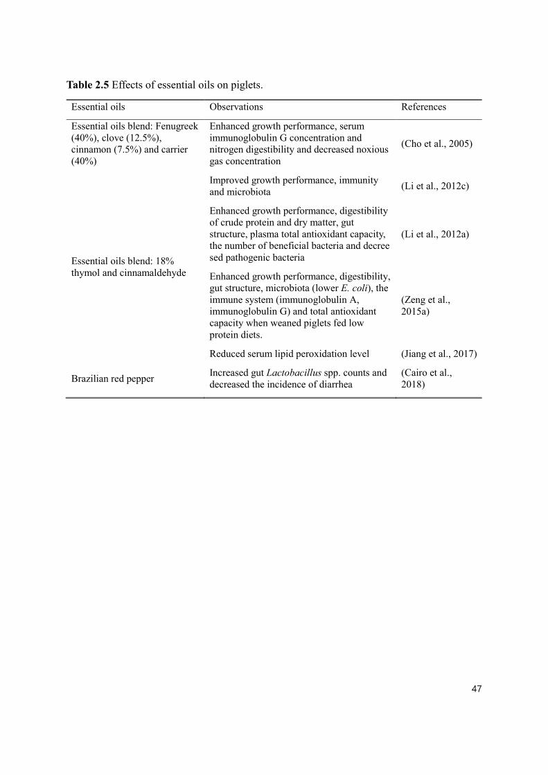
47
Table 2.5 Effects of essential oils on piglets.
Essential oils Observations References
Essential oils blend: Fenugreek (40%), clove (12.5%), cinnamon (7.5%) and carrier (40%)
Enhanced growth performance, serum immunoglobulin G concentration and nitrogen digestibility and decreased noxious gas concentration
(Cho et al., 2005)
Essential oils blend: 18% thymol and cinnamaldehyde
Improved growth performance, immunity and microbiota (Li et al., 2012c)
Enhanced growth performance, digestibility of crude protein and dry matter, gut structure, plasma total antioxidant capacity, the number of beneficial bacteria and decree sed pathogenic bacteria
(Li et al., 2012a)
Enhanced growth performance, digestibility, gut structure, microbiota (lower E. coli), the immune system (immunoglobulin A, immunoglobulin G) and total antioxidant capacity when weaned piglets fed low protein diets.
(Zeng et al., 2015a)
Reduced serum lipid peroxidation level (Jiang et al., 2017)
Brazilian red pepper Increased gut Lactobacillus spp. counts and decreased the incidence of diarrhea
(Cairo et al., 2018)

48
Plant extracts are known to have antimicrobial, anti-inflammatory, and gene regulatory
functions, which make them promising for human and veterinary medicine (Windisch et al.,
2008). There are diverse kinds of plant extracts, and their effects on animals differ depending
on the types and parts of plants. When capsicum oleoresin, garlic botanical, and turmeric
oleoresin were supplemented to weaned piglets, capsicum oleoresin and turmeric oleoresin
increased the expression of the genes related to gut barrier function and immune response,
indicating that feeding plant extracts can improve the gut ecosystem of weaned piglets (Liu et
al., 2014). The extract of Yucca schidigera, native to North America and rich in saponin, is
known to increase growth performance, improve feed efficiency, promote anti-protozoal and
nematocidal activity, and modify the microbial population in the gut ecosystem of pigs (Cheeke,
2000). According to Cromwell et al. (1985), Yucca schidigera extract improved average daily
gain (ADG) in weaned piglets. Seaweed extract contains mannitol, storage and structural
polysaccharides, phlorotannins, amino acids, high level of minerals including Ca and iodine,
and vitamins such as thiamin, ascorbic acid, tocopherols, and carotenoids (MacArtain et al.,
2007). Phlorotannins in the brown seaweed extract are thought to be an antimicrobial substance
against both Gram-positive and Gram-negative bacteria (Eom et al., 2012). Leonard et al. (2011)
showed that supplementation of seaweed extract decreased the population of E. coli and
Enterobacteriaceae in the colon digesta of weaned piglets. Dietary Chinese medicinal herbs
(composed of Panax ginseng, Dio-scoreaceae opposite, Atractylodes macrocephala,
Glycyrrhiza uralensis, Ziziphusjujube and Platycodon grandiflorum) increased the VH and
Lactobacillus counts in the ileal digesta as well as decreased coliforms counts in the colon of
weaned piglets (Huang et al., 2012). A number of secondary metabolites are produced by plants.
Among these, the group of polyphenols might be the most promising ones due to their

49
antioxidative, anti-inflammatory, and gene regulatory properties (Gessner et al., 2017).
Feeding grape seed and grape marc meal extract, which are rich in polyphenols, to weaned
piglets, decreased the expression of pro-inflammatory genes and produced a higher ratio of VH
and CD in the duodenum and jejunum than in the control pigs (Fiesel et al., 2014). Yang et al.
(2019a) showed that Red-osier dogwood (native to North America), which is rich in phenolic
compounds, improved cellular activity by directly reducing ROS and improving the cellular
antioxidant system as well as by upregulating tight junction proteins. There are many kinds of
phytochemical compounds; since each compound has different effects on the gut ecosystem,
appropriate kinds and amounts of phytochemical compounds should be applied to piglets.
Figure 2.3 Schematic diagram illustrating the four different potential mechanisms by which
essential oils improve the gut ecosystem and growth performance of piglets (Omonijo et al.,
2018c).

50
2.6.8 Organic acids (OA)
In swine production, OA are considered promising AGP alternatives and a blend of
diverse OA is known to improve gut health and growth performance of weaned piglets (Kil et
al., 2011). The mode of actions for OA includes reducing gut pH, stimulating the secretion of
enzymes, inhibiting the growth of pathogenic bacteria, and improving the growth and recovery
of the intestinal morphology (Papatsiros et al., 2012). Supplementation of 1% lactic acid and
1% formic acid reduced pH in the stomach and decreased the population of Enterobacteriaceae
in the stomach of weaned piglets (Hansen et al., 2007). The addition of citric acid in the weaned
pig diet yielded a significant decrease in fecal counts of Salmonella and E.coli and an increase
in Lactobacillus spp. and Bacillus spp. compared to the control diet without any OA (Ahmed
et al., 2014). Coated OA (benzoic acid, calcium formate, and fumaric acid) increased VH in
the duodenum and jejunum, attenuated diarrhea and improved growth performance in weaned
piglets (Xu et al., 2018). Humic substances including humic acid, fulvic acid, and humin are
organic residues generated from the decomposition of organic matter in the soil (Kaevska et
al., 2016). These humic substances are known to have antidiarrheal, analgesic,
immunostimulatory, and antimicrobial properties (Huck et al., 1991). Dietary supplementation
with sodium humate and ZnO reduced the incidence of diarrhea and increased the population
of beneficial bacteria in feces of experimental ETEC-challenged piglets compared to control
ETEC F4 and F18 challenged piglets (Kaevska et al., 2016). A study by Weber et al. (2014)
revealed that supplementation of humic acid and butyric acid improved the immune system in
LPS challenged piglets. Taken together, available evidence shows that OA are shown to have
diverse benefits to piglets, which includes decreasing the pH of the gut, activating enzyme

51
secretion, and having an antimicrobial effect on pathogenic bacteria.
2.7 Conclusion
The weaning period is the most challenging and demanding phase for pigs since piglets
have a functionally and anatomically immature gut ecosystem, and there are diverse stress
factors associated with weaning process. As a result, in the weaning phase, piglets undergo
dramatic alterations in their gut ecosystem. In order to assess the gut ecosystem of pigs, several
in vitro, in vivo, and ex vivo methods have been developed and utilized. After world-wide
restrictions were put in place with regard to using AGP in feeds, a number of antibiotic
alternatives have been introduced and evaluated in pursuit of replacing AGP. However, each
AGP replacer candidate has pros and cons and more studies are needed to completely remove
AGP from the swine industry.

52
3.0 CHAPTER 3 HYPOTHESES AND OBJECTIVES
3.1 Hypotheses
The following hypotheses were tested in this thesis:
1. Lipid matrix microparticles can improve the stability of essential oils during
feed pelleting and storage;
2. Lipid matrix microparticles can allow a slow release of essential oils in weaned
pig gut; and
3. Microencapsulated essential oils and organic acids by a lipid matrix can improve
gut health in weaned piglets with physiological challenges.
3.2 Objectives
The overall objective was to investigate the potential protective effects of essential oils
and organic acids microencapsulated with a lipid matrix for improving gut health in weaned
piglets. Specific objectives were to:
1. Evaluate the stability of thymol in the lipid matrix microparticles during feed
pelleting and storage;
2. Validate and demonstrate the slow release of thymol in the lipid matrix
microparticles with in vitro and in vivo approaches; and
3. To elucidate the molecular mechanisms of the function of microencapsulated OA
and EO in experimentally infected weaned piglets with enterotoxigenic E.coli F4
by measuring nutrient absorption, immune responses, microbiota, and gut barrier
function.

53
4.0 CHAPTER 4 MANUSCRIPT I
EVALUATION OF LIPID MATRIX MICRO-ENCAPSULATION FOR INTESTINAL DELIVERY OF THYMOL IN WEANED PIGLETS
4.1 Abstract
The EO are defined as plant-derived natural bioactive compounds, which can have
positive effects on animal growth and health due to their antimicrobial and antioxidative
properties. However, EO are volatile, can evaporate quickly and be rapidly absorbed in the
upper gastrointestinal tract. Also, due to their labile nature, the stability of EO during feed
processing is often questionable, leading to variations in the final concentration in feed.
Encapsulation has become one of the most popular methods of stabilizing EO during feed
processing, storage and delivery into the lower gut. The objectives of the present study were to
1) evaluate the stability of thymol microencapsulated in combination with OA in commercially
available lipid matrix microparticles during the feed pelleting process and storage; 2) validate
and demonstrate the slow release of thymol from the lipid matrix microparticles in a simulated
pig gastric fluid (SGF) and a simulated pig intestinal fluid (SIF); and 3) evaluate in vivo release
of thymol from the lipid matrix microparticles along the pig gut. The results showed that
thymol concentration was not significantly different in the mash and pelleted feeds (P > 0.05).
In the in vitro study, 26.04% thymol was released in SGF, and the rest of the thymol was
progressively released in SIF until completion, which was achieved by 24 h. The in vivo study
showed that 15.5% of thymol was released in the stomach, and 41.85% of thymol was delivered
in the mid-jejunum section. Only 2.21% of thymol was recovered in feces. In conclusion, the
lipid matrix microparticles were able to maintain the stability of thymol during a feed pelleting
process and storage and allow a slow and progressive intestinal release of thymol in weaned

54
piglets.
Keywords: Essential oils; Micro-encapsulation; Pelleting; In vitro release; In vivo release; Pigs

55
4.2 Introduction
During the weaning phase, piglets frequently have diarrheic symptoms and other
intestinal disturbances, which can result in decreased growth performance and mortality (Yang
et al., 2015; Hassan et al., 2018). Traditionally, AGP were used to reduce the complications
associated with weaning. However, there is a concern regarding the transmission and the
proliferation of resistant bacteria via the food chain, which has induced regulations and
restrictions of the use of AGP in animal feed in many countries (Zeng et al., 2015b). Various
AGP alternatives have been developed and practically used in the swine industry (Cheng et al.,
2014).
Essentials oils (EO), natural bioactive compounds obtained from plants, are known to
have antibacterial, antiviral, antifungal and antioxidative properties, and have traditionally
been used as complementary or alternative medicines to improve human health or cure human
diseases (Kim et al., 2008; Brenes and Roura, 2010; Omonijo et al., 2018b). With the
identification of active components in plant extracts and some progress in the mechanistic
studies of these components in animals, there has been an increase of studies in pursuit of using
EO to substitute AGP in animal diets (Li et al., 2012c). Many studies found that various EO
(e.g., thymol, cinnamaldehyde, eugenol, etc.) could improve growth performance (Manzanilla
et al., 2006; Nofrarias et al., 2006), gut immune system (Su et al., 2018), gut morphology (Xu
et al., 2018), and gut microbiota (Zeng et al., 2015a). However, the lipophilic and volatile
properties of EO are obstacles that must be considered when including EO in pig feed (Omonijo
et al., 2018c). Due to their volatile properties, EO may be absorbed into feed components or
air-dried and evaporated during feed processing (e.g., pelleting), leading to reduced potency

56
(Si et al., 2006). Several studies indicated that EO were mainly or almost entirely absorbed in
the stomach and the proximal small intestine of piglets after oral intake (Michiels et al., 2008).
Thus, the majority of the EO, without proper protection, will be lost during feed processing
and delivery to the pig gut and may not be able to reach the lower gut of pigs where most
pathogens reside and propagate (Chen et al., 2017), which will reduce the profitability of feed
mills and become one of the major barriers for EO application in feed. Thus, it is crucial to
develop an effective and practically feasible delivery method for the use of EO in the feed.
Encapsulation, which provides a physical barrier for bioactive compounds and
separates the core material from the environment until release, is thought to improve the
stability of bioactive compounds and enable the slow release of EO in animals (Vidhyalakshmi
et al., 2009). Lipid matrix micro-encapsulation has been popularly used to deliver bioactive
compounds (e.g., EO, OA and vitamins) to the animal’s gut (Liu et al., 2017b; Gottschalk et
al., 2018; Yang et al., 2018; Kaur et al., 2019; Yang et al., 2019b). However, there is a lack of
information on the stability of EO during feed processing and storage and the intestinal release
of EO from the lipid matrix microparticles in animals. This study hypothesized that EO
embedded (micro-encapsulated) in a commercially available lipid matrix microparticles as a
blend of EO and OA will maintain their stability during the pelleting process and storage and
EO may be slowly released in the pigs’ gut. Therefore, the objectives of this research were to
evaluate the stability of thymol in the lipid matrix microparticles during feed pelleting and feed
storage and to determine the intestinal release of thymol using in vitro and in vivo approaches.
4.3 Materials and Methods

57
4.3.1 Materials
Thymol (≥98.5%), α-methyl-trans-cinnamaldehyde (≥98%), hexane (HPLC grade,
95%), pepsin derived from porcine gastric mucosa (≥250 units·mg-1), bile salts, pancreatin
originated from porcine pancreas (≥3 USP), and titanium dioxide (≥99% trace metal basis)
were purchased from Sigma-Aldrich (Oakville, ON, Canada). A blend of EO and OA was
embedded in lipid matrix microparticles (Jefo Nutrition Inc., Saint-Hyacinthe, QC, Canada).
The components of the lipid matrix microparticles were hydrogenated vegetable oil for the
matrix material and fumaric acid, sorbic acid, malic acid, citric acid, soya lecithin, thymol,
vanillin and eugenol as active ingredients embedded (microencapsulated) within the lipid
matrix.
4.3.2 Thymol stability in the lipid matrix microparticles during feed pelleting process
and storage
The stability of thymol in the lipid matrix microparticles was determined during the
pelleting process. A wheat-soybean meal (SBM) basal diet was formulated as shown in Table
4.1. The treatments included: 1) a control mash basal diet (CM), 2) CM + 0.2% of the lipid
matrix microparticles (EOM), 3) CM pelleted (CP) and 4) EOM pelleted (EOP). The lipid
matrix microparticles were premixed with corn (approximately 8 kg) before being added to the
whole diets. The pelleting process was conducted with a Master Model California Pellet Mill
(California Pellet Mill Co., San Francisco, CA, USA) at the Glenlea Swine Research Unit at
the University of Manitoba. The air temperature during conditioning and pelleting was
measured with a non-contact infrared thermometer (Fluke 62 mini infrared thermometer, Fluke
Corporation, Everett, WA, USA). Conditioning before pelleting was conducted at 69 - 74°C by

58
directly adding steam to a mixer where feed and steam were thoroughly mixed and after 4
seconds the first feed particles moved to the pelleting part. The steam and feed mixture was
pressed with a pressor that has a 4 mm diameter and 10 mm length and the pelleting temperature
reached to 61°C. The total pelleting time of each batch was less than 2 min to pellet 50 kg of
feed. Six samples were obtained from mash feed, and 6 samples after pelleting were collected.
Every batch, the mash feed mixing was followed by the pelleting procedure. The pelleting
process was conducted independently three times. The samples were kept at -80°C until further
analyses.

59
Table 4.1 The composition of a wheat-soybean meal basal diet for the feed pelleting experiment
(kg, as-fed basis).
Ingredients kg
Wheat 400
Barley 60
Corn 250
Soybean meal (480 g crude protein·kg-1) 215
Soybean oil 10
Fish meal 40
Limestone 10
Vitamin-Minerals premix1 14
L-Lysine HCl 1
Total 1,000
Calculated net energy and nutrient content (g·kg-1)
Net energy (kcal·kg-1) 2,272
Crude protein (%) 22.0
1Supplied the following per kilogram of diet: 2200 IU vitamin A, 220 IU D3, 16IU E, 0.5 mg
vitamin K, 1.5 mg vitamin B1, 4 mg vitamin B2, 12 mg calcium pantothenate, 600 mg choline
chloride, 30 mg niacin, 7 mg pyridoxine, 0.02 mg vitamin B12, 0.2 mg biotin, 0.3 mg folic
acid, 0.14 mg calcium iodate, 6 mg copper sulphate, 100 mg ferrous sulfate, 4 mg manganese
oxide, 0.3 mg sodium selenite, and 100 mg zinc oxide.

60
The stability of thymol in the lipid matrix microparticles during feed storage was
measured for 12 weeks at room temperature. The feeds from the third batch of EOM and EOP
were used in the experiment. Six samples (400 g of feed) were taken from the EOM and EOP,
respectively. Each feed sample was placed in an opened zip bag, and a total of 12 bags were
stored in two plastic containers (45 cm × 30 cm × 40 cm) with the closed lid. The plastic
containers were stored at a temperature of 23°C - 24°C and a relative humidity of 25 to 30%.
At the time points of 0, 1, 3, 6, 9, 12 weeks, 25 g of feed were obtained from each bag and then
stored at -80°C to minimize thymol evaporation until further analyses.
4.3.3 In vitro release of thymol in simulated gastric and intestinal fluids
The in vitro release profile of thymol in the lipid matrix microparticles was determined
using a simulated pig gastric fluid (SGF) and a simulated pig intestinal fluid (SIF). Both SGF
and SIF were prepared according to the methods described by Minekus et al. (2014) with some
modifications. The SGF contained 47.2 mmol·L-1 NaCl, 25 mmol·L-1 NaHCO3, 6.9 mmol·L-1
KCl, 0.9 mmol·L-1 KH2PO4, 0.5 mmol·L-1 (NH4)2CO3, 0.1 mmol·L-1 MgCl2(H2O)6, 0.15
mmol·L-1 CaCl2(H2O)2 and 2,000 U·mL-1 pepsin originated from porcine gastric mucosa. The
SIF contained 85 mmol·L-1 NaHCO3, 38.4 mmol·L-1 NaCl, 6.8 mmol·L-1 KCl, 0.8 mmol·L-1
KH2PO4, 0.33 mmol·L-1 MgCl2(H2O)6, 0.6 mmol·L-1 CaCl2(H2O)2, 10 mM bile salts and 1%
(by vol.) pancreatin originated from porcine pancreas (Liu et al., 2017a). The pH of SGF and
SIF was adjusted to 3.0 and 7.0, respectively, using HCl or NaOH. The mixture of 9.5 mL of
pre-warmed SGF (39°C) and 0.5 g of the lipid matrix microparticles was added into each tube
and then incubated at 200 rpm for 2 h at 39°C. After that, 18 mL of pre-warmed SIF (39°C)
was added into the tubes and pH was adjusted to 7.0. Then the tubes were horizontally

61
incubated at 200 rpm for 24 h at 39°C using a forced-convection laboratory oven
(Heratherm, Thermo Scientific Inc., Waltham, MA, USA) and a shaker (MaxQ 2508, Thermo
Scientific Inc.). At SIF 0 (SGF 2h + SIF 0), 1 (SGF 2 h + SIF 1 h), 2, 3, 4, 6, 8, 12, and 24 h,
two samples (i.e. two tubes) were taken out to represent each time point and the pH of each
sample was adjusted to 5.0 to minimize enzyme activities and the samples were then stored at
–20°C until further analyses (Fig. 4.1). The two tubes collected for each time point were
considered as the technical replicates and the in vitro release profile experiment was conducted
in triplicates.
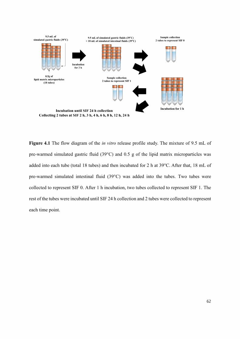
62
Figure 4.1 The flow diagram of the in vitro release profile study. The mixture of 9.5 mL of
pre-warmed simulated gastric fluid (39°C) and 0.5 g of the lipid matrix microparticles was
added into each tube (total 18 tubes) and then incubated for 2 h at 39°C. After that, 18 mL of
pre-warmed simulated intestinal fluid (39°C) was added into the tubes. Two tubes were
collected to represent SIF 0. After 1 h incubation, two tubes collected to represent SIF 1. The
rest of the tubes were incubated until SIF 24 h collection and 2 tubes were collected to represent
each time point.

63
4.3.4 In vivo recovery rate along the gut of weaned piglets
The experimental and animal care protocol (F17-018, AC11280) was reviewed and
approved by the Animal Care Committee of the University of Manitoba and the pigs were cared
for in accordance with the Canadian Council on Animal Care guidelines (CCAC, 2009). A total
of 12 male piglets (TN Tempo × TN70; 9.86 ± 0.52 kg; 28 d) were obtained from the Glenlea
Swine Research Unit at the University of Manitoba and housed individually after 4 d of group
adaptation period (1 d – 5 d) in a temperature-controlled room in the T.K. Cheung Centre for
Animal Science Research at the University of Manitoba. Room temperature was maintained at
29 ± 1ºC during the first week and then reduced by 1.5 ºC for the rest of the experimental period.
Piglets were randomly allotted to two treatments in a completely randomized design (n = 6): 1)
a control corn-SBM basal diet (CF) and 2) a corn-SBM basal diet supplemented with 6 g·kg-1
lipid matrix microparticles (EOF). The corn-SBM basal diets were formulated to meet or
exceed NRC (2012) nutrient specifications for pigs weighing 6-10 kg body weight (BW, Table
4.2). Zinc oxide was added in the diets to prevent diarrhea in pigs and titanium dioxide (3 g·kg-
1) was added as an inert marker to calculate the thymol recovery rate in the different
gastrointestinal sections. All pigs had free access to water and feed during the whole
experimental period and all experimental diets were provided in a mash form. On 8 d and 9d,
feces were collected. On 9 d, final BW and feed intake were measured and thereafter, the pigs
were anesthetized by an intramuscular injection of ketamine:xylazine (20:2 mg·kg BW-1) and
euthanized by intravenous injection of sodium pentobarbital (110 mg·kg BW-1). The whole
organs of the gastrointestinal tract were removed from the carcass and the digesta samples from
the stomach, mid-jejunum (350 to 450 cm from the stomach-duodenum junction), ileum (upper

64
0 to 80 cm of the ileum-cecum junction), cecum and colon (lower 20 cm from the ileum-cecum
junction) were collected into different sterilized containers (Adeola and King, 2006). The
samples of collected digesta were kept at -20°C to be freeze-dried later. The individual pig was
considered the experimental unit.
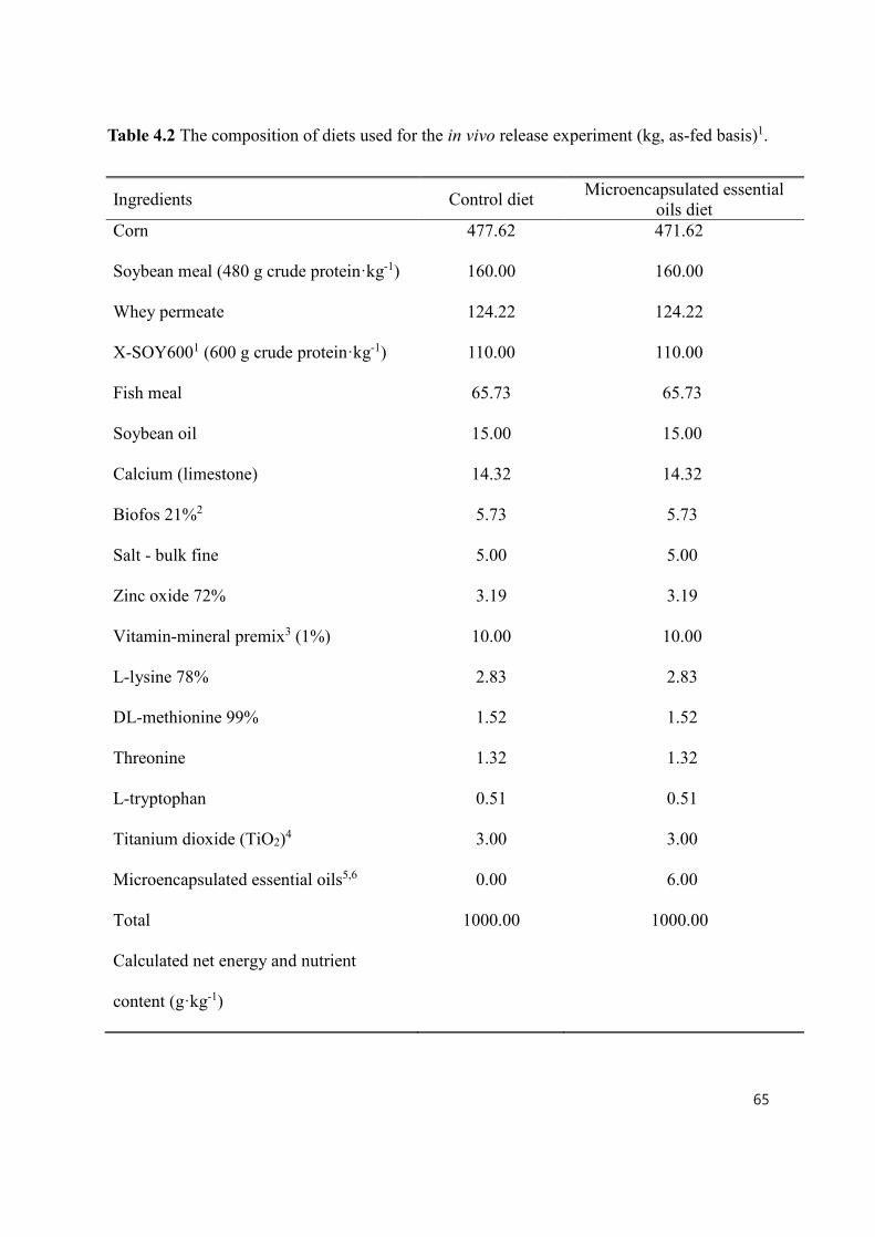
65
Table 4.2 The composition of diets used for the in vivo release experiment (kg, as-fed basis)1.
Ingredients Control diet Microencapsulated essential oils diet
Corn 477.62 471.62
Soybean meal (480 g crude protein·kg-1) 160.00 160.00
Whey permeate 124.22 124.22
X-SOY6001 (600 g crude protein·kg-1) 110.00 110.00
Fish meal 65.73 65.73
Soybean oil 15.00 15.00
Calcium (limestone) 14.32 14.32
Biofos 21%2 5.73 5.73
Salt - bulk fine 5.00 5.00
Zinc oxide 72% 3.19 3.19
Vitamin-mineral premix3 (1%) 10.00 10.00
L-lysine 78% 2.83 2.83
DL-methionine 99% 1.52 1.52
Threonine 1.32 1.32
L-tryptophan 0.51 0.51
Titanium dioxide (TiO2)4 3.00 3.00
Microencapsulated essential oils5,6 0.00 6.00
Total 1000.00 1000.00
Calculated net energy and nutrient
content (g·kg-1)

66
1Soy protein concentration (CJ Selecta, Goiania, State of Goiás, Brazil)
2Ca, 21%; P, 17% (The Mosaic Co., Plymouth, MN)
3Supplied the following per kilogram of diet: 2200 IU vitamin A, 220 IU D3, 16 IU E, 0.5 mg
vitamin K, 1.5 mg vitamin B1, 4 mg vitamin B2, 12 mg calcium pantothenate, 600 mg choline
chloride, 30 mg niacin, 7 mg pyridoxine, 0.02 mg vitamin B12, 0.2 mg biotin, 0.3 mg folic
acid, 0.14 mg calcium iodate, 6 mg copper sulphate, 100 mg ferrous sulfate, 4 mg manganese
oxide, 0.3 mg sodium selenite, and 100 mg zinc oxide.
4Titanium dioxide (TiO2; Sigma-Aldrich, Oakville, Ontario, Canada)
5Lipid matrix microparticles including hydrogenated vegetable oil, fumaric acid, sorbic acid,
malic acid, citric acid, soya lecithin, thymol, vanillin and eugenol (Jefo, Saint-Hyacinthe,
Quebec, Canada)
6The lipid matrix microparticles were premixed in corn (approximately 8 kg) before being
added to the whole diet.
Net energy (kcal·kg-1) 2475 2459
Crude protein (%) 22.4 22.3

67
4.3.5 Gas chromatographic determination of thymol
Thymol extraction from the feed or digesta samples was conducted according to the
methods described by Folch et al. (1957) and Ndou et al. (2018) with some modifications.
Samples were freeze-dried and finely ground with a coffee grinder (Applica Consumer
Products Inc., Miami Lakes, FL, USA) and 1 g of sample was weighed and added to a 50 mL
glass tube. Twenty milliliters of a mixture of chloroform/methanol (2:1, by vol.) and internal
standard (α-methyl-trans-cinnamaldehyde) were added and shaken for 1 h to break down the
lipid matrix microparticles and absorb thymol in the mixture. After shaking, 5 mL (25% by
vol.) of water was added to separate the chloroform and methanol phase and the samples were
centrifuged at 750 × g for 15 min at 4°C. The chloroform phase was obtained with a Pasteur
pipette (Fisher Scientific, Hampton, NH, USA) and was filtered with a filter paper (P5, Fisher
Scientific) and dried under nitrogen gas (N2) flux using an N-EVAP 112 evaporator
(Organomation Associates Inc., Berlin, MA, USA) at 37°C. Methylation was done according
to the method described by Ichihara and Fukubayashi (2010). Toluene (0.2 mL), methanol (1.5
mL), and 8% HCl (0.3 mL) were added sequentially and the mixture was vortexed and
incubated at 45°C overnight. After the overnight incubation, the solution was evaporated under
nitrogen gas (N2) flux using an N-EVAP 112 evaporator (Organomation Associates Inc.).
Hexane (2 mL) was added to dissolve thymol and water (2 mL) was added to wash hexane and
then the tubes were vortexed and centrifuged at 750 × g for 15 min at 4°C. Finally, the 2 mL of
the hexane phase was obtained, and thymol content was analyzed by Gas chromatography –
flame ionization detector (GC-FID).
Samples from the in vitro release experiment were thawed at room temperature and

68
centrifuged at 4700 × g for 20 min at 4°C and the supernatant was filtered with a filter paper
(P5, Fisher Scientific) and the filtered quantity of the supernatant was recorded. The filtered
supernatant was mixed with 15 mL of hexane with internal standard using a rotator (Rotator
AG, FINEPCR, Gunpo, Gyeonggi-do, Korea) for 1 h and centrifuged at 750 × g for 10 min at
4°C and the hexane phase was obtained by a Pasteur pipette (Fisher Scientific). The obtained
hexane was methylated as described above (Ichihara and Fukubayashi, 2010) and the samples
were analyzed by GC-FID.
The amount of thymol was determined by GC-FID. The samples were separated on a
CP Select Fames column (100 m × 0.25 mm diameter and 0.25 mm film thickness; Varian
Canada, Mississauga, ON, Canada) using a Bruker 450 GC with FID (Varian Canada). The
temperature program was 70°C for 2 min, the temperature was raised to 175°C at 25°C·min-1,
held for 20 min, raised to 215°C at 1.5°C·min-1, held for 10 min, and raised to 250°C at
20°C·min-1 and held for 3 min and total run time was 67.62 min. Samples were run with a 20:1
split ratio and 0.8 mL·min-1 column flow. The temperature detector was 290°C and hydrogen
was used as the carrier gas.
4.3.6 Calculation of thymol concentrations and recovery rates
The thymol concentration was calculated based on the peak area ratio between thymol
(specific compound of interest) and α-methyl-trans-cinnamaldehyde (internal standard) as
follows (FID, 2003):
Thymol concentration (mg · kg − 1) = AMOUNT × AREA × IRFAREA
(IS = internal standard, SC = specific compound of interest, IRF = internal response ratio

69
between IS and SC).
According to Zhang et al. (2016), the recovery rate of thymol in the different
gastrointestinal segments was calculated by analyzing thymol and titanium dioxide contents in
feed or digesta. Samples for titanium analysis were prepared according to the method proposed
by Lomer et al. (2000) and the titanium concentration was determined using an inductively
coupled plasma spectrometer (Vista-MPX; Varian Canada). Thymol recovery rate was
calculated based on the following equation (Zhang et al., 2016):
RECOVERY (%) = [(MARKER × THYMOL )(THYMOL × MARKER )] × 100
4.3.7 Statistical analyses
GraphPad Prism 7 (GraphPad Software, Inc., San Diego, CA, USA) was used to
perform statistical analyses. In the pelleting experiment, the differences in thymol content
between the EOM and EOP were analyzed by an unpaired t-test. In the storage stability
experiments, total thymol contents were compared by one-way analysis of variance (ANOVA)
followed by a Tukey’s test. For the in vitro release experiment, a curve fitting program (Padé
approximant) was used. In the in vivo release experiment, the mean and SEM were calculated.
Data in all figures are shown as means ± SEM. For all statistical analyses, P < 0.05 was
considered significant.
4.4 Results
The wheat-SBM basal diets either not supplemented or supplemented with thymol
microencapsulated in the lipid matrix microparticles were pelleted at up to 74°C. Thymol was

70
not detectable in the non-supplemented diets (mash feed and pelleted feed). As shown in Fig.
4.2, there was no difference in the thymol content between EOM and EOP in the three different
batches (P > 0.05) (Fig. 4.2). As shown in Fig. 4.3, the total amount of thymol in both EOM
and EOP did not change during the studied periods (12 weeks) (P > 0.05).
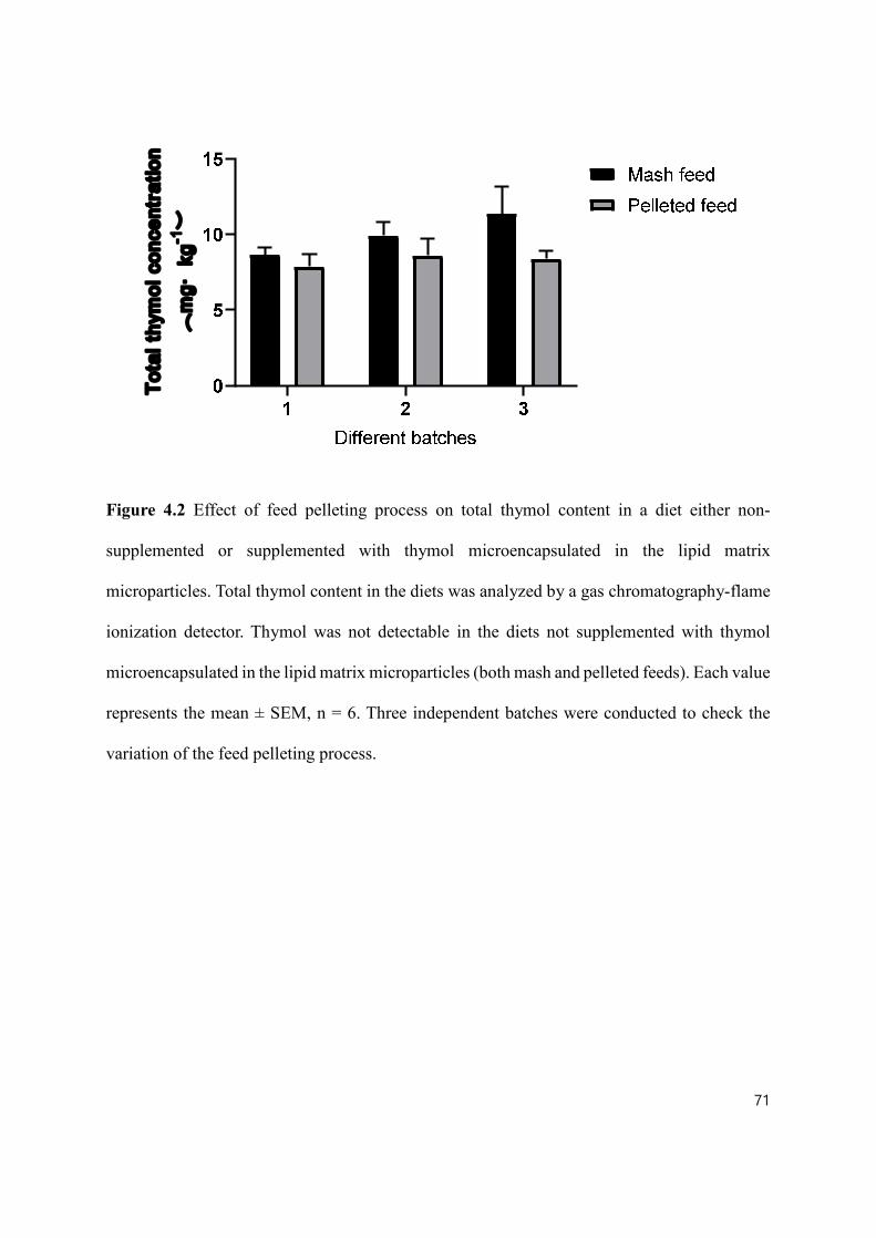
71
Tota
l thy
mol
con
cent
ratio
n(
mg·
kg-1
)
Figure 4.2 Effect of feed pelleting process on total thymol content in a diet either non-
supplemented or supplemented with thymol microencapsulated in the lipid matrix
microparticles. Total thymol content in the diets was analyzed by a gas chromatography-flame
ionization detector. Thymol was not detectable in the diets not supplemented with thymol
microencapsulated in the lipid matrix microparticles (both mash and pelleted feeds). Each value
represents the mean ± SEM, n = 6. Three independent batches were conducted to check the
variation of the feed pelleting process.
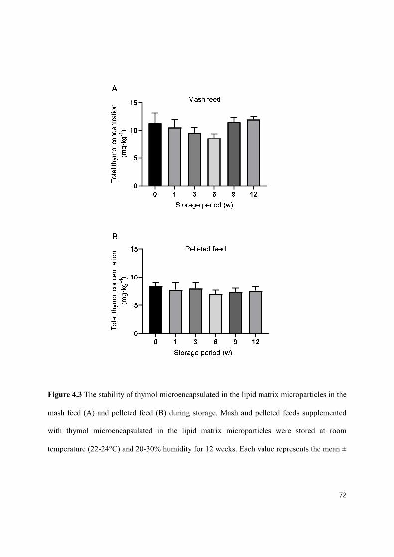
72
Figure 4.3 The stability of thymol microencapsulated in the lipid matrix microparticles in the
mash feed (A) and pelleted feed (B) during storage. Mash and pelleted feeds supplemented
with thymol microencapsulated in the lipid matrix microparticles were stored at room
temperature (22-24°C) and 20-30% humidity for 12 weeks. Each value represents the mean ±
73
SEM, n = 6.
In vitro release profile of thymol from the lipid matrix microparticles were investigated
in SGF and SIF. As shown in Fig. 4.4, 26.04% thymol was released in SGF, and the rest of the
loaded thymol was progressively released in SIF until completion, which was achieved by
around 24 h. The recovery rate of thymol was determined along the gut of weaned piglets fed
a diet either non-supplemented or supplemented with 6 g·kg-1 thymol microencapsulated in the
lipid matrix microparticles. During the whole experiment period, all pigs were healthy and
consumed the feed at the normal quantity. The average final BW of all the pigs was 11.5 ± 0.99
kg and daily feed intake (d 6 – d 9) was 0.45 ± 0.12 kg. There was no significant difference
between the CF and EOF in the final BW and daily feed intake (P > 0.05). Thymol was not
detectable along the gut of weaned piglets fed a diet non-supplemented with thymol
microencapsulated in the lipid matrix microparticles. As shown in Fig. 4.5, 15.5% of thymol
was released in the stomach, and 41.1% of thymol was delivered to the mid-jejunum section.
The thymol was recovered in the ileum, cecum, and colon at 14.36%, 14.92%, and 14.35%,
respectively. Only 2.21% of thymol was recovered in feces.

74
Figure 4.4 In vitro release profile of thymol from the lipid matrix microparticles in simulated
pig gastric fluid (SGF) and simulated pig intestinal fluid (SIF). Each value represents the mean
± SEM, n = 3.

75
Figure 4.5 The recovery rate of thymol along the gut of weaned piglets fed a diet either non-
supplemented or supplemented with thymol microencapsulated in the lipid matrix
microparticles. Thymol was not detectable along the gut of weaned piglets fed a diet non-
supplemented with thymol microencapsulated in the lipid matrix microparticles. Each value
represents the mean ± SEM and n = 6.

76
4.5 Discussion
Encapsulation, defined as a process in which micro- or nano- particles or droplets are
encircled by a coating, or embedded in a homogeneous or heterogeneous matrix, is a helpful
method to improve the potency of feed additives (Gharsallaoui et al., 2007). The benefits of
encapsulation are to 1) improve the storage stability of feed additives; 2) protect feed additives
during feed processing including mixing, conditioning and pelleting; 3) mask unpleasant odor
that can decrease feed intake; 4) improve the ease of handling of liquid feed additives (e.g., EO)
by changing liquid to solid state; 5) slowly release bioactive compounds along the gut of
animals; and 6) reduce the effective dosage of bioactive compounds that have high cost and
environmental issues. A broad range of bioactive compounds such as probiotics (Liu et al.,
2016), EO (Omonijo et al., 2018a), ZnO (Xie et al., 2011), OA (Grilli et al., 2010),
bacteriophages (Huff et al., 2013) and enzymes (Chandrasekar et al., 2017) have been
encapsulated for improving animal health.
An ideal encapsulation should not only increase the stability of EO, but also release
them specifically in the target regions of the intestine (Chen et al., 2017). Wall or matrix
materials are one of the most influential factors in controlling the release of bioactive
compounds (Carneiro et al., 2013). Many wall or matrix materials, including polysaccharides
(alginate xanthan gum), proteins (whey protein and gelatin) and lipids (milk fat and
hydrogenated fat), have been used to encapsulate EO for effective delivery to the gut (Piva et
al., 2007; Liu et al., 2016; Zhang et al., 2016; Chen et al., 2017). The benefits of encapsulated
EO with hydrogenated fat are to facilitate slow release (Mehnert and Mäder, 2012) and to have
high stability (Souto and Müller, 2010). Furthermore, solid lipid has been considered as the

77
most cost-effective material for encapsulating EO. Solid lipid has also been used for
encapsulating probiotics (Okuro et al., 2013), ZnO (Jang et al., 2014), vitamin A (Jenning et
al., 2000) and OA (Piva et al., 2007). However, the stability of EO during feed processing and
storage and the intestinal release of EO in animals are still not clear. Therefore, this study
evaluated the stability of thymol in lipid matrix microparticles during a feed pelleting process
and feed storage and determined the intestinal release of thymol using in vitro and in vivo
approaches.
In modern farming, pig diets are commonly provided in a pellet form (Fahrenholz,
2012). The pelleting process is mainly composed of conditioning and pelleting. Conditioning
refers to adding steam and heat to improve the binding property, while the purpose of pelleting
is to agglomerate small particles into large particles (Falk, 1985). It has been proven that
pelleting pig’ diets enhance palatability, growth performance, nutrient and energy digestibility,
and feed utilization efficiency compared to mash feeding (Lahaye et al., 2008; Vukmirović et
al., 2017). However, the side effects of pelleting, including the possibility of breaking down of
nutrients and feed additives, should be considered (Lewis et al., 2015; Kiarie and Mills, 2019).
The most negative effects of pelleting are from wet steam, fat addition and high energy input,
which can decrease the stability of nutrients and feed additives (Broz et al., 1997). For example,
Jongbloed and Kemme (1990) showed that when the pelleting temperature reached over 80°C,
the activity of exogenous phytase was decreased in the animal feed. In this study, the pelleting
process did not change total thymol in the feed. The melting point of hydrogenated vegetable
oil (matrix materials of the lipid matrix microparticles) is between 50 – 54°C and the range of
the measured temperature during the conditioning and pelleting process in this experiment
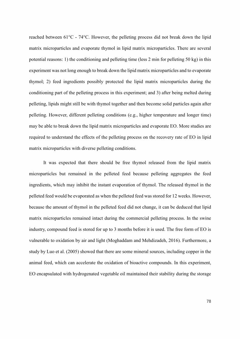
78
reached between 61°C - 74°C. However, the pelleting process did not break down the lipid
matrix microparticles and evaporate thymol in lipid matrix microparticles. There are several
potential reasons: 1) the conditioning and pelleting time (less 2 min for pelleting 50 kg) in this
experiment was not long enough to break down the lipid matrix microparticles and to evaporate
thymol; 2) feed ingredients possibly protected the lipid matrix microparticles during the
conditioning part of the pelleting process in this experiment; and 3) after being melted during
pelleting, lipids might still be with thymol together and then become solid particles again after
pelleting. However, different pelleting conditions (e.g., higher temperature and longer time)
may be able to break down the lipid matrix microparticles and evaporate EO. More studies are
required to understand the effects of the pelleting process on the recovery rate of EO in lipid
matrix microparticles with diverse pelleting conditions.
It was expected that there should be free thymol released from the lipid matrix
microparticles but remained in the pelleted feed because pelleting aggregates the feed
ingredients, which may inhibit the instant evaporation of thymol. The released thymol in the
pelleted feed would be evaporated as when the pelleted feed was stored for 12 weeks. However,
because the amount of thymol in the pelleted feed did not change, it can be deduced that lipid
matrix microparticles remained intact during the commercial pelleting process. In the swine
industry, compound feed is stored for up to 3 months before it is used. The free form of EO is
vulnerable to oxidation by air and light (Moghaddam and Mehdizadeh, 2016). Furthermore, a
study by Luo et al. (2005) showed that there are some mineral sources, including copper in the
animal feed, which can accelerate the oxidation of bioactive compounds. In this experiment,
EO encapsulated with hydrogenated vegetable oil maintained their stability during the storage
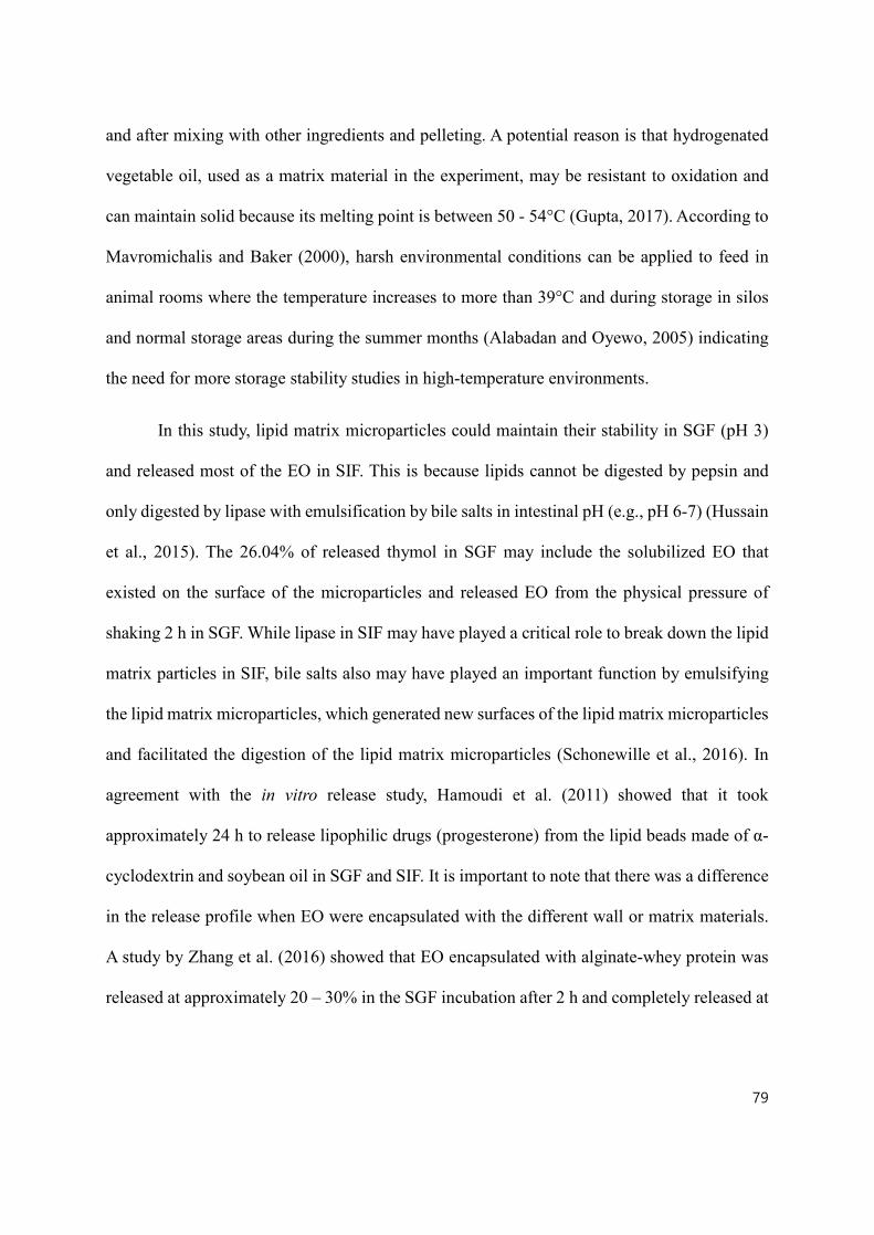
79
and after mixing with other ingredients and pelleting. A potential reason is that hydrogenated
vegetable oil, used as a matrix material in the experiment, may be resistant to oxidation and
can maintain solid because its melting point is between 50 - 54°C (Gupta, 2017). According to
Mavromichalis and Baker (2000), harsh environmental conditions can be applied to feed in
animal rooms where the temperature increases to more than 39°C and during storage in silos
and normal storage areas during the summer months (Alabadan and Oyewo, 2005) indicating
the need for more storage stability studies in high-temperature environments.
In this study, lipid matrix microparticles could maintain their stability in SGF (pH 3)
and released most of the EO in SIF. This is because lipids cannot be digested by pepsin and
only digested by lipase with emulsification by bile salts in intestinal pH (e.g., pH 6-7) (Hussain
et al., 2015). The 26.04% of released thymol in SGF may include the solubilized EO that
existed on the surface of the microparticles and released EO from the physical pressure of
shaking 2 h in SGF. While lipase in SIF may have played a critical role to break down the lipid
matrix particles in SIF, bile salts also may have played an important function by emulsifying
the lipid matrix microparticles, which generated new surfaces of the lipid matrix microparticles
and facilitated the digestion of the lipid matrix microparticles (Schonewille et al., 2016). In
agreement with the in vitro release study, Hamoudi et al. (2011) showed that it took
approximately 24 h to release lipophilic drugs (progesterone) from the lipid beads made of α-
cyclodextrin and soybean oil in SGF and SIF. It is important to note that there was a difference
in the release profile when EO were encapsulated with the different wall or matrix materials.
A study by Zhang et al. (2016) showed that EO encapsulated with alginate-whey protein was
released at approximately 20 – 30% in the SGF incubation after 2 h and completely released at

80
6 h (SGF 2 h + SIF 4 h). Omonijo et al. (2018a) showed that approximately 50% of thymol
encapsulated with starch and alginate were released within 2 h of incubation in SGF and 100%
release was observed following by incubation in SIF for an additional 2 h. These differences
might be due to using different wall/matrix materials or differences in in vitro experimental
conditions such as enzyme concentrations and incubation temperature.
In the in vivo study, pharmacological concentration of zinc oxide (above 3,000 mg/kg)
were added to the diets in order to prevent PWD, which may be able to affect release profile
along the pig gut. In vivo study showed that around 15.5% of thymol was released in the
stomach and 41.85% of thymol was delivered to the mid-jejunum section and only 2.21% of
thymol was recovered in the feces, which is considered a slow release. A slow-release can be
defined as releasing minimal amounts of bioactive compounds in the stomach and delivering
high amounts of such compounds to the mid-jejunum section and releasing most of the
bioactive compounds before they are excreted. Zhang et al. (2014) showed that approximately
38%, 19% and 4% of the non-encapsulated form of carvacrol (e.g. EO) was recovered in the
stomach, duodenum, and jejunum of weaned piglets, respectively, which indicated significant
amount carvacrol disappeared in the stomach. Thus, as 84.5% of thymol was recovered in the
stomach in the study, it can be inferred that only a minimal release occurred thus indicating a
slow-release.
A nutrient with digestibility of more than 90% is considered as very digestible for pigs
and thus 2.21% of remained thymol in the feces indicates that almost all of the thymol
disappeared in the gut of pigs (Jørgensen et al., 2000). the in vivo study, 15.5% of the released
thymol in the stomach may have included the solubilized thymol from the surface of the lipid

81
matrix microparticles and released thymol from the physical pressure of the segmentation
movement of the stomach. Also, some of the lipid matrix microparticles may have been
digested by gastric lipase, which is secreted from gastric chief cells in the fundic mucosa and
plays an important role in the digestion of lipid, especially in piglets. The possible reason for
the difference between released thymol in SGF (SIF 0, 26.04%) and the stomach in pigs (15.5%)
would be that SIF 0 represents finished incubation in the SGF 2h, but a recovery rate of thymol
in the stomach represents the released thymol during incubation in the stomach. Therefore, it
would be more accurate to calculate the thymol recovery rate in the duodenum, but it was not
feasible to collect duodenal digesta from piglets.
Most of the thymol from the lipid matrix microparticles were released in the jejunum,
which can be estimated by subtracting the recovery rate of the ileum (14.36%) from the
stomach (85.5%). Pancreatin enzymes, including lipase and bile salts, are secreted into the
duodenum and most of the lipid sources are digested before they reach the ileum (Valette et al.,
1992). However, the recovery rates of thymol in the ileum (14.36%), cecum (14.91%) and
colon (14.35%) were similar. The potential explanation could be the 14.36% of thymol in the
ileum existed as released form but thymol was not absorbed in the ileum, cecum and colon,
and after digesta were excreted as feces, most of the thymol was evaporated. There have been
a few in vivo studies that have investigated the release profile of EO in pigs. In one of those
studies, when EO microparticles encapsulated with alginate-whey protein were supplemented
to pig, roughly 75%, 68%, 51%, 17%, 5%, and 5% was recovered in the stomach, duodenum,
jejunum, ileum, cecum, and colon, respectively (Zhang et al., 2014). According to Piva et al.
(2007), encapsulated OA and natural identical flavors with hydrogenated vegetable lipids
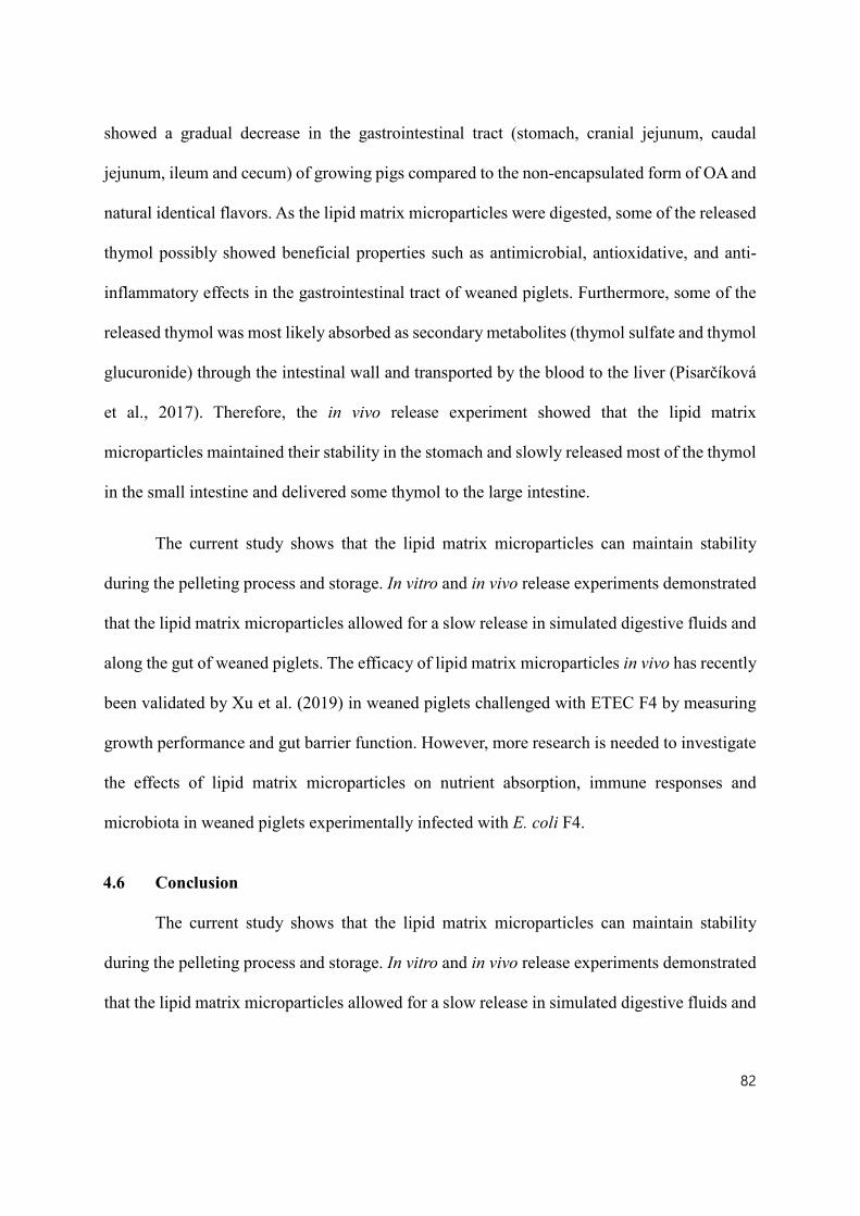
82
showed a gradual decrease in the gastrointestinal tract (stomach, cranial jejunum, caudal
jejunum, ileum and cecum) of growing pigs compared to the non-encapsulated form of OA and
natural identical flavors. As the lipid matrix microparticles were digested, some of the released
thymol possibly showed beneficial properties such as antimicrobial, antioxidative, and anti-
inflammatory effects in the gastrointestinal tract of weaned piglets. Furthermore, some of the
released thymol was most likely absorbed as secondary metabolites (thymol sulfate and thymol
glucuronide) through the intestinal wall and transported by the blood to the liver (Pisarčíková
et al., 2017). Therefore, the in vivo release experiment showed that the lipid matrix
microparticles maintained their stability in the stomach and slowly released most of the thymol
in the small intestine and delivered some thymol to the large intestine.
The current study shows that the lipid matrix microparticles can maintain stability
during the pelleting process and storage. In vitro and in vivo release experiments demonstrated
that the lipid matrix microparticles allowed for a slow release in simulated digestive fluids and
along the gut of weaned piglets. The efficacy of lipid matrix microparticles in vivo has recently
been validated by Xu et al. (2019) in weaned piglets challenged with ETEC F4 by measuring
growth performance and gut barrier function. However, more research is needed to investigate
the effects of lipid matrix microparticles on nutrient absorption, immune responses and
microbiota in weaned piglets experimentally infected with E. coli F4.
4.6 Conclusion
The current study shows that the lipid matrix microparticles can maintain stability
during the pelleting process and storage. In vitro and in vivo release experiments demonstrated
that the lipid matrix microparticles allowed for a slow release in simulated digestive fluids and

83
along the gut of weaned piglets. The efficacy of lipid matrix microparticles in vivo has recently
been validated by Xu et al. (2019) in weaned piglets challenged with ETEC F4 by measuring
growth performance and gut barrier function. However, more research is needed to investigate
the effects of lipid matrix microparticles on nutrient absorption, immune responses and
microbiota in weaned piglets experimentally infected with ETEC F4.

84
Table 4.3 The composition of a wheat-soybean meal basal diet for the feed pelleting experiment
(kg, as-fed basis).
Ingredients kg
Wheat 400
Barley 60
Corn 250
Soybean meal (480 g crude protein·kg-1) 215
Soybean oil 10
Fish meal 40
Limestone 10
Vitamin-Minerals premix1 14
L-Lysine HCl 1
Total 1,000
Calculated net energy and nutrient content (g·kg-1)
Net energy (kcal·kg-1) 2,272
Crude protein (%) 22.0
1Supplied the following per kilogram of diet: 2200 IU vitamin A, 220 IU D3, 16IU E, 0.5 mg
vitamin K, 1.5 mg vitamin B1, 4 mg vitamin B2, 12 mg calcium pantothenate, 600 mg choline
chloride, 30 mg niacin, 7 mg pyridoxine, 0.02 mg vitamin B12, 0.2 mg biotin, 0.3 mg folic
acid, 0.14 mg calcium iodate, 6 mg copper sulphate, 100 mg ferrous sulfate, 4 mg manganese
oxide, 0.3 mg sodium selenite, and 100 mg zinc oxide.

85
Table 4.4 The composition of diets used for the in vivo release experiment (kg, as-fed basis)1.
Ingredients Control diet Microencapsulated essential oils diet
Corn 477.62 471.62
Soybean meal (480 g crude protein·kg-1) 160.00 160.00
Whey permeate 124.22 124.22
X-SOY6001 (600 g crude protein·kg-1) 110.00 110.00
Fish meal 65.73 65.73
Soybean oil 15.00 15.00
Calcium (limestone) 14.32 14.32
Biofos 21%2 5.73 5.73
Salt - bulk fine 5.00 5.00
Zinc oxide 72% 3.19 3.19
Vitamin-mineral premix3 (1%) 10.00 10.00
L-lysine 78% 2.83 2.83
DL-methionine 99% 1.52 1.52
Threonine 1.32 1.32
L-tryptophan 0.51 0.51
Titanium dioxide (TiO2)4 3.00 3.00
Microencapsulated essential oils5,6 0.00 6.00
Total 1000.00 1000.00
Calculated net energy and nutrient
content (g·kg-1)

86
1Soy protein concentration (CJ Selecta, Goiania, State of Goiás, Brazil)
2Ca, 21%; P, 17% (The Mosaic Co., Plymouth, MN)
3Supplied the following per kilogram of diet: 2200 IU vitamin A, 220 IU D3, 16 IU E, 0.5 mg
vitamin K, 1.5 mg vitamin B1, 4 mg vitamin B2, 12 mg calcium pantothenate, 600 mg choline
chloride, 30 mg niacin, 7 mg pyridoxine, 0.02 mg vitamin B12, 0.2 mg biotin, 0.3 mg folic
acid, 0.14 mg calcium iodate, 6 mg copper sulphate, 100 mg ferrous sulfate, 4 mg manganese
oxide, 0.3 mg sodium selenite, and 100 mg zinc oxide.
4Titanium dioxide (TiO2; Sigma-Aldrich, Oakville, Ontario, Canada)
5Lipid matrix microparticles including hydrogenated vegetable oil, fumaric acid, sorbic acid,
malic acid, citric acid, soya lecithin, thymol, vanillin and eugenol (Jefo, Saint-Hyacinthe,
Quebec, Canada)
6The lipid matrix microparticles were premixed in corn (approximately 8 kg) before being
added to the whole diet.
Net energy (kcal·kg-1) 2475 2459
Crude protein (%) 22.4 22.3

87
5.0 CHAPTER 5 MANUSCRIPT II
EFFECTS OF MICRO-ENCAPSULATED FORMULA OF ORGANIC ACIDS AND
ESSENTIAL OILS ON THE NUTRIENT ABSORPTION, IMMUNITY,
MICROBIOTA AND GUT BARRIER FUNCTION OF WEANED PIGLETS
CHALLENGED WITH ENTEROTOXIGENIC Escherichia coli F4
5.1 Abstract
The purpose of the study was to investigate the effects of micro-encapsulated OA and
EO on growth performance, immune system, gut barrier function, nutrient absorption, and
microbiota in the weaned piglets challenged with ETEC F4. Twenty-four ETEC F4 susceptible
weaned piglets were randomly distributed to four treatments including: (1) non-challenged
negative control (NNC; piglets fed a control diet and challenged with phosphate-buffered saline
(PBS); (2) negative control (NC; piglets fed a control diet and challenged with ETEC F4); (3)
positive control (PC; NC + 55 mg·kg-1 of Aureomycin); and (4) micro-encapsulated OA and
EO (P(OA+EO); (NC + 2 g·kg-1 of micro-encapsulated OA and EO). On d 7, ETEC F4 (5 mL
of 1 × 10⁷ CFU·mL-1) or PBS was inoculated to piglets and piglets were euthanized on 5 day
post-inoculum (dpi). ETEC F4 infection significantly decreased ADG during the post-
challenge period (P < 0.05). The piglets fed micro-encapsulated OA and EO had significantly
lower core body temperature (P < 0.05) at 3 hpi (hours post-inoculum) compared to the NC
piglets (P < 0.05). Diarrhea was significantly induced at 8 hpi, 16 hpi, 28 hpi, 34 hpi, and 40
hpi (P < 0.05) and tended to increase diarrhea at the 3 hpi (P = 0.10) and 24 hpi (P = 0.09) due
to the ETEC F4 infection. The supplementation of micro-encapsulated OA and EO relieved
diarrhea at 28 and 40 hpi (P < 0.05) and tended to alleviate diarrhea at 34 hpi (P = 0.07).

88
Intestinal permeability as indicated by the flux of fluorescein isothiocyanate-dextran D4 (FITC
D4) was significantly decreased in the P(OA+EO) piglets (P < 0.05). Decreased jejunal VH
due to ETEC F4 infection (P < 0.05) was significantly attenuated in the P(OA+EO) pigs (P <
0.05). The number of goblet cells per 100 μm VH tended to decrease in the PC (P = 0.10) and
P(OA+EO) piglets (P = 0.09) compared to the NC piglets. The supplementation of OA and EO
tended to alleviate (P = 0.10) the decreased maltase activity due to ETEC F4 infection (P =
0.07). The activity of IAP and relative B0AT1 mRNA abundance was significantly increased in
the PC piglets (P < 0.05). The ETEC F4 infection significantly increased the mRNA abundance
of IL8 (P < 0.05), which was attenuated in the PC and P(OA+EO) piglets (P < 0.05). In
summary, the data suggest that supplementation of micro-encapsulated OA and EO alleviated
the induced diarrhea and inflammation response, the damaged gut barrier integrity, intestinal
morphology, enzyme activities and nutrient transport from ETEC F4 infection in weaned
piglets. Therefore, micro-encapsulated OA and EO could be used as an alternative to antibiotics
for swine production.
Keywords Micro-encapsulation, essential oils, organic acids, weaned piglets, Escherichia coli
F4

89
5.2 Introduction
Post weaning diarrhea (PWD) is one of the most economically important issues in the
swine industry (Yang et al., 2014), which is characterized by the frequent release of watery
feces resulting in retarded growth performance, increased morbidity and mortality (Pan et al.,
2017). ETEC F4 is one of the common pathogens associated with PWD. The fimbriae (F4) of
ETEC F4 can attach to epithelial receptors and release toxins in the intestine of pigs (Jacobsen
et al., 2011). Over the last half-century, AGP have been generally used to control incidences of
PWD and to improve the growth rate and feed efficiency of pigs (Cromwell, 2002). However,
the overuse of AGP could lead to the spread of antimicrobial-resistant pathogens in both
livestock and humans, posing a significant public health threat (Yang et al., 2015). European
Union prohibited the use of AGP in 2006 and worldwide authorities are also trying to restrict
the use of antibiotics in the livestock industry (Bengtsson and Wierup, 2006; Murphy et al.,
2017).
A number of AGP alternatives have been developed and practically used in the swine
industry (Heo et al., 2013), among which EO are considered as one of the most promising AGP
alternatives in the swine industry due to their benefits to gut health and growth performance of
pigs (Omonijo et al., 2018c). Antimicrobial effects to both Gram-negative and Gram-positive
bacteria of diverse EO are already well documented (Chouhan et al., 2017). An in vitro study
conducted by Si et al. (2006) showed that EO efficiently controlled the growth of pathogens
including Salmonella Typhimurium DT 104, ETEC O157:H7 and ETEC F4. In addition, an in
vitro study showed that EO (e.g. thymol) improved barrier integrity and attenuated
inflammatory responses in the porcine intestinal epithelial cells (IPEC-J2) challenged with LPS

90
(Omonijo et al., 2018b). In pigs, the supplementation of EO improved growth performance (Xu
et al., 2018), gut morphology (Zou et al., 2016a), enzyme activities (Xu et al., 2018), intestinal
barrier function (Zou et al., 2016b), immune system activation (Williams et al., 2017),
antioxidative capacity (Cheng et al., 2017) and microbiota (Cairo et al., 2018). Even though
EO have benefits of promoting the growth performance and gut health and of pigs, their
stability in the feed and along the gut restrains their application to pig diets (Omonijo et al.,
2018a). However, micro-encapsulation, which provides a physical barrier for EO from their
environment until their release, is thought to improve the stability of EO and enable the slow
release of EO along the pig gut (Vidhyalakshmi et al., 2009).
The supplementation of EO with OA showed the synergistic effects to improve the
growth performance and gut health of pigs (Yang et al., 2015). Zhou et al. (2007) reported that
EO with OA showed synergistic antimicrobial effects against Salmonella Typhimurium. The
supplementation of OA and EO improved nutrient digestibility and digestive enzyme activities
in weaned piglets (Xu et al., 2018). However, more studies are still needed to comprehensively
understand the mechanisms behind the protection of micro-encapsulated OA and EO against
pathogens in weaned piglets. Therefore, the purpose of the study was to investigate the effects
of micro-encapsulated OA and EO on growth performance, immune system, gut barrier
function, nutrient absorption, and microbiota in the weaned piglets challenged with ETEC F4.
5.3 Materials and Methods
The experimental and animal care protocol (F17-018, AC11280) were reviewed and
approved by the Animal Care Committee of the University of Manitoba and piglets were cared
for in accordance with the Canadian Council on Animal Care guidelines (CCAC, 2009).

91
5.3.1 Virulence factors of enterotoxigenic Escherichia coli (ETEC) F4
The ETEC F4 strain P4 used in this study was isolated from feces of piglets with PWD
by the Veterinary Diagnostic Services Laboratory – Government of Manitoba, Canada. In this
study, the presence and expression of 4 virulence genes associated with adhesion including
faeG (F4 fimbriae) and enterotoxins including estA (Sta, heat-stable toxin) and estB (STb, heat-
stable toxin), elt (LT, heat-labile toxin) in ETEC F4 were checked by PCR (polymerase chain
reaction) according to the method previously described by Zhu et al. (2011) with some
modifications (Table 5.1). The genomic DNA from cultured ETEC F4 (1 × 10⁹ CFU) was
extracted using PureLink® Genomic DNA Kits (Invitrogen, Carlsbad, CA, USA). Total RNA
was extracted using an Ambion RiboPureTM RNA isolation kit (Ambion Inc., Foster City, CA,
USA) and first-strand cDNA was synthesized using oligo (dT) 20 primers and Superscript II
reverse transcriptase (Invitrogen) according to the manufacturer's instructions. Each PCR
reaction mixture (20 μL) contained 7 μL of 0.1% diethylpyrocarbonate (DEPC)–treated water,
1 μL each of forward and reverse primer (10 μmol·L-1), 10 μL of DreamTaq Green PCR Master
Mix (2×) (Thermo Fisher Scientific, Waltham, MA, USA), and 1 μL genomic DNA or 1 μL
cDNA. PCR thermocycler conditions were as follows: 50°C denature 2 min, 95°C denature 5
min 40 cycles at 95°C for 45 s, 50°C for 45 s, and 72°C for 30 s, and a final extension of 72 °C
for 10 min. All PCR products were electrophoresed on a 3% agarose gel in a Tris-borate-EDTA
buffer and visualized by staining with SYBR Green (Invitrogen). All 4 virulence genes (estA,
estB, faeG and elt) were expressed in the ETEC F4 used in the study (Fig. 5.1).

92
Figure 5.1 Agarose gel electrophoresis of the amplification products of virulence genes
(Genomic DNA = A and RNA expression = B) in enterotoxigenic E. coli F4 used in the study.
The four virulence genes were associated with adhesion (faeG), and enterotoxins (estA, estB,
elt) of E. coli F4. The size of each gene was: estA = 158 bp; estB = 113 bp; faeG = 215 bp; elt
= 322 bp. GeneRuler 100 bp Plus DNA Ladder (Thermo Fisher Scientific, Waltham, MA, USA)
was included in the first lane.

93
5.3.2 Genetic susceptibility screening and piglet selection
The ETEC F4 susceptible piglets were selected according to a previous publication
(Jensen et al., 2006). Tails were collected when the pig’s tails were docked on 3 d after
farrowing. DNA was extracted using a method described by Truett et al. (2000). The PCR of
MUC4 gene was performed using DreamTaq DNA polymerase (Thermo Fisher Scientific) with
2 mmol·L-1 MgCl2, 200 µmol·L-1 of each dNTP, 400 µmol·L-1 of each primer in a total volume
of 25 µl. Thermocycling was performed using 5 min initial denaturation at 95℃ subsequently
95℃ for 30 s, annealing at 65℃ for 30 s and extension at 72℃ for 1 min for 35 cycles. The
size of the PCR product obtained from pig genomic DNA was 367 bp and 5 µl of the PCR
products were digested with FastDigest XbaI (Thermo Fisher Scientific) at 37℃ for 5 min
following the supplier’s instructions. All digested PCR products were electrophoresed on a 2%
agarose gel in a Tris-borate-EDTA buffer and visualized by staining with SYBR Green
(Invitrogen). The resistant allele (R) was indigestible by XbaI, whereas the susceptible allele
(S) was digested into 151 bp and 216 bp fragments. The piglets with susceptible alleles and
similar BW were selected.
5.3.3 Preparation of enterotoxigenic Escherichia coli F4
The ETEC F4 was streaked on tryptic soy agar (TSA) from frozen stock and grown
anaerobically at 37°C overnight. Afterward, 10 mL of tryptic soy broth (sterile) was inoculated
with a single ETEC F4 colony from the streak plate and aerobically grown overnight at 37°C
and shaking at 150 rpm. The culture was inclined at 45°C to promote enough aeration.
Thereafter, 300 µL of the overnight culture was used as an inoculant for a fresh 300 mL of
tryptic soy broth (sterile), again incubating at 37°C and shaking at 150 rpm. The culture was
grown for 2.5 h. Necessary preliminary experiments such as a growth curve and standard curve

94
were generated first before preparing with the final ETEC F4 inoculum. After incubation, a
small sample was taken for OD measurement at 600 nm (tryptic soy broth as blank) to check
the bacterial density, according to the standard curve generated earlier. Phosphate buffered
saline (PBS, pH 7.4) was used as the diluent to achieve the targeted ETEC F4 concentration
(1×10⁷ CFU·mL-1). The culture was transported with ice packs to the site for inoculation.
5.3.4 Animals and experimental design
Twenty-four ETEC F4 susceptible weaned piglets (TN Tempo × TN70; 12 female and
12 castrated male piglets with average BW of 8.52 ± 0.11 kg) at the age of 28 d were obtained
from the Glenlea Swine Research Unit at the University of Manitoba and housed individually
in a temperature-controlled room within T.K. Cheung Centre for Animal Science Research at
the University of Manitoba. Room temperature was maintained at 29 ± 1 ºC during the first
week and then reduced by 1.5 ºC for the rest of the experiment period (8-12 d). The selected
susceptible weaned piglets were randomly distributed to 4 treatments with 6 replicates for each
treatment. A corn-SBM basal diet was formulated to meet or exceed the NRC (2012)
recommendations for 6-10 kg piglets (Table 5.2). The four treatments were: 1) non-challenged
negative control (NNC; piglets fed a control basal diet and challenged with PBS; 2) negative
control (NC; piglets fed a control diet and challenged with ETEC F4; 3) positive control (PC;
NC + 55 mg·kg-1 of Aureomycin (Zoetis Canada Inc., Kirkland, QC, Canada)); and 4) micro-
encapsulated formula of OA and EO (P(OA+EO)); NC + 2 g·kg-1 of a selected formula of OA
(fumaric, citric, malic and sorbic acids) and EO (thymol, vanillin and eugenol) micro-
encapsulated in a matrix of triglycerides (Jefo Nutrition Inc., QC, Canada). Piglets were housed
in individual pens and allowed free access to mash feed and water during the whole experiment
period. During the 7 d of pre-challenge (adaptation period) and 4 day post-inoculum (dpi),
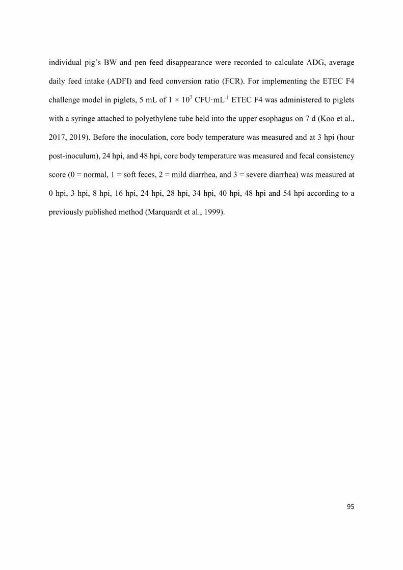
95
individual pig’s BW and pen feed disappearance were recorded to calculate ADG, average
daily feed intake (ADFI) and feed conversion ratio (FCR). For implementing the ETEC F4
challenge model in piglets, 5 mL of 1 × 10⁷ CFU·mL-1 ETEC F4 was administered to piglets
with a syringe attached to polyethylene tube held into the upper esophagus on 7 d (Koo et al.,
2017, 2019). Before the inoculation, core body temperature was measured and at 3 hpi (hour
post-inoculum), 24 hpi, and 48 hpi, core body temperature was measured and fecal consistency
score (0 = normal, 1 = soft feces, 2 = mild diarrhea, and 3 = severe diarrhea) was measured at
0 hpi, 3 hpi, 8 hpi, 16 hpi, 24 hpi, 28 hpi, 34 hpi, 40 hpi, 48 hpi and 54 hpi according to a
previously published method (Marquardt et al., 1999).

96
Table 5.1 The ingredient composition of the basal diet (kg, as-fed basis).
Ingredients, kg, as-fed basis Basal diet
Corn 483.84
Soybean meal (480 g crude protein·kg-1) 160
Whey permeate 124.2
X-SOY6002 (600 g crude protein·kg-1) 110
Fish meal 65.73
Soybean oil 15
Calcium (limestone) 14.32
Biofos 21%3 5.73
Salt - bulk fine 5
Vitamin-mineral premix4 (1%) 10
L-lysine 78% 2.83
DL-methionine 99% 1.52
Threonine 1.32
L-tryptophan 0.51
Total 1,000.00
Calculated net energy and nutrient content (g·kg-1)
Metabolizable energy (kcal·kg-1) 3,389.11
Net energy (kcal·kg-1) 2,475
Crude protein (%) 22.35
SID5 Lysine 1.34
SID5 Methionine 0.5
SID5 Threonine 0.87
SID5 Tryptophan 0.27
97
1The diet for Positive control (PC) was prepared by adding 55 mg·kg-1 of Aureomycin 220G
(Zoetis Canada Inc., Kirkland, QC, Canada) into the basal diet. The diet for micro-encapsulated
organic acids and essential oils (P(OA+EO)) was prepared by adding 2 g·kg-1 of a selected
formula of organic acids (fumaric, citric, malic and sorbic acids) and essential oils (thymol,
vanillin and eugenol) micro-encapsulated in a matrix of triglycerides (Jefo Nutrition Inc., QC,
Canada).
2Soy protein concentration (CJ Selecta, Goiania, State of Goiás, Brazil).
3Monocalcium phosphate containing Ca, 21% and P, 17% (The Mosaic Co., Plymouth, MN)
4Supplied the following per kilogram of diet: 2,200 IU vitamin A, 220 IU vitamin D3, 16 IU
vitamin E, 0.5 mg vitamin K, 1.5 mg vitamin B1, 4 mg vitamin B2, 12 mg calcium pantothenate,
600 mg choline chloride, 30 mg niacin, 7 mg pyridoxine, 0.02 mg vitamin B12, 0.2 mg biotin,
0.3 mg folic acid, 0.14 mg calcium iodate, 6 mg copper sulphate, 100 mg ferrous sulfate, 4 mg
manganese oxide, 0.3 mg sodium selenite, and 100 mg zinc oxide.
5Standardized ileal digestible amino acids.

98
5.3.5 In vivo gut permeability
On 4 dpi, 4 h after oral gavage of fluorescein isothiocyanate-dextran 70 kDa (10 mg per
pig; FITC-D70; molecular weight 70 kDa; Sigma-Aldrich Co., St. Louis, MO, USA) in 5 mL
PBS buffer, blood samples (serum) were collected from each piglets through jugular vein into
heparinized vacutainer tubes (Becton Dickinson, Rutherford, NJ, USA) wrapped in aluminum
foil to block the light and kept at room temperature for 3 h to allow clotting. The blood samples
were centrifuged at 750 × g for 15 min to separate serum from red blood cells and stored at –
80°C until further analyses. The fluorescence was measured at an excitation wavelength of 485
nm and an emission wavelength of 528 nm using a Bio-Tek PowerWaveTM HT Microplate
Scanning Spectrophotometer (BIO-TEK Instruments, Inc., Winooski, VT, USA) and the
concentrations of FITC-D70 in the serum samples (ng·mL-1) were calculated based on a
standard curve.
5.3.6 Sample collection
At the end of the experiment (on 5 dpi) all piglets were anesthetized by an intramuscular
injection of ketamine:xylazine (20:2 mg·kg-1 BW) and euthanized with a captive bolt gun. The
abdomen was immediately opened and the whole gastrointestinal tract was removed from the
carcass. Initially, the mid-jejunum (400 cm from the stomach-duodenum junction) was located
and a 10 cm of the mid-jejunum was put in an ice-cold KRB and delivered to the laboratory
for the Ussing chamber analysis. Another 10 cm of the mid-jejunum tissue was removed and
immediately snap-frozen in liquid nitrogen. The samples were stored at –80°C until further
analyses. Afterward, a 2 cm of the mid jejunum tissue was collected and fixed in a 10%
formaldehyde solution for gut morphology measurement. The digesta of the colon (20 cm from
the ileum-cecum junction) was collected and immediately frozen in liquid nitrogen. The
99
samples were stored at –80°C until further analyses.
5.3.7 Ussing chamber
The electrophysiological properties including short-circuit current and transepithelial
electrical resistance (TEER) were determined using a modified Ussing chambers (VCC-MC8;
Physiologic Instruments Inc., San Diego, CA, USA) containing pairs of current (Ag wire) and
voltage (Ag/AgCl pellet) electrodes housed in 3% agar bridges and filled with KRB buffer (in
mmol·L-1: 154 Na+, 6.3 K+, 137 Cl−, 0.3 H2PO4, 1.2 Ca2+, 0.7 Mg2+, 24 HCO3−
- pH 7.4 with
1μmol·L-1 of indomethacin). Five milliliters of the KRB buffer solution with 10 mmol·L-1 D-
glucose was added to the serosal chambers, and five milliliters of KRB buffer solution enriched
with 10 mmol·L-1 D-mannitol instead of D-glucose was added to the mucosal chambers. Both
the mucosal and serosal chambers were continuously gassed with a mixture of 95% O2 and 5%
CO2. The temperature of the chambers was maintained at 37°C by using a water-jacketed
reservoir. The possible potential difference existing between the mucosal and serosal chambers
was offset before tissue mounting. After gently stripping off serosal and longitudinal muscle
layers using micro-forceps, the tissue was mounted in Ussing chambers employing a tissue
slider with an aperture of 1 cm2. The tissue was left to equilibrate for 10 min followed by the
recording of the short-circuit current and TEER for 10 min after mounting. Afterward, 10
mmol·L-1 D-glucose was added to the mucosal chamber to measure the sodium-dependent
glucose transportation and 10 mmol·L-1 mannitol was added to the serosal chamber to maintain
osmotic balance across the tissue (Mrabti et al., 2019). The difference of short circuit current
generated by SGLT1 was determined by subtracting the short circuit current value before
stimulation from the peak after stimulation. When D-glucose was added, 0.1 mg·mL-1 of FITC-
D4 (molecular weight 4 kDa; Sigma-Aldrich Co.) was added to the mucosal side and after 1 h,

100
the sample (1 mL) were obtained from the serosal side to measure intestinal permeability.
5.3.8 Intestinal morphology analysis
After fixation in 10% neutral-buffered formalin, samples were embedded in paraffin
and 5 µm section was sliced and subsequently mounted on glass slides. Dewaxed sections were
immersed in xylene, 100% ethanol and 95% ethanol for 5 min 2 cycles in each solution. The
samples were immersed in Alcian blue solution (pH 2.5) for 15 min at room temperature and
washed by water for 2 min and placed in the Schiff reagent for 10 min and washed by water
for 10 min. Finally, the samples were counterstained in hematoxylin for 10 s and wash and
dehydrated. For the quantification of Alcian blue/The periodic acid–Schiff (AB/PAS) staining,
each sample was visualized and photographed using an Axio Scope A1 microscope (Carl Zeiss
Micro-Imaging GmbH, Göttingen, Germany) coupled with an Infinity 2 digital camera
(Lumenera Corporation, Ottawa, ON, Canada). VH, CD and VH:CD were measured and the
number of goblet cells per 100 μm VH and 100 μm CD was counted using Infinity Analyze
software (version 6.5.4; Lumenera Corporation, Ottawa, ON, Canada). All measurable villus
and crypts were measured, which was 50 to 150 measurements per each sample.
5.3.9 Total antioxidant capacity, total GSH and GSH/GSSG assays
Total antioxidant capacity (TAC) in the mid-jejunal was measured in duplicate by using
the Colorimetric Microplate Assay Kits for Total Antioxidant Capacity (TA02, Oxford
Biomedical Research, Oxford, MI, USA) (Yang, 2011). Briefly, 200 mg of liquid nitrogen
pulverized samples were weighted out with a 1.5 mL Eppendorf tube, homogenized with 1 mL
of ice-cold PBS on ice for 30 s, and then centrifuged at 3,600 × g for 12 min at 4 ºC. Aliquot
of supernatant was taken for the analysis of their protein content using the Pierce™ BCA
101
Protein Assay Kit (Thermo Fisher Scientific). The TAC in the supernatant was measured as the
capacity to convert Cu2+ to Cu+ by all antioxidants according to the manufacturer’s protocol.
Cu+ ion forms a stable complex with bathocuproine that was detected by measuring the
absorbance at 450 nm with a 96-well plate reader (Bio-Tek PowerWaveTM HT Microplate
Scanning Spectrophotometer, BIO-TEK Instruments, Inc.). The values were compared to a
standard curve obtained using uric acid as a reductant and were expressed as mM·mg protein-
1.
Total glutathione (GSH) and oxidized glutathione (GSSG) in the mid-jejunal tissues
were measured in duplicate by using the Glutathione colorimetric detection kit (Invitrogen).
Briefly, 30 mg of liquid nitrogen pulverized samples will be weighted out with a 1.5 mL
Eppendorf tube, homogenized with 750 µl of ice-cold PBS on ice for 30 s, and then centrifuged
at 3,600 × g for 10 min at 4 ºC. Aliquot of supernatant was taken for the analyses of protein
content using the Pierce™ BCA Protein Assay Kit (Thermo Fisher Scientific). Afterward, 5-
sulfo-salicylic acid dihydrate was added to the obtained supernatant to precipitate protein, and
then centrifuged at 3,600 × g for 10 min at 4 ºC. After deproteinization, total GSH and GSSG
levels in the resulting supernatant were measured according to the manufacture’s protocol.
Reduced GSH was calculated by the equation: Reduced GSH = Total GSH – 2 × GSSG.
5.3.10 Digestive enzyme activity assays
The maximal enzyme activity (Vmax) of intestinal digestive enzymes including
aminopeptidase N (APN), IAP, maltase, maltase-glucoamylase (MGA), and sucrase was
determined in the study. Specifically, about 200 mg of liquid nitrogen pulverized, and frozen
intestinal tissue samples were thawed in an ice-cold homogenizing buffer (50 mmol·L-1 D-

102
mannitol and 0.1 mmol·L-1 Phenylmethylsulfonyl fluoride at pH 7.4) and homogenized on ice
using a polytron homogenizer. The protein content of the resulting homogenate samples was
determined using a Pierce™ BCA Protein Assay Kit (Thermo Fisher Scientific). APN (EC.
3.4.11.2) activity was measured according to the method of Maroux et al. (1973) and IAP (EC
3.1.3.1) activity was measured according to Hübscher and West (1965). The activities of
disaccharidases including sucrase (EC 3.2.1.48) and maltase (EC 3.2.1.20) were determined by
the procedure of Dahlqvist (1964). MGA (EC 3.2. 1.20) activity was analyzed according to the
method of Lackeyram et al. (2012). The Vmax was expressed nmol·mg-1·min-1.
5.3.11 RNA extraction and Real-time PCR analysis
Total RNA was isolated from 50 mg of liquid nitrogen pulverized mid-jejunal tissue samples
using an RNAqueous® total RNA isolation kit (Ambion Inc.). The concentration and
OD260:OD280 ratio of extracted RNA samples were measured using a Nanodrop UV-Vis
spectrophotometer 2000 (Thermo Fisher Scientific Inc., Ottawa, ON, Canada) and the
OD260:OD280 ratios of all RNA samples were between 1.9 and 2.1. The RNA samples were
stored at -80°C for further analyses. A total of 1 µg RNA was used to synthesize the first-strand
cDNA using an iScriptTM cDNA Synthesis Kit (Biorad, Mississauga, ON, Canada) according
to the manufacturer's instructions. All Primers were designed with Primer-Blast
(https://www.ncbi.nlm.nih.gov/tools/primer-blast/) and shown in Table 5.1. The primers were
synthesized by Integrated DNA Technologies, Inc. (Coralville, IA, USA). Real-time PCR was
carried out using an SYBR Green Supermix (Biorad) on a CFX Connect™ Real-Time PCR
Detection System (Biorad) (Omonijo et al., 2018b). A total of 1 μL cDNA was added to a total
volume of 20 μL containing 10 μL SYBR Green supermix, and 300 nmol·L-1 of each forward
and reverse primers. Thermal condition for all reactions was: denaturation 3 min at 95 °C, then
103
40 cycles of 20 s at 95°C, 30 s at 60°C and 30 s at 72°C. Cyclophilin-A (CycA) was used as
the internal control to normalize the amount of RNA used in the real-time PCR for all the
samples. A melting curve program was conducted to confirm the specificity of each PCR
product. The target mRNA abundance was normalized with that of a selected reference gene
and relative mRNA abundance was determined by using the 2-ΔΔCT method (Livak and
Schmittgen, 2001). Threshold cycle (Ct) values were obtained at the cycle number at which the
gene is amplified beyond the threshold of 30 fluorescence units. Real-time PCR efficiencies
were acquired by amplification of the dilution series of DNase-treated RNA according to
formula 10(-1/slope) (Pfaffl, 2001). The efficiencies of all primers used in this study were between
96-105%. Negative controls without cDNA were conducted along with each run, and each
sample was analyzed in duplicate for each gene.
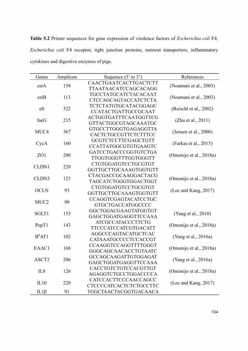
104
Table 5.2 Primer sequences for gene expression of virulence factors of Escherichia coli F4,
Escherichia coli F4 receptor, tight junction proteins, nutrient transporters, inflammatory
cytokines and digestive enzymes of pigs.
Genes Amplicon Sequence (5’ to 3’) References
estA 158 CAACTGAATCACTTGACTCTT (Noamani et al., 2003) TTAATAACATCCAGCACAGG
estB 113 TGCCTATGCATCTACACAAT (Noamani et al., 2003) CTCCAGCAGTACCATCTCTA
elt 322 TCTCTATGTGCATACGGAGC (Reischl et al., 2002) CCATACTGATTGCCGCAAT
faeG 215 ACTGGTGATTTCAATGGTTCG (Zhu et al., 2011) GTTACTGGCGTAGCAAATGC
MUC4 367 GTGCCTTGGGTGAGAGGTTA (Jensen et al., 2006) CACTCTGCCGTTCTCTTTCC
CycA 160 GCGTCTCCTTCGAGCTGTT (Farkas et al., 2015) CCATTATGGCGTGTGAAGTC
ZO1 200 GATCCTGACCCGGTGTCTGA (Omonijo et al., 2018a) TTGGTGGGTTTGGTGGGTT
CLDN1 220 CTGTGGATGTCCTGCGTGT GGTTGCTTGCAAAGTGGTGTT
CLDN3 123 CTACGACCGCAAGGACTACG (Omonijo et al., 2018a) TAGCATCTGGGTGGACTGGT
OCLN 93 CTGTGGATGTCCTGCGTGT (Lee and Kang, 2017) GGTTGCTTGCAAAGTGGTGTT
MUC2 90 CCAGGTCGAGTACATCCTGC GTGCTGACCATGGCCCC
SGLT1 153 GGCTGGACGAAGTATGGTGT (Yang et al., 2010) GAGCTGGATGAGGTTCCAAA
PepT1 143 ATCGCCATACCCTTCTG (Omonijo et al., 2018a) TTCCCATCCATCGTGACATT
B0AT1 102 AGGCCCAGTACATGCTCAC (Yang et al., 2016a) CATAAATGCCCCTCCACCGT
EAAC1 168 CCAAGGTCCAGGTTTTGGGT (Omonijo et al., 2018a) GGGCAGCAACACCTGTAATC
ASCT2 206 GCCAGCAAGATTGTGGAGAT (Yang et al., 2016a) GAGCTGGATGAGGTTCCAAA
IL8 126 CACCTGTCTGTCCACGTTGT (Omonijo et al., 2018a) AGAGGTCTGCCTGGACCCCA
IL10 220 CATCCACTTCCCAACCAGCC (Lee and Kang, 2017) CTCCCCATCACTCTCTGCCTTC IL1β 91 TGGCTAACTACGGTGACAACA

105
CCAAGGTCCAGGTTTTGGGT
TLR2 109 ACATGAAGATGATGTGGGCC (Tohno et al., 2005) TAGGAGTCCTGCTCACTGTA
TLR5 86 GTTCTTTATCCGGGTGACTT AATAAGTCAGGATCGGGAGA
TLR7 107 GCTGTTCCCACTGTTTTGCC GAGCTGGATGAGGTTCCAAA
MGA 118 GCCCCTTCTGCATGAGTTCT CGTCACTTTCTCTGCACCCT
SI 113 AGAAACTTGCCAGTGGAGCA TCCTGGCCATACCTCTCCAA
APN 114 GGACGATTGGGTCTTGCTGA GGGATGACCGACAGGTTTGT 1Note: estA: Sta, heat stable toxin A; estB: STb, heat stable toxin B; elt: LT, heat labile toxin;
faeG: F4 fimbriae; MUC4: Mucin 4; CycA: Cyclophilin-A; ZO1: Zonula occludens 1; CLDN1:
Claudin 1; CLDN3: Claudin 3; OCLN: Occludin; MUC2: Mucin 2; IL8: Interleukin 8; IL10:
Interleukin 10; IL1β: Interleukin 1β; TLR2: Toll like receptor 2; TLR5: Toll like receptor 5;
TLR7: Toll like receptor 7; SGLT1: Na+-glucose cotransporter 1; PepT1: Peptide transporter 1;
ASCT2: Neutral amino acid transporter 2; EAAC1: Excitatory amino acid transporter 1; B0AT1:
Neutral amino acid transporter; MGA: Maltase-glucoamylase; SI: Sucrase-isomaltose; APN:
Aminopeptidase N.

106
5.3.12 Western blotting
Relative protein abundance of ZO1, OCLN, and neutral amino acid transporter (B0AT1)
in the jejunum were detected by western blotting. Briefly, an aliquot of about 50 mg of liquid
nitrogen pulverized mid-jejunal tissue samples were homogenized in a
radioimmunoprecipitation assay buffer (RIPA lysis buffer; Sigma-Aldrich Co.) containing a
complete cocktail of proteinase inhibitors and protein concentration was analyzed by a BCA
protein detection kit (Thermo Fisher Scientific) following the manufacturer’s instructions.
Protein samples were then denatured in 1× Laemmli buffer with mercaptoethanol at 95℃ for
5 min and loaded into the wells of 4-12% gradient pre-made SDS-PAGE gel (Biorad) for
electrophoresis. After electrophoresis, the proteins were transferred onto the Polyvinylidene
fluoride or polyvinylidene difluoride (PVDF) membrane using a Trans-Blot® TurboTM transfer
system (Biorad). For immunoblotting, the membranes were first blocked with 5% non-fat dry
milk in tris-buffered saline with 0.1% of Tween 20 (TBST) at room temperature for 1 h and
then incubated with primary antibodies rabbit anti-ZO1 (1:1,000 dilution, Thermo Fisher
Scientific), rabbit anti-OCLN (1:500 dilution, Thermo Fisher Scientific), and rabbit anti-B0AT1
(1:2,000 dilution, provide by Dr. François Verrey at University of Zurich, Switzerland) (Romeo
et al., 2006) at 4℃ overnight. Afterward, the membranes were washed 5 times with TBST and
incubated with a horseradish peroxidase-conjugated goat anti-rabbit secondary antibody
(1:5,000 dilution, Thermo Fisher Scientific) at room temperature for 1 h, then washed 5 times
with TBST. The chemiluminescent signals were achieved by applying ClarityMax Western ECL
Substrate (Biorad) to the membranes and images were captured using a ChemiDoc MP imaging
system (Biorad). The intensity of the bands was quantified using Image Lab 6.0 software
(Biorad). β-actin (from mouse, Thermal Fisher Scientific) was used as the internal reference.

107
The relative abundance of these proteins was semi-quantified by calculating the ratio of the
band intensity of target and reference proteins. Data were presented as mean ± SEM (n=4).
5.3.13 Measuring ETEC F4 abundance by droplet digital PCR (ddPCR)
DNA from the colon digesta was extracted using a QIAamp DNA Stool Mini Kit
(Qiagen, Hilden, Germany) following the manufacture’s instruction. ETEC F4 abundance in
the colon digesta was quantified by measuring the gene copy number of F4 specific fimbriae
gene (faeG) using the droplet digital PCR system (Biorad). Briefly, 25 µl of PCR reaction
mixture containing 1 ng (ETEC F4 challenged samples) or 100 ng (control samples without
ETEC F4 challenge) of DNA templets, 100 nmol·L-1 of each faeG primer and 1× Evagreen
Supermix (Biorad) was prepared and 20 µl of the mixture was transferred into a sample well
of the droplet generator cartridge (DG8 cartridges; Biorad). Droplet Generation Oil (70 μl)
(Biorad) was added to the oil wells of DG8 cartridges. Droplets were generated using a droplet
generator (Biorad) and were gently transferred onto the 96-well PCR plate (Biorad). The faeG
gene in the droplets was amplified on the C1000 Touch thermal cycler (Biorad) using the
following thermal cycling protocol: 95°C for 5 min, 40 cycles of 95°C for 30 s and 57°C for 1
min, and followed by 4°C for 5 min, 90°C for 5 min and 4°C for 10 min. After thermal cycling,
the PCR end products were read by a QX200 droplet reader (Biorad) and data were analyzed
by QuantaSoft (Biorad). Data were presented as log10(faeG gene copies·μg DNA-1).
5.3.14 Statistical analyses
All data were analyzed using the PROC MIXED of SAS (version 9.4; SAS Inst. Inc.,
Cary, NC, USA) with an individual animal used as the experimental unit. The LSMEANS
statement with the Tukey-adjusted PDIFF option was employed to calculate and split the
treatment mean value for each treatment. The NC piglets were compared by preplanned

108
contrasts with either the NNC piglets to determine the ETEC F4 inoculation effects, or the PC
piglets or the P(OA+EO) piglets to determine the effects of Aureomycin or the micro-
encapsulated OA and EO, respectively. Results in tables were shown as least-square means and
pooled standard errors of the means and results in figures shown as mean ± SEM. Differences
were considered significant at P < 0.05, and trends (0.05 ≤ P ≤ 0.10) were also presented.
5.4 Results
5.4.1 Growth performance, rectal temperature and diarrhea score
As shown in Table 5.3, there was no significant difference in the ADG, ADFI and FCR
observed among all treatment groups during the pre-challenge period (P > 0.05). During the
post-challenge period (0 – 11 d), ETEC F4 infection significantly decreased ADG of the NC
piglets when compared to the NNC piglets (P < 0.05). There was no significant difference in
the ADG observed among the PC, P(OA+EO) and NC piglets had similar ADG with the NC
piglets (P > 0.05) although it was numerously higher in the P(OA+EO) piglets. However, there
was no significant difference in the ADFI among all treatment groups during the post-challenge
period (P > 0.05). During the whole period (0 – 11 d), there was no significant difference in
the ADG, ADFI and FCR observed among all treatment groups (P > 0.05).

109
Table 5.3 Effects of micro-encapsulated organic acids and essential oils on the growth performance of weaned piglets during the pre-challenge period (0-7 d), post-challenge period (0 – 4 days post-inoculum) and whole period (0-11 d).
ETEC F4-challenged P value1
Items NNC NC PC P(OA+EO) SEM TRT 1 2 3
Pre-challenge
ADG (g·d-1) 202 257 219 243 30.6 0.60 0.22 0.39 0.76
ADFI (g·d-1) 298 362 317 370 28.1 0.24 0.12 0.27 0.86
FCR (g·g-1) 1.73 1.42 1.55 1.58 0.20 0.76 0.29 0.66 0.59
Post-
challenge2
ADG (g·d-1) 446a 240ab 183b 354ab 53.2 0.02 0.02 0.51 0.16
ADFI (g·d-1) 635 538 477 608 58.1 0.29 0.27 0.51 0.43
Whole period
ADG (g·d-1) 291 251 195 284 34.2 0.26 0.44 0.31 0.52
ADFI (g·d-1) 420 422 364 456 34.7 0.39 0.97 0.29 0.52
FCR (g·g-1) 1.50 1.82 2.08 1.62 0.18 0.17 0.23 0.36 0.46
NNC (non-challenged negative control): pigs fed a control diet and challenged with phosphate
buffered saline; NC (negative control): pigs fed a control diet and challenged with
enterotoxigenic Escherichia coli F4; PC (positive control): NC + 55 mg·kg-1 of Aureomycin
(Zoetis Canada Inc., Kirkland, QC, Canada); P(OA+EO) (micro-encapsulated organic acids
and essential oils): NC + 2 g·kg-1 of a selected formula of organic acids (fumaric, citric, malic
and sorbic acids) and essential oils (thymol, vanillin and eugenol) micro-encapsulated in a
matrix of triglycerides (Jefo Nutrition Inc., QC, Canada); ADG: average daily gain; ADFI:
average daily feed intake; FCR: feed conversion ratio (Feed to gain ratio).
1TRT: treatment; Contrast: (1) NNC v. NC; (2) NC v. PC; (3) NC v. P(OA+EO).

110
2The FCR during the post-challenge period was unable to calculate due to the minus ADG
during the post-challenge period.
a,bValues within a row with different superscripts differ significantly at P < 0.05.

111
As shown in Fig. 5.2, the core body temperature was significantly increased in the NC
piglets when compared to the NNC piglets (P < 0.05) and the piglets fed P(OA+EO) tended to
have lower core body temperature when compared to the NC piglets (P = 0.06). At 24 hpi, the
P(OA+EO) piglets tended to have a lower core body temperature when compared to the NNC
piglets (P = 0.09).

112
As shown in Fig. 5.3, inoculation of ETEC F4 significantly induced diarrhea at 8 hpi,
16 hpi, 28 hpi, 34 hpi, 40 hpi (P < 0.05) and tended to increase diarrhea at the 3 hpi (P = 0.10)
and 24 hpi (P = 0.09) in the NC piglets when compared to the NNC piglets. The
supplementation of micro-encapsulated OA and EO significantly relieved diarrhea at 28 hpi
and 40 hpi (P < 0.05) and showed a tendency to alleviate diarrhea at 34 hpi (P = 0.07) when
compared to the NC piglets. At 48 hpi and 54 hpi, there was no significant difference in the
diarrhea index observed among all treatment groups during the pre-challenge period (P > 0.05).

113
Figure 5.2 Effects of micro-encapsulated organic acids and essential oils on the core body
temperature in weaned piglets. Core body temperature of weaned piglets was measured in the
NNC (non-challenged negative control): pigs fed a control diet and challenged with phosphate-
buffered saline; NC (negative control): pigs fed a control diet and challenged with
enterotoxigenic Escherichia coli F4; PC (positive control): NC + 55 mg·kg-1 of Aureomycin
(Zoetis Canada Inc., Kirkland, QC, Canada); P(OA+EO) (micro-encapsulated organic acids
and essential oils): NC + 2 g·kg-1 of a selected formula of organic acids (fumaric, citric, malic
and sorbic acids) and essential oils (thymol, vanillin and eugenol) micro-encapsulated in a
matrix of triglycerides (Jefo Nutrition Inc., QC, Canada) groups during 48 hpi (hour post-
inoculation). Each value represents the mean ± SEM. Bars with different letters are
significantly different (P < 0.05). At each time point, the pre-planned contrasts were designed
to compare NNC vs NC (1), NC vs PC (2), and NC vs P(OA+EO) (3). The contrasts were
presented when P ≤ 0.10. hpi: hour post-inoculation.
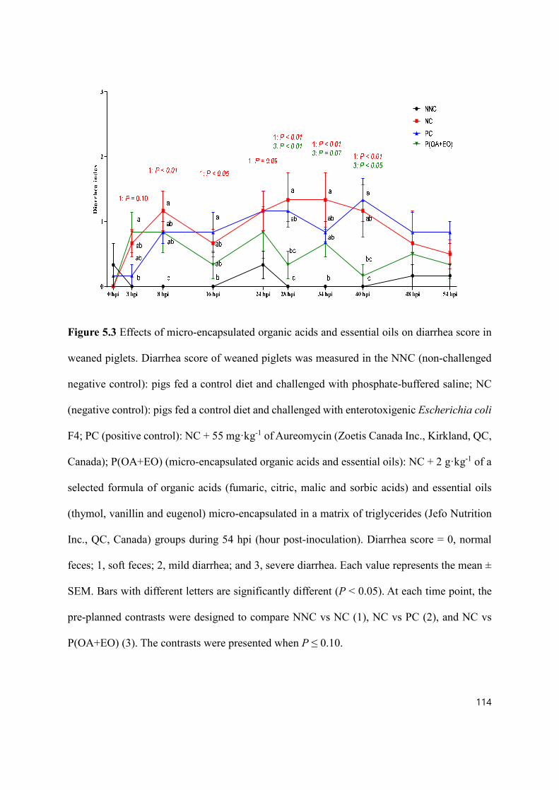
114
Figure 5.3 Effects of micro-encapsulated organic acids and essential oils on diarrhea score in
weaned piglets. Diarrhea score of weaned piglets was measured in the NNC (non-challenged
negative control): pigs fed a control diet and challenged with phosphate-buffered saline; NC
(negative control): pigs fed a control diet and challenged with enterotoxigenic Escherichia coli
F4; PC (positive control): NC + 55 mg·kg-1 of Aureomycin (Zoetis Canada Inc., Kirkland, QC,
Canada); P(OA+EO) (micro-encapsulated organic acids and essential oils): NC + 2 g·kg-1 of a
selected formula of organic acids (fumaric, citric, malic and sorbic acids) and essential oils
(thymol, vanillin and eugenol) micro-encapsulated in a matrix of triglycerides (Jefo Nutrition
Inc., QC, Canada) groups during 54 hpi (hour post-inoculation). Diarrhea score = 0, normal
feces; 1, soft feces; 2, mild diarrhea; and 3, severe diarrhea. Each value represents the mean ±
SEM. Bars with different letters are significantly different (P < 0.05). At each time point, the
pre-planned contrasts were designed to compare NNC vs NC (1), NC vs PC (2), and NC vs
P(OA+EO) (3). The contrasts were presented when P ≤ 0.10.

115
5.4.2 Gut permeability and glucose transport
As shown in Table 5.4, there was no significant difference in TEER and SGLT1
dependent short-circuit current measured by the Ussing chamber (P > 0.05). However, the
P(OA+EO) piglets tended to have a lower FITC-D4 concentration in the serosal chamber when
compared to the NC piglets (P = 0.05). There was no significant difference observed in in vivo
gut permeability measured by the oral gavaging FITC-D70 assay among the NNC, NC and
P(OA+EO) piglets (P > 0.05). However, the PC piglets had a higher concentration of FITC-
D70 in blood when compared to the NNC and P(OA+EO) piglets (P < 0.05).

116
Table 5.4 Effects of micro-encapsulated organic acids and essential oils on
electrophysiological properties including transepithelial electrical resistance (TEER, Ω·cm2)
and SGLT1 dependent short-circuit current (μA·cm-2) and flux of fluorescein isothiocyanate–
dextran 4 kDa (FITC-D4, μg·cm-2·h-1) of weaned piglets jejunum mounted in Ussing chambers
(Ex vivo) and flux of fluorescein isothiocyanate–dextran 70 kDa (FITC-D70, μg·mL-1) in
weaned piglets (In vivo)
ETEC F4-challenged P value1
Items NNC NC PC P(OA+EO) SEM TRT 1 2 3
Ex Vivo
TEER 41.77 50.00 42.19 54.70 6.35 0.44 0.39 0.44 0.62
SGLT1
dependent short-
circuit current
80.85 48.84 42.10 54.57 12.82 0.26 0.11 0.77 0.78
FITC-D4 flux 45.11 55.21 46.88 31.82 7.55 0.24 0.38 0.49 0.05
In vivo
FITC-D70 flux 1,032b 1,357ab 1,682a 1,006b 151.2 0.02 0.17 0.18 0.14
NNC (non-challenged negative control): pigs fed a control diet and challenged with phosphate
buffered saline; NC (negative control): pigs fed a control diet and challenged with
enterotoxigenic Escherichia coli F4; PC (positive control): NC + 55 mg·kg-1 of Aureomycin
(Zoetis Canada Inc., Kirkland, QC, Canada); P(OA+EO) (micro-encapsulated organic acids
and essential oils): NC + 2 g·kg-1 of a selected formula of organic acids (fumaric, citric, malic
and sorbic acids) and essential oils (thymol, vanillin and eugenol) micro-encapsulated in a
matrix of triglycerides (Jefo Nutrition Inc., QC, Canada); SGLT1: Na+-glucose cotransporter 1;
FITC-D4 and FITC-D70: fluorescein isothiocyanate–dextran 4 kDa and 70 kDa; TEER:
transepithelial electrical resistance.
117
1TRT: treatment; Contrast: (1) NNC v. NC; (2) NC v. PC; (3) NC v. P(OA+EO). a,bValues within a row with different superscripts differ significantly at P < 0.05.

118
5.4.3 Intestinal morphology and goblet cells
As shown in Table 5.5, ETEC F4 inoculation significantly decreased mid-jejunal VH in
the NC piglets when compared to the NNC piglets (P < 0.05). The piglets supplemented with
micro-encapsulated OA and EO significantly increased VH in the jejunum (P < 0.05) when
compared to the NC piglets. However, no significant difference was found in the CD and
VH:CD among all treatment groups (P > 0.05). ETEC F4 inoculation numerically increased
the number of goblet cells per 100 μm VH when compared to the NNC piglets (P = 0.12).
There was a tendency in the number of goblet cells per 100 μm VH with the supplementation
of Aureomycin (P = 0.10) and micro-encapsulated OA and EO (P = 0.09) when compared to
the NC piglets. The number of goblet cells per 100 μm CD was not affected by treatments (P
> 0.05).
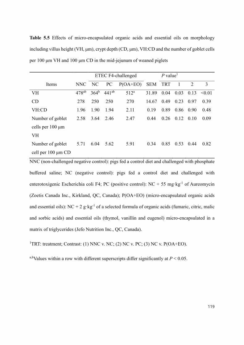
119
Table 5.5 Effects of micro-encapsulated organic acids and essential oils on morphology
including villus height (VH, μm), crypt depth (CD, μm), VH:CD and the number of goblet cells
per 100 μm VH and 100 μm CD in the mid-jejunum of weaned piglets
ETEC F4-challenged P value1
Items NNC NC PC P(OA+EO) SEM TRT 1 2 3
VH 478ab 364b 441ab 512a 31.89 0.04 0.03 0.13 <0.01
CD 278 250 250 270 14.67 0.49 0.23 0.97 0.39
VH:CD 1.96 1.90 1.94 2.11 0.19 0.89 0.86 0.90 0.48
Number of goblet
cells per 100 μm
VH
2.58 3.64 2.46 2.47 0.44 0.26 0.12 0.10 0.09
Number of goblet
cell per 100 μm CD
5.71 6.04 5.62 5.91 0.34 0.85 0.53 0.44 0.82
NNC (non-challenged negative control): pigs fed a control diet and challenged with phosphate
buffered saline; NC (negative control): pigs fed a control diet and challenged with
enterotoxigenic Escherichia coli F4; PC (positive control): NC + 55 mg·kg-1 of Aureomycin
(Zoetis Canada Inc., Kirkland, QC, Canada); P(OA+EO) (micro-encapsulated organic acids
and essential oils): NC + 2 g·kg-1 of a selected formula of organic acids (fumaric, citric, malic
and sorbic acids) and essential oils (thymol, vanillin and eugenol) micro-encapsulated in a
matrix of triglycerides (Jefo Nutrition Inc., QC, Canada).
1TRT: treatment; Contrast: (1) NNC v. NC; (2) NC v. PC; (3) NC v. P(OA+EO).
a,bValues within a row with different superscripts differ significantly at P < 0.05.

120
5.4.4 Digestive enzyme maximal activities
As shown in Table 5.6, no significant difference was found in the Vmax of APN, MGA,
sucrase among all treatment groups (P > 0.05). The PC piglets had significantly higher Vmax of
IAP when compared to the NC piglets (P < 0.05). The Vmax of maltase tended to decrease due
to the ETEC F4 inoculation in the NC piglets when compared to the NNC piglets (P = 0.07)
and the piglets supplemented with micro-encapsulated OA and EO tended to have higher Vmax
for maltase when compared to the NC piglets (P = 0.10).

121
Table 5.6 Effects of micro-encapsulated organic acids and essential oils on the activities
(nmol·L-1·mg protein-1·min-1) of brush border digestive enzymes in the mid-jejunum of weaned
piglets
ETEC F4-challenged P value1
Items NNC NC PC P(OA+EO) SEM TRT 1 2 3
Aminopeptidase N 0.13 0.10 0.11 0.14 0.02 0.39 0.34 0.84 0.13
Intestinal alkaline
phosphatase 0.45 0.40 0.52 0.46 0.04 0.22 0.42 0.04 0.25
Maltase 78.63 54.04 70.64 76.11 8.71 0.27 0.07 0.24 0.10
Maltase-
glucoamylase 4.36 3.95 4.02 4.49 0.50 0.87 0.61 0.93 0.50
Sucrase 14.43 11.57 12.90 15.93 3.02 0.81 0.56 0.79 0.37
NNC (non-challenged negative control): pigs fed a control diet and challenged with phosphate
buffered saline; NC (negative control): pigs fed a control diet and challenged with
enterotoxigenic Escherichia coli F4; PC (positive control): NC + 55 mg·kg-1 of Aureomycin
(Zoetis Canada Inc., Kirkland, QC, Canada); P(OA+EO) (micro-encapsulated organic acids
and essential oils): NC + 2 g·kg-1 of a selected formula of organic acids (fumaric, citric, malic
and sorbic acids) and essential oils (thymol, vanillin and eugenol) micro-encapsulated in a
matrix of triglycerides (Jefo Nutrition Inc., QC, Canada).
1 TRT: treatment; Contrast: (1) NNC v. NC; (2) NC v. PC; (3) NC v. P(OA+EO).

122
5.4.5 Total antioxidant capacity (TAC), total GSH and GSH/GSSG
As shown in Table 5.7, there was no significant difference in TAC, total GSH, GSSG,
reduced GSH and reduced GSH:GSSG observed among all treatment groups (P > 0.05).

123
Table 5.7 Effects of micro-encapsulated organic acids and essential oils on the total antioxidant
capacity (TAC, mmol·L-1·mg protein-1), total glutathione (GSH, nmol·L-1·mg protein-1),
oxidized glutathione (GSSG, nmol·L-1·mg protein-1), and reduced GSH:GSSG in the mid-
jejunum of weaned piglets
ETEC F4-challenged P value1
Items NNC NC PC P(OA+EO) SEM TRT 1 2 3
TAC 84.88 79.80 82.18 85.51 3.61 0.78 0.35 0.68 0.30
Total GSH 3.26 3.09 2.98 2.95 0.21 0.32 0.62 0.76 0.68
GSSG 0.44 0.42 0.50 0.55 0.05 0.42 0.79 0.31 0.10
Reduced GSH2 2.37 2.24 1.99 1.85 0.22 0.23 0.71 0.47 0.28
Reduced
GSH:GSSG 5.46 5.69 4.06 3.81 0.71 0.70 0.83 0.16 0.11
NNC (non-challenged negative control): pigs fed a control diet and challenged with phosphate
buffered saline; NC (negative control): pigs fed a control diet and challenged with
enterotoxigenic Escherichia coli F4; PC (positive control): NC + 55 mg·kg-1 of Aureomycin
(Zoetis Canada Inc., Kirkland, QC, Canada); P(OA+EO) (micro-encapsulated organic acids
and essential oils): NC + 2 g·kg-1 of a selected formula of organic acids (fumaric, citric, malic
and sorbic acids) and essential oils (thymol, vanillin and eugenol) micro-encapsulated in a
matrix of triglycerides (Jefo Nutrition Inc., QC, Canada).
1 TRT: treatment; Contrast: (1) NNC v. NC; (2) NC v. PC; (3) NC v. P(OA+EO). 2 Reduced GSH = Total GSH – 2 × GSSG.

124
5.4.6 Relative mRNA abundance in the jejunum
The relative mRNA abundance of genes associated with gut barrier function, immune
system, nutrient transport, digestive enzymes was analyzed by real-time PCR (Table 5.8). There
was no significant difference in the relative mRNA abundance of zonulna occludens 1 (ZO1)
and OCLN among all treatment groups (P > 0.05). ETEC F4 inoculation significantly
decreased the relative mRNA abundance of CLDN1 and mucin 2 (MUC2) in the NC piglets
when compared to the NNC piglets (P < 0.05). The PC piglets supplemented with Aureomycin
had a higher level of relative mRNA abundance of CLDN3 when compared to the NNC piglets
(P < 0.05). There was no significant difference in the relative mRNA abundance of peptide
transporter 1 (PepT1), excitatory amino-acid carrier 1 (EAAC1), and neutral amino acid
transporter 2 (ASCT2) observed among all treatment groups (P > 0.05). The relative mRNA
abundance of SGLT1 and B0AT1 was significantly decreased due to ETEC F4 inoculation when
compared to the NNC piglets (P < 0.05). The PC piglets had a higher relative mRNA
abundance of B0AT1 when compared to the NC piglets (P < 0.05). Among the genes related to
the immune system including IL8, IL10, IL1β, toll-like receptor 2 (TLR2), TLR5 and TLR7,
only the relative mRNA abundance of IL8 was significantly increased due to ETEC F4
inoculation (P < 0.05) in the NC piglets when compared to the NNC piglets. The PC and
P(OA+EO) piglets significantly decreased the relative mRNA abundance of IL8 when
compared to the NC piglets (P < 0.05). Compared to the NNC piglets, ETEC F4 inoculation
significantly decreased relative mRNA abundance of MGA, sucrase-isomaltase (SI), APN (P
< 0.05) in the NC piglets, however, the relative mRNA abundance of MGA, SI, and APN in
the PC and P(OA+EO) piglets was not different from those in the NC piglets (P > 0.05).

125
Table 5.8 Effects of micro-encapsulated organic acids and essential oils on the relative mRNA
abundance of genes associated with gut barrier integrity, nutrient transporters, immune system,
and digestive enzymes in the mid jejunum of weaned piglets1.
ETEC F4-challenged P value2
Items NNC NC PC P(OA+EO) SEM TRT 1 2 3
Gut barrier
integrity
ZO1 1.02 0.78 0.87 0.72 0.12 0.33 0.20 0.62 0.75
CLDN1 1.04a 0.61b 0.50b 0.41b 0.09 <0.01 0.01 0.46 0.23
CLDN3 1.01 0.75 1.15 0.92 0.10 0.23 0.15 0.05 0.36
OCLN 1.03 0.89 0.86 0.81 0.13 0.65 0.48 0.86 0.67
MUC2 1.01a 0.40ab 0.49ab 0.33b 0.15 0.02 0.02 0.68 0.74
Nutrient
transporters
SGLT1 1.01a 0.50b 0.69ab 0.52b 0.09 <0.01 <0.01 0.16 0.88
PepT1 1.05 0.64 0.80 0.73 0.16 0.37 0.11 0.53 0.73
B0AT1 1.02a 0.36b 0.84ab 0.49b 0.09 <0.01 <0.01 0.01 0.41
EAAC1 1.06 0.76 0.81 0.96 0.17 0.64 0.25 0.86 0.47
ASCT2 1.03 1.18 1.11 1.02 0.35 0.99 0.78 0.90 0.79
Immune system
IL8 1.02 1.92 1.03 1.02 0.27 0.07 0.03 0.03 0.02
IL10 1.09 1.04 0.73 1.01 0.19 0.61 0.31 0.94 0.19
IL1β 1.02 0.99 0.78 0.72 0.13 0.32 0.86 0.31 0.19
TLR2 1.04 1.35 0.82 1.12 0.21 0.41 0.32 0.11 0.51
TLR5 1.02 0.86 0.52 0.55 0.18 0.18 0.54 0.20 0.24
TLR7 1.05 0.75 0.70 0.65 0.12 0.12 0.11 0.77 0.59
Digestive

126
enzymes
MGA 1.01a 0.41b 0.61ab 0.53ab 0.11 0.02 <0.01 0.27 0.51
SI 1.04 0.56 0.65 0.63 0.15 0.15 0.04 0.71 0.76
APN 1.00 0.49 0.64 0.57 0.13 0.08 0.02 0.47 0.69
NNC (non-challenged negative control): pigs fed a control diet and challenged with phosphate
buffered saline; NC (negative control): pigs fed a control diet and challenged with
enterotoxigenic Escherichia coli F4; PC (positive control): NC + 55 mg·kg-1 of Aureomycin
(Zoetis Canada Inc., Kirkland, QC, Canada); P(OA+EO) (micro-encapsulated organic acids
and essential oils): NC + 2 g·kg-1 of a selected formula of organic acids (fumaric, citric, malic
and sorbic acids) and essential oils (thymol, vanillin and eugenol) micro-encapsulated in a
matrix of triglycerides (Jefo Nutrition Inc., QC, Canada).
1Note: ZO1: Zonula occludens-1; CLDN1: Claudin 1; CLDN3: Claudin 3; OCLN: Occludin;
MUC2: Mucin 2; SGLT1: Na+-glucose cotransporter 1; PepT1: Peptide transporter 1; ASCT2:
Neutral amino acid transporter 2; EAAC1: Excitatory amino acid transporter 1; B0AT1: Neutral
amino acid transporter; IL8: Interleukin 8; IL10: Interleukin 10; IL1β: Interleukin 1β; TLR2:
Toll like receptor 2; TLR5: Toll like receptor 5; TLR7: Toll like receptor 7; MGA: Maltase-
glucoamylase; SI: Sucrase-isomaltose; APN: Aminopeptidase N.
2TRT: treatment; Contrast: (1) NNC v. NC; (2) NC v. PC; (3) NC v. P(OA+EO).
a,bValues within a row with different superscripts differ significantly at P < 0.05.

127
5.4.7 Relative protein abundance of tight junction proteins and nutrient transporter
As shown in Fig. 5.4, ETEC F4 inoculation tended to increase the protein abundance of
OCLN (P = 0.09) and significantly decreased ZO1 (P < 0.05) in the NC piglets when compared
to the NNC piglets. The supplementation of Aureomycin significantly decreased the relative
protein abundance of OCLN (P < 0.05) when compared to the NC piglets (P < 0.05). However,
the relative protein abundance of ZO1 and OCLN in the P(OA+EO) piglets was not
significantly different from those in the NC piglets (P > 0.05). There was no significant
difference in the relative protein abundance of B0AT1 among all treatment groups (P > 0.05).

128
Figure 5.4 Effects of micro-encapsulated organic acids and essential oils on the relative
abundance of proteins associated with gut barrier integrity and nutrient transporters in weaned
piglets. Mid-jejunal relative protein abundance of zonulna occludens 1 (ZO1), occludin
(OCLN), and B0AT1 (neutral amino acid transporter) was measured in the NNC (non-
challenged negative control): pigs fed a control diet and challenged with phosphate-buffered
saline; NC (negative control): pigs fed a control diet and challenged with enterotoxigenic
Escherichia coli F4; PC (positive control): NC + 55 mg·kg-1 of Aureomycin (Zoetis Canada
Inc., Kirkland, QC, Canada); P(OA+EO) (micro-encapsulated organic acids and essential oils):
NC + 2 g·kg-1 of a selected formula of organic acids (fumaric, citric, malic and sorbic acids)
and essential oils (thymol, vanillin and eugenol) micro-encapsulated in a matrix of triglycerides
(Jefo Nutrition Inc., QC, Canada) groups. Each value represents the mean ± SEM. Bars with
different letters are significantly different (P < 0.05). The pre-planned contrasts were designed
to compare NNC vs NC (1), NC vs PC (2), and NC vs P(OA+EO) (3). The contrasts were
presented when P ≤ 0.10.

129
5.4.8 ETEC F4 abundance in the colon digesta
As shown in Fig. 5.5, ETEC F4 inoculation significantly increased the ETEC F4 gene
(faeG) (P < 0.05) in the colon digesta in the NC piglets when compared to the NNC piglets.
However, there was no significant difference in the copy number of faeG observed among the
piglets challenged with ETEC F4 (P > 0.05) although it was numerously lower in the P(OA+EO)
piglets.

130
Figure 5.5 Effects of micro-encapsulated organic acids and essential oils on DNA abundance
of faeG (F4 fimbriae) in the colon digesta in weaned piglets. DNA abundance of faeG (F4
fimbriae) in the colon digesta (20cm from the ileum-cecum junction) was measured in the NNC
(non-challenged negative control): pigs fed a control diet and challenged with phosphate-
buffered saline; NC (negative control): pigs fed a control diet and challenged with
enterotoxigenic Escherichia coli F4; PC (positive control): NC + 55 mg·kg-1 of Aureomycin
(Zoetis Canada Inc., Kirkland, QC, Canada); P(OA+EO) (micro-encapsulated organic acids
and essential oils): NC + 2 g·kg-1 of a selected formula of organic acids (fumaric, citric, malic
and sorbic acids) and essential oils (thymol, vanillin and eugenol) micro-encapsulated in a
matrix of triglycerides (Jefo Nutrition Inc., QC, Canada) groups. Data were presented as
log10(faeG gene copies·μg DNA-1). Each value represents the mean ± SEM. Bars with different
letters are significantly different (P < 0.05). The pre-planned contrasts were designed to
compare NNC vs NC (1), NC vs PC (2), and NC vs P(OA+EO) (3). The contrasts were
presented when P ≤ 0.10.
131
5.5 Discussion
This study was to investigate whether the supplementation of micro-encapsulated OA
and EO could alleviate the responses to bacterial infection (e.g. diarrhea, inflammation, and
compromised gut health) in weaned piglets. A model for inducing bacterial infection in weaned
piglets was established by inoculating ETEC F4 (Opapeju et al., 2015). The pathogenesis of
ETEC F4 in pigs depends on two major factors: ETEC F4 virulence and F4 fimbriae receptors
in piglets (Kim et al., 2019). The F4 fimbriae attach to the F4 receptors (MUC4) on the
intestinal brush borders and induce ETEC F4 colonization in the intestine and then release
toxins (estA, estB, elt) (Moonens et al., 2015). The toxins of ETEC F4, including estA, estB,
elt and lipopolysaccharides (LPS), can cause the disorders of electrolytes and fluid secretion in
the intestine, which results in watery feces (Koo et al., 2019). The presence and expression of
virulence factors in ETEC F4 strain P4's were checked in this experiment and four virulence
genes (faeG, estA, estB, and elt) were expressed in the ETEC F4 used in the current study.
The ETEC F4 susceptible piglets were selected by checking the susceptible alleles of
MUC4 according to a previous publication (Jensen et al., 2006). Gibbons et al. (1977) showed
that the susceptibility to ETEC F4 was inherited as an autosomal dominant Mendelian trait
with the two alleles: S (adhesion, dominant) and R (non-adhesion, recessive). ETEC F4 induces
more clinical symptoms if piglets have susceptible alleles of the MUC4 gene (Fairbrother et
al., 2005). So it is necessary to choose susceptible piglets for this challenge study in order to
successfully induce diarrhea and minimize variations among piglets (Trevisi et al., 2015;
Sterndale et al., 2019). In the study, the symptoms of bacterial infection were successfully
achieved, which can be indicated by increased diarrhea index and core body temperature and

132
compromised gut health.
The purpose of this study was to evaluate dietary strategies as AGP alternatives. The
AGP (low dosage of medicine) are mostly expected to show subtherapeutic effects rather than
the therapeutic effects that may alleviate clinical diarrhea (Diarrhea score 2 or 3) or mortality
(Adewole et al., 2016). Thus, an appropriate amount of ETEC F4 that may show only mild
diarrhea should be inoculated to piglets. In our pilot studies (data not published), piglets
inoculated with 10 mL of 1 × 10⁹ CFU·mL-1 and 5 mL of 3 × 10⁸ CFU·mL-1 of ETEC F4
showed 75% (15 dead piglets out of 20 piglets) of and 65% (13 dead piglets out of 20 piglets)
of mortality in all treatment groups, respectively. A pilot study showed that the oral gavage of
5 mL of 1 × 10⁷ CFU·mL-1 ETEC F4 induced mild diarrhea and thus 5 mL of 1 × 10⁷ CFU·mL-
1 was selected in this study.
In this study, during the pre-challenge period, the supplementation of micro-
encapsulated OA and EO did not affect the growth performance. The ETEC F4 infection
significantly decreased the ADG, which is consistent with the results of Trevisi et al. (2009)
and Rong et al. (2015). The potential reasons for decreased ADG of piglets due to ETEC F4 in
the current study could be 1) decreased efficiency of nutrient digestion and absorption (Chen
et al., 2018); 2) inflammation (Kim et al., 2016); 3) increased diarrhea (Cho et al., 2012); and
4) decreased available nutrients for pigs due to the inoculation of ETEC F4, which may
compete for nutrients with the host (Richards et al., 2005). In this study, the supplementation
of micro-encapsulated OA and EO numerically increased ADG although statistically difference
was not achieved in this study (P = 0.16) and the possible reason might be that the
microencapsulated OA and EO attenuated bacterial infection symptoms of piglets. Similarly,

133
Devi et al. (2015) showed that the supplementation of a blend of EO including cinnamon,
fenugreek, clove improved ADG when compared to the control group but the supplementation
of a coated OA containing formic acid, lactic acid, fumaric acid and citric acid could not
improve growth performance of weaned piglets challenged with ETEC F4. Kwak et al. (2019)
showed that micro-encapsulated OA and EO attenuated the decrease of ADG and ADFI in the
LPS-challenged piglets. However, Ahmed et al. (2013) reported that when a mixture of ETEC
KCTC 2571 and Salmonella Typhimurium was inoculated to piglets, the blend of EO including
oregano (Origanum vulgare), anise (Pimpinella anisum), orange peel (Citrus sinensis), and
chicory (Cichorium intybus) did not improve the growth performance when compared to the
control group. These inconsistent results may come from different kinds of OA and EO,
challenging inoculum or experimental designs (e.g. experimental period and replicates).
In this study, the supplementation of micro-encapsulated OA and EO attenuated the
increase of core body temperature by ETEC F4 infection. An increase in core body temperature
may imply inflammatory reactions (Kwak et al., 2019). Similarly, the relative mRNA
abundance of IL8 (pro-inflammatory cytokines) was increased due to ETEC F4 infection and
attenuated by the supplementation of microencapsulated OA and EO in this study. Pro-
inflammatory cytokines (IL8), produced by various cell types such as macrophages, endothelial
cells, B cells and mast cells, are one of the important markers for inflammation (Akira et al.,
1993). The alleviated inflammation by the supplementation microencapsulated OA and EO
could be explained by enhanced gut barrier function indicated by decreased FITC-D4 uptake
in the jejunum. Gut barrier function, the first defense line against the hostile environment,
protects noxious antigens and pathogens from permeating into the body (Wijtten et al., 2011).
It is already well documented that supplementation of OA and EO can enhance the gut barrier

134
function of pigs (Grilli et al., 2015; Zou et al., 2016b; Omonijo et al., 2018b). Alleviated core
body temperature and enhanced gut barrier function, which are achieved by the
supplementation of microencapsulated OA and EO, possibly explain the mitigated diarrhea in
the present study, which supports the fact that OA and EO have an antidiarrheal effect
(Tsiloyiannis et al., 2001; Suiryanrayna and Ramana, 2015; Omonijo et al., 2018c). The
supplementation of Aureomycin decreased mRNA expression of IL8, which is consistent with
a previous study (Koo et al., 2019). This also could be explained by the enhanced gut barrier
function indicated by the up-regulated relative abundance of mRNA of CLDN3 in the PC
piglets, one of the tight junction proteins, which are vital in the maintenance of gut barrier
function (Li et al., 2019). However, the antidiarrheal effect of Aureomycin supplementation
was not shown in the current study.
To study gut permeability of piglets, in addition to FITC-D4 assay in the Ussing
chamber, FITC-D70 in PBS (2 mg·mL-1) was orally gavaged to the piglets. Briefly, under
normal (health) conditions, FITC-D should not cross the epithelial barrier and digested by
digestive enzymes, however, once tight junctions proteins are damaged due to inflammation,
pathogens and toxins, the FITC-D molecule can enter blood circulation (Yan et al., 2009;
Baxter et al., 2017). Ussing chamber is specifically measuring intestinal permeability of the
mid-jejunum (1 cm2 in the study) but in vivo gut permeability assay may measure gut
permeability starting from the esophagus to possibly up to ileum (Baxter et al., 2017). This
may explain why the different results from in vivo assay and Ussing chamber analysis were
obtained. In the analysis, a higher molecule of FITC-D has a lower conjugated fluorescence
substances compared to a lower molecule of FITC-D, therefore there was no need to dilute the
135
samples.
Enterocytes in the villus play crucial roles in nutrient digestion and absorption,
therefore the increased VH may imply better nutrient digestion and absorption (Kong et al.,
2018). In this study, ETEC F4 infection decreased the VH, which is consistent with a study by
Yi et al. (2005). The possible reason is that toxins produced from ETEC F4 could induce villous
atrophy in pigs (Rong et al., 2015). In this study, the supplementation of micro-encapsulated
OA and EO significantly increased VH in the weaned piglets. Similarly, a micro-encapsulated
OA and EO product also improved VH in the broiler chickens (Liu et al., 2017b). The increased
VH in this study may partially explain the enhanced maltase activity in piglets fed P(OA+EO).
Brush border digestive enzymes, including APN, IAP, maltase, sucrose, are expressed from
enterocytes in villus. Therefore, the activities of digestive enzymes are closely related to the
gut morphology (Hedemann et al., 2006). The activities of brush border digestive enzymes,
which are responsible for the final stage of digestion before absorption, are one of the indicators
of the gut health of piglets (He et al., 2016). According to Platel and Srinivasan (2000), EO
including curcumin, capsaicin and piperine have been documented to enhance the brush border
enzyme activities in animals. A study by Diao et al. (2015) reported that the dietary
supplementation of 2,000 mg·kg-1 benzoic acid and 100 mg·kg-1 of thymol increased activities
of brush border enzymes in the jejunal digesta of piglets. In the study, the piglets fed PC
treatment showed significantly higher IAP activity when compared to the NC piglets. IAP is a
gut health-related enzyme because it plays an important role in regulation of gut inflammation,
digestion of organic phosphate and fat in pigs, and also IAP is an intrinsic enzyme, which its
increased activity may represent the matured gut morphology and gut functions of piglets

136
(Ghafoorunissa, 2001; Lackeyram et al., 2010). The ETEC F4 infection reduced relative
mRNA abundance of digestive enzymes including MGA, SI, APN, which may be closely
related to the decreased VH due to ETEC infection in this study.
Goblets cells produce mucus that provides a physical barrier to inhibit the penetration
of pathogenic bacteria and to protect epithelial cells from digestive enzymes secreted by
microbiota (Neutra, 1987). In addition, mucus provides lubrication for nutrients to be
transported across enterocytes (Specian and Oliver, 1991). According to Brown et al. (2006),
bacterial infection may stimulate the production and release of mucus from goblet cells as a
defense system. Furthermore, an increased number of goblet cells in the villus was thought to
be a marker of diarrhea in piglets (Claus et al., 2001), and the increased number of the goblet
cells per 100 μm VH may have decreased the number of enterocytes, which is crucial for
nutrient digestion and absorption in the villus. In this study, the NC piglets had the highest
number of goblet cells per 100 μm VH, and the supplementation of micro-encapsulated OA
and EO decreased goblet cells per 100 μm VH. Potentially, the absence of the antimicrobials
(e.g. ZnO) in the NC diets stimulated the increase of goblet cells as a defense mechanism and
the possible protective effect of micro-encapsulated OA and EO may have decreased the
number of goblet cells per 100 μm VH.
The inoculation of ETEC F4 significantly decreased the relative mRNA abundance of
mid-jejunal SGLT1 and B0AT1 in the present study. The down-regulated mRNA expression of
SGLT1 due to ETEC F4 infection showed a similar pattern in SGLT1 activity in the Ussing
chamber but statistical significance was not achieved (P = 0.11). The SGLT1 is the main sugar
transport system in pigs and B0AT1 transports leucine, valine and isoleucine, methionine and
137
proline (Hwang et al., 1991; Yang et al., 2016a). In this study, decreased nutrient transporters
and their reduced activities may be possibly associated with villous atrophy and secreted toxins
from ETEC F4 (Wu et al., 2015). The other explanation for decreased B0AT1 would be the
decreased available proteins in the gut because inoculated ETEC F4 possibly competed for
available proteins with the host (Jha and Berrocoso, 2016). Decreased available nutrients can
decrease the expression of apical membrane nutrient transporters (Zhang et al., 2013).
The ETEC F4 infection increased the expression of OCLN and ZO1. These findings
show different numerical patterns with relative mRNA expression of OCLN and ZO1 and are
contradictory to the results of the gut and intestinal permeability assays in this study. In addition,
it has already shown that ETEC infection decreased the protein expression of tight junctions
(Yang et al., 2014; Wu et al., 2016). However, the current data can be supported by Wu and Su
(2018) reporting that ETEC infection increased the expression of tight junction proteins via
myosin light chain kinase (MLCK)-myosin II regulatory light chain (MLC20) pathways. These
differences may be associated with the difference in ETEC strains, infectious models and
analytical methods used.
The droplet digital PCR (ddPCR) assay is a novel and promising absolute
quantification method in the animal science field due to its sensitivity, specificity, and speed
(Sui et al., 2019). According to the ddPCR analysis in this study, ETEC F4 also existed in the
NNC piglets and ETEC F4 inoculation significantly increased the number of ETEC F4 in the
colon digesta, but the supplementation of Aureomycin or micro-encapsulated EO an OA did
not decrease the number of ETEC F4 in the colon digesta of piglets.

138
In summary, overall data suggest that the supplementation of micro-encapsulated OA
and EO alleviated the induced diarrhea and inflammatory responses, the compromised gut
barrier integrity, intestinal morphology, enzyme activities and nutrient transport from ETEC F4
infection in weaned piglets. Therefore, micro-encapsulated OA and EO could be used as an
alternative to antibiotics for swine production.

139
6.0 CHAPTER 6 GENERAL DISCUSSION AND CONCLUSION
6.1 General discussion
In this study, the lipid matrix microparticles were able to maintain the stability of
thymol in feeds during the pelleting process and storage. Moreover, in vitro and in vivo release
experiments suggested that the lipid matrix microparticles allowed for a slow release of thymol
in simulated digestive fluids and along the gut of weaned piglets. Therefore, we could assume
that the lipid matrix microparticles are able to maintain the stability of other compounds in the
product (e.g., vanillin, eugenol, fumaric acid, and sorbic acid) in feeds during the pelleting
process and storage, and allow for a slow release of these compounds in simulated digestive
fluids and along the gut of weaned piglets. However, this assumption can be true only when
these compounds have the same behavior with thymol in the lipid matrix microparticles, feeds
and gut. It is also not clear if these compounds could interfere with each other on the release
along the gut of weaned piglets. It is necessary to further investigate the stability and release
of other compounds in the lipid matrix microparticles during feed process, storage and in
simulated digestive fluids and along the gut of weaned piglets. Furthermore, the
physicochemical and molecular properties (e.g., the distribution of encapsulated bioactive
ingredients) of the lipid matrix microparticles are required to be investigated, which will
provide the mechanisms underlying the phenomenon of stability or release of bioactive
ingredients and will help to further optimize the lipid matrix microparticles to better protect
and deliver bioactive ingredients.
In vitro and in vivo release studies were conducted to investigate the release profile of
thymol in the lipid matrix microparticles in weaned piglets. Microencapsulated OA and EO
also can be used for growing and finishing pigs to improve growth performance and nutrient

140
digestibility (Cho et al., 2014). There could be slight changes in the amount of released thymol
from the lipid matrix microparticles in the stomach because gastric lipase activity decreases,
the physical pressure of the segmentation movement and transit time can be altered as pigs
grow (Jensen et al., 1997; Snoeck et al., 2004). Possibly, more thymol can be released from the
lipid matrix microparticles in the intestine because pancreatic enzyme activity increases as pigs
grow (Hedemann and Jensen, 2004). Thus, the release profile of thymol from the lipid matrix
microparticles should be investigated in growing or finishing pigs.
The microencapsulated OA and EO showed the attenuating effects from ETEC F4
infection by improving gut barrier function, intestinal morphology, immune system, nutrient
absorption, enzyme activities, and showing anti-diarrhea effect in weaned piglets. However,
ddPCR assays showed that microencapsulated OA and EO could not reduce the DNA
abundance of ETEC F4 in the study. However, it is thought that encapsulated OA and EO would
have shown that antimicrobial effect to piglets in the study because gut health-promoting
effects are closely associated with gut microbiota (Dowarah et al., 2017). Although ddPCR is
a powerful method for absolute quantification of bacterial populations within the gut
microbiota (Gong et al., 2018), more microbiota analyses (e.g. 16s rRNA gene sequence or
microbiome) are required to investigate the effects of microencapsulated OA and EO on the
abundance of all bacterial species including pathogenic and commensal bacteria in the colon
digesta. Moreover, it is necessary to further elucidate the molecular mechanisms through the
determination of host serum metabolite profiles in weaned piglets fed microencapsulated OA
and EO using metabolomics.
There is growing advocacy for antibiotic-free pig production that leaves pigs at
considerable risk of exposure to disease (Dee et al., 2018). The use of micro-encapsulated OA

141
and EO may improve animal health based on our study. However, there is a need to use a large
scale study to investigate the effects of micro-encapsulated OA and EO on health status,
nutrient utilization, and growth performance in weaned piglets in antibiotic-free pig production.
It was reported that the use of EO can increase 3 to 19% of feed intake (Zeng et al., 2015b).
Several studies also investigated the effects of EO on growth performance and carcass merit in
growing-finishing pigs (Janz et al., 2007; Yan et al., 2010). Although there are concerns if the
concentration of EO within diets could alter the flavor of pork product, sensory panelists were
unable to detect a flavor or aroma differences between the conventional-fed and essential oil
diets (Janz et al., 2007). Moreover, carcass and meat quality attributes were not affected in the
finisher pigs fed with oregano EO diets when compared with conventional diets (Janz et al.,
2007). The cost-effectiveness of using antibiotic alternatives is an important factor for
producers to consider (Yang et al., 2015). Therefore, more research and information are needed
to understand the effects of micro-encapsulated OA and EO on health status, nutrient utilization,
growth performance and pork quality in growing-finishing pigs in antibiotic-free pig
production.
The development of antibiotic-resistant microorganisms is one of the main reasons
why AGP is banned and restricted in the swine industry (Manso et al., 2011). The mechanisms
of inducing the growth of antibiotic-resistant bacteria include 1) reducing permeability by
modulating outer membrane of porin proteins 2) increasing efflux of antibiotics by changing
bacterial efflux pumps 3) altering the antibiotic target by mutating the target structure 4)
protecting target structure by synthesizing the target site binding protein 5) and directly
inactivating of antibiotics by hydrolysis (Blair et al., 2015). Those resistant mechanisms of
microorganisms are closely related to modulation of genes because the microorganisms that

142
had resistant genes to translate vital proteins for microorganisms (e.g. porin proteins, efflux
pump proteins, target site proteins) against antibiotics can be naturally selected, and also the
microorganisms directly mutate genes to modulate protein expression as a defense system
against antibiotics (Liu et al., 2010). These naturally selected or mutates genes in bacteria can
be delivered intrinsically and transferred horizontally to other bacteria easily (Blair et al., 2015).
Thus, ideally, alternatives for AGP should optimize antibiotic alternative effectiveness as well
as to minimize the development of resistance mechanisms.
The development of the microencapsulated OA and EO is to reduce antibiotic use.
However, relatively little information or consideration has been given to the development of
resistance to alternatives (e.g., OA and EO) (Willing et al., 2018). EO may not induce instantly
the growth of resistant bacteria because EO mostly show the antibacterial effect by modulating
membrane structure and/or composition including LPS and lipoprotein of Gram-negative
bacteria due to lipophilic property and peptidoglycan layer of Gram-positive bacteria because
of hydrophobicity of EO and peptidoglycan layer, which may be hard to develop instant
resistant system for microorganisms (Hurdle et al., 2011; Langeveld et al., 2014). However,
according to Becerril et al. (2012), after bacteria exposed to oregano EO, Serratia marcescens,
Morganella morganii, and Proteus mirabilis changed their antibiotic resistance profile and/or
increased their resistance to oregano essential oils while cinnamon oils did not induce the
development of resistant bacteria. In addition, resistant bacteria can be also generated because
of OA because pH less-sensitive pathogens can be naturally selected (Ricke, 2003). However,
the use of diverse antimicrobial substances (e.g., a blend of OA and EO) may reduce the
possibility of the development of resistant microorganisms because microorganisms are
hampered to develop resistant systems against numerous targets at the same time (Yap et al.,

143
2014). It is also not clear if the microencapsulated OA and EO could support or reduce the
development of antibiotic resistance. Therefore, it is necessary to investigate the effects of
microencapsulated OA and EO used in weaned piglets on the development of antibiotic
resistance and the development of resistance to OA and EO themselves.
6.2 General conclusion
The lipid matrix microparticles were able to maintain the stability of thymol during a
feed pelleting process and storage (12 weeks) and allow a slow and progressive intestinal
release of thymol in the weaned piglets. Moreover, the supplementation of micro-encapsulated
OA and EO alleviated diarrhea and inflammation response, and improved gut barrier integrity,
intestinal morphology, enzyme activities, and nutrient transport in the weaned piglets
experimentally infected with ETEC F4. In conclusion, micro-encapsulated OA and EO can
improve gut health in weaned piglets with physiological challenges and can be used as an
alternative to antibiotics for swine production.

144
7.0 CHAPTER 7 FUTURE DIRECTIONS
Future directions include:
1. To investigate the physicochemical and molecular properties (e.g., the distribution
of encapsulated bioactive ingredients) of the lipid matrix microparticles in order
to provide the mechanisms underlying the phenomenon of stability or release of
bioactive ingredients;
2. To further elucidate the molecular mechanisms of the function of micro-
encapsulated OA and EO in experimentally infected weaned piglets by
microbiome and metabolomics analyses;
3. To evaluate the effects of micro-encapsulated OA and EO on health status,
nutrient utilization, and growth performance in weaned piglets in antibiotic-free
pig production; and
4. To evaluate the effects of micro-encapsulated OA and EO on health status,
nutrient utilization, growth performance and pork quality in growing-finishing
pigs in antibiotic-free pig production.
145
8.0 REFERENCES
Abraham, S. N., and A. L. S. John. 2010. Mast cell-orchestrated immunity to pathogens. Nat. Rev. Immunol. 10(6):440.
Adeola, O., and D. King. 2006. Developmental changes in morphometry of the small intestine and jejunal sucrase activity during the first nine weeks of postnatal growth in pigs. J. Anim. Sci. 84(1):112-118.
Adewole, D., I. Kim, and C. Nyachoti. 2016. Gut health of pigs: challenge models and response criteria with a critical analysis of the effectiveness of selected feed additives—a review. Asian Australas. J. Anim. Sci. 29(7):909.
Agyekum, A. K., and C. M. Nyachoti. 2017. Nutritional and metabolic consequences of feeding high-fiber diets to swine: a review. Engineering 3(5):716-725.
Ahmed, S., M. Hossain, G. Kim, J. Hwang, H. Ji, and C. Yang. 2013. Effects of resveratrol and essential oils on growth performance, immunity, digestibility and fecal microbial shedding in challenged piglets. Asian Australas. J. Anim. Sci. 26(5):683.
Ahmed, S., J. Hwang, J. Hoon, H. Mun, and C. Yang. 2014. Comparison of single and blend acidifiers as alternative to antibiotics on growth performance, fecal microflora, and humoral immunity in weaned piglets. Asian Australas. J. Anim. Sci. 27(1):93.
Akira, S., T. Taga, and T. Kishimoto. 1993. Interleukin-6 in biology and medicine, Advances in immunology No. 54. Elsevier. p. 1-78.
Al-Sadi, R., M. Boivin, and T. Ma. 2009. Mechanism of cytokine modulation of epithelial tight junction barrier. Front. Biosci. 14:2765.
Alabadan, B., and O. Oyewo. 2005. Temperature variations within wooden and metal grain silos in the tropics during storage of maize (Zea mays). Leonardo J. Sci. 6(1):59-67.
Allen, H. K., U. Y. Levine, T. Looft, M. Bandrick, and T. A. Casey. 2013. Treatment, promotion, commotion: antibiotic alternatives in food-producing animals. Trends Microbiol. 21(3):114-119.
Anany, H., W. Chen, R. Pelton, and M. Griffiths. 2011. Biocontrol of Listeria monocytogenes and E. coli O157: H7 in meat using phage immobilized on modified cellulose membranes. Appl. Environ. Microbiol. 77(18):6379-6387.
Annamalai, T., L. J. Saif, Z. Lu, and K. Jung. 2015. Age-dependent variation in innate immune responses to porcine epidemic diarrhea virus infection in suckling versus weaned pigs. Vet. Immunol. Immunopathol. 168(3-4):193-202.
Arnardottir, H. H., J. Freysdottir, and I. Hardardottir. 2012. Dietary Fish Oil Decreases the Proportion of Classical Monocytes in Blood in Healthy Mice but Increases Their Proportion upon Induction of Inflammation–3. J. Nutr. 142(4):803-808.
Balimane, P. V., S. Chong, and R. A. Morrison. 2000. Current methodologies used for evaluation of intestinal permeability and absorption. J. Pharmacol. Toxicol. Methods 44(1):301-312.
Balouiri, M., M. Sadiki, and S. K. Ibnsouda. 2016. Methods for in vitro evaluating antimicrobial activity: A review. J. Pharm. Biomed. Anal. 6(2):71-79.
Baltić, B., M. Starčević, J. Đorđević, B. Mrdović, and R. Marković. 2017. Importance of medium chain fatty acids in animal nutrition. In: IOP Conf. Ser. Earth Environ. Sci. p 012048.
Barba-Vidal, E., S. Martín-Orúe, and L. Castillejos. 2018. Are we using probiotics correctly in
146
post-weaning piglets? Animal:1-10. Barko, P., M. McMichael, K. Swanson, and D. Williams. 2018. The gastrointestinal
microbiome: a review. J. Vet. Intern. Med. 32(1):9-25. Baxter, M. F., R. Merino-Guzman, J. D. Latorre, B. D. Mahaffey, Y. Yang, K. D. Teague, L. E.
Graham, A. D. Wolfenden, X. Hernandez-Velasco, and L. R. Bielke. 2017. Optimizing fluorescein isothiocyanate dextran measurement as a biomarker in a 24-h feed restriction model to induce gut permeability in broiler chickens. Front. Vet. Sci. 4:56.
Becerril, R., C. Nerín, and R. Gómez-Lus. 2012. Evaluation of bacterial resistance to essential oils and antibiotics after exposure to oregano and cinnamon essential oils. Foodborne Pathog. Dis. 9(8):699-705.
Bengtsson, B., and M. Wierup. 2006. Antimicrobial resistance in Scandinavia after a ban of antimicrobial growth promoters. Anim. Biotechnol. 17(2):147-156.
Bergsson, G., J. Arnfinnsson, Ó. SteingrÍmsson, and H. Thormar. 2001. Killing of Gram‐positive cocci by fatty acids and monoglycerides Note. Apmis 109(10):670-678.
Beumer, C., M. Wulferink, W. Raaben, D. Fiechter, R. Brands, and W. Seinen. 2003. Calf intestinal alkaline phosphatase, a novel therapeutic drug for lipopolysaccharide (LPS)-mediated diseases, attenuates LPS toxicity in mice and piglets. J. Pharmacol. Exp. Ther. 307(2):737-744.
Bhandari, S., B. Xu, C. Nyachoti, D. Giesting, and D. Krause. 2008. Evaluation of alternatives to antibiotics using an Escherichia coli K88+ model of piglet diarrhea: effects on gut microbial ecology. J. Anim. Sci. 86(4):836-847.
Blair, J. M., M. A. Webber, A. J. Baylay, D. O. Ogbolu, and L. J. Piddock. 2015. Molecular mechanisms of antibiotic resistance. Nat. Rev. Microbiol. 13(1):42.
Bosi, P., L. Casini, A. Finamore, C. Cremokolini, G. Merialdi, P. Trevisi, F. Nobili, and E. Mengheri. 2004. Spray-dried plasma improves growth performance and reduces inflammatory status of weaned pigs challenged with enterotoxigenic Escherichia coli K88. J. Anim. Sci. 82(6):1764-1772.
Boyen, F., F. Haesebrouck, D. Maes, F. Van Immerseel, R. Ducatelle, and F. Pasmans. 2008. Non-typhoidal Salmonella infections in pigs: a closer look at epidemiology, pathogenesis and control. Vet. Microbiol. 130(1-2):1-19.
Britton, G. 2008. Functions of intact carotenoids, carotenoids. Springer. p. 189-212. Brosnahan, A. J., and D. R. Brown. 2012. Porcine IPEC-J2 intestinal epithelial cells in
microbiological investigations. Vet. Microbiol. 156(3-4):229-237. Brown, D., C. Maxwell, G. Erf, M. Davis, S. Singh, and Z. Johnson. 2006. The influence of
different management systems and age on intestinal morphology, immune cell numbers and mucin production from goblet cells in post-weaning pigs. Vet. Immunol. Immunopathol. 111(3-4):187-198.
Broz, J., E. Schai, and M. Gadient. 1997. Micronutrient stability in feed processing. ASA Tech. Bull., FT 42(1):8.
Burt, S. A., S. J. Adolfse, D. S. Ahad, M. H. Tersteeg‐Zijderveld, B. G. Jongerius‐Gortemaker, J. A. Post, H. Brüggemann, and R. R. Santos. 2016. Cinnamaldehyde, carvacrol and organic acids affect gene expression of selected oxidative stress and inflammation markers in IPEC‐J2 cells exposed to Salmonella typhimurium. Phytother. Res. 30(12):1988-2000.
Cairo, P. L. G., F. D. Gois, M. Sbardella, H. Silveira, R. M. de Oliveira, I. B. Allaman, V. S.

147
Cantarelli, and L. B. Costa. 2018. Effects of dietary supplementation of red pepper (Schinus terebinthifolius Raddi) essential oil on performance, small intestinal morphology and microbial counts of weanling pigs. J. Sci. Food Agric. 98(2):541-548.
Campos, L. A., and J. Sancho. 2003. The active site of pepsin is formed in the intermediate conformation dominant at mildly acidic pH. FEBS Lett. 538(1-3):89-95.
Cao, S., H. Wu, C. Wang, Q. Zhang, L. Jiao, F. Lin, and C. H. Hu. 2018. Diquat-induced oxidative stress increases intestinal permeability, impairs mitochondrial function, and triggers mitophagy in piglets. J. Anim. Sci. 96(5):1795-1805.
Carlson, M. S., and C. A. Boren. 2001. Mineral Requirements for Growing Swine. Extension publications (MU)
Carneiro, H. C., R. V. Tonon, C. R. Grosso, and M. D. Hubinger. 2013. Encapsulation efficiency and oxidative stability of flaxseed oil microencapsulated by spray drying using different combinations of wall materials. J. Food Eng. 115(4):443-451.
Castella, M.-E., M. Reist, J. M. Mayer, J.-J. Turban, B. Testa, C. Boursier-Neyret, B. Walther, J.-M. Delbos, and P.-A. Carrupt. 2006. Development of an in vitro rat intestine segmental perfusion model to investigate permeability and predict oral fraction absorbed. Pharmacol. Res. 23(7):1543-1553.
Cavaillon, J.-M. 2001. Pro-versus anti-inflammatory cytokines: myth or reality. Cell. Mol. Biol. - Paris-Wegmann- 47(4):695-702.
CCAC. 2009. CCAC guidelines on: The care and use of farm animals in research, teaching and testing.
Cera, K., D. Mahan, and G. Reinhart. 1990. Effect of weaning, week postweaning and diet composition on pancreatic and small intestinal luminal lipase response in young swine. J. Anim. Sci. 68(2):384-391.
Cha, S. B., A. N. Yoo, W. J. Lee, M. K. Shin, M. H. Jung, S. W. SHIN, Y. W. Cho, and H. S. Yoo. 2012. Effect of bacteriophage in enterotoxigenic Escherichia coli (ETEC) infected pigs. J. Vet. Med. Sci. 74(8):1037-1039.
Chandrasekar, S., P. Das, Y. Bashir, M. Karthigan, and S. Saravanan. 2017. Comparative Effects of Coated Compound and Mono-component Proteases on Growth Performance and Nutritional Efficiency in Broiler Diets. J. Agr. Sci. Tech. 7:432-439.
Chaucheyras-Durand, F., N. Walker, and A. Bach. 2008. Effects of active dry yeasts on the rumen microbial ecosystem: Past, present and future. Anim. Feed Sci. Technol. 145(1-4):5-26.
Cheeke, P. 2000. Actual and potential applications of Yucca schidigera and Quillaja saponaria saponins in human and animal nutrition, Saponins in food, feedstuffs and medicinal plants. Springer. p. 241-254.
Chen, C., Z. Wang, J. Li, Y. Li, P. Huang, X. Ding, J. Yin, S. He, H. Yang, and Y. Yin. 2019. Dietary vitamin E affects small intestinal histomorphology, digestive enzyme activity, and the expression of nutrient transporters by inhibiting proliferation of intestinal epithelial cells within jejunum in weaned piglets. J. Anim. Sci. 97(3):1212-1221.
Chen, J., Y. Li, B. Yu, D. Chen, X. Mao, P. Zheng, J. Luo, and J. He. 2018. Dietary chlorogenic acid improves growth performance of weaned pigs through maintaining antioxidant capacity and intestinal digestion and absorption function. J. Anim. Sci. 96(3):1108-1118.
Chen, J., Q. Wang, C.-M. Liu, and J. Gong. 2017. Issues deserve attention in encapsulating probiotics: Critical review of existing literature. Crit. Rev. Food Sci. Nutr. 57(6):1228-

148
1238. Cheng, C., Z. Liu, Y. Zhou, H. Wei, X. Zhang, M. Xia, Z. Deng, Y. Zou, S. Jiang, and J. Peng.
2017. Effect of oregano essential oil supplementation to a reduced-protein, amino acid-supplemented diet on meat quality, fatty acid composition, and oxidative stability of Longissimus thoracis muscle in growing-finishing pigs. Meat Sci. 133:103-109.
Cheng, G., H. Hao, S. Xie, X. Wang, M. Dai, L. Huang, and Z. Yuan. 2014. Antibiotic alternatives: the substitution of antibiotics in animal husbandry? Front. Microbiol. 5:217.
Cho, J., Y. Chen, B. Min, H. Kim, O. Kwon, K. Shon, I. Kim, S. Kim, and A. Asamer. 2005. Effects of essential oils supplementation on growth performance, IgG concentration and fecal noxious gas concentration of weaned pigs. Asian Australas. J. Anim. Sci. 19(1):80-85.
Cho, J., S. Zhang, and I.-H. Kim. 2012. Effects of anti-diarrhoeal herbs on growth performance, nutrient digestibility, and meat quality in pigs. Asian Australas. J. Anim. Sci. 25(11):1595.
Cho, J. H., M. H. Song, and I. H. Kim. 2014. Effect of microencapsulated blends of organic acids and essential oils supplementation on growth performance and nutrient digestibility in finishing pigs. Rev. Colom. Cienc. Pecua. 27(4):264-272.
Choi, Y., A. Goel, A. Hosseindoust, S. Lee, K. Kim, S. Jeon, H. Noh, I. Kyong Kwon, and B. Chae. 2016. Effects of dietary supplementation of Ecklonia cava with or without probiotics on the growth performance, nutrient digestibility, immunity and intestinal health in weanling pigs. Ital. J. Anim. Sci. 15(1):62-68.
Chou, H.-T., H.-W. Wen, T.-Y. Kuo, C.-C. Lin, and W.-J. Chen. 2010. Interaction of cationic antimicrobial peptides with phospholipid vesicles and their antibacterial activity. Peptides. 31(10):1811-1820.
Chouhan, S., K. Sharma, and S. Guleria. 2017. Antimicrobial activity of some essential oils—present status and future perspectives. Medicines 4(3):58.
Ciesinski, L., S. Guenther, R. Pieper, M. Kalisch, C. Bednorz, and L. H. Wieler. 2018. High dietary zinc feeding promotes persistence of multi-resistant E. coli in the swine gut. PloS one 13(1):e0191660
Cilieborg, M. S., S. B. Bering, M. V. Østergaard, M. L. Jensen, Ł. Krych, D. S. Newburg, and P. T. Sangild. 2016. Minimal short-term effect of dietary 2'-fucosyllactose on bacterial colonisation, intestinal function and necrotising enterocolitis in preterm pigs. Br. J. Nutr. 116(5):834-841.
Clarke, L. L. 2009. A guide to Ussing chamber studies of mouse intestine. Am. J. Physiol. Gastrointest. Liver Physiol. 296(6):G1151-G1166.
Claus, R., J. Mentschel, B. Blazey, and O. Munz. 2001. Preliminary studies on epidermal growth factor (EGF) immunoreactivity in goblet cells of the small intestine by a species-specific antiserum in healthy piglets and piglets with diarrhoea. J. Anim. Feed Sci. 10(2)
Clemens, M., C. Müller-Ladner, and K. Gey. 1992. Vitamins during high dose chemo-and radiotherapy. Eur. J. Nutr. 31(2):110-120.
Cone, J., A. Jongbloed, A. Van Gelder, and L. De Lange. 2005. Estimation of protein fermentation in the large intestine of pigs using a gas production technique. Anim. Feed Sci. Technol. 123:463-472.

149
Cromwell, G., T. Stahly, and H. Monegue. 1985. Efficacy of sarsaponin for weanling and growing-finishing swine housed at two animal densities. J. Anim. Sci. 61(Suppl 1):111.
Cromwell, G. L. 2002. Why and how antibiotics are used in swine production. Anim. Biotechnol. 13(1):7-27.
Dahlqvist, A. 1964. Method for assay of intestinal disaccharaidases. Anal. Biochem. 7:18-25. Daly, K., M. Al-Rammahi, A. Moran, M. Marcello, Y. Ninomiya, and S. P. Shirazi-Beechey.
2012. Sensing of amino acids by the gut-expressed taste receptor T1R1-T1R3 stimulates CCK secretion. Am. J. Physiol. Gastrointest. Liver Physiol. 304(3):G271-G282.
Debier, C. 2007. Vitamin E during pre‐and postnatal periods. Vitam. Horm. 76:357-373. Dębski, B. 2016. Supplementation of pigs diet with zinc and copper as alternative to
conventional antimicrobials. Pol. J. Vet. Sci. 19(4):917-924. Dee, S., J. E. Guzman, D. Hanson, N. Garbes, R. Morrison, D. Amodie, and L. G. Pantoja.
2018. A randomized controlled trial to evaluate performance of pigs raised in antibiotic-free or conventional production systems following challenge with porcine reproductive and respiratory syndrome virus. PloS one 13(12):e0208430.
Deng, Q., J. Xu, B. Yu, J. He, K. Zhang, X. Ding, and D. Chen. 2010. Effect of dietary tea polyphenols on growth performance and cell-mediated immune response of post-weaning piglets under oxidative stress. Arch. Anim. Nutr. 64(1):12-21.
Denyer, M. S., T. E. Wileman, C. M. Stirling, B. Zuber, and H.-H. Takamatsu. 2006. Perforin expression can define CD8 positive lymphocyte subsets in pigs allowing phenotypic and functional analysis of natural killer, cytotoxic T, natural killer T and MHC un-restricted cytotoxic T-cells. Vet. Immunol. Immunopathol. 110(3-4):279-292.
Deusch, S., B. Tilocca, A. Camarinha-Silva, and J. Seifert. 2015. News in livestock research—use of Omics-technologies to study the microbiota in the gastrointestinal tract of farm animals. Comput. Struct. Bio.Tec. 13:55-63.
Devi, S. M., S. Lee, and I. Kim. 2015. Effect of phytogenics on growth performance, fecal score, blood profiles, fecal noxious gas emission, digestibility, and intestinal morphology of weanling pigs challenged with Escherichia coli K88. Pol. J. Vet. Sci. 18(3):557-564.
Diao, H., P. Zheng, B. Yu, J. He, X. Mao, J. Yu, and D. Chen. 2015. Effects of benzoic acid and thymol on growth performance and gut characteristics of weaned piglets. Asian Australas. J. Anim. Sci. 28(6):827.
DiPalma, J., C. L. Kirk, M. Hamosh, A. R. Colon, S. B. Benjamin, and P. Hamosh. 1991. Lipase and pepsin activity in the gastric mucosa of infants, children, and adults. Gastroenterology 101(1):116-121.
Domeneghini, C., A. G. Di, S. Arrighi, and G. Bosi. 2006. Gut-trophic feed additives and their effects upon the gut structure and intestinal metabolism. State of the art in the pig, and perspectives towards humans. Histol. Histopathol. 21(3):273-283.
Dong, L., J. Liu, Z. Zhong, S. Wang, H. Wang, Y. Huo, Z. Wei, and L. Yu. 2019. Dietary tea tree oil supplementation improves the intestinal mucosal immunity of weanling piglets. Anim. Feed Sci. Technol. 255:114209.
Dowarah, R., A. Verma, N. Agarwal, B. Patel, and P. Singh. 2017. Effect of swine based probiotic on performance, diarrhoea scores, intestinal microbiota and gut health of grower-finisher crossbred pigs. Livest. Sci. 195:74-79.

150
Drulis-Kawa, Z., G. Majkowska-Skrobek, B. Maciejewska, A.-S. Delattre, and R. Lavigne. 2012. Learning from bacteriophages-advantages and limitations of phage and phage-encoded protein applications. Curr. Protein Pept. Sci. 13(8):699-722.
Duan, J., J. Yin, W. Ren, T. Liu, Z. Cui, X. Huang, L. Wu, S. W. Kim, G. Liu, and X. Wu. 2016. Dietary supplementation with l-glutamate and l-aspartate alleviates oxidative stress in weaned piglets challenged with hydrogen peroxide. Amino acids 48(1):53-64.
Duquette, S. C., C. D. Fischer, T. D. Feener, G. P. Muench, D. W. Morck, D. R. Barreda, J. G. Nickerson, and A. G. Buret. 2014. Anti-inflammatory effects of retinoids and carotenoid derivatives on caspase-3–dependent apoptosis and efferocytosis of bovine neutrophils. Am. Vet. Vet. Res 75(12):1064-1075.
Durand, M., and S. Komisarczuk. 1988. Influence of major minerals on rumen microbiota. J. Nutr. 118(2):249-260.
Efird, R. C., W. D. Armstrong, and D. L. Herman. 1982. The development of digestive capacity in young pigs: effects of age and weaning system. J. Anim. Sci. 55(6):1380-1387.
El Asbahani, A., K. Miladi, W. Badri, M. Sala, E. A. Addi, H. Casabianca, A. El Mousadik, D. Hartmann, A. Jilale, and F. Renaud. 2015. Essential oils: from extraction to encapsulation. Int. J. Pharm. 483(1-2):220-243.
Eom, S.-H., Y.-M. Kim, and S.-K. Kim. 2012. Antimicrobial effect of phlorotannins from marine brown algae. Food Chem. Toxicol. 50(9):3251-3255.
Ewbank, J. J., and O. Zugasti. 2011. C. elegans: model host and tool for antimicrobial drug discovery. Dis Model Mech 4(3):300-304. doi: 10.1242/dmm.006684
Fahrenholz, A. C. 2012. Evaluating factors affecting pellet durability and energy consumption in a pilot feed mill and comparing methods for evaluating pellet durability, Kansas State University.
Fahy, E., D. Cotter, M. Sud, and S. Subramaniam. 2011. Lipid classification, structures and tools. Biochim. Biophys. Acta Molecular and cell biology of lipids 1811(11):637-647.
Fairbrother, J. M., É. Nadeau, and C. L. Gyles. 2005. Escherichia coli in postweaning diarrhea in pigs: an update on bacterial types, pathogenesis, and prevention strategies. Anim. Health. Res. Rev. 6(1):17-39.
Falk, D. 1985. Pelleting cost center. Feed Manufacturing Technology III’.(Ed. RR Mcellhiney) pp:167-190.
Farkas, O., O. Palócz, E. Pászti-Gere, and P. Gálfi. 2015. Polymethoxyflavone apigenin-trimethylether suppresses LPS-induced inflammatory response in nontransformed porcine intestinal cell line IPEC-J2. Oxid, Med. Cell Longev. 2015
Feng, W., Y. Wu, G. Chen, S. Fu, B. Li, B. Huang, D. Wang, W. Wang, and J. Liu. 2018. Sodium butyrate attenuates diarrhea in weaned piglets and promotes tight junction protein expression in colon in a GPR109A-dependent manner. Cell. Physiol. Biochem. 47(4):1617-1629.
Feng, Y., J. Gong, H. Yu, Y. Jin, J. Zhu, and Y. Han. 2010. Identification of changes in the composition of ileal bacterial microbiota of broiler chickens infected with Clostridium perfringens. Vet. Microbiol. 140(1-2):116-121.
Festin, R., B. Björklund, and T. H. Tötterman. 1987. Detection of triple antibody-binding lymphocytes in standard single laser flow cytometry using colloidal gold, fluorescein and phycoerythrin as labels. J. Immunol. Methods 101(1):23-28.
FID, E. 2003. Method 8000C determinative chromatographic seperation

151
Fiesel, A., D. K. Gessner, E. Most, and K. Eder. 2014. Effects of dietary polyphenol-rich plant products from grape or hop on pro-inflammatory gene expression in the intestine, nutrient digestibility and faecal microbiota of weaned pigs. BMC Vet. Res. 10(1):196.
Flint, H. J., K. P. Scott, P. Louis, and S. H. Duncan. 2012. The role of the gut microbiota in nutrition and health. Nat. Rev. Gastroenterol. Hepatol. 9(10):577.
Folch, J., M. Lees, and G. Sloane Stanley. 1957. A simple method for the isolation and purification of total lipides from animal tissues. J. Biol. Chem. 226(1):497-509.
Fuller, R. 2012. Probiotics: the scientific basis. Springer Science & Business Media. Gargouri, Y., G. Pieroni, C. Riviere, J.-F. Sauniere, P. A. Lowe, L. Sarda, and R. Verger. 1986.
Kinetic assay of human gastric lipase on short-and long-chain triacylglycerol emulsions. Gastroenterology 91(4):919-925.
Gaskins, H., C. Collier, and D. Anderson. 2002. Antibiotics as growth promotants: mode of action. Anim. Biotechnol. 13(1):29-42.
Gebru, E., J. Lee, J. Son, S. Yang, S. Shin, B. Kim, M. Kim, and S. Park. 2010. Effect of probiotic-, bacteriophage-, or organic acid-supplemented feeds or fermented soybean meal on the growth performance, acute-phase response, and bacterial shedding of grower pigs challenged with Salmonella enterica serotype Typhimurium. J. Anim. Sci. 88(12):3880-3886.
Gessner, D., R. Ringseis, and K. Eder. 2017. Potential of plant polyphenols to combat oxidative stress and inflammatory processes in farm animals. J. Anim. Physiol. Anim. Nutr. (Berl) 101(4):605-628.
Ghafoorunissa, S. A. I. 2001. Influence of dietary partially hydrogenated fat high in trans fatty acids on lipid composition and function of intestinal brush border membrane in rats. J. Nutr. Biochem. 12(2):116-120.
Gharsallaoui, A., G. Roudaut, O. Chambin, A. Voilley, and R. Saurel. 2007. Applications of spray-drying in microencapsulation of food ingredients: An overview. Food Res. Int. 40(9):1107-1121.
Gibbons, R., R. Sellwood, M. Burrows, and P. Hunter. 1977. Inheritance of resistance to neonatal E. coli diarrhoea in the pig: examination of the genetic system. Theor. Appl. Genet. 51(2):65-70.
Gibson, G. R., and M. B. Roberfroid. 1995. Dietary modulation of the human colonic microbiota: introducing the concept of prebiotics. J. Nutr. 125(6):1401-1412.
Gong, J., R. J. Forster, H. Yu, J. R. Chambers, R. Wheatcroft, P. M. Sabour, and S. Chen. 2002. Molecular analysis of bacterial populations in the ileum of broiler chickens and comparison with bacteria in the cecum. FEMS Microbiol. Ecol. 41(3):171-179.
Gong, J., and C. Yang. 2012. Advances in the methods for studying gut microbiota and their relevance to the research of dietary fiber functions. Food Res. Int. 48(2):916-929.
Gong, J., C. Yang, and E. Khafipour. 2018. Molecular and “Omics” Techniques for Studying Gut Microbiota Relevant to Food Animal Production. Molecular Techniques in Food Biology: Safety, Biotechnology, Authenticity and Traceability:71.
Gottschalk, P., B. Brodesser, D. Poncelet, H. Jaeger, H. Rennhofer, and S. Cole. 2018. Formation of essential oil containing microparticles comprising a hydrogenated vegetable oil matrix and characterisation thereof. J. Microencapsul. 35(6):513-521.
Grilli, E., M. Messina, M. Tedeschi, and A. Piva. 2010. Feeding a microencapsulated blend of organic acids and nature identical compounds to weaning pigs improved growth

152
performance and intestinal metabolism. Livest. Sci. 133(1-3):173-175. Grilli, E., B. Tugnoli, C. Foerster, and A. Piva. 2016. Butyrate modulates inflammatory
cytokines and tight junctions components along the gut of weaned pigs. J. Anim. Sci. 94(suppl_3):433-436.
Grilli, E., B. Tugnoli, J. L. Passey, C. H. Stahl, A. Piva, and A. J. Moeser. 2015. Impact of dietary organic acids and botanicals on intestinal integrity and inflammation in weaned pigs. BMC Vet. Res. 11(1):96.
Gupta, M. 2017. Practical guide to vegetable oil processing. Elsevier. Hamoudi, M., E. Fattal, C. Gueutin, V. Nicolas, and A. Bochot. 2011. Beads made of
cyclodextrin and oil for the oral delivery of lipophilic drugs: In vitro studies in simulated gastro-intestinal fluids. Int. J. Pharm. 416(2):507-514.
Han, M., P. Song, C. Huang, A. Rezaei, S. Farrar, M. A. Brown, and X. Ma. 2016. Dietary grape seed proanthocyanidins (GSPs) improve weaned intestinal microbiota and mucosal barrier using a piglet model. Oncotarget 7(49):80313.
Hancock, R., and A. Patrzykat. 2002. Clinical development of cationic antimicrobial peptides: from natural to novel antibiotics. Curr. Drug Targets 2(1):79-83.
Hanczakowska, E., M. Świątkiewicz, M. Natonek-Wiśniewska, and K. Okoń. 2016. Medium chain fatty acids (MCFA) and/or probiotic Enterococcus faecium as a feed supplement for piglets. Livest. Sci. 192:1-7.
Hanczakowska, E., A. Szewczyk, and K. Okoń. 2011. Effects of dietary caprylic and capric acids on piglet performance and mucosal epithelium structure of the ileum. J. Anim. Feed Sci. 20:556-565.
Hanhineva, K., T. Barri, M. Kolehmainen, J. Pekkinen, J. Pihlajamäki, A. Vesterbacka, G. Solano-Aguilar, H. Mykkänen, L. O. Dragsted, and J. F. Urban Jr. 2013. Comparative nontargeted profiling of metabolic changes in tissues and biofluids in high-fat diet-fed Ossabaw pig. J. Proteome Res. 12(9):3980-3992.
Hansen, C. F., A. L. Riis, S. Bresson, O. Højbjerg, and B. B. Jensen. 2007. Feeding organic acids enhances the barrier function against pathogenic bacteria of the piglet stomach. Livest. Sci. 108(1-3):206-209.
Hassan, Y. I., L. Lahaye, M. M. Gong, J. Peng, J. Gong, S. Liu, C. G. Gay, and C. Yang. 2018. Innovative drugs, chemicals, and enzymes within the animal production chain. Vet. Res. 49(1):71.
He, J., G. Feng, X. Ao, Y. Li, H. Qian, J. Liu, G. Bai, and Z. He. 2016. Effects of L-glutamine on growth performance, antioxidant ability, immunity and expression of genes related to intestinal health in weanling pigs. Livest. Sci. 189:102-109.
He, L., Y. Yin, T. Li, R. Huang, M. Xie, Z. Wu, and G. Wu. 2013. Use of the Ussing chamber technique to study nutrient transport by epithelial tissues. Front Biosci. 18(3):1266-1275.
He, T., Y.-H. Zhu, J. Yu, B. Xia, X. Liu, G.-Y. Yang, J.-H. Su, L. Guo, M.-L. Wang, and J.-F. Wang. 2019. Lactobacillus johnsonii L531 reduces pathogen load and helps maintain short-chain fatty acid levels in the intestines of pigs challenged with Salmonella enterica Infantis. Vet. Microbiol. 230:187-194.
Hedemann, M. S., M. Eskildsen, H. N. Lærke, C. Pedersen, J. E. Lindberg, P. Laurinen, and K. B. Knudsen. 2006. Intestinal morphology and enzymatic activity in newly weaned pigs fed contrasting fiber concentrations and fiber properties. J. Anim. Sci. 84(6):1375-1386.

153
Hedemann, M. S., and B. B. Jensen. 2004. Variations in enzyme activity in stomach and pancreatic tissue and digesta in piglets around weaning. Arch. Anim. Nutr. 58(1):47-59.
Heinritz, S. N., E. Weiss, M. Eklund, T. Aumiller, S. Louis, A. Rings, S. Messner, A. Camarinha-Silva, J. Seifert, and S. C. Bischoff. 2016. Intestinal microbiota and microbial metabolites are changed in a pig model fed a high-fat/low-fiber or a low-fat/high-fiber diet. PLoS One 11(4):e0154329.
Heo, J., F. Opapeju, J. Pluske, J. Kim, D. Hampson, and C. Nyachoti. 2013. Gastrointestinal health and function in weaned pigs: a review of feeding strategies to control post‐weaning diarrhoea without using in‐feed antimicrobial compounds. J. Anim. Physiol. Anim. Nutr. 97(2):207-237.
Herfel, T. M., S. K. Jacobi, X. Lin, V. Fellner, D. C. Walker, Z. E. Jouni, and J. Odle. 2011. Polydextrose Enrichment of Infant Formula Demonstrates Prebiotic Characteristics by Altering Intestinal Microbiota, Organic Acid Concentrations, and Cytokine Expression in Suckling Piglets1, 2. J. Nutr. 141(12):2139-2145.
Heyer, C. M., E. Weiss, S. Schmucker, M. Rodehutscord, L. E. Hoelzle, R. Mosenthin, and V. Stefanski. 2015. The impact of phosphorus on the immune system and the intestinal microbiota with special focus on the pig. Nutr. Res. Rev. 28(1):67-82.
Hooper, L. V. 2015. Epithelial cell contributions to intestinal immunity, Advances in immunology No. 126. Elsevier. p. 129-172.
Hotel, A. C. P., and A. Cordoba. 2001. Health and nutritional properties of probiotics in food including powder milk with live lactic acid bacteria. Prevention 5(1):1-10.
Hou, Y., L. Wang, B. Ding, Y. Liu, H. Zhu, J. Liu, Y. Li, X. Wu, Y. Yin, and G. Wu. 2010. Dietary α-ketoglutarate supplementation ameliorates intestinal injury in lipopolysaccharide-challenged piglets. Amino acids 39(2):555-564.
Hübscher, G., and G. West. 1965. Specific assays of some phosphatases in subcellular fractions of small intestinal mucosa. Nature 205(4973):799-800.
Hu, C., J. Song, Y. Li, Z. Luan, and K. Zhu. 2013a. Diosmectite–zinc oxide composite improves intestinal barrier function, modulates expression of pro-inflammatory cytokines and tight junction protein in early weaned pigs. Br. J. Nutr. 110(4):681-688.
Hu, C., K. Xiao, Z. Luan, and J. Song. 2013b. Early weaning increases intestinal permeability, alters expression of cytokine and tight junction proteins, and activates mitogen-activated protein kinases in pigs. J. Anim. Sci. 91(3):1094-1101.
Huang, B., D. Xiao, B. Tan, H. Xiao, J. Wang, J. Yin, J. Duan, R. Huang, C. Yang, and Y. Yin. 2015. Chitosan oligosaccharide reduces intestinal inflammation that involves calcium-sensing receptor (CaSR) activation in lipopolysaccharide (LPS)-challenged piglets. J. Agric. Food Chem. 64(1):245-252.
Huang, C., T. Lee, Y. Shih, and B. Yu. 2012. Effects of dietary supplementation of Chinese medicinal herbs on polymorphonuclear neutrophil immune activity and small intestinal morphology in weanling pigs. J. Anim. Physiol. An. N. 96(2):285-294.
Huck, J., N. Porter, and M. Bushell. 1991. Effect of humates on microbial activity. J. Gen. Microbiol. 137(10):2321-2329.
Huff, W., G. Huff, N. Rath, and A. Donoghue. 2013. Method of administration affects the ability of bacteriophage to prevent colibacillosis in 1-day-old broiler chickens. Poult. Sci. 92(4):930-934.
Højberg, O., N. Canibe, H. D. Poulsen, M. S. Hedemann, and B. B. Jensen. 2005. Influence of
154
dietary zinc oxide and copper sulfate on the gastrointestinal ecosystem in newly weaned piglets. Appl. Environ. Microbiol. 71(5):2267-2277.
Hurdle, J. G., A. J. O'neill, I. Chopra, and R. E. Lee. 2011. Targeting bacterial membrane function: an underexploited mechanism for treating persistent infections. Nat. Rev. Microbiol. 9(1):62.
Hussain, A., A. Samad, M. Usman Mohd Siddique, and S. Beg. 2015. Lipid Microparticles for Oral Bioavailability Enhancement. Rec. Patents. on Nanomedic. 5(2):104-110.
Hussain, M. A., H. Liu, Q. Wang, F. Zhong, Q. Guo, and S. Balamurugan. 2017. Use of encapsulated bacteriophages to enhance farm to fork food safety. Crit. Rev. Food Sci. Nutr. 57(13):2801-2810.
Hwang, E.-S., B. A. Hirayama, and E. M. Wright. 1991. Distribution of the SGLT1 Na+ glucose cotransporter and mRNA along the crypt-villus axis of rabbit small intestine. Biochem. Biophys. Res. Commun. 181(3):1208-1217.
Ichihara, K. i., and Y. Fukubayashi. 2010. Preparation of fatty acid methyl esters for gas-liquid chromatography. J. Lipid. Res. 51(3):635-640.
Jacobsen, M., S. Cirera, D. Joller, G. Esteso, S. S. Kracht, I. Edfors, C. Bendixen, A. L. Archibald, P. Vogeli, and S. Neuenschwander. 2011. Characterisation of five candidate genes within the ETEC F4ab/ac candidate region in pigs. BMC Res. Notes 4(1):225.
Jang, I., C. H. Kwon, D. M. Ha, D. Y. Jung, S. Y. Kang, M. J. Park, J. H. Han, B.-C. Park, and C. Y. Lee. 2014. Effects of a lipid-encapsulated zinc oxide supplement on growth performance and intestinal morphology and digestive enzyme activities in weanling pigs. J. Anim. Sci. Technol. 56(1):29.
Janssen, S., and I. Depoortere. 2013. Nutrient sensing in the gut: new roads to therapeutics? Trends Endocrinol. Metab. 24(2):92-100.
Janz, J., P. Morel, B. Wilkinson, and R. Purchas. 2007. Preliminary investigation of the effects of low-level dietary inclusion of fragrant essential oils and oleoresins on pig performance and pork quality. Meat Sci. 75(2):350-355.
Jenning, V., M. Schäfer-Korting, and S. Gohla. 2000. Vitamin A-loaded solid lipid nanoparticles for topical use: drug release properties. J. Control. Release 66(2-3):115-126.
Jensen, B. B. 2016. Extensive Literature Search on the ‘Effects of Copper intake levels in the gut microbiota profile of target animals, in particular piglets’. EFSA Support. Publ. 13(5):1024E.
Jensen, G. M., K. Frydendahl, O. Svendsen, C. B. Jørgensen, S. Cirera, M. Fredholm, J.-P. Nielsen, and K. Møller. 2006. Experimental infection with Escherichia coli O149: F4ac in weaned piglets. Vet. Microbiol. 115(1-3):243-249.
Jensen, M. S., S. K. Jensen, and K. Jakobsen. 1997. Development of digestive enzymes in pigs with emphasis on lipolytic activity in the stomach and pancreas. J. Anim. Sci. 75(2):437-445.
Jha, R., and J. F. Berrocoso. 2016. Dietary fiber and protein fermentation in the intestine of swine and their interactive effects on gut health and on the environment: A review. Anim. Feed Sci. Technol. 212:18-26.
Jiang, X., X. Li, A. Awati, H. Bento, H. Zhang, and V. Bontempo. 2017. Effect of an essential oils blend on growth performance, and selected parameters of oxidative stress and antioxidant defence of Escherichia coli challenged piglets. J. Anim. Feed Sci 26(1):38-

155
43. Johnson, R., C. Gyles, W. Huff, S. Ojha, G. Huff, N. Rath, and A. Donoghue. 2008.
Bacteriophages for prophylaxis and therapy in cattle, poultry and pigs. Anim. Health. Res. Rev. 9(2):201-215.
Jongbloed, A., and P. Kemme. 1990. Effect of pelleting mixed feeds on phytase activity and the apparent absorbability of phosphorus and calcium in pigs. Anim. Feed Sci. Technol. 28(3-4):233-242.
Jørgensen, H., V. M. Gabert, M. S. Hedemann, and S. K. Jensen. 2000. Digestion of fat does not differ in growing pigs fed diets containing fish oil, rapeseed oil or coconut oil. J. Nutr. 130(4):852-857.
Just, F., M. Oster, K. Büsing, L. Borgelt, E. Murani, S. Ponsuksili, P. Wolf, and K. Wimmers. 2018. Lowered dietary phosphorus affects intestinal and renal gene expression to maintain mineral homeostasis with immunomodulatory implications in weaned piglets. BMC Genomics 19(1):207.
Kaevska, M., A. Lorencova, P. Videnska, K. Sedlar, I. Provaznik, and M. Trckova. 2016. Effect of sodium humate and zinc oxide used in prophylaxis of post-weaning diarrhoea on faecal microbiota composition in weaning piglets. Vet. Med. - CZECH 61(6):328-336.
Kang, M., J. Oh, S. Cha, W. Kim, H. Cho, and H. Jang. 2018. Efficacy of polymers from spontaneous carotenoid oxidation in reducing necrotic enteritis in broilers. Poult. Sci. 97(9):3058-3062.
Karasov, W. H. 2017. Integrative physiology of transcellular and paracellular intestinal absorption. J. Exp. Biol. 220(14):2495-2501.
Kaur, M., I. Hartling, T. A. Burnett, L. B. Polsky, C. R. Donnan, H. Leclerc, D. Veira, and R. L. Cerri. 2019. Rumen-protected B vitamin complex supplementation during the transition period and early lactation alters endometrium mRNA expression on day 14 of gestation in lactating dairy cows. J. Dairy. Sci. 102(2):1642-1657.
Kelly, D., J. Smyth, and K. McCracken. 1990. Effect of creep feeding on structural and functional changes of the gut of early weaned pigs. Res. Vet. Sci. 48(3):350-356.
Kelly, d. D., J. Smyth, and K. McCracken. 1991. Digestive development of the early-weaned pig: 2. Effect of level of food intake on digestive enzyme activity during the immediate post-weaning period. Br. J. Nutr. 65(2):181-188.
Kiarie, E. G., and A. Mills. 2019. Role of feed processing on gut health and function in pigs and poultry: conundrum of optimal particle size and hydrothermal regimens. Front. Vet. Sci. 6
Kil, D. Y., W. B. Kwon, and B. G. Kim. 2011. Dietary acidifiers in weanling pig diets: a review. Rev. Colomb. Cienc. Pec. 24(3):231-247.
Kim, J., A. Hosseindoust, S. Lee, Y. Choi, M. Kim, J. Lee, I. Kwon, and B. Chae. 2017a. Bacteriophage cocktail and multi-strain probiotics in the feed for weanling pigs: effects on intestine morphology and targeted intestinal coliforms and Clostridium. Animal 11(1):45-53.
Kim, J. C., B. P. Mullan, J. L. Black, R. J. Hewitt, R. J. van Barneveld, and J. R. Pluske. 2016. Acetylsalicylic acid supplementation improves protein utilization efficiency while vitamin E supplementation reduces markers of the inflammatory response in weaned pigs challenged with enterotoxigenic E. coli. J. Anim. Sci. Biotechnol. 7(1):58.
Kim, K., A. Ehrlich, V. Perng, J. A. Chase, H. Raybould, X. Li, E. R. Atwill, R. Whelan, A.

156
Sokale, and Y. Liu. 2019. Algae-derived β-glucan enhanced gut health and immune responses of weaned pigs experimentally infected with a pathogenic E. coli. Anim. Feed Sci. Technol. 248:114-125.
Kim, K., S. Ingale, J. Kim, S. Lee, J. Lee, I. Kwon, and B. Chae. 2014. Bacteriophage and probiotics both enhance the performance of growing pigs but bacteriophage are more effective. Anim. Feed Sci. Technol. 196:88-95.
Kim, W., G. L. Hendricks, K. Lee, and E. Mylonakis. 2017b. An update on the use of C. elegans for preclinical drug discovery: screening and identifying anti-infective drugs. Expert Opin. Drug Discov. 12(6):625-633.
Kim, S., C. H. Kwon, B. C. Park, C. Y. Lee, and J. H. Han. 2015. Effects of a lipid-encapsulated zinc oxide dietary supplement, on growth parameters and intestinal morphology in weanling pigs artificially infected with enterotoxigenic Escherichia coli. J. Anim. Sci. Technol. 57(1):4.
Kim, Y. S., and S. B. Ho. 2010. Intestinal goblet cells and mucins in health and disease: recent insights and progress. Curr. Gastroenterol. Rep. 12(5):319-330.
Klasing, K. 2007. Nutrition and the immune system. Br. Poult. Sci. 48(5):525-537. Kong, C., S.-A. Eng, M.-P. Lim, and S. Nathan. 2016. Beyond traditional antimicrobials: A
Caenorhabditis elegans model for discovery of novel anti-infectives. Front. Microbiol. 7:1956.
Kong, S., Y. H. Zhang, and W. Zhang. 2018. Regulation of intestinal epithelial cells properties and functions by amino acids. Biomed Res. Int. 2018
Koo, B., D. Bustamante-García, J. Kim, and C. Nyachoti. 2019. Health-promoting effects of Lactobacillus-fermented barley in weaned pigs challenged with Escherichia coli K88+. Animal:1-11.
Koo, B., M. Hossain, and C. Nyachoti. 2017. Effect of dietary wheat bran inclusion on nutrient and energy digestibility and microbial metabolites in weaned pigs. Livest. Sci. 203:110-113.
Kornegay, E. T., A. F. Harper, R. Jones, and L. Boyd. 1997. Environmental nutrition: Nutrient management strategies to reduce nutrient excretion of swine. Prof. Anim. Scient. 13(3):99-111.
Kuang, Y., Y. Wang, Y. Zhang, Y. Song, X. Zhang, Y. Lin, L. Che, S. Xu, D. Wu, and B. Xue. 2015. Effects of dietary combinations of organic acids and medium chain fatty acids as a replacement of zinc oxide on growth, digestibility and immunity of weaned pigs. Anim. Feed Sci. Technol. 208:145-157.
Kunavue, N., and T. Lien. 2012. Effects of fulvic acid and probiotic on growth performance, nutrient digestibility, blood parameters and immunity of pigs. J. Anim. Sci. Adv. 2(8):711-721.
Kwak, W., M. Song, D. H. Lee, W. YUN, J. Lee, C. Lee, H. j. Oh, S. LIU, J. S. An, and H. B. Kim. 2019. The effects of microencapsulate compounds supplementation on growth performance, immune cells, rectal temperature in weaned pigs by lipopolysaccharides. Can. J. Anim. Sci.
Lackeyram, D., Y. Mine, T. Widowski, T. Archbold, and M. Fan. 2012. The in vivo infusion of hydrogen peroxide induces oxidative stress and differentially affects the activities of small intestinal carbohydrate digestive enzymes in the neonatal pig. J. Anim. Sci. 90(suppl_4):418-420.
157
Lackeyram, D., C. Yang, T. Archbold, K. C. Swanson, and M. Z. Fan. 2010. Early weaning reduces small intestinal alkaline phosphatase expression in pigs. J. Nutr. 140(3):461-468.
Lahaye, L., P. Ganier, J. Thibault, Y. Riou, and B. Seve. 2008. Impact of wheat grinding and pelleting in a wheat–rapeseed meal diet on amino acid ileal digestibility and endogenous losses in pigs. Anim. Feed Sci. Technol. 141(3-4):287-305.
Lan, R., T. Li, and I. Kim. 2017. Effects of xylanase supplementation on growth performance, nutrient digestibility, blood parameters, fecal microbiota, fecal score and fecal noxious gas emission of weaning pigs fed corn‐soybean meal‐based diet. Anim. Sci. J. 88(9):1398-1405.
Langeveld, W. T., E. J. Veldhuizen, and S. A. Burt. 2014. Synergy between essential oil components and antibiotics: a review. Critical reviews in microbiology 40(1):76-94.
Le, P., A. Aarnink, A. Jongbloed, C. Van der Peet-Schwering, N. Ogink, and M. Verstegen. 2008. Interactive effects of dietary crude protein and fermentable carbohydrate levels on odour from pig manure. Livest. Sci. 114(1):48-61.
Lee, C., S. Kim, B. Park, and J. Han. 2017. Effects of dietary supplementation of bacteriophages against enterotoxigenic Escherichia coli (ETEC) K88 on clinical symptoms of post‐weaned pigs challenged with the ETEC pathogen. J. Anim. Physiol. Anim. Nutr. 101(1):88-95.
Lee, D., Y. Chuang, H. Chiou, F. Wu, H. Yen, and C. Weng. 2008. Oral administration recombinant porcine epidermal growth factor enhances the jejunal digestive enzyme genes expression and activity of early‐weaned piglets. J. Anim. Physiol. Anim. Nutr. 92(4):463-470.
Lee, G.-S., N. Subramanian, A. I. Kim, I. Aksentijevich, R. Goldbach-Mansky, D. B. Sacks, R. N. Germain, D. L. Kastner, and J. J. Chae. 2012a. The calcium-sensing receptor regulates the NLRP3 inflammasome through Ca 2+ and cAMP. Nature. 492(7427):123.
Lee, J., E. Awji, S. Lee, D. Tassew, Y. Park, K. Park, M. Kim, B. Kim, and S. Park. 2012b. Effect of Lactobacillus plantarum CJLP243 on the growth performance and cytokine response of weaning pigs challenged with enterotoxigenic Escherichia coli. J. Anim. Sci. 90(11):3709-3717.
Lee, S., A. Hosseindoust, A. Goel, Y. Choi, I. K. Kwon, and B. Chae. 2016. Effects of dietary supplementation of bacteriophage with or without zinc oxide on the performance and gut development of weanling pigs. Ital. J. Anim. Sci. 15(3):412-418.
Lee, S. I., and K. S. Kang. 2017. Function of capric acid in cyclophosphamide-induced intestinal inflammation, oxidative stress, and barrier function in pigs. Sci. Rep. 7(1):16530.
León, M., and R. Bastías. 2015. Virulence reduction in bacteriophage resistant bacteria. Front. Microbiol. 6:343.
Leonard, S., T. Sweeney, B. Bahar, B. Lynch, and J. O'Doherty. 2011. Effects of dietary seaweed extract supplementation in sows and post-weaned pigs on performance, intestinal morphology, intestinal microflora and immune status. Br. J. Nutr. 106(5):688-699.
Lessard, M., C. Savard, K. Deschene, K. Lauzon, V. A. Pinilla, C. A. Gagnon, J. Lapointe, F. Guay, and Y. Chorfi. 2015. Impact of deoxynivalenol (DON) contaminated feed on intestinal integrity and immune response in swine. Food Chem. Toxicol. 80:7-16.
158
Lewis, L., C. Stark, A. Fahrenholz, J. Bergstrom, and C. Jones. 2015. Evaluation of conditioning time and temperature on gelatinized starch and vitamin retention in a pelleted swine diet. J. Anim. Sci. 93(2):615-619.
Li, M., J. Gong, M. Cottrill, H. Yu, C. de Lange, J. Burton, and E. Topp. 2003. Evaluation of QIAamp® DNA Stool Mini Kit for ecological studies of gut microbiota. J. Microbiol. Methods 54(1):13-20.
Li, P., X. Piao, Y. Ru, X. Han, L. Xue, and H. Zhang. 2012a. Effects of adding essential oil to the diet of weaned pigs on performance, nutrient utilization, immune response and intestinal health. Asian Australas. J. Anim. Sci. 25(11):1617.
Li, Q., J. H. Brendemuhl, K. C. Jeong, and L. Badinga. 2014. Effects of dietary omega-3 polyunsaturated fatty acids on growth and immune response of weanling pigs. J. Anim. Sci. Technol. 56(1):7.
Li, Q., E. R. Burrough, N. K. Gabler, C. L. Loving, O. Sahin, S. A. Gould, and J. F. Patience. 2019. A soluble and highly fermentable dietary fiber with carbohydrases improved gut barrier integrity markers and growth performance in F18 ETEC challenged pigs. J. Anim. Sci. 97(5):2139-2153.
Li, Q., Y. Liu, Z. Che, H. Zhu, G. Meng, Y. Hou, B. Ding, Y. Yin, and F. Chen. 2012b. Dietary L-arginine supplementation alleviates liver injury caused by Escherichia coli LPS in weaned pigs. Innate Immun. 18(6):804-814.
Li, S., Y. Ru, M. Liu, B. Xu, A. Péron, and X. Shi. 2012c. The effect of essential oils on performance, immunity and gut microbial population in weaner pigs. Livest. Sci. 145(1-3):119-123.
Li, X., S. Akhtar, and M. A. Choudhry. 2012d. Alteration in intestine tight junction protein phosphorylation and apoptosis is associated with increase in IL-18 levels following alcohol intoxication and burn injury. Biochim. Biophys. Acta Molecular Basis of Disease 1822(2):196-203.
Li, Y., S. L. Hansen, L. B. Borst, J. W. Spears, and A. J. Moeser. 2016. Dietary Iron Deficiency and Oversupplementation Increase Intestinal Permeability, Ion Transport, and Inflammation in Pigs–3. J. Nutr. 146(8):1499-1505.
Li, Y., M. Rajput, K. M. Fernandez, and A. J. Moeser. 2018. Early weaning in pigs induces long-term alterations in intestinal nutrient transporter function and expression. FASEB J. 32(1_supplement):873.817-873.817.
Li, Y., H. Zhang, L. Yang, L. Zhang, and T. Wang. 2015. Effect of medium-chain triglycerides on growth performance, nutrient digestibility, plasma metabolites and antioxidant capacity in weanling pigs. Anim. Nutr. 1(1):12-18.
Lidbeck, A., and C. E. Nord. 1993. Lactobacilli and the normal human anaerobic microflora. Clin. Infect. Dis. 16(Supplement_4):S181-S187.
Lin, H.-C., and W. J. Visek. 1991. Colon mucosal cell damage by ammonia in rats. J. Nutr. 121(6):887-893.
Lin, M., B. Zhang, C. Yu, J. Li, L. Zhang, H. Sun, F. Gao, and G. Zhou. 2014. L-Glutamate supplementation improves small intestinal architecture and enhances the expressions of jejunal mucosa amino acid receptors and transporters in weaning piglets. PLoS one. 9(11):e111950.
Lindberg, J. E. 2014. Fiber effects in nutrition and gut health in pigs. J. Anim. Sci. Biotechnol. 5(1):15.

159
Lindemann, M., S. Cornelius, S. El Kandelgy, R. Moser, and J. Pettigrew. 1986. Effect of age, weaning and diet on digestive enzyme levels in the piglet. J. Anim. Sci. 62(5):1298-1307.
Liu, A., L. Tran, E. Becket, K. Lee, L. Chinn, E. Park, K. Tran, and J. H. Miller. 2010. Antibiotic sensitivity profiles determined with an Escherichia coli gene knockout collection: generating an antibiotic bar code. Antimicrob. Agents Chemother. 54(4):1393-1403.
Liu, H., J. Gong, D. Chabot, S. S. Miller, S. W. Cui, J. Ma, F. Zhong, and Q. Wang. 2016. Incorporation of polysaccharides into sodium caseinate-low melting point fat microparticles improves probiotic bacterial survival during simulated gastrointestinal digestion and storage. Food Hydrocoll. 54:328-337.
Liu, J., S. Cao, J. Liu, Y. Xie, and H. Zhang. 2018. Effect of probiotics and xylo-oligosaccharide supplementation on nutrient digestibility, intestinal health and noxious gas emission in weanling pigs. Asian-Australas. J. Anim. Sci. 31(10):1660.
Liu, L. J., J. Zhu, B. Wang, C. Cheng, Y. J. Du, and M. Q. Wang. 2017a. In vitro stability evaluation of coated lipase. Asian Australas. J. Anim. Sci. 30(2):192.
Liu, Y. 2015. Fatty acids, inflammation and intestinal health in pigs. J. Anim. Sci. Biotechnol. 6(1):41.
Liu, Y., F. Chen, J. Odle, X. Lin, S. K. Jacobi, H. Zhu, Z. Wu, and Y. Hou. 2012a. Fish oil enhances intestinal integrity and inhibits TLR4 and NOD2 signaling pathways in weaned pigs after LPS challenge. J. Nutr. 142(11):2017-2024.
Liu, Y., M. Song, T. Che, D. Bravo, C. W. Maddox, and J. Pettigrew. 2014. Effects of capsicum oleoresin, garlic botanical, and turmeric oleoresin on gene expression profile of ileal mucosa in weaned pigs. J. Anim. Sci. 92(8):3426-3440.
Liu, Y., X. Yang, H. Xin, S. Chen, C. Yang, Y. Duan, and X. Yang. 2017b. Effects of a protected inclusion of organic acids and essential oils as antibiotic growth promoter alternative on growth performance, intestinal morphology and gut microflora in broilers. Anim. Sci. J. 88(9):1414-1424.
Livak, K. J., and T. D. Schmittgen. 2001. Analysis of relative gene expression data using real-time quantitative PCR and the 2− ΔΔCT method. Methods 25(4):402-408.
Ljungh, A., and T. Wadstrom. 2006. Lactic acid bacteria as probiotics. Curr. Issues Mol. Biol. 7(2):73-90.
Lodemann, U., B. M. Lorenz, K. D. Weyrauch, and H. Martens. 2008. Effects of Bacillus cereus var. toyoi as probiotic feed supplement on intestinal transport and barrier function in piglets. Arch. Anim. Nutr. 62(2):87-106.
Lomer, M. C., R. P. Thompson, J. Commisso, C. L. Keen, and J. J. Powell. 2000. Determination of titanium dioxide in foods using inductively coupled plasma optical emission spectrometry. Analyst 125(12):2339-2343.
Looft, T., H. K. Allen, B. L. Cantarel, U. Y. Levine, D. O. Bayles, D. P. Alt, B. Henrissat, and T. B. Stanton. 2014. Bacteria, phages and pigs: the effects of in-feed antibiotics on the microbiome at different gut locations. ISME J. 8(8):1566.
López-Canut, V., S. Marti, J. Bertrán, V. Moliner, and I. Tunon. 2009. Theoretical modeling of the reaction mechanism of phosphate monoester hydrolysis in alkaline phosphatase. J. Phys. Chem. B 113(22):7816-7824.
Lu, H., I. Kühn, M. R. Bedford, H. Whitfield, C. Brearley, O. Adeola, and K. M. Ajuwon. 2019. Effect of phytase on intestinal phytate breakdown, plasma inositol concentrations, and

160
glucose transporter type 4 abundance in muscle membranes of weanling pigs. J. Anim. Sci. 97(9):3907-3919.
Luo, X., F. Ji, Y. Lin, F. Steward, L. Lu, B. Liu, and S. Yu. 2005. Effects of dietary supplementation with copper sulfate or tribasic copper chloride on broiler performance, relative copper bioavailability, and oxidation stability of vitamin E in feed. Poult. Sci. 84(6):888-893.
Luo, Y., U. Nguyen, P. Y. F. Rodriguez, B. Devriendt, and E. Cox. 2015. F4+ ETEC infection and oral immunization with F4 fimbriae elicits an IL-17-dominated immune response. Vet. Res. 46(1):121.
Lyutskanov, M. 2011. Epidemiolog ical characteristics of post-weaning diarrhoea associated with toxin-producing Escherichia coli in large intensive pig farms. Trakia J. Sci. 9:68-73.
Ma, Y., and T. Guo. 2008. Intestinal morphology, brush border and digesta enzyme activities of broilers fed on a diet containing Cu2+-loaded montmorillonite. Br. Poult. Sci. 49(1):65-73.
Ma, Y., J. C. Pacan, Q. Wang, Y. Xu, X. Huang, A. Korenevsky, and P. M. Sabour. 2008. Microencapsulation of bacteriophage Felix O1 into chitosan-alginate microspheres for oral delivery. Appl. Environ. Microbiol. 74(15):4799-4805.
MacArtain, P., C. I. Gill, M. Brooks, R. Campbell, and I. R. Rowland. 2007. Nutritional value of edible seaweeds. Nutr. Rev. 65(12):535-543.
Makkink, C. A., G. P. Negulescu, Q. Guixin, and M. W. Verstegen. 1994. Effect of dietary protein source on feed intake, growth, pancreatic enzyme activities and jejunal morphology in newly-weaned piglets. Br. J. Nutr. 72(3):353-368.
Mann, E., S. Schmitz-Esser, Q. Zebeli, M. Wagner, M. Ritzmann, and B. U. Metzler-Zebeli. 2014. Mucosa-associated bacterial microbiome of the gastrointestinal tract of weaned pigs and dynamics linked to dietary calcium-phosphorus. PLoS one. 9(1):e86950.
Manso, S., C. Nerin, and R. Gómez-Lus. 2011. Antifungal activity of the essential oil of cinnamon (Cinnamomum zeylanicum), oregano (Origanum vulgare) and lauramide argine ethyl ester (LAE) against the mold Aspergillus flavus Cect 2949. Ital. J. Food Sci. 23:151.
Manzanilla, E., M. Nofrarias, M. Anguita, M. Castillo, J. Perez, S. Martin-Orue, C. Kamel, and J. Gasa. 2006. Effects of butyrate, avilamycin, and a plant extract combination on the intestinal equilibrium of early-weaned pigs. J. Anim. Sci. 84(10):2743-2751.
Mao, X., M. Liu, J. Tang, H. Chen, D. Chen, B. Yu, J. He, J. Yu, and P. Zheng. 2015. Dietary leucine supplementation improves the mucin production in the jejunal mucosa of the weaned pigs challenged by porcine rotavirus. PLoS one. 10(9):e0137380.
Mardones, P., D. Andrinolo, A. Csendes, and N. Lagos. 2004. Permeability of human jejunal segments to gonyautoxins measured by the Ussing chamber technique. Toxicon. 44(5):521-528.
Maroux, S., H. Feracci, A. Benajiba, J. Gorvel, D. Louvard, and A. Bernadac. 2018. AMINOPEPTIDASES OF INTESTINAL BRUSH BORDER MEMBRANE. Attachment Of Organisms To The Gut Mucosa 2
Maroux, S., D. Louvard, and J. Barath. 1973. The aminopeptidase from hog intestinal brush border. Biochim. Biophys. Acta-Enzymology 321(1):282-295.
Marquardt, R. R., L. Jin, J.-W. Kim, L. Fang, A. A. Frohlich, and S. K. Baidoo. 1999. Passive

161
protective effect of egg-yolk antibodies against enterotoxigenic Escherichia coli K88+ infection in neonatal and early-weaned piglets. FEMS Immunol. Med. Microbiol. 23(4):283-288.
Martín, M. J., F. Lara-Villoslada, M. A. Ruiz, and M. E. Morales. 2015. Microencapsulation of bacteria: A review of different technologies and their impact on the probiotic effects. Innov. Food Sci. Emerg. Technol. 27:15-25.
Mavromichalis, I., and D. Baker. 2000. Effects of pelleting and storage of a complex nursery pig diet on lysine bioavailability. J. Anim. Sci. 78(2):341-347.
McLamb, B. L., A. J. Gibson, E. L. Overman, C. Stahl, and A. J. Moeser. 2013. Early weaning stress in pigs impairs innate mucosal immune responses to enterotoxigenic E. coli challenge and exacerbates intestinal injury and clinical disease. PLoS one. 8(4):e59838.
Mehnert, W., and K. Mäder. 2012. Solid lipid nanoparticles: production, characterization and applications. Adv. Drug. Deliv. Rev. 64:83-101.
Metzler-Zebeli, B. U., M. G. Gänzle, R. Mosenthin, and R. T. Zijlstra. 2012. Oat β-glucan and dietary calcium and phosphorus differentially modify intestinal expression of proinflammatory cytokines and monocarboxylate transporter 1 and cecal morphology in weaned pigs–3. J. Nutr. 142(4):668-674.
Michiels, J., J. Missotten, N. Dierick, D. Fremaut, P. Maene, and S. De Smet. 2008. In vitro degradation and in vivo passage kinetics of carvacrol, thymol, eugenol and trans‐cinnamaldehyde along the gastrointestinal tract of piglets. J. Sci. Food Agric. 88(13):2371-2381.
Miller, B., P. James, M. Smith, and F. Bourne. 1986. Effect of weaning on the capacity of pig intestinal villi to digest and absorb nutrients. J. Agric. Sci. 107(3):579-590.
Miller, B., T. Newby, C. Stokes, and F. Bourne. 1984. Influence of diet on postweaning malabsorption and diarrhoea in the pig. Res. Vet. Sci. 36(2):187-193.
Minekus, M., M. Alminger, P. Alvito, S. Ballance, T. Bohn, C. Bourlieu, F. Carriere, R. Boutrou, M. Corredig, and D. Dupont. 2014. A standardised static in vitro digestion method suitable for food–an international consensus. Food Funct. 5(6):1113-1124.
Miyoshi, Y., S. Tanabe, and T. Suzuki. 2016. Cellular zinc is required for intestinal epithelial barrier maintenance via the regulation of claudin-3 and occludin expression. Am. J. Physiol. Gastrointest. Liver Physiol. 311(1):G105-G116.
Moeser, A. J., C. S. Pohl, and M. Rajput. 2017. Weaning stress and gastrointestinal barrier development: Implications for lifelong gut health in pigs. Anim. Nutr. 3(4):313-321.
Moghaddam, M., and L. Mehdizadeh. 2016. Essential oil and antifungal therapy, Recent trends in antifungal agents and antifungal therapy. Springer. p. 29-74.
Montagne, L., J. Pluske, and D. Hampson. 2003. A review of interactions between dietary fibre and the intestinal mucosa, and their consequences on digestive health in young non-ruminant animals. Anim. Feed. Sci. Technol. 108(1-4):95-117.
Moonens, K., I. Van den Broeck, E. Okello, E. Pardon, M. De Kerpel, H. Remaut, and H. De Greve. 2015. Structural insight in the inhibition of adherence of F4 fimbriae producing enterotoxigenic Escherichia coli by llama single domain antibodies. Vet. Res. 46(1):14.
Moran, A. W., M. A. Al-Rammahi, D. K. Arora, D. J. Batchelor, E. A. Coulter, K. Daly, C. Ionescu, D. Bravo, and S. P. Shirazi-Beechey. 2010a. Expression of Na+/glucose co-transporter 1 (SGLT1) is enhanced by supplementation of the diet of weaning piglets with artificial sweeteners. Br. J. Nutr. 104(5):637-646.

162
Moran, A. W., M. A. Al-Rammahi, D. K. Arora, D. J. Batchelor, E. A. Coulter, C. Ionescu, D. Bravo, and S. P. Shirazi-Beechey. 2010b. Expression of Na+/glucose co-transporter 1 (SGLT1) in the intestine of piglets weaned to different concentrations of dietary carbohydrate. Br. J. Nutr. 104(5):647-655.
Mrabti, H. N., M. E. A. Faouzi, F. M. Mayuk, H. Makrane, N. Limas-Nzouzi, S. D. Dibong, Y. Cherrah, F. K. Elombo, B. Gressier, and J.-F. Desjeux. 2019. Arbutus unedo L.,(Ericaceae) inhibits intestinal glucose absorption and improves glucose tolerance in rodents. J. Ethnopharmacol. 235:385-391.
Müller, R. H., K. Mäder, and S. Gohla. 2000. Solid lipid nanoparticles (SLN) for controlled drug delivery–a review of the state of the art. Eur. J. Pharm. Biopharm. 50(1):161-177.
Murphy, D., A. Ricci, Z. Auce, J. G. Beechinor, H. Bergendahl, R. Breathnach, J. Bureš, J. P. Duarte Da Silva, and J. Hederová. 2017. EMA and EFSA Joint Scientific Opinion on measures to reduce the need to use antimicrobial agents in animal husbandry in the European Union, and the resulting impacts on food safety (RONAFA). ESFA J. 15(1):e04666.
Ndou, S., H. Tun, E. Kiarie, M. Walsh, E. Khafipour, and C. Nyachoti. 2018. Dietary supplementation with flaxseed meal and oat hulls modulates intestinal histomorphometric characteristics, digesta-and mucosa-associated microbiota in pigs. Sci. Rep. 8(1):5880.
Nemcova, R., A. Bomba, S. Gancarcikova, R. Herich, and P. Guba. 1999. Study of the effect of Lactobacillus paracasei and fructooligosaccharides on the faecal microflora in weanling piglets. Berl. Munch. Tierarztl. 112(6-7):225-228.
Neutra, M. 1987. Gastrointestinal mucus: synthesis, secretion, and function. Physiology of the gastrointestinal tract:975-1009.
Newport, M., and G. Howarth. 1985. Contribution of gastric lipolysis to the digestion of fat in the neonatal pig. Beretning fra Statens Husdyrbrugsforsoeg
Noamani, B. N., J. M. Fairbrother, and C. L. Gyles. 2003. Virulence genes of O149 enterotoxigenic Escherichia coli from outbreaks of postweaning diarrhea in pigs. Vet. Microbiol. 97(1-2):87-101.
Nofrarias, M., E. Manzanilla, J. Pujols, X. Gibert, N. Majo, J. Segalés, and J. Gasa. 2006. Effects of spray-dried porcine plasma and plant extracts on intestinal morphology and on leukocyte cell subsets of weaned pigs. J. Anim. Sci. 84(10):2735-2742.
Nousiainen, J. 1991. Comparative observations on selected probiotics and olaquindox as feed additives for piglets around weaning: 2. Effect on villus length and crypt depth in the jejunum, ileum, caecum and colon. J. Anim. Physiol. An. N. 66(1‐5):224-230.
NRC. 2012. Nutrient requirements of swine. National Academies Press. Nyachoti, C., E. Kiarie, S. Bhandari, G. Zhang, and D. Krause. 2012. Weaned pig responses to
Escherichia coli K88 oral challenge when receiving a lysozyme supplement. J. Anim. Sci. 90(1):252-260.
Odenwald, M. A., and J. R. Turner. 2013. Intestinal permeability defects: is it time to treat? Clin. Gastroenterol. Hepatol. 11(9):1075-1083.
Okuro, P. K., M. Thomazini, J. C. Balieiro, R. D. Liberal, and C. S. Fávaro-Trindade. 2013. Co-encapsulation of Lactobacillus acidophilus with inulin or polydextrose in solid lipid microparticles provides protection and improves stability. Food Res. Int. 53(1):96-103.
Omonijo, F. A., S. Kim, T. Guo, Q. Wang, J. Gong, L. Lahaye, J.-C. Bodin, M. Nyachoti, S.

163
Liu, and C. Yang. 2018a. Development of novel microparticles for effective delivery of thymol and lauric acid to pig intestinal tract. J. Agric. Food Chem. 66(37):9608-9615.
Omonijo, F. A., S. Liu, Q. Hui, H. Zhang, L. Lahaye, J.-C. Bodin, J. Gong, M. Nyachoti, and C. Yang. 2018b. Thymol improves barrier function and attenuates inflammatory responses in porcine intestinal epithelial cells during lipopolysaccharide (LPS)-induced inflammation. J. Agric. Food Chem. 67(2):615-624.
Omonijo, F. A., L. Ni, J. Gong, Q. Wang, L. Lahaye, and C. Yang. 2018c. Essential oils as alternatives to antibiotics in swine production. Anim. Nutr. 4(2):126-136.
Omonijo, F. 2018. Microencapsulation for effective delivery of essential oils to improve gut health in pigs.
Opapeju, F., J. Rodriguez-Lecompte, M. Rademacher, D. Krause, and C. Nyachoti. 2015. Low crude protein diets modulate intestinal responses in weaned pigs challenged with Escherichia coli K88. Can. J. Anim. Sci. 95(1):71-78.
Pan, L., P. Zhao, X. Ma, Q. Shang, Y. Xu, S. Long, Y. Wu, F. Yuan, and X. Piao. 2017. Probiotic supplementation protects weaned pigs against enterotoxigenic Escherichia coli K88 challenge and improves performance similar to antibiotics. J. Anim. Sci. 95(6):2627-2639.
Pandey, K. R., S. R. Naik, and B. V. Vakil. 2015. Probiotics, prebiotics and synbiotics-a review. J. Food Sci. Technol. 52(12):7577-7587.
Pandol, S., M. Schoeffield, G. Sachs, and S. Muallem. 1985. Role of free cytosolic calcium in secretagogue-stimulated amylase release from dispersed acini from guinea pig pancreas. J. Biol. Chem. 260(18):10081-10086.
Papatsiros, V., G. Christodoulopoulos, and L. Filippopoulos. 2012. The use of organic acids in monogastric animals (swine and rabbits). J. Cell Anim. Biol. 6(10):154-159.
Partanen, K. H., and Z. Mroz. 1999. Organic acids for performance enhancement in pig diets. Nutr. Res. Rev. 12(1):117-145.
Peace, R. M., J. Campbell, J. Polo, J. Crenshaw, L. Russell, and A. Moeser. 2011. Spray-dried porcine plasma influences intestinal barrier function, inflammation, and diarrhea in weaned pigs. J. Nutr. 141(7):1312-1317.
Pérez-Bosque, A., J. Polo, and D. Torrallardona. 2016. Spray dried plasma as an alternative to antibiotics in piglet feeds, mode of action and biosafety. Porcine Health Manag. 2(1):16.
Perez-Roses, R., E. Risco, R. Vila, P. Penalver, and S. Canigueral. 2016. Biological and nonbiological antioxidant activity of some essential oils. J. Agric. Food Chem. 64(23):4716-4724.
Pfaffl, M. W. 2001. A new mathematical model for relative quantification in real-time RT–PCR. Nucleic Acids Res. 29(9):e45-e45.
Pieper, R., C. Boudry, J. Bindelle, W. Vahjen, and J. Zentek. 2014. Interaction between dietary protein content and the source of carbohydrates along the gastrointestinal tract of weaned piglets. Arch. Anim. Nutr. 68(4):263-280.
Pieper, R., P. Janczyk, R. Schumann, and W. Souffrant. 2006. The intestinal microflora of piglets around weaning with emphasis on lactobacilli. Arch. Zootech. 9:28-40.
Pisarčíková, J., V. Oceľová, Š. Faix, I. Plachá, and A. I. Calderón. 2017. Identification and quantification of thymol metabolites in plasma, liver and duodenal wall of broiler chickens using UHPLC‐ESI‐QTOF‐MS. Biomed. Chromatogr. 31(5):e3881.

164
Piva, A., V. Pizzamiglio, M. Morlacchini, M. Tedeschi, and G. Piva. 2007. Lipid microencapsulation allows slow release of organic acids and natural identical flavors along the swine intestine. J. Anim. Sci. 85(2):486-493.
Platel, K., and K. Srinivasan. 2000. Influence of dietary spices and their active principles on pancreatic digestive enzymes in albino rats. Food/Nahrung 44(1):42-46.
Pluske, J. R., D. W. Pethick, D. E. Hopwood, and D. J. Hampson. 2002. Nutritional influences on some major enteric bacterial diseases of pig. Nutr. Res. Rev. 15(2):333-371.
Pohl, C., J. Medland, E. Mackey, L. Edwards, K. Bagley, M. DeWilde, K. Williams, and A. Moeser. 2017. Early weaning stress induces chronic functional diarrhea, intestinal barrier defects, and increased mast cell activity in a porcine model of early life adversity. Neurogastroenterol. Motil. 29(11):e13118.
Poulsen, H. D. 1998. Zinc and copper as feed additives, growth factors or unwanted environmental factors. J. Anim. Feed Sci 7(1):135-142.
Qian, L., X. Yue, L. Hu, Y. Ma, and X. Han. 2016. Changes in diarrhea, nutrients apparent digestibility, digestive enzyme activities of weaned piglets in response to chitosan‐zinc chelate. Anim. Sci. J. 87(4):564-569.
Rajagopalan, G., K. W. Yew, J. He, and K. L. Yang. 2013. Production, Purification, and Characterization of a Xylooligosaccharides-forming Xylanase from High-butanol-producing Strain Clostridium sp. BOH3. Bioenergy Res. 6(2):448-457.
Ramos, M., A. Beltrán, A. Valdés, M. A. Peltzer, A. Jiménez, M. C. Garrigós, A. Kochnev, and G. Zaikov. 2013. Carvacrol and thymol for fresh food packaging. Вестник Казанского технологического университета 16(3)
Rasschaert, G., J. Michiels, M. Tagliabue, J. Missotten, S. De Smet, and M. Heyndrickx. 2016. Effect of organic acids on Salmonella shedding and colonization in pigs on a farm with high Salmonella prevalence. J. Food Prot. 79(1):51-58.
Reischl, U., M. T. Youssef, J. Kilwinski, N. Lehn, W. L. Zhang, H. Karch, and N. A. Strockbine. 2002. Real-time fluorescence PCR assays for detection and characterization of Shiga toxin, intimin, and enterohemolysin genes from Shiga toxin-producing Escherichia coli. J. Clin. Microbiol. 40(7):2555-2565.
Ren, J., X. Yan, H. Ai, Z. Zhang, X. Huang, J. Ouyang, M. Yang, H. Yang, P. Han, and W. Zeng. 2012. Susceptibility towards enterotoxigenic Escherichia coli F4ac diarrhea is governed by the MUC13 gene in pigs. PLoS one. 7(9):e44573.
Rey, A., C. López-Bote, and G. Litta. 2017. Effects of dietary vitamin E (DL-α-tocopheryl acetate) and vitamin C combination on piglets oxidative status and immune response at weaning. J. Anim. Feed Sci. 26(3):226-235.
Richards, J., J. Gong, and C. De Lange. 2005. The gastrointestinal microbiota and its role in monogastric nutrition and health with an emphasis on pigs: Current understanding, possible modulations, and new technologies for ecological studies. Can. J. Anim. Sci. 85(4):421-435.
Ricke, S. 2003. Perspectives on the use of organic acids and short chain fatty acids as antimicrobials. Poult. Sci. 82(4):632-639.
Rieckmann, K., A. Seydel, K. Szewczyk, K. Klimke, V. Rungelrath, and C. G. Baums. 2018. Streptococcus suis cps7: an emerging virulent sequence type (ST29) shows a distinct, IgM-determined pattern of bacterial survival in blood of piglets during the early adaptive immune response after weaning. Vet. Res. 49(1):48.

165
Romeo, E., M. H. Dave, D. Bacic, Z. Ristic, S. M. Camargo, J. Loffing, C. A. Wagner, and F. Verrey. 2006. Luminal kidney and intestine SLC6 amino acid transporters of B0AT-cluster and their tissue distribution in Mus musculus. Am. J. Physiol. Renal Physiol. 290(2):F376-F383.
Rong, Y., Z. Lu, H. Zhang, L. Zhang, D. Song, and Y. Wang. 2015. Effects of casein glycomacropeptide supplementation on growth performance, intestinal morphology, intestinal barrier permeability and inflammatory responses in Escherichia coli K88 challenged piglets. Anim. Nutr. 1(2):54-59.
Rossi, R., G. Pastorelli, S. Cannata, and C. Corino. 2010. Recent advances in the use of fatty acids as supplements in pig diets: a review. Anim. Feed Sci. Technol. 162(1-2):1-11.
Roura, E., S.-J. Koopmans, J.-P. Lallès, I. Le Huerou-Luron, N. De Jager, T. Schuurman, and D. Val-Laillet. 2016. Critical review evaluating the pig as a model for human nutritional physiology. Nutr. Res. Rev. 29(1):60-90.
Sabouri, S., Z. Sepehrizadeh, S. Amirpour-Rostami, and M. Skurnik. 2017. A minireview on the in vitro and in vivo experiments with anti-Escherichia coli O157: H7 phages as potential biocontrol and phage therapy agents. Int. J. Food Microbiol. 243:52-57.
Scheller, J., A. Chalaris, D. Schmidt-Arras, and S. Rose-John. 2011. The pro-and anti-inflammatory properties of the cytokine interleukin-6. Biochim. Biophys. Acta Molecular Cell Research 1813(5):878-888.
Schena, M., D. Shalon, R. W. Davis, and P. O. Brown. 1995. Quantitative monitoring of gene expression patterns with a complementary DNA microarray. Sci. 270(5235):467-470.
Schierack, P., M. Nordhoff, M. Pollmann, K. D. Weyrauch, S. Amasheh, U. Lodemann, J. Jores, B. Tachu, S. Kleta, and A. Blikslager. 2006. Characterization of a porcine intestinal epithelial cell line for in vitro studies of microbial pathogenesis in swine. Histochem. Cell. Biol. 125(3):293-305.
Schonewille, M., J. F. de Boer, and A. K. Groen. 2016. Bile salts in control of lipid metabolism. Curr. Opin. Lipidol. 27(3):295-301.
Scott, K. P., S. W. Gratz, P. O. Sheridan, H. J. Flint, and S. H. Duncan. 2013. The influence of diet on the gut microbiota. Pharmacol. Res. 69(1):52-60.
Sekirov, I., S. L. Russell, L. C. M. Antunes, and B. B. Finlay. 2010. Gut microbiota in health and disease. Physiol. Rev. 90(3):859-904.
Shah, N. P., W. E. Lankaputhra, M. L. Britz, and W. S. Kyle. 1995. Survival of Lactobacillus acidophilus and Bifidobacterium bifidum in commercial yoghurt during refrigerated storage. Int. Dairy. J. 5(5):515-521.
Shao, Y.-X., Z. Lei, P. G. Wolf, Y. Gao, Y.-M. Guo, and B.-K. Zhang. 2017. Zinc supplementation, via GPR39, upregulates PKCζ to protect intestinal barrier integrity in Caco-2 cells challenged by salmonella enterica serovar typhimurium. J. Nutr. 147(7):1282-1289.
Shen, J., Y. Chen, Z. Wang, A. Zhou, M. He, L. Mao, H. Zou, Q. Peng, B. Xue, and L. Wang. 2014. Coated zinc oxide improves intestinal immunity function and regulates microbiota composition in weaned piglets. Br. J. Nutr. 111(12):2123-2134.
Shen, Y., X. Piao, S. Kim, L. Wang, P. Liu, I. Yoon, and Y. Zhen. 2009. Effects of yeast culture supplementation on growth performance, intestinal health, and immune response of nursery pigs. J. Anim. Sci. 87(8):2614-2624.
Shirazi-Beechey, S. P., K. Daly, M. Al-Rammahi, A. W. Moran, and D. Bravo. 2014. Role of
166
nutrient-sensing taste 1 receptor (T1R) family members in gastrointestinal chemosensing. Br. J. Nutr. 111(S1):S8-S15.
Shokryazdan, P., M. F. Jahromi, B. Navidshad, and J. B. Liang. 2017. Effects of prebiotics on immune system and cytokine expression. Med. Microbiol. Immunol. 206(1):1-9.
Si, W., J. Gong, C. Chanas, S. Cui, H. Yu, C. Caballero, and R. Friendship. 2006. In vitro assessment of antimicrobial activity of carvacrol, thymol and cinnamaldehyde towards Salmonella serotype Typhimurium DT104: effects of pig diets and emulsification in hydrocolloids. J. Appl. Microbiol. 101(6):1282-1291.
Skřivanová, E., Z. Molatová, V. Skřivanová, and M. Marounek. 2009. Inhibitory activity of rabbit milk and medium-chain fatty acids against enteropathogenic Escherichia coli O128. Vet. Microbiol. 135(3-4):358-362.
Smith, H. W., M. B. Huggins, and K. M. Shaw. 1987. Factors influencing the survival and multiplication of bacteriophages in calves and in their environment. Microbiology 133(5):1127-1135.
Snoeck, V., N. Huyghebaert, E. Cox, A. Vermeire, J. Saunders, J. P. Remon, F. Verschooten, and B. Goddeeris. 2004. Gastrointestinal transit time of nondisintegrating radio-opaque pellets in suckling and recently weaned piglets. J. Control. Release 94(1):143-153.
Song, J., Y. li Li, and C. hong Hu. 2013. Effects of copper-exchanged montmorillonite, as alternative to antibiotic, on diarrhea, intestinal permeability and proinflammatory cytokine of weanling pigs. Appl. Clay Sci. 77:52-55.
Song, T., J. Peng, J. Ren, H.-k. Wei, and J. Peng. 2015. Cloning and characterization of spliced variants of the porcine G protein coupled receptor 120. Biomed Res. Int. 2015
Souto, E. B., and R. H. Müller. 2010. Lipid nanoparticles: effect on bioavailability and pharmacokinetic changes, Drug delivery. Springer. p. 115-141.
Specian, R. D., and M. G. Oliver. 1991. Functional biology of intestinal goblet cells. Am. J. Physiol., Cell Physiol. 260(2):C183-C193.
Stanley, D., R. J. Hughes, M. S. Geier, and R. J. Moore. 2016. Bacteria within the gastrointestinal tract microbiota correlated with improved growth and feed conversion: challenges presented for the identification of performance enhancing probiotic bacteria. Front. Microbiol. 7:187.
Steed, H., and S. Macfarlane. 2009. Mechanisms of prebiotic impact on health, Prebiotics and Probiotics Science and Technology. Springer. p. 135-161.
Sterndale, S., D. Miller, J. Mansfield, J. Kim, M. O’Dea, and J. Pluske. 2019. novel delivery methods for an enterotoxigenic Escherichia coli infection model in MUC4-locus sequenced weaner pigs. J. Anim. Sci.
Su, G., X. Zhou, Y. Wang, D. Chen, G. Chen, Y. Li, and J. He. 2018. Effects of plant essential oil supplementation on growth performance, immune function and antioxidant activities in weaned pigs. Lipids. Health. Dis. 17(1):139.
Sugiharto, S., C. Lauridsen, and B. B. Jensen. 2015. Gastrointestinal ecosystem and immunological responses in E. coli challenged pigs after weaning fed liquid diets containing whey permeate fermented with different lactic acid bacteria. Anim. Feed Sci. Technol. 207:278-282.
Sui, Z., S. Liu, S. Liu, J. Wang, L. Xue, X. Liu, B. Wang, S. Gu, and Y. Wang. 2019. Evaluation of digital PCR for absolute and accurate quantification of Hepatitis A virus.
Suiryanrayna, M. V., and J. Ramana. 2015. A review of the effects of dietary organic acids fed

167
to swine. J. Anim. Sci. Biotechnol. 6(1):45. Suttle, N. F. 2010. Mineral nutrition of livestock. Cabi. Swamy, M. K., M. S. Akhtar, and U. R. Sinniah. 2016. Antimicrobial properties of plant
essential oils against human pathogens and their mode of action: an updated review. Evid. Based Complement. Alternat. Med. 2016
Takeyama, N., Y. Yuki, D. Tokuhara, K. Oroku, M. Mejima, S. Kurokawa, M. Kuroda, T. Kodama, S. Nagai, and S. Ueda. 2015. Oral rice-based vaccine induces passive and active immunity against enterotoxigenic E. coli-mediated diarrhea in pigs. Vaccine 33(39):5204-5211.
Tan, Z., S. M. Chekabab, H. Yu, X. Yin, M. S. Diarra, C. Yang, and J. Gong. 2019. Growth and Virulence of Salmonella Typhimurium Mutants Deficient in Iron Uptake. ACS omega
Tang, Y., L. He, C. Nyachoti, and L. Yin. 2012. Applications of small intestinal segment perfusion and Ussing chambers technique in pig intestinal function. Res. Agric. Modern. 33:741-744.
Thacker, P. A. 2013. Alternatives to antibiotics as growth promoters for use in swine production: a review. Journal of animal science and biotechnology 4(1):35.
Theander, O., P. Aman, E. Westerlund, and H. Graham. 1994. Enzymatic/chemical analysis of dietary fiber. J. AOAC Int. 77(3):703-709.
Thu, T., T. C. Loh, H. Foo, H. Yaakub, and M. Bejo. 2011. Effects of liquid metabolite combinations produced by Lactobacillus plantarum on growth performance, faeces characteristics, intestinal morphology and diarrhoea incidence in postweaning piglets. Trop. Anim. Health Pro. 43(1):69-75.
Tilocca, B., K. Burbach, C. M. Heyer, L. E. Hoelzle, R. Mosenthin, V. Stefanski, A. Camarinha-Silva, and J. Seifert. 2017. Dietary changes in nutritional studies shape the structural and functional composition of the pigs’ fecal microbiome—from days to weeks. Microbiome 5(1):144.
Timmerman, H., C. Koning, L. Mulder, F. Rombouts, and A. Beynen. 2004. Monostrain, multistrain and multispecies probiotics—a comparison of functionality and efficacy. Int. J. Food Microbiol. 96(3):219-233.
Tohno, M., T. Shimosato, H. Kitazawa, S. Katoh, I. D. Iliev, T. Kimura, Y. Kawai, K. Watanabe, H. Aso, and T. Yamaguchi. 2005. Toll-like receptor 2 is expressed on the intestinal M cells in swine. Biochem. Biophys. Res. Commun. 330(2):547-554.
Torres-Pitarch, A., D. Hermans, E. G. Manzanilla, J. Bindelle, N. Everaert, Y. Beckers, D. Torrallardona, G. Bruggeman, G. E. Gardiner, and P. G. Lawlor. 2017. Effect of feed enzymes on digestibility and growth in weaned pigs: a systematic review and meta-analysis. Anim. Feed Sci. Technol. 233:145-159.
Tous, N., R. Lizardo, B. Vilà, M. G. Martinell, M. F. Furnols, and E. Esteve-Garcia. 2016. Addition of arginine and leucine to low or normal protein diets: performance, carcass characteristics and intramuscular fat of finishing pigs. Span. J. Agric. Res. 14(4):9.
Trevisi, P., E. Corrent, M. Mazzoni, S. Messori, D. Priori, Y. Gherpelli, A. Simongiovanni, and P. Bosi. 2015. Effect of added dietary threonine on growth performance, health, immunity and gastrointestinal function of weaning pigs with differing genetic susceptibility to E scherichia coli infection and challenged with E. coli K88ac. J. Anim. Physiol. An. N. 99(3):511-520.
Trevisi, P., D. Melchior, M. Mazzoni, L. Casini, S. De Filippi, L. Minieri, G. Lalatta-Costerbosa,

168
and P. Bosi. 2009. A tryptophan-enriched diet improves feed intake and growth performance of susceptible weanling pigs orally challenged with Escherichia coli K88. J. Anim. Sci. 87(1):148-156.
Truett, G., P. Heeger, R. Mynatt, A. Truett, J. Walker, and M. Warman. 2000. Preparation of PCR-quality mouse genomic DNA with hot sodium hydroxide and tris (HotSHOT). Biotechniques 29(1):52-54.
Tsiloyiannis, V., S. Kyriakis, J. Vlemmas, and K. Sarris. 2001. The effect of organic acids on the control of porcine post-weaning diarrhoea. Res. Vet. Sci. 70(3):287-293.
Upadhaya, S. D., K. Y. Lee, and I. H. Kim. 2016. Effect of protected organic acid blends on growth performance, nutrient digestibility and faecal micro flora in growing pigs. J. Appl. Anim. Res. 44(1):238-242.
Valette, P., H. Malouin, T. Corring, L. Savoie, A. Gueugneau, and S. Berot. 1992. Effects of diets containing casein and rapeseed on enzyme secretion from the exocrine pancreas in the pig. Br. J. Nutr. 67(2):215-222.
van Beers‐Schreurs, H., L. Vellenga, T. Wensing, and H. Breukink. 1992. The pathogenesis of the post‐weaning syndrome in weaned piglets; a review. Vet. Quart. 14(1):29-34.
Van Beers, E. H., H. A. Büller, R. J. Grand, A. W. Einerhand, and J. Dekker. 1995. Intestinal brush border glycohydrolases: structure, function, and development. Crit. Rev. Biochem. Mol. Bio 30(3):197-262.
Van der Fels-Klerx, H., L. Puister-Jansen, E. Van Asselt, and S. Burgers. 2011. Farm factors associated with the use of antibiotics in pig production. J. Anim. Sci. 89(6):1922-1929.
Van Kempen, T., E. Van Heugten, A. Moeser, N. Muley, and V. Sewalt. 2006. Selecting soybean meal characteristics preferred for swine nutrition. J. Anim. Sci. 84(6):1387-1395.
Vente-Spreeuwenberg, M., J. Verdonk, A. Beynen, and M. Verstegen. 2003. Interrelationships between gut morphology and faeces consistency in newly weaned piglets. Anim. Sci. 77(1):85-94.
Verstappen, K. M., P. Tulinski, B. Duim, A. C. Fluit, J. Carney, A. Van Nes, and J. A. Wagenaar. 2016. The effectiveness of bacteriophages against methicillin-resistant staphylococcus aureus ST398 nasal colonization in pigs. PLoS one. 11(8):e0160242.
Vidhyalakshmi, R., R. Bhakyaraj, and R. Subhasree. 2009. Encapsulation “the future of probiotics”-a review. Adv. Biol. Res. 3(3-4):96-103.
Vieco-Saiz, N., Y. Belguesmia, R. Raspoet, E. Auclair, F. Gancel, I. Kempf, and D. Drider. 2019. Benefits and inputs from lactic acid bacteria and their bacteriocins as alternatives to antibiotic growth promoters during food-animal production. Front. Microbiol. 10:57.
Vukmirović, Đ., R. Čolović, S. Rakita, T. Brlek, O. Đuragić, and D. Solà-Oriol. 2017. Importance of feed structure (particle size) and feed form (mash vs. pellets) in pig nutrition–A review. Anim. Feed Sci. Technol. 233:133-144.
Wallner, G., R. Erhart, and R. Amann. 1995. Flow cytometric analysis of activated sludge with rRNA-targeted probes. Appl. Environ. Microbiol. 61(5):1859-1866.
Wang, C. C., H. Wu, F. H. Lin, R. Gong, F. Xie, Y. Peng, J. Feng, and C. H. Hu. 2018a. Sodium butyrate enhances intestinal integrity, inhibits mast cell activation, inflammatory mediator production and JNK signaling pathway in weaned pigs. Innate Immun. 24(1):40-46.
Wang, G. 2017. Antimicrobial peptides: discovery, design and novel therapeutic strategies. Cabi.
169
Wang, H., Y. Liu, H. Shi, X. Wang, H. Zhu, D. Pi, W. Leng, and S. Li. 2017. Aspartate attenuates intestinal injury and inhibits TLR4 and NODs/NF-κB and p38 signaling in weaned pigs after LPS challenge. Eur. J. Nutr. 56(4):1433-1443.
Wang, H., C. Zhang, G. Wu, Y. Sun, B. Wang, B. He, Z. Dai, and Z. Wu. 2014. Glutamine Enhances Tight Junction Protein Expression and Modulates Corticotropin-Releasing Factor Signaling in the Jejunum of Weanling Piglets, 2. J. Nutr. 145(1):25-31.
Wang, J., C. Qin, T. He, K. Qiu, W. Sun, X. Zhang, N. Jiao, W. Zhu, and J. Yin. 2018b. Alfalfa-containing diets alter luminal microbiota structure and short chain fatty acid sensing in the caecal mucosa of pigs. J. Anim. Sci. Biotechnol. 9(1):11.
Wang, X., S. Qiao, Y. Yin, L. Yue, Z. Wang, and G. Wu. 2007. A deficiency or excess of dietary threonine reduces protein synthesis in jejunum and skeletal muscle of young pigs. J. Nutr. 137(6):1442-1446.
Wayhs, M. L. C., F. Patrício, O. M. S. Amancio, M. Z. Pedroso, U. Fagundes Neto, and M. B. d. Morais. 2004. Morphological and functional alterations of the intestine of rats with iron-deficiency anemia. Braz. J. Med. Biol. Res. 37(11):1631-1635.
Weber, T., D. van Sambeek, N. Gabler, B. Kerr, S. Moreland, S. Johal, and M. Edmonds. 2014. Effects of dietary humic and butyric acid on growth performance and response to lipopolysaccharide in young pigs. J. Anim. Sci. 92(9):4172-4179.
Weiner, M., H. Ferguson, B. Thorsrud, K. Nelson, W. Blakemore, B. Zeigler, M. Cameron, A. Brant, L. Cochrane, and M. Pellerin. 2015. An infant formula toxicity and toxicokinetic feeding study on carrageenan in preweaning piglets with special attention to the immune system and gastrointestinal tract. Food Chem. Toxicol. 77:120-131.
Wijtten, P. J., J. van der Meulen, and M. W. Verstegen. 2011. Intestinal barrier function and absorption in pigs after weaning: a review. Br. J. Nutr. 105(7):967-981.
Williams, A. R., T. V. Hansen, L. Krych, H. F. B. Ahmad, D. S. Nielsen, K. Skovgaard, and S. M. Thamsborg. 2017. Dietary cinnamaldehyde enhances acquisition of specific antibodies following helminth infection in pigs. Vet. Immunol. Immunopathol. 189:43-52.
Willing, B. P., G. Malik, and A. G. Van Kessel. 2013. Nutrition and gut health in swine. Sustain Swine Nutri.:197-213.
Willing, B. P., D. M. Pepin, C. S. Marcolla, A. J. Forgie, N. E. Diether, and B. C. Bourrie. 2018. Bacterial resistance to antibiotic alternatives: a wolf in sheep’s clothing? Anim. Front. 8(2):39-47.
Wilmes, P., and P. Bond. 2006. Towards exposure of elusive metabolic mixed-culture processes: the application of metaproteomic analyses to activated sludge. Water Sci. Technol. 54(1):217-226.
Windisch, W., K. Schedle, C. Plitzner, and A. Kroismayr. 2008. Use of phytogenic products as feed additives for swine and poultry 1. J. Anim. Sci. 86(14_suppl):E140-E148.
Woyengo, T. 2011. Gut secretions and nutrient absorption responses to dietary phytic acid and phytase in piglets.
Wu, G., S. A. Meier, and D. A. Knabe. 1996. Dietary glutamine supplementation prevents jejunal atrophy in weaned pigs. J. Nutr. 126(10):2578-2584.
Wu, L., P. Liao, L. He, W. Ren, J. Yin, J. Duan, and T. Li. 2015. Growth performance, serum biochemical profile, jejunal morphology, and the expression of nutrients transporter genes in deoxynivalenol (DON)-challenged growing pigs. BMC Vet. Res. 11(1):144.

170
Wu, S., F. Zhang, Z. Huang, H. Liu, C. Xie, J. Zhang, P. A. Thacker, and S. Qiao. 2012. Effects of the antimicrobial peptide cecropin AD on performance and intestinal health in weaned piglets challenged with Escherichia coli. Peptides. 35(2):225-230.
Wu, X., D. Chen, B. Yu, Y. Luo, P. Zheng, X. Mao, J. Yu, and J. He. 2018. Effect of different dietary non-starch fiber fractions on growth performance, nutrient digestibility, and intestinal development in weaned pigs. Nutrition 51:20-28.
Wu, X., and D. Su. 2018. Enterotoxigenic Escherichia coli infection induces tight junction proteins expression in mice. Iran. J Vet. Res. 19(1):35.
Wu, Y., L. Pan, Q. Shang, X. Ma, S. Long, Y. Xu, and X. Piao. 2017. Effects of isomalto-oligosaccharides as potential prebiotics on performance, immune function and gut microbiota in weaned pigs. Anim. Feed Sci. Technol. 230:126-135.
Wu, Y., C. Zhu, Z. Chen, Z. Chen, W. Zhang, X. Ma, L. Wang, X. Yang, and Z. Jiang. 2016. Protective effects of Lactobacillus plantarum on epithelial barrier disruption caused by enterotoxigenic Escherichia coli in intestinal porcine epithelial cells. Vet. Immunol. Immunopathol. 172:55-63.
Xia, X., D. Zheng, H. Zhong, B. Qin, G. M. Gurr, L. Vasseur, H. Lin, J. Bai, W. He, and M. You. 2013. DNA sequencing reveals the midgut microbiota of diamondback moth, Plutella xylostella (L.) and a possible relationship with insecticide resistance. PLoS one. 8(7):e68852.
Xiao, H., M. Wu, B. Tan, Y. Yin, T. Li, D. Xiao, and L. Li. 2013. Effects of composite antimicrobial peptides in weanling piglets challenged with deoxynivalenol: I. Growth performance, immune function, and antioxidation capacity. J. Anim. Sci. 91(10):4772-4780.
Xie, Y., Y. He, P. L. Irwin, T. Jin, and X. Shi. 2011. Antibacterial activity and mechanism of action of zinc oxide nanoparticles against Campylobacter jejuni. Appl. Environ. Microbiol. 77(7):2325-2331.
Xiong, X., H. Yang, X. Wang, Q. Hu, C. Liu, X. Wu, D. Deng, Y. Hou, C. Nyachoti, and D. Xiao. 2015a. Effect of low dosage of chito-oligosaccharide supplementation on intestinal morphology, immune response, antioxidant capacity, and barrier function in weaned piglets. J. Anim. Sci. 93(3):1089-1097.
Xiong, X., H. Yang, X. Wang, Q. Hu, C. Liu, X. Wu, D. Deng, Y. Hou, C. Nyachoti, and D. Xiao. 2015b. Effect of low dosage of chito-oligosaccharide supplementation on intestinal morphology, immune response, antioxidant capacity, and barrier function in weaned piglets. J. Anim. Sci. 93(3):1089-1097.
Xu, Y., L. Lahaye, Z. He, J. Zhang, C. Yang, X. Piao, and J.-C. Bodin. 2019. 137 Effects of micro-encapsulated formula of organic acids and essential oils on performance and gut integrity of weaned piglets challenged with ETEC K88. J. Anim. Sci. 97(Supplement_2):77-78.
Xu, Y., L. Liu, S. Long, L. Pan, and X. Piao. 2018. Effect of organic acids and essential oils on performance, intestinal health and digestive enzyme activities of weaned pigs. Anim. Feed Sci. Technol. 235:110-119.
Yan, L., J. Wang, H. Kim, Q. Meng, X. Ao, S. Hong, and I. Kim. 2010. Influence of essential oil supplementation and diets with different nutrient densities on growth performance, nutrient digestibility, blood characteristics, meat quality and fecal noxious gas content in grower–finisher pigs. Livest. Sci. 128(1-3):115-122.

171
Yan, Y., V. Kolachala, G. Dalmasso, H. Nguyen, H. Laroui, S. V. Sitaraman, and D. Merlin. 2009. Temporal and spatial analysis of clinical and molecular parameters in dextran sodium sulfate induced colitis. PloS one 4(6):e6073.
Yang, C. 2011. Expression of porcine intestinal nutrient transporters along crypt-villus axis and during postnatal development.
Yang, C., D. M. Albin, Z. Wang, B. Stoll, D. Lackeyram, K. C. Swanson, Y. Yin, K. A. Tappenden, Y. Mine, and R. Y. Yada. 2010. Apical Na+-D-glucose cotransporter 1 (SGLT1) activity and protein abundance are expressed along the jejunal crypt-villus axis in the neonatal pig. Am. J. Physiol. Gastrointest. Liver Physiol. 300(1):G60-G70.
Yang, C., M. Chowdhury, Y. Huo, and J. Gong. 2015. Phytogenic compounds as alternatives to in-feed antibiotics: potentials and challenges in application. Pathog. 4(1):137-156.
Yang, C., P. Ferket, Q. Hong, J. Zhou, G. Cao, L. Zhou, and A. Chen. 2012. Effect of chito-oligosaccharide on growth performance, intestinal barrier function, intestinal morphology and cecal microflora in weaned pigs. J. Anim. Sci. 90(8):2671-2676.
Yang, C., X. Yang, D. Lackeyram, T. C. Rideout, Z. Wang, B. Stoll, Y. Yin, D. G. Burrin, and M. Z. Fan. 2016a. Expression of apical Na+–l-glutamine co-transport activity, B 0-system neutral amino acid co-transporter (B 0 AT1) and angiotensin-converting enzyme 2 along the jejunal crypt–villus axis in young pigs fed a liquid formula. Amino acids 48(6):1491-1508.
Yang, K., Z. Jiang, C. Zheng, L. Wang, and X. Yang. 2014. Effect of Lactobacillus plantarum on diarrhea and intestinal barrier function of young piglets challenged with enterotoxigenic Escherichia coli K88. J. Anim. Sci. 92(4):1496-1503.
Yang, R., Q. Hui, Q. Jiang, S. Liu, H. Zhang, J. Wu, F. Lin, and C. Yang. 2019a. Effect of Manitoba-Grown Red-Osier Dogwood Extracts on Recovering Caco-2 Cells from H2O2-Induced Oxidative Damage. Antioxidants 8(8):250.
Yang, X., Y. Liu, F. Yan, C. Yang, and X. Yang. 2019b. Effects of encapsulated organic acids and essential oils on intestinal barrier, microbial count, and bacterial metabolites in broiler chickens. Poult. Sci. 98(7):2858-2865.
Yang, X., H. Xin, C. Yang, and X. Yang. 2018. Impact of essential oils and organic acids on the growth performance, digestive functions and immunity of broiler chickens. Ani. Nutri. 4(4):388-393.
Yang, Y., Q. Wang, M. S. Diarra, H. Yu, Y. Hua, and J. Gong. 2016b. Functional assessment of encapsulated citral for controlling necrotic enteritis in broiler chickens. Poult. Sci. 95(4):780-789.
Yao, K., S. Guan, T. Li, R. Huang, G. Wu, Z. Ruan, and Y. Yin. 2011. Dietary L-arginine supplementation enhances intestinal development and expression of vascular endothelial growth factor in weanling piglets. Br. J. Nutr. 105(5):703-709.
Yap, P. S. X., B. C. Yiap, H. C. Ping, and S. H. E. Lim. 2014. Essential oils, a new horizon in combating bacterial antibiotic resistance. The open microbiology journal 8:6.
Yeung, A. T., S. L. Gellatly, and R. E. Hancock. 2011. Multifunctional cationic host defence peptides and their clinical applications. Cell. Mol. Life Sci. 68(13):2161.
Yi, G., J. Carroll, G. Allee, A. Gaines, D. Kendall, J. Usry, Y. Toride, and S. Izuru. 2005. Effect of glutamine and spray-dried plasma on growth performance, small intestinal morphology, and immune responses of Escherichia coli K88+-challenged weaned pigs. J. Anim. Sci. 83(3):634-643.

172
Yoon, J. H., S. L. Ingale, J. S. Kim, K. H. Kim, J. Lohakare, Y. K. Park, J. C. Park, I. K. Kwon, and B. J. Chae. 2013. Effects of dietary supplementation with antimicrobial peptide‐P5 on growth performance, apparent total tract digestibility, faecal and intestinal microflora and intestinal morphology of weanling pigs. J. Sci. Food Agric. 93(3):587-592.
Yu, T., C. Zhu, S. Chen, L. Gao, H. Lv, R. Feng, Q. Zhu, J. Xu, Z. Chen, and Z. Jiang. 2017. Dietary high zinc oxide modulates the microbiome of ileum and colon in weaned piglets. Front. Microbiol. 8:825.
Zasloff, M. 2002. Antimicrobial peptides of multicellular organisms. Nature. 415(6870):389. Zeng, Z., X. Xu, Q. Zhang, P. Li, P. Zhao, Q. Li, J. Liu, and X. Piao. 2015a. Effects of essential
oil supplementation of a low‐energy diet on performance, intestinal morphology and microflora, immune properties and antioxidant activities in weaned pigs. Anim. Sci. J. 86(3):279-285.
Zeng, Z., S. Zhang, H. Wang, and X. Piao. 2015b. Essential oil and aromatic plants as feed additives in non-ruminant nutrition: a review. J. Anim. Sci. Biotechnol. 6(1):7.
Zentek, J., S. Buchheit-Renko, F. Ferrara, W. Vahjen, A. Van Kessel, and R. Pieper. 2011. Nutritional and physiological role of medium-chain triglycerides and medium-chain fatty acids in piglets. Anim. Health. Res. Rev. 12(1):83-93.
Zentek, J., S. Buchheit-Renko, K. Männer, R. Pieper, and W. Vahjen. 2012. Intestinal concentrations of free and encapsulated dietary medium-chain fatty acids and effects on gastric microbial ecology and bacterial metabolic products in the digestive tract of piglets. Arch. Anim. Nutr. 66(1):14-26.
Zhang, H., Y. Chen, Y. Li, T. Zhang, Z. Ying, W. Su, and L. Z. T. Wang. 2018a. L-Threonine improves intestinal mucin synthesis and immune function of intrauterine growth retarded weanling piglets. Nutrition
Zhang, S., S. Qiao, M. Ren, X. Zeng, X. Ma, Z. Wu, P. Thacker, and G. Wu. 2013. Supplementation with branched-chain amino acids to a low-protein diet regulates intestinal expression of amino acid and peptide transporters in weanling pigs. Amino acids 45(5):1191-1205.
Zhang, S., X. Zhang, H. Qiao, J. Chen, C. Fang, Z. Deng, and W. Guan. 2018b. Effect of timing of post-weaning supplementation of soybean oil and exogenous lipase on growth performance, blood biochemical profiles, intestinal morphology and caecal microbial composition in weaning pigs. Ital. J. Anim, Sci.:1-9.
Zhang, Y., J. Gong, H. Yu, Q. Guo, C. Defelice, M. Hernandez, Y. Yin, and Q. Wang. 2014. Alginate-whey protein dry powder optimized for target delivery of essential oils to the intestine of chickens. Poult. Sci. 93(10):2514-2525.
Zhang, Y., Q. C. Wang, H. Yu, J. Zhu, K. de Lange, Y. Yin, Q. Wang, and J. Gong. 2016. Evaluation of alginate–whey protein microcapsules for intestinal delivery of lipophilic compounds in pigs. J. Sci. Food Agric. 96(8):2674-2681.
Zhang, Y., and R. Xu. 2006. Anatomy and histology of the gastrointestinal tract. The neonatal pig: Gastrointest. Physiol. Nutr.
Zhao, X., B. Schindell, C. M. Nyachoti, S. Liu, W. Li, C. Yang, L. Ni, C. U. B. Wijerathne, K. O, and J. Gong. 2019. Distribution and localization of porcine calcium sensing receptor (pCaSR) in different tissues of weaned piglets. doi: 10.1093/jas/skz096
Zhao, Y., G. Qin, Z. Sun, D. Che, N. Bao, and X. Zhang. 2011. Effects of soybean agglutinin
173
on intestinal barrier permeability and tight junction protein expression in weaned piglets. Int. J. Mol. Sci. 12(12):8502-8512.
Zheng, P., Y. Song, Y. Tian, H. Zhang, B. Yu, J. He, X. Mao, J. Yu, Y. Luo, and J. Luo. 2018. Dietary arginine supplementation affects intestinal function by enhancing antioxidant capacity of a nitric oxide–independent pathway in low-ibrth-weight piglets. J. Nutr. 148(11):1751-1759.
Zheng, P., B. Yu, J. He, J. Yu, X. Mao, Y. Luo, J. Luo, Z. Huang, G. Tian, and Q. Zeng. 2017. Arginine metabolism and its protective effects on intestinal health and functions in weaned piglets under oxidative stress induced by diquat. Br. J. Nutr. 117(11):1495-1502.
Zhou, D., Y.-H. Zhu, W. Zhang, M.-L. Wang, W.-Y. Fan, D. Song, G.-Y. Yang, B. B. Jensen, and J.-F. Wang. 2015. Oral administration of a select mixture of Bacillus probiotics generates Tr1 cells in weaned F4ab/acR− pigs challenged with an F4+ ETEC/VTEC/EPEC strain. Vet. Res. 46(1):95.
Zhou, F., B. Ji, H. Zhang, H. Jiang, Z. Yang, J. Li, J. Li, Y. Ren, and W. Yan. 2007. Synergistic effect of thymol and carvacrol combined with chelators and organic acids against Salmonella Typhimurium. J. Food Prot. 70(7):1704-1709.
Zhou, H., D.-w. Chen, X.-b. Mao, J. He, J. Yu, P. Zheng, J.-q. Luo, J. Gao, J. Htoo, and B. Yu. 2018. Effects of dietary lysine levels on jejunal expression of amino acids transporters and hindgut microflora in weaned pigs. J. Anim. Feed Sci. 27(3):238-247.
Zhu, J., X. Yin, H. Yu, L. Zhao, P. Sabour, and J. Gong. 2011. Involvement of quorum sensing and heat-stable enterotoxin a in cell damage caused by a porcine enterotoxigenic Escherichia coli strain. Infect. Immun. 79(4):1688-1695.
Zhu, L., K. Zhao, X. Chen, and J. Xu. 2012. Impact of weaning and an antioxidant blend on intestinal barrier function and antioxidant status in pigs. J. Anim. Sci. 90(8):2581-2589.
Zong, E., P. Huang, W. Zhang, J. Li, Y. Li, X. Ding, X. Xiong, Y. Yin, and H. Yang. 2018. The effects of dietary sulfur amino acids on growth performance, intestinal morphology, enzyme activity, and nutrient transporters in weaning piglets. J. Anim. Sci. 96(3):1130-1139.
Zou, Y., X. M. Hu, T. Zhang, H. K. Wei, Y. F. Zhou, Z. X. Zhou, and J. Peng. 2016a. Effects of dietary oregano essential oil and vitamin E supplementation on meat quality, stress response and intestinal morphology in pigs following transport stress. J. Vet. Med. Sci.:16-0576.
Zou, Y., Q. Xiang, J. Wang, J. Peng, and H. Wei. 2016b. Oregano essential oil improves intestinal morphology and expression of tight junction proteins associated with modulation of selected intestinal bacteria and immune status in a pig model. Biomed Res. Int. 2016
Zuo, J., B. Ling, L. Long, T. Li, L. Lahaye, C. Yang, and D. Feng. 2015. Effect of dietary supplementation with protease on growth performance, nutrient digestibility, intestinal morphology, digestive enzymes and gene expression of weaned piglets. Ani. Nutri. 1(4):276-282.

174
Appendix 1. Partial gas chromatography-flame ionization detector (GC-FID) chromatogram
of thymol (compounds of interested) in the feed and α-methyl-trans-cinnamaldehyde (internal
standard)