Lineage tracing demonstrates the venous origin of the ...
12
Lineage tracing demonstrates the venous origin of the mammalian lymphatic vasculature R. Sathish Srinivasan, 1 Miriam E. Dillard, 1 Oleg V. Lagutin, 1 Fu-Jung Lin, 2 Sophia Tsai, 2,3 Ming-Jer Tsai, 2,3 Igor M. Samokhvalov, 4 and Guillermo Oliver 1,5 1 Department of Genetics and Tumor Cell Biology, St. Jude Children’s Hospital, Memphis, Tennessee 38105, USA; 2 Department of Molecular and Cellular Biology, Baylor College of Medicine, Houston, Texas 77030, USA; 3 Developmental Biology Program, Baylor College of Medicine, Houston, Texas 77030, USA; 4 Laboratory for Stem Cell Biology, Center for Developmental Biology, RIKEN Kobe, Kobe 650-0047, Japan The origin of the mammalian lymphatic vasculature has been debated for more than 100 years. Whether lymphatic endothelial cells have a single or dual, venous or mesenchymal origin remains controversial. To resolve this debate, we performed Cre/loxP-based lineage-tracing studies using mouse strains expressing Cre recombinase under the control of the Tie2, Runx1, or Prox1 promoter elements. These studies, together with the analysis of Runx1-mutant embryos lacking definitive hematopoiesis, conclusively determined that from venous-derived lymph sacs, lymphatic endothelial cells sprouted, proliferated, and migrated to give rise to the entire lymphatic vasculature, and that hematopoietic cells did not contribute to the developing lymph sacs. We conclude that the mammalian lymphatic system has a solely venous origin. [Keywords: Lymphatic endothelial cells; lymphangiogenesis; Prox1; mouse; lineage tracing; Runx1] Supplemental material is available at http://www.genesdev.org. Received June 29, 2007; revised version accepted August 10, 2007. The lymphatic vasculature returns extravasated fluids to the blood circulation, thereby maintaining tissue fluid homeostasis. It also facilitates immune surveillance and lipid absorption from the intestine. Furthermore, the lymphatic vasculature is a major route for tumor metas- tasis (Oliver and Alitalo 2005). Recently, the identifica- tion of lymphatic markers has greatly increased our un- derstanding of the genes and mechanisms that regulate the development of the lymphatic vasculature (lymph- angiogenesis) (Oliver and Alitalo 2005). Despite this progress, our knowledge of the lymphatic system is still rudimentary, and some aspects remain unresolved. A century-old debate persists about the origin of em- bryonic lymph sacs, the structures from which the lym- phatic vasculature is derived. Studies performed in the early 1900s proposed that early during development, lymph sacs originate from budding venous endothelial cells (ECs); from these initial structures, the entire lym- phatic system then spreads into surrounding tissues and organs (Sabin 1902). An alternative view proposed that lymph sacs arise from mesoderm-derived endothelial precursors and secondarily establish venous connections (Huntington and McClure 1910). We previously showed that in mice, starting around embryonic day 9.5 (E9.5), the homeobox gene Prox1 is expressed in a subpopulation of blood ECs (BECs) in the anterior cardinal vein (Wigle and Oliver 1999). On the basis of our expression and functional analyses, we fa- vored Sabin’s venous model and proposed that Prox1- expressing ECs bud from the veins and form embryonic lymph sacs and the lymphatic vasculature (Wigle and Oliver 1999). In addition, our finding that Prox1-null em- bryos are devoid of lymphatic vasculature (Wigle and Ol- iver 1999) due to a failure in lymphatic cell-type speci- fication (Wigle et al. 2002) definitively determined the crucial role of Prox1 in developmental lymphangiogen- esis. Although these initial studies demonstrated that Prox1 activity confers a lymphatic endothelial cell (LEC) phenotype on venous LEC progenitors (Oliver and Det- mar 2002; Wigle et al. 2002), they did not determine the origin of the Prox1-expressing progenitors, nor did they exclude the possibility that sources other than Prox1- expressing venous LEC progenitors contribute to mam- malian lymphangiogenesis. Recent work in different model organisms has pro- vided mixed results about the origin of LECs. In chicken and frog embryos, LECs arise from venous-derived ECs and mesenchymal lymphangioblasts (Wilting et al. 2000, 2006; Ny et al. 2005). In zebrafish, time-lapse imaging revealed that LECs of the main thoracic duct-like vessel arise from primitive veins (Yaniv et al. 2006). However, 5 Corresponding author. E-MAIL [email protected]; FAX (901) 526-2907. Article is online at http://www.genesdev.org/cgi/doi/10.1101/gad.1588407. 2422 GENES & DEVELOPMENT 21:2422–2432 © 2007 by Cold Spring Harbor Laboratory Press ISSN 0890-9369/07; www.genesdev.org Cold Spring Harbor Laboratory Press on February 11, 2022 - Published by genesdev.cshlp.org Downloaded from
Transcript of Lineage tracing demonstrates the venous origin of the ...
Lineage tracing demonstrates the venousorigin of the mammalian lymphaticvasculatureR. Sathish Srinivasan,1 Miriam E. Dillard,1 Oleg V. Lagutin,1 Fu-Jung Lin,2 Sophia Tsai,2,3
Ming-Jer Tsai,2,3 Igor M. Samokhvalov,4 and Guillermo Oliver1,5
1Department of Genetics and Tumor Cell Biology, St. Jude Children’s Hospital, Memphis, Tennessee 38105, USA;2Department of Molecular and Cellular Biology, Baylor College of Medicine, Houston, Texas 77030, USA; 3DevelopmentalBiology Program, Baylor College of Medicine, Houston, Texas 77030, USA; 4Laboratory for Stem Cell Biology, Center forDevelopmental Biology, RIKEN Kobe, Kobe 650-0047, Japan
The origin of the mammalian lymphatic vasculature has been debated for more than 100 years. Whetherlymphatic endothelial cells have a single or dual, venous or mesenchymal origin remains controversial. Toresolve this debate, we performed Cre/loxP-based lineage-tracing studies using mouse strains expressing Crerecombinase under the control of the Tie2, Runx1, or Prox1 promoter elements. These studies, together withthe analysis of Runx1-mutant embryos lacking definitive hematopoiesis, conclusively determined that fromvenous-derived lymph sacs, lymphatic endothelial cells sprouted, proliferated, and migrated to give rise to theentire lymphatic vasculature, and that hematopoietic cells did not contribute to the developing lymph sacs.We conclude that the mammalian lymphatic system has a solely venous origin.
[Keywords: Lymphatic endothelial cells; lymphangiogenesis; Prox1; mouse; lineage tracing; Runx1]
Supplemental material is available at http://www.genesdev.org.
Received June 29, 2007; revised version accepted August 10, 2007.
The lymphatic vasculature returns extravasated fluids tothe blood circulation, thereby maintaining tissue fluidhomeostasis. It also facilitates immune surveillance andlipid absorption from the intestine. Furthermore, thelymphatic vasculature is a major route for tumor metas-tasis (Oliver and Alitalo 2005). Recently, the identifica-tion of lymphatic markers has greatly increased our un-derstanding of the genes and mechanisms that regulatethe development of the lymphatic vasculature (lymph-angiogenesis) (Oliver and Alitalo 2005). Despite thisprogress, our knowledge of the lymphatic system is stillrudimentary, and some aspects remain unresolved.
A century-old debate persists about the origin of em-bryonic lymph sacs, the structures from which the lym-phatic vasculature is derived. Studies performed in theearly 1900s proposed that early during development,lymph sacs originate from budding venous endothelialcells (ECs); from these initial structures, the entire lym-phatic system then spreads into surrounding tissues andorgans (Sabin 1902). An alternative view proposed thatlymph sacs arise from mesoderm-derived endothelialprecursors and secondarily establish venous connections(Huntington and McClure 1910).
We previously showed that in mice, starting around
embryonic day 9.5 (E9.5), the homeobox gene Prox1 isexpressed in a subpopulation of blood ECs (BECs) in theanterior cardinal vein (Wigle and Oliver 1999). On thebasis of our expression and functional analyses, we fa-vored Sabin’s venous model and proposed that Prox1-expressing ECs bud from the veins and form embryoniclymph sacs and the lymphatic vasculature (Wigle andOliver 1999). In addition, our finding that Prox1-null em-bryos are devoid of lymphatic vasculature (Wigle and Ol-iver 1999) due to a failure in lymphatic cell-type speci-fication (Wigle et al. 2002) definitively determined thecrucial role of Prox1 in developmental lymphangiogen-esis. Although these initial studies demonstrated thatProx1 activity confers a lymphatic endothelial cell (LEC)phenotype on venous LEC progenitors (Oliver and Det-mar 2002; Wigle et al. 2002), they did not determine theorigin of the Prox1-expressing progenitors, nor did theyexclude the possibility that sources other than Prox1-expressing venous LEC progenitors contribute to mam-malian lymphangiogenesis.
Recent work in different model organisms has pro-vided mixed results about the origin of LECs. In chickenand frog embryos, LECs arise from venous-derived ECsand mesenchymal lymphangioblasts (Wilting et al. 2000,2006; Ny et al. 2005). In zebrafish, time-lapse imagingrevealed that LECs of the main thoracic duct-like vesselarise from primitive veins (Yaniv et al. 2006). However,
5Corresponding author.E-MAIL [email protected]; FAX (901) 526-2907.Article is online at http://www.genesdev.org/cgi/doi/10.1101/gad.1588407.
2422 GENES & DEVELOPMENT 21:2422–2432 © 2007 by Cold Spring Harbor Laboratory Press ISSN 0890-9369/07; www.genesdev.org
Cold Spring Harbor Laboratory Press on February 11, 2022 - Published by genesdev.cshlp.orgDownloaded from
this mosaic analysis did not determine whether the en-tire zebrafish lymphatic vasculature is solely venous de-rived or has other contributing sources (e.g., mesen-chyme-derived lymphangioblasts). Furthermore, unlikemammals, zebrafish do not appear to have lymph sacs;thus, the critical steps leading to the formation of theentire lymphatic networks probably differ in these twomodel systems. In mammals, the current data proposethat venous-derived LECs, hematopoietic cell-derivedcirculating endothelial progenitors (CEPs), and transdif-ferentiating leukocytes and macrophages are putativesources of LECs during embryonic and adult lymphan-giogenesis (in health and disease) (Wigle and Oliver 1999;Wigle et al. 2002; Salven et al. 2003; Maruyama et al.2005; Religa et al. 2005; Buttler et al. 2006; Kerjaschki etal. 2006; Sebzda et al. 2006). These results indicate thatthe source(s) of LECs and the mechanisms of lymphaticvasculature formation are species specific, and theyhighlight the importance of resolving this century-longquestion concerning the origin(s) of the lymphatic sys-tem to facilitate our understanding of normal and patho-logical lymphangiogenesis.
To this end, we used tamoxifen (TM)-inducible Cre/LoxP-based tracing systems to genetically label earlyProx1-expressing murine LECs and determine their ori-gin and fate. Fate-mapping studies were also performedto evaluate the contribution(s) of venous ECs and hema-topoietic cells to the developing lymphatic vasculatureand to elucidate the stepwise mechanisms of lymphan-giogenesis.
Results
Generation of the Prox1-CreERT2 mouse strain
In the mouse, Prox1 expression in ECs initiates aroundE9.5 in the anterior cardinal vein (Wigle and Oliver1999). To irreversibly mark and follow the fate of Prox1+
cells that contribute to developmental lymphangiogen-esis, we genetically labeled Prox1-expressing cells with aTM-inducible Cre-ERT2/LoxP-based tracing system(Danielian et al. 1998; Indra et al. 1999). Gene targetinginserted Cre-ERT2 into the mouse Prox1 locus (Supple-mentary Fig. 1) to generate the Prox1-CreERT2 allele. Togenerate the targeting construct and avoid the haploin-sufficiency phenotype observed in Prox1+/LacZ mice(Wigle and Oliver 1999; Harvey et al. 2005), we insertedat the second intron of Prox1 a cassette containing afusion of a synthetic splice acceptor site, a fragment ofProx1 cDNA containing exons 3 and 4, an internal ribo-some entry site, Cre-ERT2, and a poly(A) transcription-stop signal (Supplementary Fig. 1). This modified Prox1allele should express Prox1 and Cre-ERT2 from a singlebicistronic transcript, thereby recapitulating the normalpattern of Prox1 expression. Targeted embryonic stemcells were used to generate the Prox1-CreERT2 mousestrain. As expected, mice that were either heterozygousor homozygous for the modified Prox1 allele were viableand showed no obvious phenotypic alteration.
To evaluate whether the modified allele functioned asexpected, we used the R26R reporter line (Soriano 1999)
to monitor the activation of �-galactosidase (lacZ) in de-scendants of Prox1-expressing cells at different timepoints. No lacZ labeling was detected prior to TM ad-ministration (data not shown); instead, most Prox1-ex-pressing cells were lacZ+ after TM exposure (Supplemen-tary Fig. 2).
Next, we determined the optimal TM dose required tovisualize the progeny of Prox1-expressing cells in thedeveloping lymphatics. We determined that intraperito-neal injection of 3–5 mg of TM per 40 g of body weightinto pregnant dams was sufficient to label embryoniccells without affecting embryonic viability. As expected,the higher the dose of TM, the more efficient and rapidthe extent of cell labeling (data not shown). Accordingly,our choice of TM dose depended on the goal of the par-ticular experiment: To label the maximum number ofLECs, we used 5 mg of TM, and to label the Prox1-ex-pressing LEC lineage within the narrowest window oftime, we used 3 mg of TM. Therefore, in an initial ex-periment to determine whether the lymphatic expres-sion of Prox1-CreERT2 recapitulates that of endogenousProx1, we exposed Prox1-CreERT2;R26R embryos to 5mg of TM at different embryonic stages and comparedtheir X-gal expression patterns with those of comparablystaged control Prox1+/LacZ embryos (SupplementaryFig. 2).
In addition to the developing lymphatics, we also de-termined the extent of lacZ labeling in other Prox1-ex-pressing embryonic cell types (data not shown). With theexception of the heart and CNS, where only a few lacZ+
cells were detected, all other cell types appeared to faith-fully recapitulate the Prox1 expression profile (Supple-mentary Fig. 2; data not shown). Similar to what wasreported for other inducible Cre strains (Dor et al. 2004;Zhang et al. 2005), the efficiency of cell labeling wasvariable. Therefore, the pattern of Prox1 expression wasmosaic in the generated Prox1-CreERT2 mouse strain.This feature was most likely caused by multiple factorssuch as small experimental variations in the effectivedose of TM available at any certain time to any particu-lar cell of interest, variations in the susceptibility of thespecific locus, and the transient nature of the cell’s ac-cess to TM. Nevertheless, the labeled domain was repro-ducible with only small variations in the percentage ofmarked cells. The reduced number of lacZ+ cells de-tected in the heart and CNS could be explained by thefact that most Prox1-expressing cells in those tissues arepost-mitotic.
Having determined the optimal dose of TM, we nextperformed detailed cell-labeling time-course studies todetermine the kinetics of cell labeling mediated byProx1-CreERT2. We have previously shown that Prox1-expressing LECs are normally detected in and near thecardinal vein at around E9.5 (Wigle and Oliver 1999).Considering 9 a.m. of the day the vaginal plug was de-tected as E0.5, we have now precisely determined thatProx1 expression initiates in the anterior cardinal vein atE9.75. No lacZ+ LECs were detected in E10.5 embryosexposed to 3 mg of TM that was administered to preg-nant Prox1-CreERT2 × R26R females at E9.0 (Supple-
Lymphatic vasculature is venous derived
GENES & DEVELOPMENT 2423
Cold Spring Harbor Laboratory Press on February 11, 2022 - Published by genesdev.cshlp.orgDownloaded from
mentary Fig. 3A). This finding suggested that 3 mg of TMmediates Cre-ERT2-mediated cell labeling for <24 h. In-stead, lacZ+ LECs were detected in and around the car-dinal vein of E10.5 embryos exposed to TM at E9.5(Supplementary Fig. 3B,C, arrows). Next, to precisely de-termine the kinetics of Cre-mediated R26R activation,we administered 3 mg of TM at E10.5, when Prox1 isstrongly expressed in the cardinal vein, and isolated em-bryos 4, 6, 8, and 12 h later. The lacZ+ LECs were firstdetected 6 h after TM exposure; labeling was found inand near the cardinal vein (Supplementary Fig. 3D; datanot shown).
Venous ECs are the earliest Prox1-expressing LECprogenitors
An initial step in developmental lymphangiogenesis isthe formation of primitive lymph sacs. Based on expres-sion analysis, we previously proposed that in mice thesesacs arise from Prox1+ LECs located in the cardinal veins(Wigle and Oliver 1999). Therefore, we first wanted toconclusively determine whether mammalian primitivelymph sacs are formed by Prox1-expressing venous ECs.To do this, we first aimed to exclusively label the earliestProx1+ LECs located in the anterior cardinal vein atE9.75. We performed a kinetic analysis (similar to theone described above) by exposing embryos to 3 mg of TMat E9.5 (6 h prior to the first appearance of Prox1+ ECs inthe anterior cardinal vein). In agreement with the aboveresults, we first detected scattered lacZ+ LECs in someembryos 12 h after TM administration (or 6 h afterProx1-CreERT2 locus expression); by 24 h and once TMwas no longer active, lacZ+ LECs were detected in ornear the cardinal vein in all embryos (Supplementary Fig.3C,E). These results demonstrate that administration of3 mg of TM at E9.5 exclusively labels the earliest Prox1+
LECs located in the embryonic veins.
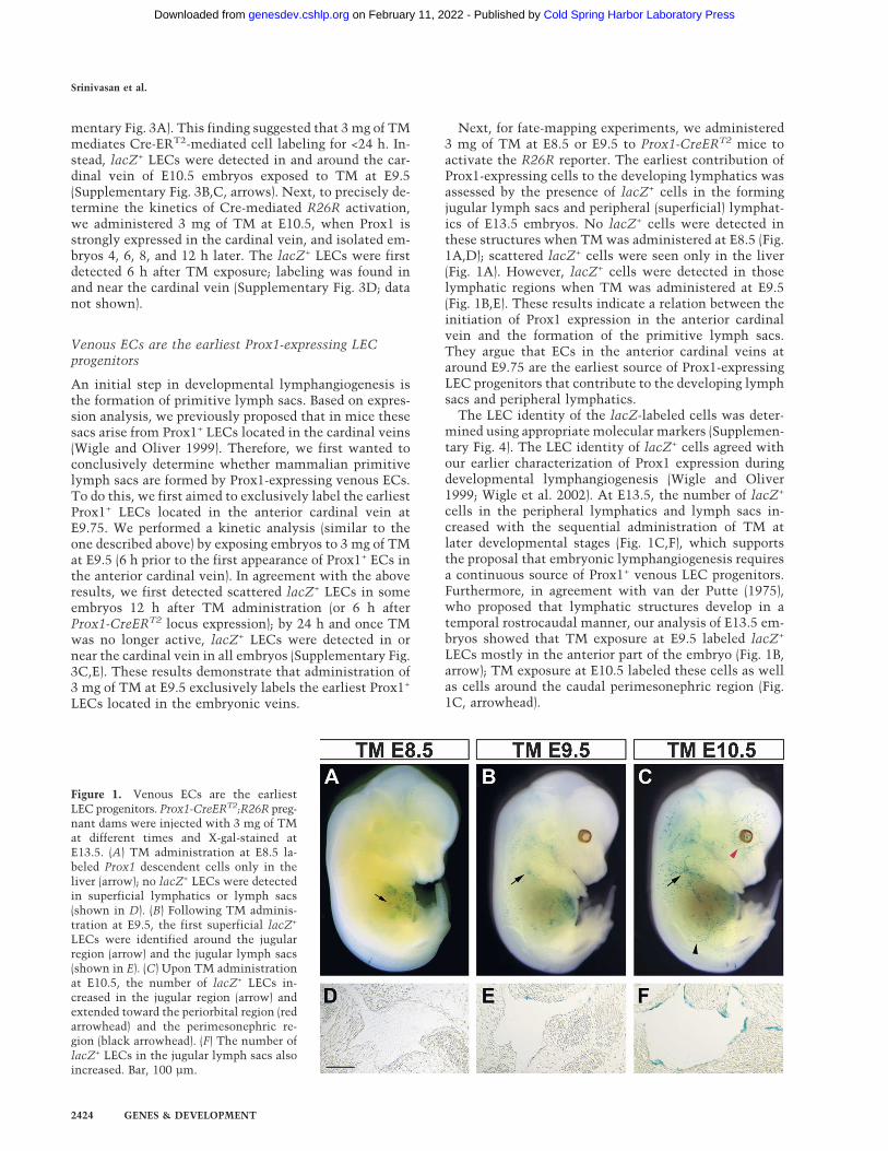
Next, for fate-mapping experiments, we administered3 mg of TM at E8.5 or E9.5 to Prox1-CreERT2 mice toactivate the R26R reporter. The earliest contribution ofProx1-expressing cells to the developing lymphatics wasassessed by the presence of lacZ+ cells in the formingjugular lymph sacs and peripheral (superficial) lymphat-ics of E13.5 embryos. No lacZ+ cells were detected inthese structures when TM was administered at E8.5 (Fig.1A,D); scattered lacZ+ cells were seen only in the liver(Fig. 1A). However, lacZ+ cells were detected in thoselymphatic regions when TM was administered at E9.5(Fig. 1B,E). These results indicate a relation between theinitiation of Prox1 expression in the anterior cardinalvein and the formation of the primitive lymph sacs.They argue that ECs in the anterior cardinal veins ataround E9.75 are the earliest source of Prox1-expressingLEC progenitors that contribute to the developing lymphsacs and peripheral lymphatics.
The LEC identity of the lacZ-labeled cells was deter-mined using appropriate molecular markers (Supplemen-tary Fig. 4). The LEC identity of lacZ+ cells agreed withour earlier characterization of Prox1 expression duringdevelopmental lymphangiogenesis (Wigle and Oliver1999; Wigle et al. 2002). At E13.5, the number of lacZ+
cells in the peripheral lymphatics and lymph sacs in-creased with the sequential administration of TM atlater developmental stages (Fig. 1C,F), which supportsthe proposal that embryonic lymphangiogenesis requiresa continuous source of Prox1+ venous LEC progenitors.Furthermore, in agreement with van der Putte (1975),who proposed that lymphatic structures develop in atemporal rostrocaudal manner, our analysis of E13.5 em-bryos showed that TM exposure at E9.5 labeled lacZ+
LECs mostly in the anterior part of the embryo (Fig. 1B,arrow); TM exposure at E10.5 labeled these cells as wellas cells around the caudal perimesonephric region (Fig.1C, arrowhead).
Figure 1. Venous ECs are the earliestLEC progenitors. Prox1-CreERT2;R26R preg-nant dams were injected with 3 mg of TMat different times and X-gal-stained atE13.5. (A) TM administration at E8.5 la-beled Prox1 descendent cells only in theliver (arrow); no lacZ+ LECs were detectedin superficial lymphatics or lymph sacs(shown in D). (B) Following TM adminis-tration at E9.5, the first superficial lacZ+
LECs were identified around the jugularregion (arrow) and the jugular lymph sacs(shown in E). (C) Upon TM administrationat E10.5, the number of lacZ+ LECs in-creased in the jugular region (arrow) andextended toward the periorbital region (redarrowhead) and the perimesonephric re-gion (black arrowhead). (F) The number oflacZ+ LECs in the jugular lymph sacs alsoincreased. Bar, 100 µm.
Srinivasan et al.
2424 GENES & DEVELOPMENT
Cold Spring Harbor Laboratory Press on February 11, 2022 - Published by genesdev.cshlp.orgDownloaded from
These results support the proposal that in the devel-oping mouse embryo, Prox1+ LEC progenitors are notpresent prior to the appearance of the Prox1-expressingvenous ECs in the anterior cardinal vein (around E9.75).In addition, sequential, stage-dependent administrationof TM correlates with an increasing number of lacZ+
cells in the lymph sacs and peripheral lymphatics, sug-gesting that ECs in the cardinal veins are a continuoussource of Prox1-expressing LECs, at least until thelymph sacs form.
The lymphatic vasculature arises by sprouting,proliferation, and migration of LECs
Once we determined that Prox1-expressing venous ECsare the earliest LEC progenitors contributing to the de-veloping lymph sacs and peripheral lymphatics, weaimed to assess the mechanisms that control LECsprouting into peripheral tissues, thereby giving rise tothe whole lymphatic vasculature. To do this, we per-formed similar lineage-tracing studies using Prox1-CreERT2;R26R embryos. TM (5 mg) remains active for<2.5 d; therefore, to efficiently label as many Prox1+ cellsas possible in the forming lymph sacs, TM was admin-istered at E10.5, and embryos were isolated and analyzeddaily between E11.5 and E15.5.
At E11.5, most superficial lacZ+ cells were located an-terior to the developing forelimbs; a few were scatteredmore caudally (Fig. 2A). Most lacZ+ cells were LECs thathad originated from the anterior (Supplementary Fig. 5A)and posterior cardinal veins and the iliac veins (data notshown). Therefore, these lacZ+ cells were the earliestdescendants of Prox1-expressing venous ECs. At E12.5,the number of lacZ+ cells along the anteroposterior axis
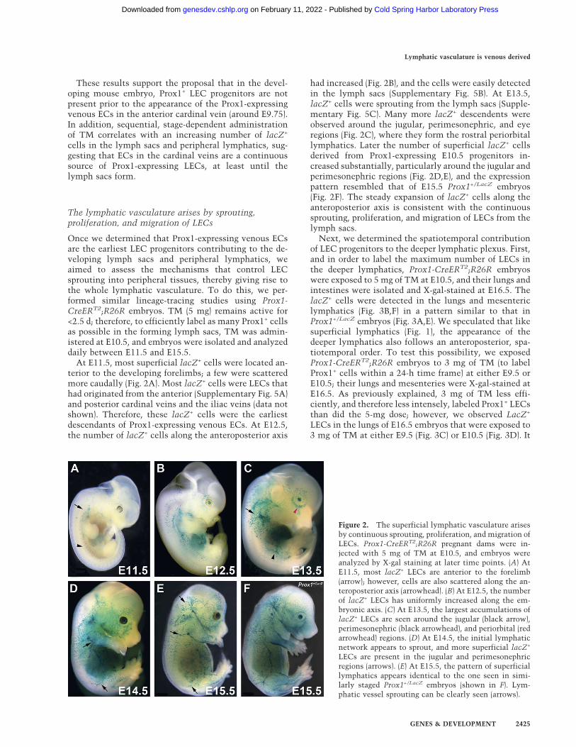
had increased (Fig. 2B), and the cells were easily detectedin the lymph sacs (Supplementary Fig. 5B). At E13.5,lacZ+ cells were sprouting from the lymph sacs (Supple-mentary Fig. 5C). Many more lacZ+ descendents wereobserved around the jugular, perimesonephric, and eyeregions (Fig. 2C), where they form the rostral periorbitallymphatics. Later the number of superficial lacZ+ cellsderived from Prox1-expressing E10.5 progenitors in-creased substantially, particularly around the jugular andperimesonephric regions (Fig. 2D,E), and the expressionpattern resembled that of E15.5 Prox1+/LacZ embryos(Fig. 2F). The steady expansion of lacZ+ cells along theanteroposterior axis is consistent with the continuoussprouting, proliferation, and migration of LECs from thelymph sacs.
Next, we determined the spatiotemporal contributionof LEC progenitors to the deeper lymphatic plexus. First,and in order to label the maximum number of LECs inthe deeper lymphatics, Prox1-CreERT2;R26R embryoswere exposed to 5 mg of TM at E10.5, and their lungs andintestines were isolated and X-gal-stained at E16.5. ThelacZ+ cells were detected in the lungs and mesentericlymphatics (Fig. 3B,F) in a pattern similar to that inProx1+/LacZ embryos (Fig. 3A,E). We speculated that likesuperficial lymphatics (Fig. 1), the appearance of thedeeper lymphatics also follows an anteroposterior, spa-tiotemporal order. To test this possibility, we exposedProx1-CreERT2;R26R embryos to 3 mg of TM (to labelProx1+ cells within a 24-h time frame) at either E9.5 orE10.5; their lungs and mesenteries were X-gal-stained atE16.5. As previously explained, 3 mg of TM less effi-ciently, and therefore less intensely, labeled Prox1+ LECsthan did the 5-mg dose; however, we observed LacZ+
LECs in the lungs of E16.5 embryos that were exposed to3 mg of TM at either E9.5 (Fig. 3C) or E10.5 (Fig. 3D). It
Figure 2. The superficial lymphatic vasculature arisesby continuous sprouting, proliferation, and migration ofLECs. Prox1-CreERT2;R26R pregnant dams were in-jected with 5 mg of TM at E10.5, and embryos wereanalyzed by X-gal staining at later time points. (A) AtE11.5, most lacZ+ LECs are anterior to the forelimb(arrow); however, cells are also scattered along the an-teroposterior axis (arrowhead). (B) At E12.5, the numberof lacZ+ LECs has uniformly increased along the em-bryonic axis. (C) At E13.5, the largest accumulations oflacZ+ LECs are seen around the jugular (black arrow),perimesonephric (black arrowhead), and periorbital (redarrowhead) regions. (D) At E14.5, the initial lymphaticnetwork appears to sprout, and more superficial lacZ+
LECs are present in the jugular and perimesonephricregions (arrows). (E) At E15.5, the pattern of superficiallymphatics appears identical to the one seen in simi-larly staged Prox1+/LacZ embryos (shown in F). Lym-phatic vessel sprouting can be clearly seen (arrows).
Lymphatic vasculature is venous derived
GENES & DEVELOPMENT 2425
Cold Spring Harbor Laboratory Press on February 11, 2022 - Published by genesdev.cshlp.orgDownloaded from
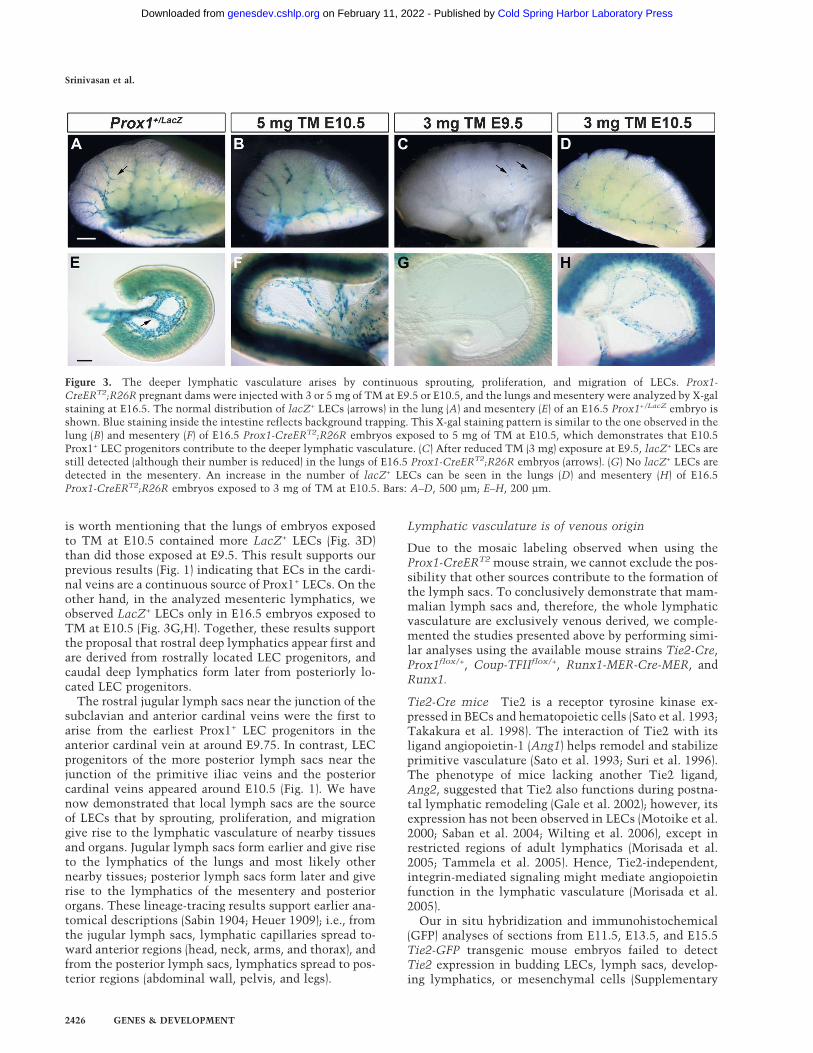
is worth mentioning that the lungs of embryos exposedto TM at E10.5 contained more LacZ+ LECs (Fig. 3D)than did those exposed at E9.5. This result supports ourprevious results (Fig. 1) indicating that ECs in the cardi-nal veins are a continuous source of Prox1+ LECs. On theother hand, in the analyzed mesenteric lymphatics, weobserved LacZ+ LECs only in E16.5 embryos exposed toTM at E10.5 (Fig. 3G,H). Together, these results supportthe proposal that rostral deep lymphatics appear first andare derived from rostrally located LEC progenitors, andcaudal deep lymphatics form later from posteriorly lo-cated LEC progenitors.
The rostral jugular lymph sacs near the junction of thesubclavian and anterior cardinal veins were the first toarise from the earliest Prox1+ LEC progenitors in theanterior cardinal vein at around E9.75. In contrast, LECprogenitors of the more posterior lymph sacs near thejunction of the primitive iliac veins and the posteriorcardinal veins appeared around E10.5 (Fig. 1). We havenow demonstrated that local lymph sacs are the sourceof LECs that by sprouting, proliferation, and migrationgive rise to the lymphatic vasculature of nearby tissuesand organs. Jugular lymph sacs form earlier and give riseto the lymphatics of the lungs and most likely othernearby tissues; posterior lymph sacs form later and giverise to the lymphatics of the mesentery and posteriororgans. These lineage-tracing results support earlier ana-tomical descriptions (Sabin 1904; Heuer 1909); i.e., fromthe jugular lymph sacs, lymphatic capillaries spread to-ward anterior regions (head, neck, arms, and thorax), andfrom the posterior lymph sacs, lymphatics spread to pos-terior regions (abdominal wall, pelvis, and legs).
Lymphatic vasculature is of venous origin
Due to the mosaic labeling observed when using theProx1-CreERT2 mouse strain, we cannot exclude the pos-sibility that other sources contribute to the formation ofthe lymph sacs. To conclusively demonstrate that mam-malian lymph sacs and, therefore, the whole lymphaticvasculature are exclusively venous derived, we comple-mented the studies presented above by performing simi-lar analyses using the available mouse strains Tie2-Cre,Prox1flox/+, Coup-TFIIflox/+, Runx1-MER-Cre-MER, andRunx1.
Tie2-Cre mice Tie2 is a receptor tyrosine kinase ex-pressed in BECs and hematopoietic cells (Sato et al. 1993;Takakura et al. 1998). The interaction of Tie2 with itsligand angiopoietin-1 (Ang1) helps remodel and stabilizeprimitive vasculature (Sato et al. 1993; Suri et al. 1996).The phenotype of mice lacking another Tie2 ligand,Ang2, suggested that Tie2 also functions during postna-tal lymphatic remodeling (Gale et al. 2002); however, itsexpression has not been observed in LECs (Motoike et al.2000; Saban et al. 2004; Wilting et al. 2006), except inrestricted regions of adult lymphatics (Morisada et al.2005; Tammela et al. 2005). Hence, Tie2-independent,integrin-mediated signaling might mediate angiopoietinfunction in the lymphatic vasculature (Morisada et al.2005).
Our in situ hybridization and immunohistochemical(GFP) analyses of sections from E11.5, E13.5, and E15.5Tie2-GFP transgenic mouse embryos failed to detectTie2 expression in budding LECs, lymph sacs, develop-ing lymphatics, or mesenchymal cells (Supplementary
Figure 3. The deeper lymphatic vasculature arises by continuous sprouting, proliferation, and migration of LECs. Prox1-CreERT2;R26R pregnant dams were injected with 3 or 5 mg of TM at E9.5 or E10.5, and the lungs and mesentery were analyzed by X-galstaining at E16.5. The normal distribution of lacZ+ LECs (arrows) in the lung (A) and mesentery (E) of an E16.5 Prox1+/LacZ embryo isshown. Blue staining inside the intestine reflects background trapping. This X-gal staining pattern is similar to the one observed in thelung (B) and mesentery (F) of E16.5 Prox1-CreERT2;R26R embryos exposed to 5 mg of TM at E10.5, which demonstrates that E10.5Prox1+ LEC progenitors contribute to the deeper lymphatic vasculature. (C) After reduced TM (3 mg) exposure at E9.5, lacZ+ LECs arestill detected (although their number is reduced) in the lungs of E16.5 Prox1-CreERT2;R26R embryos (arrows). (G) No lacZ+ LECs aredetected in the mesentery. An increase in the number of lacZ+ LECs can be seen in the lungs (D) and mesentery (H) of E16.5Prox1-CreERT2;R26R embryos exposed to 3 mg of TM at E10.5. Bars: A–D, 500 µm; E–H, 200 µm.
Srinivasan et al.
2426 GENES & DEVELOPMENT
Cold Spring Harbor Laboratory Press on February 11, 2022 - Published by genesdev.cshlp.orgDownloaded from
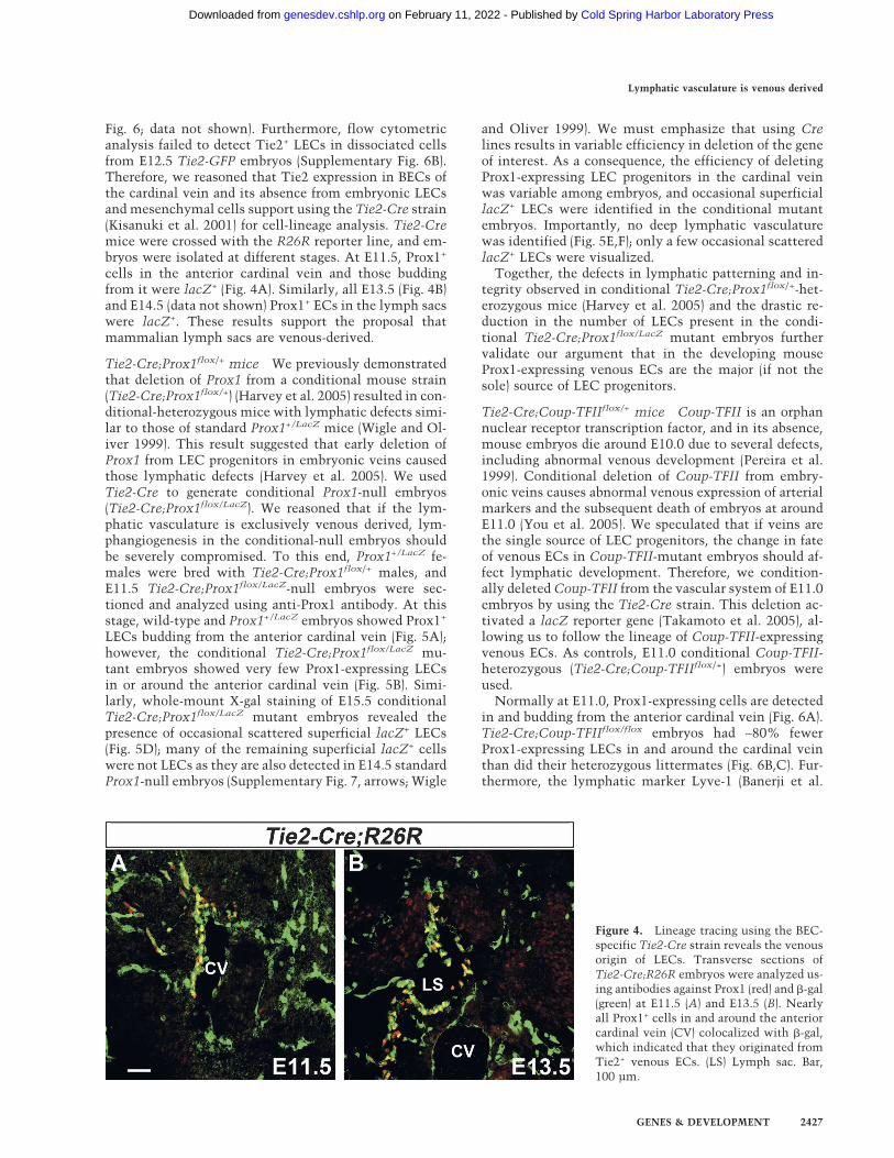
Fig. 6; data not shown). Furthermore, flow cytometricanalysis failed to detect Tie2+ LECs in dissociated cellsfrom E12.5 Tie2-GFP embryos (Supplementary Fig. 6B).Therefore, we reasoned that Tie2 expression in BECs ofthe cardinal vein and its absence from embryonic LECsand mesenchymal cells support using the Tie2-Cre strain(Kisanuki et al. 2001) for cell-lineage analysis. Tie2-Cremice were crossed with the R26R reporter line, and em-bryos were isolated at different stages. At E11.5, Prox1+
cells in the anterior cardinal vein and those buddingfrom it were lacZ+ (Fig. 4A). Similarly, all E13.5 (Fig. 4B)and E14.5 (data not shown) Prox1+ ECs in the lymph sacswere lacZ+. These results support the proposal thatmammalian lymph sacs are venous-derived.
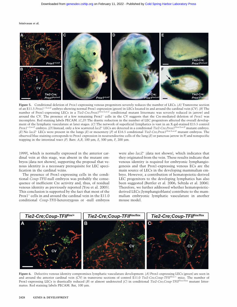
Tie2-Cre;Prox1flox/+ mice We previously demonstratedthat deletion of Prox1 from a conditional mouse strain(Tie2-Cre;Prox1flox/+) (Harvey et al. 2005) resulted in con-ditional-heterozygous mice with lymphatic defects simi-lar to those of standard Prox1+/LacZ mice (Wigle and Ol-iver 1999). This result suggested that early deletion ofProx1 from LEC progenitors in embryonic veins causedthose lymphatic defects (Harvey et al. 2005). We usedTie2-Cre to generate conditional Prox1-null embryos(Tie2-Cre;Prox1flox/LacZ). We reasoned that if the lym-phatic vasculature is exclusively venous derived, lym-phangiogenesis in the conditional-null embryos shouldbe severely compromised. To this end, Prox1+/LacZ fe-males were bred with Tie2-Cre;Prox1flox/+ males, andE11.5 Tie2-Cre;Prox1flox/LacZ-null embryos were sec-tioned and analyzed using anti-Prox1 antibody. At thisstage, wild-type and Prox1+/LacZ embryos showed Prox1+
LECs budding from the anterior cardinal vein (Fig. 5A);however, the conditional Tie2-Cre;Prox1flox/LacZ mu-tant embryos showed very few Prox1-expressing LECsin or around the anterior cardinal vein (Fig. 5B). Simi-larly, whole-mount X-gal staining of E15.5 conditionalTie2-Cre;Prox1flox/LacZ mutant embryos revealed thepresence of occasional scattered superficial lacZ+ LECs(Fig. 5D); many of the remaining superficial lacZ+ cellswere not LECs as they are also detected in E14.5 standardProx1-null embryos (Supplementary Fig. 7, arrows; Wigle
and Oliver 1999). We must emphasize that using Crelines results in variable efficiency in deletion of the geneof interest. As a consequence, the efficiency of deletingProx1-expressing LEC progenitors in the cardinal veinwas variable among embryos, and occasional superficiallacZ+ LECs were identified in the conditional mutantembryos. Importantly, no deep lymphatic vasculaturewas identified (Fig. 5E,F); only a few occasional scatteredlacZ+ LECs were visualized.
Together, the defects in lymphatic patterning and in-tegrity observed in conditional Tie2-Cre;Prox1flox/+-het-erozygous mice (Harvey et al. 2005) and the drastic re-duction in the number of LECs present in the condi-tional Tie2-Cre;Prox1flox/LacZ mutant embryos furthervalidate our argument that in the developing mouseProx1-expressing venous ECs are the major (if not thesole) source of LEC progenitors.
Tie2-Cre;Coup-TFIIflox/+ mice Coup-TFII is an orphannuclear receptor transcription factor, and in its absence,mouse embryos die around E10.0 due to several defects,including abnormal venous development (Pereira et al.1999). Conditional deletion of Coup-TFII from embry-onic veins causes abnormal venous expression of arterialmarkers and the subsequent death of embryos at aroundE11.0 (You et al. 2005). We speculated that if veins arethe single source of LEC progenitors, the change in fateof venous ECs in Coup-TFII-mutant embryos should af-fect lymphatic development. Therefore, we condition-ally deleted Coup-TFII from the vascular system of E11.0embryos by using the Tie2-Cre strain. This deletion ac-tivated a lacZ reporter gene (Takamoto et al. 2005), al-lowing us to follow the lineage of Coup-TFII-expressingvenous ECs. As controls, E11.0 conditional Coup-TFII-heterozygous (Tie2-Cre;Coup-TFIIflox/+) embryos wereused.
Normally at E11.0, Prox1-expressing cells are detectedin and budding from the anterior cardinal vein (Fig. 6A).Tie2-Cre;Coup-TFIIflox/flox embryos had ∼80% fewerProx1-expressing LECs in and around the cardinal veinthan did their heterozygous littermates (Fig. 6B,C). Fur-thermore, the lymphatic marker Lyve-1 (Banerji et al.
Figure 4. Lineage tracing using the BEC-specific Tie2-Cre strain reveals the venousorigin of LECs. Transverse sections ofTie2-Cre;R26R embryos were analyzed us-ing antibodies against Prox1 (red) and �-gal(green) at E11.5 (A) and E13.5 (B). Nearlyall Prox1+ cells in and around the anteriorcardinal vein (CV) colocalized with �-gal,which indicated that they originated fromTie2+ venous ECs. (LS) Lymph sac. Bar,100 µm.
Lymphatic vasculature is venous derived
GENES & DEVELOPMENT 2427
Cold Spring Harbor Laboratory Press on February 11, 2022 - Published by genesdev.cshlp.orgDownloaded from
1999), which is normally expressed in the anterior car-dinal vein at this stage, was absent in the mutant em-bryos (data not shown), supporting the proposal that ve-nous identity is a necessary prerequisite for LEC speci-fication in the cardinal veins.
The presence of Prox1-expressing cells in the condi-tional Coup-TFII-null embryos was probably the conse-quence of inefficient Cre activity and, thus, of residualvenous identity as previously reported (You et al. 2005).This conclusion is supported by the fact that most of theProx1+ cells in and around the cardinal vein in the E11.0conditional Coup-TFII-heterozygous or -null embryos
were also lacZ+ (data not shown), which indicates thatthey originated from the vein. These results indicate thatvenous identity is required for embryonic lymphangio-genesis and that Prox1-expressing venous ECs are themain source of LECs in the developing mammalian em-bryo. However, a contribution of hematopoietic-derivedLEC progenitors to the developing lymphatics has alsobeen suggested (Buttler et al. 2006; Sebzda et al. 2006).Therefore, we further addressed whether hematopoietic-derived LECs (lymphangioblasts) contribute to the mam-malian embryonic lymphatic vasculature in anothermouse model.
Figure 6. Defective venous identity compromises lymphatic vasculature development. (A) Prox1-expressing LECs (green) are seen inand around the anterior cardinal vein (CV) in transverse sections of control E11.0 Tie2-Cre;Coup-TFIIflox/+ mice. The number ofProx1-expressing LECs is drastically reduced (B) or almost undetected (C) in conditional Tie2-Cre;Coup-TFIIflox/flox mutant litter-mates. Red staining labels PECAM. Bar, 100 µm.
Figure 5. Conditional deletion of Prox1-expressing venous progenitors severely reduces the number of LECs. (A) Transverse sectionof an E11.5 Prox1+/LacZ embryo showing normal Prox1 expression (green) in LECs located in and around the cardinal vein (CV). (B) Thenumber of Prox1-expressing LECs in a Tie2-Cre;Prox1flox/LacZ conditional mutant littermate was severely reduced in (arrow) andaround the CV. The presence of a few remaining Prox1+ cells in the CV suggests that the Cre-mediated deletion of Prox1 wasincomplete. Red staining labels PECAM. (C,D) The drastic reduction in the number of LEC progenitors affected the overall develop-ment of the lymphatic vasculature at later stages. (C) The network of superficial lymphatics is vast in an X-gal-stained E15.5 controlProx1+/LacZ embryo. (D) Instead, only a few scattered lacZ+ LECs are detected in a conditional Tie2-Cre;Prox1flox/LacZ mutant embryo.(E) No lacZ+ LECs were present in the lungs (E) or mesentery (F) of E16.5 conditional Tie2-Cre;Prox1flox/LacZ mutant embryos. Theobserved blue staining corresponds to Prox1 expression in neuroendocrine cells of the lung (E) or pancreas (arrow in F) and nonspecifictrapping in the intestinal tract (F). Bars: A,B, 100 µm; E, 500 µm; F, 200 µm.
Srinivasan et al.
2428 GENES & DEVELOPMENT
Cold Spring Harbor Laboratory Press on February 11, 2022 - Published by genesdev.cshlp.orgDownloaded from
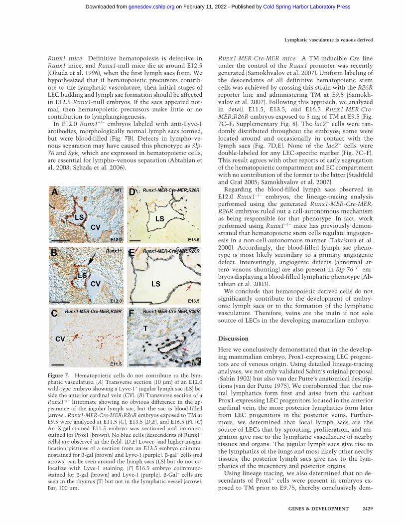
Runx1 mice Definitive hematopoiesis is defective inRunx1 mice, and Runx1-null mice die at around E12.5(Okuda et al. 1996), when the first lymph sacs form. Wehypothesized that if hematopoietic precursors contrib-ute to the lymphatic vasculature, then initial stages ofLEC budding and lymph sac formation should be affectedin E12.5 Runx1-null embryos. If the sacs appeared nor-mal, then hematopoietic precursors make little or nocontribution to lymphangiogenesis.
In E12.0 Runx1−/− embryos labeled with anti-Lyve-1antibodies, morphologically normal lymph sacs formed,but were blood-filled (Fig. 7B). Defects in lympho–ve-nous separation may have caused this phenotype as Slp-76 and Syk, which are expressed in hematopoietic cells,are essential for lympho–venous separation (Abtahian etal. 2003; Sebzda et al. 2006).
Runx1-MER-Cre-MER mice A TM-inducible Cre lineunder the control of the Runx1 promoter was recentlygenerated (Samokhvalov et al. 2007). Uniform labeling ofthe descendants of all definitive hematopoietic stemcells was achieved by crossing this strain with the R26Rreporter line and administering TM at E9.5 (Samokh-valov et al. 2007). Following this approach, we analyzedin detail E11.5, E13.5, and E16.5 Runx1-MER-Cre-MER;R26R embryos exposed to 5 mg of TM at E9.5 (Fig.7C–F; Supplementary Fig. 8). The lacZ+ cells were ran-domly distributed throughout the embryos; some werelocated around and occasionally in contact with thelymph sacs (Fig. 7D,E). None of the lacZ+ cells weredouble-labeled for any LEC-specific marker (Fig. 7C–F).This result agrees with other reports of early segregationof the hematopoietic compartment and EC compartmentwith no contribution of the former to the latter (Stadtfeldand Graf 2005; Samokhvalov et al. 2007).
Regarding the blood-filled lymph sacs observed inE12.0 Runx1−/− embryos, the lineage-tracing analysisperformed using the generated Runx1-MER-Cre-MER;R26R embryos ruled out a cell-autonomous mechanismas being responsible for that phenotype. In fact, workperformed using Runx1−/− mice has previously demon-strated that hematopoietic stem cells regulate angiogen-esis in a non-cell-autonomous manner (Takakura et al.2000). Accordingly, the blood-filled lymph sac pheno-type is most likely secondary to a primary angiogenicdefect. Interestingly, angiogenic defects (abnormal ar-tero–venous shunting) are also present in Slp-76−/− em-bryos displaying a blood-filled lymphatic phenotype (Ab-tahian et al. 2003).
We conclude that hematopoietic-derived cells do notsignificantly contribute to the development of embry-onic lymph sacs or to the formation of the lymphaticvasculature. Therefore, veins are the main if not solesource of LECs in the developing mammalian embryo.
Discussion
Here we conclusively demonstrated that in the develop-ing mammalian embryo, Prox1-expressing LEC progeni-tors are of venous origin. Using detailed lineage-tracinganalyses, we not only validated Sabin’s original proposal(Sabin 1902) but also van der Putte’s anatomical descrip-tions (van der Putte 1975). We corroborated that the ros-tral lymphatics form first and arise from the earliestProx1-expressing LEC progenitors located in the anteriorcardinal vein; the more posterior lymphatics form laterfrom LEC progenitors in the posterior veins. Further-more, we determined that local lymph sacs are thesource of LECs that by sprouting, proliferation, and mi-gration give rise to the lymphatic vasculature of nearbytissues and organs. The jugular lymph sacs give rise tothe lymphatics of the lungs and most likely other nearbytissues; the posterior lymph sacs give rise to the lym-phatics of the mesentery and posterior organs.
Using lineage tracing, we also determined that no de-scendants of Prox1+ cells were present in embryos ex-posed to TM prior to E9.75, thereby conclusively dem-
Figure 7. Hematopoietic cells do not contribute to the lym-phatic vasculature. (A) Transverse section (10 µm) of an E12.0wild-type embryo showing a Lyve-1+ jugular lymph sac (LS) be-side the anterior cardinal vein (CV). (B) Transverse section of aRunx1−/− littermate showing no obvious difference in the ap-pearance of the jugular lymph sac, but the sac is blood-filled(arrow). Runx1-MER-Cre-MER;R26R embryos exposed to TM atE9.5 were analyzed at E11.5 (C), E13.5 (D,E), and E16.5 (F). (C)An X-gal-stained E11.5 embryo was sectioned and immuno-stained for Prox1 (brown). No blue cells (descendents of Runx1+
cells) are observed in the field. (D,E) Lower- and higher-magni-fication pictures of a section from an E13.5 embryo coimmu-nostained for �-gal (brown) and Lyve-1 (purple). �-gal+ cells (redarrows) can be seen around the lymph sacs (LS) but do not co-localize with Lyve-1 staining. (F) E16.5 embryo coimmuno-stained for �-gal (brown) and Lyve-1 (purple). �-Gal+ cells areseen in the thymus (T) but not in the lymphatic vessel (arrow).Bar, 100 µm.
Lymphatic vasculature is venous derived
GENES & DEVELOPMENT 2429
Cold Spring Harbor Laboratory Press on February 11, 2022 - Published by genesdev.cshlp.orgDownloaded from
onstrating that at around E9.75, ECs in the anterior car-dinal vein are the earliest source of Prox1-expressingLEC progenitors. To demonstrate that the mammalianlymphatic vasculature has a solely venous origin, weused several mouse strains.
Tie2-Cre;Coup-TFIIflox/flox-mutant embryos showedthat venous identity is required at least during the earlystages of lymphatic development. Conditional Tie2-Cre;Prox1flox/LacZ mutant embryos showed that thenumber of LECs was significantly reduced and that ve-nous-derived Tie2 progeny contribute to the forminglymphatics. These results support the argument thatLECs have a venous origin and that Prox1-expressing ve-nous ECs are the main (if not the sole) source of LECprogenitors in developing mice.
Although our results show that developing lymphaticvasculature arises mostly from venous-derived Prox1-ex-pressing progenitor cells, two recent reports haveclaimed that other cell types contribute to developinglymphatics in mice. Buttler et al. (2006) proposed thatscattered mesenchymal cells with leukocyte and lym-phoendothelial characteristics that are first detected af-ter E10.5 eventually integrate into the lymphatics. Se-bzda et al. (2006) identified a subpopulation of Syk- andSlp-76-expressing hematopoietic-derived CEPs that ac-quire a lymphatic fate. However, two other reports foundno evidence of hematopoietic contribution to the mam-malian endothelial compartment (Stadtfeld and Graf2005; Samokhvalov et al. 2007).
We did not identify descendents of Prox1+ cells in em-bryos exposed to TM prior to E9.5. This result not onlydemonstrated that E9.75 venous Prox1-expressing ECsare the earliest source of LEC progenitors but also elimi-nated the possibility that other hematopoietic- or mes-enchyme-derived Prox1+ lymphatic progenitors are pre-sent before Prox1 expression in the cardinal vein. Fur-thermore, Runx1−/− embryos defective in definitivehematopoiesis had morphologically normal lymph sacs,and lineage tracing using the Runx1-MER-Cre-MERstrain demonstrated that descendents of Runx1+ cells donot contribute to the developing lymph sacs. These re-sults conclusively demonstrate that hematopoietic cellsdo not significantly contribute to the forming lymphsacs or lymphatic vasculature. Therefore, if mesenchy-mal cells with leukocyte and lymphoendothelial charac-teristics or hematopoietic-derived CEPs exist in themouse embryo, they are so rare they probably cannotdirectly contribute to the developing murine lymphaticnetwork. We must emphasize that additional LECsources such as transdifferentiating macrophages (Ma-ruyama et al. 2005) and bone marrow (Salven et al. 2003;Religa et al. 2005; Kerjaschki et al. 2006) probably con-tribute to postnatal lymphangiogenesis that occurs innormal and pathological conditions.
In summary, our work validates Sabin’s model, whichalmost 100 years ago proposed that from venous-derivedprimary lymph sacs, the peripheral lymphatic systemoriginates and spreads by endothelial sprouting into thesurrounding tissues and organs, where capillaries form(Sabin 1902, 1904). We conclusively determined that
during mammalian embryonic lymphangiogenesis,Prox1-expressing LEC progenitors in early veins are themain, and most likely unique source of LECs required forthe formation of the lymphatic vasculature.
Materials and methods
Mice
Tie2-Cre mice were provided by Dr. M. Yanagisawa (Universityof Texas Southwestern Medical Center, Dallas, TX); Tie2-GFPmice were supplied by Dr. T. Sato (Cornell University, NewYork); R26R mice were provided by Dr. G. Grosveld (St. JudeChildren’s Research Hospital, Memphis, TN); and Runx1-mu-tant embryos were supplied by Dr. J. Downing (St. Jude Chil-dren’s Research Hospital, Memphis, TN). The methods for gen-erating Prox1+/LacZ, Prox1flox/flox, Coup-TFIIflox/flox, and Runx1-MER-Cre-MER mice have been reported previously (Wigle et al.1999; Harvey et al. 2005; Takamoto et al. 2005; Samokhvalov etal. 2007). To generate the Prox1-CreERT2 embryonic stem cellline, we fused a synthetic splice acceptor site, Prox1 exons 3 and4, an internal ribosome entry site, and Cre-ERT2 and Poly(A)tail. This fusion was targeted for insertion into intron 2 of themouse Prox1 locus by electroporation into the W9.5 embryonicstem cell line. Following selection and standard screening, thecorrectly targeted cells were used to generate chimeric mice.The developmental stage of mouse embryos was determined byconsidering 9 a.m. of the day the vaginal plug was detected inthe pregnant dam as E0.5. All of the mouse experiments wereapproved by the St. Jude Children’s Research Hospital AnimalCare and Use Committee.
Immunohistochemistry
Fluorescent or horseradish peroxidase staining using 3,3�-diami-nobenzidene (DAB) as a substrate was performed on frozen orparaffin-embedded sections (10 µm) as described previously(Harvey et al. 2005). Primary antibodies were rabbit anti-�-gal(MP Biomedicals), rabbit (AngioBio), and guinea pig (G. Oliver,unpubl.); anti-mouse Prox1; rat anti-mouse PECAM (BD Pharm-ingen); guinea pig anti-Lyve-1 (G. Oliver, unpubl.); and rabbitanti-GFP (Molecular Probes). Secondary antibodies were Alexa488-conjugated donkey anti-rabbit (Molecular Probes), Cy3-con-jugated donkey anti-guinea pig (Jackson ImmunoResearch Labo-ratories), and Cy3-conjugated donkey anti-rat (Jackson Immu-noResearch Laboratories).
TM injection
For lineage tracing using the Prox1-CreERT2 line, TM (20 mg/mL; Sigma) was dissolved in corn oil. Pregnant mice were in-jected intraperitoneally with either 3 or 5 mg per 40 g of bodyweight at the indicated time points. Lineage tracing using theRunx1-MER-Cre-MER line has been described previously(Samokhvalov et al. 2007).
Detection of �-galactosidase activity in embryos and tissues
To detect �-gal activity in embryos, we performed X-gal stainingas described previously (Harvey et al. 2005). Embryos were post-fixed in 4% paraformaldehyde overnight at 4°C and then clearedby soaking in sequentially increasing concentrations of glyceroldissolved in a solution of PBS and 0.1% Tween 20. Alter-natively, embryos were embedded in paraffin and sectioned(10 µm).
Srinivasan et al.
2430 GENES & DEVELOPMENT
Cold Spring Harbor Laboratory Press on February 11, 2022 - Published by genesdev.cshlp.orgDownloaded from
Acknowledgments
We thank N. Lenny and J. Downing for Runx1-mutant embryos;M. Yanagisawa for the Tie2-Cre line; T. Sato for the Tie2-GFP;D. Dumont for the Tie2 cDNA; N. Gale, N. Papadopoulos, andG. Yancopoulos for the Lyve-1 fusion protein; M. Self and X.Geng for assistance; N. Johnson for critical reading; and AngelaMcArthur for editing the manuscript. This work was supportedby NIH grant HL 076448 (to S.Y.T.) and R01-HL073402 (toG.O.), Cancer Center Support Grant CA-21765, and the Ameri-can Lebanese Syrian Associated Charities (ALSAC). R.S.S. issupported by a post-doctoral fellowship grant from the Lym-phatic Research Foundation.
References
Abtahian, F., Guerriero, A., Sebzda, E., Lu, M.M., Zhou, R.,Mocsai, A., Myers, E.E., Huang, B., Jackson, D.G., Ferrari,V.A., et al. 2003. Regulation of blood and lymphatic vascularseparation by signaling proteins SLP-76 and Syk. Science299: 247–251.
Banerji, S., Ni, J., Wang, S.X., Clasper, S., Su, J., Tammi, R.,Jones, M., and Jackson, D.G. 1999. LYVE-1, a new homo-logue of the CD44 glycoprotein, is a lymph-specific receptorfor hyaluronan. J. Cell Biol. 144: 789–801.
Buttler, K., Kreysing, A., von Kaisenberg, C.S., Schweigerer, L.,Gale, N., Papoutsi, M., and Wilting, J. 2006. Mesenchymalcells with leukocyte and lymphendothelial characteristics inmurine embryos. Dev. Dyn. 235: 1554–1562.
Danielian, P.S., Muccino, D., Rowitch, D.H., Michael, S.K., andMcMahon, A.P. 1998. Modification of gene activity inmouse embryos in utero by a tamoxifen-inducible form ofCre recombinase. Curr. Biol. 8: 1323–1326.
Dor, Y., Brown, J., Martinez, O.I., and Melton, D.A. 2004. Adultpancreatic �-cells are formed by self-duplication rather thanstem-cell differentiation. Nature 429: 41–46.
Gale, N.W., Thurston, G., Hackett, S.F., Renard, R., Wang, Q.,McClain, J., Martin, C., Witte, C., Witte, M.H., Jackson, D.,et al. 2002. Angiopoietin-2 is required for postnatal angio-genesis and lymphatic patterning, and only the latter role isrescued by Angiopoietin-1. Dev. Cell 3: 411–423.
Harvey, N.L., Srinivasan, R.S., Dillard, M.E., Johnson, N.C.,Witte, M.H., Boyd, K., Sleeman, M.W., and Oliver, G. 2005.Lymphatic vascular defects promoted by Prox1 haploinsuf-ficiency cause adult-onset obesity. Nat. Genet. 37: 1072–1081.
Heuer, G. 1909. The development of the lymphatics in thesmall intestine of the pig. Am. J. Anat. 9: 93–118. doi:10.1002/aja.1000090105.
Huntington, G.S. and McClure, C.F.W. 1910. The anatomy anddevelopment of the jugular lymph sac in the domestic cat(Felis domestica). Am. J. Anat. 10: 177–312. doi: 10.1002/aja.1000100108.
Indra, A.K., Warot, X., Brocard, J., Bornert, J.M., Xiao, J.H.,Chambon, P., and Metzger, D. 1999. Temporally-controlledsite-specific mutagenesis in the basal layer of the epidermis:Comparison of the recombinase activity of the tamoxifen-inducible Cre-ER(T) and Cre-ER(T2) recombinases. NucleicAcids Res. 27: 4324–4327.
Kerjaschki, D., Huttary, N., Raab, I., Regele, H., Bojarski-Nagy,K., Bartel, G., Krober, S.M., Greinix, H., Rosenmaier, A.,Karlhofer, F., et al. 2006. Lymphatic endothelial progenitorcells contribute to de novo lymphangiogenesis in humanrenal transplants. Nat. Med. 12: 230–234.
Kisanuki, Y.Y., Hammer, R.E., Miyazaki, J., Williams, S.C., Ri-chardson, J.A., and Yanagisawa, M. 2001. Tie2-Cre trans-
genic mice: A new model for endothelial cell-lineage analy-sis in vivo. Dev. Biol. 230: 230–242.
Maruyama, K., Ii, M., Cursiefen, C., Jackson, D.G., Keino, H.,Tomita, M., Van Rooijen, N., Takenaka, H., D’Amore, P.A.,Stein-Streilein, J., et al. 2005. Inflammation-induced lymph-angiogenesis in the cornea arises from CD11b-positive mac-rophages. J. Clin. Invest. 115: 2363–2372.
Morisada, T., Oike, Y., Yamada, Y., Urano, T., Akao, M.,Kubota, Y., Maekawa, H., Kimura, Y., Ohmura, M., Miya-moto, T., et al. 2005. Angiopoietin-1 promotes LYVE-1-posi-tive lymphatic vessel formation. Blood 105: 4649–4656.
Motoike, T., Loughna, S., Perens, E., Roman, B.L., Liao, W.,Chau, T.C., Richardson, C.D., Kawate, T., Kuno, J., Wein-stein, B.M., et al. 2000. Universal GFP reporter for the studyof vascular development. Genesis 28: 75–81.
Ny, A., Koch, M., Schneider, M., Neven, E., Tong, R.T., Maity,S., Fischer, C., Plaisance, S., Lambrechts, D., Heligon, C., etal. 2005. A genetic Xenopus laevis tadpole model to studylymphangiogenesis. Nat. Med. 11: 998–1004.
Okuda, T., van Deursen, J., Hiebert, S.W., Grosveld, G., andDowning, J.R. 1996. AML1, the target of multiple chromo-somal translocations in human leukemia, is essential fornormal fetal liver hematopoiesis. Cell 84: 321–330.
Oliver, G. and Alitalo, K. 2005. The lymphatic vasculature: Re-cent progress and paradigms. Annu. Rev. Cell Dev. Biol. 21:457–483.
Oliver, G. and Detmar, M. 2002. The rediscovery of the lymph-atic system: Old and new insights into the development andbiological function of the lymphatic vasculature. Genes &Dev. 16: 773–783.
Pereira, F.A., Qiu, Y., Zhou, G., Tsai, M.J., and Tsai, S.Y. 1999.The orphan nuclear receptor COUP-TFII is required for an-giogenesis and heart development. Genes & Dev. 13: 1037–1049.
Religa, P., Cao, R., Bjorndahl, M., Zhou, Z., Zhu, Z., and Cao, Y.2005. Presence of bone marrow-derived circulating progeni-tor endothelial cells in the newly formed lymphatic vessels.Blood 106: 4184–4190.
Saban, M.R., Memet, S., Jackson, D.G., Ash, J., Roig, A.A., Is-rael, A., and Saban, R. 2004. Visualization of lymphatic ves-sels through NF-�B activity. Blood 104: 3228–3230.
Sabin, F. 1902. On the origin of the lymphatics system from theveins and the development of the lymph hearts and the tho-racic duct in the pig. Am. J. Anat. 1: 367–389. doi: 10.1002/aja.1000010310.
Sabin, F. 1904. On the development of superficial lymphatics inthe skin of the pig. Am. J. Anat. 3: 183–195. doi: 10.1002/aja.1000030205.
Salven, P., Mustjoki, S., Alitalo, R., Alitalo, K., and Rafii, S.2003. VEGFR-3 and CD133 identify a population of CD34+
lymphatic/vascular endothelial precursor cells. Blood 101:168–172.
Samokhvalov, I.M., Samokhvalova, N.I., and Nishikawa, S.2007. Cell tracing shows the contribution of the yolk sac toadult haematopoiesis. Nature 446: 1056–1061.
Sato, T.N., Qin, Y., Kozak, C.A., and Audus, K.L. 1993. Tie-1and tie-2 define another class of putative receptor tyrosinekinase genes expressed in early embryonic vascular system.Proc. Natl. Acad. Sci. 90: 9355–9358.
Sebzda, E., Hibbard, C., Sweeney, S., Abtahian, F., Bezman, N.,Clemens, G., Maltzman, J.S., Cheng, L., Liu, F., Turner, M.,et al. 2006. Syk and Slp-76 mutant mice reveal a cell-autono-mous hematopoietic cell contribution to vascular develop-ment. Dev. Cell 11: 349–361.
Soriano, P. 1999. Generalized lacZ expression with the ROSA26Cre reporter strain. Nat. Genet. 21: 70–71.
Lymphatic vasculature is venous derived
GENES & DEVELOPMENT 2431
Cold Spring Harbor Laboratory Press on February 11, 2022 - Published by genesdev.cshlp.orgDownloaded from
Stadtfeld, M. and Graf, T. 2005. Assessing the role of hemato-poietic plasticity for endothelial and hepatocyte develop-ment by non-invasive lineage tracing. Development 132:203–213.
Suri, C., Jones, P.F., Patan, S., Bartunkova, S., Maisonpierre,P.C., Davis, S., Sato, T.N., and Yancopoulos, G.D. 1996. Req-uisite role of angiopoietin-1, a ligand for the TIE2 receptor,during embryonic angiogenesis. Cell 87: 1171–1180.
Takakura, N., Huang, X.L., Naruse, T., Hamaguchi, I., Dumont,D.J., Yancopoulos, G.D., and Suda, T. 1998. Critical role ofthe TIE2 endothelial cell receptor in the development of de-finitive hematopoiesis. Immunity 9: 677–686.
Takakura, N., Watanabe, T., Suenobu, S., Yamada, Y., Noda, T.,Ito, Y., Satake, M., and Suda, T. 2000. A role for hematopoi-etic stem cells in promoting angiogenesis. Cell 102: 199–209.
Takamoto, N., You, L.R., Moses, K., Chiang, C., Zimmer, W.E.,Schwartz, R.J., DeMayo, F.J., Tsai, M.J., and Tsai, S.Y. 2005.COUP-TFII is essential for radial and anteroposterior pat-terning of the stomach. Development 132: 2179–2189.
Tammela, T., Saaristo, A., Lohela, M., Morisada, T., Tornberg,J., Norrmen, C., Oike, Y., Pajusola, K., Thurston, G., Suda,T., et al. 2005. Angiopoietin-1 promotes lymphatic sproutingand hyperplasia. Blood 105: 4642–4648.
van der Putte, S.C. 1975. The early development of the lym-phatic system in mouse embryos. Acta Morphol. Neerl.Scand. 13: 245–286.
Wigle, J.T. and Oliver, G. 1999. Prox1 function is required forthe development of the murine lymphatic system. Cell 98:769–778.
Wigle, J.T., Chowdhury, K., Gruss, P., and Oliver, G. 1999.Prox1 function is crucial for mouse lens-fibre elongation.Nat. Genet. 21: 318–322.
Wigle, J.T., Harvey, N., Detmar, M., Lagutina, I., Grosveld, G.,Gunn, M.D., Jackson, D.G., and Oliver, G. 2002. An essen-tial role for Prox1 in the induction of the lymphatic endo-thelial cell phenotype. EMBO J. 21: 1505–1513.
Wilting, J., Papoutsi, M., Schneider, M., and Christ, B. 2000. Thelymphatic endothelium of the avian wing is of somitic ori-gin. Dev. Dyn. 217: 271–278.
Wilting, J., Aref, Y., Huang, R., Tomarev, S.I., Schweigerer, L.,Christ, B., Valasek, P., and Papoutsi, M. 2006. Dual origin ofavian lymphatics. Dev. Biol. 292: 165–173.
Yaniv, K., Isogai, S., Castranova, D., Dye, L., Hitomi, J., andWeinstein, B.M. 2006. Live imaging of lymphatic develop-ment in the zebrafish. Nat. Med. 12: 711–716.
You, L.R., Lin, F.J., Lee, C.T., DeMayo, F.J., Tsai, M.J., and Tsai,S.Y. 2005. Suppression of Notch signalling by the COUP-TFII transcription factor regulates vein identity. Nature 435:98–104.
Zhang, H., Fujitani, Y., Wright, C.V., and Gannon, M. 2005.Efficient recombination in pancreatic islets by a tamoxifen-inducible Cre-recombinase. Genesis 42: 210–217.
Srinivasan et al.
2432 GENES & DEVELOPMENT
Cold Spring Harbor Laboratory Press on February 11, 2022 - Published by genesdev.cshlp.orgDownloaded from
10.1101/gad.1588407Access the most recent version at doi: 21:2007, Genes Dev.
R. Sathish Srinivasan, Miriam E. Dillard, Oleg V. Lagutin, et al. lymphatic vasculatureLineage tracing demonstrates the venous origin of the mammalian
Material
Supplemental
http://genesdev.cshlp.org/content/suppl/2007/10/01/21.19.2422.DC1
References
http://genesdev.cshlp.org/content/21/19/2422.full.html#ref-list-1
This article cites 44 articles, 13 of which can be accessed free at:
License
ServiceEmail Alerting
click here.right corner of the article or
Receive free email alerts when new articles cite this article - sign up in the box at the top
Copyright © 2007, Cold Spring Harbor Laboratory Press
Cold Spring Harbor Laboratory Press on February 11, 2022 - Published by genesdev.cshlp.orgDownloaded from