Limiting the proliferation and reactivity of retinal Müller cells during experimental retinal...
8
Limiting the Proliferation and Reactivity of Retinal Mu ¨ ller Cells During Experimental Retinal Detachment: The Value of Oxygen Supplementation GEOFFREY LEWIS, PHD, KYLE MERVIN, BSC (HONS), KRISZTINA VALTER, MBBS, JULIANI MASLIM, MBBS, PHD, PETER J. KAPPEL, BS, JONATHAN STONE, DSC, AND STEVEN FISHER, PHD ● PURPOSE: To assess the role of hypoxia in inducing the proliferation, hypertrophy, and dysfunction of Mu ¨ ller cells in detached retina and the effectiveness of supple- mental oxygen in limiting these reactions. See also pp. 155–164 and 231. ● METHODS: Retinal detachments were produced in the right eye of each of 13 cats; the cats survived surgery for 3 days, during which six were kept in normoxia (room air, 21%) and seven in hyperoxia (70% oxygen). Retinas were labeled for proliferation with an antibody (MIB-1) to a cell cycle protein (Ki-67), for evidence of hypertro- phy employing antibodies to the intermediate filament protein glial fibrillary acidic protein (GFAP) and to b-tubulin and for disturbance of glutamate neurochem- istry employing antibodies to glutamate to a glutamate receptor (GluR-2) and to glutamine synthetase. ● RESULTS: Results from the two animals kept in nor- moxia after retinal detachment confirmed previous re- ports that detachment caused the proliferation of Mu ¨ ller cells, the hypertrophy of Mu ¨ ller cell processes, and the disruption of glutamate recycling by Mu ¨ ller cells. Oxygen supplementation during detachment reduced Mu ¨ ller cell proliferation and hypertrophy and reduced the abnormal- ities in the distributions of glutamate, GluR-2, and glutamine synthetase. ● CONCLUSIONS: Oxygen supplementation reduced the reaction of retinal Mu ¨ ller cells to retinal detachment, limiting their proliferation and helping to maintain their normal structure and function. In the clinical setting, oxygen supplementation between diagnosis and reattach- ment surgery may reduce the incidence and severity of glial-based complications, such as proliferative vitreoreti- nopathy. (Am J Ophthalmol 1999;128:165–172. © 1999 by Elsevier Science Inc. All rights reserved.) A MAJOR COMPLICATION OF RETINAL DETACHMENT is the development of proliferative vitreoretinopa- thy even after seemingly successful reattachment. The reattached portion of retina becomes a focus for glial scarring, which spreads into the surrounding retina, includ- ing regions not previously detached. 1,2 One factor in proliferative vitreoretinopathy may be the response of retinal glial cells, particularly Mu ¨ ller cells, to detachment. 3 The Mu ¨ller cell response resembles the “reactive” re- sponse of astrocytes to injury of other parts of the central nervous system and involves three principal components. The Mu ¨ller cells proliferate, with the rate of their prolif- eration rising sharply to a maximum within 3 to 4 days of retinal detachment but continuing for weeks or months. 4,5 The processes of Mu ¨ller cells hypertrophy and grow abnor- mally into the vitreous humor and along the outer surface of the retina, between the photoreceptors and the retinal pigment epithelium. 2,6 –9 In addition, detachment changes the distributions of amino acid transmitters within the retina. 10,11 It was hypothesized that the glial changes induced by detachment are caused by hypoxia and hypo- Accepted for publication March 17, 1999. From the Department of Anatomy and Histology, NSW Retinal Dystrophy Research Centre, University of Sydney, Sydney, Australia (Mr Mervin, Ms Valter, Dr Maslim, and Dr Stone); Neuroscience Research Institute (Mr Kappel, Dr Lewis, and Dr Fisher); and Department of Molecular, Cellular and Developmental Biology, University of California Santa Barbara, Santa Barbara, California (Dr Fisher). This study was supported by the Australian Retinitis Pigmentosa Associ- ation, the National Health and Medical Research Council (Australia), the Medical Foundation of the University of Sydney, National Eye Institute Research grant EY00888, and the Santa Barbara Cottage Hospital. Reprint requests to Jonathan J. Stone, DSc, NSW Retinal Dystrophy Research Center, Department of Anatomy and Histology, University of Sydney F13, NSW 2006, Australia; fax: 61 2 9351 5664; e-mail: [email protected] © 1999 BY ELSEVIER SCIENCE INC.ALL RIGHTS RESERVED. 0002-9394/99/$20.00 165 PII S0002-9394(99)00103-8
-
Upload
geoffrey-lewis -
Category
Documents
-
view
212 -
download
0
Transcript of Limiting the proliferation and reactivity of retinal Müller cells during experimental retinal...
Limiting the Proliferation and Reactivity ofRetinal Muller Cells During Experimental
Retinal Detachment: The Value ofOxygen Supplementation
GEOFFREY LEWIS, PHD, KYLE MERVIN, BSC (HONS), KRISZTINA VALTER, MBBS,JULIANI MASLIM, MBBS, PHD, PETER J. KAPPEL, BS, JONATHAN STONE, DSC,
AND STEVEN FISHER, PHD
● PURPOSE: To assess the role of hypoxia in inducing theproliferation, hypertrophy, and dysfunction of Mullercells in detached retina and the effectiveness of supple-mental oxygen in limiting these reactions.
See also pp. 155–164 and 231.
● METHODS: Retinal detachments were produced in theright eye of each of 13 cats; the cats survived surgery for3 days, during which six were kept in normoxia (roomair, 21%) and seven in hyperoxia (70% oxygen). Retinaswere labeled for proliferation with an antibody (MIB-1)to a cell cycle protein (Ki-67), for evidence of hypertro-phy employing antibodies to the intermediate filamentprotein glial fibrillary acidic protein (GFAP) and tob-tubulin and for disturbance of glutamate neurochem-istry employing antibodies to glutamate to a glutamatereceptor (GluR-2) and to glutamine synthetase.● RESULTS: Results from the two animals kept in nor-moxia after retinal detachment confirmed previous re-ports that detachment caused the proliferation of Mullercells, the hypertrophy of Muller cell processes, and the
disruption of glutamate recycling by Muller cells. Oxygensupplementation during detachment reduced Muller cellproliferation and hypertrophy and reduced the abnormal-ities in the distributions of glutamate, GluR-2, andglutamine synthetase.● CONCLUSIONS: Oxygen supplementation reduced thereaction of retinal Muller cells to retinal detachment,limiting their proliferation and helping to maintain theirnormal structure and function. In the clinical setting,oxygen supplementation between diagnosis and reattach-ment surgery may reduce the incidence and severity ofglial-based complications, such as proliferative vitreoreti-nopathy. (Am J Ophthalmol 1999;128:165–172.© 1999 by Elsevier Science Inc. All rights reserved.)
AMAJOR COMPLICATION OF RETINAL DETACHMENT
is the development of proliferative vitreoretinopa-thy even after seemingly successful reattachment.
The reattached portion of retina becomes a focus for glialscarring, which spreads into the surrounding retina, includ-ing regions not previously detached.1,2 One factor inproliferative vitreoretinopathy may be the response ofretinal glial cells, particularly Muller cells, to detachment.3
The Muller cell response resembles the “reactive” re-sponse of astrocytes to injury of other parts of the centralnervous system and involves three principal components.The Muller cells proliferate, with the rate of their prolif-eration rising sharply to a maximum within 3 to 4 days ofretinal detachment but continuing for weeks or months.4,5
The processes of Muller cells hypertrophy and grow abnor-mally into the vitreous humor and along the outer surfaceof the retina, between the photoreceptors and the retinalpigment epithelium.2,6–9 In addition, detachment changesthe distributions of amino acid transmitters within theretina.10,11 It was hypothesized that the glial changesinduced by detachment are caused by hypoxia and hypo-
Accepted for publication March 17, 1999.From the Department of Anatomy and Histology, NSW Retinal
Dystrophy Research Centre, University of Sydney, Sydney, Australia (MrMervin, Ms Valter, Dr Maslim, and Dr Stone); Neuroscience ResearchInstitute (Mr Kappel, Dr Lewis, and Dr Fisher); and Department ofMolecular, Cellular and Developmental Biology, University of CaliforniaSanta Barbara, Santa Barbara, California (Dr Fisher).
This study was supported by the Australian Retinitis Pigmentosa Associ-ation, the National Health and Medical Research Council (Australia), theMedical Foundation of the University of Sydney, National Eye InstituteResearch grant EY00888, and the Santa Barbara Cottage Hospital.
Reprint requests to Jonathan J. Stone, DSc, NSW Retinal DystrophyResearch Center, Department of Anatomy and Histology, University ofSydney F13, NSW 2006, Australia; fax: 61 2 9351 5664; e-mail:[email protected]
© 1999 BY ELSEVIER SCIENCE INC. ALL RIGHTS RESERVED.0002-9394/99/$20.00 165PII S0002-9394(99)00103-8
glycemia that result when the outer layers of retina areseparated from the choriocapillaris, their source of nutri-ents. Evidence has been found that supplementing theoxygen available to the retina during retinal detachmentlimits the degeneration of photoreceptors.12 In these ex-periments, we tested whether supplementing the oxygenavailable to the outer retina, by enriching the air inspiredto 70% oxygen, would mitigate the proliferative responseof retinal glia.
METHODS
THE STUDY WAS PERFORMED USING ADULT CATS (FELIS
domesticus), and protocols were approved by the AnimalEthics Committee of the University of Sydney and theAnimal Care Council of the University of California SantaBarbara. All procedures conformed to the standards setforth by the Association for Research in Vision andOphthalmology Statement for the Use of Animals inOphthalmic and Vision Research.
Surgery to produce experimental detachment of theretina, animal management, and the conditions of oxygenmanagement, have been described.12 The detachmentswere all of 3 days’ duration. Observations were made in atotal of 13 animals, of which six remained in room air (thatis, 21% oxygen) after the detachment had been made, andthe other seven were kept in air enriched with oxygento 70%.
Cryostat, Vibratome (Technical Products International,Polysciences, Warrington, Pennsylvania), and wax sec-tions were employed to analyze the retinal tissue. Thetechniques used to prepare and label cryostat andVibratome sections have been described.13
The primary antibodies involved in this study wereanti-glial fibrillary acidic protein (GFAP; Dako, Carpinte-ria, California), 1:500; anti-Ki67 (MIB-1) (Immunotech,Inc, Westbrook, Maine), 1:100; anti-b tubulin (gift of DrM. Klymkowsky, University of Colorado, Boulder, Colo-rado), 1:1,000; antiglutamate (gift of Dr R. Marc, Univer-sity of Utah, Salt Lake City, Utah) 1:100; anti-GluR-2(Chemicon, Temecula, California), 5 mg per ml; andantiglutamine synthetase (gift of Dr P. Linser, WhitneyLaboratories, St Augustine, Florida), 1:600. Secondaryantibodies conjugated to Cy2 or Cy3 were employed at1:200 (Jackson ImmunoResearch Laboratories, Inc, WestGrove, Pennsylvania).
Proliferating cells in detached retina were fixed in waxto obtain thin sections for quantitation. Sections were cutat 4 mm, dewaxed in xylenes, rehydrated, and steamed for20 minutes in a 10-mM citrate buffer (pH 6) to increaseantigenicity.5 Sections were blocked in 2% bovine serumalbumin in phosphate-buffered saline (PBS) for 30 minutesand incubated in the primary antibody (MIB-1, or anti-Ki67) for 2 hours and in the biotin-conjugated secondaryantibody for 1 hour. Endogenous peroxidases were
quenched by incubation with 0.3% hydrogen peroxide inPBS for 15 minutes followed by incubation in avidin-horseradish peroxidase for 1 hour. Finally, after beingrinsed in PBS, the tissue was incubated in diaminobenzi-dene and 0.02% hydrogen peroxide in PBS for 15 minutesto yield a brown precipitate.
MIB-1 recognizes a large nuclear-associated protein(Ki-67) that is present in all stages of the cell cycle exceptG0, thereby labeling all proliferating cells.5
The observations regarding MIB-1 and antibodies forglutamine synthetase, glutamate, and b-tubulin were madeon four animals kept in room air and four kept inhyperoxia; the observation of GFAP labeling was made infour animals kept in room air and seven kept in hyperoxia.
Labeling was compared for each of the markers betweenareas of retina in the same section, with one region fromdetached retina and another from retina still attached. Thedetachments were made in the midperipheral retina, wellaway from the area centralis and the peripheral margin ofthe retina. To quantify proliferation, the number ofMIB-11 profiles were counted based on millimeter ofretinal length, employing a calibrated ocular micrometer.Counts were made in three cases of normoxic detachmentand four cases of hyperoxic detachment, with counts forattached and detached regions recorded separately. In eachcase, counts were averaged from a total of six sections,three from each of two separate areas along the availableseries of sections.
RESULTS
PROLIFERATING CELLS WERE RECOGNIZED BY THEIR LABEL-
ing with the MIB-1 antibody. Labeled nuclei were ex-tremely rare in attached retina (Figure 1A), but they werenumerous in retina detached in normoxia (Figure 1B). Aspreviously reported, most proliferating cells were confinedto the inner nuclear layer,4,5 and their numbers anddistribution within the layer suggest that most if not all arethe nuclei of Muller cells. MIB-11 cells were also seen inhyperoxic detached retina, again concentrating in theinner nuclear layer (Figure 1C). However, their numberswere much lower than in normoxic detached retina (com-pare Figure 1, B and C). These trends are shown quanti-tatively in Figure 1D, which summarizes counts overseveral sections in three normoxic and four hyperoxicdetachments. The mean frequency of MIB-11 cells inhyperoxic detached retina was 12% of that in normoxicdetached retina. On a two-tailed t test, the probability thatthe normoxic and hyperoxic samples could have beendrawn from the same sample was low (P , 0.002).
Glial fibrillary acidic protein (GFAP) is an intermediatefilament protein normally prominent only in astrocytes. Inthe attached retina, GFAP was prominent in astrocytes atthe inner surface and could be detected in the radialprocesses of Muller cells near the inner surface of the retina
AMERICAN JOURNAL OF OPHTHALMOLOGY166 AUGUST 1999
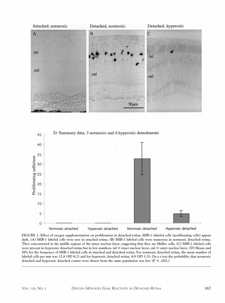
FIGURE 1. Effect of oxygen supplementation on proliferation in detached retina. MIB-1–labeled cells (proliferating cells) appeardark. (A) MIB-1 labeled cells were rare in attached retina. (B) MIB-1 labeled cells were numerous in normoxic detached retina.They concentrated in the middle regions of the inner nuclear layer, suggesting that they are Muller cells. (C) MIB-1 labeled cellswere present in hyperoxic detached retina but in low numbers. inl 5 inner nuclear layer, onl 5 outer nuclear layer. (D) Means andSDs for the frequency of MIB-1 labeled cells in attached and detached retina. For normoxic detached retina, the mean number oflabeled cells per mm was 32.8 (SD 8.2) and for hyperoxic detached retina, 4.9 (SD 1.5). On a t test the probability that normoxicdetached and hyperoxic detached counts were drawn from the same population was low (P < .002.)
OXYGEN MITIGATES GLIAL REACTIVITY IN DETACHED RETINAVOL. 128, NO. 2 167
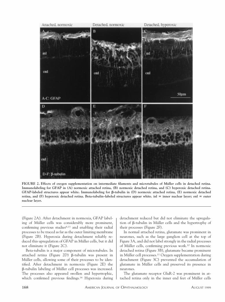
(Figure 2A). After detachment in normoxia, GFAP label-ing of Muller cells was considerably more prominent,confirming previous studies8,13 and enabling their radialprocesses to be traced as far as the outer limiting membrane(Figure 2B). Hyperoxia during detachment reliably re-duced this upregulation of GFAP in Muller cells, but it didnot eliminate it (Figure 2C).
Beta-tubulin is a major component of microtubules. Inattached retina (Figure 2D) b-tubulin was present inMuller cells, allowing some of their processes to be iden-tified. After detachment in normoxia (Figure 2E) theb-tubulin labeling of Muller cell processes was increased.The processes also appeared swollen and hypertrophic,which confirmed previous findings.14 Hyperoxia during
detachment reduced but did not eliminate the upregula-tion of b-tubulin in Muller cells and the hypertrophy oftheir processes (Figure 2F).
In normal attached retina, glutamate was prominent inneurones, such as the large ganglion cell at the top ofFigure 3A, and did not label strongly in the radial processesof Muller cells, confirming previous work.10 In normoxicdetached retina (Figure 3B), glutamate became prominentin Muller cell processes.11 Oxygen supplementation duringdetachment (Figure 3C) prevented the accumulation ofglutamate in Muller cells and preserved its presence inneurones.
The glutamate receptor GluR-2 was prominent in at-tached retina only in the inner end feet of Muller cells
FIGURE 2. Effects of oxygen supplementation on intermediate filaments and microtubules of Muller cells in detached retina.Immunolabeling for GFAP in (A) normoxic attached retina, (B) normoxic detached retina, and (C) hyperoxic detached retina.GFAP-labeled structures appear white. Immunolabeling for b-tubulin in (D) normoxic attached retina, (E) normoxic detachedretina, and (F) hyperoxic detached retina. Beta-tubulin–labeled structures appear white. inl 5 inner nuclear layer; onl 5 outernuclear layer.
AMERICAN JOURNAL OF OPHTHALMOLOGY168 AUGUST 1999

FIGURE 3. Effects of oxygen supplementation on glutamate neurochemistry in detached retina. Immunolabeling for glutamate in(A) normoxic attached retina, (B) normoxic detached retina, and (C) hyperoxic detached retina. Immunolabeling for the glutamatereceptor GluR-2 in (D) normoxic attached retina, (E) normoxic detached retina, and (F) hyperoxic detached retina. Immunolabelingfor glutamine synthetase in (G) normoxic attached retina, (H) normoxic detached retina, and (I) hyperoxic detached retina. inl 5inner nuclear layer; onl 5 outer nuclear layer.
OXYGEN MITIGATES GLIAL REACTIVITY IN DETACHED RETINAVOL. 128, NO. 2 169

(Figure 3D), but in detached retina that were kept innormoxia it was prominent as far outward as the outernuclear layer (Figure 3E). Hyperoxia during detachmentreduced but did not eliminate this upregulation of GluR-2(Figure 3F).
Detachment caused a reduction of the glutamine syn-thetase labeling in the inner processes of Muller cells(Figure 3, G and H), consistent with a previous report.13
This loss was prevented by hyperoxia during detachment(Figure 3I). In an unexpected response, glutamine syn-
FIGURE 4. Hypothesis of the genesis of retinopathy of detachment. The first three stages of genesis have a relatively strongempirical base.12 The sequence of events at stage 4 is more speculative. We have suggested that the glial responses to detachmentare mediated by signals from surrounding neurones as they undergo death or deconstruction. Although speculative, this suggestioncan be tested, which should clarify the interactions of glia and neurones in detachment retinopathy.
AMERICAN JOURNAL OF OPHTHALMOLOGY170 AUGUST 1999

thetase was upregulated in hyperoxic detached retina to alevel above those found in attached retina, either nor-moxic (Figure 3G) or hyperoxic (not shown).
DISCUSSION
THESE RESULTS PROVIDE EVIDENCE THAT OXYGEN SUPPLE-
mentation during retinal detachment reduces the prolifer-ation of Muller cells caused by detachment, reduces thehypertrophy of their processes, and stabilizes glutamateneurochemistry. These effects of oxygen supplementationconfirm that the glial reaction to detachment results partlyfrom hypoxia. They suggest that in humans the glialproliferation that sometimes follows retinal reattachmentmay be reduced in occurrence and severity if the patientreceives oxygen supplementation between diagnosis andsurgery.
It is striking that the reactions of retinal neurones andglia to detachment are diametrically opposed; neurones(photoreceptors) degenerate,12 whereas glia proliferate.Both responses are driven at least in part by hypoxia,suggesting that they are closely connected. Two furtherelements of the response of the retina to detachment, bothdriven by hypoxia, are suggested. One is the dispersal ofbFGF from its normal storage sites in both glia (astrocytesand Muller cells) and neurones, which has been de-scribed.12 Given the protective effect of bFGF on photo-receptors,15,16 this dispersal may be an important factor inphotoreceptor death. The second element is a disturbanceof the neurochemistry of glutamate, which involves theaccumulation of glutamate and a loss of glutamine syn-thetase activity in Muller cells.11 Recognition that thesevarious components of the retina’s reaction to detachmenthave a common cause in nutrient starvation leads to thesuggestion that the detached retina is undergoing a linkedseries of reactions that could be called the retinopathy ofdetachment.11
An important function of glial cells in the centralnervous system is the recycling of transmitters. Muller cellsare known to express glutamate receptors in the normallyfunctioning retina and to contain high levels of theenzyme glutamine synthetase, whose action is to transformglutamate to glutamine (which has no transmitter activity)and to make the glutamine available to neurones for theresynthesis of glutamate.17,18 Presumably, because theirglutamine synthetase activity is high, Muller cells do notnormally accumulate glutamate.10 In the detached retina,glutamine synthetase levels in Muller cells fall (Figure 3B),and GluR-2 labeling indicates that glutamate receptorlevels in Muller cells increase. Presumably as a result,glutamate levels rise in Muller cells. It is unclear whetherthese changes play a role in inducing photoreceptordegeneration in detached retina.
These results, in combination with a long series of
results from earlier studies,2,12,19 permit the proposal of atentative theory as to the genesis of the retinopathy ofdetachment (Figure 4).
● Detachment separates the photoreceptors from thechoriocapillaris, the source of their energy nutrients,inducing hypoxia and hypoglycemia in the outer layersof neural retina.
● Hypoxia and hypoglycemia induce the programmeddeath of some photoreceptors and the programmeddeconstruction of the survivors. Dying and damagedphotoreceptors release still unknown signals, whichcause the dispersal of bFGF from its normal storagesites, the proliferation and hypertrophy of Muller cellsto repair damage, and the downregulation of glutaminesynthetase activity in Muller cells, causing a redistri-bution of retinal glutamate and glutamate receptors.
● The dispersal of bFGF increases the vulnerability ofphotoreceptors to damage.
● The proliferation and hypertrophy of Muller cellscause gliosis of the outer limiting membrane, whichprevents re-establishment of normal relations betweenphotoreceptors and retinal pigment epithelial (RPE)cells when reattached.
The model in Figure 4 is specific to the neural retina. Afuller model of the retinopathy of detachment may need toinclude structures adjacent to the neural retina, especiallythe RPE.20
These results come from an experimental model thatdeserves further exploration to include longer periods ofdetachment and the effect of delay in supplementation.Clinical trials of oxygen supplementation in the treatmentof retinal detachment also deserve consideration.
There would appear to be few obstacles to such trials,other than those of case selection and the resourcesrequired for any such trial. Oxygen is toxic, and currentpractice in respiratory medicine would need to be incor-porated in any trial, including the limiting of oxygenexposure and the use of antioxidants. Current practice inrespiratory medicine is to limit the use of high oxygenlevels to a few days, whereas current ophthalmic practice isto repair attachments within 1 or a few days of diagnosis;these two constraints seem compatible.
The reaction of Muller cells to retinal detachment wasgreatly reduced by oxygen supplementation, as was theproliferation of Muller cells. Their neurochemical signa-tures were stabilized, suggesting that the Muller cellsmaintained their normal role in transmitter recycling andthat their hypertrophy was limited. In the treatment ofretinal detachment, oxygen supplementation between di-agnosis and reattachment surgery may reduce the inci-dence and severity of glial-based complications, such asproliferative vitreoretinopathy. Further experimental stud-ies and careful clinical trials will be necessary to test thispossibility.
OXYGEN MITIGATES GLIAL REACTIVITY IN DETACHED RETINAVOL. 128, NO. 2 171

ACKNOWLEDGMENT
The authors thank Tania Novikova for her skilled tech-nical assistance.
REFERENCES
1. Michels R, Wilkinson C, Rice T. Results of retinal reattach-ment surgery. In: Retinal detachment. Chapter 15. St Louis:CV Mosby 1990:917–938.
2. Fisher S, Anderson D. Cellular effects of detachment on theneural retina and the retinal pigment epithelium. In: GlaserBM, editor. Retina, Volume 3, Surgical retina. St Louis: CVMosby, 1989:165–190.
3. Guerin CJ, Wolfshagen RW, Eifrig DE, Anderson DH.Immunocytochemical identification of Muller’s glia as acomponent of human epiretinal membranes. Invest Ophthal-mol Vis Sci 1990;31:1483–1491.
4. Fisher S, Erickson P, Lewis G, Anderson D. Intraretinalproliferation induced by retinal detachment. Invest Ophthal-mol Vis Sci 1991;32:1739–1748.
5. Geller S, Lewis G, Anderson D, Fisher S. Use of the MIB-1antibody for detecting proliferating cells in the retina. InvestOphthalmol Vis Sci 1995;36:737–744.
6. Erickson P, Fisher S, Anderson D, Stern W, Borgula G.Retinal detachment in the cat: the outer nuclear and outerplexiform layers. Invest Ophthalmol Vis Sci 1983;24:927–942.
7. Anderson DH, Guerin CJ, Erickson PA, Stern WH, FisherSK. Morphological recovery in re-attached retina. InvestOphthalmol Vis Sci 1986;27:168–183.
8. Lewis GP, Guerin CJ, Anderson DH, Matsumoto B, FisherSK. Rapid changes in the expression of glial cell proteinscaused by experimental retinal detachment. Am J Ophthal-mol 1994;118:368–376.
9. Hale I, Fisher S, Matsumoto B. The actin network in the
ciliary stalk of photoreceptors functions in the generation ofnew outer segment discs. J Comp Neurol 1996;376:128–142.
10. Marc R, Murry R, Fisher S, Linberg K, Lewis G, KalloniatisM. Amino acid signatures in the normal cat retina. InvestOphthalmol Vis Sci 1998;39:1685–1693.
11. Marc RE, Murry RF, Fisher SK, Linberg KA, Lewis GP.Amino acid signatures in the detached cat retina. InvestOphthalmol Vis Sci 1998;39:1694–1702.
12. Mervin K, Valter K, Maslim J, Lewis G, Fisher S, Stone J.Limiting photoreceptor death and deconstruction duringexperimental retinal detachment: the value of oxygen sup-plementation. Am J Ophthalmol 1999;128:155–164.
13. Erickson P, Fisher S, Guerin C, Anderson D, Kaska D. Glialfibrillary acidic protein increases in Muller cells after retinaldetachment. Exp Eye Res 1987;44:37–48.
14. Lewis GP, Matsumoto B, Fisher SK. Changes in the organi-zation and expression of cytoskeletal proteins during retinaldegeneration induced by retinal detachment. Invest Oph-thalmol Vis Sci 1995;36:2404–2416.
15. Faktorovich EG, Steinberg RH, Yasumura D, Matthes MT,LaVail MM. Photoreceptor degeneration in inherited retinaldystrophy delayed by basic fibroblast growth factor. Nature1990;347:83–86.
16. Stone J, Maslim J, Valter-Kocsi K, et al. Mechanisms ofphotoreceptor death and survival in mammalian retina. ProgRet Eye Res. Forthcoming.
17. Moscona AA. On glutamine synthetase, carbonic anhydraseand Muller glia in the retina. Prog Ret Res 1983;2:111–135.
18. Pow D, Robinson S. Glutamate in some retinal neurons isderived solely from glia. Neuroscience 1994;60:355–366.
19. Lewis G, Linberg K, Fisher S. Neurite outgrowth from bipolarand horizontal cells after experimental retinal detachment.Invest Ophthalmol Vis Sci 1998;39:424–434.
20. Glaser B, Lemor M. Pathobiology of proliferative vitreoreti-nopathy. In: Ryan SJ, editor. Retina. St Louis: Mosby-YearBook, 1989:369–383.
Authors InteractivetWe encourage questions and comments regarding this article via the Interneton Authors Interactivet at http://www.ajo.com/ Questions, comments, andauthor responses are posted.
AMERICAN JOURNAL OF OPHTHALMOLOGY172 AUGUST 1999