Light-Emitting Diodes (LEDs) in Dermatology -...
12
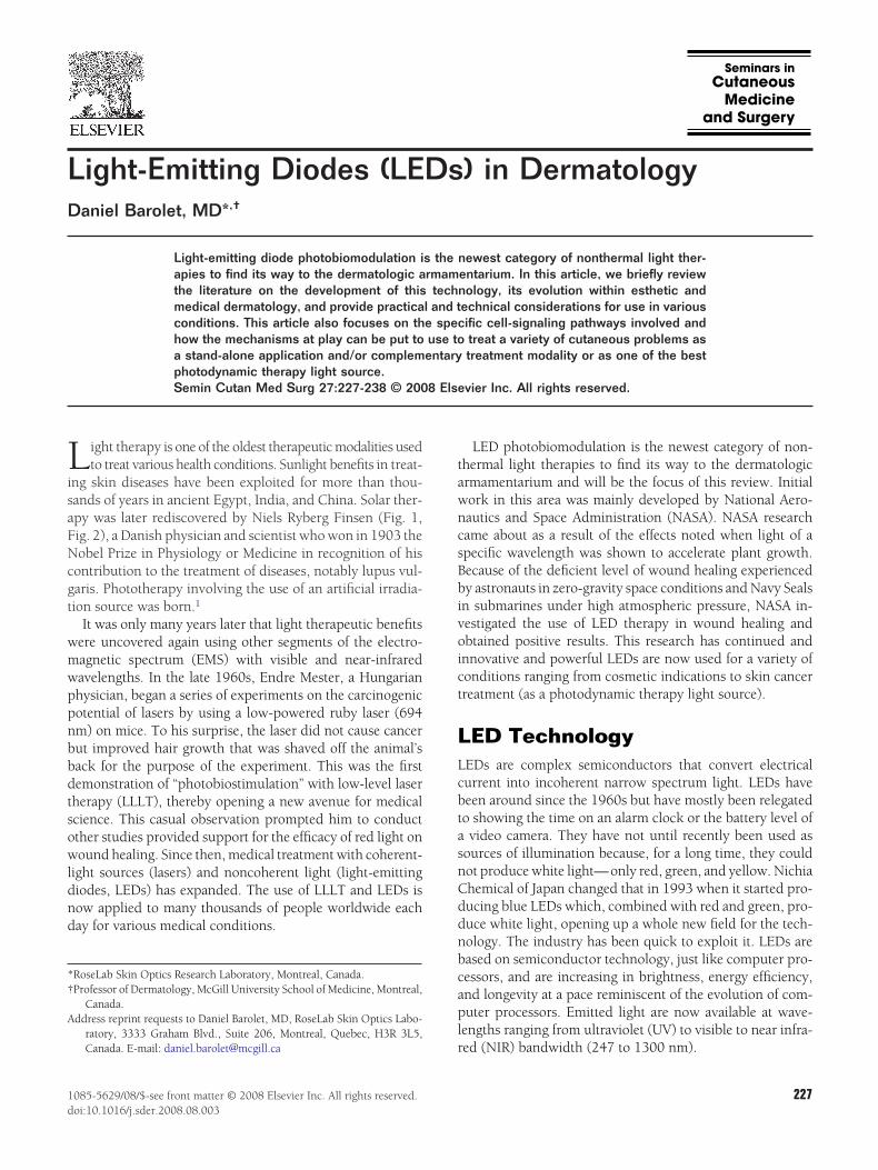
Light-Emitting Diodes (LEDs) in Dermatology Daniel Barolet, MD* ,† Light-emitting diode photobiomodulation is the newest category of nonthermal light ther- apies to find its way to the dermatologic armamentarium. In this article, we briefly review the literature on the development of this technology, its evolution within esthetic and medical dermatology, and provide practical and technical considerations for use in various conditions. This article also focuses on the specific cell-signaling pathways involved and how the mechanisms at play can be put to use to treat a variety of cutaneous problems as a stand-alone application and/or complementary treatment modality or as one of the best photodynamic therapy light source. Semin Cutan Med Surg 27:227-238 © 2008 Elsevier Inc. All rights reserved. L ight therapy is one of the oldest therapeutic modalities used to treat various health conditions. Sunlight benefits in treat- ing skin diseases have been exploited for more than thou- sands of years in ancient Egypt, India, and China. Solar ther- apy was later rediscovered by Niels Ryberg Finsen (Fig. 1, Fig. 2), a Danish physician and scientist who won in 1903 the Nobel Prize in Physiology or Medicine in recognition of his contribution to the treatment of diseases, notably lupus vul- garis. Phototherapy involving the use of an artificial irradia- tion source was born. 1 It was only many years later that light therapeutic benefits were uncovered again using other segments of the electro- magnetic spectrum (EMS) with visible and near-infrared wavelengths. In the late 1960s, Endre Mester, a Hungarian physician, began a series of experiments on the carcinogenic potential of lasers by using a low-powered ruby laser (694 nm) on mice. To his surprise, the laser did not cause cancer but improved hair growth that was shaved off the animal’s back for the purpose of the experiment. This was the first demonstration of “photobiostimulation” with low-level laser therapy (LLLT), thereby opening a new avenue for medical science. This casual observation prompted him to conduct other studies provided support for the efficacy of red light on wound healing. Since then, medical treatment with coherent- light sources (lasers) and noncoherent light (light-emitting diodes, LEDs) has expanded. The use of LLLT and LEDs is now applied to many thousands of people worldwide each day for various medical conditions. LED photobiomodulation is the newest category of non- thermal light therapies to find its way to the dermatologic armamentarium and will be the focus of this review. Initial work in this area was mainly developed by National Aero- nautics and Space Administration (NASA). NASA research came about as a result of the effects noted when light of a specific wavelength was shown to accelerate plant growth. Because of the deficient level of wound healing experienced by astronauts in zero-gravity space conditions and Navy Seals in submarines under high atmospheric pressure, NASA in- vestigated the use of LED therapy in wound healing and obtained positive results. This research has continued and innovative and powerful LEDs are now used for a variety of conditions ranging from cosmetic indications to skin cancer treatment (as a photodynamic therapy light source). LED Technology LEDs are complex semiconductors that convert electrical current into incoherent narrow spectrum light. LEDs have been around since the 1960s but have mostly been relegated to showing the time on an alarm clock or the battery level of a video camera. They have not until recently been used as sources of illumination because, for a long time, they could not produce white light— only red, green, and yellow. Nichia Chemical of Japan changed that in 1993 when it started pro- ducing blue LEDs which, combined with red and green, pro- duce white light, opening up a whole new field for the tech- nology. The industry has been quick to exploit it. LEDs are based on semiconductor technology, just like computer pro- cessors, and are increasing in brightness, energy efficiency, and longevity at a pace reminiscent of the evolution of com- puter processors. Emitted light are now available at wave- lengths ranging from ultraviolet (UV) to visible to near infra- red (NIR) bandwidth (247 to 1300 nm). *RoseLab Skin Optics Research Laboratory, Montreal, Canada. †Professor of Dermatology, McGill University School of Medicine, Montreal, Canada. Address reprint requests to Daniel Barolet, MD, RoseLab Skin Optics Labo- ratory, 3333 Graham Blvd., Suite 206, Montreal, Quebec, H3R 3L5, Canada. E-mail: [email protected] 227 1085-5629/08/$-see front matter © 2008 Elsevier Inc. All rights reserved. doi:10.1016/j.sder.2008.08.003
Transcript of Light-Emitting Diodes (LEDs) in Dermatology -...
LD
LisaFNcgt
wmwppnbbdtsowldnd
*†
A
1d
ight-Emitting Diodes (LEDs) in Dermatologyaniel Barolet, MD*,†
Light-emitting diode photobiomodulation is the newest category of nonthermal light ther-apies to find its way to the dermatologic armamentarium. In this article, we briefly reviewthe literature on the development of this technology, its evolution within esthetic andmedical dermatology, and provide practical and technical considerations for use in variousconditions. This article also focuses on the specific cell-signaling pathways involved andhow the mechanisms at play can be put to use to treat a variety of cutaneous problems asa stand-alone application and/or complementary treatment modality or as one of the bestphotodynamic therapy light source.Semin Cutan Med Surg 27:227-238 © 2008 Elsevier Inc. All rights reserved.
tawncsBbivoict
LLcbtasnCddnbcapl
ight therapy is one of the oldest therapeutic modalities usedto treat various health conditions. Sunlight benefits in treat-
ng skin diseases have been exploited for more than thou-ands of years in ancient Egypt, India, and China. Solar ther-py was later rediscovered by Niels Ryberg Finsen (Fig. 1,ig. 2), a Danish physician and scientist who won in 1903 theobel Prize in Physiology or Medicine in recognition of his
ontribution to the treatment of diseases, notably lupus vul-aris. Phototherapy involving the use of an artificial irradia-ion source was born.1
It was only many years later that light therapeutic benefitsere uncovered again using other segments of the electro-agnetic spectrum (EMS) with visible and near-infraredavelengths. In the late 1960s, Endre Mester, a Hungarianhysician, began a series of experiments on the carcinogenicotential of lasers by using a low-powered ruby laser (694m) on mice. To his surprise, the laser did not cause cancerut improved hair growth that was shaved off the animal’sack for the purpose of the experiment. This was the firstemonstration of “photobiostimulation” with low-level laserherapy (LLLT), thereby opening a new avenue for medicalcience. This casual observation prompted him to conductther studies provided support for the efficacy of red light onound healing. Since then, medical treatment with coherent-
ight sources (lasers) and noncoherent light (light-emittingiodes, LEDs) has expanded. The use of LLLT and LEDs isow applied to many thousands of people worldwide eachay for various medical conditions.
RoseLab Skin Optics Research Laboratory, Montreal, Canada.Professor of Dermatology, McGill University School of Medicine, Montreal,
Canada.ddress reprint requests to Daniel Barolet, MD, RoseLab Skin Optics Labo-
ratory, 3333 Graham Blvd., Suite 206, Montreal, Quebec, H3R 3L5,
rCanada. E-mail: [email protected]085-5629/08/$-see front matter © 2008 Elsevier Inc. All rights reserved.oi:10.1016/j.sder.2008.08.003
LED photobiomodulation is the newest category of non-hermal light therapies to find its way to the dermatologicrmamentarium and will be the focus of this review. Initialork in this area was mainly developed by National Aero-autics and Space Administration (NASA). NASA researchame about as a result of the effects noted when light of apecific wavelength was shown to accelerate plant growth.ecause of the deficient level of wound healing experiencedy astronauts in zero-gravity space conditions and Navy Seals
n submarines under high atmospheric pressure, NASA in-estigated the use of LED therapy in wound healing andbtained positive results. This research has continued andnnovative and powerful LEDs are now used for a variety ofonditions ranging from cosmetic indications to skin cancerreatment (as a photodynamic therapy light source).
ED TechnologyEDs are complex semiconductors that convert electricalurrent into incoherent narrow spectrum light. LEDs haveeen around since the 1960s but have mostly been relegatedo showing the time on an alarm clock or the battery level ofvideo camera. They have not until recently been used as
ources of illumination because, for a long time, they couldot produce white light—only red, green, and yellow. Nichiahemical of Japan changed that in 1993 when it started pro-ucing blue LEDs which, combined with red and green, pro-uce white light, opening up a whole new field for the tech-ology. The industry has been quick to exploit it. LEDs areased on semiconductor technology, just like computer pro-essors, and are increasing in brightness, energy efficiency,nd longevity at a pace reminiscent of the evolution of com-uter processors. Emitted light are now available at wave-
engths ranging from ultraviolet (UV) to visible to near infra-
ed (NIR) bandwidth (247 to 1300 nm).227
oba(sffpBfcrTabf
fdNmpc
tTwmpLndsblaag
MIibs
FdC
Filpu1
FC
228 D. Barolet
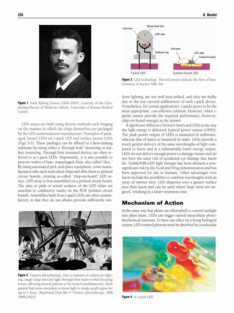
LED arrays are built using diverse methods each hingingn the manner in which the chips themselves are packagedy the LED semiconductor manufacturer. Examples of pack-ged, lensed LEDs are t-pack LED and surface mount LEDsFigs 3-5). These packages can be affixed to a heat-sinkingubstrate by using either a “through hole” mounting or sur-ace mounting. Through hole mounted devices are often re-erred to as t-pack LEDs. Importantly, it is also possible torocure wafers of bare, unpackaged chips, also called “dice.”y using automated pick-and-place equipment, some manu-acturers take such individual chips and affix them to printedircuit boards, creating so-called “chip-on-board” LED ar-ays. LED array is thus assembled on a printed circuit board.he pins or pads or actual surfaces of the LED chips arettached to conductive tracks on the PCB (printed circuitoard). Assemblies built from t-pack LEDs are often unsatis-actory in that they do not always provide sufficiently uni-
igure 1 Niels Ryberg Finsen (1860-1904). Courtesy of the Clen-ening History of Medicine Library, University of Kansas Medicalenter.
igure 2 Finsen’s phototherapy. Due to expense of carbon arc light-ng, single lamp directed light through four water-cooled focusingenses, allowing several patients to be treated simultaneously. Eachatient had nurse attendant to focus light to single small region forp to 1 hour. (Reprinted from Bie V: Finsen’s phototherapy. BMJ
899;2:825) Form lighting, are not well heat-sinked, and they are bulkyue to the size (several millimeters) of each t-pack device.onetheless, for certain applications, t-packs prove to be theost appropriate, cost-effective solution. However, when t-acks cannot provide the required performance, however,hip-on-board emerges as the answer.
A significant difference between lasers and LEDs is the wayhe light energy is delivered [optical power output (OPD)].he peak power output of LEDs is measured in milliwatts,hereas that of lasers is measured in watts. LEDs provide auch gentler delivery of the same wavelengths of light com-ared to lasers and at a substantially lower energy output.EDs do not deliver enough power to damage tissues and doot have the same risk of accidental eye damage that laserso. Visible/NIR-LED light therapy has been deemed a non-ignificant risk by the Food and Drug Administration and haseen approved for use in humans. Other advantages over
asers include the possibility to combine wavelengths with anrray of various sizes. LED disperses over a greater surfacerea than lasers and can be used where large areas are tar-eted, resulting in a faster treatment time.
echanism of Actionn the same way that plants use chlorophyll to convert sunlightnto plant tissue, LEDs can trigger natural intracellular photo-iochemical reactions. To have any effect on a living biologicalystem, LED-emitted photons must be absorbed by a molecular
igure 3 LED technology. The red arrows indicate the flow of heat.ourtesy of Stocker Yale, Inc.
igure 4 A t-pack LED.
cwfld
ldopectyNrcc(op
achgtnrclsatWfreficu
sfpaac
LgcpLmglotttfa
tcLecdcff
OIedt(tpftt
WPLnmWa(lsd
F
LEDs in dermatology 229
hromophore or photoacceptor. Light, at appropriate doses andavelengths, is absorbed by chromophores such as porphyrins,avins, and other light-absorbing entities within the mitochon-ria and cell membranes of cells.A growing body of evidence suggests that photobiomodu-
ation mechanism is ascribed to the activation of mitochon-rial respiratory chain components resulting in the initiationf a cascade of cellular reactions. It has been postulated thathotoacceptors in the red to NIR region are the terminalnzyme of the respiratory chain cytochrome c oxidase with 2opper elements. The first absorption peak is in the red spec-rum and the second peak in the NIR range. Seventy-fiveears ago, Otto Warburg, a German biochemist, was given aobel prize for his ingenious work unmasking the enzyme
esponsible for the critical steps of cell respiration, especiallyytochrome oxidase governing the last reaction in this pro-ess. Two chemical quirks are exploited: carbon monoxideCO) that can block respiration by binding to cytochromexidase in place of oxygen, and a flash of light that can dis-lace it, allowing oxygen to bind again.Nowadays, it has been reported that cells often use CO
nd, to an even greater extent, nitric oxide (NO) binding toytochrome oxidase to hinder cell respiration.2 Mitochondriaarbor an enzyme that synthesizes NO. So why would cellso out of their way to produce NO right next to the respira-ory enzymes? Evolution crafted cytochrome oxidase to bindot only to oxygen but also to NO. One effect of slowingespiration in some locations is to divert oxygen elsewhere inells and tissues, preventing oxygen sinking to dangerouslyow levels. Fireflies use a similar strategy to flash light (seeection “Pulsing and Continuous Modes”). Respiration isbout generating energy but also about generating feedbackhat allows a cell to monitor and respond to its environment.
hen respiration is blocked, chemical signals in the form ofree radicals or reactive oxygen species are generated. Freeadicals had a bad reputation, but now they can be consid-red signals. The activity of many proteins, or transcriptionactors, depends, at least in part, on free radicals.3 Thesenclude many proteins such as those involved in the p53ell-signaling pathway. Further, to bring free radical leak
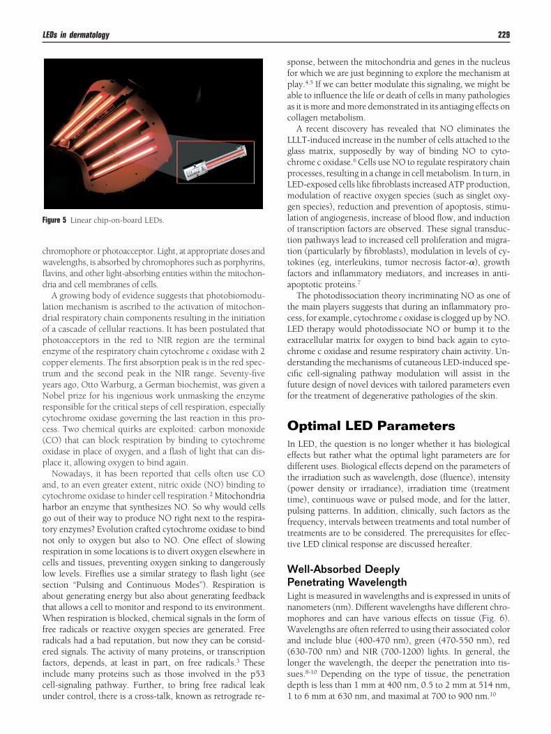
igure 5 Linear chip-on-board LEDs.
nder control, there is a cross-talk, known as retrograde re- 1
ponse, between the mitochondria and genes in the nucleusor which we are just beginning to explore the mechanism atlay.4,5 If we can better modulate this signaling, we might beble to influence the life or death of cells in many pathologiess it is more and more demonstrated in its antiaging effects onollagen metabolism.
A recent discovery has revealed that NO eliminates theLLT-induced increase in the number of cells attached to thelass matrix, supposedly by way of binding NO to cyto-hrome c oxidase.6 Cells use NO to regulate respiratory chainrocesses, resulting in a change in cell metabolism. In turn, inED-exposed cells like fibroblasts increased ATP production,odulation of reactive oxygen species (such as singlet oxy-
en species), reduction and prevention of apoptosis, stimu-ation of angiogenesis, increase of blood flow, and inductionf transcription factors are observed. These signal transduc-ion pathways lead to increased cell proliferation and migra-ion (particularly by fibroblasts), modulation in levels of cy-okines (eg, interleukins, tumor necrosis factor-�), growthactors and inflammatory mediators, and increases in anti-poptotic proteins.7
The photodissociation theory incriminating NO as one ofhe main players suggests that during an inflammatory pro-ess, for example, cytochrome c oxidase is clogged up by NO.ED therapy would photodissociate NO or bump it to thextracellular matrix for oxygen to bind back again to cyto-hrome c oxidase and resume respiratory chain activity. Un-erstanding the mechanisms of cutaneous LED-induced spe-ific cell-signaling pathway modulation will assist in theuture design of novel devices with tailored parameters evenor the treatment of degenerative pathologies of the skin.
ptimal LED Parametersn LED, the question is no longer whether it has biologicalffects but rather what the optimal light parameters are forifferent uses. Biological effects depend on the parameters ofhe irradiation such as wavelength, dose (fluence), intensitypower density or irradiance), irradiation time (treatmentime), continuous wave or pulsed mode, and for the latter,ulsing patterns. In addition, clinically, such factors as therequency, intervals between treatments and total number ofreatments are to be considered. The prerequisites for effec-ive LED clinical response are discussed hereafter.
ell-Absorbed Deeplyenetrating Wavelengthight is measured in wavelengths and is expressed in units ofanometers (nm). Different wavelengths have different chro-ophores and can have various effects on tissue (Fig. 6).avelengths are often referred to using their associated color
nd include blue (400-470 nm), green (470-550 nm), red630-700 nm) and NIR (700-1200) lights. In general, theonger the wavelength, the deeper the penetration into tis-ues.8-10 Depending on the type of tissue, the penetrationepth is less than 1 mm at 400 nm, 0.5 to 2 mm at 514 nm,
to 6 mm at 630 nm, and maximal at 700 to 900 nm.10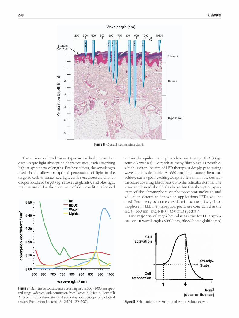
olutdm
wawwatwtwumr
c
l pene
FtAt
230 D. Barolet
The various cell and tissue types in the body have theirwn unique light absorption characteristics, each absorbingight at specific wavelengths. For best effects, the wavelengthsed should allow for optimal penetration of light in theargeted cells or tissue. Red light can be used successfully foreeper localized target (eg, sebaceous glands), and blue lightay be useful for the treatment of skin conditions located
Figure 6 Optica
igure 7 Main tissue constituents absorbing in the 600–1000 nm spec-ral range. Adapted with permission from Taroni P, Pifferi A, Torricelli, et al: In vivo absorption and scattering spectroscopy of biological
issues. Photochem Photobio Sci 2:124-129, 2003. F
ithin the epidermis in photodynamic therapy (PDT) (eg,ctinic keratoses). To reach as many fibroblasts as possible,hich is often the aim of LED therapy, a deeply penetratingavelength is desirable. At 660 nm, for instance, light can
chieve such a goal reaching a depth of 2.3 mm in the dermis,herefore covering fibroblasts up to the reticular dermis. Theavelength used should also be within the absorption spec-
rum of the chromophore or photoacceptor molecule andill often determine for which applications LEDs will besed. Because cytochrome c oxidase is the most likely chro-ophore in LLLT, 2 absorption peaks are considered in the
ed (�660 nm) and NIR (�850 nm) spectra.6
Two major wavelength boundaries exist for LED appli-ations: at wavelengths �600 nm, blood hemoglobin (Hb)
tration depth.
igure 8 Schematic representation of Arndt-Schulz curve.
ivapmta
FTdrdaactsisHL
mttitwt
eesin
ss
weit
PBabptffloqhC
dfitethech
aLnasodm
PVemAtibtpLl(
TTirh
F
LEDs in dermatology 231
s a major obstacle to photon absorption because bloodessels are not compressed during treatment. Futhermore,t wavelengths �1000 nm, water is also absorbing manyhotons, reducing their availability for specific chro-ophores located, for instance, in dermal fibroblasts. Be-
ween these 2 boundaries, there is a valley of LED possiblepplications (see Fig. 7).
luence and Irradiancehe Arndt-Schulz law states that there is only a narrow win-ow of opportunity where you can actually activate a cellularesponse using precise sets of parameters, i.e. the fluence orose (see Fig. 8). The challenge remains to find the appropri-te combinations of LED treatment time and irradiance tochieve optimal target tissue effects. Fluence or dose is, indi-ated in joules per cm2 (J/cm2). The law of reciprocity stateshat the dose is equal to the intensity � time. Therefore, theame exposure should result from reducing duration andncreasing light intensity, and vice versa. Reciprocity is as-umed and routinely used in LED and LLLT experiments.owever, the scientific evidence supporting reciprocity inED therapy is unclear.11
Dose reciprocity effects were examined in a wound healingodel and showed that varying irradiance and exposure time
o achieve a constant specified energy density affects LEDherapy outcomes.12 In practice, if light intensity (irradiance)s lower than the physiological threshold value for a givenarget, it does not produce photostimulatory effects evenhen irradiation time is extended. Moreover, photoinhibi-
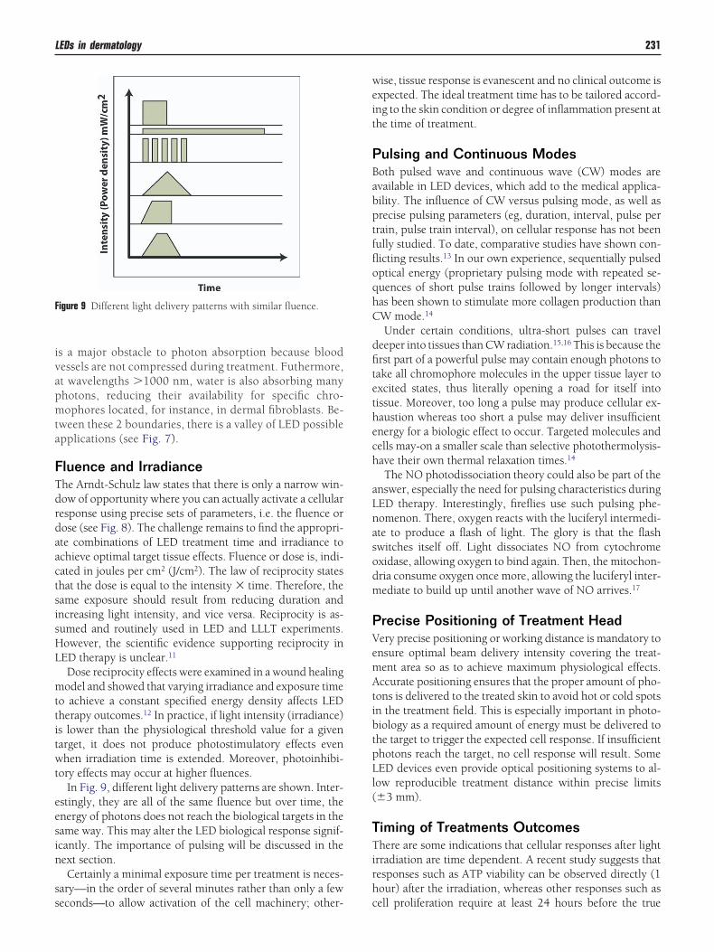
ory effects may occur at higher fluences.In Fig. 9, different light delivery patterns are shown. Inter-
stingly, they are all of the same fluence but over time, thenergy of photons does not reach the biological targets in theame way. This may alter the LED biological response signif-cantly. The importance of pulsing will be discussed in theext section.Certainly a minimal exposure time per treatment is neces-
ary—in the order of several minutes rather than only a few
igure 9 Different light delivery patterns with similar fluence.
econds—to allow activation of the cell machinery; other- c
ise, tissue response is evanescent and no clinical outcome isxpected. The ideal treatment time has to be tailored accord-ng to the skin condition or degree of inflammation present athe time of treatment.
ulsing and Continuous Modesoth pulsed wave and continuous wave (CW) modes arevailable in LED devices, which add to the medical applica-ility. The influence of CW versus pulsing mode, as well asrecise pulsing parameters (eg, duration, interval, pulse perrain, pulse train interval), on cellular response has not beenully studied. To date, comparative studies have shown con-icting results.13 In our own experience, sequentially pulsedptical energy (proprietary pulsing mode with repeated se-uences of short pulse trains followed by longer intervals)as been shown to stimulate more collagen production thanW mode.14
Under certain conditions, ultra-short pulses can traveleeper into tissues than CW radiation.15,16 This is because therst part of a powerful pulse may contain enough photons toake all chromophore molecules in the upper tissue layer toxcited states, thus literally opening a road for itself intoissue. Moreover, too long a pulse may produce cellular ex-austion whereas too short a pulse may deliver insufficientnergy for a biologic effect to occur. Targeted molecules andells may-on a smaller scale than selective photothermolysis-ave their own thermal relaxation times.14
The NO photodissociation theory could also be part of thenswer, especially the need for pulsing characteristics duringED therapy. Interestingly, fireflies use such pulsing phe-omenon. There, oxygen reacts with the luciferyl intermedi-te to produce a flash of light. The glory is that the flashwitches itself off. Light dissociates NO from cytochromexidase, allowing oxygen to bind again. Then, the mitochon-ria consume oxygen once more, allowing the luciferyl inter-ediate to build up until another wave of NO arrives.17
recise Positioning of Treatment Headery precise positioning or working distance is mandatory tonsure optimal beam delivery intensity covering the treat-ent area so as to achieve maximum physiological effects.ccurate positioning ensures that the proper amount of pho-
ons is delivered to the treated skin to avoid hot or cold spotsn the treatment field. This is especially important in photo-iology as a required amount of energy must be delivered tohe target to trigger the expected cell response. If insufficienthotons reach the target, no cell response will result. SomeED devices even provide optical positioning systems to al-
ow reproducible treatment distance within precise limits�3 mm).
iming of Treatments Outcomeshere are some indications that cellular responses after light
rradiation are time dependent. A recent study suggests thatesponses such as ATP viability can be observed directly (1our) after the irradiation, whereas other responses such as
ell proliferation require at least 24 hours before the true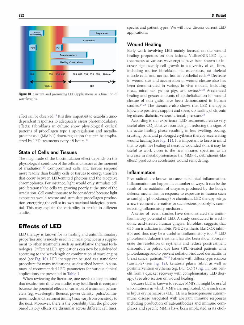
edepps
STpomtcpietts
ELpmnaupma
tbeuto
sa
WEhtcimibthcskl
utcwtuie
IFIrdaat
fld6ipedpb(pea
iimi
Fw
232 D. Barolet
ffect can be observed.18 It is thus important to establish time-ependent responses to adequately assess photomodulatoryffects. Fibroblasts in culture show physiological cyclicalatterns of procollagen type I up-regulation and metallo-roteinase-1 (MMP-1) down-regulation that can be empha-ized by LED treatments every 48 hours.19
tate of Cells and Tissueshe magnitude of the biostimulation effect depends on thehysiological condition of the cells and tissues at the momentf irradiation.20 Compromised cells and tissues respondore readily than healthy cells or tissues to energy transfers
hat occur between LED-emitted photons and the receptivehromophores. For instance, light would only stimulate cellroliferation if the cells are growing poorly at the time of the
rradiation. Cell conditions are to be considered because lightxposures would restore and stimulate procollagen produc-ion, energizing the cell to its own maximal biological poten-ial. This may explain the variability in results in differenttudies.
ffects of LEDED therapy is known for its healing and antiinflammatoryroperties and is mostly used in clinical practice as a supple-ent to other treatments such as nonablative thermal tech-ologies. Different LED applications can now be subdividedccording to the wavelength or combination of wavelengthssed (see Fig. 10). LED therapy can be used as a standalonerocedure for many indications, as described herein. A sum-ary of recommended LED parameters for various clinical
pplications are presented in Table 1.When reviewing the literature, one needs to keep in mind
hat results from different studies may be difficult to compareecause the potential effects of variation of treatment param-ters (eg, wavelength, fluence, power density, pulse/contin-ous mode and treatment timing) may vary from one study tohe next. Moreover, there is the possibility that the photobi-
igure 10 Current and promising LED applications as a function ofavelengths.
modulatory effects are dissimilar across different cell lines, p
pecies and patient types. We will now discuss current LEDpplications.
ound Healingarly work involving LED mainly focused on the woundealing properties on skin lesions. Visible/NIR-LED lightreatments at various wavelengths have been shown to in-rease significantly cell growth in a diversity of cell lines,ncluding murine fibroblasts, rat osteoblasts, rat skeletal
uscle cells, and normal human epithelial cells.21 Decreasen wound size and acceleration of wound closure also haseen demonstrated in various in vivo models, includingoads, mice, rats, guinea pigs, and swine.22,23 Acceleratedealing and greater amounts of epithelialization for woundlosure of skin grafts have been demonstrated in humantudies.24,25 The literature also shows that LED therapy isnown to positively support and speed up healing of chronic
eg ulcers: diabetic, venous, arterial, pressure.26
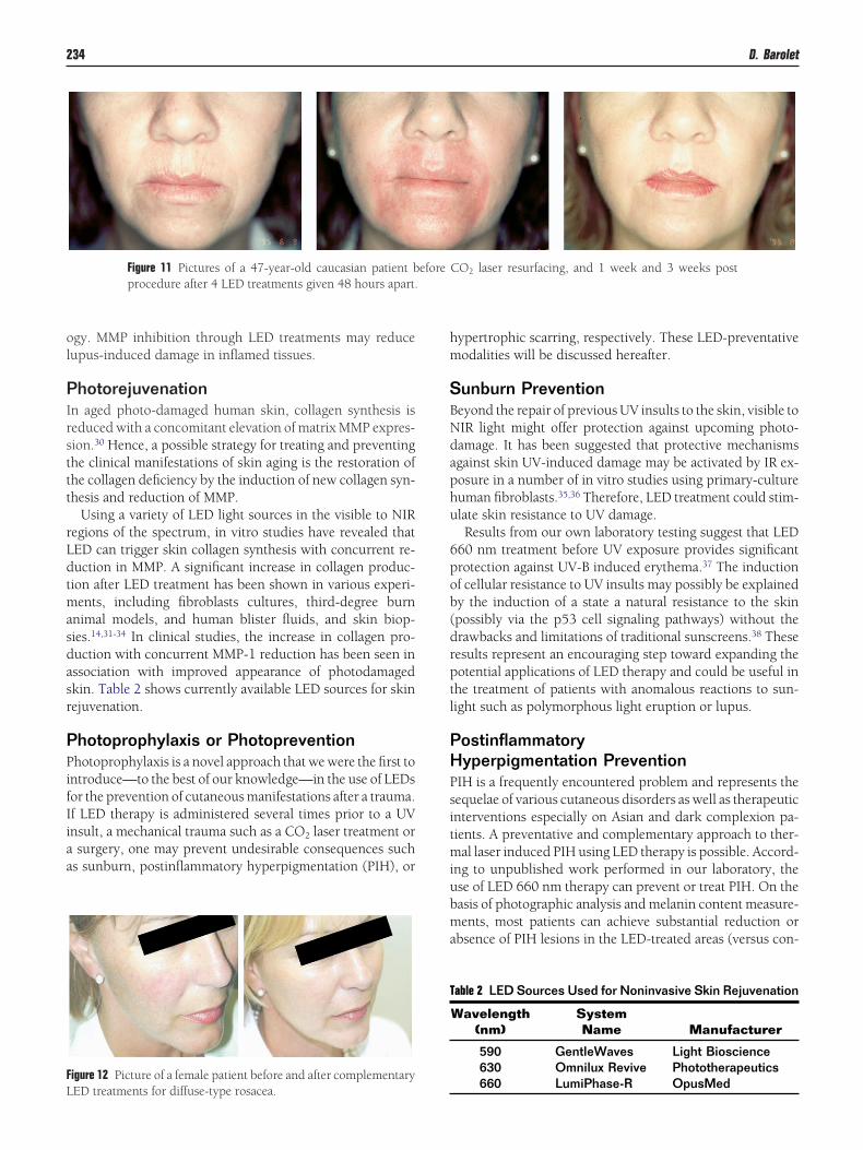
According to our experience, LED treatments are also veryseful after CO2 ablative resurfacing in reducing the signs ofhe acute healing phase resulting in less swelling, oozing,rusting, pain, and prolonged erythema thereby acceleratingound healing (see Fig. 11). It is important to keep in mind
hat to optimize healing of necrotic wounded skin, it may beseful to work closer to the near infrared spectrum as an
ncrease in metalloproteinases (ie, MMP-1, debridment-likeffect) production accelerates wound remodeling.
nflammationree radicals are known to cause subclinical inflammation.nflammation can happen in a number of ways. It can be theesult of the oxidation of enzymes produced by the body’sefense mechanism in response to exposure to trauma suchs sunlight (photodamage) or chemicals. LED therapy bringsnew treatment alternative for such lesions possibly by coun-
eracting inflammatory mediators.A series of recent studies have demonstrated the antiin-
ammatory potential of LED. A study conducted in arachi-onic acid-treated human gingival fibroblast suggests that35 nm irradiation inhibits PGE 2 synthesis like COX inhib-
tor and thus may be a useful antiinflammatory tool.27 LEDhotobiomodulation treatment has also been shown to accel-rate the resolution of erythema and reduce posttreatmentiscomfort in pulsed dye laser (IPL)-treated patients withhotodamage and to prevent radiation-induced dermatitis inreast cancer patients.28,29 Patients with diffuse type rosaceaunstable) (see Fig. 12), keratosis pilaris rubra, as well asostintervention erythema (eg, IPL, CO2) (Fig. 11) can ben-fit from a quicker recovery with complementary LED ther-py. (See also section on wound healing).
Because LED is known to reduce MMPs, it might be usefuln conditions in which MMPs are implicated. One such cases lupus erythematosus (LE). LE is a heterogeneous autoim-
une disease associated with aberrant immune responsesncluding production of autoantibodies and immune com-
lexes and specific MMPs have been implicated in its etiol-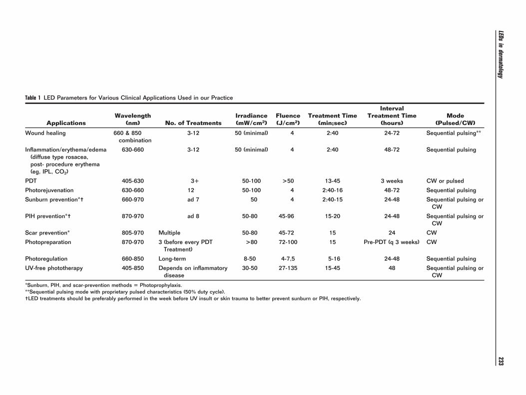
Table 1 LED Parameters for Various Clinical Applications Used in our Practice
ApplicationsWavelength
(nm) No. of TreatmentsIrradiance(mW/cm2)
Fluence(J/cm2)
Treatment Time(min;sec)
IntervalTreatment Time
(hours)Mode
(Pulsed/CW)
Wound healing 660 & 850combination
3-12 50 (minimal) 4 2:40 24-72 Sequential pulsing**
Inflammation/erythema/edema(diffuse type rosacea,post- procedure erythema(eg, IPL, CO2)
630-660 3-12 50 (minimal) 4 2:40 48-72 Sequential pulsing
PDT 405-630 3� 50-100 >50 13-45 3 weeks CW or pulsed
Photorejuvenation 630-660 12 50-100 4 2:40-16 48-72 Sequential pulsing
Sunburn prevention*† 660-970 ad 7 50 4 2:40-15 24-48 Sequential pulsing orCW
PIH prevention*† 870-970 ad 8 50-80 45-96 15-20 24-48 Sequential pulsing orCW
Scar prevention* 805-970 Multiple 50-80 45-72 15 24 CW
Photopreparation 870-970 3 (before every PDTTreatment)
>80 72-100 15 Pre-PDT (q 3 weeks) CW
Photoregulation 660-850 Long-term 8-50 4-7,5 5-16 24-48 Sequential pulsing
UV-free phototherapy 405-850 Depends on inflammatorydisease
30-50 27-135 15-45 48 Sequential pulsing orCW
*Sunburn, PIH, and scar-prevention methods � Photoprophylaxis.**Sequential pulsing mode with proprietary pulsed characteristics (50% duty cycle).†LED treatments should be preferably performed in the week before UV insult or skin trauma to better prevent sunburn or PIH, respectively.
LEDsin
dermatology
233
ol
PIrsttt
rLdtmasdasr
PPifIiaa
hm
SBNdaphu
6pob(drptl
PHPsitmiubma
T
FL
234 D. Barolet
gy. MMP inhibition through LED treatments may reduceupus-induced damage in inflamed tissues.
hotorejuvenationn aged photo-damaged human skin, collagen synthesis iseduced with a concomitant elevation of matrix MMP expres-ion.30 Hence, a possible strategy for treating and preventinghe clinical manifestations of skin aging is the restoration ofhe collagen deficiency by the induction of new collagen syn-hesis and reduction of MMP.
Using a variety of LED light sources in the visible to NIRegions of the spectrum, in vitro studies have revealed thatED can trigger skin collagen synthesis with concurrent re-uction in MMP. A significant increase in collagen produc-ion after LED treatment has been shown in various experi-ents, including fibroblasts cultures, third-degree burn
nimal models, and human blister fluids, and skin biop-ies.14,31-34 In clinical studies, the increase in collagen pro-uction with concurrent MMP-1 reduction has been seen inssociation with improved appearance of photodamagedkin. Table 2 shows currently available LED sources for skinejuvenation.
hotoprophylaxis or Photopreventionhotoprophylaxis is a novel approach that we were the first to
ntroduce—to the best of our knowledge—in the use of LEDsor the prevention of cutaneous manifestations after a trauma.f LED therapy is administered several times prior to a UVnsult, a mechanical trauma such as a CO2 laser treatment orsurgery, one may prevent undesirable consequences such
s sunburn, postinflammatory hyperpigmentation (PIH), or
Figure 11 Pictures of a 47-year-old caucasian patient bprocedure after 4 LED treatments given 48 hours apart.
igure 12 Picture of a female patient before and after complementary
ED treatments for diffuse-type rosacea.ypertrophic scarring, respectively. These LED-preventativeodalities will be discussed hereafter.
unburn Preventioneyond the repair of previous UV insults to the skin, visible toIR light might offer protection against upcoming photo-amage. It has been suggested that protective mechanismsgainst skin UV-induced damage may be activated by IR ex-osure in a number of in vitro studies using primary-cultureuman fibroblasts.35,36 Therefore, LED treatment could stim-late skin resistance to UV damage.Results from our own laboratory testing suggest that LED
60 nm treatment before UV exposure provides significantrotection against UV-B induced erythema.37 The inductionf cellular resistance to UV insults may possibly be explainedy the induction of a state a natural resistance to the skinpossibly via the p53 cell signaling pathways) without therawbacks and limitations of traditional sunscreens.38 Theseesults represent an encouraging step toward expanding theotential applications of LED therapy and could be useful inhe treatment of patients with anomalous reactions to sun-ight such as polymorphous light eruption or lupus.
ostinflammatoryyperpigmentation Prevention
IH is a frequently encountered problem and represents theequelae of various cutaneous disorders as well as therapeuticnterventions especially on Asian and dark complexion pa-ients. A preventative and complementary approach to ther-al laser induced PIH using LED therapy is possible. Accord-
ng to unpublished work performed in our laboratory, these of LED 660 nm therapy can prevent or treat PIH. On theasis of photographic analysis and melanin content measure-ents, most patients can achieve substantial reduction or
bsence of PIH lesions in the LED-treated areas (versus con-
able 2 LED Sources Used for Noninvasive Skin Rejuvenation
Wavelength(nm)
SystemName Manufacturer
590 GentleWaves Light Bioscience630 Omnilux Revive Phototherapeutics
CO2 laser resurfacing, and 1 week and 3 weeks post
efore660 LumiPhase-R OpusMed
t1lissit
SHtet
iirppfdists
PPwtptple
(armi
F1s
FPlp
Fdcb
LEDs in dermatology 235
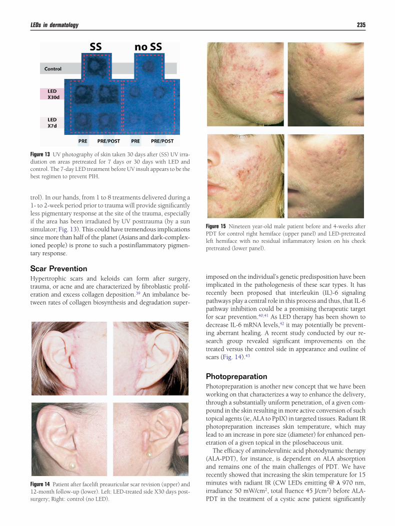
rol). In our hands, from 1 to 8 treatments delivered during a- to 2-week period prior to trauma will provide significantly
ess pigmentary response at the site of the trauma, especiallyf the area has been irradiated by UV posttrauma (by a sunimulator; Fig. 13). This could have tremendous implicationsince more than half of the planet (Asians and dark-complex-oned people) is prone to such a postinflammatory pigmen-ary response.
car Preventionypertrophic scars and keloids can form after surgery,
rauma, or acne and are characterized by fibroblastic prolif-ration and excess collagen deposition.39 An imbalance be-ween rates of collagen biosynthesis and degradation super-
igure 14 Patient after facelift preauricular scar revision (upper) and2-month follow-up (lower). Left: LED-treated side X30 days post-
igure 13 UV photography of skin taken 30 days after (SS) UV irra-iation on areas pretreated for 7 days or 30 days with LED andontrol. The 7-day LED treatment before UV insult appears to be theest regimen to prevent PIH.
Purgery; Right: control (no LED).
mposed on the individual’s genetic predisposition have beenmplicated in the pathologenesis of these scar types. It hasecently been proposed that interleukin (IL)-6 signalingathways play a central role in this process and thus, that IL-6athway inhibition could be a promising therapeutic targetor scar prevention.40,41 As LED therapy has been shown toecrease IL-6 mRNA levels,42 it may potentially be prevent-
ng aberrant healing. A recent study conducted by our re-earch group revealed significant improvements on thereated versus the control side in appearance and outline ofcars (Fig. 14).43
hotopreparationhotopreparation is another new concept that we have beenorking on that characterizes a way to enhance the delivery,
hrough a substantially uniform penetration, of a given com-ound in the skin resulting in more active conversion of suchopical agents (ie, ALA to PpIX) in targeted tissues. Radiant IRhotopreparation increases skin temperature, which may
ead to an increase in pore size (diameter) for enhanced pen-tration of a given topical in the pilosebaceous unit.
The efficacy of aminolevulinic acid photodynamic therapyALA-PDT), for instance, is dependent on ALA absorptionnd remains one of the main challenges of PDT. We haveecently showed that increasing the skin temperature for 15inutes with radiant IR (CW LEDs emitting @ � 970 nm,
rradiance 50 mW/cm2, total fluence 45 J/cm2) before ALA-
igure 15 Nineteen year-old male patient before and 4-weeks afterDT for control right hemiface (upper panel) and LED-pretreated
eft hemiface with no residual inflammatory lesion on his cheekretreated (lower panel).
DT in the treatment of a cystic acne patient significantly
dt
PPtabatsoiihtd
UUmssmte
cwskuwh
aptssertr
PPpttpebPrlborbd(pbde—tib
Ft
T
W
P
W
T
P
236 D. Barolet
ecreased the number of cystic lesions in comparison withhe non IR-heated side (Fig. 15).44
hotoregulationhotoregulation involves an exciting new 2-level (impor-ance of dermal–epidermal communication via cytokines)pproach that we have evaluated with success to enhance theiological effects of a given topical. The main goal of thispplication would be to synergistically optimize any bioac-ive compound trajectory/route to ultimately up-regulatepecific gene expression with simultaneous down-regulationf undesired ones via cell signaling pathways. In the estheticsndustry, we believe such a method—even though still in itsnfancy—will become applicable in such applications asome-use skin rejuvenation and the treatment of inflamma-ory acne, hyperpigmentation disorders, oily skin, hyperhi-rosis, eczema, etc.
V-Free PhototherapyV radiation phototherapy has been used for decades in theanagement of common skin diseases.45 However, there are
ide effects associated with UV deleterious effects as well aseveral contra-indications, including the long-term manage-ent of children and young adults and patients receiving
opical or systemic immunosuppressive drugs. The primaryffectors of UV phototherapy in the treatment of various skin
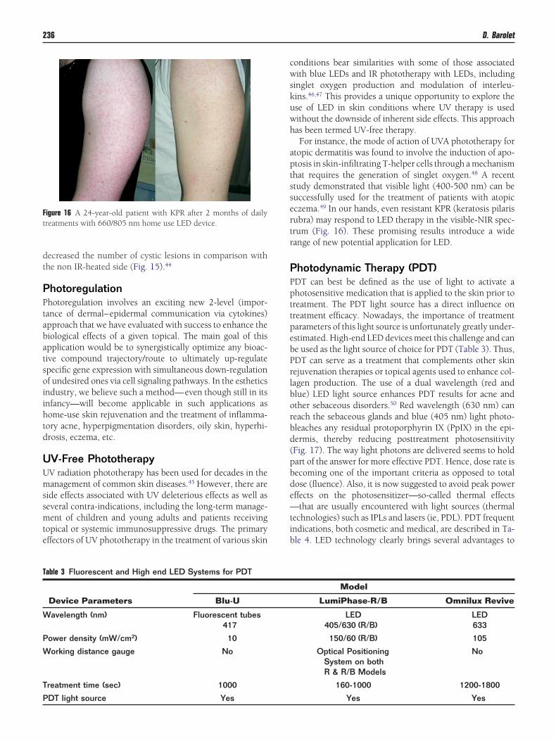
igure 16 A 24-year-old patient with KPR after 2 months of dailyreatments with 660/805 nm home use LED device.
able 3 Fluorescent and High end LED Systems for PDT
Device Parameters Blu-U
avelength (nm) Fluorescent tubes417
ower density (mW/cm2) 10
orking distance gauge No
reatment time (sec) 1000
DT light source Yes
onditions bear similarities with some of those associatedith blue LEDs and IR phototherapy with LEDs, including
inglet oxygen production and modulation of interleu-ins.46,47 This provides a unique opportunity to explore these of LED in skin conditions where UV therapy is usedithout the downside of inherent side effects. This approachas been termed UV-free therapy.For instance, the mode of action of UVA phototherapy for
topic dermatitis was found to involve the induction of apo-tosis in skin-infiltrating T-helper cells through a mechanismhat requires the generation of singlet oxygen.48 A recenttudy demonstrated that visible light (400-500 nm) can beuccessfully used for the treatment of patients with atopicczema.49 In our hands, even resistant KPR (keratosis pilarisubra) may respond to LED therapy in the visible-NIR spec-rum (Fig. 16). These promising results introduce a wideange of new potential application for LED.
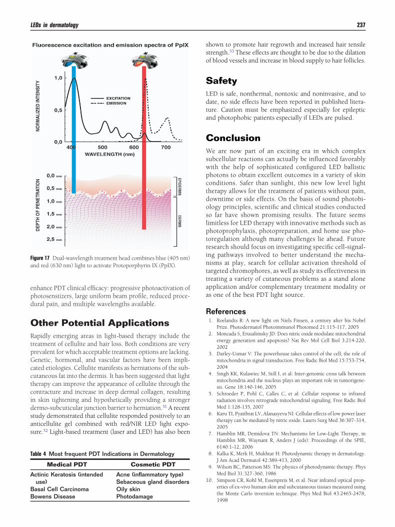
hotodynamic Therapy (PDT)DT can best be defined as the use of light to activate ahotosensitive medication that is applied to the skin prior toreatment. The PDT light source has a direct influence onreatment efficacy. Nowadays, the importance of treatmentarameters of this light source is unfortunately greatly under-stimated. High-end LED devices meet this challenge and cane used as the light source of choice for PDT (Table 3). Thus,DT can serve as a treatment that complements other skinejuvenation therapies or topical agents used to enhance col-agen production. The use of a dual wavelength (red andlue) LED light source enhances PDT results for acne andther sebaceous disorders.50 Red wavelength (630 nm) caneach the sebaceous glands and blue (405 nm) light photo-leaches any residual protoporphyrin IX (PpIX) in the epi-ermis, thereby reducing posttreatment photosensitivityFig. 17). The way light photons are delivered seems to holdart of the answer for more effective PDT. Hence, dose rate isecoming one of the important criteria as opposed to totalose (fluence). Also, it is now suggested to avoid peak powerffects on the photosensitizer—so-called thermal effects
that are usually encountered with light sources (thermalechnologies) such as IPLs and lasers (ie, PDL). PDT frequentndications, both cosmetic and medical, are described in Ta-le 4. LED technology clearly brings several advantages to
Model
LumiPhase-R/B Omnilux Revive
LED LED405/630 (R/B) 633
150/60 (R/B) 105
Optical PositioningSystem on bothR & R/B Models
No
160-1000 1200-1800
Yes Yes
epd
ORtpGcctcidsas
sso
SLdta
CWswpctdoslptrinttaa
R
1
Fa
T
A
BB
LEDs in dermatology 237
nhance PDT clinical efficacy: progressive photoactivation ofhotosenstizers, large uniform beam profile, reduced proce-ural pain, and multiple wavelengths available.
ther Potential Applicationsapidly emerging areas in light-based therapy include the
reatment of cellulite and hair loss. Both conditions are veryrevalent for which acceptable treatment options are lacking.enetic, hormonal, and vascular factors have been impli-ated etiologies. Cellulite manifests as herniations of the sub-utaneous fat into the dermis. It has been suggested that lightherapy can improve the appearance of cellulite through theontracture and increase in deep dermal collagen, resultingn skin tightening and hypothetically providing a strongerermo-subcuticular junction barrier to herniation.51 A recenttudy demonstrated that cellulite responded positively to annticellulite gel combined with red/NIR LED light expo-ure.52 Light-based treatment (laser and LED) has also been
igure 17 Dual-wavelength treatment head combines blue (405 nm)nd red (630 nm) light to activate Protoporphyrin IX (PpIX).
able 4 Most frequent PDT Indications in Dermatology
Medical PDT Cosmetic PDT
ctinic Keratosis (intendeduse)
Acne (inflammatory type)Sebaceous gland disorders
asal Cell Carcinoma Oily skin
owens Disease Photodamagehown to promote hair regrowth and increased hair tensiletrength.53 These effects are thought to be due to the dilationf blood vessels and increase in blood supply to hair follicles.
afetyED is safe, nonthermal, nontoxic and noninvasive, and toate, no side effects have been reported in published litera-ure. Caution must be emphasized especially for epilepticnd photophobic patients especially if LEDs are pulsed.
onclusione are now part of an exciting era in which complex
ubcellular reactions can actually be influenced favorablyith the help of sophisticated configured LED ballistichotons to obtain excellent outcomes in a variety of skinonditions. Safer than sunlight, this new low level lightherapy allows for the treatment of patients without pain,owntime or side effects. On the basis of sound photobi-logy principles, scientific and clinical studies conductedo far have shown promising results. The future seemsimitless for LED therapy with innovative methods such ashotoprophylaxis, photopreparation, and home use pho-oregulation although many challenges lie ahead. Futureesearch should focus on investigating specific cell-signal-ng pathways involved to better understand the mecha-isms at play, search for cellular activation threshold ofargeted chromophores, as well as study its effectiveness inreating a variety of cutaneous problems as a stand alonepplication and/or complementary treatment modality ors one of the best PDT light source.
eferences1. Roelandts R: A new light on Niels Finsen, a century after his Nobel
Prize. Photodermatol Photoimmunol Photomed 21:115-117, 20052. Moncada S, Erusalimsky JD: Does nitric oxide modulate mitochondrial
energy generation and apoptosis? Nat Rev Mol Cell Biol 3:214-220,2002
3. Darley-Usmar V: The powerhouse takes control of the cell; the role ofmitochondria in signal transduction. Free Radic Biol Med 15:753-754,2004
4. Singh KK, Kulawiec M, Still I, et al: Inter-genomic cross talk betweenmitochondria and the nucleus plays an important role in tumorigene-sis. Gene 18:140-146, 2005
5. Schroeder P, Pohl C, Calles C, et al: Cellular response to infraredradiation involves retrograde mitochondrial signaling. Free Radic BiolMed 1:128-135, 2007
6. Karu TI, Pyatibrat LV, Afanasyeva NI: Cellular effects of low power lasertherapy can be mediated by nitric oxide. Lasers Surg Med 36:307-314,2005
7. Hamblin MR, Demidova TN: Mechanisms for Low-Light Therapy, inHamblin MR, Waynant R, Anders J (eds): Proceedings of the SPIE,6140:1-12, 2006
8. Kalka K, Merk H, Mukhtar H: Photodynamic therapy in dermatology.J Am Acad Dermatol 42:389-413, 2000
9. Wilson BC, Patterson MS: The physics of photodynamic therapy. PhysMed Biol 31:327-360, 1986
0. Simpson CR, Kohl M, Essenpreis M, et al. Near infrared optical prop-erties of ex-vivo human skin and subcutaneous tissues measured usingthe Monte Carlo inversion technique. Phys Med Biol 43:2465-2478,
1998
1
1
1
1
1
1
1
1
1
2
2
2
2
2
2
2
2
2
2
3
3
3
3
3
3
3
3
3
3
4
4
4
4
4
4
4
4
4
4
5
5
5
5
238 D. Barolet
1. Sommer AP, Pinheiro AL, Mester AR, et al: Biostimulatory windows inlow-intensity laser activation: lasers, scanners, and NASA’s light-emit-ting diode array system. J Clin Laser Med Surg 19:29-33, 2001
2. Lanzafame RJ, Stadler I, Kurtz AF, et al: Reciprocity of exposure timeand irradiance on energy density during photoradiation on woundhealing in a murine pressure ulcer model. Lasers Surg Med 39:534-542, 2007
3. Al-Watban FA: The comparison of effects between pulsed and CWlasers on wound healing. J Clin Laser Med Surg 22:15-18, 2004
4. Barolet D, Boucher A, Bjerring P: In vivo human dermal collagen pro-duction following LED-based therapy: The importance of treatmentparameters. Lasers Surg Med 17:76, 2005 (suppl) (abstr)
5. Pogue BW, Lilge L, Patterson MS, et al: Absorbed photodynamic dosefrom pulsed versus continuous wave light examined with tissue-simu-lating dosimeters. Appl Opt 36:7257-7269, 1997
6. Sterenborg HJ, van Gemert MJ: Photodynamic therapy with pulsedlight sources: A theoretical analysis. Phys Med Biol 41:835-49, 1996
7. Trimmer BA, Aprille JR, Dudzinski DM, et al: Nitric oxide and thecontrol of firefly flashing. Science 29:2486-2488, 2001
8. Hawkins DH, Abrahamse H: Time-dependent responses of woundedhuman skin fibroblasts following phototherapy. J Photochem Photo-biol B 25:147-155, 2007
9. Barolet D, Roberge C, Germain L, et al: Rythid improvement by non-ablative, non-thermal LED photoinduction: In vitro and in vivo aspects,Lasers Surg Med 16:75, 2004 (suppl) (abstr)
0. Karu TI, Pyatibrat LV, Kalendo GS: Photobiological modulation of cellattachment via cytochrome c oxidase. Photochem Photobiol Sci 3:211-216, 2004
1. Whelan HT, Smits RL Jr., Buchman EV, et al: Effect of NASA light-emitting diode irradiation on wound healing. J Clin Laser Med Surg19:305-314, 2001
2. Bibikova A, Oron U: Regeneration in denervated toad (Bufo viridis)gastrocnemius muscle and the promotion of the process by low energylaser irradiation. Anat Rec 241:123-128, 1995
3. Al-Watban FA: Laser acceleration of open skin wound closure in ratsand its dosimetric dependence. Lasers Life Sci 7:237-247, 1997
4. Conlan MJ, Rapley JW, Cobb CM: Biostimulation of wound healing bylow-energy laser irradiation. J Clin Periodontol 23:492-496, 1996
5. Miller M, Truhe T: Lasers in dentistry: An overview. Am Dent Assoc124:32-35, 1993
6. Whelan HT, Buchmann EV, Whelan NT, et al: NASA light emittingdiode medical applications: From deep space to deep sea. Space Tech-nology and Applications International Forum, p. 35-45, 2001
7. Lim W, Lee S, Kim I, et al: The anti-inflammatory mechanism of 635nm light-emitting-diode irradiation compared with existing COX in-hibitors. Lasers Surg Med 39:614-621, 2007
8. Khoury JG, Goldman MP: Use of light-emitting diode photomodula-tion to reduce erythema and discomfort after intense pulsed light treat-ment of photodamage. J Cosmet Dermatol 7:30-34, 2008
9. DeLand MM, Weiss RA, McDaniel DH, et al: Treatment of radiation-induced dermatitis with light-emitting diode (LED) photomodulation.Lasers Surg Med 39:164-168, 2007
0. Fisher GJ, Kang S, Varani J, et al: Mechanisms of photoaging andchronological skin aging. Arch Dermatol 138:1462-1470, 2002
1. McDaniel DH, Weiss RA, Geronemus R, et al: Light-tissue interactionsI: Photothermolysis vs photomodulation laboratory findings. LasersSurg Med 14:25, 2002 (abstr)
2. Meirelles GC, Santos JN, Chagas PO, et al: A comparative study of theeffects of laser photobiomodulation on the healing of third-degreeburns: A histological study in rats. Photomed Laser Surg 26:159-166,
20083. Weiss RA, McDaniel DH, Geronemus R, et al: Clinical trial of a novelnon-thermal LED array for reversal of photoaging: Clinical, histologic,and surface profilometric results. Lasers Surg Med 36:85-91, 2005
4. Lee SY, Park KH, Choi JW, et al: A prospective, randomized, placebo-controlled, double-blinded, and split-face clinical study on LED pho-totherapy for skin rejuvenation: Clinical, profilometric, histologic, ul-trastructural, and biochemical evaluations and comparison of threedifferent treatment settings. J Photochem Photobiol B 27:51-67, 2007
5. Menezes S, Coulomb B, Lebreton C, et al: Non-coherent near infraredradiation protects normal human dermal fibroblasts from solar ultravi-olet toxicity. J Invest Dermatol 111:629-633, 1998
6. Frank S, Oliver L, Lebreton-De Coster C, et al: Infrared radiation affectsthe mitochondrial pathway of apoptosis in human fibroblasts. J InvestDermatol 123:823-831, 2004
7. Barolet D, Boucher A: LED Photoprevention: Reduced MED responsefollowing multiple LED exposures. Lasers Surg Med 40:106-112, 2008
8. Frank S, Menezes S, Lebreton-De Coster C, et al: Infrared radiationinduces the p53 signaling pathway: Role in infrared prevention of ul-traviolet B toxicity. Exp Dermatol 15:130-137, 2006
9. Uitto J, Kouba D: Cytokine modulation of extracellular matrix geneexpression: Relevance to fibrotic skin diseases. J Dermatol Sci 24:S60-69, 2000 (suppl)
0. Uitto J: IL-6 signaling pathway in keloids: a target for pharmacologicintervention? J Invest Dermatol 127:6-8, 2007
1. Ghazizadeh M, Tosa M, Shimizu H, et al: Functional implications of theIL-6 signaling pathway in keloid pathogenesis. J Invest Dermatol 127:98-110, 2007
2. Lee SY, Park KH, Choi JW, et al: A prospective, randomized, placebo-controlled, double-blinded, and split-face clinical study on LED pho-totherapy for skin rejuvenation: Clinical, profilometric, histologic, ul-trastructural, and biochemical evaluations and comparison of threedifferent treatment settings. J Photochem Photobiol B 27:51-67, 2007
3. Barolet D, Boucher A: LED therapy for the prevention of post-surgicalhypertrophic scars and keloids, Lasers Surg Med 20:97, 2008 (suppl)(abstr)
4. Barolet D, Boucher A: Pre-PDT use of radiant IR LED exposure as skinpreparation to enhance cystic acne treatment outcome, Lasers SurgMed 20:73, 2008 (suppl) (abstr)
5. Krutmann J, Hönigsmann H, Elmets CA, et al: Dermatological Photo-therapy and Photodiagnostic Methods. New York, Springer, 2001
6. Morita A, Werfel T, Stege H, et al: Evidence that singlet oxygen-inducedhuman T helper cell apoptosis is the basic mechanism of ultraviolet-Aradiation phototherapy. J Exp Med 17:1763-1768, 1997
7. Kramer M, Sachsenmaier C, Herrlich P, et al: UV irradiation-inducedinterleukin-1 and basic fibroblast growth factor synthesis and releasemediate part of the UV response. J Biol Chem 268:6734-6741, 1993
8. Morita A, Werfel T, Stege H, et al: Evidence that singlet oxygen-inducedhuman T helper cell apoptosis is the basic mechanism of ultraviolet-Aradiation phototherapy. J Exp Med 186:1763-1768, 1997
9. Krutmann J, Medve-Koenigs K, Ruzicka T, et al: Ultraviolet-free pho-totherapy. Photodermatol Photoimmunol Photomed 21:59-61, 2005
0. Barolet D, Boucher A: Dual wavelength high power LEDs enhance PDTacne treatment. Lasers Surg 19:37, 2007 (suppl) (abstr)
1. Alexiades-Armenakas M: Laser and light-based treatment of cellulite. JDrugs Dermatol 6:83-84, 2007
2. Sasaki GH, Oberg K, Tucker B, et al: The effectiveness and safety oftopical PhotoActif phosphatidylcholine-based anti-cellulite gel andLED (red and near-infrared) light on Grade II-III thigh cellulite: Arandomized, double-blinded study. J Cosmet Laser Ther 9:87-96, 2007
3. Satino JL, Markou M: Hair regrowth and increased hair tensile strengthusing the HairMax LeserComb for Low-level laser therapy. Int J Cosm
Surg and Aesthetic Dermatol 5:113-117, 2003