Light and melatonin schedule neuronal differentiation in the habenular nuclei
11
Light and melatonin schedule neuronal differentiation in the habenular nuclei Nancy Hernandez de Borsetti 1, 2 , Benjamin J. Dean 2 , Emily J. Bain, Joshua A. Clanton, Robert W. Taylor, Joshua T. Gamse ⁎ Vanderbilt University, Department of Biological Sciences, Nashville, TN 37235, USA abstract article info Article history: Received for publication 11 January 2011 Revised 26 July 2011 Accepted 28 July 2011 Available online 5 August 2011 Keywords: Epithalamus Photoperiod Circadian rhythm Neuropil Zebrafish Neurogenesis Pineal organ The formation of the embryonic brain requires the production, migration, and differentiation of neurons to be timely and coordinated. Coupling to the photoperiod could synchronize the development of neurons in the embryo. Here, we consider the effect of light and melatonin on the differentiation of embryonic neurons in zebrafish. We examine the formation of neurons in the habenular nuclei, a paired structure found near the dorsal surface of the brain adjacent to the pineal organ. Keeping embryos in constant darkness causes a temporary accumulation of habenular precursor cells, resulting in late differentiation and a long-lasting reduction in neuronal processes (neuropil). Because constant darkness delays the accumulation of the neurendocrine hormone melatonin in embryos, we looked for a link between melatonin signaling and habenular neurogenesis. A pharmacological block of melatonin receptors delays neurogenesis and reduces neuropil similarly to constant darkness, while addition of melatonin to embryos in constant darkness restores timely neurogenesis and neuropil. We conclude that light and melatonin schedule the differentiation of neurons and the formation of neural processes in the habenular nuclei. © 2011 Elsevier Inc. All rights reserved. Introduction The light–dark cycle synchronizes the circadian clock of organisms with their environment (Vallone et al., 2007). In zebrafish, light can be perceived not only by the eyes and pineal gland (photoreceptive organs) but, uniquely among model vertebrates, also by other organs and cultured cells (Kaneko et al., 2006; Tamai et al., 2004, 2007; Whitmore et al., 1998, 2000). Light has been shown to initiate mole- cular oscillations in the zebrafish embryo (Dekens and Whitmore, 2008; Kazimi and Cahill, 1999; Vatine et al., 2009; Vuilleumier et al., 2006) and affect the timing of the cell cycle (Dekens et al., 2003), as well as modulate predator avoidance behavior in zebrafish larvae (Budaev and Andrew, 2009). However, the consequences of light on neurogenesis have only recently begun to be characterized (D'Autilia et al., 2010; Dulcis and Spitzer, 2008; Toyama et al., 2009). Melatonin acts as a marker of photoperiod in vertebrates, regu- lating both daily and seasonal behavior in adults via receptors found in specific brain regions (Pandi-Perumal et al., 2008). In the zebrafish pineal organ, melatonin is synthesized from serotonin by a series of enzymes including arylalkylamine-N-acetyltransferase (aanat2). Transcription of aanat2 is cyclic, with peaks during the night and troughs during the day. Under conditions of alternating light:dark (L:D) periods, aanat2 is expressed by 22 hours post fertilization (hpf) in zebrafish embryos (Gothilf et al., 1999; Zilberman-Peled et al., 2007) and robust, cyclic melatonin production can be detected by 37 hpf (Kazimi and Cahill, 1999). This rhythmic expression depends on the synchronization of oscillations so that aanat2 expression is in phase in all pineal cells. The oscillators are synchronized by Period-2 (Per2), a transcriptional repressor induced by light in cells of the zebrafish pineal organ. In the absence of Per2 activity due to constant darkness, aanat2 expression and melatonin production reach a constant, intermediate level (Kazimi and Cahill, 1999; Ziv et al., 2005). Melatonin receptors are present at high levels in the embryonic brain (Rivkees and Reppert, 1991; Seron-Ferre et al., 2007), and in mammals, melatonin can be transferred to the developing fetus via the placenta (Klein, 1972) and to the newborn via milk (Reppert and Klein, 1978). Low melatonin synthesis due to mutation of the biosynthetic enzyme acetylserotonin O-methyltransferase (ASMT) has been linked to autism spectrum disorders (Melke et al., 2008). Melatonin treatment of mammalian neural stem cells induces their differentiation (Bellon et al., 2007; Kong et al., 2008; Moriya et al., 2007). Finally, melatonin stimulates increased cell division in zebrafish embryos (Danilova et al., 2004). Therefore, a link between light stimulation, gene expression and melatonin exists during early development, but its influence on neurogenesis is not well understood. In order to investigate the effects of light and melatonin on neuro- genesis, we examined the development of the habenular nuclei. These are a pair of brain nuclei that are adjacent to the pineal organ and make up part of the highly conserved dorsal diencephalic conduction Developmental Biology 358 (2011) 251–261 ⁎ Corresponding author at: VU Station B, Box 35-1634, Nashville, TN 37235-1634, USA. Fax: +1 615 343 0651. E-mail address: [email protected] (J.T. Gamse). 1 Current address: Universidad Nacional de Jujuy (UNJU), San Salvador de Jujuy, Jujuy, 4600, Argentina. 2 These authors contributed equally to this work. 0012-1606/$ – see front matter © 2011 Elsevier Inc. All rights reserved. doi:10.1016/j.ydbio.2011.07.038 Contents lists available at ScienceDirect Developmental Biology journal homepage: www.elsevier.com/developmentalbiology
-
Upload
nancy-hernandez-de-borsetti -
Category
Documents
-
view
215 -
download
2
Transcript of Light and melatonin schedule neuronal differentiation in the habenular nuclei
Developmental Biology 358 (2011) 251–261
Contents lists available at ScienceDirect
Developmental Biology
j ourna l homepage: www.e lsev ie r.com/deve lopmenta lb io logy
Light and melatonin schedule neuronal differentiation in the habenular nuclei
Nancy Hernandez de Borsetti 1,2, Benjamin J. Dean 2, Emily J. Bain, Joshua A. Clanton,Robert W. Taylor, Joshua T. Gamse ⁎Vanderbilt University, Department of Biological Sciences, Nashville, TN 37235, USA
⁎ Corresponding author at: VU Station B, Box 35-16USA. Fax: +1 615 343 0651.
E-mail address: [email protected] (J.T. Gam1 Current address: Universidad Nacional de Jujuy (U
Jujuy, 4600, Argentina.2 These authors contributed equally to this work.
0012-1606/$ – see front matter © 2011 Elsevier Inc. Aldoi:10.1016/j.ydbio.2011.07.038
a b s t r a c t
a r t i c l e i n f oArticle history:Received for publication 11 January 2011Revised 26 July 2011Accepted 28 July 2011Available online 5 August 2011
Keywords:EpithalamusPhotoperiodCircadian rhythmNeuropilZebrafishNeurogenesisPineal organ
The formation of the embryonic brain requires the production, migration, and differentiation of neurons to betimely and coordinated. Coupling to the photoperiod could synchronize the development of neurons in theembryo. Here, we consider the effect of light and melatonin on the differentiation of embryonic neurons inzebrafish. We examine the formation of neurons in the habenular nuclei, a paired structure found near thedorsal surface of the brain adjacent to the pineal organ. Keeping embryos in constant darkness causes atemporary accumulation of habenular precursor cells, resulting in late differentiation and a long-lastingreduction in neuronal processes (neuropil). Because constant darkness delays the accumulation of theneurendocrine hormone melatonin in embryos, we looked for a link between melatonin signaling andhabenular neurogenesis. A pharmacological block of melatonin receptors delays neurogenesis and reducesneuropil similarly to constant darkness, while addition of melatonin to embryos in constant darkness restorestimely neurogenesis and neuropil. We conclude that light and melatonin schedule the differentiation ofneurons and the formation of neural processes in the habenular nuclei.
34, Nashville, TN 37235-1634,
se).NJU), San Salvador de Jujuy,
l rights reserved.
© 2011 Elsevier Inc. All rights reserved.
Introduction
The light–dark cycle synchronizes the circadian clock of organismswith their environment (Vallone et al., 2007). In zebrafish, light can beperceived not only by the eyes and pineal gland (photoreceptiveorgans) but, uniquely among model vertebrates, also by other organsand cultured cells (Kaneko et al., 2006; Tamai et al., 2004, 2007;Whitmore et al., 1998, 2000). Light has been shown to initiate mole-cular oscillations in the zebrafish embryo (Dekens and Whitmore,2008; Kazimi and Cahill, 1999; Vatine et al., 2009; Vuilleumier et al.,2006) and affect the timing of the cell cycle (Dekens et al., 2003), aswell as modulate predator avoidance behavior in zebrafish larvae(Budaev and Andrew, 2009). However, the consequences of light onneurogenesis have only recently begun to be characterized (D'Autiliaet al., 2010; Dulcis and Spitzer, 2008; Toyama et al., 2009).
Melatonin acts as a marker of photoperiod in vertebrates, regu-lating both daily and seasonal behavior in adults via receptors foundin specific brain regions (Pandi-Perumal et al., 2008). In the zebrafishpineal organ, melatonin is synthesized from serotonin by a series ofenzymes including arylalkylamine-N-acetyltransferase (aanat2).Transcription of aanat2 is cyclic, with peaks during the night and
troughs during the day. Under conditions of alternating light:dark(L:D) periods, aanat2 is expressed by 22 hours post fertilization(hpf) in zebrafish embryos (Gothilf et al., 1999; Zilberman-Peledet al., 2007) and robust, cyclic melatonin production can be detectedby 37 hpf (Kazimi and Cahill, 1999). This rhythmic expression dependson the synchronization of oscillations so that aanat2 expression is inphase in all pineal cells. The oscillators are synchronized by Period-2(Per2), a transcriptional repressor induced by light in cells of thezebrafish pineal organ. In the absence of Per2 activity due to constantdarkness, aanat2 expression andmelatoninproduction reacha constant,intermediate level (Kazimi and Cahill, 1999; Ziv et al., 2005). Melatoninreceptors are present at high levels in the embryonic brain (Rivkees andReppert, 1991; Seron-Ferreet al., 2007), and inmammals,melatonin canbe transferred to the developing fetus via the placenta (Klein, 1972) andto the newborn via milk (Reppert and Klein, 1978). Low melatoninsynthesis due to mutation of the biosynthetic enzyme acetylserotoninO-methyltransferase (ASMT) has been linked to autism spectrumdisorders (Melke et al., 2008). Melatonin treatment of mammalianneural stem cells induces their differentiation (Bellon et al., 2007; Konget al., 2008;Moriya et al., 2007). Finally, melatonin stimulates increasedcell division in zebrafish embryos (Danilova et al., 2004). Therefore, alink between light stimulation, gene expression and melatonin existsduring early development, but its influence on neurogenesis is not wellunderstood.
In order to investigate the effects of light and melatonin on neuro-genesis, we examined the development of the habenular nuclei. Theseare a pair of brain nuclei that are adjacent to the pineal organ andmake up part of the highly conserved dorsal diencephalic conduction
252 N.H. de Borsetti et al. / Developmental Biology 358 (2011) 251–261
system (DDCS) implicated in modulation of the dopamine andserotonin systems (Hikosaka, 2010; Sutherland, 1982). The habenularnuclei express opsin proteins in fish and amphibians (Bertolucci andFoa, 2004) and receive projections from pinealocytes in the Djun-garian hamster (Korf et al., 1986). In addition, neurons of the ha-benular nuclei express melatonin receptors in mice (Weaver et al.,1989) and undergo seasonal changes in morphology in frogs (Kemaliet al., 1990). We examined neuronal differentiation and gene ex-pression in the zebrafish habenular nuclei and find that light andmelatonin control the timing of neuronal differentiation. In particular,reduction of light and melatonin produce a delay in differentiationwhich ultimately alters the DDCS by reducing the extension ofneuronal processes in the habenular nuclei. Our results demonstratethat light and melatonin have significant effects on vertebrate brainformation.
Materials and methods
Zebrafish
Zebrafish were raised at 28.5 °C on a 14/10-hour light/dark cycle(LD), a 10/14-hour dark/light cycle (DL), constant light (LL) or constantdarkness (DD). For DL and DD conditions, embryos were put intodarkness by 5 minpost-fertilization. Embryos and larvae were stagedaccording to hours (h) or days (d) post-fertilization. The wild-type ABstrain (Walker, 1999) was used. To prevent melanosome darkening,embryos were raised in water containing 0.003% phenylthiourea.
Drug treatments
Embryos were treated by placing them in egg water containingmelatonin (0.001 or 0.02 μM, Sigma), U0126 (100 μM, Sigma), orluzindole (5, 7.5 or 10 μM, Sigma) for the duration of the treatment.The peak concentration of melatonin used in egg water (0.02 μM) isequivalent to the peak concentration of endogenous melatonin inuntreated LD embryos at 67 hpf (0.46 pg/embryo [from Fig. 4]=0.02 μM;molarity calculated using the molecular weight of melatoninas 232.28 g/mol and the volume of an embryo at 128 nl volume basedon Cheung et al., 2006). For controls, embryos were placed in eggwater with vehicle alone (ethanol for melatonin or DMSO forluzindole and U0126).
Melatonin receptor cloning
For cloning of melatonin receptor mtnr1aa by RT-PCR, total RNAwas isolated from 24 hpf zebrafish embryos using Trizol (Invitrogen),and cDNA prepared using Superscript II reverse transcriptase(Invitrogen). cDNA was amplified using primers within the ORF ofmtnr1aa and cloned into the pCRII-Topo vector (Invitrogen). Forcloning of melatonin receptors mtnr1a-like and mtnr1ba, totalgenomic DNA was isolated from zebrafish caudal fin samples usingalkaline lysis. The largest exon of each gene was amplified usingprimers within the exon and cloned into the pCRII-Topo vector. AnEST for mtnr1bb was purchased from Open Biosystems.
RNA in situ hybridization
Whole-mount RNA in situ hybridizationwas performed as describedpreviously (Snelson et al., 2008), using reagents from Roche AppliedBioscience. RNA probes were labeled using fluorescein-UTP or digox-ygenin-UTP. To synthesize antisense RNA probes, pBK-CMV-leftover(kctd12.1) (Gamse et al., 2003) was linearized with EcoRI andtranscribed with T7 RNA polymerase; pBK-CMV-right on (kctd12.1)(Gamse et al., 2005) with BamHI and T7 RNA polymerase; pBS-gfi1(Dufourcq et al., 2004) with SacII and T3 RNA polymerase. pBK-CMV-cpd2 (cadps2) (Gamse et al., 2005) with Sal I and T7 RNA polymerase,
pCR4-nrp1a (Kuan et al., 2007b) with NotI and T3 RNA polymerase,cxcr4b (Chong et al., 2001) with EcoRV and SP6 RNA polymerase, pBS-otx5 (Gamse et al., 2002) with Not1 and T7 RNA polymerase, mtnr1aawith XhoI and SP6 RNA polymerase, mtnr1bb with EcoRI and SP6 RNApolymerase, and mtnr1a-like and mtnr1ba with EcoRV and SP6 RNApolymerase. Embryos were incubated at 70 °C with probe andhybridization solution containing 50% formamide. Hybridized probeswere detected using alkaline phosphatase-conjugated antibodies andvisualized by 4-nitro blue tetrazolium (NBT) and 5-bromo-4-chloro-3-indolyl-phosphate (BCIP) staining for single labeling, or NBT/BCIPfollowed by iodonitrotetrazolium (INT) and BCIP staining for doublelabeling. All in situ data was collected on a Leica DM6000B microscopewith a 10× or 20× objective.
Melatonin ELISA
Melatonin was isolated from zebrafish embryos as previouslydescribed (Kazimi and Cahill, 1999) with the following modifications:Methylene chloride was evaporated under vacuum using a rotaryevaporator with the collection vial semi-submerged in a room-temperature water bath. Dried extracts were eluted in 0.2 ml 0.1%porcine gelatin (type a) in PBS. This volume was used in full togenerate duplicate samples that were subsequently analyzed using aDirect Saliva Melatonin ELISA (Alpco) following manufacturer'sinstructions, beginning with acid/base pretreatment.
In order to validate the use of the ELISA assay for detectingmelatonin from zebrafish embryos, we quantified the amount ofmelatonin in 43 hpf embryos raised in LD conditions, with a sample of5 versus 15 embryos. The amount of melatonin that was reported bythe ELISA increased by 2.7 times when the number of embryos wasincreased 3-fold, indicating that the assay is valid for use withzebrafish embryos.
For LL experiments, the concentration of melatonin per embryo isextremely low (b0.01 pg/embryo). In order to ensure that the ELISAwas able to detect the small amount of melatonin in these samples, alarge number of embryos were pooled for each time point. A circadianvariation in melatonin concentration was detected in these samples,indicating that the ELISA was working properly.
Immunofluorescence
For whole-mount immunohistochemistry with rabbit or mouse-derived antibodies, larvaewerefixed overnight in 4%paraformaldehydeor Prefer fixative (Anatech). Paraformaldehyde-fixed samples werepermeabilized by treatmentwith 10 μg/ml Proteinase K (Roche AppliedBioscience) and refixed in 4% paraformaldehyde. Prefer-fixed sampleswere not permeabilized. All samples were blocked in PBS with 0.1%TritonX100, 10% sheep serum, 1% DMSO, and 1% BSA (PBSTrS). Forantibody labeling, rabbit anti-Lov (Kctd12.1) rabbit anti-Ron (Kctd12.2)(1:500; Gamse et al., 2005), rabbit anti-GFP (1:1000, Torrey PinesBiolabs), mouse anti-HuC-D (1:200, Invitrogen), mouse anti-SV2(1:500, Developmental Studies Hybridoma Bank), or mouse anti-acetylated alpha-tubulin (1:1000, Sigma) were used. Larvae wereincubated overnight in primary antibody diluted in PBSTrS. Primaryantibody was detected using goat anti-rabbit or goat anti-mousesecondary antibodies conjugated to the Alexa 568 or Alexa 488fluorophores (1:350, Invitrogen). Samples were counterstained withTOPRO3 (1:10,000, Invitrogen).
For quantitation of neuropil, confocal data was imported intoVolocity (Improvision), and the lasso tool was used to select all anti-acetylated tubulin fluorescence within the left or right habenularnucleus, excluding the habenular commissure. The volume of thisregion was calculated using Quantitation module of Volocity. Werecorded the volume of the largest contiguous labeled region as thevolume of neuropil in the habenula (in order to exclude the largeamount of small speckle artifacts).
A
B C N O
P Q
R S
D E
F G
H I
J K
L M
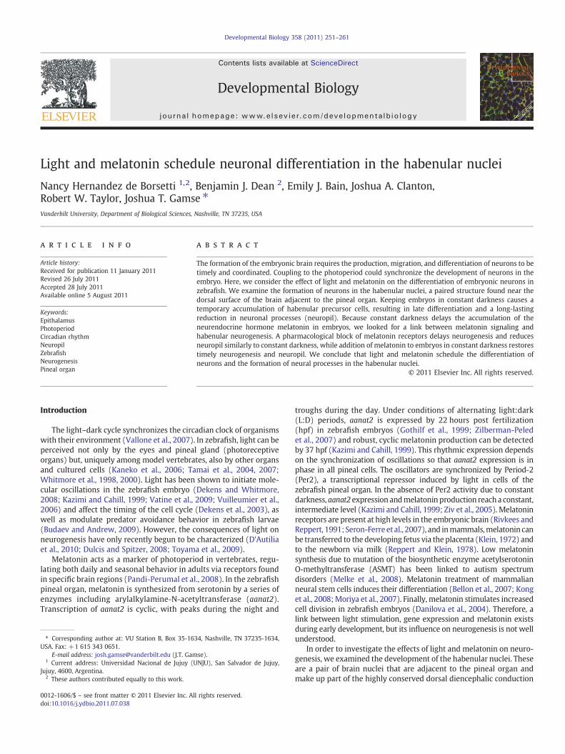
Fig. 1. Markers of differentiating habenular neurons are delayed by constant darkness. (A) Zebrafish embryos were raised in 14/10 light/dark (LD) or constant darkness (DD)conditions beginning at 5 min post-fertilization. (B–E) Expression of kctd12.1 initiates in the habenular nuclei (black arrows) at 38 h post-fertilization (hpf) in LD but does not initiateuntil 48 hpf in DD. (F–M) Similarly, expression of kctd12.2 and cadps2 initiates earlier in LD than in DD conditions. Insets in F–I are magnified views of the left habenula. (N–O) Bycontrast, expression of nrp1a in the habenular nuclei (white arrowheads) is not delayed by DD conditions; (P–S) nor is otx5 expression in the pineal and parapineal or gfi1 expressionin the parapineal (black arrows). All views are dorsal except for frontal views in B–E and lateral views in F–I. Scale bar=50 μm except for insets in F–I (25 μm).
253N.H. de Borsetti et al. / Developmental Biology 358 (2011) 251–261
254 N.H. de Borsetti et al. / Developmental Biology 358 (2011) 251–261
All immunofluorescence data were collected on a Zeiss LSM510confocal microscopewith a 40× oil-immersion objective and analyzedwith Volocity software (Improvision).
A
B C
D E
F G
H I
J K
L M
Results
Constant darkness causes delayed gene expression in the habenularnuclei
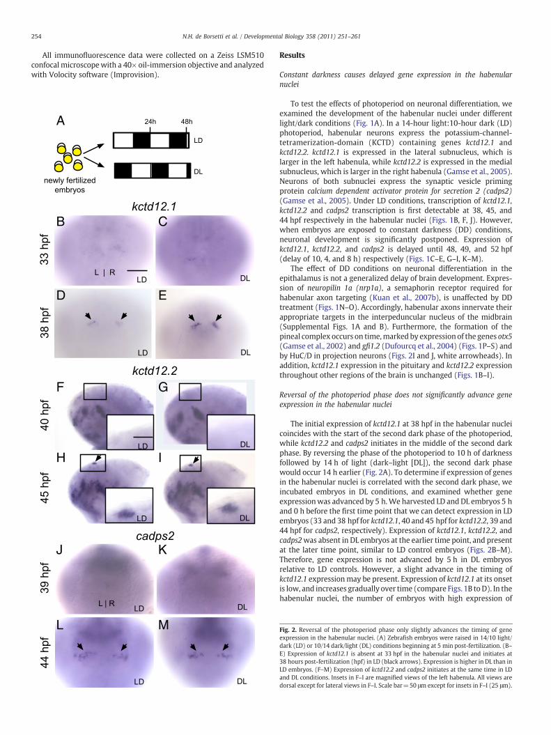
To test the effects of photoperiod on neuronal differentiation, weexamined the development of the habenular nuclei under differentlight/dark conditions (Fig. 1A). In a 14-hour light:10-hour dark (LD)photoperiod, habenular neurons express the potassium-channel-tetramerization-domain (KCTD) containing genes kctd12.1 andkctd12.2. kctd12.1 is expressed in the lateral subnucleus, which islarger in the left habenula, while kctd12.2 is expressed in the medialsubnucleus, which is larger in the right habenula (Gamse et al., 2005).Neurons of both subnuclei express the synaptic vesicle primingprotein calcium dependent activator protein for secretion 2 (cadps2)(Gamse et al., 2005). Under LD conditions, transcription of kctd12.1,kctd12.2 and cadps2 transcription is first detectable at 38, 45, and44 hpf respectively in the habenular nuclei (Figs. 1B, F, J). However,when embryos are exposed to constant darkness (DD) conditions,neuronal development is significantly postponed. Expression ofkctd12.1, kctd12.2, and cadps2 is delayed until 48, 49, and 52 hpf(delay of 10, 4, and 8 h) respectively (Figs. 1C–E, G–I, K–M).
The effect of DD conditions on neuronal differentiation in theepithalamus is not a generalized delay of brain development. Expres-sion of neuropilin 1a (nrp1a), a semaphorin receptor required forhabenular axon targeting (Kuan et al., 2007b), is unaffected by DDtreatment (Figs. 1N–O). Accordingly, habenular axons innervate theirappropriate targets in the interpeduncular nucleus of the midbrain(Supplemental Figs. 1A and B). Furthermore, the formation of thepineal complex occurs on time,marked by expression of the genes otx5(Gamse et al., 2002) and gfi1.2 (Dufourcq et al., 2004) (Figs. 1P–S) andby HuC/D in projection neurons (Figs. 2I and J, white arrowheads). Inaddition, kctd12.1 expression in the pituitary and kctd12.2 expressionthroughout other regions of the brain is unchanged (Figs. 1B–I).
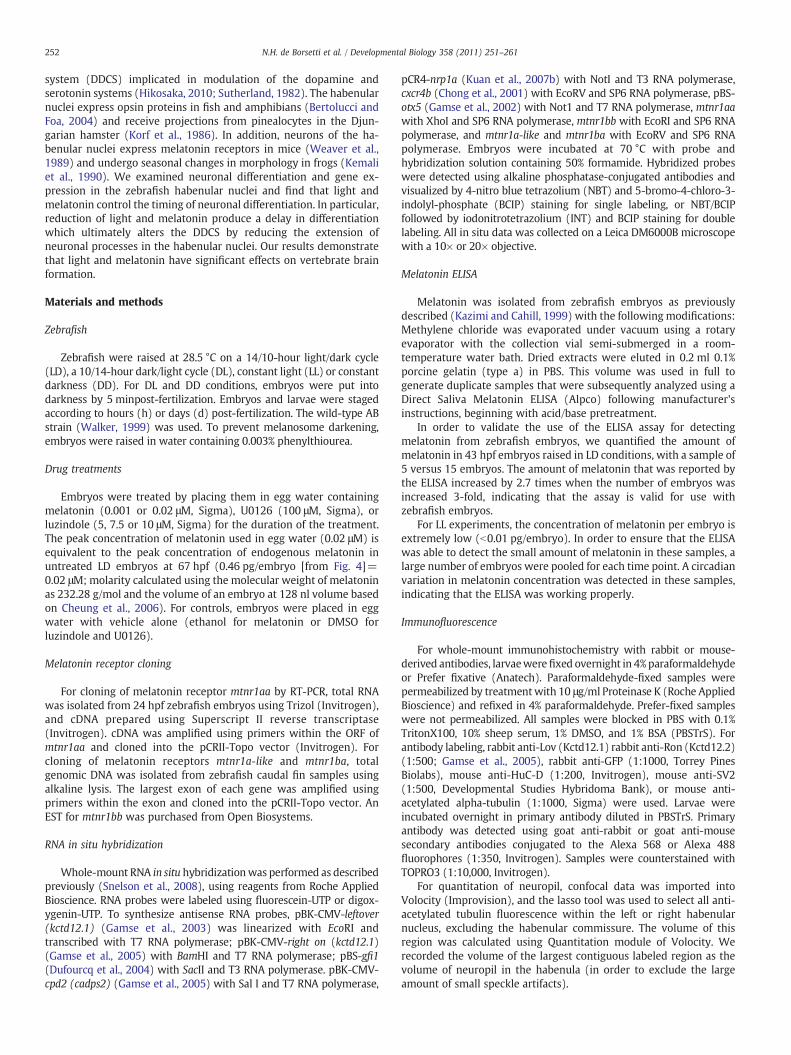
Reversal of the photoperiod phase does not significantly advance geneexpression in the habenular nuclei
The initial expression of kctd12.1 at 38 hpf in the habenular nucleicoincides with the start of the second dark phase of the photoperiod,while kctd12.2 and cadps2 initiates in the middle of the second darkphase. By reversing the phase of the photoperiod to 10 h of darknessfollowed by 14 h of light (dark–light [DL]), the second dark phasewould occur 14 h earlier (Fig. 2A). To determine if expression of genesin the habenular nuclei is correlated with the second dark phase, weincubated embryos in DL conditions, and examined whether geneexpressionwas advanced by 5 h.We harvested LD andDL embryos 5 hand 0 h before the first time point that we can detect expression in LDembryos (33 and 38 hpf for kctd12.1, 40 and 45 hpf for kctd12.2, 39 and44 hpf for cadps2, respectively). Expression of kctd12.1, kctd12.2, andcadps2was absent in DL embryos at the earlier time point, and presentat the later time point, similar to LD control embryos (Figs. 2B–M).Therefore, gene expression is not advanced by 5 h in DL embryosrelative to LD controls. However, a slight advance in the timing ofkctd12.1 expressionmay be present. Expression of kctd12.1 at its onsetis low, and increases gradually over time (compare Figs. 1B toD). In thehabenular nuclei, the number of embryos with high expression of
Fig. 2. Reversal of the photoperiod phase only slightly advances the timing of geneexpression in the habenular nuclei. (A) Zebrafish embryos were raised in 14/10 light/dark (LD) or 10/14 dark/light (DL) conditions beginning at 5 min post-fertilization. (B–E) Expression of kctd12.1 is absent at 33 hpf in the habenular nuclei and initiates at38 hours post-fertilization (hpf) in LD (black arrows). Expression is higher in DL than inLD embryos. (F–M) Expression of kctd12.2 and cadps2 initiates at the same time in LDand DL conditions. Insets in F–I are magnified views of the left habenula. All views aredorsal except for lateral views in F–I. Scale bar=50 μm except for insets in F–I (25 μm).
255N.H. de Borsetti et al. / Developmental Biology 358 (2011) 251–261
kctd12.1 was greater in DL than in LD embryos at 38 hpf (compareFigs. 2D to E; 100% of LD embryos (n=30) had expression equal or lessthan the example shown in Fig. 2D, whereas 83% of DL embryos(n=18) had expression equal or greater to the example shown inFig. 2E, and the remainder resembled Fig. 2D; pb0.0001, two-tailedFisher's exact test). We find evidence for slightly premature kctd12.1expression in the habenular nuclei of DL embryos compared to LDsiblings, and no change in the timing of expression for kctd12.2 andcadps2.
A B
C D
In constant darkness, habenular cells remain in a progenitor state for anextended time
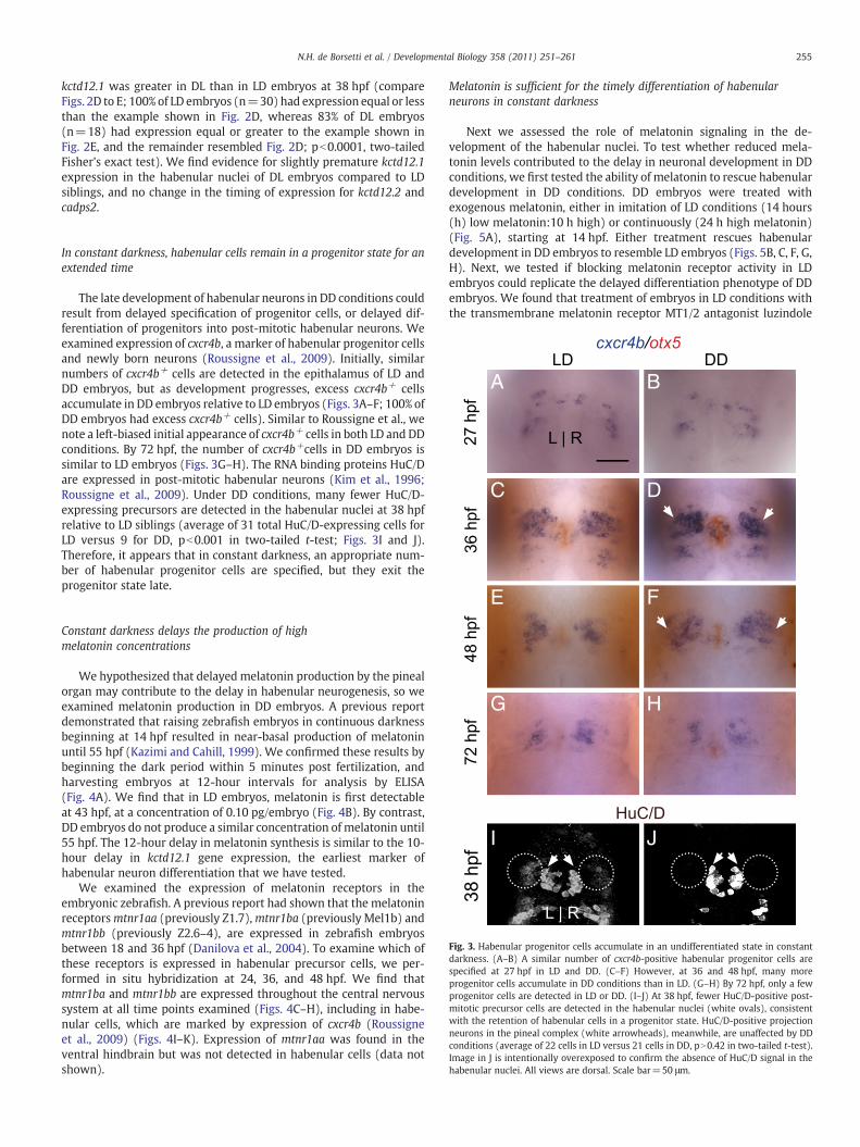
The late development of habenular neurons in DD conditions couldresult from delayed specification of progenitor cells, or delayed dif-ferentiation of progenitors into post-mitotic habenular neurons. Weexamined expression of cxcr4b, a marker of habenular progenitor cellsand newly born neurons (Roussigne et al., 2009). Initially, similarnumbers of cxcr4b+ cells are detected in the epithalamus of LD andDD embryos, but as development progresses, excess cxcr4b+ cellsaccumulate in DD embryos relative to LD embryos (Figs. 3A–F; 100% ofDD embryos had excess cxcr4b+ cells). Similar to Roussigne et al., wenote a left-biased initial appearance of cxcr4b+ cells in both LD and DDconditions. By 72 hpf, the number of cxcr4b+cells in DD embryos issimilar to LD embryos (Figs. 3G–H). The RNA binding proteins HuC/Dare expressed in post-mitotic habenular neurons (Kim et al., 1996;Roussigne et al., 2009). Under DD conditions, many fewer HuC/D-expressing precursors are detected in the habenular nuclei at 38 hpfrelative to LD siblings (average of 31 total HuC/D-expressing cells forLD versus 9 for DD, pb0.001 in two-tailed t-test; Figs. 3I and J).Therefore, it appears that in constant darkness, an appropriate num-ber of habenular progenitor cells are specified, but they exit theprogenitor state late.
E F
G H
I J
Fig. 3. Habenular progenitor cells accumulate in an undifferentiated state in constantdarkness. (A–B) A similar number of cxcr4b-positive habenular progenitor cells arespecified at 27 hpf in LD and DD. (C–F) However, at 36 and 48 hpf, many moreprogenitor cells accumulate in DD conditions than in LD. (G–H) By 72 hpf, only a fewprogenitor cells are detected in LD or DD. (I–J) At 38 hpf, fewer HuC/D-positive post-mitotic precursor cells are detected in the habenular nuclei (white ovals), consistentwith the retention of habenular cells in a progenitor state. HuC/D-positive projectionneurons in the pineal complex (white arrowheads), meanwhile, are unaffected by DDconditions (average of 22 cells in LD versus 21 cells in DD, pN0.42 in two-tailed t-test).Image in J is intentionally overexposed to confirm the absence of HuC/D signal in thehabenular nuclei. All views are dorsal. Scale bar=50 μm.
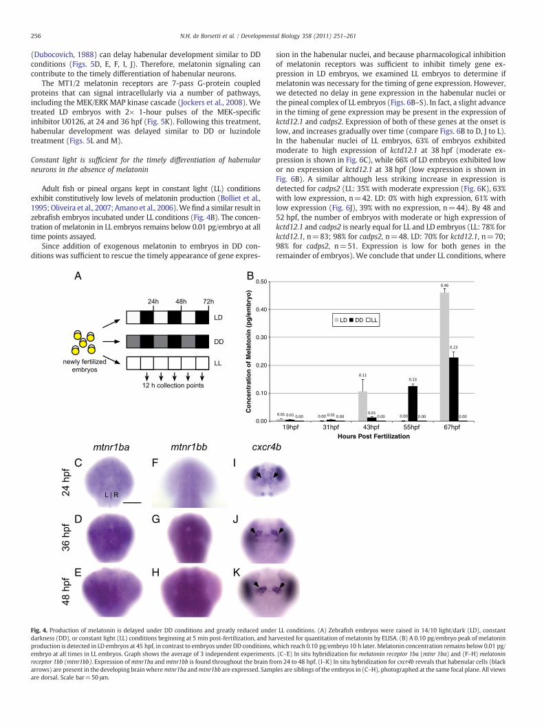
Constant darkness delays the production of highmelatonin concentrations
We hypothesized that delayed melatonin production by the pinealorgan may contribute to the delay in habenular neurogenesis, so weexamined melatonin production in DD embryos. A previous reportdemonstrated that raising zebrafish embryos in continuous darknessbeginning at 14 hpf resulted in near-basal production of melatoninuntil 55 hpf (Kazimi and Cahill, 1999). We confirmed these results bybeginning the dark period within 5 minutes post fertilization, andharvesting embryos at 12-hour intervals for analysis by ELISA(Fig. 4A). We find that in LD embryos, melatonin is first detectableat 43 hpf, at a concentration of 0.10 pg/embryo (Fig. 4B). By contrast,DD embryos do not produce a similar concentration of melatonin until55 hpf. The 12-hour delay in melatonin synthesis is similar to the 10-hour delay in kctd12.1 gene expression, the earliest marker ofhabenular neuron differentiation that we have tested.
We examined the expression of melatonin receptors in theembryonic zebrafish. A previous report had shown that the melatoninreceptorsmtnr1aa (previously Z1.7),mtnr1ba (previously Mel1b) andmtnr1bb (previously Z2.6–4), are expressed in zebrafish embryosbetween 18 and 36 hpf (Danilova et al., 2004). To examine which ofthese receptors is expressed in habenular precursor cells, we per-formed in situ hybridization at 24, 36, and 48 hpf. We find thatmtnr1ba and mtnr1bb are expressed throughout the central nervoussystem at all time points examined (Figs. 4C–H), including in habe-nular cells, which are marked by expression of cxcr4b (Roussigneet al., 2009) (Figs. 4I–K). Expression of mtnr1aa was found in theventral hindbrain but was not detected in habenular cells (data notshown).
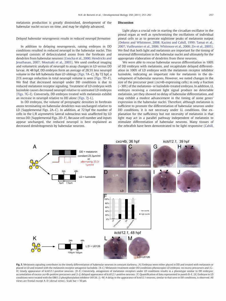
Melatonin is sufficient for the timely differentiation of habenularneurons in constant darkness
Next we assessed the role of melatonin signaling in the de-velopment of the habenular nuclei. To test whether reduced mela-tonin levels contributed to the delay in neuronal development in DDconditions, we first tested the ability of melatonin to rescue habenulardevelopment in DD conditions. DD embryos were treated withexogenous melatonin, either in imitation of LD conditions (14 hours(h) low melatonin:10 h high) or continuously (24 h high melatonin)(Fig. 5A), starting at 14 hpf. Either treatment rescues habenulardevelopment in DD embryos to resemble LD embryos (Figs. 5B, C, F, G,H). Next, we tested if blocking melatonin receptor activity in LDembryos could replicate the delayed differentiation phenotype of DDembryos. We found that treatment of embryos in LD conditions withthe transmembrane melatonin receptor MT1/2 antagonist luzindole
256 N.H. de Borsetti et al. / Developmental Biology 358 (2011) 251–261
(Dubocovich, 1988) can delay habenular development similar to DDconditions (Figs. 5D, E, F, I, J). Therefore, melatonin signaling cancontribute to the timely differentiation of habenular neurons.
The MT1/2 melatonin receptors are 7-pass G-protein coupledproteins that can signal intracellularly via a number of pathways,including the MEK/ERK MAP kinase cascade (Jockers et al., 2008). Wetreated LD embryos with 2× 1-hour pulses of the MEK-specificinhibitor U0126, at 24 and 36 hpf (Fig. 5K). Following this treatment,habenular development was delayed similar to DD or luzindoletreatment (Figs. 5L and M).
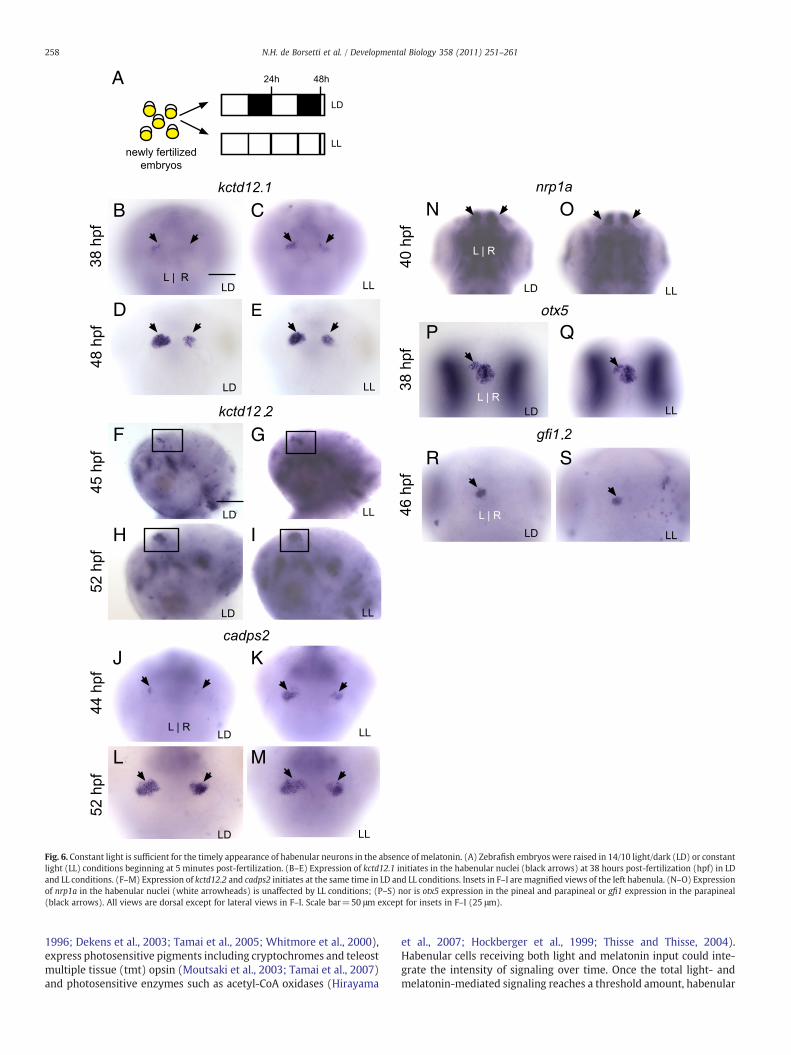
Constant light is sufficient for the timely differentiation of habenularneurons in the absence of melatonin
Adult fish or pineal organs kept in constant light (LL) conditionsexhibit constitutively low levels of melatonin production (Bolliet et al.,1995; Oliveira et al., 2007; Amano et al., 2006).Wefind a similar result inzebrafish embryos incubated under LL conditions (Fig. 4B). The concen-tration of melatonin in LL embryos remains below 0.01 pg/embryo at alltime points assayed.
Since addition of exogenous melatonin to embryos in DD con-ditions was sufficient to rescue the timely appearance of gene expres-
A
C F I
D G J
E H K
B0.50
0.40
0.30
0.20
0.10
0.00
Co
nce
ntr
atio
n o
f M
elat
on
in (
pg
/em
bry
o)
Fig. 4. Production of melatonin is delayed under DD conditions and greatly reduced undedarkness (DD), or constant light (LL) conditions beginning at 5 min post-fertilization, and haproduction is detected in LD embryos at 45 hpf, in contrast to embryos under DD conditions, wembryo at all times in LL embryos. Graph shows the average of 3 independent experimentsreceptor 1bb (mtnr1bb). Expression ofmtnr1ba andmtnr1bb is found throughout the brain froarrows) are present in the developing brain wheremtnr1ba andmtnr1bb are expressed. Sampare dorsal. Scale bar=50 μm.
sion in the habenular nuclei, and because pharmacological inhibitionof melatonin receptors was sufficient to inhibit timely gene ex-pression in LD embryos, we examined LL embryos to determine ifmelatonin was necessary for the timing of gene expression. However,we detected no delay in gene expression in the habenular nuclei orthe pineal complex of LL embryos (Figs. 6B–S). In fact, a slight advancein the timing of gene expression may be present in the expression ofkctd12.1 and cadps2. Expression of both of these genes at the onset islow, and increases gradually over time (compare Figs. 6B to D, J to L).In the habenular nuclei of LL embryos, 63% of embryos exhibitedmoderate to high expression of kctd12.1 at 38 hpf (moderate ex-pression is shown in Fig. 6C), while 66% of LD embryos exhibited lowor no expression of kctd12.1 at 38 hpf (low expression is shown inFig. 6B). A similar although less striking increase in expression isdetected for cadps2 (LL: 35% with moderate expression (Fig. 6K), 63%with low expression, n=42. LD: 0% with high expression, 61% withlow expression (Fig. 6J), 39% with no expression, n=44). By 48 and52 hpf, the number of embryos with moderate or high expression ofkctd12.1 and cadps2 is nearly equal for LL and LD embryos (LL: 78% forkctd12.1, n=83; 98% for cadps2, n=48. LD: 70% for kctd12.1, n=70;98% for cadps2, n=51. Expression is low for both genes in theremainder of embryos). We conclude that under LL conditions, where
19hpf 31hpf 43hpf 55hpf 67hpfHours Post Fertilization
LD DD LL
r LL conditions. (A) Zebrafish embryos were raised in 14/10 light/dark (LD), constantrvested for quantitation of melatonin by ELISA. (B) A 0.10 pg/embryo peak of melatoninhich reach 0.10 pg/embryo 10 h later. Melatonin concentration remains below 0.01 pg/
. (C–E) In situ hybridization for melatonin receptor 1ba (mtnr 1ba) and (F–H) melatoninm 24 to 48 hpf. (I–K) In situ hybridization for cxcr4b reveals that habenular cells (blackles are siblings of the embryos in (C–H), photographed at the same focal plane. All views
257N.H. de Borsetti et al. / Developmental Biology 358 (2011) 251–261
melatonin production is greatly diminished, development of thehabenular nuclei occurs on time, and may be slightly advanced.
Delayed habenular neurogenesis results in reduced neuropil formation
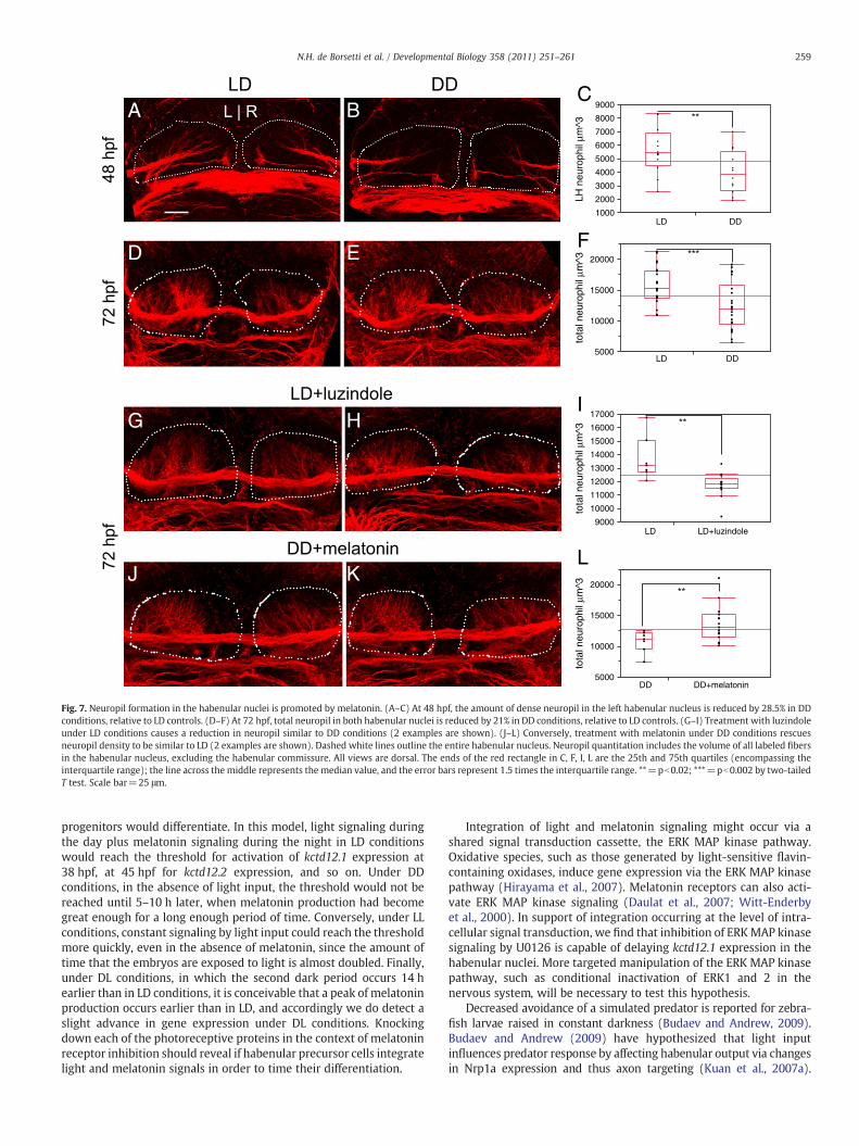
In addition to delaying neurogenesis, raising embryos in DDconditions resulted in reduced neuropil in the habenular nuclei. Thisneuropil consists of defasciculated axons from the forebrain anddendrites fromhabenular neurons (Concha et al., 2000; Hendricks andJesuthasan, 2007; Moutsaki et al., 2003). We used confocal imagingand volumetric analysis of neuropil to assay changes in LD versus DDlarvae. At 48 hpf, DD embryos form an average of 28.5% less neuropilvolume in the left habenula than LD siblings (Figs. 7A–C). By 72 hpf, a21% average reduction in total neuropil volume is seen (Figs. 7D–F).We find that decreased neuropil under DD conditions is due toreduced melatonin receptor signaling. Treatment of LD embryos withluzindole causes decreased neuropil relative to untreated LD embryos(Figs. 7G–I). Conversely, DD embryos treated with melatonin exhibitan increase in neuropil relative to DD alone (Figs. 7J–L).
In DD embryos, the volume of presynaptic densities in forebrainaxons terminating on habenular dendrites was unchanged relative toLD (Supplemental Figs. 2A–C). In addition, at 72 hpf the number ofcells in the L/R asymmetric lateral subnucleus was unaffected by LDversus DD (Supplemental Figs. 2D–F). Because cell number and inputsappear unchanged, the reduced neuropil is best explained asdecreased dendritogenesis by habenular neurons.
A
K L
B
D
F100%
80%
60%
40%
20%
0%
LD
LD+lu
Fig. 5.Melatonin signaling contributes to the timely differentiation of habenular neurons in cplaced in LD and treated with the melatonin receptor antagonist luzindole. (B–C) MelatoninH) timely appearance of kctd12.1-positive neurons. (D–E) Conversely, antagonism of melaccumulation of excess cxcr4b-positive precursors and (I–J) delayed appearance of kctd12.1-pconditions were treated with the ERK1/2 phosphorylation inhibitor U0126. (L–M) A delay in tviews are frontal except A–D (dorsal views). Scale bar=50 μm.
Discussion
Light plays a crucial role in starting the circadian oscillator in thepineal organ as well as synchronizing the oscillations of individualpineal cells so as to generate nighttime peaks of melatonin output(Dekens and Whitmore, 2008; Kazimi and Cahill, 1999; Tamai et al.,2007; Vuilleumier et al., 2006; Whitmore et al., 2000; Ziv et al., 2005).We find that both light and melatonin are important for the timing ofneuronal differentiation in the habenular nuclei and ultimately for theappropriate elaboration of dendrites from these neurons.
We were able to rescue habenular neuron differentiation in 100%of DD embryos with melatonin, and recapitulate delayed differenti-ation in 100% of LD embryos with the melatonin receptor inhibitorluzindole, indicating an important role for melatonin in the de-velopment of habenular neurons. However, we noted changes in thesize of the precursor pool (cxcr4b-expressing cells) in only a fraction(~30%) of themelatonin- or luzindole-treated embryos. In addition, LLembryos receiving a constant light signal produce no detectablemelatonin, yet they showed no delay of habenular differentiation, andmay exhibit a modest advancement in the timing of some genes’expression in the habenular nuclei. Therefore, although melatonin issufficient to promote the differentiation of habenular neurons underDD conditions, it is not necessary under LL conditions. One ex-planation for the sufficiency but not necessity of melatonin is thatlight may act in a parallel pathway independent of melatonin tostimulate differentiation of habenular neurons. Many tissues ofthe zebrafish have been demonstrated to be light responsive (Cahill,
M
C G H
E I J
DDDD+m
elat
onin
zindo
le
low cxcr4b
high cxcr4b
onstant darkness. (A) Embryos were either placed in DD and treated with melatonin ortreatment under DD conditions phenocopies LD embryos: no excess precursors and (G–atonin receptors under LD conditions results in a phenotype similar to DD embryos:ositive neurons. (F) Quantification of data represented in panels B–E. (K) Embryos in LDhe appearance of kctd12.1 neurons, similar to that seen in DD conditions, is observed. All
B C N O
D E
F G
H I
J K
L M
P Q
R S
A
Fig. 6. Constant light is sufficient for the timely appearance of habenular neurons in the absence of melatonin. (A) Zebrafish embryos were raised in 14/10 light/dark (LD) or constantlight (LL) conditions beginning at 5 minutes post-fertilization. (B–E) Expression of kctd12.1 initiates in the habenular nuclei (black arrows) at 38 hours post-fertilization (hpf) in LDand LL conditions. (F–M) Expression of kctd12.2 and cadps2 initiates at the same time in LD and LL conditions. Insets in F–I are magnified views of the left habenula. (N–O) Expressionof nrp1a in the habenular nuclei (white arrowheads) is unaffected by LL conditions; (P–S) nor is otx5 expression in the pineal and parapineal or gfi1 expression in the parapineal(black arrows). All views are dorsal except for lateral views in F–I. Scale bar=50 μm except for insets in F–I (25 μm).
258 N.H. de Borsetti et al. / Developmental Biology 358 (2011) 251–261
1996; Dekens et al., 2003; Tamai et al., 2005; Whitmore et al., 2000),express photosensitive pigments including cryptochromes and teleostmultiple tissue (tmt) opsin (Moutsaki et al., 2003; Tamai et al., 2007)and photosensitive enzymes such as acetyl-CoA oxidases (Hirayama
et al., 2007; Hockberger et al., 1999; Thisse and Thisse, 2004).Habenular cells receiving both light and melatonin input could inte-grate the intensity of signaling over time. Once the total light- andmelatonin-mediated signaling reaches a threshold amount, habenular
A B
D E
G HI
J KL
C
F
9000
8000
7000
6000
5000
4000
3000
2000
20000
15000
10000
5000
17000
16000
15000
14000
13000
12000
11000
10000
9000
20000
15000
10000
5000
1000
LH n
euro
phil
µm^3
LD DD
LD
DD
LD
DD+melatonin
LD+luzindole
DD
tota
l neu
roph
il µm
^3to
tal n
euro
phil
µm^3
tota
l neu
roph
il µm
^3
Fig. 7. Neuropil formation in the habenular nuclei is promoted by melatonin. (A–C) At 48 hpf, the amount of dense neuropil in the left habenular nucleus is reduced by 28.5% in DDconditions, relative to LD controls. (D–F) At 72 hpf, total neuropil in both habenular nuclei is reduced by 21% in DD conditions, relative to LD controls. (G–I) Treatment with luzindoleunder LD conditions causes a reduction in neuropil similar to DD conditions (2 examples are shown). (J–L) Conversely, treatment with melatonin under DD conditions rescuesneuropil density to be similar to LD (2 examples are shown). Dashed white lines outline the entire habenular nucleus. Neuropil quantitation includes the volume of all labeled fibersin the habenular nucleus, excluding the habenular commissure. All views are dorsal. The ends of the red rectangle in C, F, I, L are the 25th and 75th quartiles (encompassing theinterquartile range); the line across the middle represents the median value, and the error bars represent 1.5 times the interquartile range. **=pb0.02; ***=pb0.002 by two-tailedT test. Scale bar=25 μm.
259N.H. de Borsetti et al. / Developmental Biology 358 (2011) 251–261
progenitors would differentiate. In this model, light signaling duringthe day plus melatonin signaling during the night in LD conditionswould reach the threshold for activation of kctd12.1 expression at38 hpf, at 45 hpf for kctd12.2 expression, and so on. Under DDconditions, in the absence of light input, the threshold would not bereached until 5–10 h later, when melatonin production had becomegreat enough for a long enough period of time. Conversely, under LLconditions, constant signaling by light input could reach the thresholdmore quickly, even in the absence of melatonin, since the amount oftime that the embryos are exposed to light is almost doubled. Finally,under DL conditions, in which the second dark period occurs 14 hearlier than in LD conditions, it is conceivable that a peak of melatoninproduction occurs earlier than in LD, and accordingly we do detect aslight advance in gene expression under DL conditions. Knockingdown each of the photoreceptive proteins in the context of melatoninreceptor inhibition should reveal if habenular precursor cells integratelight and melatonin signals in order to time their differentiation.
Integration of light and melatonin signaling might occur via ashared signal transduction cassette, the ERK MAP kinase pathway.Oxidative species, such as those generated by light-sensitive flavin-containing oxidases, induce gene expression via the ERK MAP kinasepathway (Hirayama et al., 2007). Melatonin receptors can also acti-vate ERK MAP kinase signaling (Daulat et al., 2007; Witt-Enderbyet al., 2000). In support of integration occurring at the level of intra-cellular signal transduction, we find that inhibition of ERKMAP kinasesignaling by U0126 is capable of delaying kctd12.1 expression in thehabenular nuclei. More targeted manipulation of the ERK MAP kinasepathway, such as conditional inactivation of ERK1 and 2 in thenervous system, will be necessary to test this hypothesis.
Decreased avoidance of a simulated predator is reported for zebra-fish larvae raised in constant darkness (Budaev and Andrew, 2009).Budaev and Andrew (2009) have hypothesized that light inputinfluences predator response by affecting habenular output via changesin Nrp1a expression and thus axon targeting (Kuan et al., 2007a).
260 N.H. de Borsetti et al. / Developmental Biology 358 (2011) 251–261
However, we do not detect a change in nrp1a expression or axonaltargeting to the IPN in LD versus DD conditions. We do find that DDconditions result in decreased neuropil density in the habenular nuclei,perhaps because habenular neurons are exposed for a shorter time tointrinsic or extrinsic signals for dendrite formation (Parrish et al., 2007).Habenular neuron function has been recently implicated in zebrafishlearning whether it is best to flee or freeze in place in response to anegative stimulus (Agetsuma et al., 2010; Lee et al., 2010), a behaviorthat is relevant in reacting to predators. It is therefore possible thatdecreased predator avoidance behavior in DD-raised larvae is aconsequence of reduced habenular dendritogenesis.
Conclusions
We show that the timing of neuronal differentiation and sub-sequently the appropriate outgrowth of dendrites during habenulardevelopment is an event that requires light and the hormone mela-tonin. Intriguingly, alteration of melatonin production is a symptom ofsome neurological diseases, including autism and Smith-Magenissyndrome (Elsea and Girirajan, 2008; Kulman et al., 2000; Nir, 1995;Tordjman et al., 2005). In addition, mutations of the melatoninbiosynthetic enzyme ASMT are linked to increased autism risk (Melkeet al., 2008). It has been proposed that altered melatonin during earlypostnatal development may be causative rather than simply symp-tomatic of these diseases, by altering formation of brain circuits(Bourgeron, 2007). The zebrafish embryo, with its easily manipulatedpathway for melatonin signaling, now provides a platform to explorehow melatonin influences brain development.
Supplementarymaterials related to this article can be found onlineat doi:10.1016/j.ydbio.2011.07.038.
Acknowledgments
We thank Qiang Guan, Heidi Beck, and Gena Gustin for expert fishcare, Erin Booton for excellent in situ hybridization support, HugoBorsetti for discussion of experiments, Jeff Johnston for help withmelatonin extraction from embryos, and Marnie Halpern for in situclones. This work was funded by NIH grant HD054534 to J.T.G. B.J.D.was supported by the Vanderbilt Medical-Scientist Training Program(T32 GM07347 from the NIH) and J.A.C. was supported by theVanderbilt Training Program in Developmental Biology (T32HD007402 from the NIH).
References
Agetsuma, M., Aizawa, H., Aoki, T., Nakayama, R., Takahoko,M., Goto,M., Sassa, T., Amo, R.,Shiraki, T., Kawakami, K., Hosoya, T., Higashijima, S., Okamoto, H., 2010. The habenulais crucial for experience-dependent modification of fear responses in zebrafish. Nat.Neurosci. 13, 1354–1356.
Amano, M., Iigo, M., Kitamura, S., Amiya, N., Yamamori, K., 2006. Changes in melatoninbinding sites under artificial light-dark, constant light and constant dark conditionsin the masu salmon brain. Comp. Biochem. Physiol. Part A. Mol. Integr. Physiol. 144,509–513.
Bellon, A., Ortiz-Lopez, L., Ramirez-Rodriguez, G., Anton-Tay, F., Benitez-King, G., 2007.Melatonin induces neuritogenesis at early stages in N1E-115 cells through actinrearrangements via activation of protein kinase C and Rho-associated kinase.J. Pineal Res. 42, 214–221.
Bertolucci, C., Foa, A., 2004. Extraocular photoreception and circadian entrainment innonmammalian vertebrates. Chronobiol. Int. 21, 501–519.
Bolliet, V., Falcón, J., Ali, M.A., 1995. Regulation of melatonin secretion by light in theisolated pineal organ of the white sucker (Catostomus commersoni). J. Neuroendo-crinol. 7, 535–542.
Bourgeron, T., 2007. The possible interplay of synaptic and clock genes in autismspectrum disorders. Cold Spring Harb. Symp. Quant. Biol. 72, 645–654.
Budaev, S., Andrew, R.J., 2009. Patterns of early embryonic light exposure determinebehavioural asymmetries in zebrafish: a habenular hypothesis. Behav. Brain Res.200, 91–94.
Cahill, G.M., 1996. Circadian regulation of melatonin production in cultured zebrafishpineal and retina. Brain Res. 708, 177–181.
Cheung, C.Y., Webb, S.E., Meng, A., Miller, A.L., 2006. Transient expression ofapoaequorin in zebrafish embryos: extending the ability to image calciumtransients during later stages of development. Int. J. Dev. Biol. 50, 561–569.
Chong, S.W., Emelyanov, A., Gong, Z., Korzh, V., 2001. Expression pattern of twozebrafish genes, cxcr4a and cxcr4b. Mech. Dev. 109, 347–354.
Concha, M.L., Burdine, R.D., Russell, C., Schier, A.F., Wilson, S.W., 2000. A nodal signalingpathway regulates the laterality of neuroanatomical asymmetries in the zebrafishforebrain. Neuron 28, 399–409.
Danilova, N., Krupnik, V.E., Sugden, D., Zhdanova, I.V., 2004. Melatonin stimulates cellproliferation in zebrafish embryo and accelerates its development. FASEB J. 18,751–753.
Daulat, A.M., Maurice, P., Froment, C., Guillaume, J.L., Broussard, C., Monsarrat, B.,Delagrange, P., Jockers, R., 2007. Purification and identification of G protein-coupled receptor protein complexes under native conditions. Mol. Cell. Proteomics6, 835–844.
D'Autilia, S., Broccoli, V., Barsacchi, G., Andreazzoli, M., 2010. Xenopus Bsx links dailycell cycle rhythms and pineal photoreceptor fate. Proc. Natl. Acad. Sci. U.S.A. 107,6352–6357.
Dekens, M.P., Whitmore, D., 2008. Autonomous onset of the circadian clock in thezebrafish embryo. EMBO J. 27, 2757–2765.
Dekens, M.P., Santoriello, C., Vallone, D., Grassi, G., Whitmore, D., Foulkes, N.S., 2003.Light regulates the cell cycle in zebrafish. Curr. Biol. 13, 2051–2057.
Dubocovich, M.L., 1988. Luzindole (N-0774): a novel melatonin receptor antagonist. J.Pharmacol. Exp. Ther. 246, 902–910.
Dufourcq, P., Rastegar, S., Strahle, U., Blader, P., 2004. Parapineal specific expression ofgfi1 in the zebrafish epithalamus. Gene Expr. Patterns 4, 53–57.
Dulcis, D., Spitzer, N.C., 2008. Illumination controls differentiation of dopamine neuronsregulating behaviour. Nature 456, 195–201.
Elsea, S.H., Girirajan, S., 2008. Smith–Magenis syndrome. Eur. J. Hum. Genet. 16,412–421.
Gamse, J.T., Shen, Y.C., Thisse, C., Thisse, B., Raymond, P.A., Halpern, M.E., Liang, J.O.,2002. Otx5 regulates genes that show circadian expression in the zebrafish pinealcomplex. Nat. Genet. 30, 117–121.
Gamse, J.T., Thisse, C., Thisse, B., Halpern, M.E., 2003. The parapineal mediates left-rightasymmetry in the zebrafish diencephalon. Development 130, 1059–1068.
Gamse, J.T., Kuan, Y.S., Macurak, M., Brosamle, C., Thisse, B., Thisse, C., Halpern, M.E.,2005. Directional asymmetry of the zebrafish epithalamus guides dorsoventralinnervation of the midbrain target. Development 132, 4869–4881.
Gothilf, Y., Coon, S.L., Toyama, R., Chitnis, A., Namboodiri, M.A., Klein, D.C., 1999.Zebrafish serotonin N-acetyltransferase-2: marker for development of pinealphotoreceptors and circadian clock function. Endocrinology 140, 4895–4903.
Hendricks, M., Jesuthasan, S., 2007. Asymmetric innervation of the habenula inzebrafish. J. Comp. Neurol. 502, 611–619.
Hikosaka, O., 2010. The habenula: from stress evasion to value-based decision-making.Nat. Rev. Neurosci. 11, 503–513.
Hirayama, J., Cho, S., Sassone-Corsi, P., 2007. Circadian control by the reduction/oxidation pathway: catalase represses light-dependent clock gene expression inthe zebrafish. Proc. Natl. Acad. Sci. U.S.A. 104, 15747–15752.
Hockberger, P.E., Skimina, T.A., Centonze, V.E., Lavin, C., Chu, S., Dadras, S., Reddy, J.K.,White, J.G., 1999. Activation of flavin-containing oxidases underlies light-inducedproduction of H2O2 in mammalian cells. Proc. Natl. Acad. Sci. U.S.A. 96, 6255–6260.
Jockers, R., Maurice, P., Boutin, J.A., Delagrange, P., 2008. Melatonin receptors,heterodimerization, signal transduction and binding sites: what's new? Br. J.Pharmacol. 154, 1182–1195.
Kaneko, M., Hernandez-Borsetti, N., Cahill, G.M., 2006. Diversity of zebrafish peripheraloscillators revealed by luciferase reporting. Proc. Natl. Acad. Sci. U.S.A. 103,14614–14619.
Kazimi, N., Cahill, G.M., 1999. Development of a circadian melatonin rhythm inembryonic zebrafish. Brain Res. Dev. Brain Res. 117, 47–52.
Kemali, M., Guglielmotti, V., Fiorino, L., 1990. The asymmetry of the habenular nuclei offemale and male frogs in spring and in winter. Brain Res. 517, 251–255.
Kim, C.H., Ueshima, E., Muraoka, O., Tanaka, H., Yeo, S.Y., Huh, T.L., Miki, N., 1996.Zebrafish elav/HuC homologue as a very early neuronal marker. Neurosci. Lett. 216,109–112.
Klein, D.C., 1972. Evidence for the placental transfer of 3 H-acetyl-melatonin. Nat. NewBiol. 237, 117–118.
Kong, X., Li, X., Cai, Z., Yang, N., Liu, Y., Shu, J., Pan, L., Zuo, P., 2008. Melatonin regulatesthe viability and differentiation of rat midbrain neural stem cells. Cell. Mol.Neurobiol. 28, 569–579.
Korf, H.W., Oksche, A., Ekstrom, P., Gery, I., Zigler Jr., J.S., Klein, D.C., 1986. Pinealocyteprojections into the mammalian brain revealed with S-antigen antiserum. Science231, 735–737.
Kuan, Y.S., Gamse, J.T., Schreiber, A.M., Halpern, M.E., 2007a. Selective asymmetry in aconserved forebrain to midbrain projection. J. Exp. Zool. B Mol. Dev. Evol. 308,669–678.
Kuan, Y.S., Yu, H.H., Moens, C.B., Halpern, M.E., 2007b. Neuropilin asymmetry mediatesa left-right difference in habenular connectivity. Development 134, 857–865.
Kulman, G., Lissoni, P., Rovelli, F., Roselli, M.G., Brivio, F., Sequeri, P., 2000. Evidence ofpineal endocrine hypofunction in autistic children. Neuro Endocrinol. Lett. 21,31–34.
Lee, A., Mathuru, A.S., Teh, C., Kibat, C., Korzh, V., Penney, T.B., Jesuthasan, S., 2010. Thehabenula prevents helpless behavior in larval zebrafish. Curr. Biol. 20, 2211–2216.
Melke, J., Goubran Botros, H., Chaste, P., Betancur, C., Nygren, G., Anckarsater, H.,Rastam, M., Stahlberg, O., Gillberg, I.C., Delorme, R., Chabane, N., Mouren-Simeoni,M.C., Fauchereau, F., Durand, C.M., Chevalier, F., Drouot, X., Collet, C., Launay, J.M.,Leboyer, M., Gillberg, C., Bourgeron, T., 2008. Abnormal melatonin synthesis inautism spectrum disorders. Mol. Psychiatry 13, 90–98.
Moriya, T., Horie, N., Mitome, M., Shinohara, K., 2007. Melatonin influences theproliferative anddifferentiative activity of neural stem cells. J. Pineal Res. 42, 411–418.
261N.H. de Borsetti et al. / Developmental Biology 358 (2011) 251–261
Moutsaki, P., Whitmore, D., Bellingham, J., Sakamoto, K., David-Gray, Z.K., Foster, R.G.,2003. Teleost multiple tissue (tmt) opsin: a candidate photopigment regulating theperipheral clocks of zebrafish? Brain Res. Mol. Brain Res. 112, 135–145.
Nir, I., 1995. Biorhythms and the biological clock involvement of melatonin and thepineal gland in life and disease. Biomed. Environ. Sci. 8, 90–105.
Oliveira, C., Ortega, A., Lopez-Olmeda, J.F., Vera, L.M., Sánchez-Vázquez, F.J., 2007.Influence of constant light and darkness, light intensity, and light spectrum onplasma melatonin rhythms in senegal sole. Chronobiol. Int. 24, 615–627.
Pandi-Perumal, S.R., Trakht, I., Srinivasan, V., Spence, D.W., Maestroni, G.J., Zisapel, N.,Cardinali, D.P., 2008. Physiological effects of melatonin: role of melatonin receptorsand signal transduction pathways. Prog. Neurobiol. 85, 335–353.
Parrish, J.Z., Emoto, K., Kim, M.D., Jan, Y.N., 2007. Mechanisms that regulateestablishment, maintenance, and remodeling of dendritic fields. Annu. Rev.Neurosci. 30, 399–423.
Reppert, S.M., Klein, D.C., 1978. Transport of maternal[3H]melatonin to suckling ratsand the fate of [3H]melatonin in the neonatal rat. Endocrinology 102, 582–588.
Rivkees, S.A., Reppert, S.M., 1991. Appearance of melatonin receptors during embryoniclife in Siberian hamsters (Phodopus sungorous). Brain Res. 568, 345–349.
Roussigne, M., Bianco, I.H., Wilson, S.W., Blader, P., 2009. Nodal signalling imposes left–right asymmetry upon neurogenesis in the habenular nuclei. Development 136,1549–1557.
Seron-Ferre, M., Valenzuela, G.J., Torres-Farfan, C., 2007. Circadian clocks duringembryonic and fetal development. Birth Defects Res. C Embryo Today 81, 204–214.
Snelson, C.D., Santhakumar, K., Halpern, M.E., Gamse, J.T., 2008. Tbx2b is required forthe development of the parapineal organ. Development 135, 1693–1702.
Sutherland, R.J., 1982. The dorsal diencephalic conduction system: a review of theanatomy and functions of the habenular complex. Neurosci. Biobehav. Rev. 6, 1–13.
Tamai, T.K., Vardhanabhuti, V., Foulkes, N.S., Whitmore, D., 2004. Early embryonic lightdetection improves survival. Curr. Biol. 14, R104–R105.
Tamai, T.K., Carr, A.J., Whitmore, D., 2005. Zebrafish circadian clocks: cells that see light.Biochem. Soc. Trans. 33, 962–966.
Tamai, T.K., Young, L.C., Whitmore, D., 2007. Light signaling to the zebrafish circadianclock by Cryptochrome 1a. Proc. Natl. Acad. Sci. U.S.A. 104, 14712–14717.
Thisse, B., Thisse, C., 2004. Fast Release Clones: A High Throughput ExpressionAnalysis.
Tordjman, S., Anderson, G.M., Pichard, N., Charbuy, H., Touitou, Y., 2005. Nocturnalexcretion of 6-sulphatoxymelatonin in children and adolescents with autisticdisorder. Biol. Psychiatry 57, 134–138.
Toyama, R., Chen, X., Jhawar, N., Aamar, E., Epstein, J., Reany, N., Alon, S., Gothilf, Y.,Klein, D.C., Dawid, I.B., 2009. Transcriptome analysis of the zebrafish pineal gland.Dev. Dyn. 238, 1813–1826.
Vallone, D., Lahiri, K., Dickmeis, T., Foulkes, N.S., 2007. Start the clock! Circadianrhythms and development. Dev. Dyn. 236, 142–155.
Vatine, G., Vallone, D., Appelbaum, L., Mracek, P., Ben-Moshe, Z., Lahiri, K., Gothilf, Y.,Foulkes, N.S., 2009. Light directs zebrafish period2 expression via conserved D andE boxes. PLoS Biol. 7, e1000223.
Vuilleumier, R., Besseau, L., Boeuf, G., Piparelli, A., Gothilf, Y., Gehring, W.G., Klein, D.C.,Falcon, J., 2006. Starting the zebrafish pineal circadian clock with a single photictransition. Endocrinology 147, 2273–2279.
Walker, C., 1999. Haploid screens and gamma-ray mutagenesis. Methods in CellBiology. Elsevier, London, pp. 43–70.
Weaver, D.R., Rivkees, S.A., Reppert, S.M., 1989. Localization and characterization ofmelatonin receptors in rodent brain by in vitro autoradiography. J. Neurosci. 9,2581–2590.
Whitmore, D., Foulkes, N.S., Strahle, U., Sassone-Corsi, P., 1998. Zebrafish clockrhythmic expression reveals independent peripheral circadian oscillators. Nat.Neurosci. 1, 701–707.
Whitmore, D., Foulkes, N.S., Sassone-Corsi, P., 2000. Light acts directly on organs andcells in culture to set the vertebrate circadian clock. Nature 404, 87–91.
Witt-Enderby, P.A., MacKenzie, R.S., McKeon, R.M., Carroll, E.A., Bordt, S.L., Melan, M.A.,2000. Melatonin induction of filamentous structures in non-neuronal cells that isdependent on expression of the human mt1 melatonin receptor. Cell Motil.Cytoskeleton 46, 28–42.
Zilberman-Peled, B., Appelbaum, L., Vallone, D., Foulkes, N.S., Anava, S., Anzulovich, A.,Coon, S.L., Klein, D.C., Falcon, J., Ron, B., Gothilf, Y., 2007. Transcriptional regulationof arylalkylamine-N-acetyltransferase-2 gene in the pineal gland of the giltheadseabream. J. Neuroendocrinol. 19, 46–53.
Ziv, L., Levkovitz, S., Toyama, R., Falcon, J., Gothilf, Y., 2005. Functional development ofthe zebrafish pineal gland: light-induced expression of period2 is required foronset of the circadian clock. J. Neuroendocrinol. 17, 314–320.


![Feasibility of melatonin for treatment (MEL-T) of …...Perioperative melatonin & delirium • >20 years; elective Sx with planned post-op ICU stay >48h [plasma] melatonin 08:00 before](https://static.fdocuments.us/doc/165x107/5f1f61cce84d081c1e42da29/feasibility-of-melatonin-for-treatment-mel-t-of-perioperative-melatonin-.jpg)