Levels or Strength of Evidence Ratings b-1... · VU = Venous ulcer, also called venous...
103
International Consolidated Guideline Task Force (2015 Update of the 2010 Association for the Advancement of Wound Care (AAWC) Venous Ulcer Guideline) Evidence Accessible at aawconline.org/professional-resources/resources/ Last updated: August 23, 2017 Page 1 of 51 ©AAWC 2016 International Consolidated Venous Ulcer Guideline Abbreviations & Evidence Page 1 of 103 8/23/2017 Definition of a Venous Ulcer (VU) A venous ulcer is a 'leg ulcer with either clinical or vascular findings consistent with venous hypertension ' It is considered chronic and is likely to experience delayed healing if it reduces in area less than 40% during 3 weeks of best evidence-based care (Phillips et al., 2000). If not adequately managed, associated local edema can progress to dermatitis and ulceration. Causative factors include, but are not limited to incompetent venous valves in the superficial, perforator or deep vein systems and/or inadequate calf muscle pump function (O’Donnell et al., 2014; Wound Ostomy Continence Nurses Society, 2005). Strength of Evidence Ratings A. Results of a meta-analysis or two or more venous ulcer (VU)-related randomized controlled trials (RCT) on humans provide support. For diagnostics or risk assessment screening: prospective cohort (CO) studies and/or controlled studies reporting recognized diagnostic (e.g. sensitivity or specificity) or screening (e.g. + or - predictive validity) measures. B. Results of one VU-related RCT in humans plus one or more similar Historically Controlled Trials (HCT) or Convenience Controlled Trials (CCT) or one HCT and one CCT provide support or when appropriate, results of two or more RCT in animal model validated as clinically relevant to VU provide indirect support. For diagnostics or risk assessment one VU-related prospective cohort (CO) study and/or a controlled study reporting recognized diagnostic or predictive screening validity measures. C. This rating requires one or more of the following: C1: Results of one controlled VU trial, e.g. RCT, CCT or HCT (or for diagnostics or risk prediction one prospective CO study may be substituted for a controlled trial C2: Results of at least two clinical VU case series (CS) or descriptive studies or a cohort study in humans C3: Expert opinion (EO) Abbreviations used in Evidence Table Below and Annotated Venous Ulcer Algorithm: AAWC = Association for the Advancement of Wound Care ABI = Ankle systolic blood pressure divided by brachial systolic blood pressure AM = Animal Model ASPS = American Society of Plastic Surgeons BWAT = Bates-Jensen Wound Assessment Tool CAK = Cryopreserved allogeneic keratinocyte CC = Case Controlled Epidemiology Study CEAP = Clinical Etiologic Anatomic Physiologic venous ulcer staging scale of the American Venous Forum CFU = Colony Forming Units (visible colonies of microorganisms counted on agar plates) CCT = Convenience-Assigned or Non-randomized Controlled Trial CO = Cohort study e.g. of all consecutive patients admitted to a facility studied prospectively CS = Case series or descriptive uncontrolled study of performance of one modality CVI = Chronic Venous Insufficiency DVT = Deep vein thrombosis EMLA = Eutectic Mixture of Local Anesthetic (lidocaine-prilocaine 5% cream) EO = Expert opinion, Content Validation Study or Consensus Statement EVLT = Endovenous laser therapy typically used for vein stripping FT = Full-thickness wound (through the fascia) G = Guideline GSV = Greater saphenous vein HCD = Hydrocolloid dressing HCT = Historically Controlled Trial with successive measure on a series of patients HRCT = Historically baseline data comparisons included in a randomized controlled clinical trial.
Transcript of Levels or Strength of Evidence Ratings b-1... · VU = Venous ulcer, also called venous...

International Consolidated Guideline Task Force (2015 Update of the 2010 Association for the Advancement of Wound Care (AAWC) Venous Ulcer Guideline) Evidence
Accessible at aawconline.org/professional-resources/resources/ Last updated: August 23, 2017 Page 1 of 51
©AAWC 2016 International Consolidated Venous Ulcer Guideline Abbreviations & Evidence
Page 1 of 103 8/23/2017
Definition of a Venous Ulcer (VU)
A venous ulcer is a 'leg ulcer with either clinical or vascular findings consistent with venous hypertension ' It is considered chronic and is likely to experience delayed healing if it reduces in area less than 40% during 3 weeks of best evidence-based care (Phillips et al., 2000). If not adequately managed, associated local edema can progress to dermatitis and ulceration. Causative factors include, but are not limited to incompetent venous valves in the superficial, perforator or deep vein systems and/or inadequate calf muscle pump function (O’Donnell et al., 2014; Wound Ostomy Continence Nurses Society, 2005). Strength of Evidence Ratings A. Results of a meta-analysis or two or more venous ulcer (VU)-related randomized controlled trials (RCT) on humans provide support. For diagnostics or risk assessment screening: prospective cohort (CO) studies and/or controlled studies reporting recognized diagnostic (e.g. sensitivity or specificity) or screening (e.g. + or - predictive validity) measures. B. Results of one VU-related RCT in humans plus one or more similar Historically Controlled Trials (HCT) or Convenience Controlled Trials (CCT) or one HCT and one CCT provide support or when appropriate, results of two or more RCT in animal model validated as clinically relevant to VU provide indirect support. For diagnostics or risk assessment one VU-related prospective cohort (CO) study and/or a controlled study reporting recognized diagnostic or predictive screening validity measures. C. This rating requires one or more of the following:
C1: Results of one controlled VU trial, e.g. RCT, CCT or HCT (or for diagnostics or risk prediction one prospective CO study may be substituted for a controlled trial C2: Results of at least two clinical VU case series (CS) or descriptive studies or a cohort study in humans C3: Expert opinion (EO)
Abbreviations used in Evidence Table Below and Annotated Venous Ulcer Algorithm: AAWC = Association for the Advancement of Wound Care ABI = Ankle systolic blood pressure divided by brachial systolic blood pressure AM = Animal Model ASPS = American Society of Plastic Surgeons BWAT = Bates-Jensen Wound Assessment Tool CAK = Cryopreserved allogeneic keratinocyte CC = Case Controlled Epidemiology Study CEAP = Clinical Etiologic Anatomic Physiologic venous ulcer staging scale of the American Venous Forum CFU = Colony Forming Units (visible colonies of microorganisms counted on agar plates) CCT = Convenience-Assigned or Non-randomized Controlled Trial CO = Cohort study e.g. of all consecutive patients admitted to a facility studied prospectively CS = Case series or descriptive uncontrolled study of performance of one modality CVI = Chronic Venous Insufficiency DVT = Deep vein thrombosis EMLA = Eutectic Mixture of Local Anesthetic (lidocaine-prilocaine 5% cream) EO = Expert opinion, Content Validation Study or Consensus Statement EVLT = Endovenous laser therapy typically used for vein stripping FT = Full-thickness wound (through the fascia) G = Guideline GSV = Greater saphenous vein HCD = Hydrocolloid dressing HCT = Historically Controlled Trial with successive measure on a series of patients HRCT = Historically baseline data comparisons included in a randomized controlled clinical trial.

International Consolidated Guideline Task Force (2015 Update of the 2010 Association for the Advancement of Wound Care (AAWC) Venous Ulcer Guideline) Evidence
Accessible at aawconline.org/professional-resources/resources/ Last updated: August 23, 2017 Page 2 of 51
©AAWC 2016 International Consolidated Venous Ulcer Guideline Abbreviations & Evidence
Page 2 of 103 8/23/2017
HRQoL = Health-related quality of life IPC = Intermittent Pneumatic Compression ITT = Intent to treat analysis LDF = Lazer- Doppler Flow measure of local blood circulation LR[n RCTs] = Literature Review [number of randomized controlled trials supporting the recommendation] MA = Meta-analysis: number of patients with data supporting the modality added if known MLD = Manual lymph drainage MVTR = moisture vapor transmission rate NMES = neuromuscular electrical stimulation NPV = Negative predictive value, proportion of enrolled subjects correctly predicted not to have the outcome NPWT = Negative pressure woud therapy; if used with instillation, abbreviation is NPWTi NS = Not statistically significant according to the criterion p< 0.05 PCT = Within-patient Controlled Trial PPV = Positive predictive value, proportion of enrolled subjects correctly predicted to have the outcome PRP = Platelet-rich plasma PT = Partial-thickness wound (epidermal and dermal tissue involved but not through underlying fascia) PTS = Post thrombotic syndrome PU = Pressure ulcer QoL = Quality of life RCO = Retrospective cohort study RCT = Randomized Controlled Trial: RCT = Human, ARCT = Animal RFA = Radiofrequency ablation of a vein SC = Standard of Care SEPS = Subfascial Endoscopic Perforator Surgery to correct perforator vein insufficiency SR [n RCTs] = Systematic Reveiw [number of RCTs supporting recommendation] SSB = Short-stretch Bandage SUR = Survey TCPO2 = Transcutaneous partial pressure of oxygen US = Ultrasound VAS = Visual Analogue Scale VCSS = Venous Clinical SeverityScale VI = Venous insufficiency VRT = Venous refill time VU = Venous ulcer, also called venous insufficiency (or stasis or leg) ulcers or ulcus cruris
ICVUG Guidelines Were Derived from Systematic Literature Reviews Plus Specialty Sources Below:
1. Alguire PC, Mathes BM. Chronic venous insufficiency and venous ulceration. J Gen Internal Med 1997; 12:374-383.
2. Alexanderhouse Group Consensus paper on venous leg ulcers Phlebology 1992; 7:48-58.
3. Angel D. Sieunarine K, Flexman J, Fraser D, Tibbett, P, Nyal L. Nurse practitoner management of lower leg ulcers in the adult population clinical protocol. Royal Perth Hospital and South Metropolitan Area Health Service, Department of Health, Government of Western Australia. 2007.
4. Black SR Venous stasis ulcers: A review. Ostomy/Wound Management, 1995; 41(8):20-29.
5. Burton CS. Venous ulcers. Amer J Surg 1994;167(Suppl 1A):37S-39S.
6. Cherry GW, Cameron J, Ryan TJ. Blueprint for the treatment of leg ulcers and the prevention of recurrence. Wounds 1993; 3:2-5.
7. ConvaTec. SOLUTIONS® wound care algorithm. 1994 (revised 2013 Sep). NGC:010274 Accessed November 1, 2014 at www.guidelines.gov

International Consolidated Guideline Task Force (2015 Update of the 2010 Association for the Advancement of Wound Care (AAWC) Venous Ulcer Guideline) Evidence
Accessible at aawconline.org/professional-resources/resources/ Last updated: August 23, 2017 Page 3 of 51
©AAWC 2016 International Consolidated Venous Ulcer Guideline Abbreviations & Evidence
Page 3 of 103 8/23/2017
8. European Wound Management Association (2003) Position Document: Understanding compression therapy. MEP Ltd, London.
9. Falanga V. Venous ulceration: Assessment, classification and management. Chapter 20 in Krasner D, Kane D. Chronic Wound Care, Second Edition. Health Management Publications, Inc. Wayne PA, 1997, pp165-171.
10. Falanga V. Brem H. Ennis WJ. Wolcott R. Gould LJ. Ayello EA. Maintenance debridement in the treatment of difficult-to-heal chronic wounds. Recommendations of an expert panel. Ostomy/Wound Management. Suppl:2-13; quiz 14-5, 2008 Jun.
11. Kelechi TJ, Johnson JJ; WOCN Society. Guideline for the management of wounds in patients with lower-extremity venous disease: an executive summary. J Wound Ostomy Continence Nurs. 2012 ;39(6):598-606.
12. Kerstein MD. The non-healing leg ulcer: Peripheral vascular disease, chronic venous insufficiency and ischemic vasculitis. Ostomy/Wound Management; 1996; 42(10A Suppl): 19S-35S.
13. McGuckin M, Stineman MC, Goin JE, Williams SV. Venous Leg Ulcer Guideline. Copyright of the Trustees of the University of Pennsylvania, Philadelphia, PA, 1997. Distributed by Health Management Publications, Inc., Malvern, PA.
14. Morison M, Moffatt C, Bridel-Nixon J, Bale S. Chapter 10. Leg Ulcers in Nursing Management of Chronic Wounds, Second Edition. Mosby, London, 1987. Pp 177-220.
15. Nelson EA, Dale J. The management of leg ulcers. J Wound Care 1996; 5(2):73-76.
16. O'Donnell TF Jr, Passman MA, Marston WA, Ennis WJ, Dalsing M, Kistner RL, Lurie F, Henke PK, Gloviczki ML, Eklöf BG, Stoughton J, Raju S, Shortell CK, Raffetto JD, Partsch H, Pounds LC, Cummings ME, Gillespie DL, McLafferty RB,Murad MH, Wakefield TW, Gloviczki P; Society for Vascular Surgery; American Venous Forum. Management of venous leg ulcers: clinical practice guidelines of the Society for Vascular Surgery ® and the American Venous Forum. J Vasc Surg.2014 Aug;60(2 Suppl):3S-59S.
17. Phillips T. Successful methods of treating leg ulcers. Postgraduate Medicine 1999; 105(5):159-180
18. Registered Nurses Association of Ontario (RNAO). Assessment and management of venous leg ulcers. Toronto (ON): Registered Nurses Association of Ontario (RNAO); 2004 Mar. Accessed October 1, 2010, www.guidelines.gov
19. Robson MC, Cooper DM, Aslam R, Gould LJ, Harding KG, Margolis DJ, Ochs DE, Serena TE, Snyder RJ, Steed DL, Thomas DR, Wiersema-Bryant L. Guidelines for the prevention of venous ulcers.Wound Repair Regen. 2008;16(2):147-50.
20. Robson MC, Cooper DM, Aslam R, Gould LJ, Harding KG, Margolis DJ, Ochs DE, Serena TE, Snyder RJ, Steed DL, Thomas DR, Wiersma-Bryant L. Guidelines for the treatment of venous ulcers. Wound Repair Regen. 2006;14(6):649-62.
21. Royal College of Nursing. The management of patients with venous leg ulcers: Clinical Practice Guideline. 1998; The RCN Institute, Center for Evidence-based Nursing, Univ. of York & School of Nursing, Midwifery and Health Visiting, Univ. of Manchester. Accessed October 1, 2010 at http://www.rcn.org.uk/development/practice/clinicalguidelines/venous_leg_ulcers
22. Rudolph D. Standards of care for venous leg ulcers: Compression therapy and moist wound healing. J Vasc Nurs 2001; 19:20-27.
23. Scottish Intercollegiate Guidelines Network (SIGN). Management of chronic venous leg ulcers. A national clinical guideline. Edinburgh (Scotland): Scottish Intercollegiate Guidelines Network (SIGN); 2010 Aug. 44 p. (SIGN publication; no. 120). Accessed October 1, 2010, www.guidelines.gov
24. Silberzweig JE, Funaki BS, Ray CE Jr, Burke CT, Kinney TB, Kostelic JK, Loesberg A, Lorenz JM, Mansour MA, Millward SF, Nemcek AA Jr, Owens CA, Reinhart RD, Vatakencherry G, Expert Panel on Interventional Radiology. ACR Appropriateness Criteria® treatment of lower-extremity venous insufficiency. [online publication]. Reston (VA): American College of Radiology (ACR); 2009. 7 p. [70 references] Accessed August 1, 2010, www.guidelines.gov
25. Wound Ostomy Continence Nurses Society. Clinical Practice Guideline #4. Management of Wounds in Patients with Lower-Extremity Venous Disease, 2005. http://www.guideline.gov Accessed Nov 10, 2010.

International Consolidated Guideline Task Force (2015 Update of the 2010 Association for the Advancement of Wound Care (AAWC) Venous Ulcer Guideline) Evidence
Accessible at aawconline.org/professional-resources/resources/ Last updated: August 23, 2017 Page 4 of 51
©AAWC 2016 International Consolidated Venous Ulcer Guideline Abbreviations & Evidence
Page 4 of 103 8/23/2017
APPENDIX I. Reference Summary to Accompany AAWC VU Algorithms (References Are In Alphabetic Order)
References Study Groups (N Subjects) Study Design Results (p<0.05 if not specified)
Abu-Own A, Scurr JH, Coleridge Smith PD. Effect of leg elevation on the skin microcirculation in chronic venous insufficiency. J Vasc Surg. 1994;20(5):705–10.
Venous insufficiency (VI) lipodermatosclerosis (15 patients) Normal volunteers (15)
Prospective CCT Laser Doppler (LD) blood flow velocity 8 cm above medial maleolus before and after foot elevation 30 cm above heart
During limb elevation, blood cell velocity rose 45% in VI patients, but not in controls (p<0.01). reflecting increased microcirculatory flow velocity
Adera HM, James K, Castronuovo JJ Jr, Byrne M, Deshmukh R, Lohr J. Prediction of amputation wound healing with skin perfusion pressure. J Vasc Surg. 1995; 21(5):823-8; discussion 828-9.
62 limbs on 52 patients Prospective CO study predicting healing failure, healing, major and minor amputations using laser Doppler (LD) skin perfusion pressure (SPP)
LD-SPP < 30 predicted non-healing (75%; p<0.001) , major (NPvalue 100%; PPV 83%) and less significantly minor amputations.
Agrifoglio G, Domanin M, Baggio E, Cao, P, Alberti AN, Bonn AR, Caserini M. EMLA anaesthetic cream for sharp debridement of venous leg ulcers: a double masked placebo controlled study. Phlebology. 2000;15(2):81–3.
EMLA Cream (54) or Placebo (56) patients with uninfected non-diabetic VU <50 cm
2 . Both held in place
by occlusive food wrap 30 -45 minutes before surgical debridement
RCT in Italy wound clinics Baseline pain was not reported. VAS measured patient-reported pain was reported immediately after debridement and clinicians rated difficulty of debridement on a 3-point scale.
Mean VAS for EMLA Cream 25 compared to 49 for Placebo. (p< 0.01)
Aharinejad S, Nedwed S, Michlits W, Dunn R, Abraham D, Vernadakis A, Marks SC Jr. Valvular density alone cannot account for sites of chronic venous insufficiency and ulceration in the lower extremity. Microcirculation. 2001;8(5): 347-54.
Venous valves on 6 subjects with normal legs.
Case series. Anatomical examination of density of venous valves in lower leg.
Valvular density was higher over bones and tendons where VUs are common, than in muscular areas where VUs are rare. So valve quantity alone can’t account for increased incidence of VUs.
Alavi A, Kirsner RS. Hemoglobinopathies and leg ulcers. Int J Low Extrem Wounds. 2015;14(3):213-6.
Literature review Review of leg ulcer hemoglobinopathies including sickle cell anemia.
Sickle cell ulcers are very painful. The cause must be treated to prevent sickling of the red blood cells. Differential diagnosis is essential.
Alexanderhouse Group. Consensus paper on venous leg ulcers Phlebology. 1992; 7:48-58
Literature search combined with expert opinion (EO)
LR with 203 references supporting aspects of VU diagnosis and care.
Best diagnostic tools: air and photo-plethysmography. Best microcirculation measure: TCPO2. Recommends Compression, elevation and walking
Alguire PC, Mathes BM. Chronic venous insufficiency and venous ulceration. J Gen Internal Med 1997; 12:374-83.
Review of venous ulcer literature: Source guideline.
Literature search and EO Stasis dermititis is diagnostic for venous ulceration
Al-Kurdi D, Bell-Syer SE, Flemming K. Therapeutic
5 RCT Ultrasound (US) vs Sham or placebo
Pooled MA showed effect though no single RCT did
Greater % healed and faster rate of % area reduction with US

International Consolidated Guideline Task Force (2015 Update of the 2010 Association for the Advancement of Wound Care (AAWC) Venous Ulcer Guideline) Evidence
Accessible at aawconline.org/professional-resources/resources/ Last updated: August 23, 2017 Page 5 of 51
©AAWC 2016 International Consolidated Venous Ulcer Guideline Abbreviations & Evidence
Page 5 of 103 8/23/2017
References Study Groups (N Subjects) Study Design Results (p<0.05 if not specified)
ultrasound for venous leg ulcers. Cochrane Database Syst Rev. 2008 Jan 23;(1):CD001180.
3 RCT vs best practice therapy than controls.
Alvarez , O M, Phillips, T J, Menzoian, J O; Patel, M, Andriessen, A. An RCT to compare a bio-cellulose wound dressing with a non-adherent dressing in VLUs. Journal of Wound Care, 2012;21(9):448-53.
Biocellulose wound dressing (Suprasorb® n = 25 patients with a venous leg ulcer Non-adherent gauze dressing (n=23) 18 VU dressed with Biocellulose- and 15 dressed with a non-adherent dressing control stayed on trial for the full 12 weeks.
RCT comparing debridement efficacy, time to 75-100% granulation and at least 50% reepithelialisation, reduction of ulcer size and patient-reported ulcer pain, comparing the status at day 0 and weekly, over a 12-week study treatment period. This was a small study and results were not evaluated blinded to.
Autolytic debridement was faster in the BWD group: 84% removal of yellow tissue compared with 26% in the control group, during 12-weeks (p < 0.0001). Biocellulse-dressed VU achieved 75-100% granulation coverage in a median of 25 compared to 36 days for controls. VU dressed with biocellulose were 50% epithelized in a median of 36 days vs. 50 days for controls. Patient-reported ulcer pain reduced faster in the biocellulose-dressed group (p < 0.05). By week 7, 100% of patients reported no pain, compared with 63% of controls.
Alvarez OM, Kalinski C, Nusbaum J, Luz Hernandez L, Pappous E, Kyriannis C, Parker R, Chrzanowski G, Comfort CP, Incorporating wound healing strategies to improve palliation (symptom management) in patients with chronic wounds. J Palliative Medicine. 2007;10(5) : 1161-89
Consider S-P-E-C-I-A-L (below) for PU in palliative care : S-stabilizing wound, P-prevent new wounds, E-eliminate odor, C-control pain, I- infection prophylaxis, A-advanced, absorbent wound dressings, L-lessen dressing changes.
LR Level C3--EO Using wound palliation (symptom management) with current wound healing practices can provide appropriate options for palliative care providers.
Alvarez OM, Fernandez-Obregon A, Rogers RS et al. A prospective, randomized, comparative study of collagenase and papain-urea for pressure ulcer debridement. Wounds 2002;14:293-301
After a 2-week screening period of cleansing plus moist-to-moist gauze, 10 patients with a pressure ulcer in need of debridement received collagenase 11 similar patients received papain-urea.
Randomized prospective 3-center controlled trial lasting 4 weeks. Small sample size trial on pressure ulcers was not blinded to treatment during outcome measures, making this weak evidence.
NS differences in healing rates between groups. Papain-urea treated ulcers had less necrotic tissue and more granulation tissue after 4 weeks of treatment.
Amato L, Chiarini C, Berti S, Massi D, Fabbri P, Idiopathic atrophie blanche. Skinmed , 2006;5(3):151-154
Case study CS of clients with atrophie blanch.
Description of atrophie blanche noting that it is associated with venous insufficiency.
American Society of Plastic Surgeons.(ASPS). Evidence-based clinical practice guideline: Chronic wounds of the lower
Guideline Guideline All recommendations included in the ICVUG Content Validation Survey.

International Consolidated Guideline Task Force (2015 Update of the 2010 Association for the Advancement of Wound Care (AAWC) Venous Ulcer Guideline) Evidence
Accessible at aawconline.org/professional-resources/resources/ Last updated: August 23, 2017 Page 6 of 51
©AAWC 2016 International Consolidated Venous Ulcer Guideline Abbreviations & Evidence
Page 6 of 103 8/23/2017
References Study Groups (N Subjects) Study Design Results (p<0.05 if not specified)
extremity. Arlington Heights (IL): American Society of Plastic Surgeons; 2007 May. 21 pages accessed October 5, 2010, at www.guidelines.gov.
Amsler F, Willenberg T, Blättler W. In search of optimal compression therapy for venous leg ulcers: A meta-analysis of studies comparing diverse bandages with specifically designed stockings. J Vasc Surg. 2009;50(3):668-74.
8 RCT on VU patients comparing stocking compression with bandage compression 535 patients for heal time MA 7 studies 219 for pain MA in 3 studies
MA of VU healing, pain, nursing convenience
No trial showed better or faster healing with bandages than stockings. Greater proportion of VU healed with stockings than bandages (p= 0.00001) in less time (p=0.0002) with less pain, more nursing advantage
Andreozzi GM Effectiveness of mesoglycan in patients with previous deep venous thrombosis and chronic venous insufficiency. Minerva Cardioangiol. 2007;55(6):741-53.
1. 56 patients with first DVT 2. 27 patient recurrent DVT 3. 182 patients with CVI including primary (107) or secondary (75)
All patients given mean dose of 50 mg mesoglycan twice daily, followed up at 6 month intervals for up to 3 years.
18% PTS prevalence in first DVT group; 81% for recurrent DVTs, CVI patients: all venous dysfunction scores improved significantly during the follow-up, both in comparison with beginning of treatment and with immediately preceding control visit.
Angel D. Sieunarine K, Flexman J, Fraser D, Tibbett P, Nyal L. Nurse practitioner management of lower leg ulcers in the adult population clinical protocol. Royal Perth Hospital and South Metropolitan Area Health Service, Department of Health, Government of Western Australia. 2007.
Guideline All recommendations included in the ICVUG Content Validation Survey.
Angel DE, Lloyd P, Carville K, Santamaria N. The clinical efficacy of two semi-quantitative wound swabbing techniques in identifying the causative organisms in infected cutaneous wounds. Int Wound J 2011;8:176-85.
50 patients with acute and chronic wounds; comparing Levine technique with Z technique for wound culutre
RCT of two paired wound-swabbing techniques (Levine versus Z) was conducted to establish which method was more effective in determining the presence of bacteria in clinically infected wounds
Overall,the Levine technique detected significantly more organisms than the Z technique (P ≤ 0·001). When acute and chronic wounds were analysed separately, the Levine technique again detected more organisms in both acute (P ≤ 0.001) and chronic wounds (P ≤ 0·001). Conclusion:We the Levine technique is superior to the Z technique and this result may bpossiblyebe because the Levine technique expressesexpress fluid from the wound bed, sampling a greater concentration
International Consolidated Guideline Task Force (2015 Update of the 2010 Association for the Advancement of Wound Care (AAWC) Venous Ulcer Guideline) Evidence
Accessible at aawconline.org/professional-resources/resources/ Last updated: August 23, 2017 Page 7 of 51
©AAWC 2016 International Consolidated Venous Ulcer Guideline Abbreviations & Evidence
Page 7 of 103 8/23/2017
References Study Groups (N Subjects) Study Design Results (p<0.05 if not specified)
of microorganisms from both the wound surface and slightly below it the
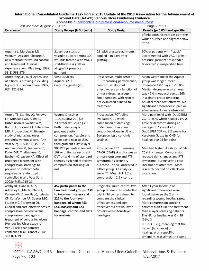
Argenta L, Morykwas MJ Vacuum- Assisted Closure: A new method for wound control and treatment: Clinical experience. Ann Plas Surg. 1997; 38(8):563-576.
31 venous stasis or vasculitic ulcers among 300 wounds treated with VAC + split-thickness graft or allograft + pressure garment
CS with pressure garments applied ~10 days after grafting
90% of patients with “stasis” ulcers treated with VAC + graft + pressure garment “responded favorably” in unspecified time.
Armstrong SH, Ruckley CV. Use of a fibrous dressing in exuding leg ulcers. J Wound Care. 1997; 6(7):322-324.
Venous ulcers Aquacel (21) Calcium alginate (23)
Prospective, multi-center, RCT measuring performance, comfort, safety, cost effectiveness as a function of primary dressing group. Small samples, with results not evaluated blinded to treatment.
Mean wear time in the Aquacel group was longer (mean difference 1.02 days, p < 0.05). Median decrease in ulcer area was 42% in Aquacel versus 26% in alginate group rendering Aquacel more cost effective. No significant differences in pain or adverse events were observed.
Arnold TE, Stanley JC, Fellows EP, Moncada GA, Allen R., Hutchinson JJ, Swartz WM, Bolton LL, Vickers CFH, Kerstein MD. Prospective, Multicenter study of managing lower extremity venous ulcers. Ann Vasc Surg. 1994;9(4):356-62.
Wound Dressings: 1.DuoDERM CGF (35) 2.Xeroform® Gauze (35) Both under 2-layer gradient elastic compression: flexible zinc oxide paste next to skin, then gradient elastic layer.
Prospective, RCT, blind evaluation, 10 week comparison of dressings under compression on venous leg ulcers in US and European leg ulcer clinic settings
More pain relief with DuoDERM CGF ulcers, which healed 71% vs 43% for Xeroform during an average of 7.2 weeks for DuoDERM CGF vs. 9.2 weeks for Xeroform Gauze (p>0.05 for healing; p<0.05 for pain)
Aschwanden M, Jeanneret C, Koller MT, Thalhammer C, Bucher HC, Jaeger KA. Effect of prolonged treatment with compression stockings to prevent post-thrombotic sequelae: a randomized controlled trial. J Vasc Surg. 2008;47(5):1015-21.
900 PTS patients screened 169 with first or recurrent DVT after 6 mo of standard therapy assigned to receive compression stockings or not
Prospective RCT measuring C4-C6 (CEAP) skin changes as primary outcome and PTS symptoms as secondry outcome. No VU observed in either group. All analysis were ITT. Mean FU 3.2 y compression, 2.9 y control
Men had higher likelihood of C4-C6 skin changes. Compression reduced skin changes and PTS symptoms during year 1 post DVT but not after that. More research needed on effects on ulceration.
Ashby RL, Gabe R, Ali S, Adderley U, Martin Bland J, Cullum NA, Dumville JC, Iglesias CP, Kang’ombe AR, Soares MO, Stubbs NC, Torgerson DJ. Clinical and cost-effectiveness of compression hosiery versus compression bandages in treatment of venous leg ulcers (Venous leg Ulcer Study IV, VenUS IV); a randomized controlled trial. Lancet 2014; 383:871-79.
457 participants to the two treatment groups: 230 to two-layer hosiery and 227 to the four-layer bandage, of whom 453 (230 hosiery and 223 bandage) contributed data for analysis.
Pragmatic, multi-centre, two group randomized controlled trial in 34 centers aimed to compare the clinical effectiveness and cost-effectiveness of two-layer hosiery versus four-layer bandages.
After 1 year followup no significant differences were found between the 2 arms regarding wound healing rates. More compression stocking patients didn’t like the treatment than 4 layers dressing patients.
The HR for healing was 0・99 (95% CI
0・79 (・25), meaning that the hazard (ie, chance) of healing, at any specifi c timepoint, was almost the same

International Consolidated Guideline Task Force (2015 Update of the 2010 Association for the Advancement of Wound Care (AAWC) Venous Ulcer Guideline) Evidence
Accessible at aawconline.org/professional-resources/resources/ Last updated: August 23, 2017 Page 8 of 51
©AAWC 2016 International Consolidated Venous Ulcer Guideline Abbreviations & Evidence
Page 8 of 103 8/23/2017
References Study Groups (N Subjects) Study Design Results (p<0.05 if not specified)
in the two groups. The 95% CI indicates that hosiery might reduce the chance of healing by as much as 21% or increase it by as much as 25%.
Atillasoy E. The safety and efficacy of graftskin (Apligraf) in the treatment of venous leg ulcers: a multicenter, randomized controlled clinical trial. Wounds. 2000;12(Suppl):20A-6A
Human skin equivalent (146 patients with a VU) Dressing similar in appearance (123 similar patients) Not ITT analysis.
RCT with 8 weeks treatment measuring % completely healed and time to heal within 6 months. 18 of 309 patients randomized were lost to follow up.
VU dressed with skin equivalent healed in a mean of 61 days; 63% completely healed in 6 months. Controls healed in a mean of 181 days; 49% healed in 6 months
Baker S, Fletcher A, Glanville J, Press P, Sharp F, Sheldon T, Collum N, Semlyen A. Compression therapy for venous leg ulcers. Effective Health Care. 1997; 3(4):2-12
Review of compression studies and questions addressed by Cullum et al. More studies and different information.
Systematic review with different interpretations of some studies than Cullum et al.
-compression > no compression -elastic high > low compression - and " > inelastic compression -NS difference between different multilayer high compression systems -multilayer high compression > 1-layer bandage systems -Insufficient evidence for *stockings vs bandages * intermittent or sequential pneumatic compression
Barron GS, Jacob SE, Kirsner RS. Dermatologic complications of chronic venous disease: medical management and beyond. Ann Vasc Surg. 2007;21(5):652-62.
Case studies. LR of atrophie blanche Description and case study evidence that atrophie blanche is associated with venous insufficiency.
Barwell JR, Davies CE, Deacon J, Harvey K, Minor J, Sassano A, Taylor M, Usher J, Wakely C, Earnshaw JJ, Heather BP, Mitchell, DC, Shyman MR, Poskitt KR. Comparison of surgery and compression with compression alone in chronic venous ulceration (ESCHAR study): Random control trial. Lancet. 2004, June 5(363):1854-1858.
500 patients from three centers received venous color duplex imaging of ulcerated or recently healed wounds. These were used to guide surgical decisions.
RCT.Multilayer compression with or without superficial vein surgery or deep vein stripping, avulsion of varicosities or junction disconnection. Comparison of recurrence rates at 24 weeks and 12 months.
Surgery with compression vs. compression alone: at 24 weeks no difference (65% vs65% recurrence.At 12 months surgical: 12% vs 28% for compression alone.Surgical correction of venous reflux with compression reduces 12 month venous recurrence.
Bates-Jensen B, McNees P. The Wound Intelligence System early issues and findings from multi-site tests. Ostomy/Wound Manage. 1996; 41(10A Suppl): 53S-61S
Patients with wounds including pressure or venous ulcers
Prospective cohort of electronic wound care record system including wound and peri-ulcer skin erythema and edema.
Validation of wound care procedure decisions guided by Bates-Jensen Wound Assessment Test. (BWAT)
Beele H, de la Brassine M, Lambert J, Suys E, De Cuyper C,
CryoCeal 9 applications allogeneic human
CS of patients treated for up to 9 applications measuring
11 patients (41%) healed.
International Consolidated Guideline Task Force (2015 Update of the 2010 Association for the Advancement of Wound Care (AAWC) Venous Ulcer Guideline) Evidence
Accessible at aawconline.org/professional-resources/resources/ Last updated: August 23, 2017 Page 9 of 51
©AAWC 2016 International Consolidated Venous Ulcer Guideline Abbreviations & Evidence
Page 9 of 103 8/23/2017
References Study Groups (N Subjects) Study Design Results (p<0.05 if not specified)
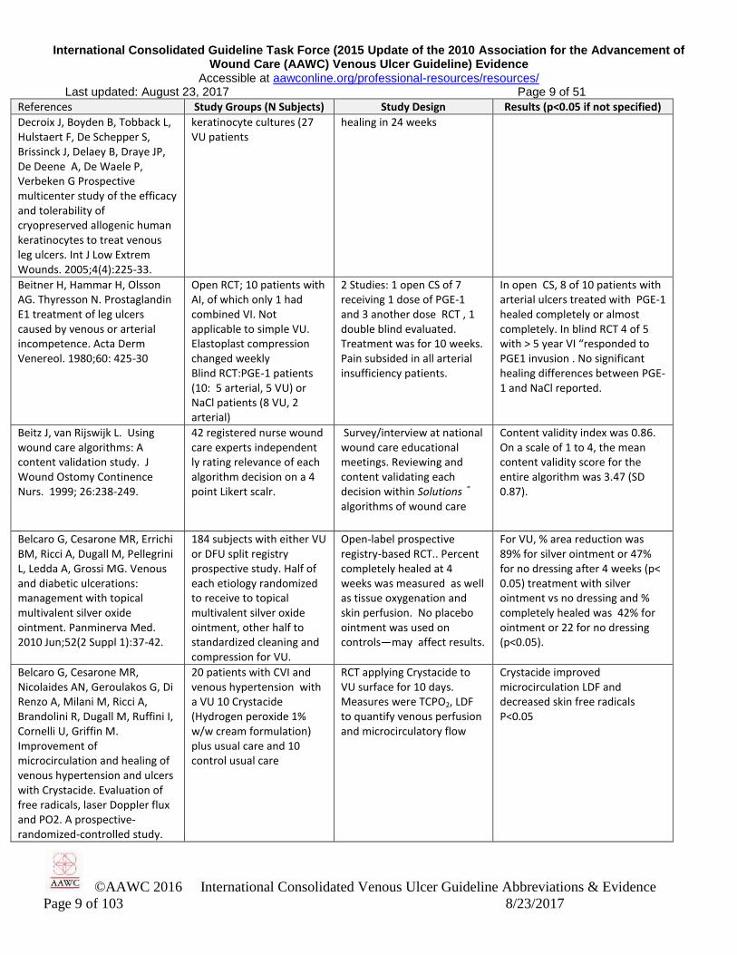
Decroix J, Boyden B, Tobback L, Hulstaert F, De Schepper S, Brissinck J, Delaey B, Draye JP, De Deene A, De Waele P, Verbeken G Prospective multicenter study of the efficacy and tolerability of cryopreserved allogenic human keratinocytes to treat venous leg ulcers. Int J Low Extrem Wounds. 2005;4(4):225-33.
keratinocyte cultures (27 VU patients
healing in 24 weeks
Beitner H, Hammar H, Olsson AG. Thyresson N. Prostaglandin E1 treatment of leg ulcers caused by venous or arterial incompetence. Acta Derm Venereol. 1980;60: 425-30
Open RCT; 10 patients with AI, of which only 1 had combined VI. Not applicable to simple VU. Elastoplast compression changed weekly Blind RCT:PGE-1 patients (10: 5 arterial, 5 VU) or NaCl patients (8 VU, 2 arterial)
2 Studies: 1 open CS of 7 receiving 1 dose of PGE-1 and 3 another dose RCT , 1 double blind evaluated. Treatment was for 10 weeks. Pain subsided in all arterial insufficiency patients.
In open CS, 8 of 10 patients with arterial ulcers treated with PGE-1 healed completely or almost completely. In blind RCT 4 of 5 with > 5 year VI “responded to PGE1 invusion . No significant healing differences between PGE-1 and NaCl reported.
Beitz J, van Rijswijk L. Using wound care algorithms: A content validation study. J Wound Ostomy Continence Nurs. 1999; 26:238-249.
42 registered nurse wound care experts independent ly rating relevance of each algorithm decision on a 4 point Likert scalr.
Survey/interview at national wound care educational meetings. Reviewing and content validating each decision within Solutions ® algorithms of wound care
Content validity index was 0.86. On a scale of 1 to 4, the mean content validity score for the entire algorithm was 3.47 (SD 0.87).
Belcaro G, Cesarone MR, Errichi BM, Ricci A, Dugall M, Pellegrini L, Ledda A, Grossi MG. Venous and diabetic ulcerations: management with topical multivalent silver oxide ointment. Panminerva Med. 2010 Jun;52(2 Suppl 1):37-42.
184 subjects with either VU or DFU split registry prospective study. Half of each etiology randomized to receive to topical multivalent silver oxide ointment, other half to standardized cleaning and compression for VU.
Open-label prospective registry-based RCT.. Percent completely healed at 4 weeks was measured as well as tissue oxygenation and skin perfusion. No placebo ointment was used on controls—may affect results.
For VU, % area reduction was 89% for silver ointment or 47% for no dressing after 4 weeks (p< 0.05) treatment with silver ointment vs no dressing and % completely healed was 42% for ointment or 22 for no dressing (p<0.05).
Belcaro G, Cesarone MR, Nicolaides AN, Geroulakos G, Di Renzo A, Milani M, Ricci A, Brandolini R, Dugall M, Ruffini I, Cornelli U, Griffin M. Improvement of microcirculation and healing of venous hypertension and ulcers with Crystacide. Evaluation of free radicals, laser Doppler flux and PO2. A prospective-randomized-controlled study.
20 patients with CVI and venous hypertension with a VU 10 Crystacide (Hydrogen peroxide 1% w/w cream formulation) plus usual care and 10 control usual care
RCT applying Crystacide to VU surface for 10 days. Measures were TCPO2, LDF to quantify venous perfusion and microcirculatory flow
Crystacide improved microcirculation LDF and decreased skin free radicals P<0.05

International Consolidated Guideline Task Force (2015 Update of the 2010 Association for the Advancement of Wound Care (AAWC) Venous Ulcer Guideline) Evidence
Accessible at aawconline.org/professional-resources/resources/ Last updated: August 23, 2017 Page 10 of 51
©AAWC 2016 International Consolidated Venous Ulcer Guideline Abbreviations & Evidence
Page 10 of 103 8/23/2017
References Study Groups (N Subjects) Study Design Results (p<0.05 if not specified)
Angiology. 2003;54(3):325-30.
Belcaro G, Cesarone R, Nicolaides AN, De Sanctis, MT. Treatment of venous ulcers with pentoxifylline: a 6-month, double blinded placebo controlled trial. Angiology 2002;53(Supp 1): s45-7
Placebo (88 VU patients) pentoxifylline (PXF; 400 mg 82 VU patients), both groups comparable in age and gender were treated 3 times daily with similar elastic compression
Double-blind RCT of 6 month duration measured number of limbs completely healed and % wound area reduction.
After 6 months 67% of PXF patients healed and 31% of placebo-treated patients healed (p< 0.02). 87% area reduction from baseline in PXF group compared to 47% in placebo group. PXF added cost (21%) was less than added cost of delayed healing in placebo group.
Bello M, Scriven M, Hartshorne T, Bell, PRF, Naylor AR, London NJM. Role of superficial venous surgery in the treatment of venous ulceration. Brit J Surg. 1999; 86:755-59.
122 legs with VU and normal deep veins underwent superficial venous surgery
Prospective case series Post op treatment :non-adherent gauze and Tubigrip (8mm) Ulcers assessed every 8 weeks
VU post-op healing rates: Median time to healing 18 weeks, Cumulative 12 month healing rate 82% No recurrence data
Benigni, J.P., Sadoun. S, Allaert FA, Vin F. Comparative Study of the effectiveness of Class 1 compression stockings on the symptomatology of early chronic venous disease. Phlebologie 2003;56:117-125.
125 subjects- Comparison of class 1 compression stockings with identically looking, non-active stockings (pressure < 7mmHg) in patients with early stages of venous disease
Randomized, multi-center cross-over study
Statistically highly significant differences in favor of the class 1 stockings were found for pain, for all other parameters of discomfort except parasthesia and for the QOL dimensions for mood and every day work. The relief of symptoms with the class 1 stockings was 2x that of the control.
Bennett ML, Jackson JM, Jorizzo JL, Fleischer Jr. AB, White WL, Callen JP. Pyoderma gangrenosum. A comparison of typical and atypical forms with an emphasis on time to remission. Case review of 86 patients from 2 institutions. Medicine(Baltimore) 2000;79(1):37-46.
86 patients from two institutions with venous insufficiency and Pyoderma gangrenosum.
Clinical Study – Case series Description of typical and atypical wound progress.
Bérard A, Abenhaim L, Platt R, Kahn SR, Steinmetz O. Risk factors for the first-time development of venous ulcers of the lower limbs: the influence of heredity and physical activity.
200 clients with a first VU 200 matched subjects with no VU
Prospective Case Control study of 200 subjects with first VU compared to 200 subjects matched on referring physician, age + 5 years and gender presenting
Significant predictors of VU are: Family history of maternal VU Vigorous exercise History of DVT Multiple pregnancy
International Consolidated Guideline Task Force (2015 Update of the 2010 Association for the Advancement of Wound Care (AAWC) Venous Ulcer Guideline) Evidence
Accessible at aawconline.org/professional-resources/resources/ Last updated: August 23, 2017 Page 11 of 51
©AAWC 2016 International Consolidated Venous Ulcer Guideline Abbreviations & Evidence
Page 11 of 103 8/23/2017
References Study Groups (N Subjects) Study Design Results (p<0.05 if not specified)
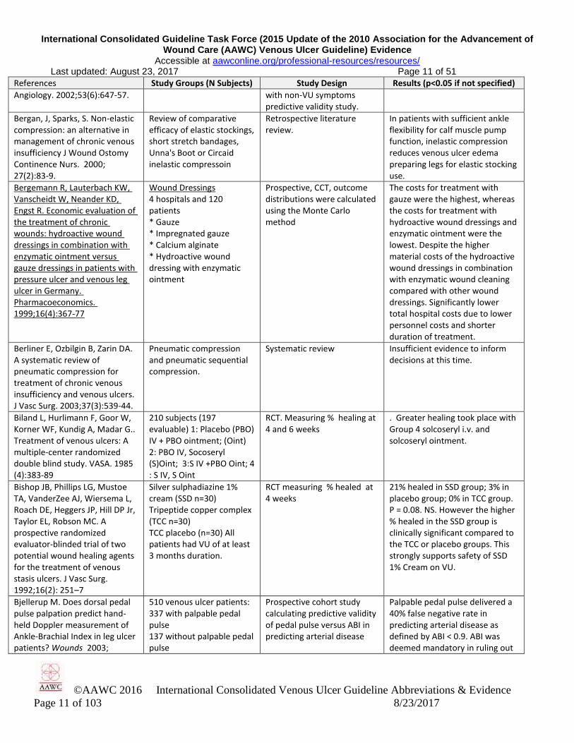
Angiology. 2002;53(6):647-57. with non-VU symptoms predictive validity study.
Bergan, J, Sparks, S. Non-elastic compression: an alternative in management of chronic venous insufficiency J Wound Ostomy Continence Nurs. 2000; 27(2):83-9.
Review of comparative efficacy of elastic stockings, short stretch bandages, Unna's Boot or Circaid inelastic compressoin
Retrospective literature review.
In patients with sufficient ankle flexibility for calf muscle pump function, inelastic compression reduces venous ulcer edema preparing legs for elastic stocking use.
Bergemann R, Lauterbach KW, Vanscheidt W, Neander KD, Engst R. Economic evaluation of the treatment of chronic wounds: hydroactive wound dressings in combination with enzymatic ointment versus gauze dressings in patients with pressure ulcer and venous leg ulcer in Germany. Pharmacoeconomics. 1999;16(4):367-77
Wound Dressings 4 hospitals and 120 patients * Gauze * Impregnated gauze * Calcium alginate * Hydroactive wound dressing with enzymatic ointment
Prospective, CCT, outcome distributions were calculated using the Monte Carlo method
The costs for treatment with gauze were the highest, whereas the costs for treatment with hydroactive wound dressings and enzymatic ointment were the lowest. Despite the higher material costs of the hydroactive wound dressings in combination with enzymatic wound cleaning compared with other wound dressings. Significantly lower total hospital costs due to lower personnel costs and shorter duration of treatment.
Berliner E, Ozbilgin B, Zarin DA. A systematic review of pneumatic compression for treatment of chronic venous insufficiency and venous ulcers. J Vasc Surg. 2003;37(3):539-44.
Pneumatic compression and pneumatic sequential compression.
Systematic review Insufficient evidence to inform decisions at this time.
Biland L, Hurlimann F, Goor W, Korner WF, Kundig A, Madar G.. Treatment of venous ulcers: A multiple-center randomized double blind study. VASA. 1985 (4):383-89
210 subjects (197 evaluable) 1: Placebo (PBO) IV + PBO ointment; (Oint) 2: PBO IV, Socoseryl (S)Oint; 3:S IV +PBO Oint; 4 : S IV, S Oint
RCT. Measuring % healing at 4 and 6 weeks
. Greater healing took place with Group 4 solcoseryl i.v. and solcoseryl ointment.
Bishop JB, Phillips LG, Mustoe TA, VanderZee AJ, Wiersema L, Roach DE, Heggers JP, Hill DP Jr, Taylor EL, Robson MC. A prospective randomized evaluator-blinded trial of two potential wound healing agents for the treatment of venous stasis ulcers. J Vasc Surg. 1992;16(2): 251–7
Silver sulphadiazine 1% cream (SSD n=30) Tripeptide copper complex (TCC n=30) TCC placebo (n=30) All patients had VU of at least 3 months duration.
RCT measuring % healed at 4 weeks
21% healed in SSD group; 3% in placebo group; 0% in TCC group. P = 0.08. NS. However the higher % healed in the SSD group is clinically significant compared to the TCC or placebo groups. This strongly supports safety of SSD 1% Cream on VU.
Bjellerup M. Does dorsal pedal pulse palpation predict hand-held Doppler measurement of Ankle-Brachial Index in leg ulcer patients? Wounds 2003;
510 venous ulcer patients: 337 with palpable pedal pulse 137 without palpable pedal pulse
Prospective cohort study calculating predictive validity of pedal pulse versus ABI in predicting arterial disease
Palpable pedal pulse delivered a 40% false negative rate in predicting arterial disease as defined by ABI < 0.9. ABI was deemed mandatory in ruling out

International Consolidated Guideline Task Force (2015 Update of the 2010 Association for the Advancement of Wound Care (AAWC) Venous Ulcer Guideline) Evidence
Accessible at aawconline.org/professional-resources/resources/ Last updated: August 23, 2017 Page 12 of 51
©AAWC 2016 International Consolidated Venous Ulcer Guideline Abbreviations & Evidence
Page 12 of 103 8/23/2017
References Study Groups (N Subjects) Study Design Results (p<0.05 if not specified)
15(7):237-240. arterial disease.
Black SR Venous stasis ulcers: A review. Ostomy/Wound Manage. 1995; 41(8):20-9.
Review
Blair SD, Wright DDI, Backhouse CM, Riddle E, McCollum CN. Sustained compression and healing of chronic venous ulcers. BMJ. 1988;297(6657):1159-61.
1. Adhesive plaster (AP) control compression (20) 2. 4-layer elastic compression bandage (4LB) (20) 126 consecutive patients whose ulcers had not healed in a mean of 27.2 (StdErr 8) months were subsequently managed with 4LB for 12 weeks.
Compression was measured every 2 hours up to 8 hours after application, at 24 and 7 days after application as well as healing and recurrence. Correlated compression to ankle circumference reduction. % healed was measured after 12 weeks on 4LB
Initially applied ankle—knee compression: 4LB 43—17 mmHg, maintained at ankle >35 mmHg for 7 days versus AP 30—6 mmHg on AP application declining to <20 mmHg after 8 hours. 4LB applied more consistent compression across appliers. 4LB reduced edema more and healed 74% of ulcers on 110 patients (not patients) at 12 weeks
Blair SD, Backhouse CM, Wright DDI, Riddle E, McCollum CN. Do dressings influence the healing of chronic venous ulcers? Phlebology. 1988;3(2):129–34.
1% Silver sulphadiazine cream (30) Non-adhering gauze (30)
RCT comparing % of VU healed at 12 weeks and % VU area reduction
12 week 63 % SSD or NADressing 80% % healed (NS—also NS % area reduction) subjects in silver sulphadiazine experienced erythema , pruritis vs. 0 in NA group (NS)
Bland, JM, Dumville JC, Ashby RL, Gabe R , Stubbs N, Adderley U, Kang’ombe AR, Cullum NA. Validation of the VEINES-QOL quality of life instrument in venous leg ulcers: repeatability and validity study embedded in a randomized clinical trial. BMC Cardiovascular Disorders. 2015;15(1):85-9.
451 participants in the VenUS IV trial which compared 2-layer to 4-layer compression effects on healing.
Prospective RCT validation of quality of life measures including pain, SF-12 items, and healing after 2 weeks and 4 months. Integrity of a VEINES-SYM subscale was tested by factor analysis of correlations among items.
No floor- or ceiling-effects were observed. Item-item correlations were weak to moderate. Item-total score corelations were moderate. Internal reliability was good. WEINES-SYM subscale was confirmed by factor analysis. Internal reliability was good and test-retest satisfactory to good. Healed clients reported higher scores than those not healed.
Blecken SR, Villavicencio JL, Kao TC. Comparison of elastic versus nonelastic compressiion in bilateral venous ulcers: a randomized trial. J Vasc Surg. 2005;42(6):1150-5.
12 patients with bilateral VUs randomly assigned to either: -Circaid™ (12 legs) -4-layer bandage (12 legs)
12-week RCT of same-subject different leg VUs. Measures: % healed, patient satisfaction every 4 weeks, duplex ultrasound, phlebography, air plethysmography documented nature and site of obstruction
Circaid™ group healed 4.14 cm
2/week; 4LB 1.22 cm
2/week
(p=0.011. Cox proportional hazard ratio for healing greater for Circaid (p=0.017)
Blomgren L, Johansson G, Siegbahn A, Bergqvist D. Coagulation and fibrinolysis in chronic venous insufficiency, Vasa, 2001; 30(3):184-7.
20 patients with CVI 20 matched controls
Blood samples were analyzed to correlate plasma markers with ulcer development.
Increased levels of PAI-I and tPA in patients with CVI compared to controls
Blumberg SN, Maggi J, Melamed J, Golinko M, Ross F, Chen W. A Histopathologic basis for surgical debridement to
26 consecutive clients with a lower extremity VU of at least 4 weeks duration. Only 13 had surgical
Prospectie evaluation of biopsies obtained during surgical debridement to the subcutaneous level. Biopsies
Baseline area was 34.7 cm2
. 89% had continuously decreasing VU area. Specimens with dense fibrosis, decreased cellularity,
International Consolidated Guideline Task Force (2015 Update of the 2010 Association for the Advancement of Wound Care (AAWC) Venous Ulcer Guideline) Evidence
Accessible at aawconline.org/professional-resources/resources/ Last updated: August 23, 2017 Page 13 of 51
©AAWC 2016 International Consolidated Venous Ulcer Guideline Abbreviations & Evidence
Page 13 of 103 8/23/2017
References Study Groups (N Subjects) Study Design Results (p<0.05 if not specified)
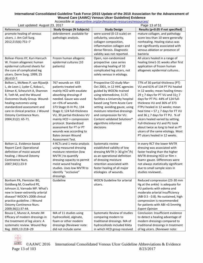
promote healing of venous ulcers. J. Am Coll Surg. 2012;215(6):751–7
debridement available for pathologic analysis.
were scored (0-13 scale) on cellularity, vascularity, collagen composition, inflammation collagen and dense fibrosis. Diagnostic validity was not reported.
mature collagen, and pathology score less than 10 were generally nonhealing. Healing status was not significantly associated with venous ablation or presence of bacteria
Bolivar-Flores XY, Kuri-Harcuch W. Frozen allogeneic human epidermal cultured sheets for the cure of complicated leg ulcers. Derm Surg. 1999; 25 (8):610-7.
Frozen human allogenic epidermal cultures (10 patients)
Open, non-randomized prospective case series observing healing of 10 complicated leg ulcers, not solely venous in etiology.
All ulcers healed in a range of healing time1-31 weeks after first application of frozen human allogeneic epidermial cultures..
Bolton L, McNees P, van Rijswijk L, de Leon J, Lyder C, Kobza L, Edman K, Scheurich A, Shannon R, Toth M, and the Wound Outcomes Study Group. Wound-healing outcomes using standardized assessment and care in clinical practice. J Wound Ostomy Continence Nurs. 2004;31(2): 65-71.
767 wounds on 433 patients treated with mainly HCD with exudate absorbing dressings if needed. Gauze was used on <5% of wounds. 373 Stage III-IV PU, 134 Stage II; 124 full-thickness VU, 30 partial-thickness VU mainly HCD + compression protocol. Standardized assessment for FT or PT wounds was according to Bates-Jensen Wound Assessment Test.
Prospective CO study Mar-Oct 2001, in 12 HHC agencies guided by WOCNs trained using telemedicine, 3 LTC facilities a University hospital based Long Term Acute Care setting avoiding gauze, using moisture retentive dressings and compression for VU. Content validated Solutions® algorithms informed decisions
77% of 30 partial-thickness (PT) VU and 61% of 134 PT PU healed in 12 weeks; mean healing times: 29 + 7 days for PT VU and 31+ 7 days for PT PU. 44% of 124 full-thickness VU and 36% of 373 FTPU healed in 12 weeks; mean heal time = 57 + 7 days for FT VU and 36 + 7 days for FT PU. % of ulcers healed varied by setting. Full-thickness VU and PU took about twice as long to heal as PT ulcers of the same etiology. Most PT ulcers healed in 12 weeks.
Bolton LL. Evidence-based Report Card: Operational definition of moist wound healing. J Wound Ostomy Continence Nurs. 2007;34(1):23-9
4 RCTs and 1 meta-analysis using measured dressing moisture retention (low MVTR ) to quantify dressing capacity to permit moist wound healing studies. Uses low MVTR to identify “occlusive” dressings.
Systematic review established validity of low dressing MVTR (< 30 g/m
2/h)
as an operational definition of dressing moisture retention associated with faster healing of all major etiologies of wounds.
In every RCT the lower MVTR dressing was associated with faster healing than the higher MVTR dressing HCD or film > foam> gauze. Differences were not always statistically significant due to small sample sizes in studies reviewed..
Bonham PA, Flemister BG, Goldberg M, Crawford PE, Johnson JJ, Varnado MF. What's new in lower-extremity arterial disease? WOCN's 2008 clinical practice guideline. J Wound Ostomy Continence Nurs. 2009;36(1):37-44.
Guideline WOCN Guideline for arterial ulcers.
Reduced compression (23-30 mm Hg at the ankle) is adequate for VU patients with edema and moderate arterial insufficiency (ABI 0.5 - 0.8). No sustained, high compression is recommended for patients with ABI <0.5mmHg Expert Opinion
Bouza C, Munoz A, Amate JM Efficacy of modern dressings in the treatment of leg ulcers: A systematic review. Wound Rep Reg. 2005;13:218–29
MA of 11 studies using hydrocolloid, alginate, foam or other modern dressings (Reviewer note: did not include some
Systematic Review of studies comparing modern to traditional dressings. MA of hydrocolloids included Kikta in which HCD group received
Conclusion: Insufficient evidence to detect a healing advantage of modern dressings compared to traditional dressings in treatment of leg ulcers. (Reviewer note:

International Consolidated Guideline Task Force (2015 Update of the 2010 Association for the Advancement of Wound Care (AAWC) Venous Ulcer Guideline) Evidence
Accessible at aawconline.org/professional-resources/resources/ Last updated: August 23, 2017 Page 14 of 51
©AAWC 2016 International Consolidated Venous Ulcer Guideline Abbreviations & Evidence
Page 14 of 103 8/23/2017
References Study Groups (N Subjects) Study Design Results (p<0.05 if not specified)
studies which which significantly favored hydrocolloid healing, e.g. Meredith, 1988, )
no compression and was compared to compression.
Inappropriate inclusion of Kikta study may have biased this SR against hydrocolloid dressings.)
Bradley M, Cullum N, Sheldon T. Systematic reviews of wound care management: (2) Dressings and topical agents used in the healing of chronic wounds. Health Technol Assess2001;3(17 Pt 1)
SR of dressings and topical agents in wound care
SR of topical agents and dressings used in healing all chronic wounds, not just VU
Supports using topical agents and wound dressings that minimize pain, manage wound fluid and protect skin from physical or chemical trauma.
Brassard, A. A Prospective, multi-centre, randomized, controlled clinical investigation of Dermagraft in patients with venous leg ulcers: a feasibility study. Canadian J Plastic Surg. 2002;10:17A-22A.
Dermagraft + multilayer compression bandage, 13 patients Multilayer compression bandage, 13 patients
Prospective, multicentre, pilot Randomized, Controlled, feasibility study (not sufficiently powered for statistical significance).
38% (5/13) healed with Dermagraft + compression, 15% (2/13) healed with compression alone (control group).
Brem, H, Balledux, J, Sukkarieh, T, Carson, P, Falanga, V. Healing of venous ulcers of long duration with a bilayered living skin substitute: results from a general surgery and dermatology department. J Foot Ankle Surg. 1999:38(6):388-93.
33 patients with 54 VU >1 year duration at a general surgery department of a major medical center and a dermatology department of a university-based hospital during the study were treated with fenestrated living skin equivalent
CS Retrospective chart review of healing results. Repeated surgical debridement and treatment with living skin equivalend after 7 days was practiced.
74% of VUs completely healed in 6 months, after a median of 2 living skin equivelane applications. Mean healing time was 55 to 61 days. VU treated in the surgery and dermatology departments were similar in wound size and duration and patient population.
Breuing KH, Bayer L, Neuwalder J, Orgill DP. Early experience using low-frequency ultrasound in chronic wounds. Ann Plast Surg. 2005;55(2):183-7.
Low Frequency Ultrasound Debridement (17 VU patients)
CS over 8 months debridement and bacterial biofilm destruction with minimum follow up of 3 months
20-30% reduction in wound area during up to 3 months. No patient required antibiotics.
Briggs M, Nelson EA, Martyn-St James M. Topical agents or dressings for pain in venous leg ulcers. Cochrane Database of Systematic Reviews 2012(11): CD001177.
6 RCTs of EMLA lidocaine-prilocaine cream 2 RCT on 470 participants studied Foam w/without ibubrofen (studied only on first evening of use.
MA of 6 RCTs on 343 participants measured debridement pain reported using 5% Eutectic Mixture of Local Anesthetic (EMLA) lidocaine-prilocaine cream
EMLA significantly reduced pain. Effect on healing is uncertain. No effect of Ibuprofen on VU pain first evening of use. (Both RCTs effectively reduced pain during the first week of use)
Brizzio E, Amsler F, Lun B, Blättler W. Comparison of low-strength compression stockings with bandages for the treatment of recalcitrant venous ulcers. J Vasc Surg. 2010;51(2):410-6
Medical compression stockings (28) Short Stretch bandages (27)
RCT of healing within 90 days NS difference between the two groups in any healing, pain or QoL parameter, time to heal identical. Both alleviated pain promptly. QoL improved only in patients who healed.
Brodovicz KG, McNaughton K, Uemura N, Meininger G, Girman CJ, Yale SH. Reliability and
Convenience sample of 20 patients with type 2 diabetes with varying levels
Compared 8 methods of edema assessment evaluated independently by 3 nurses:
Water displacement and ankle circumference had high inter-examiner agreement (intraclass

International Consolidated Guideline Task Force (2015 Update of the 2010 Association for the Advancement of Wound Care (AAWC) Venous Ulcer Guideline) Evidence
Accessible at aawconline.org/professional-resources/resources/ Last updated: August 23, 2017 Page 15 of 51
©AAWC 2016 International Consolidated Venous Ulcer Guideline Abbreviations & Evidence
Page 15 of 103 8/23/2017
References Study Groups (N Subjects) Study Design Results (p<0.05 if not specified)
feasibility of methods to quantitatively assess peripheral edema. Clin Med Res. 2009;7(1-2):21-31.
of edema (1) clinical assessment of pit depth and recovery at three locations, (2) patient questionnaire, (3) ankle circumference, (4) 8-point (5) edema tester using a plastic card with holes of varying size pressed to the ankle with a blood pressure cuff), (6) modified edema tester (with bumps), (7) indirect leg volume (by series of ankle/leg circumferences), (8) foot/ankle volumetry by water displacement.
correlation coefficient 0.93, 0.96 right; 0.97, 0.97 left). Less consistent for figure-of-eight (0.64, 0.86), or indirect leg volume (0.53, 0.66), which had low edema results and clinical assessments at all sites. Results varied by pressure administered. Classic subjective clinical assessment correlated well for nurse-performed assessments and patient questionnaire. Ankle circumference and patient questionnaires each took 1 minute to complete. Other tools took >5 minutes.
Burnand K, Clemenson G, Morland M, Jarret PE. Venous lipodermato-sclerosis: treatment by fibrinolytic enhancement and elastic compression. Br Med J. 1980;280:7-11
34 legs of 23 patients being treated for lipodermato-sclerosis randomly assigned to receive either oral stanazalol (14) or placebo (9) first with elastic stocking compression.
Randomized crossover trial, with patients crossed over to the other treatment after 3 months, who then received 3 months with the other oral randomized treatment. Area of lipodermatosclerosis was measured at 0, 3, 6 months.
Both groups reduced areas of lipodermatosclerosis (p< 0.001), the stanozolol group slightly more than the placebo group (p = 0.10) Sample size was too small for statistical significance.
Burton C. Venous ulcers. Amer J Surg. 1994;167(1A Suppl): 37S-41S.
Hydrocolloid Dressing DuoDERM® or DuoDERM® CGF (5 studies: 181 subjects) Hydrocolloid Dressing Comfeel® (1 study: 30 subjects) Gauze or Unna's boot (3 studies: 54 subjects)
Review of venous ulcer studies using compression and reporting healing times and/or % wound contraction per week and summary of protocol of care and infection rates experienced in Duke University ambulatory leg ulcer clinic.
Infections noted at 1% of weekly dressing changes despite heavy colonization. Healing review: HCD D family: 50% healed in 12 weeks to 82% healed in 50 days. HCD C: 43% healed in 12 weeks Gauze/Unna's boot: 23-43% healed in 12 weeks.
Burton CS. Treatment of leg ulcers. Dermatol Clinics. 1993; 11(2):315-323.
LR LR and Expert opinion Venous disease is associated with venous hypertension and responds poorly to diuretic therapy.
Callam MJ, Harper DR, Dale JJ, Brown D, Gibson B, Prescott RJ, Ruckley CV. Lothian and forth valley leg ulcer healing trial .1. Elastic versus nonelastic bandaging in the treatment of chronic leg ulceration. Phlebology.1992;7:136-141.
Compression: 1. Elastic: orthopaedic wool (Soffban), Tensopress + Tensoshape (65) 2.Non-elastic: orthopaedic wool (Soffban), Elastocrepe + Tensoplus Forte; (67)
Prospective RCT for 12 weeks in leg ulcer clinics in Scotland UK
% completely healed at 12 weeks was: 54% for elastic compression 28% for non-elastic compression
Callam MJ, Ruckley CV, Dale JJ, Harper DR. Hazards of compression treatment of the leg: an estimate from Scottish
Review of safety of compression stockings and bandages
Literature review by surgical specialists in Scotland.
High levels of compression in patients with arterial disease can lead to adverse effects and amputation.
International Consolidated Guideline Task Force (2015 Update of the 2010 Association for the Advancement of Wound Care (AAWC) Venous Ulcer Guideline) Evidence
Accessible at aawconline.org/professional-resources/resources/ Last updated: August 23, 2017 Page 16 of 51
©AAWC 2016 International Consolidated Venous Ulcer Guideline Abbreviations & Evidence
Page 16 of 103 8/23/2017
References Study Groups (N Subjects) Study Design Results (p<0.05 if not specified)
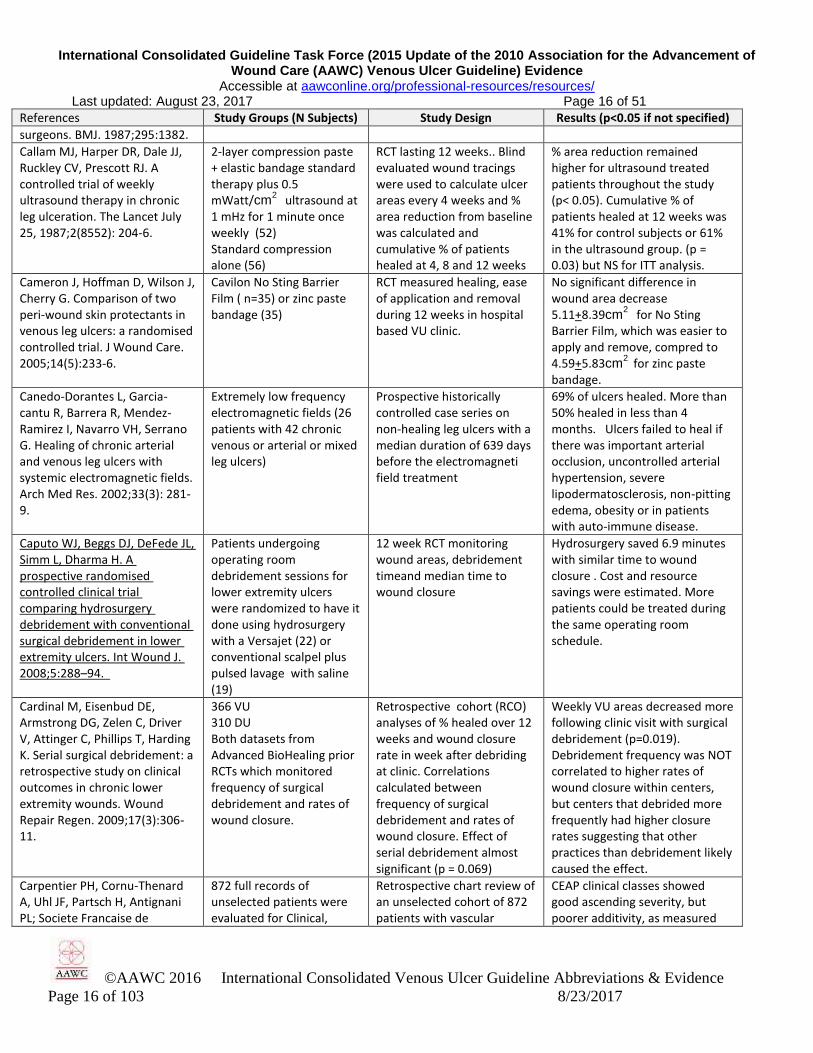
surgeons. BMJ. 1987;295:1382.
Callam MJ, Harper DR, Dale JJ, Ruckley CV, Prescott RJ. A controlled trial of weekly ultrasound therapy in chronic leg ulceration. The Lancet July 25, 1987;2(8552): 204-6.
2-layer compression paste + elastic bandage standard therapy plus 0.5 mWatt/cm
2 ultrasound at
1 mHz for 1 minute once weekly (52) Standard compression alone (56)
RCT lasting 12 weeks.. Blind evaluated wound tracings were used to calculate ulcer areas every 4 weeks and % area reduction from baseline was calculated and cumulative % of patients healed at 4, 8 and 12 weeks
% area reduction remained higher for ultrasound treated patients throughout the study (p< 0.05). Cumulative % of patients healed at 12 weeks was 41% for control subjects or 61% in the ultrasound group. (p = 0.03) but NS for ITT analysis.
Cameron J, Hoffman D, Wilson J, Cherry G. Comparison of two peri-wound skin protectants in venous leg ulcers: a randomised controlled trial. J Wound Care. 2005;14(5):233-6.
Cavilon No Sting Barrier Film ( n=35) or zinc paste bandage (35)
RCT measured healing, ease of application and removal during 12 weeks in hospital based VU clinic.
No significant difference in wound area decrease 5.11+8.39cm
2 for No Sting
Barrier Film, which was easier to apply and remove, compred to 4.59+5.83cm
2 for zinc paste
bandage.
Canedo-Dorantes L, Garcia-cantu R, Barrera R, Mendez-Ramirez I, Navarro VH, Serrano G. Healing of chronic arterial and venous leg ulcers with systemic electromagnetic fields. Arch Med Res. 2002;33(3): 281-9.
Extremely low frequency electromagnetic fields (26 patients with 42 chronic venous or arterial or mixed leg ulcers)
Prospective historically controlled case series on non-healing leg ulcers with a median duration of 639 days before the electromagneti field treatment
69% of ulcers healed. More than 50% healed in less than 4 months. Ulcers failed to heal if there was important arterial occlusion, uncontrolled arterial hypertension, severe lipodermatosclerosis, non-pitting edema, obesity or in patients with auto-immune disease.
Caputo WJ, Beggs DJ, DeFede JL, Simm L, Dharma H. A prospective randomised controlled clinical trial comparing hydrosurgery debridement with conventional surgical debridement in lower extremity ulcers. Int Wound J. 2008;5:288–94.
Patients undergoing operating room debridement sessions for lower extremity ulcers were randomized to have it done using hydrosurgery with a Versajet (22) or conventional scalpel plus pulsed lavage with saline (19)
12 week RCT monitoring wound areas, debridement timeand median time to wound closure
Hydrosurgery saved 6.9 minutes with similar time to wound closure . Cost and resource savings were estimated. More patients could be treated during the same operating room schedule.
Cardinal M, Eisenbud DE, Armstrong DG, Zelen C, Driver V, Attinger C, Phillips T, Harding K. Serial surgical debridement: a retrospective study on clinical outcomes in chronic lower extremity wounds. Wound Repair Regen. 2009;17(3):306-11.
366 VU 310 DU Both datasets from Advanced BioHealing prior RCTs which monitored frequency of surgical debridement and rates of wound closure.
Retrospective cohort (RCO) analyses of % healed over 12 weeks and wound closure rate in week after debriding at clinic. Correlations calculated between frequency of surgical debridement and rates of wound closure. Effect of serial debridement almost significant (p = 0.069)
Weekly VU areas decreased more following clinic visit with surgical debridement (p=0.019). Debridement frequency was NOT correlated to higher rates of wound closure within centers, but centers that debrided more frequently had higher closure rates suggesting that other practices than debridement likely caused the effect.
Carpentier PH, Cornu-Thenard A, Uhl JF, Partsch H, Antignani PL; Societe Francaise de
872 full records of unselected patients were evaluated for Clinical,
Retrospective chart review of an unselected cohort of 872 patients with vascular
CEAP clinical classes showed good ascending severity, but poorer additivity, as measured

International Consolidated Guideline Task Force (2015 Update of the 2010 Association for the Advancement of Wound Care (AAWC) Venous Ulcer Guideline) Evidence
Accessible at aawconline.org/professional-resources/resources/ Last updated: August 23, 2017 Page 17 of 51
©AAWC 2016 International Consolidated Venous Ulcer Guideline Abbreviations & Evidence
Page 17 of 103 8/23/2017
References Study Groups (N Subjects) Study Design Results (p<0.05 if not specified)
Medicine Vasculaire; European Working Group on the Clinical Characterization of Venous Disorders. J Vasc Surg. 2003; 37(4):827-33.
Etiologic, Anatomic and Physiologic variables of the CEAP.
disease were abstracted to determine validity of ascending severity and additivity of CEAP clinical scores.
with the Cronbach alpha coefficient. Additivity was satisfactory in highest clinical severity cases, but poorer in the lower 3 classes.
Carter MJ, Fylling CP, Li WW, de Leon J, Driver VR, Serena TE, Wilson J. Analysis of run-in and treatment data in a wound outcomes registry: clinical impact of topical platelet-rich plasma gel on healing trajectory Int Wound J. 2011;8(6):638-50.
(AutoloGel™, Cytomedix, Inc) treatment registry of 285 chronic wounds, 46 had run-in and post-treatment data. Seven chronic wound categories were identified with mean duration of 52.4 days. Pre-treatment (baseline) was compared with post treatment results.
General linear model repeated measures analysis of robust Autologel™ data set Registry compared pre-post treatment changes in % ulcer area or mm depth reduction during run-in period to those during platelet-rich plasma (PRP) gel therapy protocol. Unblinded application and evaluation—potential bias.
Improvements (p< 0.05) were observed between run-in and post-treatment period at multiple time points for % area reduction and depth reduction ≥50%. Kaplan-Meier analysis showed during run-in, 15% wound area reduction vs. 28% post-treatment. 11% of wounds reduced in depth during run-in compared to 39% post-treatment.
CDC. Steps to prevent antimicrobial resistance. Accessed February 2, 2016, at www.cdc.gov/ drugresistance/healthcare/ha/12steps_HA.htm
Campaign to prevent antimicrobial resistance in healthcare settings Fact Sheet; general guidelines
EO consensus of expert opinion
Target definitive antibiotic therapy to known pathogens identified through C&S. Treat infection, not contaminants or colonization. Monitor response to treatment & adjust or stop when indicated.
Chaby G, Senet P, Vaneau M, Martel P, Guillaume JC, Meaume S, Téot L, Debure C, Dompmartin A, Bachelet H, Carsin H, Matz V, Richard JL, Rochet JM, Sales-Aussias N, Zagnoli A, Denis C, Guillot B, Chosidow O. Dressings for acute and chronic wounds: a systematic review. Arch Dermatol. 2007;143(10):1297-304
Acute or chronic wounds including some VU dressed with all “modern dressings” regardless of their capacity to retain moisture, including HCDs, alginates, films, hydrofiber or gauze
Review of MEDLINE, EMBASE and Cochrane databases 1990-2006 and derivative references for studies reporting wound healing, pain, infection or dressing exudate management, and trauma on removal or ease of use.
11 RCTs and 3 meta-analyses led to conclusion that HCD were only form of dressing with strong evidence of healing advantage over impregnated gauze
Chaby G. Management of leg ulcers. Rev Prat. 2010;20;60(7):970-8.
Review of comparative studies using any systemic or local therapy for treating a leg ulcer of any etiology.
LR of comparative studies. VU cleansing does not require antiseptics. Debridement is an accepted practice but no RCTs tested efficacy on VU. No systemic treatment has any indication in treatment or prevention of ulcers. Consider systemic antibiotics only if VU presents clinically significant infection
Chakrabarty A, Phillips T. Leg ulcers of unusual causes. Int J Low Extrem Wounds
Review of studies publishing unusual causes of leg ulcers
LR List of unusual causes of leg ulcers and diagnostic cues.

International Consolidated Guideline Task Force (2015 Update of the 2010 Association for the Advancement of Wound Care (AAWC) Venous Ulcer Guideline) Evidence
Accessible at aawconline.org/professional-resources/resources/ Last updated: August 23, 2017 Page 18 of 51
©AAWC 2016 International Consolidated Venous Ulcer Guideline Abbreviations & Evidence
Page 18 of 103 8/23/2017
References Study Groups (N Subjects) Study Design Results (p<0.05 if not specified)
2003;21:207-16
Chan CLH, Meyer FJ, Hay RJ, Burnand KG. Toe ulceration associated with compression bandaging: observational study. BMJ 2001;323:1099.
Cohort of 194 patients with at least one VU, managed with weekly changed 3-layer or 4-layer elastic compression bandages.
Prospective cohort study of patients with VU etiology confirmed with duplex Doppler ultrasonography , ascending phlebography and, after healing, foot voume.
12 (6%) treated with the 4-layer bandage acquired toe and/or cleft ulceration during treatment, despite confirmed absence of ischemia or vasculitis. One required amputation which then healed successfully.
Charles H, Callicot C, Mathurin D, Ballard K, Hart J. Randomised, comparative study of three primary dressings for the treatment of venous ulcers. Br J Community Nurs. 2002; 7(6):48-52.
Short-stretch bandage (91) randomized to 1 of 3 primary dressings: + DuoDERM CGF (31) or + Cutinova Foam (31) or + Comfeel (29)
Prospective RCT of VU pain and healing over 12 weeks. Small group sizes, may be underpowered for healing differences.
67% of VU patients initially reported mean 0-10 VAS pain of 4.1, dropping to 1.4 during first 2 weeks of all dressings. No differences for pain or healing among the dressing groups.
Charles H. Compression healing of ulcers. J District Nurs. 1991;4:6-7.
Compression intervention: 1.Short stretch bandage (Rosidal K) applied by project nurse (27) 2.'Usual treatment' applied by district nurse (26)
Prospective RCT, of 3 months duration in home care, London, UK
71% healed with Rosidal K 25% with usual treatment Ulcers increased in size 0% with Rosidal K versus 21% with usual treatment
Cherry GW, Cameron J, Ryan TJ. Blueprint for the treatment of leg ulcers and the prevention of recurrence. Wounds 1993; 3:2-5.
Algorithm for VU management
EO Stasis dermititis is diagnositc for VU and CVI
Choh CT, Wall ML, Brown MD, Nicolson AM, Simms MH Use of durometry in assessment of venous disease. Phlebology. 2010;25(2):94-9.
107 people with 203 lower limbs with or without venous insufficiency with CEAP score 0,1 or 2 or 4,5 or 6
A durometer probe resting perpendicular to the skin tested hardness of the skin to assess induration.4 measurements were averaged.
Age and CEAP classification correlated (p<0.0001) with durometry testing skin hardness.
Chrisman CA. Care of chronic wounds in palliative care and end-of-life patients. Int Wound J. 2010;7(4):214-35
LR: early recognition of delayed healing, quality of life measurement tools related to chronic wounds, and comfort care strategies aligned with patient wishes
LR of practices for wound palliative care . Include realistic expectations for wound improvement
Wound related symptoms: pain, exudate, odour, infection, bleeding, dressing comfort, low psychological & social functioning. Closure may not be realistic.
Christiansen, J, Ek, L., Tegner, E. Pinch grafting of leg ulcers. A retrospective study of 412 treated ulcers in 146 patients Acta Derm Venreol. 1997;77(6):471-3.
Pinch Ggafts 412 leg ulcers in 146 patients
CS Retrospective uncontrolled stcase series of leg ulcers treated with pinch grafts..
Overall healing rate was 38%. Mean duration of follow-up was 32 months. In ulcers stilled healed at the close of the study (27%), the remission time was > or = 26.6 months.
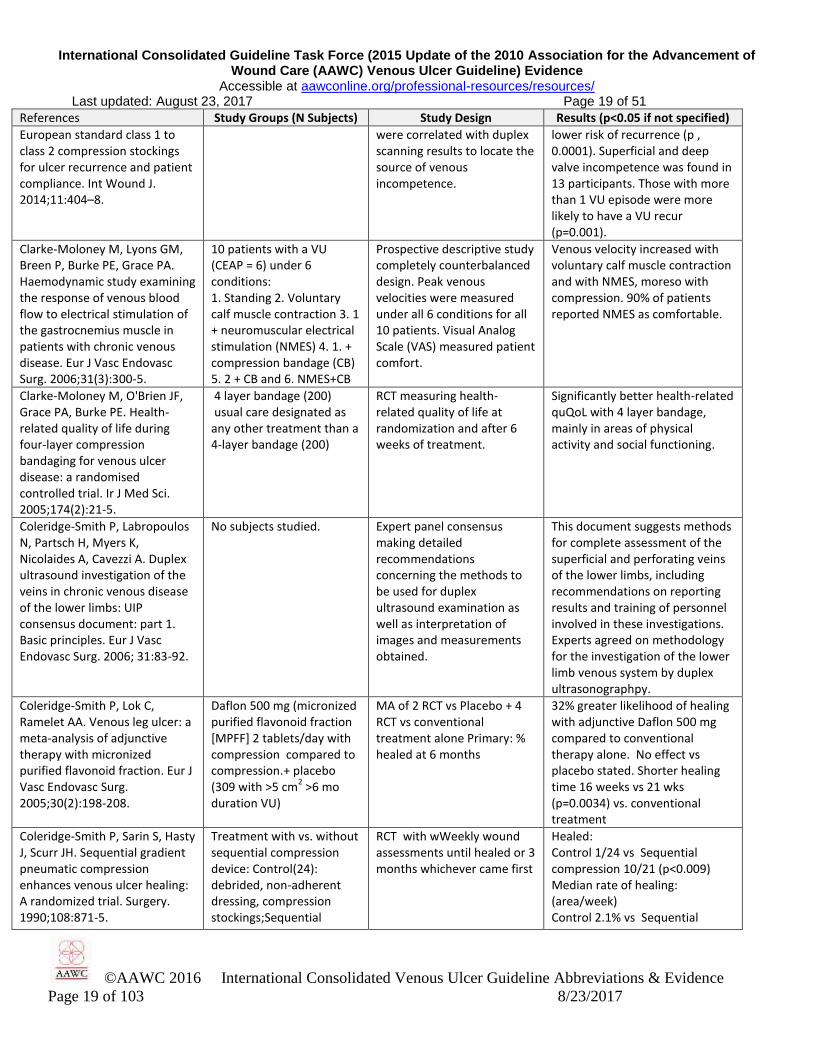
Clarke-Moloney M, Keane N, O’Connor V, Ryan MA, Meagher H, Grace PA, Kavanagh E, Walsh SR, Burke PE. Randomised controlled trial comparing
European clients with a healed VU were treated with Class 1 (50) versus Class 2 (50) compression stockings
RCT measuring VU recurrence and compliance with stocking use at 1,3,6,9 and 12 months after enrollment in study. Findings
After 12 months, 16.1% of patients had a recurrent VU, with NS difference between groups (p = 0.287). Compliant participants (88.9%) were at significantly
International Consolidated Guideline Task Force (2015 Update of the 2010 Association for the Advancement of Wound Care (AAWC) Venous Ulcer Guideline) Evidence
Accessible at aawconline.org/professional-resources/resources/ Last updated: August 23, 2017 Page 19 of 51
©AAWC 2016 International Consolidated Venous Ulcer Guideline Abbreviations & Evidence
Page 19 of 103 8/23/2017
References Study Groups (N Subjects) Study Design Results (p<0.05 if not specified)
European standard class 1 to class 2 compression stockings for ulcer recurrence and patient compliance. Int Wound J. 2014;11:404–8.
were correlated with duplex scanning results to locate the source of venous incompetence.
lower risk of recurrence (p , 0.0001). Superficial and deep valve incompetence was found in 13 participants. Those with more than 1 VU episode were more likely to have a VU recur (p=0.001).
Clarke-Moloney M, Lyons GM, Breen P, Burke PE, Grace PA. Haemodynamic study examining the response of venous blood flow to electrical stimulation of the gastrocnemius muscle in patients with chronic venous disease. Eur J Vasc Endovasc Surg. 2006;31(3):300-5.
10 patients with a VU (CEAP = 6) under 6 conditions: 1. Standing 2. Voluntary calf muscle contraction 3. 1 + neuromuscular electrical stimulation (NMES) 4. 1. + compression bandage (CB) 5. 2 + CB and 6. NMES+CB
Prospective descriptive study completely counterbalanced design. Peak venous velocities were measured under all 6 conditions for all 10 patients. Visual Analog Scale (VAS) measured patient comfort.
Venous velocity increased with voluntary calf muscle contraction and with NMES, moreso with compression. 90% of patients reported NMES as comfortable.
Clarke-Moloney M, O'Brien JF, Grace PA, Burke PE. Health-related quality of life during four-layer compression bandaging for venous ulcer disease: a randomised controlled trial. Ir J Med Sci. 2005;174(2):21-5.
4 layer bandage (200) usual care designated as any other treatment than a 4-layer bandage (200)
RCT measuring health-related quality of life at randomization and after 6 weeks of treatment.
Significantly better health-related quQoL with 4 layer bandage, mainly in areas of physical activity and social functioning.
Coleridge-Smith P, Labropoulos N, Partsch H, Myers K, Nicolaides A, Cavezzi A. Duplex ultrasound investigation of the veins in chronic venous disease of the lower limbs: UIP consensus document: part 1. Basic principles. Eur J Vasc Endovasc Surg. 2006; 31:83-92.
No subjects studied. Expert panel consensus making detailed recommendations concerning the methods to be used for duplex ultrasound examination as well as interpretation of images and measurements obtained.
This document suggests methods for complete assessment of the superficial and perforating veins of the lower limbs, including recommendations on reporting results and training of personnel involved in these investigations. Experts agreed on methodology for the investigation of the lower limb venous system by duplex ultrasonographpy.
Coleridge-Smith P, Lok C, Ramelet AA. Venous leg ulcer: a meta-analysis of adjunctive therapy with micronized purified flavonoid fraction. Eur J Vasc Endovasc Surg. 2005;30(2):198-208.
Daflon 500 mg (micronized purified flavonoid fraction [MPFF] 2 tablets/day with compression compared to compression.+ placebo (309 with >5 cm
2 >6 mo
duration VU)
MA of 2 RCT vs Placebo + 4 RCT vs conventional treatment alone Primary: % healed at 6 months
32% greater likelihood of healing with adjunctive Daflon 500 mg compared to conventional therapy alone. No effect vs placebo stated. Shorter healing time 16 weeks vs 21 wks (p=0.0034) vs. conventional treatment
Coleridge-Smith P, Sarin S, Hasty J, Scurr JH. Sequential gradient pneumatic compression enhances venous ulcer healing: A randomized trial. Surgery. 1990;108:871-5.
Treatment with vs. without sequential compression device: Control(24): debrided, non-adherent dressing, compression stockings;Sequential
RCT with wWeekly wound assessments until healed or 3 months whichever came first
Healed: Control 1/24 vs Sequential compression 10/21 (p<0.009) Median rate of healing: (area/week) Control 2.1% vs Sequential

International Consolidated Guideline Task Force (2015 Update of the 2010 Association for the Advancement of Wound Care (AAWC) Venous Ulcer Guideline) Evidence
Accessible at aawconline.org/professional-resources/resources/ Last updated: August 23, 2017 Page 20 of 51
©AAWC 2016 International Consolidated Venous Ulcer Guideline Abbreviations & Evidence
Page 20 of 103 8/23/2017
References Study Groups (N Subjects) Study Design Results (p<0.05 if not specified)
compression device (21): as above + sequential compression 3-4 hours/day
compression 19.8% (p<0.05)
Colgan MP, Teevan M, McBride C, O'Sullivan L, Moore D, Shanik G. Cost comparisons in the management of venous ulceration. Proc. 5th European Conf Adv Wound Management. London: Macmillan Magazines, 1996.
1. 4-layer elastic Unna's Boot (10) 2. 4-layer elastic Profore® (10) 3. Lyofoam primary dressing +1-layer Setopress™ elastic bandage (10)
RCT Dressing changes in clinic by nurse. Outcome measured was cost and % of patients healed at 12 weeks.
60% healed 4-layer elastic Unna's Boot group; 70% healed in Profore group; 20% healed in Setopress group (which had 3 dropped due to poor patient compliance or inadequate application. Setopress protocol cost less. Study did not report significance of differences .
Collins L, Seraj S. Diagnosis and treatment of venous ulcers. Am Fam Physician. 2010;81(8):989-96.
Guideline literature search LR with evidence base for each intervention
Compression 5 RCT A Level Elevation: 1 CCT: C Level Dressings beneficial: A Level Pentoxifylline (Trental) A Level Aspirin (1 RCT)
Combemale P, Bousquet M, Kanitakis J, Bernard P, Angiodermatology Group, French Society of Dermatology. Malignant transformation of leg ulcers: a retrospective study of 85 cases. J Eur Acad Dermatol Venereol. 2007;21 (7):935-41.
85 cases of malignant transformation of leg ulcers
Retrospective case series. Description, many were long term VUs. Correlational study makes it difficult to tell whether the long duration of a VU causes cells to transform or whether the ulcer was due to original undiagnosed malignant transformation
Combemale P, Bousquet M, Kanitakis J, Bernard P; Angiodermatology Group ,French Society of Dermatology. Malignant transformation of leg ulcers: a retrospective study of 85 cases. J Eur Acad Dermatol Venereol. 2007;21(7):935-41.
80 French patients with squamous cell carcinoma mean age 75, 88% with VU of mean duration 27.5 years.
Retrospective cohort of chronic ulcers of duration longer than 3 years. For basal cell carcinoma (BCC) a negative prior biopsy was considered.
Abnormal granulation tissue found clinically in 76%; absence of healing in 14% with abnormal extension in 6% 98% were squamous cell carcinoma of which 82% were well differentiated. Simple surgical excision was preferred to amputation.
Comerota AJ. Intermittent pneumatic compression: physiologic and clinical basis to improve management of venous ulcers. J of Vascular Surg.2011;53 (4); 1121-9.
LR (7 studies of Intermittent Pneumatic Compression on patients with a VU)
LR on VU pathophysiology and effects of Intermittent Pneumatic Compression, RCTs reported increased venous return, reduced leg edema, increased endogenous fibrinolysis, reduced intra-vascular coagulation and improved arterial (skin) perfusion resulting in increased TcPO2, combining to foster healing.
In VU patients, Intermittent Pneumatic Compression produces effects compatible with VU healing, wound care and compression therapy show improved rates of VU healing, supporting recommendations from the American College of Chest Physicians that this intervention be used to speed healing of large VUs and those recalcitrant to healing for 6

International Consolidated Guideline Task Force (2015 Update of the 2010 Association for the Advancement of Wound Care (AAWC) Venous Ulcer Guideline) Evidence
Accessible at aawconline.org/professional-resources/resources/ Last updated: August 23, 2017 Page 21 of 51
©AAWC 2016 International Consolidated Venous Ulcer Guideline Abbreviations & Evidence
Page 21 of 103 8/23/2017
References Study Groups (N Subjects) Study Design Results (p<0.05 if not specified)
months.
ConvaTec. SOLUTIONS®
wound care algorithm. 1994 (revised 2013 Sep). NGC:010274 Accessed November 1, 2014 at www.guidelines.gov
Guideline for all wound treatment
Content-validated evidence-based guideline for wound cleansing, debriding, dressing managing excess exudate and, hydrating dry wounds.
Care decision algorithms based on reliable, validated wound assessment. Re-evaluate care plan/ address etiology if the wound has not decreased in area during 2 to 4 weeks of care.
Cordts PR, Hanrahan LM, Rodriguez AA, Woodson J, LaMorte WW, Menzoian JO. A prospective, randomized trial of Unna’s boot versus Duoderm CGF hydroactive dressing plus compression in the management of venous leg ulcers. J Vascular Surg 1992;15:480-6.
1. Duoderm CGF + Coban (16) 2. Unna’s boot with gauze VU dressing (14)
RCT with weekly assessments of VU healing rates until healed or 12 weeks. Patient ratings of comfort, adhesion, cosmesis and odor.
Healing rates faster with Duoderm + Coban than Unna’s boot (p<0.002) when corrected for initial perimeter differences. 50% healed at 12 weeks in Group 1; 43% in Group 2 (p=0.18) Group 1 patients rated dressing higher. (p<0.05) in comfort, adhesion, cosmesis and with more odor
Cullum NA, Al-Kurdi D, Bell-Syer SE. Therapeutic ultrasound for venous leg ulcers. Cochrane Database Syst Rev. 2010;(6):CD001180
8 RCTon ultrasound for VU 6 RCT on High frequency 2 RCT on Low frequency
Systematic review High frequency ultrasound healed more patients with VU at 7-8 weeks than none (5 RCT). The effect did not last to 12 weeks. 2 RCT reported NS effect of low frequency ultrasound.
Cullum. N, Nelson EA, Fletcher AW The Cochrane library 2001, Compression for venous ulcers (Cochrane Review) In The Cochrane Library, Issue 3, 2002: Update Software
1. Compression vs dressings [Kitka, 1988; Rubin, 1990; Sikes, 1985] 2. Compression wraps vs noncompression wraps [Charles, 1991; Erikson, 1984; Taylor, 1998]
Analyses of literature for each numbered question investigated.
1. Compression healed more VU than dressings alone 2. Compression wraps healed more VU than noncompression 3. Multi-layer elastic compression healed more than non-elastic or than single-layer elastic compression.
Cushman M, Callas PW, Denenberg JO, Bovill EG, Criqui MH. Risk factors for peripheral venous disease resemble those for venous thrombosis: the San Diego Population Study. Thromb Haemost. 2010;8(8):1730-5.
2404 men and women in the San Diego Population Study: 308 cases developing a DVT of graded severity compared to 346 controls with no venous abnormality, frequency case-matched in 10 yr age groups
Case-control study to identify patients at risk of post thrombotic syndrome (PTS). Peripheral venous disease was evaluated using physical exam, symptom assessment and venous ultrasound. All had no prior DVT initially.
Risk factors for PTS are: Age, obesity, family history, elevated factor VIII, von Willebrand factor, D-dimer and factor V Leiden, all similar to those for DVT. PTS may result from a previously unrecognized DVT. Prothrombin 20210A was unrelated to venous disease. DVT risk factors are associated with presence and severity of peripheral venous disease.
Cushman M. Epidemiology and risk factors for venous thrombosis. Semin Hematol. 2007;44(2):62-9
Literature review of 79 references
LR: Venous thrombosis (DVT pulmonary embolism and post-thrombotic syndrome [PTS]) risk factors.
PTS occurs in 20-50% after 1st
DVT (2 CO). Risk factors include older age, male gender, proximal (as compared to distal) DVT and higher D-dimer.
da Silva JL, Lopes MJ. [Health Not stated in abstract. HCT in Portuguese Leg Club model improved

International Consolidated Guideline Task Force (2015 Update of the 2010 Association for the Advancement of Wound Care (AAWC) Venous Ulcer Guideline) Evidence
Accessible at aawconline.org/professional-resources/resources/ Last updated: August 23, 2017 Page 22 of 51
©AAWC 2016 International Consolidated Venous Ulcer Guideline Abbreviations & Evidence
Page 22 of 103 8/23/2017
References Study Groups (N Subjects) Study Design Results (p<0.05 if not specified)
education for varicose ulcer patients through group activities] Rev Gaucha Enferm. 2006;27(2):240-50.
adherence to treatment, attitude of health care team.
Dall L, Peterson S, Simmons T, Dall A. Rapid resolution of cellulitis in patients managed with combination antibiotic and anti-inflammatory therapy. Cutis. 2005;75:177-80
Patients presenting in the emergency department with signs and symptoms of class II cellulitis treatment with either antibiotic therapy alone (intravenous, supplemented with oral cephalexin or an equivalent) for 10 days (n = 33) or antibiotic therapy for 10 days plus an oral anti-inflammatory (ibuprofen 400 mg every 6 hours) for 5 days (n = 31)
RCT measuring time to resolve signs and symptoms of class II cellulitis. This small preliminary study provides some promising data, suggesting that the supplemental use of anti-inflammatory therapy may hasten the time to regression of inflammation and complete resolution of cellulitis.
The addition of an oral anti-inflammatory agent significantly (P < .05) shortened the time to regression of inflammation and complete resolution of cellulitis. Twenty-four of 29 evaluable patients (82.8%) who received supplemental anti-inflammatory treatment showed regression of inflammation within 1 to 2 days compared with only 3 of 33 patients (9.1%) treated without an anti-inflammatory in the same time frame. All patients receiving adjunctive anti-inflammatory treatment experienced complete resolution of cellulitis in 4 to 5 days or less, while 24.2% (8/33) of patients treated with antibiotic alone required 6 to 7 days, and 6.1% (2/33) required 7 days or more (P < .05).
Daniels S, Sibbald RG, Ennis W, Eager CA. Evaluation of a new composite dressing for the management of chronic leg ulcer wounds. J Wound Care. 2002;11(8):290-4.
75 dressing changes of 11 ulcers dressed with Versiva® (ConvaTec). Historic control data taken for each patient.
Prospective HCT, open-label, multicentre, phase II study assessed weartime, absorption, dressing integrity, ease of use and wound progression during up to 10 dressing changes within five-week study period of patients with venous leg ulcers
Healing or marked improvement was observed in 82% of leg ulcers within the five-week study.93%, rated the dressing 'very easy' to remove, with no trauma to surrounding skin. Minimal to no leakage was observed in 81% of changes. Most dressing changes (77%) were painless.
Danielsen L, Madsen SM, Henriksen L. Venous leg ulcer healing: a randomised prospective study of long-stretch versus short-stretch compression bandages. Phleboloby. 1998;13:59-63.
1. 2-layer long-stretch elastic high compression with gauze + Setopress® (21) 2. 2-layer short-stretch high compression with gauze + Comprilan non-adhesive bandage (19)
Prospective RCT with number of patients healed/total assessed at that time interval (%) and ulcer area reported at 1, 6 and 12 months. 5 Withdrew in Group 1; 9 withdrew in Group 2.
% healed at 1, 6, 12 months: Group 1: 27%, 50%, 71% Group 2: 5%, 36%, 30% % area remaining at 1,6,12 mo. Group 1: 45%, 81%, 25%. Group 2: 72% 60% 95%. Cullum et al. report 9/21 Group 1 healed (43%), 5/19 (26%) healed in Group 2. NS.
Davies CE, Hill KE, Newcombe RG, Stephens P, Wilson MJ, Harding KG, Thomas DW. A prospective study of the
70 patients with chronic venous leg ulcers
This study determined whether comprehensive microbiological analysis offered real predictive value
Bioplsies did not add prognostic information compared to analysis of surface microflora)
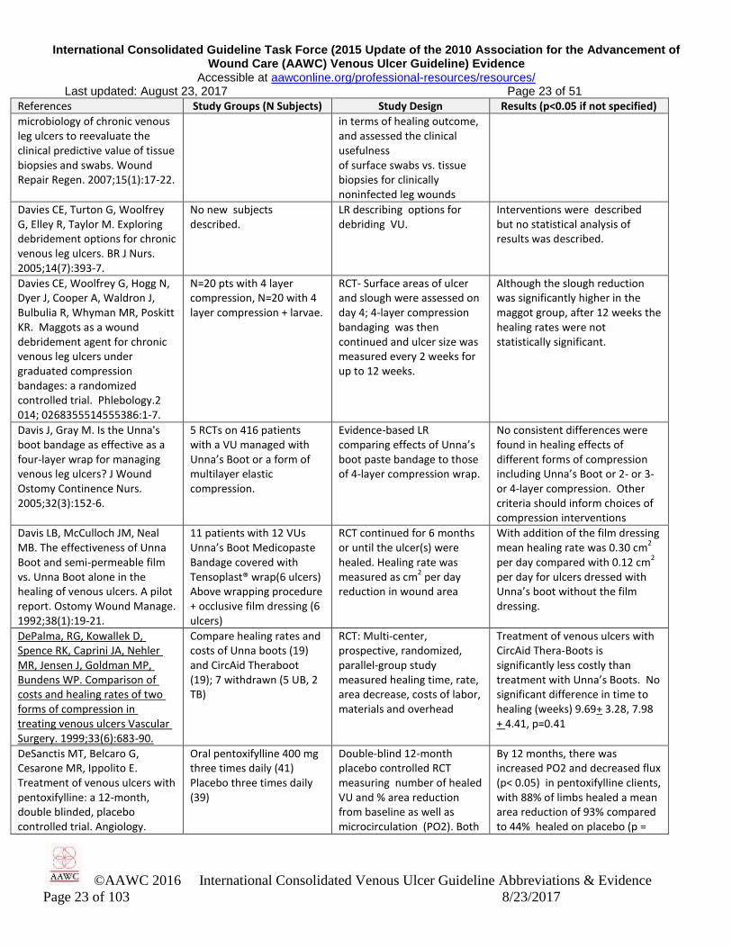
International Consolidated Guideline Task Force (2015 Update of the 2010 Association for the Advancement of Wound Care (AAWC) Venous Ulcer Guideline) Evidence
Accessible at aawconline.org/professional-resources/resources/ Last updated: August 23, 2017 Page 23 of 51
©AAWC 2016 International Consolidated Venous Ulcer Guideline Abbreviations & Evidence
Page 23 of 103 8/23/2017
References Study Groups (N Subjects) Study Design Results (p<0.05 if not specified)
microbiology of chronic venous leg ulcers to reevaluate the clinical predictive value of tissue biopsies and swabs. Wound Repair Regen. 2007;15(1):17-22.
in terms of healing outcome, and assessed the clinical usefulness of surface swabs vs. tissue biopsies for clinically noninfected leg wounds
Davies CE, Turton G, Woolfrey G, Elley R, Taylor M. Exploring debridement options for chronic venous leg ulcers. BR J Nurs. 2005;14(7):393-7.
No new subjects described.
LR describing options for debriding VU.
Interventions were described but no statistical analysis of results was described.
Davies CE, Woolfrey G, Hogg N, Dyer J, Cooper A, Waldron J, Bulbulia R, Whyman MR, Poskitt KR. Maggots as a wound debridement agent for chronic venous leg ulcers under graduated compression bandages: a randomized controlled trial. Phlebology.2 014; 0268355514555386:1-7.
N=20 pts with 4 layer compression, N=20 with 4 layer compression + larvae.
RCT- Surface areas of ulcer and slough were assessed on day 4; 4-layer compression bandaging was then continued and ulcer size was measured every 2 weeks for up to 12 weeks.
Although the slough reduction was significantly higher in the maggot group, after 12 weeks the healing rates were not statistically significant.
Davis J, Gray M. Is the Unna's boot bandage as effective as a four-layer wrap for managing venous leg ulcers? J Wound Ostomy Continence Nurs. 2005;32(3):152-6.
5 RCTs on 416 patients with a VU managed with Unna’s Boot or a form of multilayer elastic compression.
Evidence-based LR comparing effects of Unna’s boot paste bandage to those of 4-layer compression wrap.
No consistent differences were found in healing effects of different forms of compression including Unna’s Boot or 2- or 3- or 4-layer compression. Other criteria should inform choices of compression interventions
Davis LB, McCulloch JM, Neal MB. The effectiveness of Unna Boot and semi-permeable film vs. Unna Boot alone in the healing of venous ulcers. A pilot report. Ostomy Wound Manage. 1992;38(1):19-21.
11 patients with 12 VUs Unna’s Boot Medicopaste Bandage covered with Tensoplast® wrap(6 ulcers) Above wrapping procedure + occlusive film dressing (6 ulcers)
RCT continued for 6 months or until the ulcer(s) were healed. Healing rate was measured as cm
2 per day
reduction in wound area
With addition of the film dressing mean healing rate was 0.30 cm
2
per day compared with 0.12 cm2
per day for ulcers dressed with Unna’s boot without the film dressing.
DePalma, RG, Kowallek D, Spence RK, Caprini JA, Nehler MR, Jensen J, Goldman MP, Bundens WP. Comparison of costs and healing rates of two forms of compression in treating venous ulcers Vascular Surgery. 1999;33(6):683-90.
Compare healing rates and costs of Unna boots (19) and CircAid Theraboot (19); 7 withdrawn (5 UB, 2 TB)
RCT: Multi-center, prospective, randomized, parallel-group study measured healing time, rate, area decrease, costs of labor, materials and overhead
Treatment of venous ulcers with CircAid Thera-Boots is significantly less costly than treatment with Unna’s Boots. No significant difference in time to healing (weeks) 9.69+ 3.28, 7.98 + 4.41, p=0.41
DeSanctis MT, Belcaro G, Cesarone MR, Ippolito E. Treatment of venous ulcers with pentoxifylline: a 12-month, double blinded, placebo controlled trial. Angiology.
Oral pentoxifylline 400 mg three times daily (41) Placebo three times daily (39)
Double-blind 12-month placebo controlled RCT measuring number of healed VU and % area reduction from baseline as well as microcirculation (PO2). Both
By 12 months, there was increased PO2 and decreased flux (p< 0.05) in pentoxifylline clients, with 88% of limbs healed a mean area reduction of 93% compared to 44% healed on placebo (p =
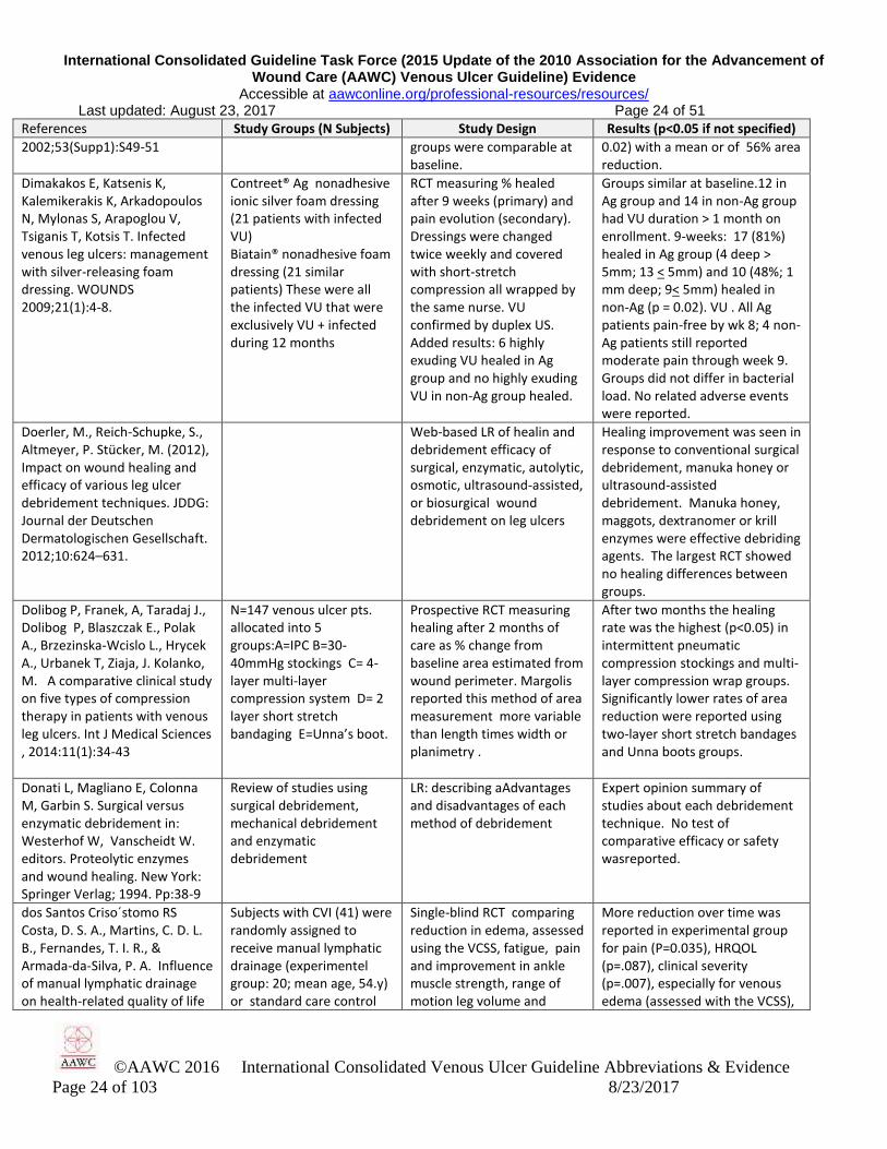
International Consolidated Guideline Task Force (2015 Update of the 2010 Association for the Advancement of Wound Care (AAWC) Venous Ulcer Guideline) Evidence
Accessible at aawconline.org/professional-resources/resources/ Last updated: August 23, 2017 Page 24 of 51
©AAWC 2016 International Consolidated Venous Ulcer Guideline Abbreviations & Evidence
Page 24 of 103 8/23/2017
References Study Groups (N Subjects) Study Design Results (p<0.05 if not specified)
2002;53(Supp1):S49-51 groups were comparable at baseline.
0.02) with a mean or of 56% area reduction.
Dimakakos E, Katsenis K, Kalemikerakis K, Arkadopoulos N, Mylonas S, Arapoglou V, Tsiganis T, Kotsis T. Infected venous leg ulcers: management with silver-releasing foam dressing. WOUNDS 2009;21(1):4-8.
Contreet® Ag nonadhesive ionic silver foam dressing (21 patients with infected VU) Biatain® nonadhesive foam dressing (21 similar patients) These were all the infected VU that were exclusively VU + infected during 12 months
RCT measuring % healed after 9 weeks (primary) and pain evolution (secondary). Dressings were changed twice weekly and covered with short-stretch compression all wrapped by the same nurse. VU confirmed by duplex US. Added results: 6 highly exuding VU healed in Ag group and no highly exuding VU in non-Ag group healed.
Groups similar at baseline.12 in Ag group and 14 in non-Ag group had VU duration > 1 month on enrollment. 9-weeks: 17 (81%) healed in Ag group (4 deep > 5mm; 13 < 5mm) and 10 (48%; 1 mm deep; 9< 5mm) healed in non-Ag (p = 0.02). VU . All Ag patients pain-free by wk 8; 4 non-Ag patients still reported moderate pain through week 9. Groups did not differ in bacterial load. No related adverse events were reported.
Doerler, M., Reich-Schupke, S., Altmeyer, P. Stücker, M. (2012), Impact on wound healing and efficacy of various leg ulcer debridement techniques. JDDG: Journal der Deutschen Dermatologischen Gesellschaft. 2012;10:624–631.
Web-based LR of healin and debridement efficacy of surgical, enzymatic, autolytic, osmotic, ultrasound-assisted, or biosurgical wound debridement on leg ulcers
Healing improvement was seen in response to conventional surgical debridement, manuka honey or ultrasound-assisted debridement. Manuka honey, maggots, dextranomer or krill enzymes were effective debriding agents. The largest RCT showed no healing differences between groups.
Dolibog P, Franek, A, Taradaj J., Dolibog P, Blaszczak E., Polak A., Brzezinska-Wcislo L., Hrycek A., Urbanek T, Ziaja, J. Kolanko, M. A comparative clinical study on five types of compression therapy in patients with venous leg ulcers. Int J Medical Sciences , 2014:11(1):34-43
N=147 venous ulcer pts. allocated into 5 groups:A=IPC B=30-40mmHg stockings C= 4-layer multi-layer compression system D= 2 layer short stretch bandaging E=Unna’s boot.
Prospective RCT measuring healing after 2 months of care as % change from baseline area estimated from wound perimeter. Margolis reported this method of area measurement more variable than length times width or planimetry .
After two months the healing rate was the highest (p<0.05) in intermittent pneumatic compression stockings and multi-layer compression wrap groups. Significantly lower rates of area reduction were reported using two-layer short stretch bandages and Unna boots groups.
Donati L, Magliano E, Colonna M, Garbin S. Surgical versus enzymatic debridement in: Westerhof W, Vanscheidt W. editors. Proteolytic enzymes and wound healing. New York: Springer Verlag; 1994. Pp:38-9
Review of studies using surgical debridement, mechanical debridement and enzymatic debridement
LR: describing aAdvantages and disadvantages of each method of debridement
Expert opinion summary of studies about each debridement technique. No test of comparative efficacy or safety wasreported.
dos Santos Criso´stomo RS Costa, D. S. A., Martins, C. D. L. B., Fernandes, T. I. R., & Armada-da-Silva, P. A. Influence of manual lymphatic drainage on health-related quality of life
Subjects with CVI (41) were randomly assigned to receive manual lymphatic drainage (experimentel group: 20; mean age, 54.y) or standard care control
Single-blind RCT comparing reduction in edema, assessed using the VCSS, fatigue, pain and improvement in ankle muscle strength, range of motion leg volume and
More reduction over time was reported in experimental group for pain (P=0.035), HRQOL (p=.087), clinical severity (p=.007), especially for venous edema (assessed with the VCSS),

International Consolidated Guideline Task Force (2015 Update of the 2010 Association for the Advancement of Wound Care (AAWC) Venous Ulcer Guideline) Evidence
Accessible at aawconline.org/professional-resources/resources/ Last updated: August 23, 2017 Page 25 of 51
©AAWC 2016 International Consolidated Venous Ulcer Guideline Abbreviations & Evidence
Page 25 of 103 8/23/2017
References Study Groups (N Subjects) Study Design Results (p<0.05 if not specified)
and symptoms of chronic venous insufficiency: a randomized controlled trial. Arch Physical Medicine Rehab. 2015;96:283-91
(21) mean age, 46.y). health-related quality of life (HRQoL) over time for subjects receiving manual lymphatic drainage (MLD). Plus standard care vs standard care controls.
fatigue (p =.012), and heaviness (p=.001), with the MLD group improving more over time in all of these outcomes. Effect was not significant for ankle muscle strength, ankle ROM, or leg volume.
Duby T, Cherry G, Hoffman D, Cameron J, Dobloff-Brown D, Ryan T. A randomized trial in the treatment of venous leg ulcers comparing short stretch bandages, four layer bandage system, and a long stretch-paste bandage system. Wounds 1993;5(6):276-9.
1. Short stretch: Comprilan+Tricofix net (20) 2. Zinc paste Icthopaste + Elastocrepe (long-stretch) + Tubigrip (24) 4-Layer compression bandage (23)
RCT comparing treatment for 1-12 weeks, Measurements:
% change in leg volume
% of legs changing volume
% of V.Ulcers healed
Mean % change in area
Reduction in leg volume (edema reduction) is strongly correlated with % reduction in ulcer area and % of ulcers healed, with 4-Layer compression 44% of ulcers healed (mean area reduction 76%); short stretch 40% (60% mean area reduction) and zinc paste bandage 23% healed (43% mean area reduction.
Dumville JC, Land L, Evans D, Peinemann F. Negative pressure wound therapy for treating leg ulcers. Cochrane Database of Systematic Reviews 2015, Issue 7. Art. No.: CD011354.
One study met inclusion criteria: RCT comparing NPWT to control
1 RCT on 60 subjects with leg ulcers reported % healed at 12 weeks: Vuerstock., 2006
There were no difference in primary outcome % healed at 12 months or costs or pain or quality of life.
Dumville JC, Worthy G, Soares MO, Bland JM, Cullum N, Dowson C, Iglesias C, McCaughan D, Mitchell JL, Nelson EA, Torgerson DJ; VenUS II team. VenUS II: a randomised controlled trial of larval therapy in the management of leg ulcers. Health Technol Assess. 2009;13(55):1-182, iii-iv.
267 subjects with VU or mixed AU-VU ulcer: ABI<0.6 and > 25% covered with slough or necrotic material assigned randomly to 1 of 3 groups: 1.Loose larval therapy 2. Bagged larval therapy 3. hydrogel
Multicenter 3-arm RCT measuring time to complete healing pain and costs of care.
Maggot debridement (larval therapy) was associated with faster debridement of VU, but more pain, equal healing time and costs of care. No differences in health-related quality of life or bacteriology.
Dunn RM, Fudam GM, Walton RL, Anderson FA Jr., Malhotra R. Free flap valvular transplantation for refractory venous ulceration. J Vasc Surg. 1994;19:525–31
6 patients with CVI Prospective case series debriding, VU then performing fasciocutaneous free flap valve transplants. Venous reflux was measured using photoplethysmography
All 6 VU healed with no recurrence after 2 or 7 years of any of the 6 VU or of associated lipodermatosclerosis.
Edlich RF, Winters KL, Britt LD, Long III WB. Bacterial diseases of the skin. J Long Term Eff Med Implants 2005;15(5):499-510.
Case examples described. Lit Rev describing bacterial diseases of the skin
Many bacterial diseases of the skin can occur on the lower leg and should be distinguished from a VU as they require different treatment.
Edwards H, Courtney M, Finlayson K, Lewis C, Lindsay E, Dumble J. Improved healing rates for chronic venous leg ulcers: pilot study results from a
33 VU patients: half randomized to Leg Club and half to Home care with standardized compression
RCT of care for 12 weeks by a team of wound care nurses following evidence-based assessment & treatment guidelines. Data collected at
More healing in Leg club group (p<0.05)

International Consolidated Guideline Task Force (2015 Update of the 2010 Association for the Advancement of Wound Care (AAWC) Venous Ulcer Guideline) Evidence
Accessible at aawconline.org/professional-resources/resources/ Last updated: August 23, 2017 Page 26 of 51
©AAWC 2016 International Consolidated Venous Ulcer Guideline Abbreviations & Evidence
Page 26 of 103 8/23/2017
References Study Groups (N Subjects) Study Design Results (p<0.05 if not specified)
randomized controlled trial of a community nursing intervention. Int J Nurs Pract. 2005;11(4):169-76.
and baseline and week 12 on healing, and PUSH score
Edwards H, Courtney M, Finlayson K, Shuter P, Lindsay E. A randomised controlled trial of a community nursing intervention: improved quality of life and healing for clients with chronic leg ulcers. J Clin Nurs. 2009;18(11):1541-9.
Nurse led Leg Club (34 VU patients) with peer support Nurse led home care (33) All patients had identical research protocols with short-stretch bandages
RCT HRQoL, healing, functional capacity collected at baseline, 12 and 24 weeks.
Leg club improved outcomes in quality of life (p = 0.014), morale (p < 0.001), self-esteem (p = 0.006), healing (p = 0.004), pain (p = 0.003) and functional ability (p = 0.044)
Edwards H, Finlayson K, Courtney M, Graves N, Gibb M, Parker C. Health service pathways for patients with chronic leg ulcers: identifying effective pathways for facilitation of evidence based wound care. BMC Health Services Research 2013, 13:86
N= 70- lower limb or foot ulcer seen in specialist wound clinics
HCT: Observational study with retrospectice and prospective arm aimed to identify effective health service pathways of care which facilitated evidence-based management of chronic leg ulcers.
Retrospective data indicated that evidence based guidelines were poorly implemented prior to admission to the study. Only 31% of clients with a lower limb ulcer had ABPI or duplex assessment in the previous 12 months. After admission to wound clinics , key indicators of evidence based care increased (p < 0.001). Kaplan-Meier survival analysis found the median time to healing was 12 weeks (95% CI 9.3–14.7). Use of evidence-based care was significantly related to improved healing outcomes (p < 0.001). “Results demonstrate that access to wound management expertise can promote streamlined health services and evidence based wound care, leading to efficient use of health resources and improved health.”
Eklöf B, Rutherford RB, Bergan JJ, Carpentier PH, Gloviczki P, Kistner RL, Meissner MH, Moneta GL, Myers K, Padberg FT, Perrin M, Ruckley CV, Smith PC, Wakefield TW; American Venous Forum International Ad Hoc Committee for Revision of the CEAP Classification. Revision of the CEAP classification for chronic venous disorders: consensus statement. J Vasc Surg. 2004;40(6):1248-52.
Literature review Consensus statement and literature review updating CEAP classification system.
CEAP is a descriptive classification only for documenting symptoms. For longitudinal outcomes measurements use venous severity score and quality of life instruments.
Emmons KR, Dale, B & Crouch C. No individual patient data LR of palliative care issues. Palliative care patients are often

International Consolidated Guideline Task Force (2015 Update of the 2010 Association for the Advancement of Wound Care (AAWC) Venous Ulcer Guideline) Evidence
Accessible at aawconline.org/professional-resources/resources/ Last updated: August 23, 2017 Page 27 of 51
©AAWC 2016 International Consolidated Venous Ulcer Guideline Abbreviations & Evidence
Page 27 of 103 8/23/2017
References Study Groups (N Subjects) Study Design Results (p<0.05 if not specified)
Palliative wound care: application of principles. Home Healthcare Nurse 2014;32 (4) :210-22.
complex with comorbidities. They may not be able to heal, so their caregivers must learn and meet client goals to improve quality of life.
Ennis WJ, Meneses P. Clinical evaluation: Outcomes, benchmarking, introspec-tion, and quality improve-ment. Ostomy/Wound Manage. 1996; 42(10A Suppl 10A):40S-47S.
147 patients with leg or foot ulcers 1993-4: 57 patients 1995: 101 with prophylactic antibiotics, and 103 similar patients without
Prospective CO study measuring median heal time and % of patients healed at each week of care
Median 12 week heal time in 1993-4; median 10 week heal time 1995 p< 0.001. With antibiotics median 19 weeks, without median 9 weeks.
Ennis WJ, Meneses P. Leg ulcers: a practical approach to the leg ulcer patient. Ostomy/Wound Management 1995;41(7)Suppl A:52S-63S.
Compression plus moist wound healing.
Expert opinion validated by clinical healing outcomes.from a CO of successive VU patients.
VU associated with non-pitting, tender, tight edema of lower extremity.
Eriksson G, Eklund AE, Liden S, Zetterquist S. Comparison of different treatments of venous leg ulcers: a controlled study using stereophotogrammetry. Current Therapeutic Research 1984;35: 4:678-684.
1. Metallina aluminium foil dressing (20) 2. Two layer bandage: ACO paste bandage +Tensoplast (13)
Multicenter 8-week study in Sweden, setting unclear. A third group crossed over during study from porcine skin dressing to 2-layer compression (not included)
% Area reduction at 8 weeks: 10% with aluminum foil 80% with 2-layer bandage % volume reduction 8 weeks: 0% with aluminum foil 90% with 2-layer bandage
Eriksson G. Comparative study of hydrocolloid dressing and double layer bandage in treatment of venous stasis ulceration. In: Ryan TJ (Ed) An environment for healing: The role of occlusion. London: Royal Society of Medicine International Congress and Symposium Series, Number 88: 111-3, 1984.
1. 2-layer elastic compression (ACO Salvstrumpa® zinc paste stocking + Tensoplast® elastic bandage (Smith & Nephew) changed every 1-2 weeks (17) 2. 1-layer elastic bandage removed nightly, reapplied am, + DuoDERM (ConvaTec) changed 1-2 / week (17)
8-week RCT same as in Br J Dermatol 1986. Objective evaluation with stereophotogrammetric measurement of ulcer area and volume and bacterial count.
No significant difference in 8-week healing between DuoDERM + 1-layer elastic bandage and double layer bandage. (Note: may besame patients as Erikkson BrJ Dermatol, 1986)
Eriksson G. Comparison of two occlusive bandages in the treatment of venous leg ulceration. Br J Derm. 1986;(114):227-30.
34 patients with venous ulcers. 17 patients treated with 1-layer compression + hydrocolloid dressing and 17 patients treated with double layered elastic compression bandage.
RCT. Objective evaluation with stereophotogrammetric measurement of ulcer area and volume and bacterial count. 8 week study.
No significant difference in 8-week healing between DuoDERM + one-layer elastic bandage (9/17 healed) or double layer bandage (7 /17 healed).Both provided compression and a moist wound environment.
European Wound Management Association (EWMA) Position document: understanding compression therapy. MEP Ltd, London, 2003.
Model of costs per patient year with compression with 4Layer compression or usual care based on LR
LR of pathophysiology of venous disease, lymphedema and role and outcomes of various forms of compression therapy, factors to consider before applying compression
Cost effectiveness, efficacy and safety evidence of compression therapy is reviewed. Mean cost /y of care with 4L Bandage € 1,205 or € 2,135 with usual care.
Evangelista MTP, Casintahan 66 pts- randomized into 2 RCT double blind pacebo Among ulcers < 5 cm, 100% of
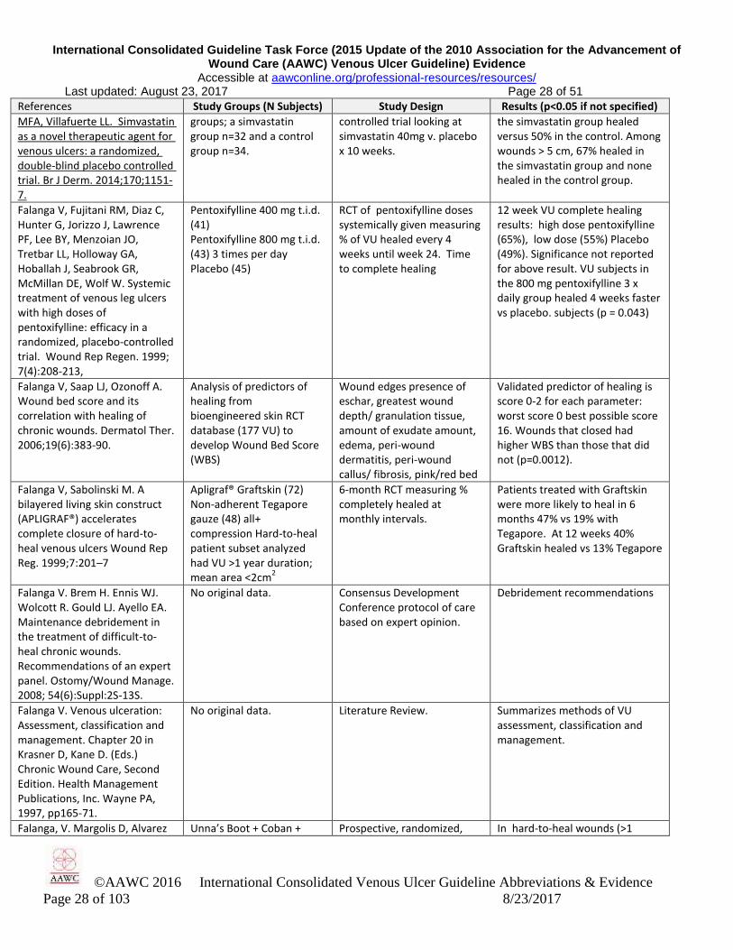
International Consolidated Guideline Task Force (2015 Update of the 2010 Association for the Advancement of Wound Care (AAWC) Venous Ulcer Guideline) Evidence
Accessible at aawconline.org/professional-resources/resources/ Last updated: August 23, 2017 Page 28 of 51
©AAWC 2016 International Consolidated Venous Ulcer Guideline Abbreviations & Evidence
Page 28 of 103 8/23/2017
References Study Groups (N Subjects) Study Design Results (p<0.05 if not specified)
MFA, Villafuerte LL. Simvastatin as a novel therapeutic agent for venous ulcers: a randomized, double-blind placebo controlled trial. Br J Derm. 2014;170;1151-7.
groups; a simvastatin group n=32 and a control group n=34.
controlled trial looking at simvastatin 40mg v. placebo x 10 weeks.
the simvastatin group healed versus 50% in the control. Among wounds > 5 cm, 67% healed in the simvastatin group and none healed in the control group.
Falanga V, Fujitani RM, Diaz C, Hunter G, Jorizzo J, Lawrence PF, Lee BY, Menzoian JO, Tretbar LL, Holloway GA, Hoballah J, Seabrook GR, McMillan DE, Wolf W. Systemic treatment of venous leg ulcers with high doses of pentoxifylline: efficacy in a randomized, placebo-controlled trial. Wound Rep Regen. 1999; 7(4):208-213,
Pentoxifylline 400 mg t.i.d. (41) Pentoxifylline 800 mg t.i.d. (43) 3 times per day Placebo (45)
RCT of pentoxifylline doses systemically given measuring % of VU healed every 4 weeks until week 24. Time to complete healing
12 week VU complete healing results: high dose pentoxifylline (65%), low dose (55%) Placebo (49%). Significance not reported for above result. VU subjects in the 800 mg pentoxifylline 3 x daily group healed 4 weeks faster vs placebo. subjects (p = 0.043)
Falanga V, Saap LJ, Ozonoff A. Wound bed score and its correlation with healing of chronic wounds. Dermatol Ther. 2006;19(6):383-90.
Analysis of predictors of healing from bioengineered skin RCT database (177 VU) to develop Wound Bed Score (WBS)
Wound edges presence of eschar, greatest wound depth/ granulation tissue, amount of exudate amount, edema, peri-wound dermatitis, peri-wound callus/ fibrosis, pink/red bed
Validated predictor of healing is score 0-2 for each parameter: worst score 0 best possible score 16. Wounds that closed had higher WBS than those that did not (p=0.0012).
Falanga V, Sabolinski M. A bilayered living skin construct (APLIGRAF®) accelerates complete closure of hard-to-heal venous ulcers Wound Rep Reg. 1999;7:201–7
Apligraf® Graftskin (72) Non-adherent Tegapore gauze (48) all+ compression Hard-to-heal patient subset analyzed had VU >1 year duration; mean area <2cm
2
6-month RCT measuring % completely healed at monthly intervals.
Patients treated with Graftskin were more likely to heal in 6 months 47% vs 19% with Tegapore. At 12 weeks 40% Graftskin healed vs 13% Tegapore
Falanga V. Brem H. Ennis WJ. Wolcott R. Gould LJ. Ayello EA. Maintenance debridement in the treatment of difficult-to-heal chronic wounds. Recommendations of an expert panel. Ostomy/Wound Manage. 2008; 54(6):Suppl:2S-13S.
No original data. Consensus Development Conference protocol of care based on expert opinion.
Debridement recommendations
Falanga V. Venous ulceration: Assessment, classification and management. Chapter 20 in Krasner D, Kane D. (Eds.) Chronic Wound Care, Second Edition. Health Management Publications, Inc. Wayne PA, 1997, pp165-71.
No original data. Literature Review. Summarizes methods of VU assessment, classification and management.
Falanga, V. Margolis D, Alvarez Unna’s Boot + Coban + Prospective, randomized, In hard-to-heal wounds (>1
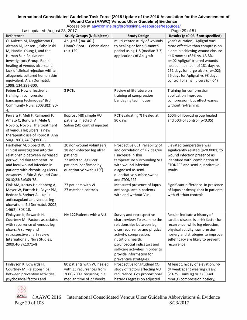
International Consolidated Guideline Task Force (2015 Update of the 2010 Association for the Advancement of Wound Care (AAWC) Venous Ulcer Guideline) Evidence
Accessible at aawconline.org/professional-resources/resources/ Last updated: August 23, 2017 Page 29 of 51
©AAWC 2016 International Consolidated Venous Ulcer Guideline Abbreviations & Evidence
Page 29 of 103 8/23/2017
References Study Groups (N Subjects) Study Design Results (p<0.05 if not specified)
O, Auletta M, Maggiacomo F, Altman M, Jensen J, Sabolinski M, Hardin-Young J, and the Human Skin Equivalent Investigators Group. Rapid healing of venous ulcers and lack of clinical rejection with an allogeneic cultured human skin equivalent. Arch Dermatol, 1998; 134:293-300.
Apligraf ( n=146 ) Unna’s Boot + Coban alone (n = 129 )
multi-center study of wounds to healing or for a 6-month period using 1-5 (median 3.3) applications of Apligraft
year’s duration), Apligraf was more effective than compression alone in achieving wound closure at 6 months (63% vs. 48.8%, p=.02 Apligraf-treated wounds healed in a mean of 181 days vs 231 days for large ulcers (p=.02); 56 days for Apligraf vs 98 days control for small ulcers (p=.04)
Feben K. How effective is training in compression bandaging techniques? Br J Community Nurs. 2003;8(2):80-4.
3 RCTs
Review of literature on training of compression bandaging techniques.
Training for compression application improves compression, but effect wanes without re-training.
Ferrara F, Meli F, Raimondi F, Amato C, Bonura F, Mulè G, Novo G, Novo S. The treatment of venous leg ulcers: a new therapeutic use of iloprost. Ann Surg. 2007;246(5):860-5.
Iloprost (48) simple VU patients injected IV Saline (50) control injected
RCT evaluating % healed at 90 days
100% of iloprost group healed and 50% of control (p<0.05)
Fierheller M, Sibbald RG. A clinical investigation into the relationship between increased periwound skin temperature and local wound infection in patients with chronic leg ulcers. Advances in Skin & Wound Care. 2010;23(8):369-78.
20 non-wound volunteers 18 non-infected leg ulcer patients 22 infected leg ulcer patients (confirmed by quantitative swab >10
5)
Prospective CCT reliability of and correlation of > 2 degree F increase in skin temperature surrounding VU with wound infection diagnosed as semi-quantitative surface swabs and STONEES
Elevated temperature was significantly related (p<0.0001) to wound infection presence as identified with combination of STONEES and semi-quantitative swabs
Fink AM, Kottas-Heldenberg A, Mayer W, Partsch H, Bayer PM, Bednar R, Steiner A. Lupus anticoagulant and venous leg ulceration. B J Dermatol. 2002; 146(2): 308-10.
27 patients with VU 27 matched controls
Measured presence of lupus anticoagulant in patients with and without Vus
Significant difference in presence of lupus anticoagulant in patients with VU than controls
Finlayson K, Edwards H, Courtney M. Factors associated with recurrence of venous leg ulcers: A survey and retrospective chart review International J Nurs Studies. 2009;46(8):1071–8
N= 122Patients with a VU Survey and retrospective chart review: To examine the relationships between leg ulcer recurrence and physical activity, compression, nutrition, health, psychosocial indicators and self-care activities in order to provide information for preventive strategies.
Results indicate a history of cardiac disease is a risk factor for recurrence; while leg elevation, physical activity, compression hosiery and strategies to improve selfefficacy are likely to prevent recurrence.
Finlayson K, Edwards H, Courtney M. Relationships between preventive activities, psychosocial factors and
80 patients with VU healed with 35 recurrences from 2006-2009, recurring in a median time of 27 weeks
Prospective longitudinal CO study of factors affecting VU recurrence. Cox proportional hazards regression adjusted
At least 1 h/day of elevation, >6 d/ week spent wearing class2 (20-25 mmHg) or 3 (30-40 mmHg) compression hosiery,

International Consolidated Guideline Task Force (2015 Update of the 2010 Association for the Advancement of Wound Care (AAWC) Venous Ulcer Guideline) Evidence
Accessible at aawconline.org/professional-resources/resources/ Last updated: August 23, 2017 Page 30 of 51
©AAWC 2016 International Consolidated Venous Ulcer Guideline Abbreviations & Evidence
Page 30 of 103 8/23/2017
References Study Groups (N Subjects) Study Design Results (p<0.05 if not specified)
recurrence of venous leg ulcers: a prospective study. J Adv Nurs. 2011;67(10):2180-90
for confounders determined effects of preventive factors
higher social support scale and higher General Self-Efficacy scores predicted lower recurrence p<0.05
Finlayson K, Edwards H, Courtney M. The impact of psychosocial factors on adherence to compression therapy to prevent recurrence of venous leg ulcers. J Clin Nurs. 2010 ;19(9-10):1289-97.
122 VU patients RCO of factors that deter compliance with compression therapy. Adjusted for covariates and confounders in multiple regression model
Main self-care activities related to VU were application of topical skin treatments, wearing compression hosiery, covering legs to prevent trauma. Depression decreased compliance, education or knowledge about condition and self-efficacy increased compliance
Finlayson K, Wu ML, Edwards HE. Identifying risk factors and protective factors for venous leg ulcer recurrence using a theoretical approach: A longitudinal study. Internat J Nurs Stud. 2015;52(6)1042–51.
N=250 adults, with a leg ulcer of primarily venous aetiology, who were followed after ulcer healing for a median follow-up time of 17 months after healing (range: 3–36 months).
Data from the three previous prospective longitudinal studies were combined. The original participant data were collected through medical records and self-reported questionnaires upon healing and every 3 months thereafter. A Cox proportion-hazards regression analysis was undertaken to determine the influential factors on leg ulcer recurrence based on the proposed conceptual framework.
The median time to recurrence was 42 weeks (95% CI 31.9–52.0), with an incidence of 22% (54 of 250 participants) recurrence within three months of healing, 39% (91 of 235 participants) for those who were followed for six months, 57% (111 of 193) by 12 months, 73% (53 of 72) by two years and 78% (41 of 52) of those who were followed up for three years. Regression model revealed that the risk factors for recurrence included a history of deep vein thrombosis (HR 1.7, 95% CI 1.07–2.67, p = 0.024), history of multiple previous leg ulcers (HR 4.4, 95% CI 1.84–10.5, p = 0.001), and longer duration (in weeks) of previous ulcer (HR 1.01, 95% CI 1.003–1.01, p < 0.001); while the protective factors were elevating legs for at least 30 min per day (HR 0.33, 95% CI 0.19– 0.56, p < 0.001), higher levels of self-efficacy (HR 0.95, 95% CI 0.92–0.99, p = 0.016), and walking around for at least 3 h/day (HR 0.66, 95% CI 0.44–0.98, p = 0.040).
Finlayson KJ, Courtney MD, Gibb MA, O’Brien JA, Parker CN, Edwards HE. The effectiveness of a four-layer compression bandage system in comparison
Compression using : Four-Layer Bandage (4LB) N=49 Class 3 hosiery N= 46
RCT lasting 24 weeks to compare the effectiveness of a four-layer compression bandage system and Class 3 compression
After 24 weeks of treatment, 84% of 4-layer patients and 72% of those in Class 3 hosiery were healed (χ2 = 2·16, P = 0·14). Mean of 95% reduction in ulcer

International Consolidated Guideline Task Force (2015 Update of the 2010 Association for the Advancement of Wound Care (AAWC) Venous Ulcer Guideline) Evidence
Accessible at aawconline.org/professional-resources/resources/ Last updated: August 23, 2017 Page 31 of 51
©AAWC 2016 International Consolidated Venous Ulcer Guideline Abbreviations & Evidence
Page 31 of 103 8/23/2017
References Study Groups (N Subjects) Study Design Results (p<0.05 if not specified)
with Class 3 compression hosiery on healing and quality of life in patients with venous leg ulcers: a randomised controlled trial. Int Wound J. 2014;11(1) :21–27.
hosiery on healing and quality of life (QL) in patients with venous leg ulcers. Cox proportional hazards regression calculated the odds ratio for likelihood of healing in 24 weeks. X
2 was
used to compare % healed.
area (SD 15·6) for four-layer or 93% (SD 14·9) for Class 3 hosiery groups (P = 0·27). Median time to healing was 10 weeks with 4-layer compression or 14 weeks for hosiery group (P = 0·018). The four-layer system clients were 2.1 times (95% CI 1·2–3·5) more likely to heal than those in hosiery. Longer ulcer duration, larger ulcer area and higher depression scores significantly delayed healing. NS differences between groups were found in QoL or pain measures. Findings indicate similar healing outcomes by 24 weeks, with faster healing for clients using 4-layer bandage.
Finnane A, Janda M, Hayes SC: Review of the evidence of lymphedema treatment effect. Am J Phys Med Rehabil. 2015;94(6):483-98.
LR of over 160 studies included in 20 LRs on lymphedema treatment.
A critique of these reviews was undertaken to summarize efficacy finding.s
Mainly poor quality study design and reporting characterized studies in prior reviews. Reviews consistently concluded that complex physical therapy is effective at reducing limb volume. Greatest improvements were reported using a combined treatment program. Large, well-designed, evaluated, and reported RCTs are needed to , compare treatment efficacy.
Flemming K, Cullum N. Laser therapy for venous leg ulcers (Cochrane Review ) The Cochrane Library, Issue 2, 2002, Oxford: Update Software Ltd. Accessed 3 July 2002.(b)
2 RCTs compared laser with sham therapy (88). 1 RCT compared laser with ultraviolet therapy (45) and one with non-coherent unpolarized light (6)
Three trials were pooled for a meta analysis. The fourth trial compared laser and UV light.
The three-arm analysis found more ulcers completely healed in the laser + IR group compared with non-coherent unpolarized light. No differences were significant in the fourth trial. Reviewer conclusion: no evidence of laser light efficacy by itself.
Flemming K, Cullum N. Therapeutic ultrasound for venous leg ulcers. The Cochrane Library, Issue 2, 2002, Oxford: Update Software Ltd. Accessed 3 July 2002.(a)
Of 7 small RCTs found, 4 compared ultrasound (US) with sham US and 3 compared US with standard therapy.
SR of healing rates of VU were compared at various time points.
While no single study reached statistical significance, "available evidence does suggest a possible benefit of US therapy in the healing of venous leg ulcers."
FlourM, Clark M, Partsch H, Mosti G, Uhl JF, Chauveau M, Cros F, Gelade P, Bender D, Andriessen A, Schuren J. Dogmas and controversies in
No original VU data analyzed. One day meeting of International Compression Club
Consensus publication on evidence-based compression issues
Discussion of materials and application techniqes and indications for using compression on VU patients.
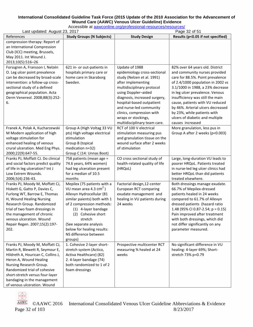
International Consolidated Guideline Task Force (2015 Update of the 2010 Association for the Advancement of Wound Care (AAWC) Venous Ulcer Guideline) Evidence
Accessible at aawconline.org/professional-resources/resources/ Last updated: August 23, 2017 Page 32 of 51
©AAWC 2016 International Consolidated Venous Ulcer Guideline Abbreviations & Evidence
Page 32 of 103 8/23/2017
References Study Groups (N Subjects) Study Design Results (p<0.05 if not specified)
compression therapy: Report of an International Compression Club (ICC) meeting, Brussels, May 2011. Int Wound J. 2013;10(5):516–26
Forssgren A, Fransson I, Nelzén O. Leg ulcer point prevalence can be decreased by broad-scale intervention: a follow-up cross-sectional study of a defined geographical population. Acta Derm Venereol. 2008;88(3):252-6.
621 in- or out-patients in hospitals primary care or home care in Skaraborg Sweden.
Update of 1988 epidemiology cross-sectional study (Nelzen et al. 1991) after implementing multidisciplinary protocol using Doppler–aided diagnosis, increased surgery, hospital-based outpatient and nurse-led community clinics, compression with wraps or stockings, multidisciplinary team care.
82% over 64 years old. District and community nurses provided care for 88.5%. Point prevalence of 2.4/1000 population in 2002 vs 3.1/1000 in 1988, a 23% decrease in leg ulcer prevalence. Venous insufficiency was still the main cause, patients with VU reduced by 46%. Arterial ulcers decreased by 23%, while patients with ulcers of diabetic and multiple causes increased
Franek A, Polak A, Kucharzewski M Modern application of high voltage stimulation for enhanced healing of venous crural ulceration. Med Eng Phys. 2000;22(9):647-55.
Group A (High Voltag 33 VU pts) High voltage electrical stimulation Group B (topical medication n=32) Group C (14: Unnas Boot)
RCT of 100 V electrical stimulation measuring pus and granulation tissue on the wound surface after 2 weeks of stimulation
More granulation, less pus in Group A after 2 weeks (p<0.003)
Franks PJ, Moffatt CJ. Do clinical and social factors predict quality of life in leg ulceration? Int J Low Extrem Wounds. 2006;5(4):236-43.
758 patients (mean age = 74.6 years, 64% women) had leg ulceration present for a median of 10.5 months
CO cross sectional study of health-related quality of life (HRQoL)
Large, long-duration VU leads to poorer HRQoL. Patients treated in nurse-led leg ulcer clinics had better HRQoL than did patients treated elsewhere.
Franks PJ, Moody M, Moffatt CJ, Hiskett G, Gatto P, Davies C, Furlong WT, Barrow E, Thomas H, Wound Healing Nursing Research Group. Randomized trial of two foam dressings in the management of chronic venous ulceration. Wound Repair Regen. 2007;15(2):197-202.
Mepilex (75 patients with a VU mean area 4.3 cm
2 )
Allevyn Hydrocellular (81 similar paients) both with 1 of 2 compression methods:
(1) 4-layer bandage (2) Cohesive short
stretch (See separate analysis below for healing results: NS difference between groups)
Factorial design,12-center European RCT comparing exudate management and healing in VU patients during 24 weeks
Both dressings manage exudate. 66.7% of Mepilex-dressed patients healed in 24 weeks compared to 61.7% of Allevyn dressed patients (hazard ratio 1.48 (95% CI 0.87-2.54; p = 0.15) Pain improved after treatment with both dressings, which did not differ significantly on any parameter measured.
Franks PJ, Moody M, Moffatt CJ, Martin R, Blewett R, Seymour E, Hildreth A, Hourican C, Collins J, Heron A, Wound Healing Nursing Research Group. Randomized trial of cohesive short-stretch versus four-layer bandaging in the management of venous ulceration. Wound
1. Cohesive 2-layer short-stretch system (Actico, Activa Healthcare) (82) 2. 4-layer bandage (74) both randomized to 1 of 2 foam dressings
Prospective multicenter RCT measuring % healed at 24 weeks
No significant difference in VU healing: 4-layer 69%; Short-stretch 73% p=0.79

International Consolidated Guideline Task Force (2015 Update of the 2010 Association for the Advancement of Wound Care (AAWC) Venous Ulcer Guideline) Evidence
Accessible at aawconline.org/professional-resources/resources/ Last updated: August 23, 2017 Page 33 of 51
©AAWC 2016 International Consolidated Venous Ulcer Guideline Abbreviations & Evidence
Page 33 of 103 8/23/2017
References Study Groups (N Subjects) Study Design Results (p<0.05 if not specified)
Repair Regen 2004;12:157–62
Friedman SJ, Su DS. Management of leg ulcers with hydrocolloid occlusive dressing. Arch Dermatol 1984;120:1329-36
Study 1: 15 patients with 19 VU or mixed etiology leg ulcers Study 2: 7 hospitalized patients confined to bed with 2 VU each, the worst one randomized to receive hydrocolloid and the other VU received wet-dry gauze
CCT. All Study 1 leg ulcers were treated solely with hydrocolloid dressing for up to 259 days. In Study 2, the largest or deepest VU on each of 7 patients with 2 leg ulcers each received HCD and the other leg ulcer received Sweitzers wet-to-dry gauze.
12 of 19 leg ulcers healed on the patients receiving DuoDERM® hydrocolloid. Study 2 all ulcers healed in mean of 44 days for HCD or 42 days with Sweitzer’s moist gauze. Patients reported much greater pain relief in the HCD-dressed leg ulcers than in those dressed with moist gauze.
Gallenkemper G, Rabe E, Bauer R. Contact sensitization in chronic venous insufficiency: modern wound dressings. Contact Dermatitis 1998;38(5): 274–8.
Secondary studies only. LR citing sensitization reactions in patients with a VU some confirmed with patch tests.
Be alert for sensitization reactions in VU patients skin surrounding VU
Garcia-Rinaldi R, Soltero E, Gaviria J, Sosa J, Tucker P. Implantation of cryopreserved allograft pulmonary monocusp patch to treat nonthrombotic femoral vein incompetence. Texas Heart Inst J 2002; 29(2):92-9.
38 patients with 40 >3 year duration VU resulting from non-thrombotic venous insufficiency of common femoral vein received patch
CS: Ulcer healing and competence of monocusp patches implanted using Duplex scans at 30 days, 6 months and 1 yr post-op
23 ulcers remained healed at the end of 1 year with competent valves at patch sites. 27 of 36 evaluable ulcers eventually healed. 9 had monocusp insufficiency.
Gardner SE, Frantz RA, Saltzman CL, Hillis SL, Park H, Scherubel M. Diagnostic validity of three swab techniques for identifying chronic wound infection. Wound Repair Regen. 2006;14(5):548-557.
30 of 36 patients with 83 wounds, mainly non-arterial ulcers including 5 venous ulcers, 2 infected as defined by having 10
6
CFU/g of viable tissue and 3 non-infected by this definition, which is not universally accepted or practical in some settings.
Prospective cross-over study comparing CFU from Levine technique, harvesting wound exudate or a Z –swab technique not using Levine technique. Results are reported as overall accuracy of predicting clean biopsy bioburden from area under receiver-operating curve, sensitivity, specificity and positive (PPV) or negative (NPV) predictive likelihood of having a deep biopsy with >10
5 CFU / g of tissue
CFU for all 3 techniques were (p< 0.02) correlated with those of biopsies. A accuracy was highest (0.80) for Levine technique was strongest predictor of deep biopsy having >10
5/ CFU / g of
biopsy tissue. At a critical CFU of 37,000 it had sensitivity of 0.90, specificity of 0.57, PPV of 0.54, NPV of 0.91; Wound exudate: accuracy 0.67 .At a critical value of 20,000 CFU, sensitivity =0.90, specificity =0.40, PPV=0.46, NPV= 0.88. Z-technique Accuracy: 0.67; at a critical 62,500 CFU, sensitivity =0.90; specificity=0.27 PPV=0.41, NPV=0.83.
Gethin G, Cowman S, Kolbach DN. Debridement for venous leg ulcers. Cochrane Database Syst Rev. 2015 Sep 14;9:CD008599. Review. PubMed PMID: 26368002
SR of VU debridement effects on healing or debridement: 10 RCTs involving 715 participants.
8 RCTs evaluated autolytic debridement using: biocellulose wound dressing , non-adherent dressing, honey, hydrogel (amorphous), hydrofiber® dressing, hydrocolloid dressings, dextranomer
Two trials that assessed autolytic debridement methods reported the number of wounds healed at 12 weeks. One trial (n=108) reported that 24/54 (44%) ulcers treated with honey healed versus 18/54 (33%) treated with hydrogel (RR (adjusted for

International Consolidated Guideline Task Force (2015 Update of the 2010 Association for the Advancement of Wound Care (AAWC) Venous Ulcer Guideline) Evidence
Accessible at aawconline.org/professional-resources/resources/ Last updated: August 23, 2017 Page 34 of 51
©AAWC 2016 International Consolidated Venous Ulcer Guideline Abbreviations & Evidence
Page 34 of 103 8/23/2017
References Study Groups (N Subjects) Study Design Results (p<0.05 if not specified)
beads, Edinburgh University Solution of Lime (EUSOL) and paraffin gauze. Two RCTs evaluated enzymatic preparations and one evaluated biosurgical debridement. No RCTs evaluated surgical, sharp or mechanical methods of debridement, or debridement versus no debridement.
baseline wound diameter) 1.38, 95% CI 1.02 to 1.88; P value 0.037). The second trial (n=48) reported NS healing difference: 7/25 (28%) ulcers treated with biocellulose dressing healed vs. 7/23 (30%) dressed with non-adherent gauze.Reduction in wound area was assessed in five trials (n=444) comparing two autolytic methods One three-armed trial (n=153) reported a mean of 24.9 cm
2 more area
reduction using cadexomer iodine or 23.8 cm² more for hydrocolloid dressing both compared to paraffin gauze (both p values < 0.01; ) . A second trial that assessed reduction in median area at four weeks, reported faster healing with honey vs. hydrogel (both under a foam dressing). (p < 0.001)
Gethin G, Cowman S. Bacteriological changes in sloughy venous leg ulcers treated with manuka honey or hydrogel: an RCT. J Wound Care. 2008;17(6):241-4, 246-7.
54 patients with VU<100 cm
2 in each group:
Manuka Honey (MH) Hydrogel (HG) All treated with compression.
Open label multicenter RCT of VU covered with >50% slough. Wound swabs taken at baseline and after 4 weeks of topical treatment.
16 patients had MRSA at baseline eradicated in 7 / 10 of MH group by week 4 and increased from 6 at baseline to 7 at week 4 for HG group. Pain decreased more with MH than HG. (P<0.05)
Gethin G. Manuka honey versus hydrogel - a prospective, open label, multicentre, randomised controlled trial to compare desloughing efficacy and healing outcomes in venous ulcers. Unpublished PhD thesis 2007.
VU patients >18 years of age with ABI > 0.8 and a VU of area < 100 cm
2 with
ulcer bed > 50%covered with slough. Manukah honey (MH) 5 g/ 20 cm
2
(54) Hydrogel (HG) 3 g/ 20 cm
2 (54) Both covered with
hydrocellular foam, mostly 4-layer compression. At baseline mean duration:MH: 39.5 wk, HG 29.9 wk; area MH 10.5 cm
2,
HG 9.9 cm2
Open label RCT. All patients treated once weekly for at least 4 weeks. Primary outcomes % slough week 1-4 and % healed at 12 weeks. Secondary outcomes: decrease in wound size; percent epithelized; safety was measured as adverse events
At 4 weeks MH had 67% slough decrease, HG 53% (p=0.05) At 12 weeks, 44% of MH and 33% of HG patients healed (p=0.037). Epithelization was visible earlier for MH (p=0.042). Median wound size decreased from baseline to week 4 by 34% for MH or 13% for HG (p=0.001). At 4 weeks > 50% slough reduction associated with a higher 12-wk healing probability-all patients (p =0.029)
Geyer MJ, Brienza DM, Chib V, Wang J. Quantifying fibrosis in venous disease: mechanical properties of lipodermatosclerotic and
9 healthy volunteers 9 individuals with venous insufficiency
CCT quantifying fibrosis and lipodermatosclerotic tissue compared to healthy tissue using ultrasound indentometry and
Significant differences between groups on clinical and quasi-liniear viscoelastic tissue parameters and ultrasound indentometry—all measures

International Consolidated Guideline Task Force (2015 Update of the 2010 Association for the Advancement of Wound Care (AAWC) Venous Ulcer Guideline) Evidence
Accessible at aawconline.org/professional-resources/resources/ Last updated: August 23, 2017 Page 35 of 51
©AAWC 2016 International Consolidated Venous Ulcer Guideline Abbreviations & Evidence
Page 35 of 103 8/23/2017
References Study Groups (N Subjects) Study Design Results (p<0.05 if not specified)
healthy tissue. Adv Skin Wound Care. 2004;17(3):131-42.
computerized tomography. proved accurate, reliable and valid.
Gibbons GW, Orgill DP, Serena TE, Novuong A, O’Connell JB, Li WW, Driver VR. A prospective, randomized controlled trial comparing the effects of non-contact, low-frequency ultrasound to standard of care in healing venous ulcers. Ostomy/Wound Manage. 2015;61(1):16-29
N=112 with documented venous ulcers of venous or mixed venous-arterial etiology. Of these 81 were non-healing, i.e. <30% ulcer area reduction during the run-in phase and were randomized to SC (40) NLFU+SC (41)
RCT of 81 pts- randomized to standard care (SC) alone or SC and 40 kHz noncontact, low-frequency ultrasound (US) treatments 3 times per week for 4 weeks.
After 4 weeks of treatment, average wound size reduction was 61.6% ± 28.9 in the US+SC compared to 45% ± 32.5 in the SC group (P = 0.02). Reductions in median (65.7% versus 44.4%, P = 0.02) and absolute wound area (9.0 cm2 versus 4.1 cm2, P = 0.003) as well as pain scores (from 3.0 to 0.6 versus 3.0 to 2.4, P = 0.01) were also significant. NLFU therapy with guideline-defined standard VLU care should be considered for healing VLUs not responding to SC alone.
Glinski W, Chodynicka B, Roszkiewicz J, Bogdanowski T, Lecewicz-Torun B, Kaszuba A, Bowszyc J, Nowak A, Wnorowski J, Wasik F, Glinska-Ferenz M, Blaszczyk M, Strzyga P, Pachocki R. [Effectiveness of a micronized purified flavonoid fraction (MPFF) in the healing process of lower limb ulcers. An open multicentre study, controlled and randomized]. Minerva Cardioangiol. 2001 Apr;49(2):107-14.
Data from SR. Original not found. Micronized purified flavonoid fraction (oral 2 tablets daily) in addition to standardized conventional compression and dressings on 140 clients with a VU and chronic venous insufficiency
Open label multicenter RCT in Italy measuring % healed after 24 weeks and cost per VU healed.
Among patients receiving the oral flavonoid, 46.5% healed their VU in 24 weeks compared to 27.5% control clients (p<0.05). The cost per ulcer healed was €1026.2 compared to €1871.8 in the control group. % healed at 24 weeks increased in VU < 3 cm
2 in
diameter at baseline, but the group comparisons remained statistically significant for VU of all sizes.
Gloviczki P, Comerota AJ, Dalsing MC, Eklof BG, Gillespie DL, Gloviczki ML, Lohr JM, McLafferty RB, Meissner MH, Murad MH, Padberg FT, Pappas PJ, Passman MA, Raffetto JD, Vasquez MA, Wakefield TW; Society for Vascular Surgery.; American Venous Forum. The care of patients with varicose veins and associated chronic venous diseases: clinical practice guidelines of the Society for Vascular Surgery and the American Venous Forum. J Vasc Surg 2011;53(5):2S-48S.
Guideline-no new subjects Guideline. All recommendations were included in the 2015 update of the ICGTF Venous Ulcer Guideline.
Gloviczki P, Gloviczki ML LR of minimally invasive LR of RCTs reporting healing Several RCTs confirm SEPS

International Consolidated Guideline Task Force (2015 Update of the 2010 Association for the Advancement of Wound Care (AAWC) Venous Ulcer Guideline) Evidence
Accessible at aawconline.org/professional-resources/resources/ Last updated: August 23, 2017 Page 36 of 51
©AAWC 2016 International Consolidated Venous Ulcer Guideline Abbreviations & Evidence
Page 36 of 103 8/23/2017
References Study Groups (N Subjects) Study Design Results (p<0.05 if not specified)
Evidence on efficacy of treatments of venous ulcers and on prevention of ulcer recurrence. Perspect Vasc Surg Endovasc Ther. 2009;21(4):259-68.
SEPS or endoscopic laser surgery or radiofrequency ablation, vs open deep vein stripping
or prevention of recurrence of VU resulting from use of any one of these three interventions.
reduces VU recurrence and improves healing. Evidence is mounting for radiofrequency ablation and laser ablation., but is insufficient for open deep vein (saphenous) surgery.
Goedkoop R, Juliet R, You PH, Daroczy J, de Roos KP, Lijnen R, Rolland E, Hunziker T. Wound stimulation by growth-arrested human keratinocytes and fibroblasts: HP802-247, a new-generation allogeneic tissue engineering product. Dermatology. 2010;220(2):114-20.
HP802-247: Allogeneic growth arrested keratinocytes and fibroblasts sprayed on in fibrin matrix administered once/week for 12 weeks (6 doses)N=66 Vs PlaceboN = 8)
Multicenter RCT measuring 12-week % healed and 20-24 week % healed Data were obtained from SR by Jones & Nelson, 2013.
12 week: 4% rate of area reduction for all patients on HP802 vs 33% for placebo. 12 week: 31/66 = 47% allogeneic healed vs.37.5% placebo ; 20-24-week: 60% Allogeneic healed or 50% of placebo. Neither result was significant. Sample size was too small resulting in insufficient evidence of efficacy of the spray intervention.
Gohel MS, Barwell JR, Heather BP, Earnshaw JJ, Mitchell DC, Whyman MR, Poskitt KR. The predictive value of haemodynamic assessment in chronic venous leg ulceration. Eur J Vasc Endovasc Surg. 2007;33(6):742-6.
383 VU patients undertoing superficial vein surgery
Retrospective CO measuring Venous Refill Time (VRT) with photoplethysmography to predict healing and VU recurrence
Significant healing and recurrence prediction only with below knee tourniquet. Normal VRT is at least 20 seconds.
Gohel MS, Barwell JR, Taylor M, Chant T, Foy C, Earnshaw JJ, Heather BP, Mitchell DC, Whyman MR, Poskitt KR. Long term results of compression therapy alone versus compression plus surgery in chronic venous ulceration (ESCHAR): randomised controlled trial. BMJ. 2007;14;335(7610):83.
500 VU patients half each: SEPS correction of superficial V reflux + compression (~250 VU patients, 1 leg each) Compression alone (~250 same)
RCT measuring 3 yr healing and recurrence rate
Healing rates similar 93% SEPS, 89% compression alone. Less recurrence 31% for SEPS + compression vs 56% compression only
Golinko MS, Clark S, Rennert R, Flattau A, Boulton AJ, Brem H. Wound emergencies: the importance of assessment, documentation, and early treatment using a wound electronic medical record. Ostomy/ Wound Manage. 2009;55(5):54-61.
139 patients with chronic wounds (29%) with a VU
Cohort treated with sharp debridement and biopsy with pathology with wound parameters documented using an electronic medical record.
New or increasing pain, cellulitis and/or drainage or presence of significant undermining may indicate invasive infection and warrant hospital admission to explore and correct causes of tissue deterioration, including suspected infection, before wound deterioration becomes more serious.
Gottrup F, Holstein P, Jorgensen B, Lohmann M, Karlsmar T. A new concept of a
23806 patient cohort with a variety of chronic wounds
HCT after a multidisciplinary team approach was implemented. Outcomes
Leg ulcers healing rates and amputation rates improved compared to pre-team levels
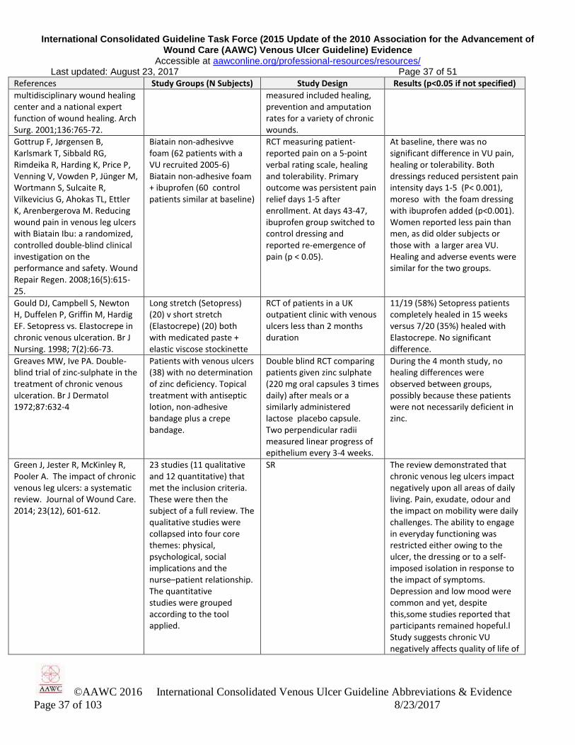
International Consolidated Guideline Task Force (2015 Update of the 2010 Association for the Advancement of Wound Care (AAWC) Venous Ulcer Guideline) Evidence
Accessible at aawconline.org/professional-resources/resources/ Last updated: August 23, 2017 Page 37 of 51
©AAWC 2016 International Consolidated Venous Ulcer Guideline Abbreviations & Evidence
Page 37 of 103 8/23/2017
References Study Groups (N Subjects) Study Design Results (p<0.05 if not specified)
multidisciplinary wound healing center and a national expert function of wound healing. Arch Surg. 2001;136:765-72.
measured included healing, prevention and amputation rates for a variety of chronic wounds.
Gottrup F, Jørgensen B, Karlsmark T, Sibbald RG, Rimdeika R, Harding K, Price P, Venning V, Vowden P, Jünger M, Wortmann S, Sulcaite R, Vilkevicius G, Ahokas TL, Ettler K, Arenbergerova M. Reducing wound pain in venous leg ulcers with Biatain Ibu: a randomized, controlled double-blind clinical investigation on the performance and safety. Wound Repair Regen. 2008;16(5):615-25.
Biatain non-adhesivve foam (62 patients with a VU recruited 2005-6) Biatain non-adhesive foam + ibuprofen (60 control patients similar at baseline)
RCT measuring patient-reported pain on a 5-point verbal rating scale, healing and tolerability. Primary outcome was persistent pain relief days 1-5 after enrollment. At days 43-47, ibuprofen group switched to control dressing and reported re-emergence of pain (p < 0.05).
At baseline, there was no significant difference in VU pain, healing or tolerability. Both dressings reduced persistent pain intensity days 1-5 (P< 0.001), moreso with the foam dressing with ibuprofen added (p<0.001). Women reported less pain than men, as did older subjects or those with a larger area VU. Healing and adverse events were similar for the two groups.
Gould DJ, Campbell S, Newton H, Duffelen P, Griffin M, Hardig EF. Setopress vs. Elastocrepe in chronic venous ulceration. Br J Nursing. 1998; 7(2):66-73.
Long stretch (Setopress) (20) v short stretch (Elastocrepe) (20) both with medicated paste + elastic viscose stockinette
RCT of patients in a UK outpatient clinic with venous ulcers less than 2 months duration
11/19 (58%) Setopress patients completely healed in 15 weeks versus 7/20 (35%) healed with Elastocrepe. No significant difference.
Greaves MW, Ive PA. Double-blind trial of zinc-sulphate in the treatment of chronic venous ulceration. Br J Dermatol 1972;87:632-4
Patients with venous ulcers (38) with no determination of zinc deficiency. Topical treatment with antiseptic lotion, non-adhesive bandage plus a crepe bandage.
Double blind RCT comparing patients given zinc sulphate (220 mg oral capsules 3 times daily) after meals or a similarly administered lactose placebo capsule. Two perpendicular radii measured linear progress of epithelium every 3-4 weeks.
During the 4 month study, no healing differences were observed between groups, possibly because these patients were not necessarily deficient in zinc.
Green J, Jester R, McKinley R, Pooler A. The impact of chronic venous leg ulcers: a systematic review. Journal of Wound Care. 2014; 23(12), 601-612.
23 studies (11 qualitative and 12 quantitative) that met the inclusion criteria. These were then the subject of a full review. The qualitative studies were collapsed into four core themes: physical, psychological, social implications and the nurse–patient relationship. The quantitative studies were grouped according to the tool applied.
SR The review demonstrated that chronic venous leg ulcers impact negatively upon all areas of daily living. Pain, exudate, odour and the impact on mobility were daily challenges. The ability to engage in everyday functioning was restricted either owing to the ulcer, the dressing or to a self-imposed isolation in response to the impact of symptoms. Depression and low mood were common and yet, despite this,some studies reported that participants remained hopeful.l Study suggests chronic VU negatively affects quality of life of

International Consolidated Guideline Task Force (2015 Update of the 2010 Association for the Advancement of Wound Care (AAWC) Venous Ulcer Guideline) Evidence
Accessible at aawconline.org/professional-resources/resources/ Last updated: August 23, 2017 Page 38 of 51
©AAWC 2016 International Consolidated Venous Ulcer Guideline Abbreviations & Evidence
Page 38 of 103 8/23/2017
References Study Groups (N Subjects) Study Design Results (p<0.05 if not specified)
the patient and that such issues receive inadequate attention during current consultations. If such negative implications are to be effectively addressed, key issues need to be considered during every consultation.
Greguric S, Budimicic, Soldo-Belic A, Tudoric M, Baricevic B, Cajkovac V, Dobric I. Hydrocolloid dressing versus a conventional dressing using magnesium sulphate paste in the management of venous leg ulcers. Acta Dermatovenerol. Croat. 1994;2(2):65-71
Hydrocolloid dressing (HCD) DuoDERM® CGF® + two layers of tubular compression bandages (55) Conventional magnesium sulfate paste with gauze + double layer elastic compression bandage (55)
Open label controlled, prospective parallel group study of venous ulcers for 10 dressing changes at 2 dermatology hospital clinics.
HCD-dressed ulcers healed 32 sq mm/day; 21 sq mm/day for gauze. 50% epithelization was achieved in 6 visits for HCD or in 10 visits for gauze. 3 healed in HCD group, 0 in gauze group. HCD had less (p<0.05) discomfort and longer intervals between dressing changes.
Grey JE, Enoch S, Harding KG. Venous and arterial leg ulcers. In Enoch Grey and Keith Harding (Eds.) ABC of Wound Healing BMJ 2006; 332(7537):347-50.
Educational text article with minimal references.
EO Differential diagnosis of arterial and venous ulcers includes reduced hair follicles and sweat glands in legs with arterial disease.
Griffiths , RD, Fernandez, RS, Ussia, CA. Is tap water a safe alternative to normal saline for wound irrigation in the community setting? Journal of Wound Care. 2001:10(10) 407-11.
35 patients with 49 acute or chronic wounds including VU in metropolitan community health care in New South Wales, Australia.
Double blind RCT comparing tap water or normal saline wound cleansing effects on healing or clinically defined wound infection.
There was no significant difference in healing or infection rates between the two groups.
Guarnera G, DeRosa A, Camerini R; 8 European sites. The effect of thymosin treatment of venous ulcers. Ann N Y Acad Sci. 2010;1194:207-12.
0.01% thymosinß4 (TB4) 0.03% TB4 0.10% TB4 Total 55 for TB4 groups 17 Placebo
RCT 2 blind Phase 2 study in Italy (5 centers) Poland (3 centers) 12 week safety and healing results from clinicaltrials.gov
Healed at 12 weeks: 4/17 Placebo 12/55 TB4 (NS) No safety differences.
Guilhou JJ, Février F, Debure C, Dubeaux D, Gillet-Terver MN, Guillot B, Levesque H, Marzin L, Mignot J, Ouvry P, Pillion G, Van Landuyt H, Zuccarelli F,Nicolaïdes AN. Benefit of a 2-month treatment with a micronized, purified flavonoidic fraction on venous ulcer healing. A randomized, double-blind, controlled versus placebo trial. Int J Microcirc Clin Exp. 1997;17 Suppl 1:21-6.
micronized purified flavonoid fraction (500 mg)53 patients with a VU or Placebo (52 similar patients. All receivedcompression therapy and standardized local care.
3-month duration RCT measuring # of patients completely healed after 2 months. ITT analysis comparing % healed in the 2 groups after 2 months treatment
After 2 months treatment, % of flavonoid group healed compared to 13% (p = 0.028. No VU over 10 cm
2 in area healed during the
study. )
Gulati S, Qureshi A, Srivastava A, Honey under Tegaderm® One-center 6-week RCT Groups were comparable on
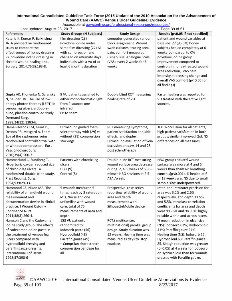
International Consolidated Guideline Task Force (2015 Update of the 2010 Association for the Advancement of Wound Care (AAWC) Venous Ulcer Guideline) Evidence
Accessible at aawconline.org/professional-resources/resources/ Last updated: August 23, 2017 Page 39 of 51
©AAWC 2016 International Consolidated Venous Ulcer Guideline Abbreviations & Evidence
Page 39 of 103 8/23/2017
References Study Groups (N Subjects) Study Design Results (p<0.05 if not specified)
Kataria K, Kumar P, Balkrishna JA. A prospective randomized study to compare the effectiveness of honey dressing vs. povidone iodine dressing in chronic wound healing. Ind J Surgery. 2014;76(3):193-8.
film dressing (23) Povidone iodine under same film diressing (22) All with compression and changed on alternate days. individuals with a Vu of at least 6 months duration
computer-generated random block assignment. Wound swab cultures, tracing area, pain, comfort measured using Visual Analogue Scale (VAS) every 2 weeks for 6 weeks
patient and wound variables at baseline. 22 (95.6%) honey subjects healed completely at 6 weeks compared to 0% in povidone iodine group. Improvement compared to controls in honey-treated wound area reduction, VAS pain intensity at dressing change and overall VAS comfort (p< 0.05 for all findings)
Gupta AK, Filonenko N, Salansky N, Sauder DN. The use of low energy photon therapy (LEPT) in venous leg ulcers: a double-blind, placebo-controlled study. Dermatol Surg. 1998;24(12):1383-6.
9 VU patients assigned to either monochromatic light from 2 sources one infrared Or to sham
Double blind RCT measuring healing rate of VU
Faster healing was reported for VU treated with the active light sources.
Hamel-Desnos CM, Guias BJ, Desnos PR, Mesgard A. Foam |py of the saphenous veins: randomised controlled trial with or without compression. Eur J Vasc Endovasc Surg. 2010;39(4):500-7
Ultrasound guided foam sclerotherapy with (29) vs without (31) compression stockings
RCT measuring symptoms, patient satisfaction and side effects and duplex Ultrasound evaluation of vein occlusion on days 14 and 28 post sclerotherapy
100 % occlusion for all patients, high patient satisfaction in both groups, similar improved QoL NS differences on all measures .
Hammarlund C. Sundberg T. Hyperbaric oxygen reduced size of chronic leg ulcers: a randomized double-blind study. Plast Reconst. Surg. 1994;93:829-33.
Patients with chronic leg ulcers: HBO (9) Control (8)
Double-bline RCT measuring wound surface area decrease during 2, 4,6 weeks of 5 90- minute HBO sessions at 2.5 ATA /week.
HBO group reduced wound surface area more at 4 and 6 weeks than sham air breathing controls(p<0.001). % healed at 6 or 18 weeks was NS due to small sample size: underpowered.
Hammond CE, Nixon MA. The reliability of a handheld wound measurement and documentation device in clinical practice. J Wound Ostomy Continence Nurs. 2011;38(3):260-4.
5 wounds measured 5 times each by 3 raters : an MD, Nurse and one unfamiliar with wound care: total of 75 measurements of area and depth
Prospective case series reporting reliability of wound area and depth measurement with SilhouetteMobile device
Inter- and intrarater precision for area was 3.2% and 2.6%, respectively, and depth 13.5% and 5.5%,intraclass correlation coefficients for area and depth were 99.76% and 98.95%: highly reliable within and across raters.
Hansson C and the Cadexomer Iodine study group. The effects of cadexomer iodine paste in the treatment of venous leg ulcers compared with hydrocolloid dressing and paraffin gauze dressing. International J of Derm. 1998;37:390-6
153 VU patients randomized to: Iodosorb paste (56) Hydrocolloid (48) Parrafin gauze (49) + Comprilan short stretch compression bandage for all
RCT,( multicenter, multinational) parallel group design. Study duration was 12 weeks. Healing time was measured as days to stop exudate.
% mean reduction in ulcer size (NS): Iodosorb 61%; Hydrocolloid 41%; Paraffin gauze 24% Healing time (NS): Iodosorb 55; Hydrocolloid 63; Paraffin gauze 85. Slough reduction was greater (p<0.05) at 4 weeks for Iodosorb or Hydrocolloid than for wounds dressed with Paraffin gauze.

International Consolidated Guideline Task Force (2015 Update of the 2010 Association for the Advancement of Wound Care (AAWC) Venous Ulcer Guideline) Evidence
Accessible at aawconline.org/professional-resources/resources/ Last updated: August 23, 2017 Page 40 of 51
©AAWC 2016 International Consolidated Venous Ulcer Guideline Abbreviations & Evidence
Page 40 of 103 8/23/2017
References Study Groups (N Subjects) Study Design Results (p<0.05 if not specified)
Harding KG, Krieg T, Eming SA, Flour ML, Jawien A, Cencora A, Kaszuba A, Noszcyk W, Willems P, De Deene A, Joos E, De Waele P, Delaey B. Efficacy and safety of the freeze-dried cultured human keratinocyte lysate, LyphoDerm 0.9%, in the treatment of hard-to-heal venous leg ulcers. Wound Repair Regen. 2005;13(2):138-47.
Std HCD +compression (43) Std + Vehicle (56) Std + Lyphoderm® (95) (XCELLentis, Belgium) dried cultured human keratinocyte lysate applied 10 weeks (a total of 8 Tx) Wk 1-4 weekly: 5 applications, followed by 3 apps @ 2 week intervals
RCT on hard-to-heal VU duration >6 weeks (mean 43 weeks) measured % healed within 24 weeks. Subgroup analysis of enlarging ulcers (17 Std; 18 Std+V; 40 Std + L) with and without the 2 control groups combined. ITT analyses.
NS effect on primary 24 week % healed (38% healed in Std+L group; 27% in Std groups pooled) in overall analysis (p = 0.114). Significantly more healed in Std+L group of enlarging VU (30% vs 11% for pooled ITT controls: p = 0.024)
Harding KG, Price P, Robinson B, Thomas S, Hofman D. Cost and dressing evaluation of hydrofiber and alginate dressings in the management of community-based patients with chronic leg ulceration. Wounds. 2001;13(6):229-36.
Leg ulcers--moderately to heavily exuding; various etiologies dressed with a hydrofiber ( AQUACEL: 66) or alginate (SORBSAN: 65) wound dressing
Prospective, 12-week multi-center, randomized, controlled--wear time, cost effectiveness, time to healing, reduction in ulcer size, ease of application and removal, exudate management, pain on dressing removal
Wear time: hydrofiber 3.63 days, alginate 3.27 days (p<0.001). 17 patients healed in each group in a mean of 42 days with hydrofiber, 57 days with alginate (p=0.053). Hydrofiber-dressed ulcers had more ease of removal (p=0.006), exudate management (p=0.002), and less pain (p=0.001) and adhesion to the wound bed (p=0.001). Costs to heal the ulcer were somewhat less for hydrofiber dressings (NS).
Harrison MB, Graham ID, Lorimer K, Friedberg E, Pierscianowski T, Brandys T. Leg ulcer care in the community before and after implementation of an evidence-based service. CMAJ. 2005;172(11);1447-52.
Mixed urban-rural population served by a single, large home-care authority. N=258. Before implementation: 78 clients and 180 clients after guideline implementation.
Prospective, observational “pre-post” study over 1 year before and 1 year after specially trained registered nurses implemented evidence-based leg ulcer guideline recommendations adapted for home care with enhanced linkages to medical specialists. Standardized outcome measures included % healed after 3-months on protocol, quality of life and resource use.
Three-month healing rates doubled between the year before implementation (23% [18/78]) and the year afterward (56% [100/180]). The number of nursing visits per case declined, from a median of 37 to 25 (p =0.041); the median supply cost per case was reduced from $1923 to $406 (p = 0.005).
International Consolidated Guideline Task Force (2015 Update of the 2010 Association for the Advancement of Wound Care (AAWC) Venous Ulcer Guideline) Evidence
Accessible at aawconline.org/professional-resources/resources/ Last updated: August 23, 2017 Page 41 of 51
©AAWC 2016 International Consolidated Venous Ulcer Guideline Abbreviations & Evidence
Page 41 of 103 8/23/2017
References Study Groups (N Subjects) Study Design Results (p<0.05 if not specified)
Harrison MB, Graham ID, Lorimer K, VandenKerkhof E, Buchanan M, Wells PS, Brandys T, Periscianowski T. Nurse clinic versus home delivery of evidence-based community leg ulcer care: A randomized health services trial. BMC Health Serv Res. 2008 Nov 26; 8:243.
N=126 pts. 65 randomized to receive care in their homes, 61 to nurse-run clinics
RCT on care delivered in the home versus a clinic. Primary outcome: 3-month % healed. Secondary outcomes: recurrence as durability of healing, ulcer-free time, satisfaction, resource use, Health Related QoL. Data were collected at base-line, every 3 months until healing occurred, with 1 year follow-up. ITT sample was analyzed.
No differences in 3-month healing rates: clinic 58.3% compared to home care at 56.7% (p = 0.5) No significant differences in secondary outcomes. Findings indicate that organization of care not the setting where care is delivered influences healing rates.
Hartung O, Loundou AD, Barthelemy P, Arnoux D, Boufi M, Alimi YS. Endovascular management of chronic disabling ilio-caval obstructive lesions: long-term results. European J Vasc Endovasc Surg. 2009; 38(1): 118-24.
72women, 17 men; median age 43 years
Long term CS of surgical endovascular management of disabling ilio-caval obstruction. ITT analysis was performed for outcomes which included length of hospital stay, technical success, patency and adverse events. Patency was verified after 3- and 10-year follow up.
Technical success was achieved in 98%. The median hospital stay was 2 days. During a median follow-up of 38 months (range: 1–144 months), one patient died and five cases of thromboses occurred. Iterative stenting was performed for restenosis in six cases. Primary, assisted-primary and secondary patency rates were 83%, 89% and 93%, respectively.
He Q, Wu G, Yu B, Zhang T, Wang W, Gu Q. [A prospective study on wound-healing hydrogel in treating chronic venous ulcer of lower extremities] [Article in Chinese: abstract reviewed] Zhongguo Xiu Fu Chong Jian Wai Ke Za Zhi. 2008;22(3):311-3.
Hydrogel (30 VU patients mean VU duration 2.9 years mean area 3.4 cm
2)
Saline (30 VU patients mean VU duration 3.3 years, mean area 3.1 cm
2)
Not different at baseline.
Prospective RCT measuring % area reduction after 7 and 14 days of treatment and days to heal.
Faster healing : 12 days mean heal time for hydrogel and 31 for saline (p<0.01)Significantly more healing in hydrogel group after 14, but not 7 days (p<0.05).
Hegarty MS, Grant E, Reid L. An overview of technologies related to care for venous leg ulcers. IEEE Transact Inf Technol Biomed. 2010;14 (2) 387-93.
Review article describing wearable, wireless vascular health monitoring systems and integrated sensing modules for use in managing chronic health conditions
This article reviews current treatment options for VU compression ( bandages, medical-grade stockings, pneumatic compression devices) and other technologies available
Heinen M, Borm G, van der Vleuten C, Evers A, Oostendorp R, van Achterberg T. The Lively Legs self-management programme increased physical activity and reduced wound days in leg ulcer patients: Results from a randomized controlled trial. Int J Nurs Stud. 2012;49(2):151-61.
92 Subjects with a healed venous ulcer randomized to “Usual care alone” 92 Similar subjects randomized to “Lively Legs” program + Usual Care
RCT comparing effects on venous ulcer recurrence during 18 months of randomly assigned care.
Subjects randomized to “lively legs” program walked and exercised more and had more VU-free months than standard care (p <0.05)
International Consolidated Guideline Task Force (2015 Update of the 2010 Association for the Advancement of Wound Care (AAWC) Venous Ulcer Guideline) Evidence
Accessible at aawconline.org/professional-resources/resources/ Last updated: August 23, 2017 Page 42 of 51
©AAWC 2016 International Consolidated Venous Ulcer Guideline Abbreviations & Evidence
Page 42 of 103 8/23/2017
References Study Groups (N Subjects) Study Design Results (p<0.05 if not specified)
Heit JA, Rooke TW, Silverstein MD, Mohr DN, Lohse CM, Petterson TM, O'Fallon WM, Melton LJ 3rd. Trends in the incidence of venous stasis syndrome and venous ulcer: a 25-year population-based study. J Vasc Surg. 2001;33(5):1022-7.
25-year study of medical records of a county in Minnesota
RCO: Retrospective review of all medical records to describe incidence of venous stasis (now called insufficiency) and venous ulcers
Venous stasis 76/100,000 person years, VU 18/100,000 person years, with no change in 25 years. Incidence was higher in women than men and increased with age for both.
Hendricks WM. Swallow RT, Management of stasis leg ulcers with Unna's boot versus elastic support stockings J Amer Acad Dermatol. 1985; 12(1 Pt. 1):90-8.
1. Unna's boot (10) 2. Open-toe, below knee graduated compression sock 24 mmHg at ankle (10) self-care between 1-2 weekly visits
RCT measuring time to heal, % healed in 78-week trial. Note: 3 Group 1 patients were transferred after not healing to Group 2 where they healed.
% complete healing on trial: Group 1: 70% Group 2: 71%
Herber OR, Schnepp W, Rieger M. A systematic review of the impact of leg ulceration on patients’ quality of life. Health Qual Life Outcomes 2007 Jul 25;5:44-.56.
24 research studies on leg ulcers of all etiologies- 11 quantitative, 11 qualitative, 2 mixed
SR of quantitative studies measuring the parameters of pain, sleep, social isolation, and physical mobility in individuals with a leg ulcer.
Those with a leg ulcer had significantly more pain, more restricted social functioning, less vitality, and morelimitations in emotional roles than the respective controls. Other problem areas reported were restrictions in work capacity, recreation, social interaction, psychological well-being and problems caused by treatment regimes. Inconclusive results were obtained regarding pain intensity, physical restrictions, and gender effects. Leg ulcer guidelines should address these outcomes with documented impact on QoL.
Herberger K, Franzke N, Blome C, Kirsten N, Augustin M. Efficacy, tolerability and patient benefit of ultrasound-assisted wound treatment versus surgical debridement: a randomized clinical study. Dermatology. 2011;222(3):244-9.
67 patients with chronic wounds in need of debridement in a Dermatologic clinic in Germany
RCT exploring healing , clinical efficacy and patient –reported pain and QoL following surgical debridement or autolytic debridement assisted with ultrasound
Wound status and pain decreased in both groups (NS) with good efficacy and tolerability and a significant increase in quality of life for both debridement techniques. 88% of ultrasound patients and 85.2% ofsurgically debrided patients reported above minimal benefit.
Hilstrom L. Iodosorb compared to standard treatment in chronic venous leg ulcers—a multicenter study. Acta Chir Scand Suppl 1988; 544:53-56.
93 patients with chronic VU unresponsive to treatment for a mean of 2 years were assigned to compression plus either Iodosorb cadexomer iodine or Standard treatment
RCT of 6 week duration Percent decrease in ulcer area during 6 weeks of treatment was measured
Cadexomer iodine-dressed wounds decreased in size 34% (p<0.05) with more pain and erythema reduction, removal of debris and pus and granulation tissue than Standard treatment which resulted in an increase in

International Consolidated Guideline Task Force (2015 Update of the 2010 Association for the Advancement of Wound Care (AAWC) Venous Ulcer Guideline) Evidence
Accessible at aawconline.org/professional-resources/resources/ Last updated: August 23, 2017 Page 43 of 51
©AAWC 2016 International Consolidated Venous Ulcer Guideline Abbreviations & Evidence
Page 43 of 103 8/23/2017
References Study Groups (N Subjects) Study Design Results (p<0.05 if not specified)
wound area. 4 patients reported transient wound pain on when cadexomer iodine was applied.
Hjerppe A, Saarinen JP, Venermo MA, Huhtala HS, Vaalasti A. Prolonged healing of venous leg ulcers: the role of venous reflux, ulcer characteristics and mobility. J Wound Care. 2010;19(11):474, 476, 478 passim..
110 successive patients with chronic VU. 62 of 90 90 healed in 12 weeks with standardized care and compression. 22 non-healed subjects were compared with 28 selected randomly from among the 62 healed controls.
3-month CCT comparing features of patients and ulcers that healed with those that did not heal. Superficial, deep and popliteal venous reflux was greater in nonhealers.
NS difference in age, gender, smoking or oral meds or venous disease severity. 62 of 90 VU in Standard care group healed VU duration 7 mo; 26 mo nonhealed. VU area of those who healed in 12 weeks was 5 cm
2 vs. 11cm
2
For those nonhealed. Nonhealers were more likely to use a walking aid device
Holloway GA, Johansen KH, Barnes RW, Pierce GE. Multicenter trial of cadexomer iodine to treat venous stasis ulcers. West J Med. 1989;151(1):35-8.
Cadexomer iodine (38 patients with a VU) Standard care control group using saline wet-to-dry gauze (37 similar patients)
RCT Healing rate measured from VU tracings as cm
2 /
week / cm of ulcer perimete . Pain, exudate, pus, debris and granulation tissue were also measured. 12 non-healing VU patients crossed over to iodine use.
Twice the rate of healing with cadexomer iodine than with the control group (p= 0.0025) with trends for reduced pain, exudate, pus and debris and more granulation tissue. Occasional mild local burning was felt in VU treated with cadexomer iodine.
Hopman, WM, Buchanan, M, VanDenKerkhof, EG., Harrison, MB., Pain and health-related quality of life in people with chronic leg ulcers. Chronic Dis Inj Can. 2013;33(3):167-74.
Combined results from 2 studies of patients with chronic leg ulcers, conducted 2001 – 2007 (n=564)
Pain measurement used the Numeric Pain Scale. Health -related QoL was measured using the Medical Outcomes Survey 12-item Short Form (SF-12), including a Physical and Mental Component Summary. Logistic and linear regression analyzed pain and QoL, respectively.
Sample (47% male) mean age was 66.5 years. Median pain was 2.2 out of 10 possible. Mental scores were normal (50). Physical scores were low (38) Younger age, arthritis and living with others were associated with pain. Low Physical status was observed infemales, those with a venous/mixed ulcer etiology, larger ulcers, longer ulcer duration, cardiovascular disease, arthritis and higher pain intensity. Poorer mental status was associated with younger age, longer ulcer duration, comorbidity and higher pain intensity.
Horakova M, Partsch H Venous leg ulcers are compression bandages indicated? Phleologie. 1994;47:53-7.
Short-stretch compression (25) Elastic high compression stockings (25)
CCT of 3 month duration. Note: groups not initially comparable in ulcer duration and size (S-s larger longer)
In the elastic high compression stocking group, 94% healed versus 52% in the short stretch bandage group.
Houghton PE, Kincaid CB, Lovell M, Campbell KE, Keast DH, Woodbury MG, Harris KA. Effect of electrical stimulation on chronic leg ulcer size and appearance. Physical Ther.
High voltage pulsed current (HVPC) 100 us, 150 V, 100 Hz (n = 14) 3 times per week versus Sham (n = 13) All patients had chronic leg ulcers, not all of venous
RCT prospective, double-blind measured healing with EZ-Graph, wound appearance with modified PSST (PWAT) 4 weeks stimulation after 2 weeks
Wound appearance and % decrease in wound area improved (p<0.05) during treatment, but difference disappeared at 4 week follow up.

International Consolidated Guideline Task Force (2015 Update of the 2010 Association for the Advancement of Wound Care (AAWC) Venous Ulcer Guideline) Evidence
Accessible at aawconline.org/professional-resources/resources/ Last updated: August 23, 2017 Page 44 of 51
©AAWC 2016 International Consolidated Venous Ulcer Guideline Abbreviations & Evidence
Page 44 of 103 8/23/2017
References Study Groups (N Subjects) Study Design Results (p<0.05 if not specified)
2003;83(1):17-28. insufficiency origin some arterial or diabetes.
with conventional therapy. 7 patients had bilateral VU compared.
Hu D, Phan TT, Cherry GW, Ryan TJ. Dermal oedema assessed by high frequency ultrasound in venous leg ulcers. Br J Dermatol. 1998;138(5):815-20.
12 venous ulcer patients with 13 VU compared to contralateral normal leg
Matched control study using high frequency ultrasound to localize edema
Edema was localized in the dermis.
Humbert P, Faivre B, Véran Y, Debure C, Truchetet F, Bécherel PA, Plantin P, Kerihuel JC, Eming SA, Dissemond J, Weyandt G, Kaspar D, Smola H, Zöllner P; CLEANSITE study group.. Protease-modulating polyacrylate-based hydrogel stimulates wound bed preparation in venous leg ulcers--a randomized controlled trial. J Eur Acad Dermatol Venereol. 2014;28(12):1742-50.
Protease-modulating polyacrylate-based hydrogel (n = 34) or to an amorphous hydrogel (n = 41) Both with compression
RCT comparing wound surface fibrin and granulationtissue from photograhs rated by days 0, 7 and 14 by three observers skilled in wound surface evaluation
After 14 days fibrin and necrotic tissue decreased by 37.6 ± 29.9 percentage points in the polyacrylate hydrogel group or by 16.8 ± 23.0 percentage (p=0.0004)points in the amorphous hydrogel group. Proportion of ulcer area covered by granulation tissue was 36.0% in the polyacrylate hydrogel group and 14.5% in the control group (p = 0.0005).
Humphreys ML, Stewart AH, Gohel MS, Taylor M, Whyman MR, Poskitt KR. Management of mixed arterial and venous leg ulcers. Br J Surg. 2007;94(9):1104-7.
N=1416 ulcers wioth venous reflux. 13.6% had moderate (ABI 0.5-0.85) and 2.2% had severe (ABI, 0.5)concomittment arterial disease.
Prospective protocol study. Consecutive patients with leg ulceration and venous reflux treated by protocol: VU with ABI>0.85 treated with MLCW. VU with ABI < 0.5 considered for immediate revasculari-zation. VU with moderate ABI >0.5 < .85 had modified compression and was considered for revascularization if non-healing.
A protocol including supervised modified compression and selective revascularization healed most mixed arterial and venous leg ulcers. By 36 weeks 87%, 68% or 53% of leg ulcers with insignificant, moderate or severe concomitant arterial disease healed (p< 0.001). Four of 17 revascularized VU with ABI 0.5-0.85 and seven of 15 VU with ABI < 0.5 healed in 36 weeks post surgery, which resulted in 6.5% combined 30-day mortality.
Hutchinson J.J. A prospective clinical trial of wound dressings to investigate the rate of infection under occlusion. In Harding KG, Turner TD. Proceedings: Advances in Wound Management. London, England: MacMillan 1993 Oct (pp. 93-6).
DuoDERM® CGF (HCD) (34 burns;37 donor sites;37 venous ulcers) Impregnated gauze (39 burns;46 donor sites;41 venous ulcers) Sivadene + HCD (29 burns;13 donor sites;16 venous ulcers)
Prospective randomized blind evaluated study of microorganisms and infections in donor sites, burns and venous ulcers (41 dressed with conventional gauze and 37 dressed with HCD alone).
There were significantly fewer clinical infections in combined groups dressed with HCD (1.9%) or HCD + Silver sulfadiazine (0 %) vs 5.38% in wounds dressed with impregnated gauze. (p<0.05)
Hutchinson JJ, McGuckin M. Occlusive dressings: A microbiologic and clinical review. Amer J Infec Control. 1990;18(4):257-68.
Controlled studies only: Occlusive (moisture-retentive) dressings (1421) all forms of gauze dressings (1013). Included 5 studies
Retrospective LR from 1962 to 1990 on moisture-retentive dressings (hydrocolloids, foams, films, gel dressings) compared to
Chronic ulcers (mainly VU) : 6.1% infections with all forms of gauze dressings; 1.1% with HCD or film dressings (p<0.05). Overall: 7.3% infections for gauze- dressed

International Consolidated Guideline Task Force (2015 Update of the 2010 Association for the Advancement of Wound Care (AAWC) Venous Ulcer Guideline) Evidence
Accessible at aawconline.org/professional-resources/resources/ Last updated: August 23, 2017 Page 45 of 51
©AAWC 2016 International Consolidated Venous Ulcer Guideline Abbreviations & Evidence
Page 45 of 103 8/23/2017
References Study Groups (N Subjects) Study Design Results (p<0.05 if not specified)
on 114 VU with impregnated gauze primary dressings and 14 studies on 504 VU with film or hydrocolloid (HCD) primary dressings
NOC dressings (gauze/impregnated gauze) Ulcer analysis also included 3 OCL Trials (131) and 1NOC Trial (42) pressure ulcers
wounds. 3.2% for HCD or film- dressed (p <0.001). This trend was similaar for ulcers, burns, donor sites and surgical/other wounds, though not statistically significant for burns.
Ieran M, Zaffuto S, Bagnacani M, Annovi M, Moratti A. Cadossi R. Effect of low frequency pulsing electromagnetic fields on skin ulcers of venous origin in humans: a double-blind study. J Orthop Res. 1990;8(2):276-82.
Electromagnetic Low Frequency Pulsed Stimulation was the Active group (22) Placebo was visually similar (22)
Double-blind RCT studying healing at 90 days and recurrence.
Significantly more healed in the active group than in the placebo group (p< 0.02). 25% recurred during 90 days in the active group; 50% recurred in 90 days in the placebo group.
Iglesias CP, Claxton K. Comprehensive decision-analytic model and Bayesian value-of-information analysis: Pentoxifylline in the treatment of chronic venous leg ulcers. Pharmacoeconomics. 2006;24(5):465-78.
No subject data. Resource use was based on a UK national audit in 2004 British pounds.
SR generated probabilities for a Bayesian model of pentoxifylline cost effective-ness compared to placebo used as an adjunct to standardized chronic VU management.
Based on prior studies analysis showed that those treated with pentoxifylline healed a mean of 8.28 weeks faster than placebo treated subjects increasing quality adjusted life years by 0.02 and decreasing costs by a mean of 153.4 British pounds.
Janković A, Binić I. Frequency rhythmic electrical modulation system in the treatment of chronic painful leg ulcers. Arch Dermatol Res. 2008;300(7):377-83.
Frequency modulated Electrical Stimulation (~1/2 of 35 VU patients) Control (remaining subjects)
RCT measuring healing, pain (VAS), ulcer fibrin, exudate, granulation and epithelization at weeks 0,1,2,3 and after 1 or 2 months post E-stim
Ulcer healing, pain and surrounding skin all improved (p<0.05) in electrical stimulation group.
Jankūnas V, Rimdeika R, Pilipaityte Treatment of the leg ulcers by skin grafting. Medicina (Kaunas). 2004;40(5):429-33.
54 Lithuania VU patients with VU of 6 months or longer duration > 50 cm
2
area, treated Jan 2001-Jan 2004
CS of 0.2-0.3 mm thick autografting using Granuflex hydrocolloid dressing to prepare the wound
Grafts took totally in 35 (65%) of cases; partial take in 19 (35%). Complete healing in 2-3 weeks in 64% of patients.
Jemec GB, Kerihuel JC, Ousey K, Lauemøller SL, Leaper DJ. Cost-effective use of silver dressings for the treatment of hard-to-heal chronic venous leg ulcers. PLoS One. 2014 Jun 19;9(6):e100582.
Cost effectiveness decision tree model based on 4 RCT meta-analysis (see Leaper et al., 2013)
Model of cost effectiveness using standardized methods and %s healed by 4 weeks in acute care, based on results of Leaper et al., 2013
4-week treatment with silver dressing for initial 4-weeks saved a total of £141.57 compared with non-silver dressings while healing a greater proportion of hard-to-heal VU.
International Consolidated Guideline Task Force (2015 Update of the 2010 Association for the Advancement of Wound Care (AAWC) Venous Ulcer Guideline) Evidence
Accessible at aawconline.org/professional-resources/resources/ Last updated: August 23, 2017 Page 46 of 51
©AAWC 2016 International Consolidated Venous Ulcer Guideline Abbreviations & Evidence
Page 46 of 103 8/23/2017
References Study Groups (N Subjects) Study Design Results (p<0.05 if not specified)
Jessup G, Lane RL. Repair of incompetent venous valves: A new technique. J Vasc Surg. 1988; 8(5):569-575.
VU patients Description of technique in venous insufficiency patients
Repairing incompetent venous valves using cuff support or constriction at the site of the valves is effective
Johannsen G, Gam AN, Karlsmark T. Ultrasound therapy in chronic leg ulceration: a meta-analysis. Wound Rep Reg 1998;6(2):121-6.
MA of 6 RCTs often lacking information on baseline ulcer area, etiology, ultrasound (US) head area and placebo treatment. Low doses to ulcer edge seem to yield better results
Pooled MA of percent area reduction from baseline for 6 RCT and of % completely healed for 3 studies.
Mean % reduction in area at 4 weeks was 16.9% higher for US treated subjects at 4 weeks ( p = 0.011) or 14.5% higher for US than control at 8 weeks ( p = 0.005) Number of ulcers healed was NS different (p = 0.06).
Johnson Jr. G, Kupper C, Farrar DJ, Swallow RT. Graded compression stockings. Custom vs. noncustom. Arch Surg 1982;117(1):69-72.
5 VU patients Prospective, convenience sample, femoral venous velocity tested with custom and non-custom gradient elastic stockings
Venous velocity reduced by 24% after removal of custom versus 22% after removal of non-custom gradient elastic stockings, with no difference in effects on venous velocity.
Jones J, Barr W, Robinson J, Carlisle C. Depression in patients with chronic venous ulceration. Br J Nurs. 2006 Jun 8-21;15(11):S17-23.
190 patients with venous ulceration within 9 Trusts in Northwestern UK
Hospital Anxiety and Depression Scale was used to measure anxiety and depression of patients with a VU
52 (26%) scored as depressed and 50 (25%) scored as anxious. Pain and odor were significantly related to depression and anxiety, but mobility and exudate were not.
Jones JE, Nelson EA, Al-Hity A. Skin grafting for venous leg ulcers. Cochrane Database Syst Rev. 2013 Jan 31;(1):CD001737.]
15 RCTS of skin grafts (579) VU patients. 8 RCT also had compression. 2 RCT (98) split thickness autografts. 4 RCT (119) cultured keratinocyte graft (3 RCT allograft, 1 autograft). 2 RCT (345) two-layer artificial skin
Systematic review of RCT reporting effects of various forms of grafting on VU healing.
Higher proportion of VU healed with bilayered artificial skin compared to no dressing under Unna Boot + Coban compression. 2RCT ( 345 VU patients). Fresh or frozen allografts healed more VU than standard care (5 RCTs on 125 subjects, but not more than placebo (Geodkoop)..No other grafting technique had A level evidence of healing efficacy..
Jones V. Comparison of the new composite dressing Versiva® with Tielle® Plus for managing venous leg ulcers: Results of an international multi-centre randomised trial. Proceedings European Wound Management Association; 2003; Pisa, Italy; 2003
Moderate to high compression plus : Versiva foam composite (53) Tielle Plus (48)
Prospective multicenter RCT comparing dressing performance, patient-reported pain and healing of venous ulcers during 12 weeks of care.
Composite foam was easier to apply (p=0.027) and remove (p<0.0001), with less trauma (p=0.0074) and was less sensitizing (p = 0.0036) All other differences were not statistically significant.
Jørgensen B, Price P, Andersen KF, Gottrup F, Bech-Thomson N, Scanlon E, Kirsner R, Rheinen H, Roed-Petersen J, Romanelli M, Jemer G, Leaper DJ, Neumann MH, Veraart J, Coerper S,
Allevyn® (64 patients with a critically colonized venous ulcer) Contreet® Foam with Silver (65 similar patients)
Prospective RCT studied for 4 weeks, measuring % of ulcers healed, odor and leakage at each week, 4-week area reduction; adverse events, systemic
No effect on % of wounds healed, but silver foam reduced mean percent area after 4 weeks (45%, compared to non-silver for 25% (p< 0.05) and odor weeks 1 – 4. No effect on adverse events or
International Consolidated Guideline Task Force (2015 Update of the 2010 Association for the Advancement of Wound Care (AAWC) Venous Ulcer Guideline) Evidence
Accessible at aawconline.org/professional-resources/resources/ Last updated: August 23, 2017 Page 47 of 51
©AAWC 2016 International Consolidated Venous Ulcer Guideline Abbreviations & Evidence
Page 47 of 103 8/23/2017
References Study Groups (N Subjects) Study Design Results (p<0.05 if not specified)
Agerslev RH, Bendz SH, Larsen JR, Sibbald RG. The silver-releasing foam dressing, Contreet Foam, promotes faster healing of critically colonized venous leg ulcers: a randomized, controlled trial. Int Wound J. 2005;2(1):64-73.
antimicrobial use,
systemic antimicrobial use.
Jørgensen B, Friis GJ, Gottrup F.Pain and quality of life for patients with venous leg ulcers: proof of concept of the efficacy of Biatain-Ibu, a new pain reducing wound dressing. Wound Repair Regen. 2006;14(3):233-9.
10 VU patients Prospective non-randomized cross-over study: 2 Biatain dressings-5 Biatain-Ibu dressings-2 Biatain dressing 3 weeks in all measuring pain & QoL
The pain levels of the Biatain-post treatment were higher than the levels of the Biatains-Ibu treatment (p <0.005).
Joseph E, Hamori CA, Bergman S, Roaf E, Swann NF, Anastasi G. A prospective randomized trial of vacuum-assisted closure versus standard therapy of chronic nonhealing wounds. Wounds. 2000;12(3):60–7.
VAC (18 chronic wounds) Wet to moist gauze secured with occlusive covering (18 chronic wounds). Only 2 patients had venous ulcers. Patient and provider were not blind to treatment.
RCT of VAC with open-cell foam dressing with continuous suction (125mmHg) changed every 48 hours. W/m gauze changed 3 times per day. Blind evaluated change in depth, length, width, volume
78% volume reduction with VAC vs 30% reduction with saline gauze dressings at 6 weeks (p=0.038). Depth reduction significant at p< 0.0001
Jull A, Parag V, Walker N, Maddison R, Kerse N, Johns T. The prepare pilot RCT of home-based progressive resistance exercises for venous leg ulcers. J Wound Care. 2009;18(12):497-503
40 VU patients (C6) 20 per group compression of patient and physician choice with or without standardized exercise regime
RCT monitoring ejection fraction and % healed at 12 weeks
Improved ejection fraction in exercise group.
Jull A, Walker N, Parag V, Molan P, Rodgers A. Honey as Adjuvant Leg Ulcer Therapy trial collaborators. Randomized clinical trial of honey-impregnated dressings for venous leg ulcers. Br J Surg. 2008;95(2):175-82.
Manukah honey (187 ulcers) Usual care (104 ulcers) Ulcers were small and short duration—would have been classified as easy to heal by Margolis criteria.
12 week RCT measuring % healed, rate of healing, adverse events
Honey did not improve VU healing at 12 weeks compared to usual care.
Jull AB, Waters J, Arroll B. Pentoxifylline for treating venous leg ulcers. Cochrane Library Syst Rev.2007;(1):CD001733
9 trials; 572 adults 8 pentoxifylline Vs placebo; 5 with compression therapy 1 pentoxifylline Vs defibrotide; with compression
LR: 9 RCT both drugs orally administered
Pentoxifylline more effective than placebo r/t heal/significant improve in 8 trials. Pentoxifyliine + compression more effective then placebo + compression No healing variance between pentoxifylline Vs defibrotide.
Jünger M, Ladwig A, Bohbot S, Haase H. Comparison of interface pressures of three
24 volunteers bandaged with 2-layer system (8)
RCT measured interface pressures, comfort and tolerability at days 1,3,7 after
2 layer partially better than short stretch, with similar sub-bandage pressure to 4-layer for 1 week.

International Consolidated Guideline Task Force (2015 Update of the 2010 Association for the Advancement of Wound Care (AAWC) Venous Ulcer Guideline) Evidence
Accessible at aawconline.org/professional-resources/resources/ Last updated: August 23, 2017 Page 48 of 51
©AAWC 2016 International Consolidated Venous Ulcer Guideline Abbreviations & Evidence
Page 48 of 103 8/23/2017
References Study Groups (N Subjects) Study Design Results (p<0.05 if not specified)
compression bandaging systems used on healthy volunteers. J Wound Care. 2009;18(11):474, 476-80.
4-layer system (8) Short-stretch system (8) on both legs for 1 week
bandage application to both legs sitting, standing and supine
Volunteers reported 2-layer more comfortable and tolerable than either 4-layer or short stretch.
Junger M, Partsch H, Ramelet A, Zuccarelli F. Efficacy of a ready-made tubular compression device versus short-stretch compression bandages in the treatment of venous leg ulcers. Wounds. 2004;16:313-20.
1.Heelless open-toed tubular compression (Tubulcus® 88) 2. Short-stretch (cotton, 90% extension) Rosidal® K (90)Patients walking >1 h/day with VU < 3 months duration
Prospective open international multicenter parallel-group RCT. Primary outcome was % healed during the 12 week study. Secondary: Kaplan-Meier healing time.
58% of tubular group healed in 12 weeks and 56.7% of short-stretch group (NS) . No significant difference in mean healing time: 43.0 days for 51 healed in tubular group; 43.6 days for 51 healed in short-stretch group. (p = 0.80)
Junger M, Wollina U, Kohnen R, Rabe E. Efficacy and tolerability of an ulcer compression stocking for therapy of chronic venous ulcer compared with a below-knee compression bandage: results from a prospective, randomized, multicentre trial. Curr Med Res Opin. 2004;20(10):1613–23.
1. Multi-layer Venotrain Ulcertec stocking (61) 2. Compression bandages (60) Both were applied for at least 8 hours per day.
Prospective open label 12-week multicenter RCT measuring % of patients healed, mean days to healing, application time and adverse events
Group 1: 47.5% healed versus 31.7% in Group 2,(p=0.0129) Both healed in a mean of 46 days. Time to apply: 5.4 minutes for stocking, 8.5 minutes for compression bandage (p=0.0001)
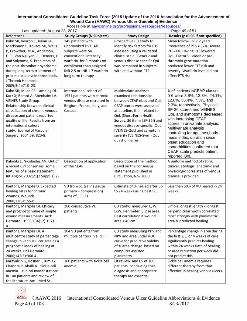
International Consolidated Guideline Task Force (2015 Update of the 2010 Association for the Advancement of Wound Care (AAWC) Venous Ulcer Guideline) Evidence
Accessible at aawconline.org/professional-resources/resources/ Last updated: August 23, 2017 Page 49 of 51
©AAWC 2016 International Consolidated Venous Ulcer Guideline Abbreviations & Evidence
Page 49 of 103 8/23/2017
References Study Groups (N Subjects) Study Design Results (p<0.05 if not specified)
Kahn SR, Kearon C, Julian JA, Mackinnon B, Kovacs MJ, Wells P, Crowther, M.A., Anderson, D.R., Van Nguyen, P., Demers, C. and Solymoss, S. Predictors of the post-thrombotic syndrome during long-term treatment of proximal deep vein thrombosis. J Thromb Haemost. 2005;3(4):718–23.
145 patients with unprovoked DVT. All subjects were on conventional intensity warfarin for 3 months on enrollment than assigned INR 2.5 or INR 1.7 warfarin long term therapy
Prospective CO study to identify risk factors for PTS assessed using a validated clinical scale. Generic and venous disease specific QoL was compared in subjects with and without PTS
Mean follow-up: 2.2 years. Prevalence of PTS = 37%; severe PTS=4%. Having PTS lowered QoL. Factor V Leiden or pro-thrombin gene mutation predicted lower PTS risk and severity. Warfarin level did not affect PTS risk
Kahn SR; M'lan CE; Lamping DL; Kurz X; Berard A; Abenhaim LA. VEINES Study Group. Relationship between clinical classification of chronic venous disease and patient reported quality of life: Results from an international cohort study. Journal of Vascular Surgery. 2004;39; 823-8.
International cohort of 1531 patients with chronic venous disease recruited in Belgium, France, Italy, and Canada.
Multivariate analyses examined relationships between CEAP class and QoL CEAP scores were assessed at baseline, then related to QoL (Short-Form Health Survey, 36 items [SF-36]) and venous disease-specific (QoL [VEINES-QoL] and symptom severity [VEINES-Sym]) QoL questionnaires..
% of patients inCEAP classes
0-6 were 3.8%, 13.3%, 24.1%, 12.8%, 36.4%, 7.3%, and 2.3%, respectively. Physical SF-36 scores and VEINES QoL and symptoms decreased with increasing CEAP scores.in univariate analysis. Multivariate analysis controlling for age, sex,body mass index, duration since onset,education and comorbidities confirmed that CEAP scale predicts patient reported QoL.
Kalodiki E, Nicolaides AN. Out of a recent CVI consensus: some features of a basic statement. Int Angiol. 2002;21(2 Suppl 1):2-11.
Description of application of the CEAP
Description of the method based on the consensus statement published in Circulation, Nov 2000
A uniform method of rating clinical, etiologic, anatomic and physiologic correlates of venous disease is provided.
Kantor J, Margolis D. Expected healing rates for chronic wounds. Wounds. 2000;12(6):155-8.
VU from SC (saline gauze primary + compression) arms of 5 RCTs
Estimate of % healed after up to 24 weeks using best SC.
Less than 50% of VU healed in 24 weeks.
Kantor J, Margolis DJ. Efficacy and prognostic value of simple wound measurements. Arch Dermatol. 1998;134(12):1571–4.
260 consecutive VU patients
CO study: measured L, W, LxW, Perimeter, Elipse area. Best correlation if wound area < 40 cm
2
Simple longest length x longest perpendicular width correlated most strongly with planimetric area & predicted healing.
Kantor J. Margolis DJ. A multicentre study of percentage change in venous ulcer area as a prognostic index of healing at 24 weeks. Br J Dermatol. 2000;142(5):960-4.
104 VU patients from multiple centers in a RCT
CO study measuring PPV and NPV and area under ROC curve for predictive validity of % area change based on computer assisted planimetry
Percentage change in area during the first 2,3, or 4 weeks of care significantly predicts healing within 24 weeks.Rate of healing or area reduction per week did not predict this.
Karayalcin G, Rosner F, Kim KY, Chandra P, Aballi AJ. Sickle cell anemia – clinical manifestations in 100 patients and review of the literature. Am J Med Sci.
100 patients with sickle cell anemia.
Lit review and CS of 100 patients, concluding that diagnosis and appropriate therapy are essential.
Sickle cell anemia requires different therapy from that effective in healing venous ulcers.
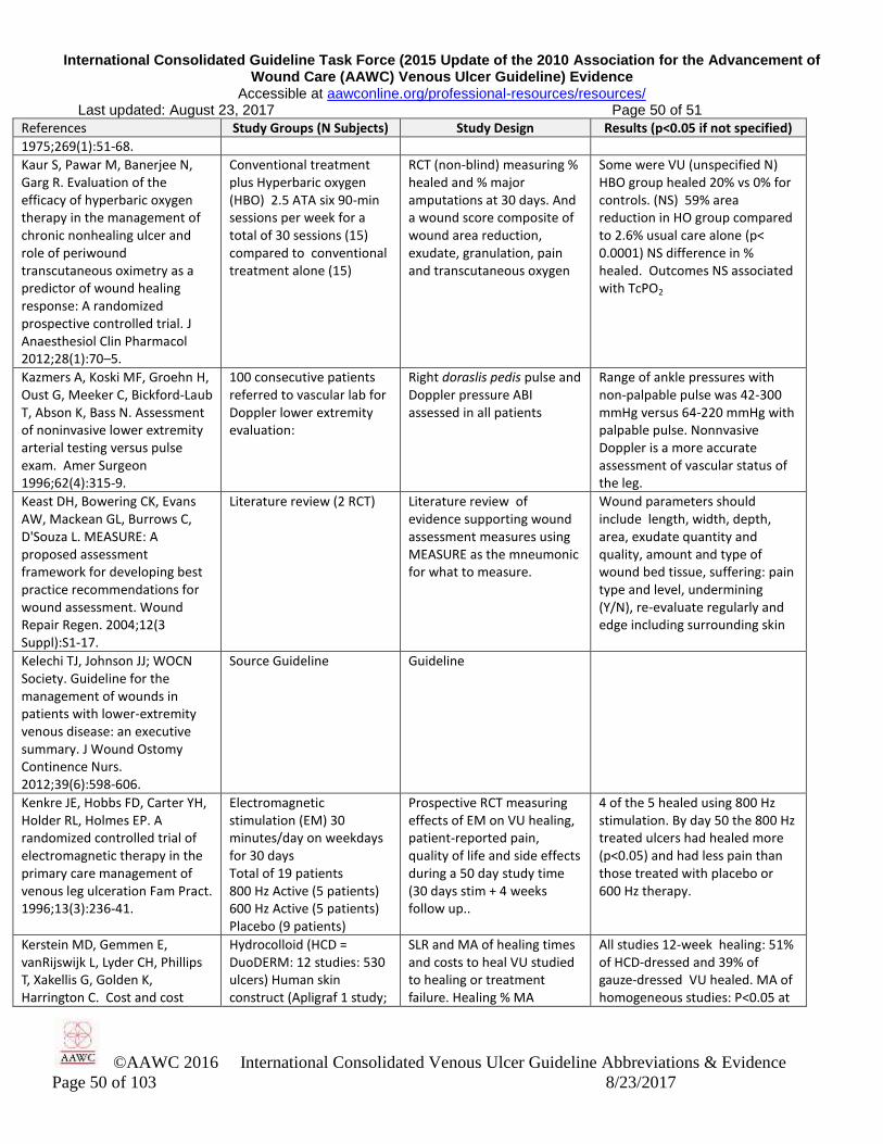
International Consolidated Guideline Task Force (2015 Update of the 2010 Association for the Advancement of Wound Care (AAWC) Venous Ulcer Guideline) Evidence
Accessible at aawconline.org/professional-resources/resources/ Last updated: August 23, 2017 Page 50 of 51
©AAWC 2016 International Consolidated Venous Ulcer Guideline Abbreviations & Evidence
Page 50 of 103 8/23/2017
References Study Groups (N Subjects) Study Design Results (p<0.05 if not specified)
1975;269(1):51-68.
Kaur S, Pawar M, Banerjee N, Garg R. Evaluation of the efficacy of hyperbaric oxygen therapy in the management of chronic nonhealing ulcer and role of periwound transcutaneous oximetry as a predictor of wound healing response: A randomized prospective controlled trial. J Anaesthesiol Clin Pharmacol 2012;28(1):70–5.
Conventional treatment plus Hyperbaric oxygen (HBO) 2.5 ATA six 90-min sessions per week for a total of 30 sessions (15) compared to conventional treatment alone (15)
RCT (non-blind) measuring % healed and % major amputations at 30 days. And a wound score composite of wound area reduction, exudate, granulation, pain and transcutaneous oxygen
Some were VU (unspecified N) HBO group healed 20% vs 0% for controls. (NS) 59% area reduction in HO group compared to 2.6% usual care alone (p< 0.0001) NS difference in % healed. Outcomes NS associated with TcPO2
Kazmers A, Koski MF, Groehn H, Oust G, Meeker C, Bickford-Laub T, Abson K, Bass N. Assessment of noninvasive lower extremity arterial testing versus pulse exam. Amer Surgeon 1996;62(4):315-9.
100 consecutive patients referred to vascular lab for Doppler lower extremity evaluation:
Right doraslis pedis pulse and Doppler pressure ABI assessed in all patients
Range of ankle pressures with non-palpable pulse was 42-300 mmHg versus 64-220 mmHg with palpable pulse. Nonnvasive Doppler is a more accurate assessment of vascular status of the leg.
Keast DH, Bowering CK, Evans AW, Mackean GL, Burrows C, D'Souza L. MEASURE: A proposed assessment framework for developing best practice recommendations for wound assessment. Wound Repair Regen. 2004;12(3 Suppl):S1-17.
Literature review (2 RCT) Literature review of evidence supporting wound assessment measures using MEASURE as the mneumonic for what to measure.
Wound parameters should include length, width, depth, area, exudate quantity and quality, amount and type of wound bed tissue, suffering: pain type and level, undermining (Y/N), re-evaluate regularly and edge including surrounding skin
Kelechi TJ, Johnson JJ; WOCN Society. Guideline for the management of wounds in patients with lower-extremity venous disease: an executive summary. J Wound Ostomy Continence Nurs. 2012;39(6):598-606.
Source Guideline Guideline
Kenkre JE, Hobbs FD, Carter YH, Holder RL, Holmes EP. A randomized controlled trial of electromagnetic therapy in the primary care management of venous leg ulceration Fam Pract. 1996;13(3):236-41.
Electromagnetic stimulation (EM) 30 minutes/day on weekdays for 30 days Total of 19 patients 800 Hz Active (5 patients) 600 Hz Active (5 patients) Placebo (9 patients)
Prospective RCT measuring effects of EM on VU healing, patient-reported pain, quality of life and side effects during a 50 day study time (30 days stim + 4 weeks follow up..
4 of the 5 healed using 800 Hz stimulation. By day 50 the 800 Hz treated ulcers had healed more (p<0.05) and had less pain than those treated with placebo or 600 Hz therapy.
Kerstein MD, Gemmen E, vanRijswijk L, Lyder CH, Phillips T, Xakellis G, Golden K, Harrington C. Cost and cost
Hydrocolloid (HCD = DuoDERM: 12 studies: 530 ulcers) Human skin construct (Apligraf 1 study;
SLR and MA of healing times and costs to heal VU studied to healing or treatment failure. Healing % MA
All studies 12-week healing: 51% of HCD-dressed and 39% of gauze-dressed VU healed. MA of homogeneous studies: P<0.05 at

International Consolidated Guideline Task Force (2015 Update of the 2010 Association for the Advancement of Wound Care (AAWC) Venous Ulcer Guideline) Evidence
Accessible at aawconline.org/professional-resources/resources/ Last updated: August 23, 2017 Page 51 of 51
©AAWC 2016 International Consolidated Venous Ulcer Guideline Abbreviations & Evidence
Page 51 of 103 8/23/2017
References Study Groups (N Subjects) Study Design Results (p<0.05 if not specified)
effectiveness of venous and pressure ulcer protocols of care. Disease Management and Health Outcomes, 2001, 9(11):651-636.
130 ulcers) Impregnated gauze (5 studies; 223 ulcers)
excluded studies not meeting homogeneity criteria, which occurred mainly at later time frames, reducing likelihood of statistical significance at 10 or 12 weeks.
8 weeks only for: 34.7% of HCD vs 25.5% of gauze-dressed VU healed. Lower cost to heal each VU with HCD. (p<0.05) Human skin construct had intermediate healing time and higher costs compared to HCD
Kerstein MD. The non-healing leg ulcer: Peripheral vascular disease, chronic venous insufficiency and ischemic vasculitis. Ostomy/Wound Manag. 1996;42(10A Suppl):19S-35S.
Literature Review with algorithms.
Referenced review of the literature on diagnosing and treating venous and ischemic leg ulcers.
Algorithms for diagnosis and treatment of venous or ischemic ulcers and those arising from arteritis or suspected blood dyscrasias are presented.
Kikta MJ, Schuler JJ, Meyer JP, Durham JR, Eldrup-Jorgensen J, Schwarcz TH, Flanigan DP. A prospective randomized trial of Unna's boots vs. hydroactive dressings in the treatment of venous stasis ulcers. .J.Vasc.Surg. 1988;7(3);478-83
Inelastic Unna Boot (42) DuoDERM (45 ) (compared dressings to compression alone)
Prospective, RCT 6 month comparison of VU healing in a US vascular surgery clinic.
70% healed withUnna's Boot 38% healed with DuoDERM dressing without compression (p = 0.01). A dressing could not compensate for the lack of compression in VU patients.
Kirsner R, Fastenau J, Falabella A, Valencia I, Long R, Eaglstein W. Clinical and economic outcomes with graftskin for hard-to-heal venous leg ulcers: A single-center experience. Dermatol Surg. 2002; 28(1):81-2.
Graftskin (Apligraf)-16 patients with 24 VUs of a mean duration of 42 months
Retrospective, open, non-randomized
A mean number of 2.25 graftskins applied per patient. All 16 patients responded to the device, with 8 patients (13 of 24 ulcers) completely healed over a mean of 13 weeks. Mean closure rate: 9.5%/week for the post graftskin group.There was an increase in ulcer size of 5.9% per week in the pregraftskin period.
Kirsner RJ, Mata SM, Falanga V, Kerdel FA. Split thickness skin grafting of leg ulcers. Dermatol Surg 1995;21(8):701-3
Kirsner RS, Pardes JB, Eaglstein WH, Falanga. The clinical spectrum of lipodermatosclerosis. J Amer Acad of Dermatol. 1993; 28(4):723-27.
Diagnosis of venous ulcers EO, LR Description of the clinical spectrum of lipodermatosclerosis.
Kjaer ML, Mainz J, Soernsen LT, Karlsmark T, Gottrup F. Clinical quality indicators of venous leg ulcers: development, feasibility and reliability. Ostomy/Wound Manage. 2005;51(5):64–74
100 VU consecutive patients tested by 1 MD for feasibility of implementation. (quality indicators based on multidisciplinary panel group nominal consensus.
CO: Multidisciplinary team generated VU Q of care indicators. Independent MD tested and feasibility of measuring these with inter-rater and intra-rater reliability CO 73% healed in
Validated, reliable quality of care indicators include VU healing, recurrence, pain, VU diagnosis with ABI and Duplex ultrasound scan recurrence, (inter-rater kappa = 0.79, P < 0.01 and intra-rater kappa = 0.89, P < 0.1).

International Consolidated Guideline Task Force (2015 Update of the 2010 Association for the Advancement of Wound Care (AAWC) Venous Ulcer Guideline) Evidence
Accessible at aawconline.org/professional-resources/resources/ Last updated: August 23, 2017 Page 52 of 51
©AAWC 2016 International Consolidated Venous Ulcer Guideline Abbreviations & Evidence
Page 52 of 103 8/23/2017
References Study Groups (N Subjects) Study Design Results (p<0.05 if not specified)
12 months.
Kobza L, Scheurich A. The impact of telemedicine on outcomes of chronic wounds in the home-care setting. Ostomy/Wound Manage. 2000;45(10):48-53.
76 chronic wounds, 52% of them VU managed by the standardized Solutions© wound care algorithm 125 matched historic control wounds
Prospective cohort compared to retrospective matched patients on % healed, healing time, home care visits and costs of care
The standardized evidence-based VU care resulted in more VU patients healed in less time with fewer visits at lower cost than matched historic controls. (p< 0.05)
Koksal C, Bozkurt AK. Combination of hydrocolloid dressing and medical compression stocking versus Unna's boot for the treatment of venous leg ulcers. Swiss Med Wkly. 2003;13325-26):364-8.
Unna's boot (30 VU patients) Comfeel Ulcer Dressing plus 30-40 mmHg Class II elastic compression stockings (30 VU patients)
Prospective RCT measuring healing of VU ulcers duration 16 .6 weeks and ease of use and patient reported pain
VU healing rates, times & recurrence not different for the two groups, but hydrocolloid plus stocking was easier to use (p < 0.0001). More pain with Unna's boot (p< 0.0001) both during application and wear. 150 min to apply Unna's Boot vs 134 to apply hydrocolloid plus elastic stocking (p >0,05).
Kopera D, Kokol R, Berger C, Haas J. Does the use of low-level laser influence wound healing in chronic venous leg ulcers? J Wound Care .2005;14(8):391-4.
44 patients with a chronic VU randomly assigned to standard care: disinfection, hydrofiber dressing and compression+ (1) Laser, (2) Placebo (3) Only standard
RCT measuring reduction in traced ulcer area during 4 weeks of treatment, then at follow up 90 days after treatment.
No significant difference was observed between the placebo and laser treated groups, as some subjects responded strongly to the placebo.
Korn P, Patel S, Heller JA Deitch JS, Krishnasastry KV, Bush HL, Kent KC. Why insurers should reimburse for compression stockings in patients with chronic venous stasis. J Vasc Surg. 2002;35(5):950-7.
Hypothetical 55-yr-old patients with prior VU receiving vs not receiving compression stockings and education (CS+Ed),
Markov decision tree analysis was conducted based on published probabilities of venous ulcer recurrence, 4.6 mo heal time, 12% chance of hospitalization and 0.4% chance of amputation after VU development.
With CS+Ed the mean time to VU recurrence was 53 months vs 18.7 mo to recurrence without CS+Ed.. CS+Ed saved costs of $5094 while saving 0.37 QALY. If considering only medical treatments, CS+Ed would save $6326 during the lifetime of each patient.
Kotz P, Fisher J, McCluskey P, Hartwell SD, Dharma H. Use of a new silver barrier dressing, ALLEVYN Ag in exuding chronic wounds. Int Wound J. 2009;6(3):186–94
N=126 pts with an exudating chronic wound dressed with a primary hydrocellular foam dressing with silver, Allevyn Ag®
Non comparative clinical evaluation to determine whether the silver foam dressing was acceptable to clinicians for indicated use and to assess dressing performance parameters.
Clinicians rated the dressings as acceptable for use in various wound types in 88% of patients. The majority of clinical signs of infection reduced between the initial and the final assessment. The condition of wound tissue and surrounding skin reportedly improved, and exudate levels reduced from initial to final assessment (p < 0.001). Clinicians rated the silver foam dressing as satisfying or exceeding expectations in over 90% of patients.
Kralj B, Kosicek MA. A randomised comparative trial of
1. 4-layer elastic compression bandage
Prospective RCT measuring % completely healed and time
At time of reporting, 7/20 (35%) healed in the Profore® group and

International Consolidated Guideline Task Force (2015 Update of the 2010 Association for the Advancement of Wound Care (AAWC) Venous Ulcer Guideline) Evidence
Accessible at aawconline.org/professional-resources/resources/ Last updated: August 23, 2017 Page 53 of 51
©AAWC 2016 International Consolidated Venous Ulcer Guideline Abbreviations & Evidence
Page 53 of 103 8/23/2017
References Study Groups (N Subjects) Study Design Results (p<0.05 if not specified)
single-layer and multi-layer bandages in the treatment of venous leg ulcers. In Leaper DJ, Cherry GW, Dealey C, Lawrence JC, Turner TD (Eds) Proc 6th European Conf on Adv Wound Management. London: Macmillan Magazines Ltd., 1996:158-160. As cited in Cullum et al. Cochrane review, 2002.
Profore® 4-layer elastic compression bandage(20) 2. 1-layer elastic compression bandage Porelast® over primary hydrocolloid dressing Tegasorb (20)
to healing. 8/20 (40%) healed in 1-layer + hydrocolloid group. Cullum meta-analysis reports 44% healed in both groups (7/16 and 8/18). 4-layer group healed in (mean 58 days) and 1-layer group healed in mean of 85 days. No statistically significant differences were reported.
Kranke P, Bennett MH, Martyn-St James M, Schnabel A, Debus SE, Weibel S. Hyperbaric oxygen therapy for chronic wounds. Cochrane Database of Sys Reviews 2015;( 6):CD004123
1 VU RCT of 16 patients managed with hyperbaric oxygen therapy at 2.5 ATA for 90 minutes 5 times weekly for 6 weeks 5 RCT on 205patients with diabetic foot ulcers
SR. 1 VU RCT (Hammarlund & Sundberg) 6 and 18 week VU size reduction and 18-week % healed. 1 6-week RCT of chronic leg ulcers (mixed chronic ulcer etiologies, unspecified number of them venous ulcers) vs usual care (Kaur)
More early reduction in VU area, only at 6 weeks 33%, 95%CI 19% to 47%, P<0.00001 Improved healing lower amputation rates for DFU.
Krizek TJ, Robson MC, Ko E. Bacterial growth and skin graft survival. Surg Forum 1967;18(518):9
30 patients receiving skin grafts. Bacterial cultures of deep tissue biopsies were made before grafting.
Good sensitivity but only 20% specificity.i.e. 80% false positives for CFU/g of tissue > 10
6 as a predictor of take
CFU /g of healthy tissue was highly sensitive predictor of graft take, but 80% of grafts that took also had high CFU
Kulkarni SR, Barwell JR, Gohel MS, Bulbulia RA, Whyman MR, Poskitt KR. Residual venous reflux after superficial venous surgery does not predict ulcer recurrence. Eur J Vasc Endovasc Surg. 2007;34(1):107-11.
144 of 185 consecutive VU patients following saphenous vein surgery with 25 having VU recurrence
CO study followed for 3 years Cox regression identified if residual venous reflux and change in reflux pattern were risk factors for ulcer recurrence.
Only increase in venous reflux time predicted non-recurrence of VU.
Kulkarni SR, Gohel MS, Wakely C, Minor J, Poskitt KR, Whyman MR. The Ulcerated Leg Severity Assessment score for prediction of venous leg ulcer healing. Br J Surg. 2007;94(2):189-93.
229 VU patients in UI 1999-2001
CO with Cox regression to predict VU healing in patients treated with compression
Patient age over 50 years, ulcer chronicity (in years) and venous refill time (VRT) of 20 s or less were identified as risk factors for non-healing
Kurd SK, Hoffstad OJ, Bilker WB, Margolis DJ. Evaluation of the use of prognostic information for the care of individuals with venous leg ulcers or diabetic neuropathic foot ulcers. Wound Repair Regen. 2009;17(3):318–25.
14 Centers with VU or DU randomized in clusters to receive feedback or not of whether VU or DU area was reducing at 4 weeks
Prospective RCT provided caregivers with feedback about healing status of their patients or similar instructions without feedback.
Feedback to caregivers about non-healing of ulcers in their care e.g. % area reduction at 4 weeks , improved VU and diabetic foot ulcer % healed after 12 weeks of care.
Labropoulos N, Landon P, Jay T. The impact of duplex scanning in phlebology. Dermatologic Surg. 2002; 28(1): 1-5
Case series of venous ulcer patients with clinical symptoms including pigmentation: hemosiderin
Literature review and case series illustrating how duplex scanning ultrasound diagnoses venous reflux
Duplex scanning ultrasonography has become the gold standard for diagnosing the location and extent of venous insufficiency.
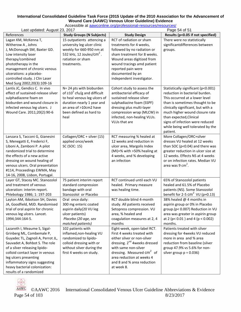
International Consolidated Guideline Task Force (2015 Update of the 2010 Association for the Advancement of Wound Care (AAWC) Venous Ulcer Guideline) Evidence
Accessible at aawconline.org/professional-resources/resources/ Last updated: August 23, 2017 Page 54 of 51
©AAWC 2016 International Consolidated Venous Ulcer Guideline Abbreviations & Evidence
Page 54 of 103 8/23/2017
References Study Groups (N Subjects) Study Design Results (p<0.05 if not specified)
Lagan KM, McKenna T, Witherow A , Johns J, McDonough SM, Baxter GD. Low intensity laser therapy/combined phototherapy in the management of chronic venous ulcerations: a placebo-controlled study. J Clin Laser Med Surg 2002;20(3):109-16
15 outpatients attencing a university leg ulcer clinic weekly for 660-950 nm at 532 kHz, 12 Joules/cm
2 .
ratiation or sham treatments.
RCT of radiation or sham treatments for 4 weeks, followed by no radiation or sham treatment for 8 weeks. Wound areas digitized from wound tracings amd patient reported pain were documented by an independent investigator.
There were no statistically significantdifferences between groups.
Lantis JC, Gendics C. In vivo effect of sustained-release silver sulphadiazene foam on bioburden and wound closure in infected venous leg ulcers. J Wound Care. 2011;20(2):90-6
N= 24 pts with bioburden of ≥10
5 cfu/g and difficult
to heal venous leg ulcers of duration nearly 1 year and an area of >10cm2 have been defined as hard to heal
Cohort study to assess the antibacterial efficacy of sustained release silver sulphadiazine foam (SSPF) dressing plus multi-layer compression wrap (MLCW) in infected, non-healing VLUs. VLUs that are
Statistically significant (p<0.001) reduction in bacterial burden. This occurred at a lower level than is sometimes thought to be clinically significant, but with a much higher wound closure rate than expected,Clinical signs of infection were reduced while being well tolerated by the patient.
Lanzara S, Tacconi G, Gianesini S, Menegatti E, Frederici F, Liboni A, Zamboni P. A pilot randomized trial to determine the effects of a new active dressing on wound healing of venous ulcers. Oral presentation #114, Proceedings EWMA, May 14-16, 2008, Lisbon, Portugal.
Collagen/ORC + silver (15) applied once/week SC (SOC: 15)
RCT measuring % healed at 12 weeks and reduction in ulcer area, Margolis Index (MI)=% with >50% healing at 4 weeks, and % developing an infection
More Collagen/ORC+silver dresses VU healed at 12 weeks than SOC (p<0.04) and there was greater reduction in ulcer size at 12 weeks. Effects NS at 4 weeks or on infection rates. Median VU area was 9 cm
2.
Layer GT, Stacey MC. Stanozolol and treatment of venous ulceration: interim report. Phlebology 1986; 1: 197-203
75 patient interim report standard compression bandage with oral Stanozolol or Placebo
RCT continued until each VU healed. Primary measure was healing time.
65% of Stanozolol patients healed and 61.5% of Placebo patients (NS). Some Stanozolol benefit for 2-5 cm
2 VU (p=0.13)
Layton AM, Ibbotson SH, Davies JA, Goodfield, MJD. Randomised trial of oral aspirin for chronic venous leg ulcers. Lancet. 1994;344:164-5.
Oral once daily: 300 mg enteric coated aspirin daily(20 VU leg ulcer patients) Placebo (20 age, sex matched patients)
RCT double blind 4-month study. All patients received Setopress compression. VU area, % healed and coagulation measures at 2, 4 months
38% healed @ 4 months in aspirin group or 0% in Placebo group.(p< 0.007) Reduction in VU area was greater in aspirin group at 2 (p< 0.01 ) and 4 (p < 0.002) months.
Lazareth I, Meaume S, Sigal-Grinberg ML, Combemale P, Guyadec TL, Zagnoli A, Perrot JL, Sauvadet A, Bohbot S. The role of a silver releasing lipido-colloid contact layer in venous leg ulcers presenting inflammatory signs suggesting heavy bacterial colonization: results of a randomized
102 patients with inflamed,non-healing VU randomized to lipido-colloid dressing with or without silver during the first 4 weeks on study.
Eight-week, open-label RCT. First 4 weeks treated with either silver or non-silver dressing. 2
nd 4weeks dressed
with same non-silver dressing. Measured cm
2 of
area reduction at weeks 4 and 8 and % area reduction at week 8.
Patients treated with silver dressing for 4weeks VU reduced more in area and % area reduction from baseline (silver group 47.9% vs 5.6% for non-silver group p = 0.036)

International Consolidated Guideline Task Force (2015 Update of the 2010 Association for the Advancement of Wound Care (AAWC) Venous Ulcer Guideline) Evidence
Accessible at aawconline.org/professional-resources/resources/ Last updated: August 23, 2017 Page 55 of 51
©AAWC 2016 International Consolidated Venous Ulcer Guideline Abbreviations & Evidence
Page 55 of 103 8/23/2017
References Study Groups (N Subjects) Study Design Results (p<0.05 if not specified)
controlled study. Wounds. 2008;20(6):158-66
Leach MJ, Pincombe J, Foster G. Clinical efficacy of horse chestnut seed extract in the treatment of venous ulceration. J Wound Care. 2006;15:159-67.
Oxerutins (Horsechestnut seed extract) (27) Placebo (27)
12-week RCT measured VU area, depth, pain, slough and exudate on enrollment and after 12 weeks of care. ANOVA analyzed effects.
NS effect on VU healing parameters. More slough reductionover time in active group (p=0.045); fewer dressing change visits week 12 (p=0.009).
Leach MJ. Making sense of the venous leg ulcer debate: a literature review. J Wound Care 2004;13(2):52-56.
Also reviews compression induced injury requiring amputation or arterial reconstruction.
Literature review including epidemiology, recurrence, financial and psychosocial implications
Prevalence: 0.11-0.63% approaching 1% if include healed VU Recurrence: 67%- 90%.
Leaper D, Munter C, Meaume S, Scalise A, Mompó B, Jakobsen B P, Gottrup F . The use of Biatain Ag in hard-to-heal venous leg ulcers: meta-analysis of randomized controlled trials. Plos one. 2013;8(7): e67083.
4 RCTs on 685 subjects with a hard-to-heal mixed or VU treated with Biatain Ag or similar Contreet or control (Biatain without silver,foam Allevyn or Algosteril calcium alginate or local best practice
Meta-analysis of % area reduction from baseline after 4 weeks of treatment and % healed at same time point.
Significantly higher % area reduction in Ag-treated VU (p= 0.001). 12% of Ag-treated subjects healed at 4 weeks compared to 6% of controls (p < 0.002)
Lee BB, Andrade M, Antignani PL, Boccardo F, Bunke N, Campisi C, Damstra R, Flour M, Forner-Cordero I, Gloviczki P, Laredo J, Partsch H, Piller N, Michelini S, Mortimer P, Rabe E, Rockson S, Scuderi A, Szolnoky G, Villavicencio JL. International Union of Phlebology. Diagnosis and treatment of primary lymphedema. Consensus document of the International Union of Phlebology (IUP)-2013. Int Angiol. 2013;32(6):541-74.
Lymphatic article summarizing state of the art of diagnosis, screening and treatment of primary lymphedema
Literature review of lymphatic signs, symptoms, risk factors diagnosis and treatment.
Summary of evidence supporting diagnosis, screening and treatment of lymphatic disorders
Lee KF, Ennis WJ, Dunn GP. Surgical palliative care of advanced wounds. Am J Hosp Palliat Care. 2007;24(2):154-60.
LR of risks and benefits of surgery. For other options see also Tippett, 2015.
LR surgical interventions for PU and VU.Treatment goals include pain reduction, managing foundexudate, preventing infection.
Discuss risks and benefits of surgery with patient and family patient’s condition and goals and VU necrosis, infection potential and underlying pathogenesis.
Lee YM, Ting AC, Cheng SW. Diagnosing deep vein thrombosis in the lower extremity: correlation of clinical and duplex scan findings. Hong Kong Med J. 2002;8(1):9-11.
313 post thrombotic patients with 345 duplex ultrasound scans (US) for deep vein thrombosis
Positive DUS were correlated with patients' demographic data (sex, age), medical history of malignancy, DVT and pulmonary embolism + clinical features (leg swelling, venous insufficiency, calf pain and leg ulcer).
Clinical features did not predict US finding of DVT, but male sex (P=0.0102), history of malignancy (P=0.0040), history of DVT P=0.0001), and history of pulmonary embolism (P=0.0265) did predict DVT US finding.
Lindgren C, Marcusson JA, Toftgard R. Treatment of venous leg ulcers with cryopreserved
Cryopreserved allogeneic keratinocyte (CAK) sheets + compression bandages ( 15
Prospective open controlled study applying compression with or without CAK , once
Mean 8-week VU percent area reduction was 35% with CAK added vs 14% with compression

International Consolidated Guideline Task Force (2015 Update of the 2010 Association for the Advancement of Wound Care (AAWC) Venous Ulcer Guideline) Evidence
Accessible at aawconline.org/professional-resources/resources/ Last updated: August 23, 2017 Page 56 of 51
©AAWC 2016 International Consolidated Venous Ulcer Guideline Abbreviations & Evidence
Page 56 of 103 8/23/2017
References Study Groups (N Subjects) Study Design Results (p<0.05 if not specified)
cultured allogeneic keratinocytes: a prospective open controlled study. Br J Dermatol. 1998; 139(2):271-5
chronic VU patients) Compression bandages alone ( 12 chronic VU patients)
weekly for 8 weeks. Primary outcome was percent area reduction after 8 weeks.
alone (p >0.05, not significant) attributed to cell-weakening by cryopreservation; more likely due to small sample size.
Lindholm C. Leg ulcer treatment in hospital and primary care in Sweden: Cost effective care and quality of life. In: Proceedings of the International Committee on Wound Management Meeting. Advances in Wound Care. 1995; 8:42-7
Std HCD Dressing (changed when needed or once a week (15); or wet-to-dry saline gauze changed twice a day (15)
Prospective RCT comparing wound pain and all costs to reduce wound area by 1 percent for the two dressing groups during 6 weeks treatment (1234 dressing changes) in the primary care setting.
There was less pain (p < .0.003) at a lower total cost of care (p<0.009) and a lower cost to achieve each percent reduction of leg ulcer area in the HCD group (p=0.026) than in subjects dressed with gauze.
Lippman HI, Fishman LM, Farrar RH, Bersnstein RK, Zybert PA. Edema control in the management of disabling chronic venous insufficiency. Arch Phys Med Rehabil 1994;75(4):436-41
762 patients with 1-8 VU 4” Unna’s boot weekly covered with tubular bandages or elastic bandages (similar to the “Duke Boot”).
Retrospective survey of a cohort of patients receiving care for at least one VU.
73.7% healing rate. Significant predictors of healing:1. # weeks to heal one VU predicts time to heal next VU 2. Age not correlated with weeks to healing; 3. Visit frequency was strongest predictor of healing
Liu JY, Hafner J, Dragieva G, Seifert B, Burg G. Autologous cultured keratinocytes on porcine gelatin microbeads effectively heal chronic venous leg ulcers. Wound Repair Regen. 2004;12(2):148-56.
15 Patients with chronic VU Autologous keratinocytes delivered on day 1 (5) or every 3 days (5) from porcine gelatin microbeads or day 1 as a monolayer on collagen pads (5)
CCT compared effects of autologous keratinocytes on VU parameters during 12 weeks of use. Percent area reduction at 12 weeks was the healing measure.
Percent area reduction at 12 weeks was 95% for microbeads delivered every 3 days; 83% for microbeads delivered once on enrollment or 50% for collagen monolayer pad delivery (p=0.003) compared to3-day microbeads .
Lo T. Sample R, Moore P, Gold P. Prediction of wound healing outcome using skin perfusion pressure and transcutaneous oximetry. Wounds. 2009; 21(11): 310-5.
100 subjects with chronic leg ulcers: CVI: 49; Peripheral arterial disease 15; Diabetic ulcers 35; unspecified etiology 1. Lazer Doppler Flow (LDF) or TcPO2 measured on all.
Prospective Comparison on CO: Skin perfusion pressure measured using an inflated pressure cuff with embedded LDF sensor compared to TcPO2 to predict healing.
LDF skin perfusion pressure had higher efficiency of prediction (p = 0.02 vs p=0.75 for TcPO2 ) PPV were both >87%. Skin perfusion pressure was more sensitive with 37% NPV compared to 14% for TcPO2. Not healing: <30 mmHg
Lok C, Paul C, Amblard P, Bessis D, Debure C, Faivre B, Guillot B, Ortonne JP, Huledal G, Kalis B. EMLA cream as a topical anesthetic for the repeated mechanical debridement of venous leg ulcers: a double-blind, placebo-controlled study. J Am Acad Dermatol. 1999;40(2 Pt 1):208-13.
69 VU patients half received mechanical debridement with gauze half had EMLA cream added
RCT of pain and debridement time using or mechanical debridement with or without EMLA cream
EMLA cream decreased median number of debridements from 15 with gauze alone to 11.5 when EMLA cream was added (p = 0.019) and decreased pain by 50% (p = 0.003). Plasma levels of lidocaine, prilocaine, and their main metabolites were low with no apparent accumulation
Lorimer KR, Harrison MB, Graham ID, Friedberg E, Davies B. Venous leg ulcer care: How evidence-based is nursing practice? J Wound Ostomy Continence Nurs.
66 VU patients CO study of adherence to EB protocols
Patients do not adhere if not educated on importance of compression and elevation to heal tissue or if their concerns such as pain or QoL were not addressed.

International Consolidated Guideline Task Force (2015 Update of the 2010 Association for the Advancement of Wound Care (AAWC) Venous Ulcer Guideline) Evidence
Accessible at aawconline.org/professional-resources/resources/ Last updated: August 23, 2017 Page 57 of 51
©AAWC 2016 International Consolidated Venous Ulcer Guideline Abbreviations & Evidence
Page 57 of 103 8/23/2017
References Study Groups (N Subjects) Study Design Results (p<0.05 if not specified)
2003;30(3):132-42.
Luebke T, Brunkwall J. Meta-analysis of subfascial endoscopic perforator vein surgery (SEPS) for chronic venous insufficiency. Phlebology. 2009;24(1):8-16.
LR and MA of: 3 RCTs comparing SEPS to conventional Linton vein surgery
MA of 3 RCT reporting % infection, LOS (length of hospital stay) VU recurrence, VU healing time,DVTor hospital re-admission@ 6 mo, or death
SEPS patients had fewer infections, shorter LOS, and less recurrence during up to 21 months post op
Lyon RT, Veith FJ, Bolton L, Machado F and the Venous Ulcer Study Collaborators. Clinical benchmark for healing of chronic venous ulcers. Am. J. Surg. 1998; 176:172-5.
Oral 250 mg/day Ifetroban Thromboxane A2 inhibitor (83) or Placebo (81) . Dressings: DuoDERM CGF + Unna Flex (boot) plus a layer of elastic graduated compression (CoFlex) + Kaltostat if high exudate. All dressings were changed at least once per week.
Prospective double blind RCT of VU with ABI > 0.7 not achieving area < 1.0 cm
2
during a 4-week screening period. VU area was measured weekly from tracings. Primary outcome was % healed at 12 weeks; secondary median time to complete healing, time to 50% area reduction or % recurring for ITT subjects.
Mean VU area was 11-13 cm2;
duration was 27-28 months. More were full-thickness than partial-thickness or superficial. NS difference in 12-week % healed (55% Ifetroban; 54% Placebo) 77% or 71% achieved > 50% healing by 12 weeks. Median time to healing was 9.6 weeks (I) or 11.0 weeks for P group. At least 40% healing by 3 weeks predicted VU healing in 12 weeks
Maessen-Visch MB, Koedam MI, Hamulyák K, Neumann HA. Atrophie blanche: a review. Int J Dermatol.1999;38(3):161–72.
Descriptive review with no patients compared.
LR of atrophie blanche Describes pathophysiology and notes it is associated with venous insufficiency.
Maida V. Wound management in patients with advanced illness. Curr Opin Support Palliat Care. 2013;7(1):73–9.
LR of wound management techniques in palliative care
LR summarizes how wounds can serve in prognosis of advancing illness and vice versa.
Wounds can heal in palliative care settings. Toronto Symptom Assessment System for Wounds may help guide care.
Maleti O, Lugli M. Neovalve construction in postthrombotic syndrome. J Vasc Surg. 2006;43(4):794-9.
16 patients with severe chronic venous insufficiency with PTS
CS evaluating neovalve cusp reconstruction: Pre- and post-operative hemodynamic flow: duplex scanning, ascending and descending venography , air plethysmography- all patients. Median follow-up of 22 months for patency and hemodynamic flow
Surgical treatment recommended in nonhealing recurrent VU (CEAP = C6). 16 VU (89%) healed in 4-25 (median 12) weeks with no recurrence. Post-op duplex scan and air plethysmograph showed significant improvement in hemodynamic flow (p<0.001) especially those with a good calf muscle pump.
Mani R, Vowden M, Nelson EA. Intermittent pneumatic compression for treating venous leg ulcers. Cochrane Database Syst. Rev. 2001; (4): CD001899. Updated with same title by: Nelson EA, Mani R, Thomas K, Vowden K. Cochrane Database Syst Rev. 2011 Feb 16;(2):CD001899.
Comparisons: 4 RCT (167): Compression wrap with vs. without IPC 1 RCT (104): Rapid IPC vs. Slow IPC 1 RCT (80): IPC vs. dressing with no compression wrap 1 RCT (16): Compression wrap vs. IPC without compression wrap
SR of RCTs comparing effects of using Intermittent Pneumatic Compression with any comparator on VU healing.
Only 1 of 4RCTs comparing IPC +compression wrap to the wrap alone found increased likelihood of healing with IPC (n=45). The other 3 RCTs (n=122) reported NS difference. IPC healed 62% of VU vs. 28% with dressings alone (p<0.002). NS healing difference comparing IPC to compression wrap. Rapid IPC healed more VU than slow IPC (p < 0.05)
Mantoni M, Larsen L, Lund JO, 39 V insufficiency patients, Triplex ultrasound (TUS) Agreement between TUS and the

International Consolidated Guideline Task Force (2015 Update of the 2010 Association for the Advancement of Wound Care (AAWC) Venous Ulcer Guideline) Evidence
Accessible at aawconline.org/professional-resources/resources/ Last updated: August 23, 2017 Page 58 of 51
©AAWC 2016 International Consolidated Venous Ulcer Guideline Abbreviations & Evidence
Page 58 of 103 8/23/2017
References Study Groups (N Subjects) Study Design Results (p<0.05 if not specified)
Henriksen L, Karlsmark T, Strandberg C, Ogstrup J, Ribel-Madsen S, Gottrup F, Danielsen L. Evaluation of chronic venous disease in the lower limbs: comparison of five diagnostic methods. Br J Radiol. 2002;75(895):578-83.
not necessarily with a VU Ascending (AP) and Descending (DP) phlebography, continuous wave Doppler (CWD) and ambulatory strain gauge plethysmography evaluated on most patients
other methods in evaluating reflux in deep veins was not better than that expected to occur by chance. Cohen's kappa was less that 0.20. TUS identified location and presence of incompetent veins most reliably.
Margolis D, Berlin J, Strom B. Which venous leg ulcers will heal with limb compression bandages? American J Medicine. 2000; 109(1): 15-9.
British database CO study of predictors of VU healing with compression during 20 weeks
Over 6 months duration adds 1 point to nonhealing score + 1 point if > 5 cm
2 Add points for
total score. Slough or non-vital tissue is also a risk factor.
Margolis D, Bilker W, Santanna J, Baumgarten M. Venous leg ulcer: Incidence and prevalence in the elderly. J Am Acad Dermatol. 2002;46(3):381-386
Incidence and prevalence determination in the General Practice Research Database
Literature review and calculation of incidence per 100 person years and annual prevalence in those 65 years of age or older
1.69% of persons >65 will have a VU on at least one visit during a given year. Incidence was 0.75%f or men and 1.45% for women per 100 person yrs
Margolis DJ, Cohen JH. Management of chronic venous ulcers: a literature guided approach. Clin Dermatol. 1994; 12(1):19-26.
Search terms venous, ulcer, leg ulcer, chronic wound, treatment, debridement, compression, topical, and dressing returned >1200 studies.
MEDLINE LR 1966-1994 of VU management techniques, summarized by categories under results at right.
Remove cause, debride VU, control infection, cleanse VU, dress VU, use adjuvant agents (topical, device, or systemic), miscellaneous treatments, and surgical closure.
Marshall JL, Mead P, Jones K, Kaba E, Roberts AP. The implementation of venous leg ulcer guidelines: process analysis of the intervention used in a multi-centre, pragmatic, randomized, controlled trial. J Clin Nurs. 2001;10(6):758-66.
13 Team intervention practices in Royal College of Nursing VU Guidelines were implemented in randomly selected centers in Northern and Yorkshire regions of the UK in 1997-1998
RCT audited Doppler assessment of VU; compression of confirmed VU and Support stocking worn to prevent recurrence of VU. Qualitative reporting of each pre- and post implementation
Supports team (District Nurse, Practice Nurse, General Practitioner) approach, training, quality improvement in implementing VU guidelines. Improved healing occurred. Involve this team + controllers of resources to make it work.
Marston WA, Vasquez MA, Lurie F, Wakefield TW, Rabe E, Shortell CK, et al. Multicenter assessment of the repeatability and reproducibility of the revised Venous Clinical Severity Score (rVCSS). J Vasc Surg Venous Lymphat Disord. 2013;1(3):219-24.
Seven centers using standardized methods, enrolled people with 136 limbs affected by chronic venous insufficiency (CVI). All were evaluated for inter-observer reliability (248 paired observations) or intra-observer reliability (258 paired observations)
Both legs of people with CVI were evaluated by the same 2 independent clinicians using the revised VCSS and CEAP scale on enrollment and by study week 6. Variability was assessed as mean VCSS difference. Cohen’s Kappa calculated agreement of intra- and inter-observer ratings.
The revised VCSS was reliable between observers (k = 0.68; p< 0.001) and within an observer (k = 0.72; p< 0.0001) with a mean variability in scores of 1.4+ 1.7 between observors or 1.3 + 1.6 within observors. This study verified the reproducibility and reliability of the revised VCSS in documenting severity of symptoms in peole with CVI.
Marston W, Tang J, Kirsner RS, Ennis W. Wound Healing Society Update on Guidelines for venous ulcers. Wound Repair Regen. 2016;24(1):136-44.
Guideline draft reviewed in 2015.
Guideline with all unique recommendations represented in the ICGTF VU Guideline.
Wound Healing Society members collaborated in assuring that all unique Wound Healing Society recommendations were accurately represented.
Marston WA, Carlin RE, 250 patients with active VU Prospective observational 57% healed at 10 weeks, 75% at
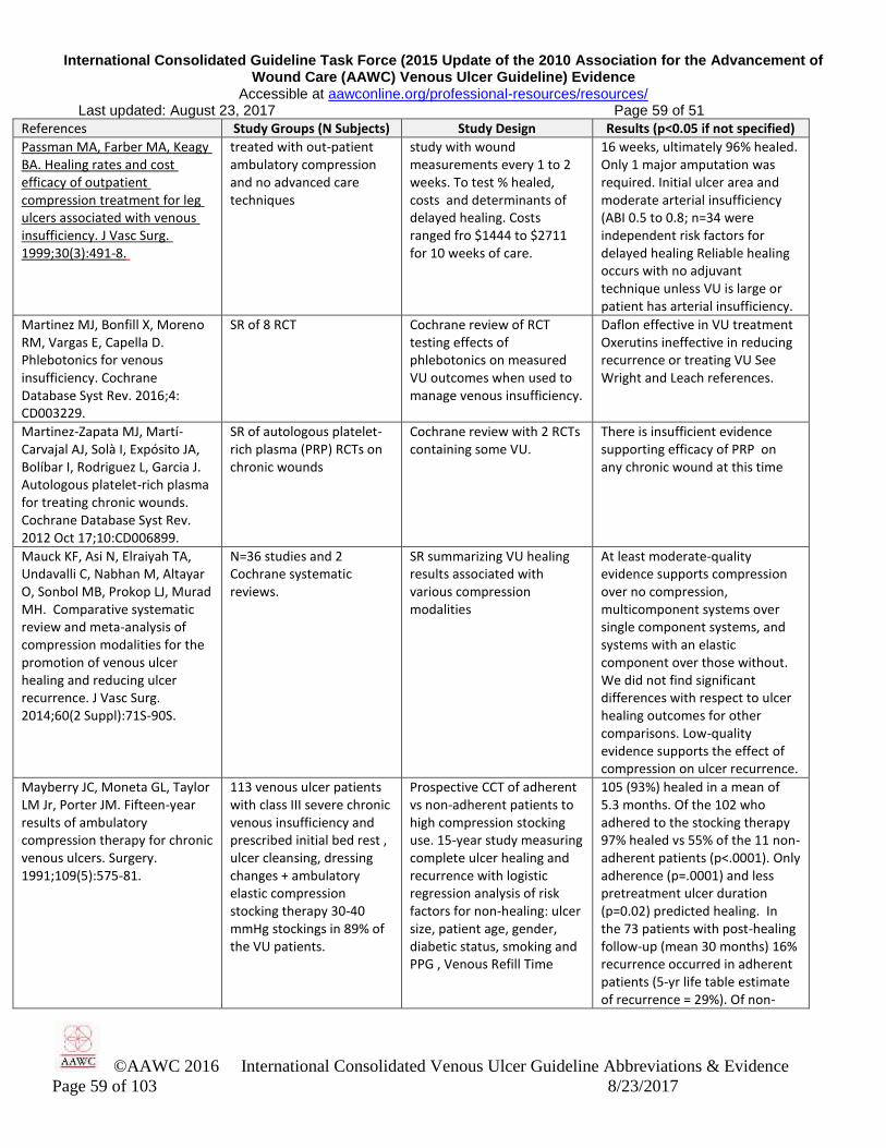
International Consolidated Guideline Task Force (2015 Update of the 2010 Association for the Advancement of Wound Care (AAWC) Venous Ulcer Guideline) Evidence
Accessible at aawconline.org/professional-resources/resources/ Last updated: August 23, 2017 Page 59 of 51
©AAWC 2016 International Consolidated Venous Ulcer Guideline Abbreviations & Evidence
Page 59 of 103 8/23/2017
References Study Groups (N Subjects) Study Design Results (p<0.05 if not specified)
Passman MA, Farber MA, Keagy BA. Healing rates and cost efficacy of outpatient compression treatment for leg ulcers associated with venous insufficiency. J Vasc Surg. 1999;30(3):491-8.
treated with out-patient ambulatory compression and no advanced care techniques
study with wound measurements every 1 to 2 weeks. To test % healed, costs and determinants of delayed healing. Costs ranged fro $1444 to $2711 for 10 weeks of care.
16 weeks, ultimately 96% healed. Only 1 major amputation was required. Initial ulcer area and moderate arterial insufficiency (ABI 0.5 to 0.8; n=34 were independent risk factors for delayed healing Reliable healing occurs with no adjuvant technique unless VU is large or patient has arterial insufficiency.
Martinez MJ, Bonfill X, Moreno RM, Vargas E, Capella D. Phlebotonics for venous insufficiency. Cochrane Database Syst Rev. 2016;4: CD003229.
SR of 8 RCT Cochrane review of RCT testing effects of phlebotonics on measured VU outcomes when used to manage venous insufficiency.
Daflon effective in VU treatment Oxerutins ineffective in reducing recurrence or treating VU See Wright and Leach references.
Martinez-Zapata MJ, Martí-Carvajal AJ, Solà I, Expósito JA, Bolíbar I, Rodriguez L, Garcia J. Autologous platelet-rich plasma for treating chronic wounds. Cochrane Database Syst Rev. 2012 Oct 17;10:CD006899.
SR of autologous platelet-rich plasma (PRP) RCTs on chronic wounds
Cochrane review with 2 RCTs containing some VU.
There is insufficient evidence supporting efficacy of PRP on any chronic wound at this time
Mauck KF, Asi N, Elraiyah TA, Undavalli C, Nabhan M, Altayar O, Sonbol MB, Prokop LJ, Murad MH. Comparative systematic review and meta-analysis of compression modalities for the promotion of venous ulcer healing and reducing ulcer recurrence. J Vasc Surg. 2014;60(2 Suppl):71S-90S.
N=36 studies and 2 Cochrane systematic reviews.
SR summarizing VU healing results associated with various compression modalities
At least moderate-quality evidence supports compression over no compression, multicomponent systems over single component systems, and systems with an elastic component over those without. We did not find significant differences with respect to ulcer healing outcomes for other comparisons. Low-quality evidence supports the effect of compression on ulcer recurrence.
Mayberry JC, Moneta GL, Taylor LM Jr, Porter JM. Fifteen-year results of ambulatory compression therapy for chronic venous ulcers. Surgery. 1991;109(5):575-81.
113 venous ulcer patients with class III severe chronic venous insufficiency and prescribed initial bed rest , ulcer cleansing, dressing changes + ambulatory elastic compression stocking therapy 30-40 mmHg stockings in 89% of the VU patients.
Prospective CCT of adherent vs non-adherent patients to high compression stocking use. 15-year study measuring complete ulcer healing and recurrence with logistic regression analysis of risk factors for non-healing: ulcer size, patient age, gender, diabetic status, smoking and PPG , Venous Refill Time
105 (93%) healed in a mean of 5.3 months. Of the 102 who adhered to the stocking therapy 97% healed vs 55% of the 11 non-adherent patients (p<.0001). Only adherence (p=.0001) and less pretreatment ulcer duration (p=0.02) predicted healing. In the 73 patients with post-healing follow-up (mean 30 months) 16% recurrence occurred in adherent patients (5-yr life table estimate of recurrence = 29%). Of non-
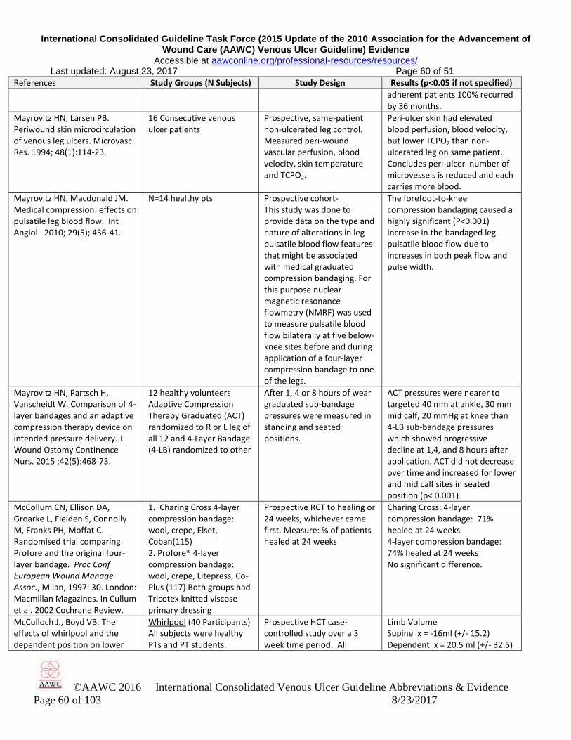
International Consolidated Guideline Task Force (2015 Update of the 2010 Association for the Advancement of Wound Care (AAWC) Venous Ulcer Guideline) Evidence
Accessible at aawconline.org/professional-resources/resources/ Last updated: August 23, 2017 Page 60 of 51
©AAWC 2016 International Consolidated Venous Ulcer Guideline Abbreviations & Evidence
Page 60 of 103 8/23/2017
References Study Groups (N Subjects) Study Design Results (p<0.05 if not specified)
adherent patients 100% recurred by 36 months.
Mayrovitz HN, Larsen PB. Periwound skin microcirculation of venous leg ulcers. Microvasc Res. 1994; 48(1):114-23.
16 Consecutive venous ulcer patients
Prospective, same-patient non-ulcerated leg control. Measured peri-wound vascular perfusion, blood velocity, skin temperature and TCPO2.
Peri-ulcer skin had elevated blood perfusion, blood velocity, but lower TCPO2 than non-ulcerated leg on same patient.. Concludes peri-ulcer number of microvessels is reduced and each carries more blood.
Mayrovitz HN, Macdonald JM. Medical compression: effects on pulsatile leg blood flow. Int Angiol. 2010; 29(5); 436-41.
N=14 healthy pts Prospective cohort- This study was done to provide data on the type and nature of alterations in leg pulsatile blood flow features that might be associated with medical graduated compression bandaging. For this purpose nuclear magnetic resonance flowmetry (NMRF) was used to measure pulsatile blood flow bilaterally at five below-knee sites before and during application of a four-layer compression bandage to one of the legs.
The forefoot-to-knee compression bandaging caused a highly significant (P<0.001) increase in the bandaged leg pulsatile blood flow due to increases in both peak flow and pulse width.
Mayrovitz HN, Partsch H, Vanscheidt W. Comparison of 4-layer bandages and an adaptive compression therapy device on intended pressure delivery. J Wound Ostomy Continence Nurs. 2015 ;42(5):468-73.
12 healthy volunteers Adaptive Compression Therapy Graduated (ACT) randomized to R or L leg of all 12 and 4-Layer Bandage (4-LB) randomized to other
After 1, 4 or 8 hours of wear graduated sub-bandage pressures were measured in standing and seated positions.
ACT pressures were nearer to targeted 40 mm at ankle, 30 mm mid calf, 20 mmHg at knee than 4-LB sub-bandage pressures which showed progressive decline at 1,4, and 8 hours after application. ACT did not decrease over time and increased for lower and mid calf sites in seated position (p< 0.001).
McCollum CN, Ellison DA, Groarke L, Fielden S, Connolly M, Franks PH, Moffat C. Randomised trial comparing Profore and the original four-layer bandage. Proc Conf European Wound Manage. Assoc., Milan, 1997: 30. London: Macmillan Magazines. In Cullum et al. 2002 Cochrane Review.
1. Charing Cross 4-layer compression bandage: wool, crepe, Elset, Coban(115) 2. Profore® 4-layer compression bandage: wool, crepe, Litepress, Co-Plus (117) Both groups had Tricotex knitted viscose primary dressing
Prospective RCT to healing or 24 weeks, whichever came first. Measure: % of patients healed at 24 weeks
Charing Cross: 4-layer compression bandage: 71% healed at 24 weeks 4-layer compression bandage: 74% healed at 24 weeks No significant difference.
McCulloch J., Boyd VB. The effects of whirlpool and the dependent position on lower
Whirlpool (40 Participants) All subjects were healthy PTs and PT students.
Prospective HCT case-controlled study over a 3 week time period. All
Limb Volume Supine x = -16ml (+/- 15.2) Dependent x = 20.5 ml (+/- 32.5)

International Consolidated Guideline Task Force (2015 Update of the 2010 Association for the Advancement of Wound Care (AAWC) Venous Ulcer Guideline) Evidence
Accessible at aawconline.org/professional-resources/resources/ Last updated: August 23, 2017 Page 61 of 51
©AAWC 2016 International Consolidated Venous Ulcer Guideline Abbreviations & Evidence
Page 61 of 103 8/23/2017
References Study Groups (N Subjects) Study Design Results (p<0.05 if not specified)
extremity volume. J Orthop Sports Ther. 1992; 16(4):169-73.
NOT VU participants had LE volume assessed pre- and post- treatment in positions of supine, dependent position with extremity in tank and with 20 minute whirlpool treatment
Whirlpool x = 44ml (+/- 30.5) This does not support using whirlpool for subjects with venous insufficiency.
McCulloch, JM, Marler KC, Neal MB, Phifer TJ. Intermittent pneumatic compression improves venous ulcer healing. Adv Wound Care. 1994;7(4):22-4, 26, passim.
Intermittent pneumatic compression (single chamber) (11) + Unna's boot 1 hour twice weekly. Unna's boot (11)
Prospective RCT measuring cm
2 VU healing per day
IPC + Unna's boot: healed 0.15 cm
2/day, Unna's boot healed
0.08 cm2/day (p<0.05)
McGuckin M, Stineman MC, Goin JE, Williams SV. Venous Leg Ulcer Guideline. Trustees of the University of Pennsylvania, Philadelphia, Pennsylvania, 1997.
Clearly specifies graduated compression descending from 30-42 mm at ankle to 12-17 mmHg below knee and moisture-retentive dressings (hydrocolloid, hydrogel or film) unless there is excess exudate or tissue maceration
Guideline that was subsequently validated in 2 RCTs one in US and one in UK by McGuckin et al, 2002.
Includes gentle ulcer cleansing, peri-ulcer skin care, patient education, debridement and many aspects of VU diagnosis, prevention and treatment.
McGuckin M, Waterman R, Brooks J, Cherry G, Porten L, Hurley S, Kerstein M. Validation of venous leg ulcer guidelines in the United States and United Kingdom. Amer J Surgery. 2002;183:132-7.
80 Retrospective pre-guideline(pre April, 1997) 80 Prospective with guideline Half from US Philadelphia Home Health Care Assns. Half from UK Oxfordshire general practice.Validates above Guideline with compression and MRD use.
Outcomes and costs of venous ulcer care were compared pre-guideline via chart abstraction versus prospective diagnosis and treatment using the content-validated guideline The guideline included patient education as well as surrounding skin and VU care.
Ankle to brachial index was performed on 8-36% of patients in US-UK pre-guideline and 93-96% with the guideline. % healed in <12 weeks increased from 23% to 70% in the US and from 40% to 65% in UK, while median cost to heal an ulcer decreased from $825 to $113 in the US and from £136 to £78 in the UK.
Meagher H, Ryan D, Clarke-Moloney M, O'Laighin G, Grace PA. An experimental study of prescribed walking in the management of venous leg ulcers. J Wound Care. 2012;21(9):421-2, 424-6, 428 passim.
40 patients with C6 VU assigned randomly to multi-layer compression with or without exercise, encouraged to take 10,000 steps per day.
RCT measuring # of steps taken per day at day 1 and week 4 after enrollment. Healing outcome was % healed at 12 weeks.
Subjects taking more steps per day by week 4 were more likely to heal at week 12 (p = 0.008)
Meaume S, Ourabah Z, Romanelli M, Manopulo R, De Vathaire F, Salomon D, Saurat JH. Efficacy and tolerance of a hydrocolloid dressing containing hyaluronic acid for the treatment of leg ulcers of venous or mixed origin. Curr
125 VU or mixed origin leg ulcers randomized to Hyaluronic acit + hydrocolloid dressing (HA+HCD) or HCD alone
6 week study of reduction in wound area, debridement
NS difference in reduction in wound area. More marked reduction in fibrinous slough (p = .04) in the HA + HCD group.)
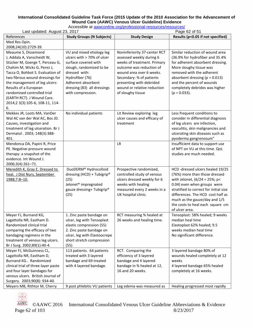
International Consolidated Guideline Task Force (2015 Update of the 2010 Association for the Advancement of Wound Care (AAWC) Venous Ulcer Guideline) Evidence
Accessible at aawconline.org/professional-resources/resources/ Last updated: August 23, 2017 Page 62 of 51
©AAWC 2016 International Consolidated Venous Ulcer Guideline Abbreviations & Evidence
Page 62 of 103 8/23/2017
References Study Groups (N Subjects) Study Design Results (p<0.05 if not specified)
Med Res Opin. 2008;24(10):2729-39.
Meaume S, Dissemond J, Addala A, Vanscheidt W, Stücker M, Goerge T, Perceau G, Chahim M, Wicks G, Perez J, Tacca O, Bohbot S. Evaluation of two fibrous wound dressings for the management of leg ulcers: Results of a European randomised controlled trial (EARTH RCT). J Wound Care. 2014;2 3(3):105-6, 108-11, 114-6.
VU and mixed etiology leg ulcers with > 70% of ulcer surface covered with slough, randomized to be dressed with: Hydrofiber (76) Adherent absorbent dressing (83) all dressings with compression.
Noninferiority 37-center RCT assessed weekly during 6 weeks of treatment. Primary outcome was reduction of wound area over 6 weeks. Secondary: % of patients presenting with debrided wound or relative reduction of sloughy tissue
Similar reduction of wound area (36.9% for hydrofiber and 35.4% for adherent absorbent dressing. More sloughy tissue was removed with the adherent absorbent dressing (p = 0.013) and the percent of wounds completely debrides was higher (p = 0.033).
Mekkes JR, Loots MA, VanDer Wal AC van der Wal AC, Bos JD. Causes, investigation and treatment of leg ulceration. Br J Dermatol . 2003; 148(3):388-401.
No individual patients Lit Review exploring leg ulcer causes and efficacy of treatment
Less frequent conditions to consider in differential diagnosis of leg ulcers are infection, vasculitis, skin malignancies and ulcerating skin diseases such as pyoderma gangrenosum”
Mendonca DA, Papini R, Price PE. Negative-pressure wound therapy: a snapshot of the evidence. Int Wound J. 2006;3(4):261–71.
LR Insufficient data to support use of NPT on VU at this time. QoL studies are much needed.
Meredith K, Gray E. Dressed to heal. J Dist Nurs, September 1988;7:8–10.
DuoDERM® Hydrocolloid dressing (HCD) + Tubigrip® (25) Jelonet® impregnated gauze dressing+ Tubigrip® (25)
Prospective randomized, controlled study of venous ulcers dressed weekly for 6 weeks with healing measured every 2 weeks in a UK hospital clinic.
HCD -dressed ulcers healed 19/25 (76%) more than those dressed with Jelonet, (6/24 = 25%; p< 0.04) even when groups were stratified to correct for initial size differences. The HCD cost half as much as the gauze/day and 1/5 the costs to heal each square cm of ulcer area.
Meyer FJ, Burnand KG, Lagattolla NR, Eastham D. Randomized clinical trial comparing the efficacy of two bandaging regimens in the treatment of venous leg ulcers. Br J Surg. 2002;89(1):40-4.
1. Zinc paste bandage on ulcer, leg with Tensoplast elastic compression (55) 2. Zinc paste bandage on ulcer, leg with Elastoocrepe short stretch compression (55)
RCT measuring % healed at 26 weeks and healing time.
Tensoplast: 58% healed; 9 weeks median heal time Elastoplast 62% healed; 9.5 weeks median heal time No significant difference.
Meyer FJ, McGuinness CL, Lagattolla NR, Eastham D, Burnand KG.. Randomized clinical trial of three-layer paste and four layer bandages for venous ulcers. British Journal of Surgery. 2003;90(8): 934-40.
113 patients. 64 patients treated with 3 layered bandage and 69 treated with 4 layered bandage.
RCT. Comparing the efficiency of 3 layered bandage and 4 layered bandage in % healed at 12, 16 and 20 weeks.
3 layered bandage 80% of wounds healed completely at 12 weeks 4 layered bandage 65% healed completely at 16 weeks.
Meyers MB, Rithtor M, Cherry 9 post phlebitic VU patients Leg edema was measured as Healing progressed most rapidly

International Consolidated Guideline Task Force (2015 Update of the 2010 Association for the Advancement of Wound Care (AAWC) Venous Ulcer Guideline) Evidence
Accessible at aawconline.org/professional-resources/resources/ Last updated: August 23, 2017 Page 63 of 51
©AAWC 2016 International Consolidated Venous Ulcer Guideline Abbreviations & Evidence
Page 63 of 103 8/23/2017
References Study Groups (N Subjects) Study Design Results (p<0.05 if not specified)
G. Relationship between edema and the healing rate of stasis ulcers of the leg. Amer J Surg. 1972; 124:686-8.
unresponsive to ligation and stripping were successively subjected to Adaptic wound dressing, Adaptic with elastic bandage, Adaptic with Unna's Boot or Adaptic + Unna's Boot + Elevation
volume by immersion and healing rate was measured as percent contraction per week.
in patients with edema reduction responses, with evidence suggesting that both ulcer and edema are due to the same cause.
Michaels JA, Campbell WB, King BM, Macintyre J, Palfreyman SJ, Shackley P, Stevenson MD. A prospective randomised controlled trial and economic modelling of antimicrobial silver dressings versus non-adherent control dressings for venous leg ulcers: the VULCAN trial. Health Technol Assess. 2009;13(56):1-114, iii.
107 patients with a VU of t lease 6 weeks duration received some sort of silver donating dressing. 106 similar patients received non-adhering gauze.Choice of dressings was left to clinician preference. Reviewer’s note: Silver was not sole variable. Dressings varied so this may not apply to 1 type of silver dressing.
12-week intervention RCT with 6-month and 1-year follow-up. Priary outcome was % completely healed at 12 weeks. Secondary measures were costs, cost effectiveness and quality-adjusted life years (QALY), time to healing and recurrence at 6 months or 1 year.
59.6% of silver-dressed or 56.7% of control patients healed in 12 weeks (NS) with NS difference between median time to heal or recurrence rates (11.6% for silver treated or 14% of controls). The groups did not differ on 5 QoL dimensions of the EuroQol-5D and Short-Form 6 (SF-6D) utility index at 1,3,6 or 12 months.The general use of silver dressings was not cost effective.
Milic DJ, Zivic SS, Bogdanovic DC, Jovanovic MM, Jankovic RJ, Milosevic ZD, Stamenkovic DM, Trenkic MS. The influence of different sub-bandage pressure values on venous leg ulcers healing when treated with compression therapy. J Vasc Surg. 2010;51(3):655-61.
189 VU patients all given multilayer high compression of which 24 failed to heal in 52 weeks. Excluded arterial disease (ABPI <0.8), heart insufficiency with ejection fraction (EF) <35, pregnancy, cancer disease, rheumatoid arthritis, and diabetes,
Prospective CO study to explore predictors of non healing by 52 weeks. Prior literature review found longer VU duration, larger surface area, > 50% VU surface covered with fibrin and ABI < 0.85 were risk factors for non-healing with compression therapy.
BMI (>33kg/m(2) short walking distance during the day (<200 m), a history of wound debridement, and ulcers >2 cm deep were indicators of slow healing. Calf:ankle circumference ratio <1.3, fixed ankle joint, and reduced ankle range of motion were the only independent parameters associated with non-healing (P < .001).
Milic DJ, Zivic SS, Bogdanovic DC, Karanovic ND, Golubovic ZV. Risk factors related to the failure of venous leg ulcers to heal with compression treatment. J Vasc Surg. 2009;49(5):1242-7.
A – (42) open-toed, elastic, class III compression device knitted in tubular form (Tubulcus, Laboratoires Innothera, Arcueil, France); B – (46) Tubulcus + 1 elastic bandage (15 cm wide and 5 cm long with 200% stretch, Niva, Novi Sad,Serbia); C – (43): Tubulcus and 2 elastic bandages
RCT measuring healing rates and median resting values in supine and standing positions of groups . Success in A group associated with smaller VU and calf circumference (CC)
Supine/standing: A -36.2 mm Hg / 43.9 mm Hg; B - 53.9 mm Hg / 68.2 mm Hg; C - 74.0 mm Hg / 87.4 mm Hg. Healing rate during 26-week treatment period was 25% (13/42) in group A, 67.4% (31/46) in group B, and 74.4% (32/43) in group C. Better results with multi component compression individualized to each patients VU & CC.
Moffat CJ, Franks PL, Oldroyd JM. Greenhalgh, RM. Randomized trial of an occlusive dressing in the treatment of chronic, non-healing leg ulcers.
Comfeel ulcer dressing (30) Nonadherent gauze dressing (30)
Prospective RCT of chronic non-healing leg ulcers studied to healing or 12 weeks, whichever came first. Primary measure was % of
Comfeel: 43% healed in 12 weeks Gauze: 23% healed in 12 weeks

International Consolidated Guideline Task Force (2015 Update of the 2010 Association for the Advancement of Wound Care (AAWC) Venous Ulcer Guideline) Evidence
Accessible at aawconline.org/professional-resources/resources/ Last updated: August 23, 2017 Page 64 of 51
©AAWC 2016 International Consolidated Venous Ulcer Guideline Abbreviations & Evidence
Page 64 of 103 8/23/2017
References Study Groups (N Subjects) Study Design Results (p<0.05 if not specified)
Phlebology 1992; 7:105-107. patients healed in 12 weeks..
Moffatt CJ, Edwards L, Collier M, Treadwell T, Miller M, Shafer L, Sibbald G, Brassard A, McIntosh A, Reyzelman A, Price P, Kraus SM, Walters SA, Harding K. A randomised controlled 8-week crossover clinical evaluation of the 3M Coban 2 Layer Compression System versus Profore to evaluate the product performance in patients with venous leg ulcers. Int Wound J. 2008;5(2):267-79.
Coban® 2-layer compression compared in cross-over trial with 4-layer Profore (81 patients with VU
RCT 8-week cross-over trial measuring % healed and HRQoL
Less pain with 2-layer, better HRQoL and Activities of daily living (p<0.03), more patients preferred 2 Layer NS difference in % healed or healing parameters.
Moffatt CJ, Franks PJ, Oldroyd M, Bosanquet N, Brown P, Greenhalgh RM, McCollum CN. Community clinics for leg ulcers and impact on healing. Br Med J 1992; 305: 1389-92
475 community care patients with 550 ulcerated legs
Case series exploring impact of nurse-led community clinics on leg ulcer healing outcomes.
The degree of compression must be reduced for those with ABI 0.5-0.8 i.e. mixed venous/arterial disease is confirmed during the diagnostic work-up
Moffatt CJ, Franks PJ. Implementation of a leg ulcer strategy. British J Dermatology. 2004;151:857-867
518 VU Home pts pre-implementation 437 same services post-implementation
Historic 12 week pre-post implementation trial of practicing multidisciplinary team EB care using Doppler US for VU ABI assessment. Measured healing, treatment frequency HRQoL using Nottingham Health Profile
Percent receiving ABI assessment rose from 11% to 94% in 12-weeks. Combined overall healing rates rose from 14% to 37% (odds ratio =3·53, P < 0·001). Frequency of treatment visits reduced: from 24 to 13.5 during over 12 weeks (P < 0·001). HRQoL improved, with improved energy, pain, sleep and mobility (P < 0·01).
Moisidis E, Heath T, Boorer C, Ho K, Deva AK. A prospective, blinded, randomized, controlled clinical trial of topical negative pressure use in skin grafting. Plast Reconstr Surg. 2004;114(4):917-22.
20 patients requiring skin grafting. Number of VU not specified. Half of each skin graft site dressed with standard bolster, other half topical negative pressure (NPWT)
RCT split half wound study. Quantity and quality of graft take was evaluated 2 weeks after graft by an observer blinded to treatment .
NS treatment effect of NPWT on quantity of graft take, but quality was better with the NPWT (p<0.05)
Mol MA, Nanninga PB, van Eendenburg JP, Westerhof W, Mekkes JR, van Ginkel CJ. Grafting of venous leg ulcers. An intraindividual comparison between cultured skin equivalents and full-thickness skin punch grafts. J Am Acad Dermatol. 1991;24(1):77-82.
noncontracted collagen gel populated with allogeneic fibroblasts and covered with autologous cultured keratinocytes were used for grafting venous leg ulcers (5) same patient punch graft controls
CCT measuring healing time and cosmetic appearance
Similar healing time: skin equivalents: 18 days, punch grafts: 15 days. The collagen gel “skin equivalents” had better cosmetic appearance.
Molski P, Ossowski R, Hagner W, Molski S. Patients with venous disease benefit from manual
Pre-surgical venous insufficiency patients receiving Manual
RCT measuring Anxiety, depression, Venous Reflux Index and CEAP Clinical stage
MLD improved venous reflux index, CEAP clinical score, anxiety and depression states. Surgery

International Consolidated Guideline Task Force (2015 Update of the 2010 Association for the Advancement of Wound Care (AAWC) Venous Ulcer Guideline) Evidence
Accessible at aawconline.org/professional-resources/resources/ Last updated: August 23, 2017 Page 65 of 51
©AAWC 2016 International Consolidated Venous Ulcer Guideline Abbreviations & Evidence
Page 65 of 103 8/23/2017
References Study Groups (N Subjects) Study Design Results (p<0.05 if not specified)
lymphatic drainage. Int Angiol. 2009;28(2):151-5.
Lymphatic Drainage (MLD) (20) or not before venous surgery(20)
at baseline, post MLD, and post surgery
improved them also.
Morison M, Moffatt C, Bridel-Nixon J, Bale S. Chapter 10. Leg Ulcers in Nursing Management of Chronic Wounds, Second Edition. Mosby, London, 1987. Pp 177-220.
Source guideline Guideline Supports patient-appropriate diagnosis, dressing and compression.
Morrell CJ, Walters J, Dixon S, Collins KA, Brereton ML, Peters, J, Brooker CGD. Cost effectiveness of community leg ulcer clinics: randomized controlled trial. BMJ 1998; 316: 1487-1491.
233 patients with venous leg ulcers allocated at random to care with 4-layer bandage in one of 8 community leg ulcer clinics (n = 120) or to control care in the home (n = 113)
RCT: Measures included percent healed during 12 weeks and 12-month follow-up
During 12 weeks of care, 34% healed in community clinic versus 24% in home care with unspecified compression (p = 0.03).
Morris EJ, Dowlen S, Cullen B. Early clinical experience with topical collagen in vascular wound care. J Wound Ostomy Continence Nurs. 1994;21(6):247-50.
1 female 82 year old with bilateral VU
CS after trying 4 years of cleansing with soap & water, SSD cream, hydrogen peroxide and Neosporin, changed to cleansing with normal saline, collagen, HCD
Cleansed with normal saline and dressed with hydrocolloid over collagen healed rapidly with leg elevation.
Morykwas MJ Argenta L, Shelton-Brown EI, McGuirt W.. Vacuum-assisted closure: A new method for wound control and treatment: Animal studies and basic foundation. Ann Plas Surg 1997; 38 (8):553-62.
5 swine partial-thickness excisions for each level of vacuum from 0.004 to 125 in 25 mmHg increments
Measured blood flow, granulation, bacterial clearance, and random-pattern flap survival
Blood flow increased four-fold at 125 mmHg. Granulation tissue increased with both continuous and intermittent application. Bacteria decreased after 4 days. Random pattern flap survival increased 21%.
Mosti G, Iabichella ML, Partsch H. Compression therapy in mixed ulcers increases venous output and arterial perfusion. J Vasc Surg.2012;55:122-8.
25 patients with mixed-etiology leg ulcers who received inelastic bandages
Case Series measuring laser-Doppler blood flow under compression bandages applied with pressures from 20 -30, 31- 40, and 41 - 50 mm Hg, with measurement before and after of Laser Doppler Fluxmetry (LDF) , transcutaneous oxygen pressure (TcPO2), toe pressure and Ejection fraction (EF) .
LDF values under the bandages increased by 33% (P < .01), 28% (P < .05), and 10% (NS), respectively, under the lower, mid level and higher pressure ranges applied. At toe level, a significant decrease in flux of –20% ( P < .05) was seen when bandage pressure exceeded 41 mm Hg. Toe pressure values and TcPO2 showed a moderate increase, excluding a restriction to arterial perfusion induced by the bandages. Inelastic bandages were highly efficient in improving venous pumping function, increasing the reduced ejection fraction by 72% ( P < .001) under pressure of 21 to 30 mm Hg and by 103% (P < .001) at 31 to 40

International Consolidated Guideline Task Force (2015 Update of the 2010 Association for the Advancement of Wound Care (AAWC) Venous Ulcer Guideline) Evidence
Accessible at aawconline.org/professional-resources/resources/ Last updated: August 23, 2017 Page 66 of 51
©AAWC 2016 International Consolidated Venous Ulcer Guideline Abbreviations & Evidence
Page 66 of 103 8/23/2017
References Study Groups (N Subjects) Study Design Results (p<0.05 if not specified)
mm Hg. In patients with mixed ulceration, an ankle-brachial pressure index >0.5 and an absolute ankle pressure of >60 mm Hg, inelastic compression of up to 40 mm Hg did not impede arterial perfusion but may normalize reduced venous pumping function.
Mosti G, Iabichella ML, Picerni P, Magliaro A, Mattaliano V. The debridement of hard to heal leg ulcers by means of a new device based on Fluidjet technology. Int Wound J 2005;2:307-14
Versajet Hydrosurgery system (68 hard-to-heal VU) “stuck in inflammatory phase” treated in a UK hospital. Traditional moist dressings (99)
Convenience controlled trial (CCT) reporting number of hydrosurgeries all at bedside, and time of procedures when compared with traditional treatment. Moist dressings worked in synergy with hydrosurgery softening necrotic tissue.
When used by experienced surgeon, the hydrosurgery was performed in a mean of 5 minutes and cleaned the VU of necrotic tissue in 46 cases during the first debridement. 17 cases took 2 procedures; 5 cases took 3. Pain was well tolerated if at “gentle” hydrosurgery setting.
Mostow EN, Araway GD, Dalsing M, Hodde JP, King D. Effectiveness of an extracellular matrix graft (OASIS Wound Matrix) in the treatment of chronic leg ulcers: a randomized clinical trial. J Vasc Surg 2005;41:837-43.
Weekly applied OASIS® + compression(62) or compression alone (58) Chronic VU Patients with a VU extending through dermis with no tendon or bone exposed, duration > 1 month
Included only VU screened for < 50% area reduction in 2 week screening period. 12-week RCT healing assessed weekly: % of patients with targed VU healed. 6-month vollow up
After 12 weeks, 55% of OASIS® dressed VU healed compared to 34% with compression alone. At 6-month follow up, no healed VU recurred or broke down.
Mudge M, Leinster SJ, Hughes LE. A prospective 10-year study of the post-thrombotic syndrome in a surgical population. Ann R Coll Surg Engl. 1988;70(4):249-52.
564 laparotomy patients Prospective 10 yr study to determine effect of post-op DVT on PTS development
35 had PTS by end of yr 10 (26 of these with prior DVT or phlebitis. New VU developed in 6 (1%). All thrombotic episodes increased risk of PTS. Most PTS occurred in patients without DVT but with lesser venous problems prior to surgery. Those with pre-existing venous problems need efforts to prevent PTS and VU.
Mulder G, Jones R, Cederholm Williams S, Cherry G, Ryan T. Fibrin cuff lysis in chronic venous ulcers treated with a hydrocolloid dressing. Int J Dermatol. 1993;32(4):304-6.
DuoDERM HCD under Unna Boot + Compression with Coban (9) Unna Boot + Compression with Coban (10)
Randomized blind evaluated, prospective controlled study of venous ulcers evaluated before and after one dressing in place for one week, in an outpatient clinic
Reduction of deep and shallow pericapillary fibrin cuffs in 40% of the group without HCD vs. 89% of the group with HCD (p < 0.05). No other histological differences were observed.
Munter KC, Beele H, Russell L, Crespi A, Grochenig E, Basse P, Aliksidic N, Fraulin F, Dahl C, Jemma AP. Effect of a sustained silver-releasing dressing on ulcers with delayed healing: the CONTOP study. J Wound Care.
Contreet® Foam with silver zeolye (326 patients with leg ulcers, pressure ulcers or diabetic foot ulcers with signs of local infection.) Local Best Practice with gauze or other silver
Prospective open-label parallel, block real-world cohortCCT 4-week study measuring healing, malodor and pain during or between dressing changes
After 4 weeks treatment,silver foam group had 47.1% wound area reduction vs. 32% for local best practice (p = 0.028), Foam had less wound bed slough (p< 0.0005), maceration (p=0.04), pain at dressing change (p =

International Consolidated Guideline Task Force (2015 Update of the 2010 Association for the Advancement of Wound Care (AAWC) Venous Ulcer Guideline) Evidence
Accessible at aawconline.org/professional-resources/resources/ Last updated: August 23, 2017 Page 67 of 51
©AAWC 2016 International Consolidated Venous Ulcer Guideline Abbreviations & Evidence
Page 67 of 103 8/23/2017
References Study Groups (N Subjects) Study Design Results (p<0.05 if not specified)
2006;15(5):199-206. dressings (n=38) 0.027). Odor decreased faster in the foam-dressed group.
Murphy RC, Robson MC, Heggers JP, Kadowaki M. The effect of microbial contamination on musculocutaneous and random flaps. J Surg Res. 1986 Jul;41(1):75-80.
Animal model of microbially inoculated granulating wounds covered with (1)musculo-cutaneous flap or (2) random flap or (3)left uncovered.
Non blind AM. Granulating wounds were inoculated with 10
4, 10
5 or 10
6 bacteria
per gram of tissue, then covered with either flap (1) or (2) or left uncovered (3). Degree of flap take or dehiscence was reported.
Flaps inoculated with 106 all
dehisced. Flaps inoculated with 10
4 bacteria all took. For those
inoculated with 105, random flaps
took, but musculoskeletal flaps dehisced and did not take. Relevance to VU is unclear.
Mustoe T. Understanding chronic wounds: a unifying hypothesis on thier pathogenesis and implications for therapy. Am J Surg. 2004;187(5A): 65s-70s
LR.of factors associated with delayed healing of chronic wounds, generating a unifying hypothesis for chronic wound management. .
LR of pressure ulcers, VU and diabetic foot ulcer causes for delayed healing and applying NPWT, growth factors, warmingor pulsed lavage to resolve these causes.
Hypothesis advanced that all chronic wounds share 3 root causes of delayed healing: cell and systemic aging; repeated ischemia-reperfusion cycles, and bacterial colonization with a resulting inflammatory response.
Myers MB, Cherry G. Zinc and healing of chronic leg ulcers. Am J Surg. 1970;120(1):77-81
Uncontrolled Case Series studying zinc in patients with chronic leg ulcers.
Zinc deficiency may be related to having a chronic leg ulcer.
Navarro TP, Dellis KT, Ribeiro AP. Clinical and hemodynamic Significance of the greater saphenous vein diameter in chronic venous insufficiency. Arch Surg. 2002; 137(11):1233-7.
85 consecutive patients with 112 lower limbs with compromised venous return were examined to test validity of CEAP and great saphenous vein diameter
Prospective cohort study investigating validity of CEAP and great sapenous vein diameter as measures of hemodynamic impairment
CEAP score and GSV diameter were well correlated with venous filling index, venous volume and residual volume fraction and with each other, validating both measures.
Neander KD, Hesse F. The protective effects of a new preparation on wound edges. J Wound Care. 2003;12(10):369-71.
227 VU patients, with half the perimeter of each VU treated with Cavilon non-sting barrier film (NSBF) or water
Double blind RCT assessing erythema daily for 4 days with a chromameter
On 88% of patients, erythema disappeared in 3 days and in 4 days for the rest on NSBF side. No observable effect of water.
Nelson EA, Bell-Syer SEM, Cullum NA. Compression for preventing recurrence of venous ulcers. Cochrane Database Syst Rev. 2000;(4):CD002303
Systematic review of the literature on venous ulcer recurrence.
No RCTs compared recurrence rates with vs without compression. Two prospective cohort studies, 1 comparing moderate to high compression hosiery and one (n=166) two types of moderate compression hose
5 yr follow up: relative risk of recurrence = 82% with both high and moderate compression hose More compliance with moderate. 74% recurrence with moderate. Not wearing compression hose was strongly associated with ulcer recurrence.
Nelson EA, Dale J. The management of leg ulcers. J Wound Care. 1996; 5(2):73-6.
No subjects. Algorithm for managing a VU Recommendations included in ICVUG .
Nelson EA, Harper DR, Ruckley CV, Prescott RJ, Gibson B, Dale JJ. A randomized trial of single-layer and multi-layer bandages in the treatment of chronic venous ulceration. Phlebologie.
1. 1-layer Granuflex® Adhesive Compression Bandage (100) 2. 4-layer: orthopedic wool, crepe, Elset ® (Seton-Scholl), Coban (3M)
RCT. Single-layer versus multi-layer compression. Primary outcome measured was % healed in 6 months.
49 % healed in 6 months in group 1 and 69% in group 2.(P<0.05). Multilayer compression healed more VU in 6 months than single layer compression.
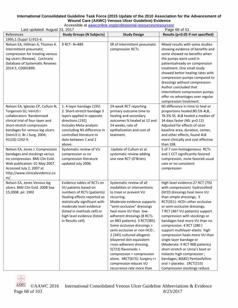
International Consolidated Guideline Task Force (2015 Update of the 2010 Association for the Advancement of Wound Care (AAWC) Venous Ulcer Guideline) Evidence
Accessible at aawconline.org/professional-resources/resources/ Last updated: August 23, 2017 Page 68 of 51
©AAWC 2016 International Consolidated Venous Ulcer Guideline Abbreviations & Evidence
Page 68 of 103 8/23/2017
References Study Groups (N Subjects) Study Design Results (p<0.05 if not specified)
1995;1 (Suppl 1):915-6.
Nelson EA, Hillman A, Thomas K. Intermittent pneumatic compression for treating venous leg ulcers (Review). Cochrane Database of Systematic Reviews 2014 5, CD001899.
9 RCT- N=489 SR of intermittent pneumatic compression RCTs
Mixed results with some studies showing evidence of benefits and some showed no benefits when the pumps were used in patientsalready on compression treatment. One small study showed better healing rates with compression pumps compared to dressings without compression. Author concluded that intermittent compression pumps offer no advantages over regular compression treatment.
Nelson EA, Iglesias CP, Cullum N, Torgerson DJ; VenUS I collaborators Randomized clinical trial of four-layer and short-stretch compression bandages for venous leg ulcers (VenUS I). Br J Surg. 2004; 91(10):1292-9.
1. 4-layer bandage (195) 2. Short-stretch bandage 3 layers applied in opposite directions (192) Includes Meta-analysis concluding NS difference in controlled literature to date between 1 and 2 above.
24-week RCT reporting primary outcome time to healing and secondary outcomes % healed at 12 and 24 weeks, rate of epithelization and cost of teatment.
NS difference in time to heal or proportions healed:80.5% 4LB, 76.5% SS. 4LB healed a median of 34 days faster (NS: p=0.12) Adjusted for effects of ulcer baseline area, duration, center, and other effects, found 4LB more clinically and cost effective than SSB.
Nelson EA, Jones J. Compression bandages and stockings versus no compression. BMJ Clin Evid. Web publication: 01 May 2007. Accessed July 2, 2007 at http://www.clinicalevidence.com/.
Systematic review of VU compression vs no compression literature updated July 2006.
.Update of Cullum et al. systematic review adding one new RCT (O'Brien)
5 of 7 non-homogeneous RCTs and 1 CCT significantly favored compression, none favored usual care or no consistent compression
Nelson EA, Jones Venous leg ulcers. BMJ Clin Evid. 2008 Sep 15;2008. pii: 1902
Evidence tables of RCTs on VU patients based on numbers of RCTs (patients) Healing effects reported as statistically significant with moderate level evidence (listed in methods cell) or high level evidence (listed in Results cell).
Systematic review of all modalities or interventions to treat or prevent VU recurring. Moderate evidence supports “semi-occlusive” dressings heal more VU than low-adherent dressings (8 RCTs on 883 patients). 3 RCT(385) Some occlusive dressings > semi-occlusive or non-HCD ; 2 (345) cultured allogenic bilayered skin equivalent >non-adherent dressing. 5(723) flavonoids + compression > compression alone. 4RCT(673): Surgery r+ compression educes VU recurrence rate more than
High level evidence:27 RCT (792 with compression) hydrocolloid (HCD) dressings heal more VU than simple dressings; 5 RCT(351): HCD> other occlusive or semi-occlusive dressings. 7 RCT (467 VU patients) support compression with stockings or bandages heal more VU than no compression. 4 RCT (280 ) support multilayer elastic high compression heals more VU than single layer bandage or (Moderate: 9 RCT 908 patients) short-stretch or Unna’s boot or inelastic high compression ; bandages; 8(682) Pentoxifylline oral > placebo. 1RCT(153) Compression stockings reduce
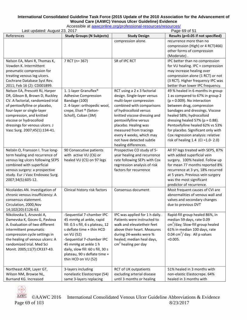
International Consolidated Guideline Task Force (2015 Update of the 2010 Association for the Advancement of Wound Care (AAWC) Venous Ulcer Guideline) Evidence
Accessible at aawconline.org/professional-resources/resources/ Last updated: August 23, 2017 Page 69 of 51
©AAWC 2016 International Consolidated Venous Ulcer Guideline Abbreviations & Evidence
Page 69 of 103 8/23/2017
References Study Groups (N Subjects) Study Design Results (p<0.05 if not specified)
compression alone. recurrence more than no compression (High) or 4 RCT(466) other forms of compression (Moderate) .
Nelson EA, Mani R, Thomas K, Vowden K. Intermittent pneumatic compression for treating venous leg ulcers. Cochrane Database Syst Rev. 2011; Feb 16 (2): CD001899.
7 RCT (n= 367) SR of IPC RCT IPC better than no compression for VU healing. IPC + compression may increase healing over compression alone (1 RCT) or not (3 RCT). Higher frequency IPC was better than lower IPC frequency.
Nelson EA, Prescott RJ, Harper DR, Gibson B, Brown D, Ruckley CV. A factorial, randomized trial of pentoxifylline or placebo, four-layer or single-layer compression, and knitted viscose or hydrocolloid dressings for venous ulcers. J Vasc Surg. 2007;45(1):134-41.
1. 1-layer Granuflex® Adhesive Compression Bandage (100) 2. 4-layer: orthopedic wool, crepe, Elset ® (Seton-Scholl), Coban (3M)
RCT using a 2 x 3 factorial design. Single-layer versus multi-layer compression combined with comparisons of hydrocolloid versus knitted viscose dressing and pentoxiifylline versus placebo. Healing was measured from tracings every 4 weeks, which may not have detected subtle healing differences.
49 % healed in 6 months in group 1 as compared to 67% in group 2 (p = 0.009). No interaction between drug, compression bandages and dressings. Viscose healed 58%; hydrocolloid dressing healed 57% (p = 0.88). Pentoxifylline healed 62% vs 53% for placebo. Significant only with Cox regression analysis: relative risk of healing 1.4 (CI =1.0- 2.0)
Nelzén O, Fransson I. True long-term healing and recurrence of venous leg ulcers following SEPS combined with superficial venous surgery: a prospective study. Eur J Vasc Endovasc Surg. 2007;34(5):605-12.
90 Consecutive patients with active VU (C6) or healed VU (C5) on 97 legs
Prospective CO study of 5-year healing and recurrence rate following SEPs with Cox regrression analysis of risk factors for recurrence
All 97 legs treated with SEPS, 87% with added superficial vein surgery. 100% healed. Follow up for mean 77 months reported 8% recurrence at 3 yrs, 18% recurred at 5 years. Previous vein surgery was the most significant predictor of recurrence.
Nicolaides AN. Investigation of chronic venous insufficiency: A consensus statement. Circulation, 2000,Nov 14;102(20):E126-63.
Clinical history risk factors Consensus document Most frequent causes of CVI are abnormalities of venous wall and valves and secondary changes due to previous DVT
Nikolovska S, Arsovski A, Damevska K, Gocev G, Pavlova A. Evaluation of two different intermittent pneumatic compression cycle settings in the healing of venous ulcers: A randomized trial. Med Sci Monit. 2005;11(7):CR337-43.
-Sequential 7-chamber IPC 45 mmHg at ankle, rapid fill: 0.5 s fill, 6 s plateau, 12 s deflate time + thin HCD on VU (52) -Sequential 7-chamber IPC 45 mmHg at ankle 1 h daily, slow fill: 60 s fill, 30 s plateau, 90 s deflate time + thin HCD on VU (52)
IPC was applied for 1 h daily. Patients were instructed to walk and elevatetheir feet above their heart. Measures during 24-weeks were % healed; median heal days, cm
2 healing per day
Rapid-fill group healed 86%, in median 59 days, rate 0.09 cm
2/day; Slow-fill group healed
61% in median 100 days, rate 0.04 cm
2/ day. All p values
<0.005.
Northeast ADR, Layer GT, Wilson NM, Browse NL, Burnand KG. Increased
3-layers including nonelastic Elastocrepe (54) same 3-layers replacing
RCT of UK outpatients excluding arterial disease until 3 months or healing
51% healed in 3 months with non-elastic Elastocrepe. 64% healed in 3 months with

International Consolidated Guideline Task Force (2015 Update of the 2010 Association for the Advancement of Wound Care (AAWC) Venous Ulcer Guideline) Evidence
Accessible at aawconline.org/professional-resources/resources/ Last updated: August 23, 2017 Page 70 of 51
©AAWC 2016 International Consolidated Venous Ulcer Guideline Abbreviations & Evidence
Page 70 of 103 8/23/2017
References Study Groups (N Subjects) Study Design Results (p<0.05 if not specified)
compression expedites venous ulcer healing. Royal Society of Med Venous Forum 1990 (Published + unpublished data cited in Cullum et al. 2001).
Elastocrepe with elastic Tensopress (52)
whichever came first. Tensopress elastic layer replacing the Elastocrepe.
O’Brien J, Edwards H, Stewart I, Gibbs H. A home-based progressive resistance exercise programme for patients with venous leg ulcers: a feasibility study. Int Wound J. 2013;10(4):389-96
13 home-based individuals with a C6 VU randomly assigned to multi-layer compression with (6) or without (7) resistance exercise: heel rises and calf hamstring stretches
RCT feasibility study of resistance exercise effect on calf muscle pump function and Range of motion (ROM) and % healed at 12 weeks.
Significant improvement in calf muscle pump function (p = 0.05) and ROM. Study was underpowered to find the 50 % healed in the exercise group vs 40% healed at 12 1weeks in the control group significant (p = 0.74)
O’Brien J, Finlayson K, Kerr G, Edwards H. The perspectives of adults with venous leg ulcers on exercise: an exploratory study. J Wound Care. 2014; 23(10): 496-509.
N=10 adults with current VLUs
Qualitative design using semi-structured interviews and discussions
The value of exercise in improving outcomes in a VU lies in its capacity to promote venousreturn and reduce the risk of secondary conditions in this population. Despite motivation and interest in doing exercise, people with VUs report many obstacles. Further exploration of mechanisms that assist this patient population and promote understanding about managing barriers, coupled with promotion of enabling (p = 0.01)factors, is vital for improving their exercise participation.
O’Meara S, Al-Kurdi D, Olugun Y, Ovington LG, Martyn-St James M, Richardson R. Antibiotics and antiseptics for venous leg ulcers. Cochrane Database Syst Rev. 2014;1: CD003557.
45 RCTs (N=4486 pts) SR of 45 RCTs comparing numerous antibiotic or antiseptic products effects on VU clinical infection and/or healing.
No evidence to support the use of systemic antibiotics or to discontinue their use. cadexomer iodine dressings to improve healing of chronic or clinically infected VU compared to conventional care, but its healing effects are accompanied by increased likelihood of adverse events and do not differ from healing responses to a hydrocolloid dressing; paraffin gauze dressing; dextranomer; and silver-impregnated dressings. Other topical products used in treating venous ulcers lack sufficient evidence to inform decisions about their use.The authors recommend limiting use of topical antibacterial products

International Consolidated Guideline Task Force (2015 Update of the 2010 Association for the Advancement of Wound Care (AAWC) Venous Ulcer Guideline) Evidence
Accessible at aawconline.org/professional-resources/resources/ Last updated: August 23, 2017 Page 71 of 51
©AAWC 2016 International Consolidated Venous Ulcer Guideline Abbreviations & Evidence
Page 71 of 103 8/23/2017
References Study Groups (N Subjects) Study Design Results (p<0.05 if not specified)
to limit emergence of resistant bacteria., and using them only in case of clinical infection.
O’Meara S, Cullum N, Nelson EA, Dumville JC. Compression for venous leg ulcers (review). Cochrane Database Syst Rev. 2012;11: CD000265.
48 RCT, N=4321 SR of all RCT evaluating effects on VU healing of compression bandages and stockings.
Compression increases ulcer healing rates compared with no compression. Multi-component systems are more effective than single-component systems. Multi-component systems containing an elastic bandage appear to be more effective than those composed mainly of inelastic constituents. Two-component bandage systems appear to perform as well as the 4LB. Patients receiving the 4LB heal faster than those allocated the Short-Stretch Bandage (SSB). More patients heal on high-compression stocking systems than with the SSB. Further data are required before the difference between high-compression stockings and the 4LB can be established.
O’Meara S, Martyn-St James M. Alginate dressings for venous leg ulcers. Cochrane Database Syst Rev. 2013;4: CD010182.
5 RCT (N=295) comparing alginate dressings to each other or other topical dressings for VU
One RCT compared different proprietary alginate dressings (20 participants), three compared alginate and hydrocolloid dressings (215 participants), and one compared alginate and plain non-adherent dressings (60 participants). Follow-up periods were six weeks in three RCTs and 12 weeks in two.
Alginates are not more effective than other dressings in healing chronic venous leg ulcers. RCTs in this area are considered to be of low or unclear methodological quality. Further, good quality evidence is required from well designed and rigorously conducted RCTs that employ - and clearly report on - methods to minimise bias, prior to any definitive conclusions being made regarding the efficacy of alginate dressings in the management of venous leg ulcers.
O’Meara S, Martyn-St James M. Foam dressings for venous leg ulcers. Cochrane Database Syst Rev. 2013;5: CD009907.
12 RCT (1023 pts) reporting 14 comparisons.
SR of foam dressings There was no difference in healing outcomes between hydrocellular foam dressings and polyurethane foam dressings (three RCTs). Pooled data across five RCTs (418 participants) showed no statistically significant

International Consolidated Guideline Task Force (2015 Update of the 2010 Association for the Advancement of Wound Care (AAWC) Venous Ulcer Guideline) Evidence
Accessible at aawconline.org/professional-resources/resources/ Last updated: August 23, 2017 Page 72 of 51
©AAWC 2016 International Consolidated Venous Ulcer Guideline Abbreviations & Evidence
Page 72 of 103 8/23/2017
References Study Groups (N Subjects) Study Design Results (p<0.05 if not specified)
difference between foam dressings and hydrocolloid dressings in the proportion of ulcers healed at 12 to 16 weeks (risk ratio (RR) 1.00, 95% confidence interval (CI) 0.81 to 1.22). No statistically significant between-group differences in healing outcomes were detected when foam dressings were compared with: paraffin gauze (two RCTs); hydrocapillary dressing (one RCT); knitted viscose dressing (one RCT); and protease modulating matrix (one RCT). No statistically significant between-group differences in the proportion of participants experiencing adverse events were detected when hydrocellular foam dressings were compared with polyurethane foam dressings, or when foam dressings were compared with hydrocapillary, hydrocolloid, or knitted viscose dressings (one RCT for each comparison). Six RCTs were considered as being at overall high risk of bias, and the remaining six RCTs were considered to be at overall unclear risk of bias. No included RCT had an overall low risk of bias.
O’Meara S, Tierney J, Cullum N, Bland JM, Franks PJ, Mole T, Scriven M. Four layer bandage compared with short stretch bandage for venous leg ulcers: systematic review and meta-analysis of randomised controlled trials with data from individual patients. BMJ. 2009 Apr 17;338:b1344
SR of 5 trials (797 patients),
Compared healing effects of four layer bandage compared to short stretch bandage in venous ulcer patients.
Four layer bandage was associated with significantly shorter time to healing: hazard ratio (95% confidence interval) from multifactorial model based on five trials was 1.31 (1.09 to 1.58), P=0.005. Larger ulcer area at baseline, more chronic ulceration, and previous ulceration were all independent predictors of delayed healing. When compared with the short stretch bandage, the four layer bandage increases the chance of

International Consolidated Guideline Task Force (2015 Update of the 2010 Association for the Advancement of Wound Care (AAWC) Venous Ulcer Guideline) Evidence
Accessible at aawconline.org/professional-resources/resources/ Last updated: August 23, 2017 Page 73 of 51
©AAWC 2016 International Consolidated Venous Ulcer Guideline Abbreviations & Evidence
Page 73 of 103 8/23/2017
References Study Groups (N Subjects) Study Design Results (p<0.05 if not specified)
healing by around 30% when independent prognostic factors are taken into account
O'Brien JF, Grace PA, Perry IJ, Hannigan A, Clarke Moloney M, Burke PE.. Randomized clinical trial and economic analysis of four-layer compression bandaging for venous ulcers. Br J Surg 2003;90(7):794–8.
1. 4-layer bandage (100) 2. No compression (100)
RCT measuring healing during 3 months and cost effectiveness. Follow up of 12 weeks for recurrence.
1. 54% healed in 3 months, with earlier healing throughout trial (p = 0.006). Lower median cost per leg healed (€ 210; p = 0.04) 2. 34% healed in 3 months; cost per leg healed € 234.
O'Donnell TF Jr, Passman MA, Marston WA, Ennis WJ, Dalsing M, Kistner RL, Lurie F, Henke PK, Gloviczki ML, Eklöf BG, Stoughton J, Raju S, Shortell CK, Raffetto JD, Partsch H, Pounds LC, Cummings ME, Gillespie DL, McLafferty RB, Murad MH, Wakefield TW, Gloviczki P; Society for Vascular Surgery; American Venous Forum. Management of venous leg ulcers: clinical practice guidelines of the Society for Vascular Surgery ® and the American Venous Forum. J Vasc Surg. 2014;60(2 Suppl):3S-59S.
Guideline source Guideline All unique recommendations listed in ICVUG
O'Donnell TF, Lau J. A systematic review of randomized controlled trials of wound dressings for chronic venous ulcer. J Vasc Surg. 2006; 44(5):1118-25.
Systematic review of 20 RCTs: 8 on moisture retentive dressings (n=687); 7 on growth factors (n=686); 5 on human skin equivalents (n=447).
RCT differences in percent healed on study and time to healing were summarized. Meta-analysis was performed only on growth factors due to heterogeneity of data in other dressing modalities.
Tegasorb®, Zinc oxide paste bandages, peri-ulcer injection of granulocyte-macrophage colony-stimulating factor, Oasis®, and Apligraf® each improved healing compared to same-study controls.
O'Donnell TF. The role of perforators in chronic venous insufficiency. Phlebology. 2010;25(1):3-10.
2 RCTs compare Greater saphenous vein (GSV) Open surgery (Linton procedure) alone to compression alone.
SR of RCTs measuring recurrence.
GSV open surgery reduces VU recurrence compared to compression alone. SEPS works only for high-volume flow veins
Oein, RF, Hansen, B.U., Hakansson, A. Pinch grafting of leg ulcers in primary care Acta Derm Venereol. 1998;78 (6): 348-9.
Pinch Grafts (45 patients with 55 ulcerated limbs and 84 skin transplantations
CCT: Open, non-randomized Healing rate after 12 weeks for venous ulcers was 45% and 44% for neuropathic ulcers. One year postoperatively, 47% (19/40) of examined ulcers remained healed. Venous ulcers represented of all ulcers.
O'Hare JL, Earnshaw JJ. Randomised clinical trial of
20 VU patients compression alone vs
RCT of healing effects. 9 of 11 evaluable patients had
Too few to support efficacy: requires more data. Supports
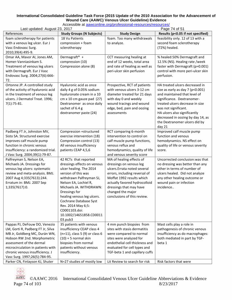
International Consolidated Guideline Task Force (2015 Update of the 2010 Association for the Advancement of Wound Care (AAWC) Venous Ulcer Guideline) Evidence
Accessible at aawconline.org/professional-resources/resources/ Last updated: August 23, 2017 Page 74 of 51
©AAWC 2016 International Consolidated Venous Ulcer Guideline Abbreviations & Evidence
Page 74 of 103 8/23/2017
References Study Groups (N Subjects) Study Design Results (p<0.05 if not specified)
foam sclerotherapy for patients with a venous leg ulcer. Eur J Vasc Endovasc Surg. 2010;39(4):495-9.
18 Vu Patients compression + foam sclerotherapy
foam. Too many withdrawals to analyze.
feasibility only. 12 of 13 with a second foam sclerotherapy (72%) healed.
Omar AA, Mavor AI, Jones AM, Homer-Vanniasinkam S. Treatment of venous leg ulcers with Dermagraft. Eur J Vasc Endovasc Surg. 2004;27(6):666-72.
Dermagraft® + compression (10) Compression alone (8)
CCT measuring healing at end of 12 weeks, total area and rate of healing as well as peri-ulcer skin perfusion
% healed:50% Dermagraft and 12.5% (NS). Healing rate /week faster with Dermagraft (p=0.001) control with more peri-ulcer skin perfusion.
Ortonne JP. A controlled study of the activity of hyaluronic acid in the treatment of venous leg ulcers. J Dermatol Treat. 1996; 7(1):75-81.
Hyaluronic acid as once daily 4 g of 0.05% sodium hyaluronate cream in a 10 cm x 10 cm gauze pad (27) Dextranomer as once daily sachet of 6.4 g dextranomer paste (24)
Prospective, RCT of patients with venous ulcers 3-12 cm diameter treated for 21 days with day 0 and weekly wound tracings and wound edge, bed, pain and oozing assessments
HA treated ulcers decreased in size as early as day 7 (p<0.001) and maintained that level of significance. Dextranomer-treated ulcers decrease in size was not significant. HA ulcers also significantly decreased in oozing by day 14, as the Dextranomer ulcers did by day 21
Padberg FT Jr, Johnston MV, Sisto SA. Structured exercise improves calf muscle pump function in chronic venous insufficiency: a randomized trial. J Vasc Surg. 2004;39(1):79-87.
Compression +structured exercise intervention (18) Compression control (13) All venous insufficiency patients CEAP 4,5,6
RCT comparing 6-month intervention to control on calf muscle pump functions, venous reflux and hemodynamics, quality of life and venous severity score
Improved calf muscle pump function and venous hemodynamics. NS effect on quality of life or venous severity score.
Palfreyman S, Nelson EA, Michaels JA. Dressings for venous leg ulcers: systematic review and meta-analysis. BMJ. 2007 Aug 4;335(7613):244. Erratum in: BMJ. 2007 Sep 1;335(7617):0.
42 RCTs that reported dressings effects on venous ulcer healing. The 2014 version of this was withdrawn Palfreyman SJ, Nelson EA, Lochiel R, Michaels JA. WITHDRAWN: Dressings for healing venous leg ulcers. Cochrane Database Syst Rev. 2014 May 6;5: CD001103.doi: 10.1002/14651858.CD001103.pub3
MA of healing effects of dressings on venous leg ulcers.Errata noted several errors, including reversal of Moffat 1992 results which actually favored hydrocolloid dressings that may have changed the major conclusions of this review.
Uncorrected conclusion:was that no dressing was better than any other in terms of number of ulcers healed. Did not analyze any other healing outcome or wound pain or infection incidence..
Pappas PJ, DeFouw DO, Venezio LM, Gorti R, Padberg FT Jr, Silva MB Jr, Goldberg MC, Durán WN, Hobson RW 2nd. Morphometric assessment of the dermal microcirculation in patients with chronic venous insufficiency. J Vasc Surg. 1997;26(5):784-95.
35 patients with venous insufficiency CEAP class 4 (n=11), class 5 (9) or class 6 (10) + 5 normal skin biopsies from normal patients without venous insufficiency.
4 mm punch biopsies from sites with stasis dermatitis were compared to normal sites were analyzed for endothelial cell thickness and evaluated for cell types and TGF-beta 1 and capillary cuffs
Mast cells play a role in pathogenesis of chronic venous insufficiency as do macrophages: both mediated in part by TGF-beta 1
Parker CN, Finlayson KJ, Shuter N=27 studies of mostly low Lit Review to search for risk Risk factors that were

International Consolidated Guideline Task Force (2015 Update of the 2010 Association for the Advancement of Wound Care (AAWC) Venous Ulcer Guideline) Evidence
Accessible at aawconline.org/professional-resources/resources/ Last updated: August 23, 2017 Page 75 of 51
©AAWC 2016 International Consolidated Venous Ulcer Guideline Abbreviations & Evidence
Page 75 of 103 8/23/2017
References Study Groups (N Subjects) Study Design Results (p<0.05 if not specified)
P, Edwards HE. Risk factors for delayed healing in venous leg ulcers: a review of the literature. Int J Clin Practice. 2015;69(9); 967-77.
level evidence factors of delayed healing in VLU
consistently identified included: larger ulcer area, longer ulcer duration, a previous history of ulceration, venous abnormalities and lack of high compression. Additional potential predictors with inconsistent or varying evidence to support their influence on delayed healing of venous leg ulcers included: decreased mobility and/or ankle range of movement, poor nutrition and increased age. Findings from this review indicate that a number of physiological risk factors are associated with delayed healing in venous leg ulcers and that social and/or psychological risk factors should also be considered and examined further.
Partsch H. [Compression stockings in treatment of lower leg venous ulcer (German)] Wien Med Wochenschr. 1994; 144(10-11):242-9.
Short stretch bandage (25) High compression elastic stockings (25)
RCT duration 3 months. High compression stockings healed 84% in 3 months versus 52% in the short-stretch bandage group.
Partsch, H & Mortimer P. Compression for leg wounds. Br J Dermatol. 2015;173(2):359–69.
Literature review Scholarly review on the use of compression therapy in leg ulcers describing available tools and modes of action.
Evidence is described supporting practical applicability and use for self management and cost effectiveness
Passman MA, McLafferty RB, Lentz MF, Nagre SB, Iafrati MD, Bohannon WT, Moore CM, Heller JA, Schneider JR, Lohr JM, Caprini JA.. Validation of Venous Clinical Severity Score (VCSS) with other venous severity assessment tools from the American Venous Forum, National Venous Screening Program. J Vasc Surg. 2011;54(6 Suppl):2S-9S.
American Venous Forum National Venous Screening Program data registry from 2007-2009, including 2907 participants with 5804 limbs screened for having a VU, all with complete datasets.
Retrospective CO study in which CEAP clinical staging, VCSS, modified Chronic Venous Insufficiency Quality of Life (CVI QoL) assessment, and venous ultrasound results were analyzed for statistical correlation trends using Spearman's rank coefficient as related to VCSS.
CO characteristics: CEAP clinical stage C0: 26%; C1: 33%; C2: 24%; C3: 9%; C4: 7%; C5: 0.5%; C6: 0.2% (mean, 1.41 ± 1.22). VCSS mean score (range, 0-3): pain 1.01 ± 0.80, compression 0.30 .+ 0.81; varicose veins 0.61 ± 0.84, edema 0.61 ± 0.81, pigmentation15 ± 0.47. CEAO-VCSS correlation was moderate r
2
= 0.49. VCSS-CVI Qol r2 =0.43 . US
screeningshowed, 38.1% of limbs had reflux and 1.5% obstruction in the femoral, saphenous, or popliteal vein segments. Between (reflux or obstruction) and VCSS correlation was 0.23; P< .0001)
Patel GK, Llewellyn M, Melhuish 50 successive venous leg CO of VUManaged with 1 19 of 29 (66%) of VU managed
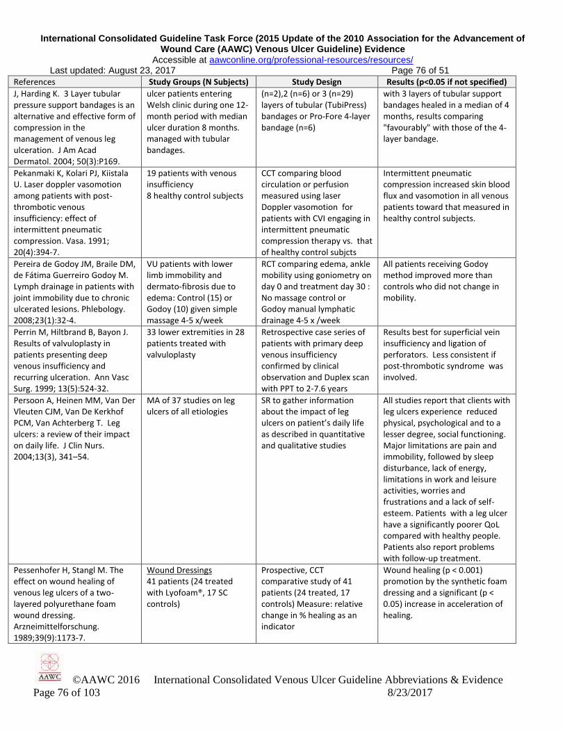
International Consolidated Guideline Task Force (2015 Update of the 2010 Association for the Advancement of Wound Care (AAWC) Venous Ulcer Guideline) Evidence
Accessible at aawconline.org/professional-resources/resources/ Last updated: August 23, 2017 Page 76 of 51
©AAWC 2016 International Consolidated Venous Ulcer Guideline Abbreviations & Evidence
Page 76 of 103 8/23/2017
References Study Groups (N Subjects) Study Design Results (p<0.05 if not specified)
J, Harding K. 3 Layer tubular pressure support bandages is an alternative and effective form of compression in the management of venous leg ulceration. J Am Acad Dermatol. 2004; 50(3):P169.
ulcer patients entering Welsh clinic during one 12-month period with median ulcer duration 8 months. managed with tubular bandages.
(n=2),2 (n=6) or 3 (n=29) layers of tubular (TubiPress) bandages or Pro-Fore 4-layer bandage (n=6)
with 3 layers of tubular support bandages healed in a median of 4 months, results comparing "favourably" with those of the 4-layer bandage.
Pekanmaki K, Kolari PJ, Kiistala U. Laser doppler vasomotion among patients with post-thrombotic venous insufficiency: effect of intermittent pneumatic compression. Vasa. 1991; 20(4):394-7.
19 patients with venous insufficiency 8 healthy control subjects
CCT comparing blood circulation or perfusion measured using laser Doppler vasomotion for patients with CVI engaging in intermittent pneumatic compression therapy vs. that of healthy control subjcts
Intermittent pneumatic compression increased skin blood flux and vasomotion in all venous patients toward that measured in healthy control subjects.
Pereira de Godoy JM, Braile DM, de Fátima Guerreiro Godoy M. Lymph drainage in patients with joint immobility due to chronic ulcerated lesions. Phlebology. 2008;23(1):32-4.
VU patients with lower limb immobility and dermato-fibrosis due to edema: Control (15) or Godoy (10) given simple massage 4-5 x/week
RCT comparing edema, ankle mobility using goniometry on day 0 and treatment day 30 : No massage control or Godoy manual lymphatic drainage 4-5 x /week
All patients receiving Godoy method improved more than controls who did not change in mobility.
Perrin M, Hiltbrand B, Bayon J. Results of valvuloplasty in patients presenting deep venous insufficiency and recurring ulceration. Ann Vasc Surg. 1999; 13(5):524-32.
33 lower extremities in 28 patients treated with valvuloplasty
Retrospective case series of patients with primary deep venous insufficiency confirmed by clinical observation and Duplex scan with PPT to 2-7.6 years
Results best for superficial vein insufficiency and ligation of perforators. Less consistent if post-thrombotic syndrome was involved.
Persoon A, Heinen MM, Van Der Vleuten CJM, Van De Kerkhof PCM, Van Achterberg T. Leg ulcers: a review of their impact on daily life. J Clin Nurs. 2004;13(3), 341–54.
MA of 37 studies on leg ulcers of all etiologies
SR to gather information about the impact of leg ulcers on patient’s daily life as described in quantitative and qualitative studies
All studies report that clients with leg ulcers experience reduced physical, psychological and to a lesser degree, social functioning. Major limitations are pain and immobility, followed by sleep disturbance, lack of energy, limitations in work and leisure activities, worries and frustrations and a lack of self-esteem. Patients with a leg ulcer have a significantly poorer QoL compared with healthy people. Patients also report problems with follow-up treatment.
Pessenhofer H, Stangl M. The effect on wound healing of venous leg ulcers of a two-layered polyurethane foam wound dressing. Arzneimittelforschung. 1989;39(9):1173-7.
Wound Dressings 41 patients (24 treated with Lyofoam®, 17 SC controls)
Prospective, CCT comparative study of 41 patients (24 treated, 17 controls) Measure: relative change in % healing as an indicator
Wound healing (p < 0.001) promotion by the synthetic foam dressing and a significant (p < 0.05) increase in acceleration of healing.
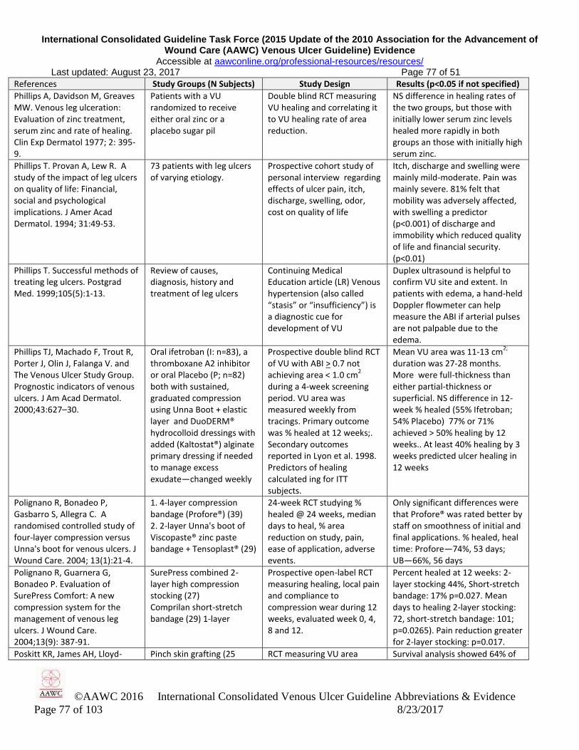
International Consolidated Guideline Task Force (2015 Update of the 2010 Association for the Advancement of Wound Care (AAWC) Venous Ulcer Guideline) Evidence
Accessible at aawconline.org/professional-resources/resources/ Last updated: August 23, 2017 Page 77 of 51
©AAWC 2016 International Consolidated Venous Ulcer Guideline Abbreviations & Evidence
Page 77 of 103 8/23/2017
References Study Groups (N Subjects) Study Design Results (p<0.05 if not specified)
Phillips A, Davidson M, Greaves MW. Venous leg ulceration: Evaluation of zinc treatment, serum zinc and rate of healing. Clin Exp Dermatol 1977; 2: 395-9.
Patients with a VU randomized to receive either oral zinc or a placebo sugar pil
Double blind RCT measuring VU healing and correlating it to VU healing rate of area reduction.
NS difference in healing rates of the two groups, but those with initially lower serum zinc levels healed more rapidly in both groups an those with initially high serum zinc.
Phillips T. Provan A, Lew R. A study of the impact of leg ulcers on quality of life: Financial, social and psychological implications. J Amer Acad Dermatol. 1994; 31:49-53.
73 patients with leg ulcers of varying etiology.
Prospective cohort study of personal interview regarding effects of ulcer pain, itch, discharge, swelling, odor, cost on quality of life
Itch, discharge and swelling were mainly mild-moderate. Pain was mainly severe. 81% felt that mobility was adversely affected, with swelling a predictor (p<0.001) of discharge and immobility which reduced quality of life and financial security. (p<0.01)
Phillips T. Successful methods of treating leg ulcers. Postgrad Med. 1999;105(5):1-13.
Review of causes, diagnosis, history and treatment of leg ulcers
Continuing Medical Education article (LR) Venous hypertension (also called “stasis” or “insufficiency”) is a diagnostic cue for development of VU
Duplex ultrasound is helpful to confirm VU site and extent. In patients with edema, a hand-held Doppler flowmeter can help measure the ABI if arterial pulses are not palpable due to the edema.
Phillips TJ, Machado F, Trout R, Porter J, Olin J, Falanga V. and The Venous Ulcer Study Group. Prognostic indicators of venous ulcers. J Am Acad Dermatol. 2000;43:627–30.
Oral ifetroban (I: n=83), a thromboxane A2 inhibitor or oral Placebo (P; n=82) both with sustained, graduated compression using Unna Boot + elastic layer and DuoDERM® hydrocolloid dressings with added (Kaltostat®) alginate primary dressing if needed to manage excess exudate—changed weekly
Prospective double blind RCT of VU with ABI > 0.7 not achieving area < 1.0 cm
2
during a 4-week screening period. VU area was measured weekly from tracings. Primary outcome was % healed at 12 weeks;. Secondary outcomes reported in Lyon et al. 1998. Predictors of healing calculated ing for ITT subjects.
Mean VU area was 11-13 cm2;
duration was 27-28 months. More were full-thickness than either partial-thickness or superficial. NS difference in 12-week % healed (55% Ifetroban; 54% Placebo) 77% or 71% achieved > 50% healing by 12 weeks.. At least 40% healing by 3 weeks predicted ulcer healing in 12 weeks
Polignano R, Bonadeo P, Gasbarro S, Allegra C. A randomised controlled study of four-layer compression versus Unna's boot for venous ulcers. J Wound Care. 2004; 13(1):21-4.
1. 4-layer compression bandage (Profore®) (39) 2. 2-layer Unna's boot of Viscopaste® zinc paste bandage + Tensoplast® (29)
24-week RCT studying % healed @ 24 weeks, median days to heal, % area reduction on study, pain, ease of application, adverse events.
Only significant differences were that Profore® was rated better by staff on smoothness of initial and final applications. % healed, heal time: Profore—74%, 53 days; UB—66%, 56 days
Polignano R, Guarnera G, Bonadeo P. Evaluation of SurePress Comfort: A new compression system for the management of venous leg ulcers. J Wound Care. 2004;13(9): 387-91.
SurePress combined 2-layer high compression stocking (27) Comprilan short-stretch bandage (29) 1-layer
Prospective open-label RCT measuring healing, local pain and compliance to compression wear during 12 weeks, evaluated week 0, 4, 8 and 12.
Percent healed at 12 weeks: 2-layer stocking 44%, Short-stretch bandage: 17% p=0.027. Mean days to healing 2-layer stocking: 72, short-stretch bandage: 101; p=0.0265). Pain reduction greater for 2-layer stocking: p=0.017.
Poskitt KR, James AH, Lloyd- Pinch skin grafting (25 RCT measuring VU area Survival analysis showed 64% of

International Consolidated Guideline Task Force (2015 Update of the 2010 Association for the Advancement of Wound Care (AAWC) Venous Ulcer Guideline) Evidence
Accessible at aawconline.org/professional-resources/resources/ Last updated: August 23, 2017 Page 78 of 51
©AAWC 2016 International Consolidated Venous Ulcer Guideline Abbreviations & Evidence
Page 78 of 103 8/23/2017
References Study Groups (N Subjects) Study Design Results (p<0.05 if not specified)
Davies ER, Walton J, McCollum C.. Pinch skin grafting or porcine dermis in venous ulcers: a randomized trial. Br. Med J (Clin Res Ed). 1987;294(6573):674-6.
mostly outpatients with a VU) Porcine dermis dressing (28 similar patients)
healed by 2 weeks post-graft and time to 100% healing analyzed using Kaplan-Meier survival analysis.
pinch grafted VU healed at six weeks and 74% by 12 weeks vs. respectively 29% and 46% of VU dressed with porcine dermis (X
2 =
4.1;p< 0.05).
Powell G. Managing a patient’s symptoms in bilateral arterial leg ulceration. Wounds UK. 2010; (3):93-8.
1 arterial ulcer patient with arterial signs:
Intermittent claudication Ischaemic rest pain and
Symptoms: Coldness of the foot Poor tissue perfusion — Purple/pink on dependence Pale on elevation Atrophic, shiny skin Lower limb loss of hair Muscle wasting Thickened toe nails Gangrene Unable to palpate pulses: absent or abnormal
CS: using Carolon multi-layer compression stockings in management of arterial ulcer Healing was not goal. Signs and symptoms are from Royal College of Nurses and British Community Health leg ulcer care pathway.
Pain and exudate were controlled and to patient’s and nursing staff delight healing followed.
Prandoni P, Lensing AWA, Cogo A, Cuppini S, Villalta S, Carta M, Cattelan AM, Polistena P, Bernardi E, Prins MH. The long-term clinical course of acute deep venous thrombosis. Ann Intern Med. 1996;125(1):1-7.
355 consecutive patients with a first episode of venography-confirmed DVT followed for up to 8 years.
CO Study: Outcomes were tracked and risk factors for ulceration included as a serious post DVT event. Follow up occurred for up to 8 years
78 had recurrent DVT, 15 were pulmonary emboli, fatal in 9 patients. 84 developed post-thrombotic syndrome (PTS). Of these 25 (30.2%) had severe PTS, listed as including a VU.
Prandoni P, Kahn SR. Post-thrombotic syndrome: prevalence, prognostication and need for progress. Br J Haematol. 2009;145(3):286-95
LR Prompt adequate elastic stocking compression in DVT patients can halve frequency of PTS, and when carefully supervised and instructed to wear proper elastic stockings > 50% of patients can remain stable or improve during long-term follow-up.
Risk factors for PTS include older age, obesity, a history of previous ipsilateral DVT, iliac-femoral location of current DVT failure to promptly recover from acute symptoms and inadequate quality of oral anticoagulant therapy
Prescott RJ, Callam MJ, Harper DR, Dale JJ, Ruckley CV. A controlled trial of weekly ultrasound therapy in chronic leg ulceration. The Lancet July 25, 1987; 204-206
Ultrasound (108 participants with chronic leg ulcers): Control Group (56) Treatment Group (52)
Prospective, RCT, Reporting % of VU healed at 12 weeks then followed up for 2 year Note:Later study by Watson et al. showed NS difference in VU healing time..
Treatment group 100% closure in 61% o f the patients in a 12 week period of time as compared to the Control group 100% closure in 41% of patient s in 12 weeks.
Price PE, Fagervik-Morton H, Mudge EJ, Beele H, Ruiz JC, Nystrøm TH, Lindholm C, Maume S, Melby-Østergaard B, Peter Y, Romanelli M, Seppänen S, Serena TE, Sibbald G, Soriano
2018 wound patients mainly VU and mixed
Survey of wound pain in patients with chronic wounds.
36.6% reported wound related pain most or all the time. Wound was most painful location for all patients. 32% experienced dressing-related pain most or all the time mostly in VU or AU
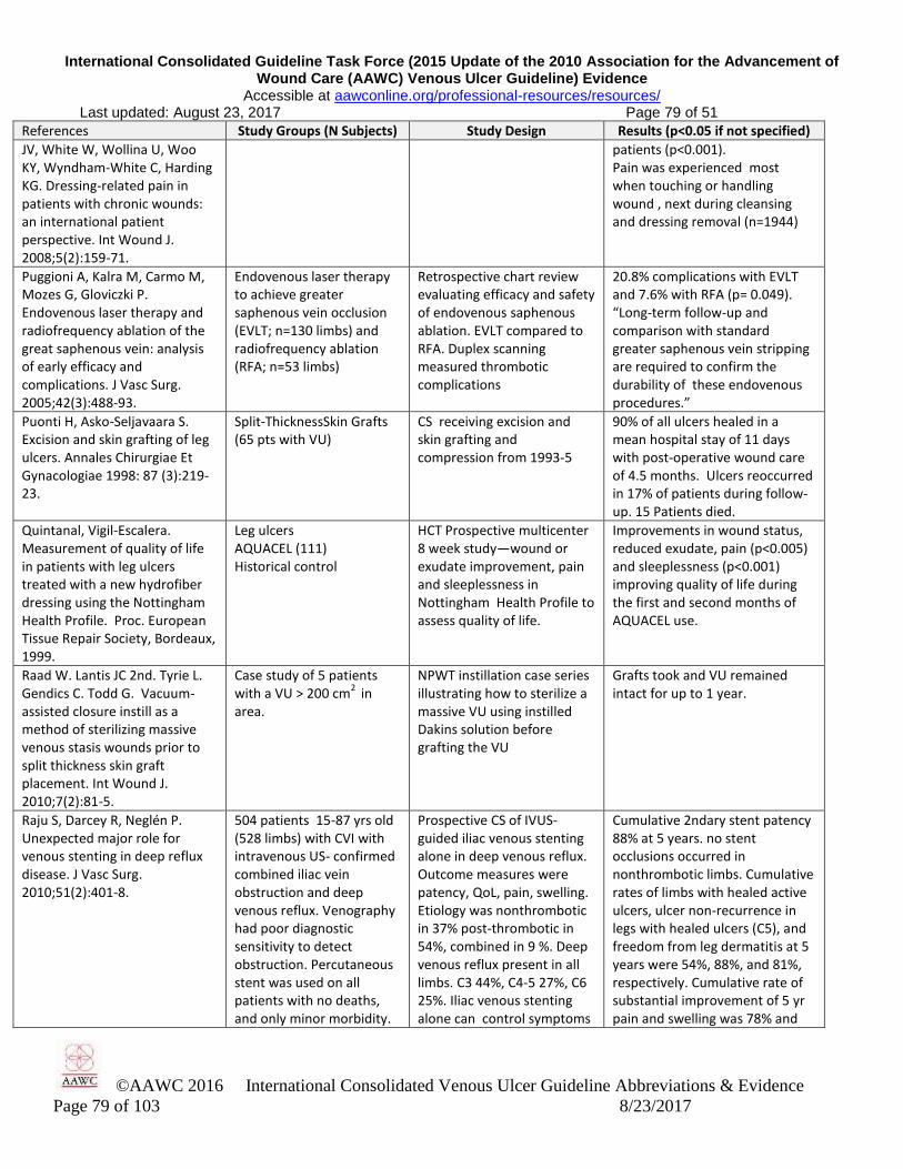
International Consolidated Guideline Task Force (2015 Update of the 2010 Association for the Advancement of Wound Care (AAWC) Venous Ulcer Guideline) Evidence
Accessible at aawconline.org/professional-resources/resources/ Last updated: August 23, 2017 Page 79 of 51
©AAWC 2016 International Consolidated Venous Ulcer Guideline Abbreviations & Evidence
Page 79 of 103 8/23/2017
References Study Groups (N Subjects) Study Design Results (p<0.05 if not specified)
JV, White W, Wollina U, Woo KY, Wyndham-White C, Harding KG. Dressing-related pain in patients with chronic wounds: an international patient perspective. Int Wound J. 2008;5(2):159-71.
patients (p<0.001). Pain was experienced most when touching or handling wound , next during cleansing and dressing removal (n=1944)
Puggioni A, Kalra M, Carmo M, Mozes G, Gloviczki P. Endovenous laser therapy and radiofrequency ablation of the great saphenous vein: analysis of early efficacy and complications. J Vasc Surg. 2005;42(3):488-93.
Endovenous laser therapy to achieve greater saphenous vein occlusion (EVLT; n=130 limbs) and radiofrequency ablation (RFA; n=53 limbs)
Retrospective chart review evaluating efficacy and safety of endovenous saphenous ablation. EVLT compared to RFA. Duplex scanning measured thrombotic complications
20.8% complications with EVLT and 7.6% with RFA (p= 0.049). “Long-term follow-up and comparison with standard greater saphenous vein stripping are required to confirm the durability of these endovenous procedures.”
Puonti H, Asko-Seljavaara S. Excision and skin grafting of leg ulcers. Annales Chirurgiae Et Gynacologiae 1998: 87 (3):219-23.
Split-ThicknessSkin Grafts (65 pts with VU)
CS receiving excision and skin grafting and compression from 1993-5
90% of all ulcers healed in a mean hospital stay of 11 days with post-operative wound care of 4.5 months. Ulcers reoccurred in 17% of patients during follow-up. 15 Patients died.
Quintanal, Vigil-Escalera. Measurement of quality of life in patients with leg ulcers treated with a new hydrofiber dressing using the Nottingham Health Profile. Proc. European Tissue Repair Society, Bordeaux, 1999.
Leg ulcers AQUACEL (111) Historical control
HCT Prospective multicenter 8 week study—wound or exudate improvement, pain and sleeplessness in Nottingham Health Profile to assess quality of life.
Improvements in wound status, reduced exudate, pain (p<0.005) and sleeplessness (p<0.001) improving quality of life during the first and second months of AQUACEL use.
Raad W. Lantis JC 2nd. Tyrie L. Gendics C. Todd G. Vacuum-assisted closure instill as a method of sterilizing massive venous stasis wounds prior to split thickness skin graft placement. Int Wound J. 2010;7(2):81-5.
Case study of 5 patients with a VU > 200 cm
2 in
area.
NPWT instillation case series illustrating how to sterilize a massive VU using instilled Dakins solution before grafting the VU
Grafts took and VU remained intact for up to 1 year.
Raju S, Darcey R, Neglén P. Unexpected major role for venous stenting in deep reflux disease. J Vasc Surg. 2010;51(2):401-8.
504 patients 15-87 yrs old (528 limbs) with CVI with intravenous US- confirmed combined iliac vein obstruction and deep venous reflux. Venography had poor diagnostic sensitivity to detect obstruction. Percutaneous stent was used on all patients with no deaths, and only minor morbidity.
Prospective CS of IVUS-guided iliac venous stenting alone in deep venous reflux. Outcome measures were patency, QoL, pain, swelling. Etiology was nonthrombotic in 37% post-thrombotic in 54%, combined in 9 %. Deep venous reflux present in all limbs. C3 44%, C4-5 27%, C6 25%. Iliac venous stenting alone can control symptoms
Cumulative 2ndary stent patency 88% at 5 years. no stent occlusions occurred in nonthrombotic limbs. Cumulative rates of limbs with healed active ulcers, ulcer non-recurrence in legs with healed ulcers (C5), and freedom from leg dermatitis at 5 years were 54%, 88%, and 81%, respectively. Cumulative rate of substantial improvement of 5 yr pain and swelling was 78% and
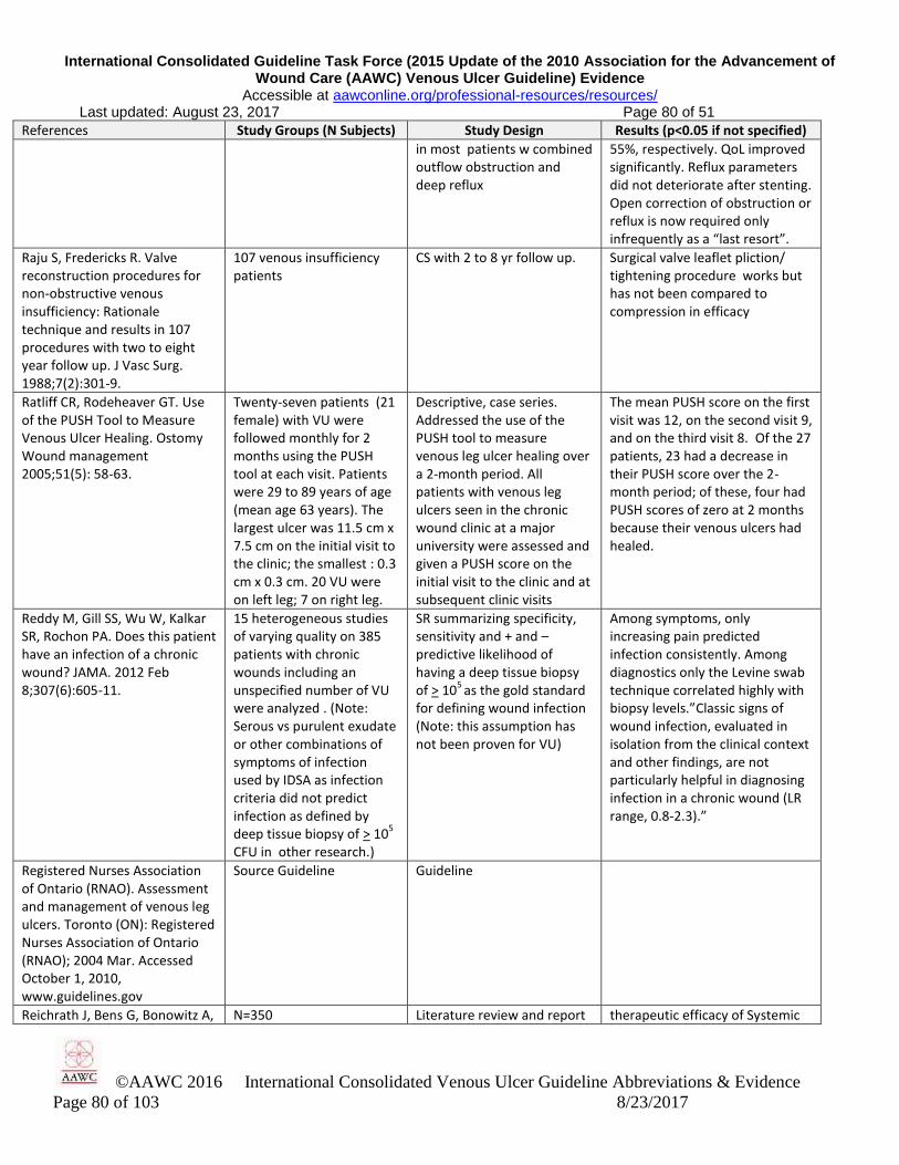
International Consolidated Guideline Task Force (2015 Update of the 2010 Association for the Advancement of Wound Care (AAWC) Venous Ulcer Guideline) Evidence
Accessible at aawconline.org/professional-resources/resources/ Last updated: August 23, 2017 Page 80 of 51
©AAWC 2016 International Consolidated Venous Ulcer Guideline Abbreviations & Evidence
Page 80 of 103 8/23/2017
References Study Groups (N Subjects) Study Design Results (p<0.05 if not specified)
in most patients w combined outflow obstruction and deep reflux
55%, respectively. QoL improved significantly. Reflux parameters did not deteriorate after stenting. Open correction of obstruction or reflux is now required only infrequently as a “last resort”.
Raju S, Fredericks R. Valve reconstruction procedures for non-obstructive venous insufficiency: Rationale technique and results in 107 procedures with two to eight year follow up. J Vasc Surg. 1988;7(2):301-9.
107 venous insufficiency patients
CS with 2 to 8 yr follow up. Surgical valve leaflet pliction/ tightening procedure works but has not been compared to compression in efficacy
Ratliff CR, Rodeheaver GT. Use of the PUSH Tool to Measure Venous Ulcer Healing. Ostomy Wound management 2005;51(5): 58-63.
Twenty-seven patients (21 female) with VU were followed monthly for 2 months using the PUSH tool at each visit. Patients were 29 to 89 years of age (mean age 63 years). The largest ulcer was 11.5 cm x 7.5 cm on the initial visit to the clinic; the smallest : 0.3 cm x 0.3 cm. 20 VU were on left leg; 7 on right leg.
Descriptive, case series. Addressed the use of the PUSH tool to measure venous leg ulcer healing over a 2-month period. All patients with venous leg ulcers seen in the chronic wound clinic at a major university were assessed and given a PUSH score on the initial visit to the clinic and at subsequent clinic visits
The mean PUSH score on the first visit was 12, on the second visit 9, and on the third visit 8. Of the 27 patients, 23 had a decrease in their PUSH score over the 2-month period; of these, four had PUSH scores of zero at 2 months because their venous ulcers had healed.
Reddy M, Gill SS, Wu W, Kalkar SR, Rochon PA. Does this patient have an infection of a chronic wound? JAMA. 2012 Feb 8;307(6):605-11.
15 heterogeneous studies of varying quality on 385 patients with chronic wounds including an unspecified number of VU were analyzed . (Note: Serous vs purulent exudate or other combinations of symptoms of infection used by IDSA as infection criteria did not predict infection as defined by deep tissue biopsy of > 10
5
CFU in other research.)
SR summarizing specificity, sensitivity and + and – predictive likelihood of having a deep tissue biopsy of > 10
5 as the gold standard
for defining wound infection (Note: this assumption has not been proven for VU)
Among symptoms, only increasing pain predicted infection consistently. Among diagnostics only the Levine swab technique correlated highly with biopsy levels.”Classic signs of wound infection, evaluated in isolation from the clinical context and other findings, are not particularly helpful in diagnosing infection in a chronic wound (LR range, 0.8-2.3).”
Registered Nurses Association of Ontario (RNAO). Assessment and management of venous leg ulcers. Toronto (ON): Registered Nurses Association of Ontario (RNAO); 2004 Mar. Accessed October 1, 2010, www.guidelines.gov
Source Guideline Guideline
Reichrath J, Bens G, Bonowitz A, N=350 Literature review and report therapeutic efficacy of Systemic

International Consolidated Guideline Task Force (2015 Update of the 2010 Association for the Advancement of Wound Care (AAWC) Venous Ulcer Guideline) Evidence
Accessible at aawconline.org/professional-resources/resources/ Last updated: August 23, 2017 Page 81 of 51
©AAWC 2016 International Consolidated Venous Ulcer Guideline Abbreviations & Evidence
Page 81 of 103 8/23/2017
References Study Groups (N Subjects) Study Design Results (p<0.05 if not specified)
Tilgen W. Treatment recommendations for pyoderma gangrenosum: an evidence-based review of the literature based on more than 350 patients. J Am Acad Dermatol. 2005; 53(2):273-83.
of pyoderma gangrenosum cases symptoms, treatment and outcomes were reported.
treatment with corticosteroids and cyclosporine have best documentation in literature, for disseminated as well as for localized disease and should be considered first-line therapy
Reich-Schupke S, Altmeyer P, Kreuter A, Stücker M. Development of spinocellular carcinoma in a long-lasting and therapy resistant venous ulcer - two case studies. J Dtsch Dermatol Ges. 2008;6(7):569-72
2 cases of long standing VU Biopsies of longstanding VU Spinous cell carcinoma in both cases.
Reich-Schupke S, Doerler M, Wollina U, Dissemond J, Horn T, Strolin A, Erfurt-Berge C, Stücker M. Squamous cell carcinomas in chronic venous leg ulcers. Data of the German Marjolin Registry and review. J Dtsch Dermatol Ges. 2015;13(10):1006-13.
Patients with VU and documented squamous cell carcinoma from 2010-2013 in the Marjolin Registry. (30 of which 20 were women) Mean age: 76 years; mean VU duration 15.9 years before squamous cell cancer diagnosis
All members of the wound healing working group of the German Society of Dermatology completed questionnaires about patients, ulcers and therapy conducted.
. Therapy resistance despite optimal care for 6-12 weeks (n=27: 90% sensitivity), “fetor” (n=12; 40% sensitivity) and atypical nodular wound bed with hypergranulation (33 % sensitivy) predicted squamous cell carcinoma.
Ricci MA, Emmerich J, Callas PW, Rosendaal FR, Stanley AC, Naud S, Vossen C, Bovill EG. Evaluating chronic venous disease with a new venous severity scoring system. J Vasc Surg. 2003;38(5):909-15.
210 patients with with protein C deficiency (420 limbs)
Venous Clinical Severity Score (VCSS) (0-3) clinically for pain, varicose veins, edema, skin pigmentation, inflammation, induration, ulcer duration and size, and compressive therapy
VCSS had sensitivity 89% and 76% specificity, + 37% predictive validity and 98% – predictive validity against ultrasound as the standard. Though designed as a severity tool, VCSS may be a useful screening tool.
Rivera-Arce E, Chávez-Soto MA, Herrera-Arellano A, Arzate S, Agüero J, Feria-Romero IA, Cruz-Guzmán A, Lozoya X. Therapeutic effectiveness of a Mimosa tenuiflora cortex extract in venous leg ulceration treatment. J Ethnopharmacol. 2007;109(3):523-8.
5% crude extract of Mimosa tenuiflora bark gel in hydrogel vehicle (22) Same hydrogel without the Mimosa extract (19)
RCT, with treatment for 8 weeks, follow up 13 weeks. Healing determined by digital area reduction as reduction in healing area and numbers of patients healed at 8 weeks
More patients healed by 8 weeks in Mimosa gel group.(p = 0.0001 chi square) Note: subsequent research by same group showed NS effects. (Int W Journal 2012
Aug;9(4):412-8.)
Robinson C, Santill S. Warm-up Active Wound Therapy: A novel approach to the management of chronic venous stasis ulcers. J Vasc Nurs. 1998;16(2):38-42.
Total of 13 VU patients assigned to either: Warm-up (8 ulcers) therapy for 1 hour 4 times daily or conventional gauze therapy (5 ulcers) followed by crossover to Warm-up
Pilot prospective RCT of inpatients for 2 weeks. Control wounds mean 64.4 cm
2 initial area. Warm-up
wounds mean 29.4 cm2
initial area.
32% decrease in wound size and 39% decrease in pain score for Warm-up patients. 25% decrease in wound size and 27% decrease in pain score for controls. Pain decreased over time for both treatments.
Robson MC, Cooper DM, Aslam R, Gould LJ, Harding KG,
Source Guideline Wound Healing Society Guideline for prevention of
Recommendations included in ICVUG.

International Consolidated Guideline Task Force (2015 Update of the 2010 Association for the Advancement of Wound Care (AAWC) Venous Ulcer Guideline) Evidence
Accessible at aawconline.org/professional-resources/resources/ Last updated: August 23, 2017 Page 82 of 51
©AAWC 2016 International Consolidated Venous Ulcer Guideline Abbreviations & Evidence
Page 82 of 103 8/23/2017
References Study Groups (N Subjects) Study Design Results (p<0.05 if not specified)
Margolis DJ, Ochs DE, Serena TE, Snyder RJ, Steed DL, Thomas DR, Wiersema-Bryant L. Guidelines for the prevention of venous ulcers. Wound Repair Regen. 2008;16(2):147-50.
venous ulcers
Robson MC, Cooper DM, Aslam R, Gould LJ, Harding KG, Margolis DJ, Ochs DE, Serena TE, Snyder RJ, Steed DL, Thomas DR, Wiersma-Bryant L. Guidelines for the treatment of venous ulcers. Wound Repair Regen. 2006;14(6):649-62.
Source Guideline Guideline for VU treatment Recommendations included in ICVUG.
Robson MC, Hanft J, Garner W, Jenson J; Serena T; Payne WG; Sussman A; Barbul A; Limova M; Snyder R; Odenheimer D J, Cooper DM. Healing of chronic venous ulcers is not enhanced by the addition of topical repifermin (KGF-2) to standardized care. J Appl Res. 2004;4(2):302-11.
94 VU patients assigned to topical treatment with Repifermin, a recombinant human KGF-2 (fibroblast growth factor-10) or placebo
12-week double blind RCT evaluating % of subjects completely healed at 12 weeks
No significant effect of repifermin (KGF-2) on 100% epithelization of chronic VU, but significantly more in the repifermin group achieved 75% closure at 12 weeks compared to placebo. Suggest if the RCT lasted 31 weeks 100% healing would have been p< 0.05.
Robson MC, Phillips LG, Cooper DM Lyle WG, Robson LE, Odom L, Hill DP, Hanham AF, Ksander GA. The safety and effect of transforming growth factor-B2 for treatment of venous stasis ulcers. Wound Rep Reg 1995;3(2):157-67.
Bovine transforming growth factor-beta(2) (2.5 ug/cm
2) in collagen matrix
or placebo vehicle or with a standard dressing. (3 groups each
Prospective blind RCT treated 3x/week with standardized compression; outcome was planimetrically determined rate of % area reduction as had shown a 5 ug/cm
2 dose to be safe in an
earlier open label study.
These doses of bovine transforming growth factor-beta(2)were safe as topically applied in a collagen matrix vehicle and “may have a positive effect on rate of VU closure”
Roche C, West J. A controlled trial investigating the effect of ultrasound on venous ulcers referred from general practitioners. Physiotherapy. 1984;70(12):475–7.
25 patients referred to investigators for management of VU received SC with or without ultrasound
RCTmeasuring % of initial VU area remaining to heal after 4 weeks of treatment
Ultrasound group reduced to 66.4 + 8.8% of original area at 4 weeks. SC group reduced to 91.6 + 8.9% (p< 0.05)
Romanelli M, Dini V, Barbanera S, Bertone MS. Evaluation of the efficacy and tolerability of a solution containing propyl betaine and polihexanide for wound irrigation. Skin Pharmacol Physiol. 2010;23(Suppl 1):41-4.
Solution containing propyl betaine and polihexanide (Prontosan® N=20 every other day or saline (N=20) every other day with polyurethane foam dressing + elastic compression
Single-blind RCT measuring safety and wound bed pH using, wound area, subject reported VAS pain each cleanser for 4 weeks
Pain at baseline ~9.5bothgroups At 4 weeks pain ~ 4.2 in test and ~7.8 in saline group (p< 0.05) Baseline pH was 8.9. After 4 weeks wound bed pH reduced to 7.0 (p< 0.05). Test cleanser reduced pain and wound bed pH.
Romanelli M, Dini V, Bertone MS. Randomized comparison of OASIS wound matrix versus
25 VU or mixed VU-AU ABI0.6-0.8, duration > 6 months duration > 50%
8-week RCT weekly assessed wound healing and dressing . 2 subjects in control group
OASIS-treated VU healed in mean 5.4 weeks of treatment, vs. 8.3 weeks for the petrolatum-treated

International Consolidated Guideline Task Force (2015 Update of the 2010 Association for the Advancement of Wound Care (AAWC) Venous Ulcer Guideline) Evidence
Accessible at aawconline.org/professional-resources/resources/ Last updated: August 23, 2017 Page 83 of 51
©AAWC 2016 International Consolidated Venous Ulcer Guideline Abbreviations & Evidence
Page 83 of 103 8/23/2017
References Study Groups (N Subjects) Study Design Results (p<0.05 if not specified)
moist wound dressing in the treatment of difficult-to-heal wounds of mixed arterial/venous etiology. Adv Skin Wound Care. 2010;23(1):34-8.
slough; > 02.5 cm2 in each
group: OASIS® or petrolatum gauze all with appropriate compression
moved away before end of study
VU (P = .02). Complete wound closure was achieved in 80% (20/25) of OASIS-VU at 8 weeks vs. 65% (15/23) of petrolatum-treated VU P < .05).
Romanelli M, Dini V, Polignano R, Bonadeo P, Maggio G. Ibuprofen slow-release foam dressing reduces wound pain in painful exuding wounds: preliminary findings from an international real-life study. J Dermatolog Treat. 2009;20(1):19-26.
Biatain-Ibu foam (98) Best practice (87)
RCT measuring pain relief over 7 days on 5 point verbal scale and 11-point numeric box scalr 0 = no pain, 10 worst
More patients in the ibuprofen foam treatment group reported wound pain relief and lower wound pain intensity values after 7 days (p < 0.0001 for both variables
Romanelli M, Kaha E, Stege H, Wnorowski JW, Vowden P, Majamaa H, Lazaro JL. Effect of amelogenin extracellular matrix protein and compression on hard-to-heal venous leg ulcers: follow-up data. J Wound Care. 2008;17(1):17-8, 20-3.
high compression therapy plus amelogenin (n=42) or high compression therapy alone (n=41) hard-to-heal
Follow up on RCT of same subjects which had shown more healing and less pain at 12 weeks in amelogenin group.
Pain and healing benefits continued past 12 weeks
Romanelli M. Objective measurement of venous ulcer debridement and granulation with a skin color reflectance analyzer. Wounds 1997; 9(4): 122-6.
Film (Opsite) dressing + elastic compression covering:
Enzymatic debridement : Elase (16)
Autolytic debridement: DuoDERM Hydroactive Gel (16)
VU covered with fibrin were rated clinically for fibrin or granulation tissue, and red or yellow colorimetry assessed on days 3, 6, 9, 14 of treatment, using a Chroma Meter CR 200 Minolta camera
Both groups were similar initially in fibrin and granulation measures. Both decreased in fibrin and increased in red granulation tissue over time. The Hydroactive Gel-dressed VUs had more granulation tissue than enzyme debrided ones from days 6-14.
Rosenthal D, Murphy F, Gottschalk R, Baxter M, Lycka B, Nevin K. Using a topical anaesthetic cream to reduce pain during sharp debridement of chronic leg ulcers. J Wound Care. 2001;10(1):503–5.
101 leg ulcer patients with leg ulcer < 50 cm
2 , of
which 61 were VU and 50 had diabetes. EMLA 5% cream (51, 30 VU) or Placebo (50, 31 VU) both held in place for 27-33 minutes with moisture retentive (occlusive) plastic wrap before surgical debridement.
RCT conducted in four Canada Dermatology centers. Participants rated pain on a 100 mm visual analogue scale after debridement and removal of cream. Also cutaneous reactions were rated on a scale of “none, mild, moderate or severe”
For VU subjects randomized to EMLA cream, VAS mean score was 31, compared to 53 for VU subjects randomized to receive Placebo (P< 0.05). Note: this study included only individuals who previously reported pain on debridement.
Rowland J. Intermittent pump versus compression bandages in the treatment of venous leg ulcers. Aust NZ J Surg. 2000; 70(2):110-3.
A randomized cross-over study of patients attending an outpatient wound clinic (n = 16) Half treated with Setopress high stretch bandages, half treated with
RCT Assessing VU healing and leg oedema and comfort in 11 of the 16 patients. Note: Limitations: too small for generalizable conclusions, though compression pumps
There was no significant difference between treatment types with regard to ulcer healing rates or control of leg oedema. Patients reported the pump easier and more comfortable

International Consolidated Guideline Task Force (2015 Update of the 2010 Association for the Advancement of Wound Care (AAWC) Venous Ulcer Guideline) Evidence
Accessible at aawconline.org/professional-resources/resources/ Last updated: August 23, 2017 Page 84 of 51
©AAWC 2016 International Consolidated Venous Ulcer Guideline Abbreviations & Evidence
Page 84 of 103 8/23/2017
References Study Groups (N Subjects) Study Design Results (p<0.05 if not specified)
IPC. Total 11 pts at conclusion of study (low power)..
and bandages were similar in efficacy for the healing of venous leg ulcers.
to use, with a trend towards increased compliance.
Royal College of Nursing. The management of patients with venous leg ulcers: Clinical Practice Guideline. 1998; The RCN Institute, Center for Evidence-based Nursing, University of York & School of Nursing, Midwifery and Health Visiting, University of Manchester. Accessed October 1, 2010 at http://www.rcn.org.uk/development/practice/clinicalguidelines/venous_leg_ulcers.
Source Guideline Guideline Recommendations addressed in ICVUG.
Rubin JR, Alexander J, Plecha EJ, Marman C. Unna's boot vs. polyurethane foam dressings for the treatment of venous ulceration. A randomized prospective study. Arch Surg. 1990;125(4):489-90.
Unna's Boot (19) SynthaDerm (17) foam dressing
Prospective, RCT unclear duration in US hospital setting. Note: compares a dressing vs. compression which is essential for VU.
94.7% healed with Unna's Boot 41.2% healed with foam Increase rate of healing with Unna’s boot of .5 cm/day vs foam of .07 cm/day.
Rudofsky G. Intravenous prostaglandin E1 in the treatment of venous ulcers: a double-blind placebo-controlled trial. Vasa. 1989;28(Suppl 1):39-43.
one intravenous infusion of 3 ampoules of Prostavasin (60 micrograms PGE1) or 3 ampoules of placebo (1940.1 micrograms alpha-CD) daily dissolved in 250 ml saline over 3 h. PGE1 group (n = 20) Placebo (n = 22)
Double blind RCT During 6 weeks daily treatment, a multivariate measure of VU healing was monitored in addition to complete healing and adverse events.
8 out of 20 patients on PGE1 (40%) compared to only 2 out of 22 patients on placebo (9%), with significant improvement in VU status (p< 0.001). No adverse events in either group. Edema completely resolved in 17 of 20 PGE-1 patients (85%) or 7 out of 20 placebo patients (35%). Calf cramps and eczema vanished in 80% and 87.5% respectively with PGE1, but only in 50% and 9% respectively with placebo. Parallel to this VU area TCPO2 increased by a mean of 45.9%.
Rudolph D. Standards of care for venous leg ulcers: Compression therapy and moist wound healing. J Vasc Nurs. 2001;19(1):20-7.
VU account for 70-90% of all lower extremity ulcers.
LR of pathophysiology and best SC for a patient with a VU
Gradient compression and moist wound healing with hydrocolloid dressings are good SC for VU despite the advent of new modalities..
Saedon M, Stansby G. Post-thrombotic syndrome: prevention is better than cure. Phlebology. 2010;25(Suppl 1):14-9.
LR of techniques to prevent or cure post thrombotic syndrome.
Villalta Scale is best for classifying post thrombotic syndrome, including VU. Prevention is better than treatment.
Risk factors for PTS include obesity and prior varicose veins. Poor quality anticoagulation control may be a factor
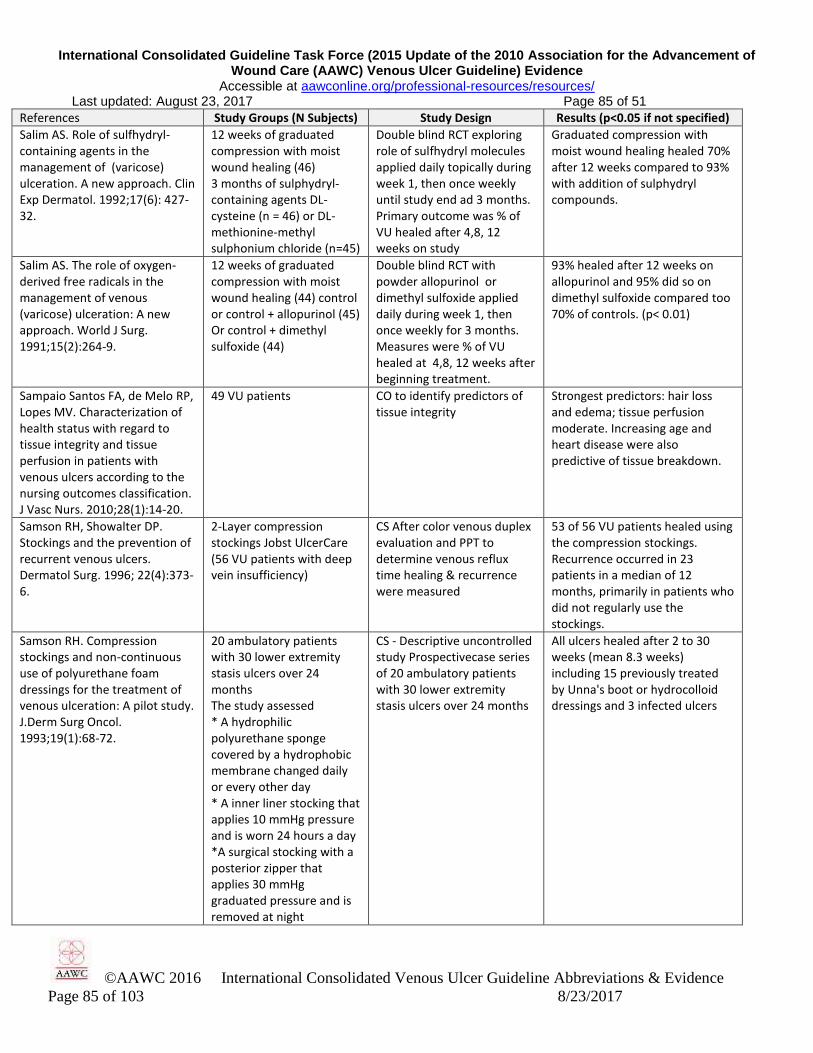
International Consolidated Guideline Task Force (2015 Update of the 2010 Association for the Advancement of Wound Care (AAWC) Venous Ulcer Guideline) Evidence
Accessible at aawconline.org/professional-resources/resources/ Last updated: August 23, 2017 Page 85 of 51
©AAWC 2016 International Consolidated Venous Ulcer Guideline Abbreviations & Evidence
Page 85 of 103 8/23/2017
References Study Groups (N Subjects) Study Design Results (p<0.05 if not specified)
Salim AS. Role of sulfhydryl-containing agents in the management of (varicose) ulceration. A new approach. Clin Exp Dermatol. 1992;17(6): 427-32.
12 weeks of graduated compression with moist wound healing (46) 3 months of sulphydryl-containing agents DL-cysteine (n = 46) or DL-methionine-methyl sulphonium chloride (n=45)
Double blind RCT exploring role of sulfhydryl molecules applied daily topically during week 1, then once weekly until study end ad 3 months. Primary outcome was % of VU healed after 4,8, 12 weeks on study
Graduated compression with moist wound healing healed 70% after 12 weeks compared to 93% with addition of sulphydryl compounds.
Salim AS. The role of oxygen-derived free radicals in the management of venous (varicose) ulceration: A new approach. World J Surg. 1991;15(2):264-9.
12 weeks of graduated compression with moist wound healing (44) control or control + allopurinol (45) Or control + dimethyl sulfoxide (44)
Double blind RCT with powder allopurinol or dimethyl sulfoxide applied daily during week 1, then once weekly for 3 months. Measures were % of VU healed at 4,8, 12 weeks after beginning treatment.
93% healed after 12 weeks on allopurinol and 95% did so on dimethyl sulfoxide compared too 70% of controls. (p< 0.01)
Sampaio Santos FA, de Melo RP, Lopes MV. Characterization of health status with regard to tissue integrity and tissue perfusion in patients with venous ulcers according to the nursing outcomes classification. J Vasc Nurs. 2010;28(1):14-20.
49 VU patients CO to identify predictors of tissue integrity
Strongest predictors: hair loss and edema; tissue perfusion moderate. Increasing age and heart disease were also predictive of tissue breakdown.
Samson RH, Showalter DP. Stockings and the prevention of recurrent venous ulcers. Dermatol Surg. 1996; 22(4):373-6.
2-Layer compression stockings Jobst UlcerCare (56 VU patients with deep vein insufficiency)
CS After color venous duplex evaluation and PPT to determine venous reflux time healing & recurrence were measured
53 of 56 VU patients healed using the compression stockings. Recurrence occurred in 23 patients in a median of 12 months, primarily in patients who did not regularly use the stockings.
Samson RH. Compression stockings and non-continuous use of polyurethane foam dressings for the treatment of venous ulceration: A pilot study. J.Derm Surg Oncol. 1993;19(1):68-72.
20 ambulatory patients with 30 lower extremity stasis ulcers over 24 months The study assessed * A hydrophilic polyurethane sponge covered by a hydrophobic membrane changed daily or every other day * A inner liner stocking that applies 10 mmHg pressure and is worn 24 hours a day *A surgical stocking with a posterior zipper that applies 30 mmHg graduated pressure and is removed at night
CS - Descriptive uncontrolled study Prospectivecase series of 20 ambulatory patients with 30 lower extremity stasis ulcers over 24 months
All ulcers healed after 2 to 30 weeks (mean 8.3 weeks) including 15 previously treated by Unna's boot or hydrocolloid dressings and 3 infected ulcers

International Consolidated Guideline Task Force (2015 Update of the 2010 Association for the Advancement of Wound Care (AAWC) Venous Ulcer Guideline) Evidence
Accessible at aawconline.org/professional-resources/resources/ Last updated: August 23, 2017 Page 86 of 51
©AAWC 2016 International Consolidated Venous Ulcer Guideline Abbreviations & Evidence
Page 86 of 103 8/23/2017
References Study Groups (N Subjects) Study Design Results (p<0.05 if not specified)
Santilli SM, Valusek PA, Robinson C. Use of a non-contact radiant heat bandage for the treatment of chronic venous stasis ulcers. Adv Wound Care; 1999; 12(2):89-92.
Warm-up Therapy (17 patients with 31 wounds)
Prospective case series in a university-affiliated VA medical center, with 18-month follow up
8/17 (44%) healed completely after discharge; 14/17 (82%) improved. One recurrence in 18 months.
Sayag J, Meaume S, Bohbot S. Healing properties of calcium alginate dressings. J Wound Care. 1996;5(8):357-62.
*Calcium alginate *Established local treatment with dextranomer paste
Prospective RCT of 92 patients with full thickness wounds
*Alginate Mean surface area reduction: 2.39 cm2 *Dextranimer paste Mean surface area reduction 0.27 cm2
Sayag J. Semi-synthetic hydrocolloids in occlusive dressings for leg ulcers. In: T J Ryan (Ed) Beyond occlusion: wound care proceedings. Royal Society of Medicine Services Ltd., 1988;136:105-108
Hydrocolloid dressing (HCD) (626 total). Before applying dressing, wound was cleansed with 3% hydrogen peroxide for at least 1 minute, then rinsed with saline and dried with sterile gauze.
Venous ulcers (356) Mixed arterio-venous (127) Arterial or diabetic (49) Trauma or burn (18) Neurotrophic foot ulcer (15) Pressure ulcer (7) Buerger’s disease (1) Connective tissue disease (3); Lymphoedema (2); Sickle cell anemia (1)
Prospective HCT of HCD or other dressings on patients hospitalized with wounds (726 episodes) from 1981-1987 HCD was applied overlapping wound edges at least 3 cm and remained in place until detachment or up to 7 days. No other local treatment or form of debridement was used. No systemic antibiotics, corticosteroids, non-steroidal anti-inflammatory agents or hyperbaric oxygen was used. Prior prescribed anticoagulants or peripheral vasodilators were continued.
During the first 6 months of HCD use, complete healing occurred in 88% of wounds with initial diameter less than 2 cm and in 78% of those with diameter more than 4 cm. Total healing occurred in 89% of wounds enduring less than 6 months, 50% of those with longer duration. Healing was “shorter than that found with traditional dressings” and reduced length of hospital stays and costs of care.
Schnirring-Judge M, Belpedio D. Malignant transformation of a chronic venous stasis ulcer to basal cell carcinoma in a diabetic patient: case study and review of the pathophysiology. J Foot Ankle Surg. 2010;49(1):75-9.
1 Case study patient with lower leg ulcer thought to be VU for 3 years
Biopsy confirmed malignant transformation.
Consider biopsy of long-standing venous ulcers to check for malignant transformation.
Schuren J, Vos A, Allen JO. Venous leg ulcer patients with low ABPIs: How much pressure is safe and tolerable?. EWMA Journal. 2010 Sep 1;10(3):1-3.
8 experienced wound care nurses in Canada and the Netherlands on patients with ABI 0.5-0.8
CS measuring pressure applied under a 2-layer compression bandage, wear time and adverse events in each ABI category
Mean sub-bandage resting pressure was 24.9 mmHg; standing pressure 32.4 mmHg standing pressure. This form of compression was safe and well tolerated. Two adverse events were reported: foot pain.
Scottish Intercollegiate Guidelines Network (SIGN). Management of chronic venous leg ulcers. A national clinical guideline. Edinburgh (Scotland):
Guideline Guideline All unique recommendations addressed.in ICVUG.

International Consolidated Guideline Task Force (2015 Update of the 2010 Association for the Advancement of Wound Care (AAWC) Venous Ulcer Guideline) Evidence
Accessible at aawconline.org/professional-resources/resources/ Last updated: August 23, 2017 Page 87 of 51
©AAWC 2016 International Consolidated Venous Ulcer Guideline Abbreviations & Evidence
Page 87 of 103 8/23/2017
References Study Groups (N Subjects) Study Design Results (p<0.05 if not specified)
Scottish Intercollegiate Guidelines Network (SIGN); 2010 Aug. 44 p. (SIGN publication; no. 120). Accessed October 1, 2010, www.guidelines.gov.
Scriven JM, Taylor LE, Wood AJ, Bell PR, Naylor AR, London NJM. A prospective randomised trial of four-layer versus short stretch compression bandages for the treatment of venous leg ulcers. Ann Royal College Surgeons. 1998; 80(3):215-20.
1. 4-layer elastic compression bandage (32 ulcers—not patients) 2. Short-stretch 3-layer compression bandage (32 ulcers—not patients)
Prospective RCT measuring complete healing at 3 months and 1 year and adverse events. (Note: 11 patients had bilateral ulcers which were randomized independently, possibly)
Healed at 3 months: 4-layer 34%; short-stretch 41% Healed at 1 year: 4-layer 53%; short-stretch 56%
Senet P, Bause R, Jørgensen B, Fogh K. Clinical efficacy of a silver-releasing foam dressing in venous leg ulcer healing: a randomised controlled trial. Int Wound J. 2014;11(6):649-55.
Biatain Ag 6 weeks (77) foam dressing with silver sulphadiazine, followed by 4 weeks same foam dressing without the silver sulphadiazene:Biatain for 10 weeks (84)
Multinational double blind RCT of only VU. Measuring % area reduction from baseline at week 6 and week 10. A subset of older patients with history of venous thrombosis larger, longer duration more recurrent VU was performed.
4-week % area reduction: Biatain Ag 35.4, Biatin: 27.5 (p=0.428). Subset of patients at higher risk of non-healing had a higher % area reduction at 10 weeks with Biatain Ag than Biatain (p< 0.05)
Senet P, Combemale P, Debure C, Baudot N, Aout M, Vicaut E, Lok C, for the Angio-Dermatology Group of the French Society of Dermatology. Malignancy of chronic leg ulcers: The value of systematic wound biopsies: a prospective, multicenter, cross-sectional study. Arch Dermatol. 2012;148(6):704-8.
January 1-May 31, 2006 144 patients consulted for chronic leg ulcers, attributed to venous and/or peripheral arterial disease(s), increasing in wound area and/or depth, despite appropriate standard treatment for at least 3 months
Prospective multicenter CO study with at least 2 6 mm wound edge biopsies taken per wound on enrollment into the study. 144 patients had 154 chronic leg ulcers with 10.4% (5% squamous cell; 3% basal cell and 2% other) Univariate analysis sensitivity, specificity, PPV and NPV reported
Numbers below are in order, sensitivity, specificity, PPV, NPV Univariate analyses found older age, abnormal excessive granulation tissue at wound edges (.81,.76,.27,.97), high clinical suspicion of cancer (.59,.91,.42,.95), and number of biopsies,but not wound area or duration, were significantly associated with skin cancer in > 1 biopsy specimens.
Serena TE, Carter MJ, Le LT, Sabo MJ, MiMarco DT and the Epifix VLU Study Group. A multicenter, randomized, controlled clinical trial evaluating the use of dehydrated human amnion/chorion membrane allografts and multilayer compression therapy vs. multilayer compression therapy alone in the treatment of venous leg ulcers. Wound Rep Regen. 2014;22(6): 688-93.
84 patients enrolled in the treatment phase all receiving multi-layer compression therapy: one dHACM amnion /chorion allograft application (26), two dHACM applications (27), and multi-layer compression therapy only control group (31)
multicenter, open-label RCT designed to evaluate the safety and efficacy of dHACM allograft (either one application or two applications) and multi-layer compression therapy vs. the same compression alone . Primary outcome was number of patients with % reduction in VU area during the first 4 weeks of allograft treatment.
4-week reduction in wound area of ≥40% occurred in more patients receiving > 1 allograft vs. controls (33/53 [62%] vs 10/31 [32%]; p = 0.005). Within the dHACM group, proportions of wound reduction ≥40% after 4 weeks were similar for those patients receiving one vs. two dHACM applications (62% [16/26] and 63.0% [17/27], each more than controls (p = 0.019 and 0.027,). Allograft patients had a mean % VU area reduction over 4-weeks of 48.1% compared with

International Consolidated Guideline Task Force (2015 Update of the 2010 Association for the Advancement of Wound Care (AAWC) Venous Ulcer Guideline) Evidence
Accessible at aawconline.org/professional-resources/resources/ Last updated: August 23, 2017 Page 88 of 51
©AAWC 2016 International Consolidated Venous Ulcer Guideline Abbreviations & Evidence
Page 88 of 103 8/23/2017
References Study Groups (N Subjects) Study Design Results (p<0.05 if not specified)
19.0% in the control group.
Shebel ND An early intervention plan for identification and control of chronic lower extremity edema. J Vasc Nurs. 2002;20(2):45-50.
Clinical experience with patients in practice.: CO
CO using an evidence-based and expert opinion-based early intervention plan to identify and control lower extremity edema
Identification and control plan for chronic lower extremity edema reduced 70-90% recurrence rate for VU
Shelling ML, Federman, DG, Kirsner RS. Clinical approach to atypical wounds with a new model for understanding hypertensive ulcers . Arch Dermatol. 2010;146 ( 9):1026-9.
Review of atypical wounds. Includes cases of VU malignant transformation
Not all leg ulcers are VU. Each etiology merits appropriate treatment.
Shi H, Liu X, Lu M, Lu X, Jiang M, Yin M. The effect of endovenous laser ablation of incompetent perforating veins and the great saphenous vein in patients with primary venous disease. Eur J Vasc Endovasc Surg. 2015;49:574-80.
311 patients (376 limbs). Among these, 132 patients (156 limbs) were treated with Edovenous Lasier Ablation (EVLA) of IPVs and varicose vein surgery, and the remaining 179 patients (220 limbs) were treated with varicose vein surgery alone and served as controls.
Retrospective analysis looking at clinical results and fate of incompetent perforating veins following treatment of superficial venous insufficiency, with or without endovenous laser ablation.
In the laser treated perforators group the ulcer healing time was significantly lower at 1.4 months versus 3.3 months in the “varicose vein surgery only” group. No significant differences were seen in the 12 months healing rate between the 2 groups. After only 1 year follow up, no differences were found in the recurrent rate between the 2 groups. This study shows only limited advantages in performing laser perforator ablation in venous ulcers. The follow up was very short relative to other studies.
Sibbald, GS, Browne, AC, Coutts, PC, Queen D. Screening evaluation of an ionized nanocrystalline silver dressing in chronic wound care. Ostomy Wound Manage. 2001; 47(10):38-43.
29 patients were dressed with a nanocrystalline silver dressing, including 6 with a VU.
Uncontrolled, open label, prospective case study.
4/6 VU patients demonstrated decreased wound size and exudate.
Sigel B, Edelstein AL, Savitch L, Hasty JH, Felix R, Jr. Type of compression for reducing venous stasis. Arch Surg. 1975; 110:171-5.
6 healthy volunteers and 1 volunteer with history of thrombophlebitis.
HCT. Common femoral vein flow was measured while subjecting supine volunteers to gradient or uniform compression.
Gradient compression descending centrally provided the greatest increment in venous flow.
Sikes E. Evaluation of a transparent dressing in the treatment of stasis ulcers of the lower limb. J Enterostomal Therapy. 1985;12(4):116-20.
Inelastic Unna's boot; (7) Opsite film dressing (6)
Convenience controlled trial for 1 year in a vascular clinic setting in the USA
81% healed with Unna's Boot 71% healed with the film dressing alone. NS difference between groups.

International Consolidated Guideline Task Force (2015 Update of the 2010 Association for the Advancement of Wound Care (AAWC) Venous Ulcer Guideline) Evidence
Accessible at aawconline.org/professional-resources/resources/ Last updated: August 23, 2017 Page 89 of 51
©AAWC 2016 International Consolidated Venous Ulcer Guideline Abbreviations & Evidence
Page 89 of 103 8/23/2017
References Study Groups (N Subjects) Study Design Results (p<0.05 if not specified)
Silberzweig JE, Funaki BS, Ray CE Jr, Burke CT, Kinney TB, Kostelic JK, Loesberg A, Lorenz JM, Mansour MA, Millward SF, Nemcek AA Jr, Owens CA, Reinhart RD, Vatakencherry G, Expert Panel on Interventional Radiology. ACR Appropriateness Criteria® treatment of lower-extremity venous insufficiency. [online publication]. Reston (VA): American College of Radiology (ACR); 2009. 7 p. [70 references] Accessed August 1, 2010, www.guidelines.gov.
Guidelne, no patient outcomes directly reported.
Guideline of the American College of Radiology
Addresses laser and other radiologic therapy for venous insufficiency and venous ulcers.
Singh A, Halder S, Menon GR, Chumber S, Misra MC, Sharma LK, Srivastava A. Meta-analysis of randomized controlled trials on hydrocolloid occlusive dressing versus conventional gauze dressing in the healing of chronic wounds. Asian J Surg. 2004;27(4):326-32.
Leg ulcer patients (693) with 819 wounds Hydrocolloid Dressings (HCD) compared to Saline gauze or Paraffin impregnated gauze
MA of 12 RCTs on 693 patients comparing healing effects of hydrocolloid to saline or paraffin-impregnated gauze
Complete healing, 51% (HCD) vs 31%,(gauze) P=.02; Odds Ratio for healing with a primary HCD dressing was significantly higher than with saline or paraffin-impregnated gauze. Odds ratio =2.45 (95% CI, 1.18-5.12)
Spence R, Cahall E Inelastic versus elastic leg compression in chronic venous insufficiency: A comparison of limb size and venous hemodynamics. J Vasc Surg. 1996; 24:783-7.
10 patients and 18 limbs all with Class III chronic venous insufficiency and ankles of sufficient flexibility to exercise the calf muscle pump
CS Patients' limb size, venous filling rate and ejection fraction was measured with no compression, with a 30 to 40 mm Hg below knee stocking and with Circaid inelastic compression 2 and 6 hours after application. Therapies were compared with baseline and over time
Inelastic compression has a significant effect on deep venous hemodynamics by decreasing venous reflux and improving calf muscle pump function (ankle circumference-at 2 vs. 6 hours:baseline,24.7+ 7 cm vs 26.1+ 1.1 cm, stocking 23.9+ 1.1cm vs 26.2+ 1.2cm when compared with compression stockings
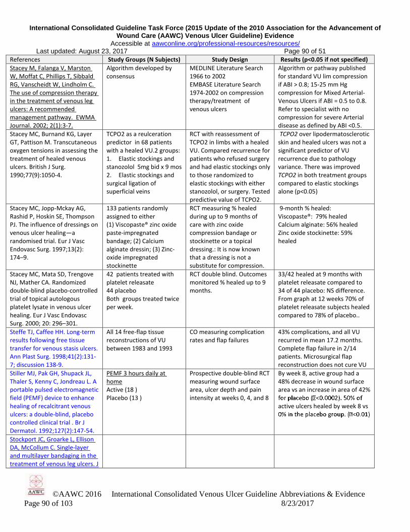
International Consolidated Guideline Task Force (2015 Update of the 2010 Association for the Advancement of Wound Care (AAWC) Venous Ulcer Guideline) Evidence
Accessible at aawconline.org/professional-resources/resources/ Last updated: August 23, 2017 Page 90 of 51
©AAWC 2016 International Consolidated Venous Ulcer Guideline Abbreviations & Evidence
Page 90 of 103 8/23/2017
References Study Groups (N Subjects) Study Design Results (p<0.05 if not specified)
Stacey M, Falanga V, Marston W, Moffat C, Phillips T, Sibbald RG, Vanscheidt W, Lindholm C. The use of compression therapy in the treatment of venous leg ulcers: A recommended management pathway. EWMA Journal. 2002; 2(1):3-7.
Algorithm developed by consensus
MEDLINE Literature Search 1966 to 2002 EMBASE Literature Search 1974-2002 on compression therapy/treatment of venous ulcers
Algorithm or pathway published for standard VU lim compression if ABI > 0.8; 15-25 mm Hg compression for Mixed Arterial-Venous Ulcers if ABI = 0.5 to 0.8. Refer to specialist with no compression for severe Arterial disease as defined by ABI <0.5.
Stacey MC, Burnand KG, Layer GT, Pattison M. Transcutaneous oxygen tensions in assessing the treatment of healed venous ulcers. British J Surg. 1990;77(9):1050-4.
TCPO2 as a reulceration predictor in 68 patients with a healed VU.2 groups: 1. Elastic stockings and stanozolol 5mg bid x 9 mos 2. Elastic stockings and surgical ligation of superficial veins
RCT with reassessment of TCPO2 in limbs with a healed VU. Compared recurrence for patients who refused surgery and had elastic stockings only to those randomized to elastic stockings with either stanozolol, or surgery. Tested predictive value of TCPO2.
TCPO2 over lipodermatosclerotic skin and healed ulcers was not a significant predictor of VU recurrence due to pathology variance. There was improved TCPO2 in both treatment groups compared to elastic stockings alone (p<0.05)
Stacey MC, Jopp-Mckay AG, Rashid P, Hoskin SE, Thompson PJ. The influence of dressings on venous ulcer healing—a randomised trial. Eur J Vasc Endovasc Surg. 1997;13(2): 174–9.
133 patients randomly assigned to either (1) Viscopaste® zinc oxide paste-impregnated bandage; (2) Calcium alginate dressin; (3) Zinc-oxide impregnated stockinette
RCT measuring % healed during up to 9 months of care with zinc oxide compression bandage or stockinette or a topical dressing.: It is now known that a dressing is not a substitute for compression.
9-month % healed: Viscopaste®: 79% healed Calcium alginate: 56% healed Zinc oxide stockinette: 59% healed
Stacey MC, Mata SD, Trengove NJ, Mather CA. Randomized double-blind placebo-controlled trial of topical autologous platelet lysate in venous ulcer healing. Eur J Vasc Endovasc Surg. 2000; 20: 296–301.
42 patients treated with platelet releasate 44 placebo Both groups treated twice per week.
RCT double blind. Outcomes monitored % healed up to 9 months.
33/42 healed at 9 months with platelet releasate compared to 34 of 44 placebo: NS difference. From graph at 12 weeks 70% of platelet releasate subjects healed compared to 78% of placebo..
Steffe TJ, Caffee HH. Long-term results following free tissue transfer for venous stasis ulcers. Ann Plast Surg. 1998;41(2):131-7; discussion 138-9.
All 14 free-flap tissue reconstructions of VU between 1983 and 1993
CO measuring complication rates and flap failures
43% complications, and all VU recurred in mean 17.2 months. Complete flap failure in 2/14 patients. Microsurgical flap reconstruction does not cure VU
Stiller MJ, Pak GH, Shupack JL, Thaler S, Kenny C, Jondreau L. A portable pulsed electromagnetic field (PEMF) device to enhance healing of recalcitrant venous ulcers: a double-blind, placebo controlled clinical trial . Br J Dermatol. 1992;127(2):147-54.
PEMF 3 hours daily at home Active (18 ) Placebo (13 )
Prospective double-blind RCT measuring wound surface area, ulcer depth and pain intensity at weeks 0, 4, and 8
By week 8, active group had a 48% decrease in wound surface area vs an increase in area of 42%
active ulcers healed by week 8 vs
Stockport JC, Groarke L, Ellison DA, McCollum C. Single-layer and multilayer bandaging in the treatment of venous leg ulcers. J
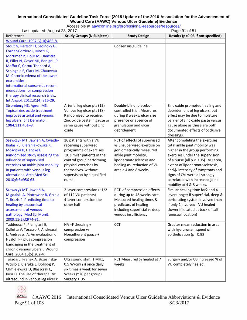
International Consolidated Guideline Task Force (2015 Update of the 2010 Association for the Advancement of Wound Care (AAWC) Venous Ulcer Guideline) Evidence
Accessible at aawconline.org/professional-resources/resources/ Last updated: August 23, 2017 Page 91 of 51
©AAWC 2016 International Consolidated Venous Ulcer Guideline Abbreviations & Evidence
Page 91 of 103 8/23/2017
References Study Groups (N Subjects) Study Design Results (p<0.05 if not specified)
Wound Care. 1997;6(10):485-8.
Stout N, Partsch H, Szolnoky G, Forner-Cordero I, Mosti G, Mortimer P, Flour M, Damstra R, Piller N, Geyer MJ, Benigni JP, Moffat C, Cornu-Thenard A, Schingale F, Clark M, Chauveau M. Chronic edema of the lower extremities: international consensus recommendations for compression therapy clinical research trials. Int Angiol. 2012;31(4):316-29.
Consensus guideline
Stromberg HE, Agren MS. Topical zinc oxide treatment improves arterial and venous leg ulcers. Br J Dermatol. 1984;111:461–8.
Arterial leg ulcer pts (19) Venous leg ulcer pts (18) Randomized to receive: Zinc oxide paste in gauze or same gauze without zinc oxide
Double-blind, placebo-controlled trial. Measures during 8 weeks: ulcer size presence or absence of granulation and ulcer debridement
Zinc oxide promoted healing and debridement of leg ulcers, but effect may be due to moisture barrier of zinc oxide paste versus gauze alone as these are both documented effects of occlusive dressings.
Szewczyk MT, Jawień A, Cwajda-Białasik J, Cierzniakowska K, Mościcka P, Hancke E. Randomized study assessing the influence of supervised exercises on ankle joint mobility in patients with venous leg ulcerations. Arch Med Sci. 2010;6(6):956-63.
16 patients with a VU receiving supervised programme of exercises 16 similar patients in the control group performing physical exercises by themselves, without supervision by a qualified nurse
RCT of effects of supervised vs unsupervised exercise on goniometrically measured ankle joint mobility, lipodermatosclerosis and healing as reduction of VU area a 4 and 8 weeks.
After completing the exercises total ankle joint mobility was higher in the group performing exercises under the supervision of a nurse (all p < 0.05). VU area, extent of lipodermatosclerosis, and↓ intensity of symptoms and signs of CVI were all strongly correlated with increased joint mobility at 4 & 8 weeks.
Szewczyk MT, Jawień A, Migdalski A, Piotrowicz R, Grzela T, Brazis P. Predicting time to healing by anatomical assessment of venous pathology. Med Sci Monit. 2009;15(2):CR74-81.
2-layer compression (~1/2 of 112 VU patients) 4-layer comprssion the other half
RCT of compression effects during up to 48 weeks care. Measured healing times & predictors of healing including superficial vs deep venous insufficiency
Similar healing time for2 and 4-layer; longer if superficial, deep & perforating system involved than if only 2 involved. VU healed slower if located at back of calf (unusual location)
Taddeucci P, Pianigiani E, Colletta V, Torasso F, Andreassi L, Andreassi A. An evaluation of Hyalofill-F plus compression bandaging in the treatment of chronic venous ulcers. J Wound Care. 2004;13(5):202-4.
HA –F dressing + compression vs Nonadherent gauze + compression
CCT Greater mean reduction in area with hyaluronan, speed of epithelization (p= 0.92
Taradaj J, Franek A, Brzezinska-Wcislo L, Cierpka L, Dolibog P, Chmielewska D, Blaszczak E, Kusz D. The use of therapeutic ultrasound in venous leg ulcers:
Ultrasound stim. 1 MHz, 0.5 W/cm(2)) once daily, six times a week for seven Weeks (~20 per group) Surgery + US
RCT Measured % healed at 7 weeks
Surgery and/or US increased % of VU completely healed.
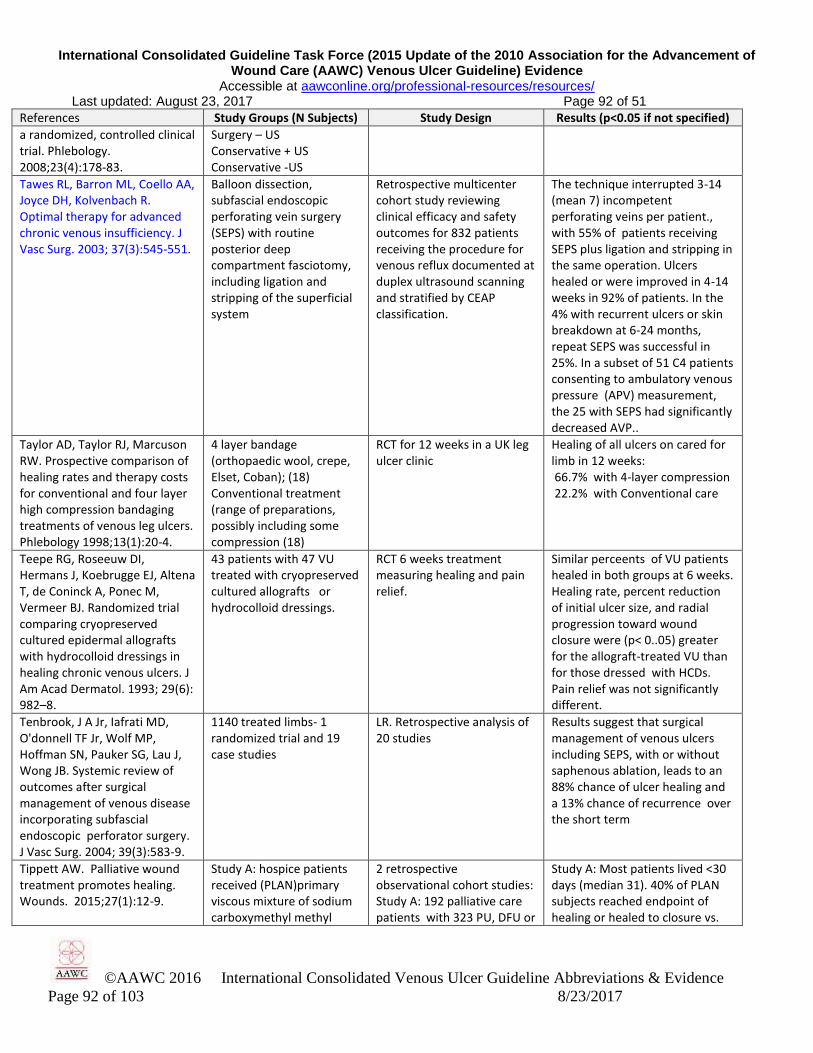
International Consolidated Guideline Task Force (2015 Update of the 2010 Association for the Advancement of Wound Care (AAWC) Venous Ulcer Guideline) Evidence
Accessible at aawconline.org/professional-resources/resources/ Last updated: August 23, 2017 Page 92 of 51
©AAWC 2016 International Consolidated Venous Ulcer Guideline Abbreviations & Evidence
Page 92 of 103 8/23/2017
References Study Groups (N Subjects) Study Design Results (p<0.05 if not specified)
a randomized, controlled clinical trial. Phlebology. 2008;23(4):178-83.
Surgery – US Conservative + US Conservative -US
Tawes RL, Barron ML, Coello AA, Joyce DH, Kolvenbach R. Optimal therapy for advanced chronic venous insufficiency. J Vasc Surg. 2003; 37(3):545-551.
Balloon dissection, subfascial endoscopic perforating vein surgery (SEPS) with routine posterior deep compartment fasciotomy, including ligation and stripping of the superficial system
Retrospective multicenter cohort study reviewing clinical efficacy and safety outcomes for 832 patients receiving the procedure for venous reflux documented at duplex ultrasound scanning and stratified by CEAP classification.
The technique interrupted 3-14 (mean 7) incompetent perforating veins per patient., with 55% of patients receiving SEPS plus ligation and stripping in the same operation. Ulcers healed or were improved in 4-14 weeks in 92% of patients. In the 4% with recurrent ulcers or skin breakdown at 6-24 months, repeat SEPS was successful in 25%. In a subset of 51 C4 patients consenting to ambulatory venous pressure (APV) measurement, the 25 with SEPS had significantly decreased AVP..
Taylor AD, Taylor RJ, Marcuson RW. Prospective comparison of healing rates and therapy costs for conventional and four layer high compression bandaging treatments of venous leg ulcers. Phlebology 1998;13(1):20-4.
4 layer bandage (orthopaedic wool, crepe, Elset, Coban); (18) Conventional treatment (range of preparations, possibly including some compression (18)
RCT for 12 weeks in a UK leg ulcer clinic
Healing of all ulcers on cared for limb in 12 weeks: 66.7% with 4-layer compression 22.2% with Conventional care
Teepe RG, Roseeuw DI, Hermans J, Koebrugge EJ, Altena T, de Coninck A, Ponec M, Vermeer BJ. Randomized trial comparing cryopreserved cultured epidermal allografts with hydrocolloid dressings in healing chronic venous ulcers. J Am Acad Dermatol. 1993; 29(6): 982–8.
43 patients with 47 VU treated with cryopreserved cultured allografts or hydrocolloid dressings.
RCT 6 weeks treatment measuring healing and pain relief.
Similar perceents of VU patients healed in both groups at 6 weeks. Healing rate, percent reduction of initial ulcer size, and radial progression toward wound closure were (p< 0..05) greater for the allograft-treated VU than for those dressed with HCDs. Pain relief was not significantly different.
Tenbrook, J A Jr, Iafrati MD, O'donnell TF Jr, Wolf MP, Hoffman SN, Pauker SG, Lau J, Wong JB. Systemic review of outcomes after surgical management of venous disease incorporating subfascial endoscopic perforator surgery. J Vasc Surg. 2004; 39(3):583-9.
1140 treated limbs- 1 randomized trial and 19 case studies
LR. Retrospective analysis of 20 studies
Results suggest that surgical management of venous ulcers including SEPS, with or without saphenous ablation, leads to an 88% chance of ulcer healing and a 13% chance of recurrence over the short term
Tippett AW. Palliative wound treatment promotes healing. Wounds. 2015;27(1):12-9.
Study A: hospice patients received (PLAN)primary viscous mixture of sodium carboxymethyl methyl
2 retrospective observational cohort studies: Study A: 192 palliative care patients with 323 PU, DFU or
Study A: Most patients lived <30 days (median 31). 40% of PLAN subjects reached endpoint of healing or healed to closure vs.
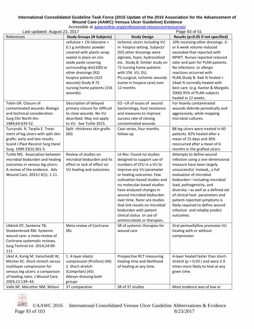
International Consolidated Guideline Task Force (2015 Update of the 2010 Association for the Advancement of Wound Care (AAWC) Venous Ulcer Guideline) Evidence
Accessible at aawconline.org/professional-resources/resources/ Last updated: August 23, 2017 Page 93 of 51
©AAWC 2016 International Consolidated Venous Ulcer Guideline Abbreviations & Evidence
Page 93 of 103 8/23/2017
References Study Groups (N Subjects) Study Design Results (p<0.05 if not specified)
cellulose + 1% lidocaine + 0.1 g antibiotic powder covered with plastic wrap sealed in place on zinc oxide paste covering surrounding skin(100) or other dressings (92) hospice patients (323 wounds) Study B 72 nursing home patients (156 wounds).
ischemic ulcers including VU in hospice setting. Subjects’ (92) other dressings were alginate, foam, hydrocolloid etc. Study B: Similar study on 72 nursing home patients with 156 VU, DU, PU,surgical, ischemic wounds (some in hospice care) over 12 months
10% receiving other dressings. 3- or 6 week volume reduced exceeded that reported with NPWT. Nurses reported reduced odor and pain for PLAN patients. No infections or allergic reactions occurred with PLAN.Study B: 4wk % healed = 24wk % normally healed with best care (e.g. Kantor & Margolis 2000) 95% of PLAN subjects healed in 12 weeks.
Tobin GR. Closure of contaminated wounds: Biologic and technical consideration. Surg Clin North Am. 1984;64:639-52.
Description of delayed primary closure for difficult to close wounds. No VU described. May not apply to VU . See Tuttle 2015.
EO –LR of issues of wound bacteriology, host resistance and measures to improve success rate of closing contaminated wounds
For heavily contaminated wounds debride periodically and aggressively, while mapping microbial cultures.
Turcynski. R, Tarpila E. Treat-ment of leg ulcers with split skin grafts: early and late results. Scand J Plast Reconst Surg Hand Surg. 1999:33(3):301-5.
Split –thickness skin grafts (60)
Case series, four months follow-up
88 leg ulcers were treated in 60 patients. 82% healed after a mean of 15 days and 36% reoccurred after a mean of 6 months in the grafted ulcers.
Tuttle MS. Association between microbial bioburden and healing outcomes in venous leg ulcers: A review of the evidence. Adv Wound Care. 2015;l 4(1): 1-11.
Review of studies on microbial bioburden and its effect or lack of effect on VU healing and outcomes.
Lit Rev. Found no studies designed to support use of numbers of CFU in a VU to improve any VU parameter or healing outcomes. Few cultivation-based studies and no molecular-based studies have analyzed changes in wound microbial bioburden over time. Rarer are studies that link results on microbial bioburden with patient clinical status or use of antimicrobials or therapies.
Attempts to define wound infection using a one-dimensional measure have been largely unsuccessful. Instead,, a full evaluation of microbial bioburden—including microbial load, pathogenicity, and diversity—as well as a defined set of clinical host parameters and patient-reported symptoms is likely required to define wound infection and reliably predict outcomes.
Ubbink DT, Santema TB, Stoekenbroek RM. Systemic wound care: a meta-review of Cochrane systematic reviews. Surg Technol Int. 2014;24:99-111.
Meta review of Cochrane SRs
SR of systemic therapies for wound care
Oral pentoxifylline promotes VU healing with or without compression
Ukat A, Konig M, Vanscheidt W, Münter KC. Short-stretch versus multilayer compression for venous leg ulcers: a comparison of healing rates. J Wound Care. 2003;12:139–43.
1. 4-layer elastic compression (Profore) (44) 2. Short-stretch (Comprilan) (45) Allevyn dressing both groups
Prospective RCT measuring healing time and likelihood of healing at any time.
4-layer healed faster than short-stretch (p = 0.03 ) and were 2.9 times more likely to heal at any given time.
Valle NF, Maruther NM, Wilson 37 comparative SR of 37 studies Most evidence was of low or

International Consolidated Guideline Task Force (2015 Update of the 2010 Association for the Advancement of Wound Care (AAWC) Venous Ulcer Guideline) Evidence
Accessible at aawconline.org/professional-resources/resources/ Last updated: August 23, 2017 Page 94 of 51
©AAWC 2016 International Consolidated Venous Ulcer Guideline Abbreviations & Evidence
Page 94 of 103 8/23/2017
References Study Groups (N Subjects) Study Design Results (p<0.05 if not specified)
LM, Malas M, Qazi U, Haberl E, Bass EB, Zenilman J, Lazarus G. Comparative effectiveness of advanced wound dressings for patients with chronic venous leg ulcers: a systematic review. Wound Rep Regen. 2014;22(2):193-204.
effectiveness studies insufficient quality. Cellular dressings, collagen, and some antimicrobial dressings may improve healing rates of chronic VU vs. compression alone or other dressings. Limited data were available on other outcomes.
Van Gent WB, Catarinella FS, Lam YL, Nieman FHM, Toonder IM, van der Ham AC, Wittens CHA. Conservative versus surgical treatment of venous leg ulcers: 10-year follow up of a randomized, multi-center trial. Phlebology. 2015;30(Suppl 1):35-41.
200 ulcerated legs, 170 pts. N=97 in the ambulatory compression therapy AND surgery group. N=103 in the ambulatory compression therapy group.
Large prospective, randomized, multi-center study comparing conservative versus surgical treatment of VU (SEPS procedure). The surgical group included treating refluxing superficial and perforating vessels.
Ulcer recurrence was 48.9% in the surgical group versus 94.3% in the conservative group. The number of refluxing perforators was a significant factor for ulcer recurrence. This study shows recurrent rate of 94% in patients with compression treatment only in 10 years
van Gent WB, Hop WC, van Praag MC, Mackaay AJ, de Boer EM, Wittens CH. Conservative versus surgical treatment of venous leg ulcers: a prospective, randomized, multicenter trial. J Vasc Surg. 2006;44(3):563–71.
Surgery : SEPS (97) Conservative ambulatory compression (103) All CEAP Score 6 (VU)
RCT measuring time to healing (NS diff), recurrence rates (NS diff) At 29 months, more SEPS ulcer free 72% than conservative 53% NS (p = 0.11)
Patients with recurrent ulceration or medially located ulcers in the surgical group had a longer ulcer-free period than those treated in the conservative group (P = .02 for both).
Van Gent WB, Wittens CHA. Influence of perforating vein surgery in patients with venous ulcerations. Phlebology. 2015;30(2):127-32.
Ninety-seven ulcerated legs (85 patients) were treated and three were lost-to-follow-up shortly after randomization of unknown cause. Analyses were performed on 94 ulcerated legs with a median follow-up of 27 (range 3–65) months.
RCT comparing surgical v. conservative treatment for venous ulcers.Measuring recurrence rate after a mean of 27 months and time to heal. The study emphasizes the importance of successful ligation of refluxing perforators in preventing ulcer recurrence.
When 1 or more perforators were missed during the SEPS procedure the recurrence rate was much higher. After mean 27 months follow up 16% recurrence was seen in the successful SEPS versus 50% in the non successful SEPS. Initial healing rates were similar between the 2 groups.
Van Hecke A, Grypdonck M, Beele H, De Bacquer D, Defloor T. How evidence-based is venous leg ulcer care? A survey in community settings. J Adv Nurs. 2009;65(2):337-47.
789 nurses who managed VU patients in community health care and private practice
Each nurse completed a survey on life-style advice. Self-perceived as more educated on EB care, gave more advice: leg elevation (68.3%), physical activity (39.8%) nutrition (16.7%)
Nurses who perceived selves to have adequate VU knowledge & skills were 3.75 times more likely to provide lifestyle advice than those lacking such knowledge and skills. They also found care more rewarding.
Van Hecke A, Grypdonck M, Beele H, Vanderwee K, Defloor T. Adherence to leg ulcer lifestyle advice: qualitative and quantitative outcomes associated with a nurse-led intervention. J Clin Nurs. 2011; 20(3-4):429-43.
26 VU patients in the community
Pre-post CO study of effects of nurses educating patients on compression wearing, leg exercise and elevation, activity level, pain and ulcer size. Wilcoxon signed rank test for differences
Leg elevation and leg exercise increased in frequency and duration. Compression wearing did not. Nurse education of patient should be incorporated into practice.

International Consolidated Guideline Task Force (2015 Update of the 2010 Association for the Advancement of Wound Care (AAWC) Venous Ulcer Guideline) Evidence
Accessible at aawconline.org/professional-resources/resources/ Last updated: August 23, 2017 Page 95 of 51
©AAWC 2016 International Consolidated Venous Ulcer Guideline Abbreviations & Evidence
Page 95 of 103 8/23/2017
References Study Groups (N Subjects) Study Design Results (p<0.05 if not specified)
Van Hecke A, Grypdonck M, Defloor T. Guidelines for the management of venous leg ulcers: a gap analysis. Eval Clin Pract. 2008;14(5):812-22.
LR and critical appraisal of 7 Evidence-based VU Guidelines
PubMED, Cinahl, Cochrane & Guideline websites were searched for VU Guidelines.
VU guidelines were evidence-based, but often failed to address patient wishes oruse multidisciplinary teams
Van Hecke A, Grypdonck M, DeFloor T. Interventions to enhance patient compliance with leg ulcer treatment: a review of the literature (2008) J Clin Nurs. 2008;17(1):29–39.
LR (20 studies) MEDLINE, Cochrane, Embase and CINAHL were explored up to April 2005
Most studies describe patient compliance as the extent to which the compression system was worn and/or the extent to which the prescribed treatment regimen was followed. Self-reporting was the most commonly used method of compliance assessment. Class III stockings for patients with a VU enhance compliance compared with short stretch compression bandages. There was no strong evidence that intermittent pneumatic compression or healthcare system interventions increased compliance. Programs combining cognitive, behavioural and affective educational components improved leg elevation, but not compliance with compression therapy.
van Rijswijk, L. The Multi-center Leg Ulcer Study Group. Full thickness leg ulcers: patient demographics and predictors of healing. J Family Practice 1993;36(6): 625-32.
DuoDERM CGF HCD DuoDERM HCD (total of 72 full-thickness leg ulcers)
Retrospective analysis of ConvaTec data on 72 full-thickness leg ulcers of venous, diabetic, arterial or mixed etiology
54% healed in average of 56 days. Risk factors for non-healing included male gender or diabetes. At least 30% area reduction after 2 weeks of treatment predicted that the ulcer would progress to healing
Vanscheidt W, Sibbald G, Eager C. Management of venous leg ulcers: Versiva: A new foam composite dressing, compared with a foam dressing. Ostomy Wound Manage. 2004;50(11):42-55.
With moderate to high compression bandaging for both groups compared: Versiva™ composite foam (55) Allevyn Adhesive (52)
Prospective RCT of dressing performance, patient-reported pain and healing during 12 weeks of care.
Composite foam was more conformable (p=0.05), less sensitizing (p=0.02) and easier to apply (p=0.01). Other variables showed no statistically significant differences.
Vanscheidt W, Ukat A, Horak V, Brüning H, Hunyadi J, Pavlicek R, Emter M, Hartmann A, Bende J, Zwingers T, Ermuth T, Eberhardt R. Treatment of recalcitrant venous leg ulcers with autologous keratinocytes in fibrin sealant: a multinational
Bioseed® (116) Autologous keratinocytes in fibrin sealant Standard treatment (109) All VU > 3 mo duration in both groups.
RCT: measuring time to complete healing in 12 weeks and % healed. Both groups received compression therapy.
Faster median healing time with autologous keratinocytes (176 d) vs > 201 d for Standard treatment (p< 0.0001) 38% healed in autologous keratinocyte group compared to 24% healed with SC.
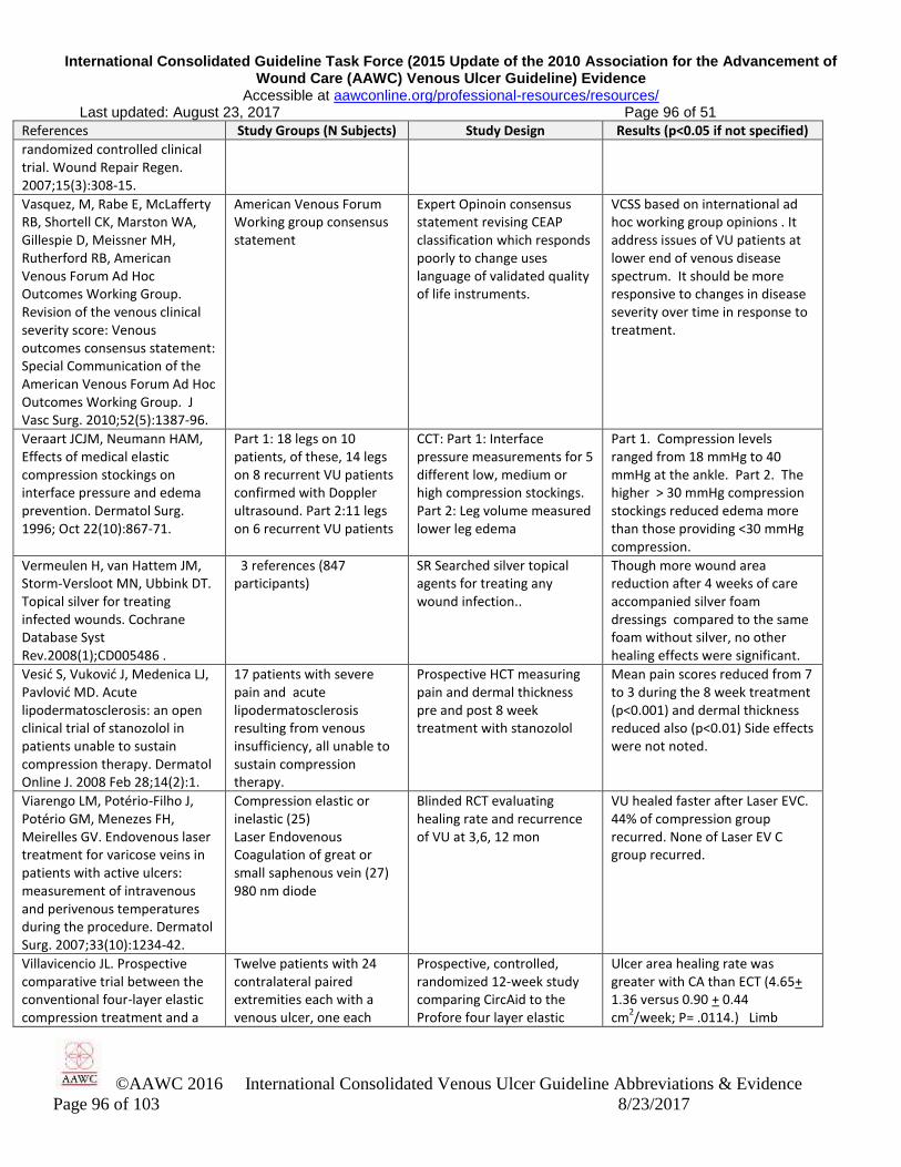
International Consolidated Guideline Task Force (2015 Update of the 2010 Association for the Advancement of Wound Care (AAWC) Venous Ulcer Guideline) Evidence
Accessible at aawconline.org/professional-resources/resources/ Last updated: August 23, 2017 Page 96 of 51
©AAWC 2016 International Consolidated Venous Ulcer Guideline Abbreviations & Evidence
Page 96 of 103 8/23/2017
References Study Groups (N Subjects) Study Design Results (p<0.05 if not specified)
randomized controlled clinical trial. Wound Repair Regen. 2007;15(3):308-15.
Vasquez, M, Rabe E, McLafferty RB, Shortell CK, Marston WA, Gillespie D, Meissner MH, Rutherford RB, American Venous Forum Ad Hoc Outcomes Working Group. Revision of the venous clinical severity score: Venous outcomes consensus statement: Special Communication of the American Venous Forum Ad Hoc Outcomes Working Group. J Vasc Surg. 2010;52(5):1387-96.
American Venous Forum Working group consensus statement
Expert Opinoin consensus statement revising CEAP classification which responds poorly to change uses language of validated quality of life instruments.
VCSS based on international ad hoc working group opinions . It address issues of VU patients at lower end of venous disease spectrum. It should be more responsive to changes in disease severity over time in response to treatment.
Veraart JCJM, Neumann HAM, Effects of medical elastic compression stockings on interface pressure and edema prevention. Dermatol Surg. 1996; Oct 22(10):867-71.
Part 1: 18 legs on 10 patients, of these, 14 legs on 8 recurrent VU patients confirmed with Doppler ultrasound. Part 2:11 legs on 6 recurrent VU patients
CCT: Part 1: Interface pressure measurements for 5 different low, medium or high compression stockings. Part 2: Leg volume measured lower leg edema
Part 1. Compression levels ranged from 18 mmHg to 40 mmHg at the ankle. Part 2. The higher > 30 mmHg compression stockings reduced edema more than those providing <30 mmHg compression.
Vermeulen H, van Hattem JM, Storm-Versloot MN, Ubbink DT. Topical silver for treating infected wounds. Cochrane Database Syst Rev.2008(1);CD005486 .
3 references (847 participants)
SR Searched silver topical agents for treating any wound infection..
Though more wound area reduction after 4 weeks of care accompanied silver foam dressings compared to the same foam without silver, no other healing effects were significant.
Vesić S, Vuković J, Medenica LJ, Pavlović MD. Acute lipodermatosclerosis: an open clinical trial of stanozolol in patients unable to sustain compression therapy. Dermatol Online J. 2008 Feb 28;14(2):1.
17 patients with severe pain and acute lipodermatosclerosis resulting from venous insufficiency, all unable to sustain compression therapy.
Prospective HCT measuring pain and dermal thickness pre and post 8 week treatment with stanozolol
Mean pain scores reduced from 7 to 3 during the 8 week treatment (p<0.001) and dermal thickness reduced also (p<0.01) Side effects were not noted.
Viarengo LM, Potério-Filho J, Potério GM, Menezes FH, Meirelles GV. Endovenous laser treatment for varicose veins in patients with active ulcers: measurement of intravenous and perivenous temperatures during the procedure. Dermatol Surg. 2007;33(10):1234-42.
Compression elastic or inelastic (25) Laser Endovenous Coagulation of great or small saphenous vein (27) 980 nm diode
Blinded RCT evaluating healing rate and recurrence of VU at 3,6, 12 mon
VU healed faster after Laser EVC. 44% of compression group recurred. None of Laser EV C group recurred.
Villavicencio JL. Prospective comparative trial between the conventional four-layer elastic compression treatment and a
Twelve patients with 24 contralateral paired extremities each with a venous ulcer, one each
Prospective, controlled, randomized 12-week study comparing CircAid to the Profore four layer elastic
Ulcer area healing rate was greater with CA than ECT (4.65+ 1.36 versus 0.90 + 0.44 cm
2/week; P= .0114.) Limb

International Consolidated Guideline Task Force (2015 Update of the 2010 Association for the Advancement of Wound Care (AAWC) Venous Ulcer Guideline) Evidence
Accessible at aawconline.org/professional-resources/resources/ Last updated: August 23, 2017 Page 97 of 51
©AAWC 2016 International Consolidated Venous Ulcer Guideline Abbreviations & Evidence
Page 97 of 103 8/23/2017
References Study Groups (N Subjects) Study Design Results (p<0.05 if not specified)
semi-rigid orthotic compression treatment and a semi-rigid orthotic compression system in patients with bilateral venous leg ulcers. American Venous Forum 21st Annual Symposium: Current Critical Problems in Vascular Surgery VI, 6.1. 1994
receiving 12 weeks treatment with: Circaid (12) (CA) Profore (12) (ECT)
treatment, measuring mean ulcer healing rate Limb circumference reduction rate, microbial burden and patient satisfaction index
circumference reduction rate was more significant with CA than with ECT (0.32+0.14 versus 0.10+ 0.14 cm/week: P=0.0385) No significant difference in patient satisfaction index or other measures of healing.
Vin F, Teot L, Meaume S. The healing properties of Promogran in venous leg ulcers. J. Wound Care. 2002;11(9):335-41.
73 Patients 37 Promogran 36 Adaptic
RCT clinical trial, multi-center study, randomized controlled trial 12 wk comparison of dressings under short- stretch compression. France
29 completed the 12 wk trial. 25 healed before 12 weeks. 19 stopped for reason unrelated to healing. Significantly more patients in Adaptic group switched to another dressing 22.2% versus 5.4%. No other differences in healing were significant.-31% (11) Healed w/ Adaptic. -41% (15) Healed w/ Promogran.-42% (15) Ulcers Improved with Adaptic -62% (23) Ulcers improved with Promogran
Voight J, Wendelken M, Driver V, Alvarez OM. Low frequency ultrasound (20-40kHz) as an adjunctive therapy for chronic wound healing: a systematic review of the literature and meta-analysis of eight RCT. Int J Low Extrem Wounds. 2011;10(4):190-9.
8 RCTs with likely bias SR of RCTs of any form of ultrasound used as adjunctive therapy measuring healing of any chronic wound.
Healing after less than 5 months of treatment in patients with a VU or diabetic foot ulcers was “favorably influenced by high- or low-intensity ultrasound delivered at low-frequency either via contact or noncontact US. Data quality may be suspect.
Volikova AI, Edwards J, Stacey MC, Wallace HJ. High-frequency ultrasound measurement for assessing post-thrombotic syndrome and monitoring compression therapy in chronic venous disease. J Vasc Surg. 2009;50(4):820-5.
20 VU patients with prior DVT not receiving compression; 20 patients with prior DVT and no VU + symptoms of post-thrombotic syndrome; 31 age-matched healthy control subjects. Association of dermal thickness with Clinical component of CEAP class examined in cross sectional analysis (n=157) of patients > 3 yr history of DVT
Prospective CCT to measure dermal thickness with 17 MHz Phillips iU22 ultrasound scanner or 20-MHz DermaScan-C medium-focus probe 7.5 cm above medial malleolus pre- and post 1,3,5 or 7 weeks of compression.
Dermal thickness (DT) of VU patients pre-compression was more than that of PTS patients without VU (p=0.002) and more for both these groups than for normal subjects (p = 0.001). Compression steadily decreased DT during first 5 weeks. DT increased with higher CEAP score > 1.985 mm DT had + predictive value of 46.9%, - predictive value of 90.3% to predict severe PTS defined as C(4b),C(5) or C(6) i.e. lipodermatoscclerosis or leg ulceration

International Consolidated Guideline Task Force (2015 Update of the 2010 Association for the Advancement of Wound Care (AAWC) Venous Ulcer Guideline) Evidence
Accessible at aawconline.org/professional-resources/resources/ Last updated: August 23, 2017 Page 98 of 51
©AAWC 2016 International Consolidated Venous Ulcer Guideline Abbreviations & Evidence
Page 98 of 103 8/23/2017
References Study Groups (N Subjects) Study Design Results (p<0.05 if not specified)
von Felbert V, Schumann H, Mercer JB, Strasser W, Daeschlein G, Hoffmann G. Therapy of chronic wounds with water-filtered infrared-A (wIRA). GMS Krankenhhyg Interdiszip. 2007;2(2) Doc 52:1-12.
Water-filtered infrared warming 30 min 3x/ week (20) or same schedule: Visible light control (20) both groups had the same cleansing, compression, antibacterial gauze dressing.
RCT for 6 weeks measuring days to heal, residual area at 42 days and pain medication required.
Infrared group healed in mean of 18 days, compared to 42 days for visible light. Infrared group had 0.4 cm
2 remaining mean area to
heal at 42 days vs 2.8 cm2 for
visible light group (p<0.001), who required more pain medication.
Vowden KR, Mason A, Wilkinson D, Vowden P. Comparison of the healing rates and complications of three four-layer bandage regimens. J Wound Care. 2000; 9(6): 269-72.
Charing Cross 4-layer bandage (n=50) Parema a4-layer bandage (n=50) Robinson 4-layer bandage (n=49)
Wound healing was measured at 12 and 20 weeks. Ulcers were mean of 4.9 to 6.76 cm
2 in area.
Overall healing rate of ulcers was 65% at 12 weeks, 80% at 20 weeks with no healing difference statistically significant among the 3 bandages.
Vu T, Harris A, Duncan G, Sussman G. Cost-effectiveness of multidisciplinary wound care in nursing homes: a pseudo-randomized pragmatic cluster trial. Family Practice. 2007; 24(4):372–9.
21 nursing homes with Multidisciplinary team care of 176 patients with VU or PU 23 nursing homes assigned randomly to control: usual care in Australia
Stratified randomized assignment of each nursing home. Healing and costs of care were measured 1999-2000.Cox regression with shared frailty predicted chances of healing
More wounds healed in team group (61.7%) than controls 52.5%, P = 0.07). Healing chance increased 73% for Team wounds (P = 0.003]. Mean treatment cost: $A616.4 for Team or $A977.9 for Control patients (P = 0.006).
Vuerstaek JD, Vainas T, Wuite J, Nelemans P, Neumann MH, Veraart JC. . State-of the-art treatment of chronic leg ulcers: a randomized controlled trial comparing vacuum assisted closure (VAC) with modern wound dressings. J Vasc Surg. 2006;44(5):1029-37.
13 VU NPWT to prepare bed until 100% granulation, then NPWT for 4 days continuous 125 mmHg VAC on pinch graft s 13 VU Std of Care with alginate- HCD to 100% granulation, then no trauma 4 d post graft to take: alginate or hydrogel
RCT measuring days to wound preparation for pinch grafting and days to healing
NPWT resulted in a 7-day wound bed preparation time (P _ .005) vs 17 days in the usual care control group. NPWT was also associated with faster time to heal and shorter hospital stay. (p<0.05) . NS difference in recurrence. First week NPWT lowered QoL .
Warriner RA 3rd, Wilcox JR, Carter MJ, Stewart DG. More frequent visits to wound care clinics result in faster times to close diabetic foot and venous leg ulcers. Adv Skin Wound Care. 2012;25(11):494-501.
206 DFU patients 215 VU patients
RCT study of visit frequency on healing outcomes
Visit frequency at least once/week healed 74.3% of VU in 4 weeks compared to 0% if visits were biweekly or less frequent.
Watson JM, Kang’ombe AR, Soares MO, Chuang L-H, Worthy G, Bland JM, Iglesias C, Cullum N, Torgerson D, Nelson EA. VenUS III: a randomized controlled trial of therapeutic ultrasound in the management of venous leg ulcers. Health Technol Assess. 2011; 15(13):1-192.
N=337 pts 1 group (n=168) received US + SC. 1 control group (n=169) received SC only.
Non-blind (open) RCT looking at the benefits of using therapeutic low frequency CONTACT ultrasound to the periulcer skin. The patients were followed for 12 weeks or until healing, whichever came first..
The study actually found that the US arm was performing worse than the control arm with fewer healed wounds, longer time to heal and more side effects (although it was not statistically significant). The conclusion was that US doesn’t aid in healing of venous ulcers.
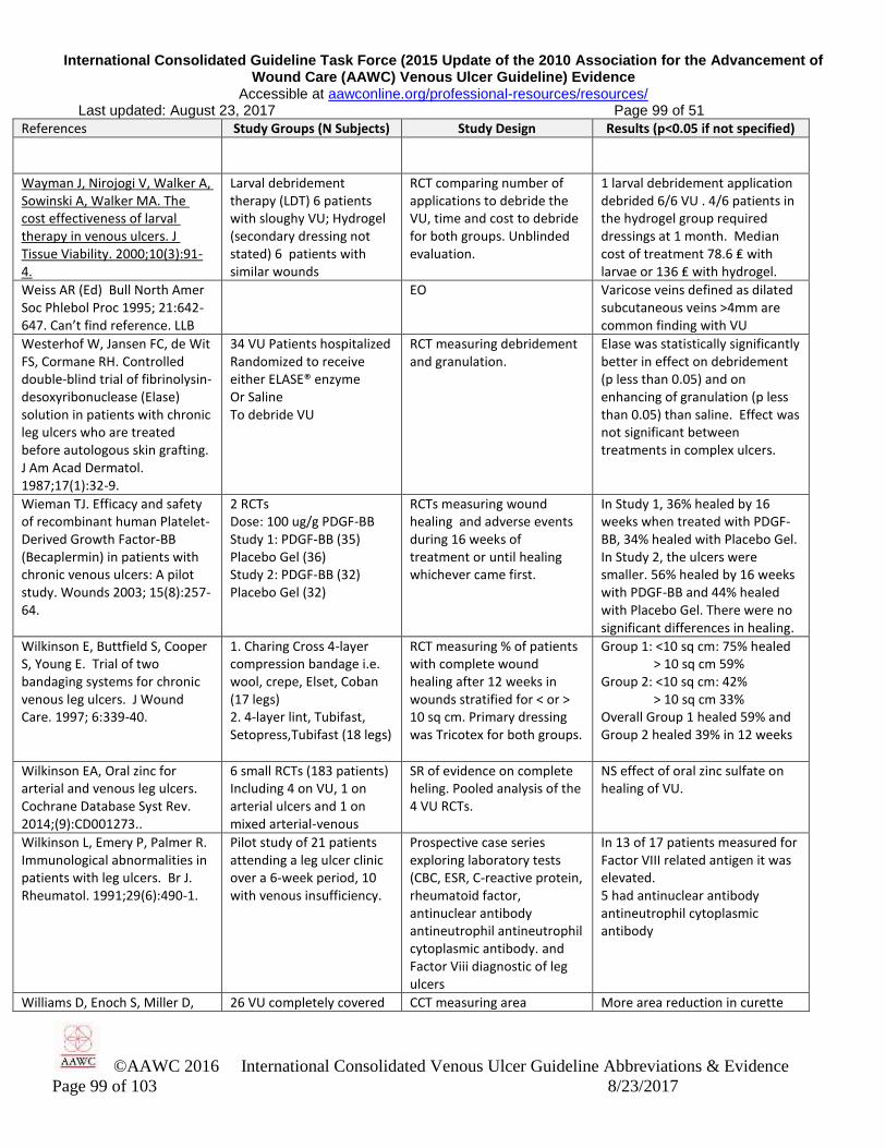
International Consolidated Guideline Task Force (2015 Update of the 2010 Association for the Advancement of Wound Care (AAWC) Venous Ulcer Guideline) Evidence
Accessible at aawconline.org/professional-resources/resources/ Last updated: August 23, 2017 Page 99 of 51
©AAWC 2016 International Consolidated Venous Ulcer Guideline Abbreviations & Evidence
Page 99 of 103 8/23/2017
References Study Groups (N Subjects) Study Design Results (p<0.05 if not specified)
Wayman J, Nirojogi V, Walker A, Sowinski A, Walker MA. The cost effectiveness of larval therapy in venous ulcers. J Tissue Viability. 2000;10(3):91-4.
Larval debridement therapy (LDT) 6 patients with sloughy VU; Hydrogel (secondary dressing not stated) 6 patients with similar wounds
RCT comparing number of applications to debride the VU, time and cost to debride for both groups. Unblinded evaluation.
1 larval debridement application debrided 6/6 VU . 4/6 patients in the hydrogel group required dressings at 1 month. Median cost of treatment 78.6 ₤ with larvae or 136 ₤ with hydrogel.
Weiss AR (Ed) Bull North Amer Soc Phlebol Proc 1995; 21:642-647. Can’t find reference. LLB
EO Varicose veins defined as dilated subcutaneous veins >4mm are common finding with VU
Westerhof W, Jansen FC, de Wit FS, Cormane RH. Controlled double-blind trial of fibrinolysin-desoxyribonuclease (Elase) solution in patients with chronic leg ulcers who are treated before autologous skin grafting. J Am Acad Dermatol. 1987;17(1):32-9.
34 VU Patients hospitalized Randomized to receive either ELASE® enzyme Or Saline To debride VU
RCT measuring debridement and granulation.
Elase was statistically significantly better in effect on debridement (p less than 0.05) and on enhancing of granulation (p less than 0.05) than saline. Effect was not significant between treatments in complex ulcers.
Wieman TJ. Efficacy and safety of recombinant human Platelet-Derived Growth Factor-BB (Becaplermin) in patients with chronic venous ulcers: A pilot study. Wounds 2003; 15(8):257-64.
2 RCTs Dose: 100 ug/g PDGF-BB Study 1: PDGF-BB (35) Placebo Gel (36) Study 2: PDGF-BB (32) Placebo Gel (32)
RCTs measuring wound healing and adverse events during 16 weeks of treatment or until healing whichever came first.
In Study 1, 36% healed by 16 weeks when treated with PDGF-BB, 34% healed with Placebo Gel. In Study 2, the ulcers were smaller. 56% healed by 16 weeks with PDGF-BB and 44% healed with Placebo Gel. There were no significant differences in healing.
Wilkinson E, Buttfield S, Cooper S, Young E. Trial of two bandaging systems for chronic venous leg ulcers. J Wound Care. 1997; 6:339-40.
1. Charing Cross 4-layer compression bandage i.e. wool, crepe, Elset, Coban (17 legs) 2. 4-layer lint, Tubifast, Setopress,Tubifast (18 legs)
RCT measuring % of patients with complete wound healing after 12 weeks in wounds stratified for < or > 10 sq cm. Primary dressing was Tricotex for both groups.
Group 1: <10 sq cm: 75% healed > 10 sq cm 59% Group 2: <10 sq cm: 42% > 10 sq cm 33% Overall Group 1 healed 59% and Group 2 healed 39% in 12 weeks
Wilkinson EA, Oral zinc for arterial and venous leg ulcers. Cochrane Database Syst Rev. 2014;(9):CD001273..
6 small RCTs (183 patients) Including 4 on VU, 1 on arterial ulcers and 1 on mixed arterial-venous
SR of evidence on complete heling. Pooled analysis of the 4 VU RCTs.
NS effect of oral zinc sulfate on healing of VU.
Wilkinson L, Emery P, Palmer R. Immunological abnormalities in patients with leg ulcers. Br J. Rheumatol. 1991;29(6):490-1.
Pilot study of 21 patients attending a leg ulcer clinic over a 6-week period, 10 with venous insufficiency.
Prospective case series exploring laboratory tests (CBC, ESR, C-reactive protein, rheumatoid factor, antinuclear antibody antineutrophil antineutrophil cytoplasmic antibody. and Factor Viii diagnostic of leg ulcers
In 13 of 17 patients measured for Factor VIII related antigen it was elevated. 5 had antinuclear antibody antineutrophil cytoplasmic antibody
Williams D, Enoch S, Miller D, 26 VU completely covered CCT measuring area More area reduction in curette

International Consolidated Guideline Task Force (2015 Update of the 2010 Association for the Advancement of Wound Care (AAWC) Venous Ulcer Guideline) Evidence
Accessible at aawconline.org/professional-resources/resources/ Last updated: August 23, 2017 Page 100 of 51
©AAWC 2016 International Consolidated Venous Ulcer Guideline Abbreviations & Evidence
Page 100 of 103 8/23/2017
References Study Groups (N Subjects) Study Design Results (p<0.05 if not specified)
Harris K, Price P, Harding KG. Effect of sharp debridement using curette on recalcitrant nonhealing venous leg ulcers: a concurrently controlled, prospective cohort study. Wound Repair Regen. 2005;13(2):131-7.
with slough or necrosis were debrided 27 VU with 15-20% granulation tissue but no slough or fibrin were not debrided
reduction 4 weeks post debridement and % healed weeks 8 & 20 post debridement. Measured area reduction, not% area reduction.
debrided VU at 4 weeks (p=0.02) and 20 weeks (p= 0.008). NS difference in % of VU healed at 20 weeks (5 in each group).
Wilson CL, Cameron J, Powell SM, Cherry G, Ryan TJ. High incidence of contact dermatitis in leg-ulcer patients--implications for management. Clin Exp Dermatol. 1991;16(4):250-3.
81 VU patients; retrospective review of patch test results performed on all new VU patients in preceding 11 months
Cohort study to explore incidence of contact dermatitis in VU patients.
67% positive for contact allergy inclusive of lanolin, topical antibiotics &/or cetearyl alcohol. Multiple allergies in 58%.
Wilson JM, Arseculeratne YM, Yang Y, Cherry GW. Healing venous ulcers with cycloidal multidirectional vibration therapy. J Wound Care. 2002; 11(10):395-8.
21 VU patients ABI >0/8 Setopress + Vibro-Pulse gentle cyclic vibration 3 x /d for 30 min each. 2x/ week NA gauze dressing changes.
Prospective CS for 12 weeks during which healing and pain were measured weekly
13 (62%) healed completely in mean of 7 weeks. Pain reduced in 17 of 18 patients completing the study, accompanied by mean 15% reduction in leg volume.
Wilson, P.D., Burroughs, D., Dunn, L.J. Methicillin Resistant Staphylococcus Aureus and hydrocolloid dressings. The Pharmaceutical Journal. December 17, 1988;243(6513):787-8.
DuoDERM (6) patients with venous or other leg ulcers populated with MRSA Conventional gauze and isolation (historic control)
Prospective open-label exploration of dispersion of Methicillin-resistant S. aureus (MRSA) from leg ulcers in a UK hospital setting.
Within 2 weeks of beginning DuoDERM dressings, 5 of the 6 patients were free of MRSA which they all had at study initiation. The dressing effectively isolated the wounds, preventing MRSA transmission.
Wipke-Tevis, D. et al. Prevalence, incidence, management and predictors of venous ulcers in the long-term care population. Adv Skin Wound Care. 2000;13(5):218-24.
venous ulcer development in 32,221 patients in long term care in Missouri 1 Jan 96 to 30 Oct 98
Retrospective cohort study from Minimum Data Set
Venous ulcer development during first year post admission was associated with lower extremity edema, peripheral vascular disease or diabetes mellitus.
Wong IKY, Andriessen A, Charles HE, Thompson D, Lee DT, So WKW, Abel M. Randomized controlled trial comparing treatment outcome of two compression bandaging systems and standard care without compression in patients with venous leg ulcers. J Eur Acad Derm Venerol. 2012;26(1):102-10.
321 elderly community care patients with venous ulcers in a community setting comparing short stretch bandaging (SSB), 4 layer bandaging and usual care with no compression
RCT measuring QOL, ulcer related pain and functional status
Compression bandaging in both groups significantly reduced pain (P < 0.0001) and improved functional status and QOL. Healing rate at 24 weeks for both compression groups was significant (P < 0.001); for SSB this was 72.0% (77 ⁄ 107) vs.67.3% in the 4-layer bandage group (72 ⁄ 107) and 29.0% (31 ⁄ 107)with usual care. The reduction in ulcer area from weeks 12 to 24 was significant only for SSB (P < 0.047). using
International Consolidated Guideline Task Force (2015 Update of the 2010 Association for the Advancement of Wound Care (AAWC) Venous Ulcer Guideline) Evidence
Accessible at aawconline.org/professional-resources/resources/ Last updated: August 23, 2017 Page 101 of 51
©AAWC 2016 International Consolidated Venous Ulcer Guideline Abbreviations & Evidence
Page 101 of 103 8/23/2017
References Study Groups (N Subjects) Study Design Results (p<0.05 if not specified)
compression with SSB or 4LB is more effective than usual care without compression. For healing time, faster reduction of ulcer area, reduction of pain SSB improved patients’ QOL faster.
Woo KY, Sibbald RG. A cross-sectional validation study of using NERDS and STONEES to assess bacterial burden. Ostomy Wound Manage. 2009;55(8):40-8.
112 patients with chronic wounds: included 44 with leg ulcers
Cohort study investigating clinical signs of critical colonization (NERDS) or infection (STONEES) compared to semiquantitative swab cultures.
Wounds with high temperature were 8 times more likely to have moderate or heavy bacterial growth. When combining any three clinical signs, sensitivity was 73.3% for scant or light and 90% for moderate and heavy bacterial growth. specificity was 80.5% and 69.4% for same
World Union of Wound Healing Societies (WUWHS). Principles of best practice: Wound exudate and the role of dressings. A consensus document. London: Medical Education Partnership, Ltd. 2007.
Best Practice Principles Consensus statement of wound care opinion leaders from different societies (EO)
Wound exudate and pain management can be managed with dressings..
World Union of Wound Healing Societies (WUWHS). Principles of best practice: Diagnostics and wounds A consensus document. London: Medical Education Partnership, Ltd., 2008.
Best Practice Principles Consensus statement of wound care opinion leaders from different societies (EO)
Wound exudate management
World Union of Wound Healing Societies. Principles of best practice. Minimising pain at wound dressing-related procedures. A consensus document. London: Medical Education Partnership, Ltd. 2004.
Best Practice Principles Consensus statement of wound care opinion leaders from different societies (EO)
Wound diagnosis and treatment
Wound Ostomy Continence Nurses Society. Clinical Practice Guideline #4. Management of wounds in patients with lower-extremity venous disease, 2005. http://www.guideline.gov Accessed Nov 10, 2010.
Venous ulcer recommendations identified.
Guideline of the WOCN. All unique recommendations are addressed in ICVUG.
Wright DD, Franks PJ, Blair SD, Backhouse CM, Moffatt C, McCollum CN. Oxerutins in the prevention of recurrence in chronic venous ulceration:
Oxerutin+ Compression Compression + dressing control
RCT for prevention of VU recurrence
NS difference from control
International Consolidated Guideline Task Force (2015 Update of the 2010 Association for the Advancement of Wound Care (AAWC) Venous Ulcer Guideline) Evidence
Accessible at aawconline.org/professional-resources/resources/ Last updated: August 23, 2017 Page 102 of 51
©AAWC 2016 International Consolidated Venous Ulcer Guideline Abbreviations & Evidence
Page 102 of 103 8/23/2017
References Study Groups (N Subjects) Study Design Results (p<0.05 if not specified)
randomized controlled trial. Br J Surg. 1991; 78(10): 1269-70.
Wright DD. The ESCHAR trial: should it change practice? Perspect Vasc Surg Endovasc Ther. 2009;21(2):69-72.
~250 VU patients SEPS+compression ~250 VU patients compression alone
RCT measuring healing at 24 weeks only (no earlier time) and VU recurrence rate
No significant difference in % healed at 24 weeks, significantly less recurrence in SEPS group regardless of deep vein incompetence.
Wunderlich U, Orfanos CE. Treatment of venous ulcera cruris with dry wound dressings. Phase overlapping use of silver impregnated activated charcoal xero-dressing. Hautarzt. 1991;42(7): 446–50.
Contreet Foam with silver sulfadiazine (19) Conventional therapy (19) All subjects had VU that was debrided.
6-week RCT measuring complete healing and mean reduction in % wound area from baseline. Study was underpowered to show significant differences.
6-Week % healed: silver sulfadiazine foam dressing 32% healed or 11% (NS) healed in conventionally dressed VU. Mean % area reduction: 75% for silver sulfadiazine foam dressing or 60%, conventional therapy (NS)
Xia ZD, Hu D, Wilson JM, Cherry GW, Ryan TJ. How echographic image analysis of venous oedema reveals the benefits of leg elevation. J Wound Care. 2004;13(4):125-8.
10 patients with venous insufficiency and leg edema ages 44-89 years
HCT using high frequency B-mode US scanning and digital image analysis pre and post 3-4 hours of leg elevation to reduce edema. Compared low echogenic pixels pre and post elevation.
Compared with pre-elevation, of lower leg volume decreased by 2.9% (138 cm3 ) after 3 to 4 hours elevation (p < 0.05). After elevation low-echogenic pixels in upper, middle and lower sites of the limb decreased by 8.8%, 15.6% and 17.3% respectively (p < 0.05)
Yager DR, Zhang LY, Liang HX , Diegelmann RF, Cohen IK. Wound fluids from human pressure ulcers contain elevated matrix metalloproteinase levels and activity compared to surgical wound fluids. J Invest Dermatol. 1996;107(5):743-8
Surgical patients undergoing mastectomy or reduction mammoplasty or abdominplasty or inpatients or outpatients with a pressure ulcer were had fluid taken from the wound.
Laboratory analysis of wound fluid from surgical wounds and pressure ulcers Samples were incubated overnight with appropriate antibodies. Matrix metalloproteinase concentrations were determined by zymography.
Levels of matrix etalloproteinase-2 and matrix metalloproteinase-9 were elevated more than 10-fold and 25-fold, respectively, in fluids from pressure ulcer compared with fluids from healing wounds.
Yamada T, Ohta T, Ishibashi H, Sugimoto I, Iwata H, Takahashi M, Kawanishi J. Clinical reliability and utility of skin perfusion pressure measurement in ischemic limbs--comparison with other noninvasive diagnostic methods. J Vasc Surg. 2008;47(2):318-23.
211 subjects with ischemic limbs (arteriosclerosis obliterans) ~ half with diabetes and/or on dialysis.
Prospective cohort study Skin perfusion pressure (SPP) correlations measured with healing, toe blood pressure (TBP), ankle blood pressure (ABP) and great toe Transcutaneous oxygen pressure (tcPO2)
Sensitivity (72%) and specificity (88%) were greatest for SPP cut-off 40 mmHg and TBP > 30 mmHg as the two best predictors of healing and strongly correlated with each other. (p<0.001) for all pairs.
Yang D, Vanongen YK, Stacey MC. Effect of exercise on calf muscle pump function in patients with chronic venous disease. Br J Surg. 1999; 86(3):338-44.
20 patients with chronic venous insufficiency
Pre-post CS exploring effect of vigorous exercise on calf muscle pump function: residual fraction, ejection fraction, venous reflux
Exercise program increased ejection fraction , decreased residual fraction (p< 0.05) and improved calf muscle strength and endurance increasedwith exercise, though NS for the latter.
Yasodhara M, Walton J, Hofman D, Cherry G. A comparison of
42 patients with venous insufficiency who had light
HCT LRR was used to measure venous refilling
All 42 patients had shortened venous refilling time of less than

International Consolidated Guideline Task Force (2015 Update of the 2010 Association for the Advancement of Wound Care (AAWC) Venous Ulcer Guideline) Evidence
Accessible at aawconline.org/professional-resources/resources/ Last updated: August 23, 2017 Page 103 of 51
©AAWC 2016 International Consolidated Venous Ulcer Guideline Abbreviations & Evidence
Page 103 of 103 8/23/2017
References Study Groups (N Subjects) Study Design Results (p<0.05 if not specified)
light reflection rheography and duplex scanning in the diagnosis of chronic venous insufficiency. Wounds 2003; 15(8):246-9.
reflection rheography and were subsequently given duplex scans
time. Duplex scans were used to measure venous reflux subsequently on the same 42 patients in a historically controlled trial
25 seconds as measured using light reflection rheography. Of these, 41 had abnormal duplex scan confirming venous reflux, and 1 had a normal duplex scan: 2.3% false negatives with DS.
Yim E, Kirsner RS, Gailey RS, Mandel DW, Chen SC, Tomic-Canic M. Effect of physical therapy on wound healing and quality of life in patients with venous leg ulcers: a systematic review. JAMA Dermatol. 2015;151(3):320-7.
10 articles (RCTs or single-arm exploring effect of exercise on healing or QoL in patients with a VU
SR of studie in which those with a VU were engaged in physical therapy or an exercise program.Measures were calf muscle pump function, range of motion and likelihood of healing after 12 weeks.
Standardized exercise or increased walking was associated with greater likelihood of healing at 12 week.s More xercise strengthens the calf muscle pump and improves ankle range of motion. Few studies have investigated effect of these interventions on QoL and healing, and few involved the supervision of a physical therapist.
Yim E, Richmond NA, Baquerizo K, Van Driessche F, Slade HB, Pieper B, Kirsner RS. The effect of ankle range of motion on venous ulcer healing rates. Wound Repair Regen. 2014 ;22(4):492-6.
49 healthy controls no VU 227 VU patients in allogeneic biologic living cell formula: 3 doses and a control group all receiving compression
Secondary data analysis from a RCT. Goniometry monitored walking all groups. VU location, ankle and plantar range of motion were analyzed as predictors of VU healing
Ankel location predicted lower % healed at 12 weeks compared to calf location of the VU. Plantar flexion and eversion increased for patients with calf vs ankle VU, RCTs are needed to explore effects on healing.
Zamboni P, Cisno C, Marchetti F, Mazza P, Fogato L, Carandina S, De Palma M, Liboni A. Minimally invasive surgical management of primary venous ulcers vs. compression treatment: a randomized clinical trial. Eur J Vasc Endovasc Surg. 2003 ;25(4):313-8.
(47 VU patients ) randomy assigned to minimally invasive surgical haemodynamic correction of reflux ( surgery) or Compression
RCT measuring healing time and quality of life at 3 years
Mean healing time (p<0.05) 31 days for surgery group 63 days in compression group. 100% of surgery group healed at 3 months compared to 96% healed with compression (NS). QoL significantly improved for surgical group.