Leucokinin and the modulation of the shunt pathway in ...directory.umm.ac.id/Data...
14
Journal of Insect Physiology 47 (2001) 263–276 www.elsevier.com/locate/jinsphys Leucokinin and the modulation of the shunt pathway in Malpighian tubules Ming-Jiun Yu, Klaus W. Beyenbach * Department of Biomedical Sciences, VRT 8014, Cornell University, Ithaca, NY 14853, USA Received 1 March 2000; accepted 20 April 2000 Abstract Transepithelial secretion in Malpighian tubules of the yellow fever mosquito (Aedes aegypti) is mediated by active transport of Na + and K + through principal cells and passive Cl 2 transport through the shunt. Permeation through the shunt was assessed by measuring transepithelial halide diffusion potentials in isolated perfused Malpighian tubules, after first inhibiting active transport with dinitrophenol. Diffusion potentials were small under control conditions, revealing Eisenman selectivity sequence I (I 2 .Br 2 .Cl 2 .F 2 ) which is the halide mobility sequence in free solution. Accordingly, electrical field strengths of the shunt are small, selecting halides for passage on the basis of hydrated size. Leucokinin-VIII (LK-VIII) significantly increased the shunt conductance from 57.1 μS/cm to 250.0 μS/cm. In parallel, the shunt selectivity sequence shifted to Eisenman sequence III (Br 2 .Cl 2 .I 2 .F 2 ), revealing increased electrical field strengths in the shunt, now capable of selecting small, dehydrated halides for passage. High concentrations of peritubular F 2 (142.5 mM) duplicated the effects of LK-VIII on shunt conductance and selec- tivity, suggesting a role for G-protein. In the presence of LK-VIII (or F 2 ), coulombic interactions between the shunt and I 2 and F 2 may be strong enough to cause binding, thereby blocking the passage of Cl 2 . Thus, LK-VIII increases both shunt conductance and selectivity, presumably via G-protein. 2001 Elsevier Science Ltd. All rights reserved. Keywords: Malpighian tubules; Transepithelial diffusion potentials; Transepithelial resistance; Shunt halide selectivity sequence 1. Introduction A fine example of the contributions of insect biochem- istry to insect physiology is the isolation and synthesis of leucokinin by Holman and his colleagues. Pursuing the isolation of myotropic agents in the cockroach Leuc- ophaea, Holman came across a family of small peptides that stimulated hindgut motility in the same species (Holman et al., 1987). Since then, leucokinin and related peptides have been found in many insects: mosquitoes (Clottens et al., 1993; Hayes et al., 1994; Veenstra, 1994), fruitflies (Terhzaz et al., 1999), houseflies (Holman et al., 1999), moths (Kim and Lee, 1998), blowflies (Na ¨ssel and Lundquist, 1991), locusts (Schoofs et al., 1992), hornworms (Cho et al., 1999), spiders (Schmid and Becherer, 1996) and crickets (Coast et al., * Corresponding author. Tel.: + 1-607-253-3482; fax: + 1-607-253- 3581. E-mail address: [email protected] (K.W. Beyenbach). 0022-1910/01/$ - see front matter 2001 Elsevier Science Ltd. All rights reserved. PII:S0022-1910(00)00084-6 1990), and also in other invertebrates such as mollusc (Cox et al., 1997; Elekes et al., 1995). In view of the association of leucokinins with peptidergic neurons, they are considered neuropeptides (Na ¨ssel et al., 1992). Holman had used contractions of the cockroach hindgut as bioassay for the isolation of the leucokinins (Holman et al., 1987). The stimulation of hindgut motility suggested to us that leucokinin might affect transport in other epithelia such as Malpighian tubules. Indeed, natural and synthetic leucokinins of the cock- roach significantly increased rates of fluid secretion in isolated Malpighian tubules of the yellow fever mosquito by increasing transepithelial secretion of both NaCl and KCl (Pannabecker et al., 1993). The increase in the secretion rate of different salts sharing the same anion suggested the effect of leucokinin on Cl 2 , the counter ion of active transepithelial secretion of Na + and K + . Electrophysiological studies in isolated perfused Mal- pighian tubules confirmed this hypothesis. Leucokinin lowered the lumen-positive transepithelial voltage from 59.3 mV to 5.7 mV and decreased the transepithelial
Transcript of Leucokinin and the modulation of the shunt pathway in ...directory.umm.ac.id/Data...

Journal of Insect Physiology 47 (2001) 263–276www.elsevier.com/locate/jinsphys
Leucokinin and the modulation of the shunt pathway inMalpighian tubules
Ming-Jiun Yu, Klaus W. Beyenbach*
Department of Biomedical Sciences, VRT 8014, Cornell University, Ithaca, NY 14853, USA
Received 1 March 2000; accepted 20 April 2000
Abstract
Transepithelial secretion in Malpighian tubules of the yellow fever mosquito (Aedes aegypti) is mediated by active transport ofNa+ and K+ through principal cells and passive Cl2 transport through the shunt. Permeation through the shunt was assessed bymeasuring transepithelial halide diffusion potentials in isolated perfused Malpighian tubules, after first inhibiting active transportwith dinitrophenol. Diffusion potentials were small under control conditions, revealing Eisenman selectivity sequence I(I2.Br2.Cl2.F2) which is the halide mobility sequence in free solution. Accordingly, electrical field strengths of the shunt aresmall, selecting halides for passage on the basis of hydrated size. Leucokinin-VIII (LK-VIII) significantly increased the shuntconductance from 57.1µS/cm to 250.0µS/cm. In parallel, the shunt selectivity sequence shifted to Eisenman sequence III(Br2.Cl2.I2.F2), revealing increased electrical field strengths in the shunt, now capable of selecting small, dehydrated halidesfor passage. High concentrations of peritubular F2 (142.5 mM) duplicated the effects of LK-VIII on shunt conductance and selec-tivity, suggesting a role for G-protein. In the presence of LK-VIII (or F2), coulombic interactions between the shunt and I2 andF2 may be strong enough to cause binding, thereby blocking the passage of Cl2. Thus, LK-VIII increases both shunt conductanceand selectivity, presumably via G-protein. 2001 Elsevier Science Ltd. All rights reserved.
Keywords:Malpighian tubules; Transepithelial diffusion potentials; Transepithelial resistance; Shunt halide selectivity sequence
1. Introduction
A fine example of the contributions of insect biochem-istry to insect physiology is the isolation and synthesisof leucokinin by Holman and his colleagues. Pursuingthe isolation of myotropic agents in the cockroachLeuc-ophaea, Holman came across a family of small peptidesthat stimulated hindgut motility in the same species(Holman et al., 1987). Since then, leucokinin and relatedpeptides have been found in many insects: mosquitoes(Clottens et al., 1993; Hayes et al., 1994; Veenstra,1994), fruitflies (Terhzaz et al., 1999), houseflies(Holman et al., 1999), moths (Kim and Lee, 1998),blowflies (Nassel and Lundquist, 1991), locusts (Schoofset al., 1992), hornworms (Cho et al., 1999), spiders(Schmid and Becherer, 1996) and crickets (Coast et al.,
* Corresponding author. Tel.:+1-607-253-3482; fax:+1-607-253-3581.
E-mail address:[email protected] (K.W. Beyenbach).
0022-1910/01/$ - see front matter 2001 Elsevier Science Ltd. All rights reserved.PII: S0022-1910 (00)00084-6
1990), and also in other invertebrates such as mollusc(Cox et al., 1997; Elekes et al., 1995). In view of theassociation of leucokinins with peptidergic neurons, theyare considered neuropeptides (Na¨ssel et al., 1992).
Holman had used contractions of the cockroachhindgut as bioassay for the isolation of the leucokinins(Holman et al., 1987). The stimulation of hindgutmotility suggested to us that leucokinin might affecttransport in other epithelia such as Malpighian tubules.Indeed, natural and synthetic leucokinins of the cock-roach significantly increased rates of fluid secretion inisolated Malpighian tubules of the yellow fever mosquitoby increasing transepithelial secretion of both NaCl andKCl (Pannabecker et al., 1993). The increase in thesecretion rate of different salts sharing the same anionsuggested the effect of leucokinin on Cl2, the counterion of active transepithelial secretion of Na+ and K+.Electrophysiological studies in isolated perfused Mal-pighian tubules confirmed this hypothesis. Leucokininlowered the lumen-positive transepithelial voltage from59.3 mV to 5.7 mV and decreased the transepithelial

264 M.-J. Yu, K.W. Beyenbach / Journal of Insect Physiology 47 (2001) 263–276
resistance from 57.8Vcm2 to 9.9 Vcm2 (Hayes et al.,1989; Pannabecker et al., 1993). Thus, leucokininchanges a moderately tight epithelium to a leaky epi-thelium, consistent with the increase in the transepi-thelial secretion of electrolytes and water. What aston-ishes is the switch-like speed of the on and off effects ofleucokinin (Beyenbach, 1995; Pannabecker et al., 1993).
An additional measure of Holman’s contribution toinsect physiology is the present controversy over the siteof action of leucokinin in Malpighian tubules. The lab-oratories of O’Donnell and Dow have strong evidencefor stellate cells providing the transepithelial shunt path-way for Cl2 secretion (Dow et al., 1998; O’Donnell etal. 1996, 1998; Terhzaz et al., 1999). Likewise, Dijkstraet al. (1995) consider Cl2 to take a transcellular pathwayfor secretion in Malpighian tubules of ants. In contrast,our laboratory has evidence for Cl2 permeating the para-cellular pathway through septate junctions (Pannabeckeret al., 1993). First, the leaky epithelium induced by leu-cokinin displays large transepithelial Cl2 diffusionpotentials that approach 85% of transepithelial Nernstpotentials. The symmetry of these diffusion potentialsfor bath-to-lumen and lumen-to-bath directed Cl2 gradi-ents suggests a single barrier, such as that expected froma junction. A transcellular pathway consisting of twomembrane barriers in series is unlikely to display sym-metrical transepithelial Cl2 diffusion potentials as itwould require similar Cl2 conductances in both basolat-eral and apical membranes. Second, the effects of leuco-kinin on transepithelial resistance can be fully restoredby removing Cl2 from the solutions on both sides of theepithelium (Pannabecker et al., 1993). Third, the effectsof leucokinin on apical and basolateral membrane volt-ages of principal cells leave but one explanation of thedata, that leucokinin increases the Cl2 conductance ofthe paracellular, septate junctional pathway.
The effects of leucokinin differ dramatically fromthose of db-cAMP which serves as second messenger ofthe mosquito natriuretic peptide (MNP). Whereas leuco-kinin causes a non-selective diuresis by increasing trans-epithelial secretion of both NaCl and KCl via theincrease in passive transepithelial Cl2 transport (Hayeset al., 1989; Pannabecker et al., 1993; Yu and Beyen-bach, 2000), db-cAMP and MNP cause a specific NaCldiuresis, in part by increasing the Na+ conductance ofthe basolateral membrane of principal cells (Beyenbach,1995; Beyenbach and Petzel, 1987; Clark et al., 1998).O’Donnell et al. (1996) confirm the separate control oftransepithelial cation and anion secretion in Malpighiantubules ofDrosophila, illustrating the functional diver-sity of Malpighian tubules.
In an attempt to resolve the present controversy overtranscellular or paracelluar pathways for Cl2, we beganin the present study a detailed investigation of the natureof the transepithelial Cl2 conductance induced by leuco-kinin-VIII. Though we have no final answer, we report
here that leucokinin-VIII increases transepithelial shuntconductance by opening an anionic pathway with the sel-ectivity sequence Br2.Cl2.I2.F2. This sequenceindicates a transepithelial pathway of high electrical fieldstrength that selects small anions with high charge den-sities, thereby optimizing the function of the shunt as aCl2 filter.
2. Materials and methods
2.1. Mosquitoes, Malpighian tubules and thecomposition of Ringer solution
The mosquito colony was maintained as described byPannabecker et al. (1993). On the day of the experimenta female mosquito (3 to 7 days post-eclosion) was coldanesthetized and decapitated. Malpighian tubules wereremoved from the abdominal cavity under Ringer sol-ution. Only segments near the blind end of the tubule,between 0.2 and 0.3 mm long, were used. Ringer sol-ution contained the following, in mM: 150 NaCl, 25HEPES (N-2-hydroxyethylpiperazine-N9-2-ethanesul-fonic acid), 3.4 KCl, 7.5 NaOH, 1.8 NaHCO3, 1 MgSO4,1.7 CaCl2, and 5 glucose. The pH was adjusted to 7.1.We used 2,4-dinitrophenol (DNP) at a concentration of0.1 mM. Leucokinin-VIII was a gift from G. Mark Hol-man. It was used at a concentration of 1µM.
2.2. In vitro microperfusion of Malpighian tubules
Fig. 1a illustrates in vitro microperfusion of Malpigh-ian tubules. The tubule lumen was cannulated with adouble-barreled perfusion pipette with an outer diameterof approximately 10 µm (Theta-Borosilicate glass,#1402401; Hilgenberg, D-34323 Malsfeld, Germany).One barrel of this pipette was used to perfuse the tubulelumen with Ringer solution and to measure transepi-thelial voltage (Vt) with respect to ground in the bath(Fig. 1). The other barrel was used to inject current (I=50nA) into the tubule lumen for measurements of transepi-thelial resistance (Rt) by cable analysis (Helman, 1972).The peritubular bath (500µl) was perfused with Ringersolution at a rate of 6 ml/min. Transepithelial voltagewas recorded continuously, and transepithelial resistancewas measured periodically when of interest. Transepi-thelial conductance is the inverse of transepithelial resist-ance.
All voltage measurements were made using custom-made high impedance amplifiers (Burr-Brown, 1011V).A permanent record of voltages was produced with theaid of a strip chart recorder (Model BD 64, Kipp andZonen, Crown Graphic, USA, or Model 220, GouldInstruments Co, Cleveland, OH, USA). Data were alsocollected in digital form using a Macintosh computerequipped with data acquisition hardware (Multifunction

265M.-J. Yu, K.W. Beyenbach / Journal of Insect Physiology 47 (2001) 263–276
Fig. 1. Transepithelial transport evaluated by electrophysiological methods in isolated perfused Malpighian tubules ofAedes aegypti. (a) A 0.25mm segment of the secretory portion of the Malpighian tubule is suspended between two holding pipettes and cannulated with a double-barrelperfusion pipette. One barrel serves for the injection of current for measurements of the transepithelial resistanceRt. The other barrel is used toperfuse the tubule lumen with normal Ringer solution and to measure the transepithelial voltageVt. Rt is determined by cable analysis fromknowledge of the length of the perfused segment and the voltage deflections ofVt andVl consequent to the injection of current (I=50 nA) into thetubule lumen. (b) Electrical model of transepithelial electrolyte secretion in Malpighian tubules after Ussing and Windhager (1964). The activetransport pathway for Na+ and K+ passes through principal cells; the passive transport pathway for Cl2 passes through the shunt located outsideprincipal cells.Ecell andRcell define respectively the electromotive force and the transcellular resistance of the active transport pathway, andRshunt
defines the resistance encountered by Cl2 during its secretion through presumably septate junctions into the tubule lumen.
I/O Board PCI-1200 and Signal Conditioning and Ter-mination Board Model SC-2071) and software(LabView for Macintosh, version 4.1, National Instru-ments Manufacturer, Austin, TX, USA).
2.3. Transepithelial diffusion potentials
Transepithelial halide selectivity sequences weredetermined by measuring bi-ionic diffusion potentials inisolated perfused Malpighian tubules. Transepithelial bi-ionic diffusion potentials were generated by 10-fold stepreductions in peritubular Cl2 concentration, where Cl2
was isosmotically replaced with the halide of interest:Br2, I2, F2, or with isethionate (Ise2). The pathwaytaken by halides is expected to have a low permeabilityto the large anion Ise2. The Cl2 concentration in thetubule lumen was held constant by perfusing the tubulelumen with normal Ringer solution at rates less than 5nl/min. In the presence of constant luminal Cl2 concen-trations, the replacement of Cl2 with another halide inthe peritubular bath always produced two transepithelialdiffusion gradients: (1) for the halide to move from thebath to the tubule lumen, reducing the lumen-positivevoltage, and/or rendering transepithelial voltage lumen-negative, and (2) for Cl2 to move from lumen to bath,increasing the transepithelial voltage to more lumen-positive values. Both diffusion gradients contribute to a
net transepithelial diffusion potential (DVt) that can bedescribed by the Goldman equation.
DVt5RT−zF
lnPClD[Cl]bath+PxD[X]bath
PCl[Cl] lumen+Px[X] lumen
(1)
whereR is the gas constant,T is the temperature (K),Fis the Faraday constant, andX is the anion replacing Cl2
in the peritubular bath. Since Cl2 is not replaced in thetubule lumen,Px[X]lumen can be neglected. Solving Eq.(1) for permeability ratios yields
Px
PCl
5e
−zFDVt
RT [Cl] lumen−D[Cl]bath
D[X]bath
(2)
In measures of ionic permeability via diffusion poten-tials care must be taken that the concentration stepchange on one side of the barrier (membrane,epithelium) does not lead to concentration changes onboth sides of the barrier. For this reason, diffusion poten-tials are measured immediately after the concentrationstep change in the peritubular medium. Furthermore,replacing anions can have additional effects, as in thepresent study in the case of Cl2 substitution by I2 andF2. Accordingly, diffusion potentials were measured assoon as the bathing Ringer solution has been replacedwith the new solution (Tabcharani et al., 1997), which

266 M.-J. Yu, K.W. Beyenbach / Journal of Insect Physiology 47 (2001) 263–276
in the present study usually took less than 20 s and oftencoincided with the peak of the voltage response.
2.4. Equivalent electrical circuit of transepithelial iontransport in Malpighian tubules
Since electrolyte secretion is electrogenic in Malpigh-ian tubules ofAedes aegypti(Beyenbach, 1995; Beyen-bach and Petzel, 1987; Williams and Beyenbach, 1984),transepithelial transport of ions can be modeled with anelectrical equivalent circuit. In its simplest form the cir-cuit distinguishes between the active transport pathwaythrough principal cells and the passive transport pathwaythrough the shunt,Rshunt (Fig. 1b). The active transportpathway is further defined by the electromotive force forcation secretion (Ecell) in series with the resistance of theactive transport pathway,Rcell.
In Malpighian tubules ofAedes aegypti, Na+ and K+
are secreted into the tubule lumen by active transportthrough principal cells, and Cl2 is secreted into thetubule lumen by passive transport through a pathwayoutside principal cells. The equivalent circuit model oftransepithelial Na+, K + and Cl2 transport in Fig. 1b illus-trates electrical coupling of transcellular and paracellulartransport pathways such that current through principalcells equals current through the shunt. Thus, an anion issecreted into the lumen for every cation moved throughprincipal cells. Indeed, rates of transepithelial cationsecretion (Na+ and K+) come close to, or equal, rates oftransepithelial Cl2 secretion (Beyenbach, 1995; Beyen-bach and Petzel, 1987). Furthermore, electrical couplingof transcellular and paracellular transport preserves elec-troneutrality of the solutions on both sides of the epi-thelium.
2.5. Statistical evaluation of data
Each tubule served as its own control. Accordingly,the data were analyzed for the differences betweenpaired samples, control versus experimental, using thepaired Studentt-test.
3. Results
3.1. Single and combined effects of dinitrophenol andleucokinin-VIII on transepithelial voltage andtransepithelial resistance
Table 1 summarizes the single and combined effectsof dinitrophenol (DNP) and leucokinin-VIII (LK-VIII)on transepithelial voltage (Vt) and resistance (Rt) in iso-lated perfused Malpighian tubules. Data from three setsof experiments are presented. In the first set of ninetubules,Vt was 34.0 mV, andRt was 12.0 KVcm onaverage. Since the tubule lumen was perfused with the
Table 1The effects of dinitrophenol (DNP, 0.1 mM) and leucokinin-VIII (LK-VIII, 1 µm), singly and combined, on transpithelial voltage (Vt) andresistance (Rt) in isolated Malpighian tubules ofAedes aegyptia
Vt (mV) Rt (KVcm)
control 34.0±7.8 (9) 12.0±3.7 (9)DNP 7.2±2.1 (9) 13.6±3.7 (9)
,0.005 ,0.05control 40.8±8.1 (6) 12.8±1.4 (6)LK-VIII 2.7 ±0.4 (6) 2.1±0.3 (6)
,0.005 ,0.001control 43.7±8.7 (6) 15.7±5.1 (6)DNP 6.6±2.5 (6) 17.5±4.8 (6)
,0.005 NSDNP+LK-VIII 21.3±0.9 (6) 4.0±1.6 (6)
,0.05 ,0.01
a Values are mean±S.E. (number of tubules).
same Ringer solution that is present in the peritubularbath, there were no transepithelial ion gradients servingas volt sources. Voltages measured under these con-ditions are due to transepithelial active transport and arecalled active transport voltages (Fro¨mter, 1974). AsDNP, an uncoupler of oxidative phosphorylation, wasadded to the peritubular Ringer solution,Vt droppedimmediately to 7.2 mV andRt rose to 13.6 KVcm, con-sistent with the inhibition of active transepithelial trans-port. The effects of DNP onVt andRt were immediatelyreversible upon washout (data not shown here but inPannabecker et al., 1992).
In the second set of six tubules, the addition of 1µMLK-VIII to the peritubular Ringer solution significantlydecreasedVt from 40.8 mV to 2.7 mV. At the same time,Rt fell from 12.8 to 2.1 KVcm. The effects of LK-VIIIwere immediately reversible upon washout of thisdiuretic neuropeptide (data not shown here but in Panna-becker et al., 1993).
In the third set of six tubules, the addition of DNPdecreasedVt from 43.7 mV to 6.6 mV, and it increasedRt from 15.7 KVcm to 17.5 KVcm. The subsequentaddition of LK-VIII in the presence of DNP significantlyreducedVt from 6.6 mV to21.3 mV and significantlydecreasedRt more than four-fold, from 17.5 KVcm to4.0 KVcm. Thus, the effects of LK-VIII on transepi-thelial resistance were observed even after inhibition ofthe active transport pathway, consistent with the effectsof LK-VIII on the shunt pathway that are independentof the effects of DNP on the active transport pathway(Fig. 1, Table 1).
3.2. Transepithelial bi-ionic diffusion potentials in theabsence and presence of transepithelial activetransport
The concept of evaluating transepithelial ion per-meability from measurements of transepithelial diffusion

267M.-J. Yu, K.W. Beyenbach / Journal of Insect Physiology 47 (2001) 263–276
potentials is perhaps best illustrated in a “passive” tubulein which active transepithelial transport has beeninhibited with dinitrophenol (Table 1). The particulartubule shown in Fig. 2b had an active transepithelialvoltage of 22.2 mV and a transepithelial resistance of5.7 KVcm under control conditions (data not shown).The addition of 0.1 mM DNP to the peritubular Ringerbath caused the transepithelial voltage to drop to zeromV and the transepithelial resistance to increase to 6.3KVcm, but only voltage data are shown in Fig. 2b. Thesubsequent 10-fold replacement of peritubular Cl2 withiodide (I2) imposed two ion diffusion gradients acrossthe epithelium: for Cl2 to diffuse from lumen to bath,and for I2 to diffuse from bath to lumen. Since thereplacement of bath Cl2 with I2 rendered the transepi-thelial voltage lumen2negative (Fig. 2b), it follows thatthe tubule wall is more permeable to I2 than to Cl2. The102fold replacement of peritubular Cl2 with Br2 alsoyielded a lumen-negative transepithelial voltage, but notas large as that generated by I2 (Fig. 2b). Thus, thetubule wall is less permeable to Br2 than it is to I2. The10-fold replacement of peritubular Cl2 with F2 yieldeda lumen-positive transepithelial voltage indicating a per-
Fig. 2. Transepithelial halide diffusion potentials inAedesMalpighian tubules under control conditions and in the presence of dinitrophenol. Toppanels show representative tubule traces of transepithelial voltage under control conditions (a) and in the presence of dinitrophenol (b). Shadingindicates the time of Cl2 replacement in the peritubular Ringer solution with the halide of interest. Bottom panel shows summaries of transepithelialdiffusion potentials under control conditions (c) and in the presence of dinitrophenol (d). Data are mean±S.E. (number of tubule experiments).Fluoride had additional effects on Malpighian tubules as indicated by the depolarization of the transepithelial voltage to zero (a). These effectsprecluded the measurement of transepithelial diffusion potentials for Cl2 replacements by F2. However, transepithelial Cl2/F2 diffusion potentialscould be evaluated after first inhibiting active transport with dinitrophenol (b).
meability for F2 less than that for Cl2 (Fig. 2b). The10-fold replacement of peritubular Cl2 with isethionate,a large organic anion, yielded the highest transepithelialCl2 diffusion potential, indicating a transepithelial per-meability for Cl2 greater than that for isethionate (Fig.2b). A quantitative comparison of halide permeabilitywas obtained using Eq. (2).
The determination of transepithelial halide selectivitysequence was not always as straight forward as duringinhibition of active transport with DNP (Fig. 2b,d).When transepithelial active transport was intact, as forexample in the tubule shown in Fig. 2a, transepithelialdiffusion potentials were much smaller and barelynoticeable. Another complication in actively transportingtubules was that the 10-fold replacement of peritubularCl2 with F2 inhibited the transepithelial voltage (Fig.2a) and increased transepithelial resistance (vide infra),indicating the inhibition of the active transport pathway(Beyenbach and Masia, 2000). Accordingly, F2 dif-fusion potentials were not measured under control con-ditions (Fig. 2a,c).
Transepithelial diffusion potentials measured in nineMalpighian tubules under control conditions and after
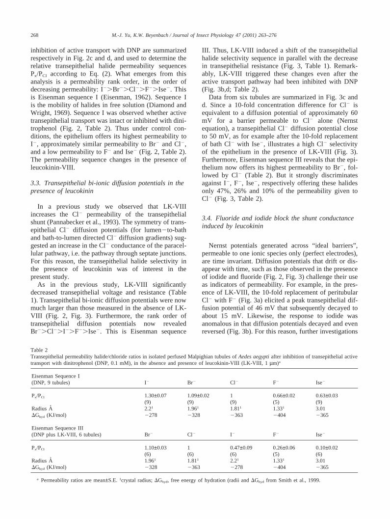
268 M.-J. Yu, K.W. Beyenbach / Journal of Insect Physiology 47 (2001) 263–276
inhibition of active transport with DNP are summarizedrespectively in Fig. 2c and d, and used to determine therelative transepithelial halide permeability sequencesPx/PCl according to Eq. (2). What emerges from thisanalysis is a permeability rank order, in the order ofdecreasing permeability: I2.Br2.Cl2.F2.Ise2. Thisis Eisenman sequence I (Eisenman, 1962). Sequence Iis the mobility of halides in free solution (Diamond andWright, 1969). Sequence I was observed whether activetransepithelial transport was intact or inhibited with dini-trophenol (Fig. 2, Table 2). Thus under control con-ditions, the epithelium offers its highest permeability toI2, approximately similar permeability to Br2 and Cl2,and a low permeability to F2 and Ise2 (Fig. 2, Table 2).The permeability sequence changes in the presence ofleucokinin-VIII.
3.3. Transepithelial bi-ionic diffusion potentials in thepresence of leucokinin
In a previous study we observed that LK-VIIIincreases the Cl2 permeability of the transepithelialshunt (Pannabecker et al., 1993). The symmetry of trans-epithelial Cl2 diffusion potentials (for lumen2to-bathand bath-to-lumen directed Cl2 diffusion gradients) sug-gested an increase in the Cl2 conductance of the paracel-lular pathway, i.e. the pathway through septate junctions.For this reason, the transepithelial halide selectivity inthe presence of leucokinin was of interest in thepresent study.
As in the previous study, LK-VIII significantlydecreased transepithelial voltage and resistance (Table1). Transepithelial bi-ionic diffusion potentials were nowmuch larger than those measured in the absence of LK-VIII (Fig. 2, Fig. 3). Furthermore, the rank order oftransepithelial diffusion potentials now revealedBr2.Cl2.I2.F2.Ise2. This is Eisenman sequence
Table 2Transepithelial permeability halide/chloride ratios in isolated perfused Malpighian tubules ofAedes aegyptiafter inhibition of transepithelial activetransport with dinitrophenol (DNP, 0.1 mM), in the absence and presence of leucokinin-VIII (LK-VIII, 1µm)a
Eisenman Sequence I(DNP, 9 tubules) I2 Br2 Cl2 F2 Ise2
Px/PCl 1.30±0.07 1.09±0.02 1 0.66±0.02 0.63±0.03(9) (9) (9) (5) (9)
Radius A 2.21 1.961 1.811 1.331 3.01DGhyd (KJ/mol) 2278 2328 2363 2404 2365
Eisenman Sequence III(DNP plus LK-VIII, 6 tubules) Br2 Cl2 I2 F2 Ise2
Px/PCl 1.10±0.03 1 0.47±0.09 0.26±0.06 0.10±0.02(6) (6) (6) (5) (6)
Radius A 1.961 1.811 2.21 1.331 3.01DGhyd (KJ/mol) 2328 2363 2278 2404 2365
a Permeability ratios are mean±S.E. 1crystal radius;DGhyd, free energy of hydration (radii andDGhyd from Smith et al., 1999.
III. Thus, LK-VIII induced a shift of the transepithelialhalide selectivity sequence in parallel with the decreasein transepithelial resistance (Fig. 3, Table 1). Remark-ably, LK-VIII triggered these changes even after theactive transport pathway had been inhibited with DNP(Fig. 3b,d; Table 2).
Data from six tubules are summarized in Fig. 3c andd. Since a 10-fold concentration difference for Cl2 isequivalent to a diffusion potential of approximately 60mV for a barrier permeable to Cl2 alone (Nernstequation), a transepithelial Cl2 diffusion potential closeto 50 mV, as for example after the 10-fold replacementof bath Cl2 with Ise2, illustrates a high Cl2 selectivityof the epithelium in the presence of LK-VIII (Fig. 3).Furthermore, Eisenman sequence III reveals that the epi-thelium now offers its highest permeability to Br2, fol-lowed by Cl2 (Table 2). But it strongly discriminatesagainst I2, F2, Ise2, respectively offering these halidesonly 47%, 26% and 10% of the permeability given toCl2 (Fig. 3, Table 2).
3.4. Fluoride and iodide block the shunt conductanceinduced by leucokinin
Nernst potentials generated across “ideal barriers”,permeable to one ionic species only (perfect electrodes),are time invariant. Diffusion potentials that drift or dis-appear with time, such as those observed in the presenceof iodide and fluoride (Fig. 2, Fig. 3) challenge their useas indicators of permeability. For example, in the pres-ence of LK-VIII, the 10-fold replacement of peritubularCl2 with F2 (Fig. 3a) elicited a peak transepithelial dif-fusion potential of 46 mV that subsequently decayed toabout 15 mV. Likewise, the response to iodide wasanomalous in that diffusion potentials decayed and evenreversed (Fig. 3b). For this reason, further investigations

269M.-J. Yu, K.W. Beyenbach / Journal of Insect Physiology 47 (2001) 263–276
Fig. 3. Transepithelial halide diffusion potentials of leucokinin-treated Malpighian tubules ofAedes aegyptiin the absence and presence ofdinitrophenol. Top panels show representative tubule traces of transepithelial voltage in the presence of leucokinin (a), and in the presence ofleucokinin and dinitrophenol (b). Shading indicates the time of Cl2 replacement in the peritubular Ringer solution with the halide of interest.Bottom panel shows summaries of transepithelial diffusion potentials in the presence of leucokinin (c), and in the presence of leucokinin anddinitrophenol (d). Data are mean±S.E. (number of tubule experiments).
of transepithelial iodide and fluoride diffusion potentialswere necessary.
In the experiments illustrated in Fig. 4, the tubuleswere first treated with DNP and then LK-VIII to inducethe transepithelial conductance with Eisenman halideselectivity sequence III. Again, DNP was used to exam-ine transepithelial diffusion potentials without inter-ference from the active transport pathway. In the pres-ence of DNP and LK-VIII transepithelial voltage was23.1 mV and transepithelial resistance was 1.6 KVcm(Fig. 4a). The 10-fold replacement of bath Cl2 with F2
elicited a sharp rise of the transepithelial voltage to 31.5mV, consistent with a transepithelial Cl2 permeabilityfar greater than that for F2. Thereafter, transepithelialvoltage returned towards zero mV even though F2
remained in the peritubular bath at high concentration(Fig. 4a). As voltage decayed towards zero, transepi-thelial resistance rose in parallel from 1.6 KVcm to apeak value of 12.5 KVcm. The disappearance of the Cl2
diffusion potential together with the increase in transepi-thelial resistance indicates that F2 blocks the transepi-thelial halide conductance induced by LK-VIII. Reversalof resistance to pre2fluoride values was not immediateafter washout of F2, but took between 2 and 5 min(Fig. 4a).
Similar observations were made in the presence ofhigh iodide concentrations in the peritubular bath (Fig.4b). Again, the tubule was first treated with DNP so thattransepithelial diffusion potentials can be studied withminimal interference from the active transport pathwaythrough principal cells (Fig. 1). Then LK-VIII was addedto induce the transepithelial halide conductance with Eis-enman selectivity sequence III. In the presence of DNPand LK-VIII transepithelial voltage was20.2 mV andtransepithelial resistance was 1.9 KVcm (Fig. 4b). The10-fold replacement of bath Cl2 with I2 elicited a sharprise of the transepithelial voltage to 16 mV and then 19mV, consistent with a transepithelial diffusion potentialfor Cl2 that is greater than that for I2. Thereafter, trans-epithelial voltage returned towards zero mV. As trans-epithelial voltage decayed, transepithelial resistance rosefrom 1.9 KVcm to values ranging from 10.3 to 11.6KVcm. The decay of the Cl2 diffusion potential togetherwith the increase in transepithelial resistance indicatesthat I2, like F2, blocks the transepithelial halide conduc-tance induced by LK-VIII (Fig. 4). The block by I2 wasmore readily reversible than that by F2, becauseRt
returned to pre-iodide values immediately after normalCl2 concentration in the peritubular bath was restored(Fig. 4). Thus, fluoride and iodide block the transepi-

270 M.-J. Yu, K.W. Beyenbach / Journal of Insect Physiology 47 (2001) 263–276
Fig. 4. Block by fluoride (a) and iodide (b) of the shunt halide con-ductance induced by leucokinin-VIII in Malpighian tubules ofAedesaegypti. Each tubule was pre-treated with leucokinin-VIII and dinitro-phenol to induce and uncover the shunt conductance. Peritubular Cl2
was then replaced 10-fold with fluoride (a) and iodide (b). Transepi-thelial voltage was measured continuously (traces) and transepithelialresistance was measured periodically (vertical bars).
thelial halide conductance induced by LK-VIII, wherethe block by F2 appears stronger than that by I2.
3.5. Fluoride mimics the effects of leucokinin
Next to blocking the conductance induced by leucoki-nin (Fig. 3, Fig. 4), F2 had additional effects on thetubule (Fig. 2a). For example, the 10-fold replacement ofbath Cl2 with F2, which increased peritubular F2 from 0mM to 142.5 mM, caused the transepithelial voltage todrop to values close to zero mV (Fig. 2a). Moreover,after restoring normal Cl2 concentrations in the peritub-ular bath, the transepithelial voltage did not recover tocontrol values but remained near zero mV. We investi-gated this phenomenon in seven additional Malpighiantubules; a representative experiment is shown in Fig. 5.
In the presence of normal Ringer solution, the tubulehad a transepithelial voltage of 25.3 mV and a transepi-thelial resistance of 15.4 KVcm. The 10-fold replace-ment of bath Cl2 with F2 caused the transepithelial volt-age to decay to zero mV in parallel with the transientrise of the transepithelial resistance to 22.0 KVcm thatleveled out at 17 KVcm, consistent with conductanceblock by F2 (Fig. 4a, Fig. 5). However, upon restoringnormal Cl2 concentration in the peritubular bath, thetransepithelial voltage remained near zero (Fig. 2a, Fig.
5), and surprisingly, after a lag time of about 4.5 min,the transepithelial resistance fell to 3.2 KVcm. Such lowtransepithelial resistances are usually observed in thepresence of LK-VIII (Fig. 5, Table 1). Indeed, transepi-thelial diffusion potentials measured at this time revealedEisenman sequence III, namely that sequence that isinduced by LK-VIII in parallel with the drop in transepi-thelial resistance (Fig. 3, Table 1, Table 2).
What is shown for a single tubule in Fig. 5 was con-sistently observed in six additional tubules. The datafrom all tubules are summarized in Fig. 6. Under controlconditions,Vt was 38.9±12.1 mV andRt was 13.5±1.7KVcm on average (Fig. 6a). The 10-fold replacement ofbath Cl2 with F2 caused the significant (P,0.05) dropof Vt to 9.0±3.8 mV and the significant (P,0.05)increase ofRt to 18.7±2.6 KVcm. Restoring normal Cl2
in the peritubular bath caused significant (P,0.05)reductions ofVt to 0.9±0.9 mV andRt to 2.9±0.6 KVcm(P,0.001). Moreover, after exposure to F2, the epi-thelium displays Eisenman halide selectivity sequenceIII (Fig. 6b). Thus, high F2 concentrations in the peritub-ular bath mimic the effects of LK-VIII by (1) decreasingthe transepithelial resistance, and (2) inducing transepi-thelial halide selectivity sequence III. Moreover, the con-ductance induced by F2 is subsequently blocked by flu-oride itself (Fig. 6a). This is illustrated by thetransepithelial resistance in the presence of F2, whichdrops from 18.3±2.6 KVcm to 2.9±0.6 KVcm afterwashout of F2. Likewise, I2 blocks the conductanceinduced by F2 (Fig. 5).
4. Discussion
4.1. Dinitrophenol uncovers the shunt pathway
The present study confirms our previous observationsof the effects of dinitrophenol on transepithelial transportin AedesMalpighian tubules (Pannabecker et al., 1992).DNP reduces the transepithelial voltage to values closeto zero mV, and it increases the transepithelial resistanceto the highest values that can be measured across theepithelium (Table 1). In principal cells of the epithelium,DNP reduces the apical membrane voltage from 111 mVto 9 mV, and it reduces the voltage across the basolateralmembrane from258 mV to23 mV (Pannabecker et al.,1992). The collapse of apical and basolateral membranevoltages to values less than 10% of control reflects theinhibition of the active transport pathway of Na+ andK + through principal cells. Stopping active transport isexpected to increase the transcellular electrical resistanceRcell (Fig. 1b). Indeed, recent two electrode voltageclamp studies in principal cells have shown that DNPincreasesRcell nearly 12-fold, from 992 KV to 11,756KV (Masia et al., 2000). Since under control conditionsRcell is approximately 3.5 times greater than the resist-

271M.-J. Yu, K.W. Beyenbach / Journal of Insect Physiology 47 (2001) 263–276
Fig. 5. Multiple effects of fluoride (F2) in Malpighian tubules ofAedes aegypti. The lumen of the isolated tubule was perfused with the same Ringersolution present in the peritubular bath. Transepithelial voltage was measured continuously. Periodic measurements of transepithelial resistance aredepicted as vertical bars. The 10-fold replacement of the peritubular Cl2 in the peritubular bath with fluoride (142.5 mM) inhibited the transepithelialvoltage while increasing transepithelial resistance, mimicking the effects of dinitrophenol. After the return to normal bath Cl2 concentration,transepithelial voltage remained near zero mV, transepithelial resistance fell, and the tubule exhibited transepithelial halide selectivity sequence III,mimicking the effects of leucokinin-VIII. Again, iodide and fluoride blocked the shunt conductance induced by high concentrations of peritubular F2.
Fig. 6. Fluoride (F2) mimics the effects of leucokinin-VIII in isolatedperfused Malpighian tubules ofAedes aegypti. In the presence of per-itubular F2 transepithelial voltage decreases, but transepithelial resist-ance remains high because of shunt conductance block by F2. Washoutof F2 lowers transepithelial resistance to values normally seen in thepresence of leucokinin-VIII, and decreases transepithelial voltagefurther, mimicking the effects of leucokinin-VIII (a). F2 treatmentinduces the halide selectivity sequence III normally seen in the pres-ence of leucokinin-VIII (b).
ance of the shunt (Masia et al., 2000), it follows that a12-fold increase ofRcell brings measures of the transepi-thelial resistanceRt within 97% of the resistance of theshunt Rsh. Thus, measures of the transepithelial resist-ance in the presence of DNP approach the resistance of
the shunt (Pannabecker et al., 1992). It follows thattransepithelial bi-ionic diffusion potentials measured inthe presence of DNP reflect the properties of the shunt.
The term ‘shunt’ defines the return pathway for cur-rent, when transepithelial secretion of NaCl and KCl isviewed as an electrical circuit consisting of an activetransport pathway for the secretion of Na+ and K+ intothe tubule lumen and by a passive transport pathway forthe secretion of Cl2 (Fig. 1). While it is clear that princi-pal cells provide the active transport pathway for trans-epithelial secretion of Na+ and K+, stellate cells and/orseptate junctions defining the paracellular transportroute, may serve as shunt.
Under control conditions, the shunt pathway exhibitshalide permeability sequence I: I2.Br2.Cl2.F2 (Fig.2, Table 2). This permeability sequence is also the rankorder of mobility these halides have in aqueous solutions(Diamond and Wright, 1969). It is the sequence wherethe ion with the smallest hydrated size, I2, is the mostmobile in free solution, and the ion with the largesthydrated size, F2, is the least mobile.
Hydration size depends on the charge density that inturn depends on the crystal radius of the ion. Since I2
has the largest crystal radius, it has the lowest chargedensity that offers weak coulombic forces for attractingwater. Thus, the hydrated size of I2 is small which isreflected in a low energy of hydration (Table 2). For thisreason, hydrated I2 is the smallest of the four hydratedhalides and therefore the most mobile halide in water.In contrast, F2 has the smallest crystal radius, the highestcharge density, and therefore the largest coulombic forcefor interacting with water. Accordingly, hydrated F2 isthe largest hydrated halide, with a high energy ofhydration (Table 2). Because of its large hydrated size,F2 is the least mobile in free solution.
Eisenman considered selectivity to derive from cou-

272 M.-J. Yu, K.W. Beyenbach / Journal of Insect Physiology 47 (2001) 263–276
lombic interactions between the ion and two players:mobile and fixed charges, where water can be consideredmobile charges, and positive or negative sites in mem-branes, resins, or filters can be considered fixed charges.For a transport pathway, such as the shunt, to be anion-selective, it must possess positive fixed sites: primary,secondary, or tertiary amino groups, or quaternaryammonium. Since, under control conditions, the halidepermeability sequence of the shunt (Eisenman sequenceI) follows the order of free-solution mobility, the shuntmust be a pathway of low positive electric field strength,too weak to remove water from hydrated halides enteringthe pathway. Hence, the shunt selects halides largely onthe basis of hydrated size. However, electric fieldstrength is sufficient to prevent cations from passingthrough. If it did allow the passage of cations, the Mal-pighian tubule could not maintain the transepithelial con-centration differences for Na+ and K+ that are generatedby active transport through principal cells (Williams andBeyenbach, 1984).
4.2. Leucokinin-VIII increases shunt conductance andselectivity
In previous studies, LK-VIII increased rates of trans-epithelial fluid secretion from 0.49 nl/min to 0.91 nl/minby stimulating transepithelial NaCl and KCl secretion(Pannabecker et al., 1993). Increasing equally the ratesof transepithelial secretion for NaCl and KCl suggestedan effect on the transport pathway taken by Cl2. Indeed,electrophysiological studies provide three lines of evi-dence for an effect on shunt Cl2 conductance. First, LK-VIII reduces transepithelial resistance by more than 80%together with the drop of the transepithelial voltage tovalues close to zero, nearly short-circuiting the transepi-thelial voltage (Pannabecker et al., 1993). A transepi-thelial short-circuit is expected from a large increase inthe shunt conductance. Second, the dramatic reductionof the transepithelial resistance can be reversed in thepresence of LK-VIII by replacing Cl2 with isethionateon both sides of the epithelium. Replacing Cl2 on onlyone side is not sufficient for restoring resistance in thepresence of LK-VIII (Pannabecker et al., 1993). Depen-dence of the transepithelial resistance on the Cl2 concen-tration in the extracellular solutions suggests an extra-cellular pathway such as that expected from a Cl2-selective paracellular pathway through tight or septatejunctions. Third, in the presence of LK-VIII the epi-thelium yields large transepithelial Cl2 diffusion poten-tials that approach 85% of Nernst potentials, and whichare symmetrical for bath-to-lumen and lumen-to-bathCl2 gradients (Pannabecker et al., 1993). Symmetry oftransepithelial diffusion potentials suggests a single dif-fusion barrier such as that expected from tight or septatejunctions. Symmetry of diffusion potentials is unlikelyto develop across two diffusion barriers in series, i.e. the
basolateral and apical membrane membranes of stellatecells. Thus, the epithelium short-circuiting itself in thepresence of LK-VIII, the dependence of the transepi-thelial resistance on the Cl2 concentration in the extra-cellular fluids, and symmetrical transepithelial Cl2 dif-fusion potentials, all point to leucokinin increasing theCl2 conductance of the paracellular shunt pathway(Fig. 1b).
In the present study, LK-VIII caused the transepi-thelial resistance to drop from 13 KVcm to 2 KVcm. Inparallel, it caused the transepithelial voltage to fall from41 mV to 3 mV, reflecting again the transepithelial short-circuit due to the increase in shunt Cl2 conductance(Table 1). Transepithelial halide diffusion potentials arenow much larger than under control condition, and dis-play the transepithelial halide selectivity sequence of theEisenman series III. Thus, LK-VIII increases the shuntconductance together with an increase in halide selec-tivity. Remarkably, LK-VIII affects conductance and sel-ectivity after the active transport pathway has beeninhibited with DNP (Table 1, Fig. 3). Hence, mediationof the effects of LK-VIII is not immediately dependenton metabolism. Since DNP brings to the fore the proper-ties of the shunt, LK-VIII is seen to reduce the shuntresistance form 17.5 KVcm to 4.0 KVcm in the presenceof DNP (Table 1). From the perspective of conductance(1/R), LK-VIII increases more than four-fold the shuntconductance, from 57µS/cm to 250µS/cm. In parallel,the shift from selectivity sequence I to III indicates thatthe pathway has acquired an electrical field strength suf-ficient to dehydrate halide ions. As a result, the shuntpathway is now more permeable to Br2 and Cl2 than itis to I2 (Table 2). Thus, LK-VIII increases shunt con-ductance four-fold while optimizing the Cl2-filter in theshunt pathway.
It is not uncommon for the “same” Cl2 channel todisplay different selectivity sequences. For example, theselectivity sequence of a channel can be dependent onthe voltage, the degree of channel block, and even onthe experimental protocol (Begenish and Melvin, 1998;Dawson et al., 1999; Tabcharani et al., 1997). Less sur-prising are mutations in the transmembrane domains(pore) of the channel that shift the selectivity sequencefrom III to I in the case of the cystic fibrosis transmem-brane regulator Cl2 channel (Anderson et al., 1991;Devidas and Guggino, 1997). To these mechanisms ofmodulating channel selectivity sequence, the presentstudy allows us to add modulation by an extracellularpeptide, leucokinin, presumably via receptor-mediatedactivation of G-protein (Cox et al., 1997).
The changes in the shunt pathway induced by LK-VIII may not require a major reorganization of the shuntpathway as suggested by Dow et al. (1998) and O’Don-nell et al. (1996, 1998) Instead, a change in the molecu-lar environment of the shunt may increase the electricfield strength of filtering sites in the shunt. Higher field

273M.-J. Yu, K.W. Beyenbach / Journal of Insect Physiology 47 (2001) 263–276
strengths may provide the energy for dehydrating smallhalides (Br2 and Cl2) with strong hydration energies,giving them passage over large halides with weakhydration energies (I2). Electric field strengths in theshunt are largely determined by electron densities thatcan change via inductive effects exerted by the immedi-ate environment, as for example during a phosphoryl-ation of a shunt protein. Modulating electrical fieldstrengths and/or conformational changes of shunt pro-teins are consistent with the reversible on-off effects ofLK-VIII with switch-like speed (Pannabecker et al.,1993).
4.3. Site of the transepithelial Cl2 shunt pathway
There is good agreement that principal cells are notthe site of the conductive transepithelial Cl2 shunt thatmediates Cl2 secretion in Malpighian tubules (Dow etal., 1998; O’Donnell et al., 1996; Pannabecker et al.,1993). But the question of exactly where this Cl2 con-ductance is located remains unanswered in spite ofpowerful experimental methods that have been broughtto bear on this question. On the basis of vibrating probeanalysis and Ca2+-imaging, the majority view considersstellate cells as providing the pathway for transepithelialCl2 secretion (Dow et al., 1998; O’Donnell et al. 1996,1998), where the leucokinins are thought to act on stel-late cells to open Ca2+-activated Cl2 channels via elev-ation of intracellular Ca2+ (O’Donnell et al., 1998).Recent patch clamp studies in our laboratory of the api-cal membrane of stellate cells ofAedes Malpighiantubules (O’Connor and Beyenbach, 2000) have revealedtwo types of Cl2 channels. The most frequently observedCl2 channel has a conductance of 27 pS, a mean opendwell time of 304 ms, and a open probability of 0.53 atO mV in excised patches. Up to five channels of thischannel were often observed in the same patch pipettewith 10–20 MV resistances, suggesting a high channeldensity in apical membrane patches. The second typehad a 5 pS conductance, a mean open dwell time of 7.5ms, and an open probability of 0.07 at O mV in excisedpatches. Whether these apical membrane Cl2 channelsparticipate in transepithelial Cl2 secretion under controlconditions and/or leucokinin-stimulated diuresis remainsto be determined.
Our laboratory holds the minority view of a paracellu-lar pathway of Cl2 through a single barrier, septate junc-tions, for the reasons discussed above. Opposition to ourview stems largely from the requirement of giving sep-tate junctions channel-like properties that are regulatedvia intracellular signal pathways. However, paracellularpathways in vertebrate epithelia are widely appreciatedto possess channel-like properties controlled and regu-lated by intracellular signaling pathways involving Ca2+,protein kinase C, G-protein, phospholipase A2 and phos-pholipase C (Anderson and Van Itallie, 1995; Denker
and Nigam, 1998; Gasbarrini and Montalto, 1999;Kovbasnjuk et al., 1998; Madara, 1998; Perez et al.,1997; Stein and Kottra, 1997). The first tight-junctionalprotein to provide a paracellular pathway for a cation,paracellin-1, has recently been discovered in the humankidney (Simon et al., 1999). Paracellin-1 offers a Mg2+-selective pathway through tight junctions of the thickascending limb of the Loop of Henle. Significantly, thethick ascending limb of the Loop of Henle is the majorsite of renal Mg2+ homeostasis in the kidney (Quamme,1997). Furthermore, nearly 100% of transepithelial Mg2+
reabsorption is passive, driven through the paracellularpathway (paracellin-1) by the transepithelial voltage(DeRouffignac et al., 1993; Quamme, 1993; Wittner etal., 1993). The situation may be similar in Malpighiantubules of Aedes aegypti, where transepithelial Cl2
secretion is also passive, driven by the transepithelialvoltage through a Cl2-selective septate junctional pro-tein. Analogues of vertebrate tight junction proteins havealready been found in insect septate junctions (Willottet al., 1993).
4.4. Block of shunt conductance by I2 and F2
Transepithelial diffusion potentials generated by thereplacement of Cl2 with isethionate climbed to maximalvalues and remained at plateau values until the bath wasreturned to control Cl2 concentration again (Fig. 2, Fig.3, Fig. 5). In contrast, diffusion potentials generated bythe replacement of Cl2 with I2 or F2 climbed briefly topeak voltages from where they decayed towards zero(Fig. 2b, Fig. 3, Fig. 4, Fig. 5). Measurements of trans-epithelial resistance give insight into the decay of dif-fusion potentials (Fig. 4). As the transepithelial diffusionpotential declines towards zero in the presence of F2
(Fig. 4a), the transepithelial shunt resistance increaseseight-fold, from 1.6 to 12.5 KVcm. Likewise, as thetransepithelial diffusion potential declines towards zeroin the presence of I2 (Fig. 4b), the transepithelial shuntresistance increases six-fold, from 1.9 to 11.6 KVcm.Such large increases in shunt resistance indicate theblock of the shunt conductance by F2 and I2. Coulombicinteractions between the halide and the shunt pathwaymay explain conductance block, where strong interac-tions may lead to binding of F2 and I2, thereby blockingthe diffusion pathway for Cl2 and decreasing its dif-fusion potential (Fig. 2b, Fig. 3, Fig. 4, Fig. 5). Thisconclusion is supported by finding that blockade of theshunt pathway by F2 and I2 is especially effective underconditions of high shunt conductance and selectivity(presence of LK-VIII) when the shunt pathway offershigh electrical field strength (Eisenman sequence III;Fig. 3, Fig. 4, Fig. 5).
After channel block by F2 or I2 the shunt selectivitysequence reverts to sequence I again, which is expectedfrom channel block by these anions. However, upon

274 M.-J. Yu, K.W. Beyenbach / Journal of Insect Physiology 47 (2001) 263–276
wash-out of F2 or I2 the shunt pathway returns to selec-tivity sequence III that was induced by leucokinin-VIII.
4.5. Fluoride, a highly reactive halide
Next to blocking the shunt conductance induced byLK-VIII, high concentrations of F2 in the peritubularbath has two additional effects in Malpighian tubules ofAedes aegypti. It inhibits the active transepithelial trans-port through principal cells, mimicking the effects ofdinitrophenol, and it stimulates the shunt pathway, mim-icking the effects of LK-VIII.
Fluoride duplicates the effects of DNP by triggeringthe fall of the transepithelial voltage to values close tozero in parallel with the increase in transepithelial resist-ance (Fig. 2a, Fig. 6). Fluoride is known to inhibit gly-colysis (Marquis, 1995) and consequently ATP syn-thesis, suggesting that withholding ATP from activetransport (by either DNP or F2) inhibits the active trans-port pathway through principal cells. Indeed, recent two-electrode voltage clamp studies in principal cells haveshown, that 142.5 mM F2 in the peritubular bath reversi-bly increases the transcellular resistance more than 10-fold, like DNP (Beyenbach and Masia, 2000; Masia etal., 2000).
High concentrations of peritubular F2 (142.5 mM)mimic the effects of LK-VIII by (1) causing transepi-thelial voltage and resistance to fall to values similar tothose observed in the presence of LK-VIII, and (2)inducing transepithelial halide selectivity sequence III,like LK-VIII (Fig. 5, Fig. 6). Mimicry by F2 of theeffects of LK-VIII is only observed after returning thetubule to its normal Ringer bath (Fig. 5, Fig. 6). It cannot be observed in the presence of F2 because of theadditional effects of F2 on cell metabolism and shuntconductance block (Fig. 4, Fig. 6).
Duplication by F2 of the effects of leucokinin-VIIIon the shunt pathway suggests mediation via G-protein.Fluoride is known to form a complex with trace quan-tities of aluminum present in commercial preparations ofnucleotides and disposable glass test tubes to yieldAlF4
2 which stimulates cellular heterotrimeric G-protein(Sternweis and Gilman, 1982). AlF4
2 is thought tostimulate G-protein by mimicking theγ-phosphate ofGTP (Bigay et al., 1987). Although mediation of theeffects of leucokinin via G-protein has not been demon-strated directly in Malpighian tubules, the circumstantialevidence is quite strong. The three leucokinins ofAedesaegypti, ALP I, II, and III, that were isolated by Veenstra(1994), all significantly increased intracellular IP3 con-centrations inAedesMalpighian tubules. Synthesis of IP3
is dependent on phospholipase C activity that increasesupon stimulation by G-protein. IP3 is known to causethe release of Ca2+ from IP3-sensitive intracellular Ca2+
stores in a variety of cells, including epithelial cells ofMalpighian tubules. O’Donnell et al. (1996) observe an
increase in cytosolic Ca2+ in stellate cells ofDrosophilaMalpighian tubules after stimulation with leucokinin.Furthermore, the effects of leucokinin-VIII on transepi-thelial voltage and resistance ofAedes Malpighiantubules can be duplicated by A23187, a Ca2+-ionohorethat is expected to increase cytosolic free Ca2+ concen-trations (Clark et al., 1998). Preliminary studies inAedesMalpighian tubules show that Ca2+ from an intracellularstore is needed to trigger the effect of LK-VIII on shuntconductance, and that extracellular Ca2+ is needed tomaintain the high conductance of the shunt pathway (Yuand Beyenbach, 2000). Finally, the first receptor of aleucokinin-like peptide that has been isolated from thecentral nervous system of the pond snail appears to becoupled to G-protein (Cox et al., 1997). Accordingly,neuropeptides of the large family of leucokinins mayemploy G-proteins not only in postsynaptic signal trans-duction (in the pond snail), but also in electrolyte andfluid transport across Malpighian tubules.
Acknowledgements
This paper is dedicated to G. Mark Holman who inthe final year of the 20th century has traded his biochem-istry lab for a log cabin home in Northern Utah. Holmandiscovered the leucokinins while isolating and charac-terizing the structure of myotropic peptides present inhead extracts of the cockroachLeucophaea madiera. Healso discovered LPK, leucosulfakinin, and LMS (theinsect FLRFamide). Altogether, Holman isolated 12 newstructures (eight of which were leucokinins) and threenew peptide families fromLeucophaeahead extracts. Heworked on isolations for more than 16 years before hegot the first structure. The authors thank the USDA forsupporting Holman’s work in the past, and also theNational Science Foundation for enabling our study(IBN 9604394).
References
Anderson, M.P., Rich, D.P., Gregory, R.J., Smith, A.E., Welsh, M.J.,1991. Generation of cAMP-activated chloride currents byexpression of CFTR. Science 251, 679–682.
Anderson, J.M., Van Itallie, C.M., 1995. Tight junctions and the mol-ecular basis for regulation of paracellular permeability. Am. J. Phy-siol. 269, G467–G475.
Begenish, T., Melvin, J.E., 1998. Regulation of chloride channels insecretory epithelia. J. Membrane Biol. 163, 77–85.
Beyenbach, K.W., Masia, R., 2000. Effects of metabolic inhibitors onprincipal cells of mosquito Malpighian tubules. ExperimentalBiology, San Diego, April 2000, Abstract #15746.
Beyenbach, K.W., 1995. Mechanism and regulation of electrolytetransport in Malpighian tubules. J. Insect Physiol. 41, 197–207.
Beyenbach, K.W., Petzel, D.H., 1987. Diuresis in mosquitoes: role ofa natriuretic factor. News Physiol. Sci. 2, 171–175.
Bigay, J., Deterre, P., Pfister, C., Chabre, M., 1987. Fluoride complex

275M.-J. Yu, K.W. Beyenbach / Journal of Insect Physiology 47 (2001) 263–276
of aluminum or beryllium act on G-proteins as reversibly boundanalogues of theγ-phosphate of GTP. EMBO Journal 6, 2907–2914.
Cho, W.J., Kim, K.S., Lee, B.H., 1999. Postembryonic localization ofleucokinin I-immunoreactive neurons in ventral ganglia of thesweet potato hornwormAgrius convolvuli. Kor. J. Entomol. 29,23–29.
Clark, T.M., Hayes, T.K., Holman, G.M., Beyenbach, K.W., 1998. Theconcentration-dependence of CRF-like diuretic peptide: mechanismof action. J. Exp. Biol. 201, 1753–1762.
Clottens, F.L., Meola, S.M., Coast, G.M., Hayes, T.K., Wright, M.S.,Nachman, R.J., Holman, G.M., 1993. Characterization of an anti-serum against an achetakinin I-analog and its use for the localiz-ation of Culekinin Depolarizing Peptide II in the mosquitoCulexsalinarius. Regul. Pept. 49, 145–157.
Coast, G.M., Holman, G.M., Nachman, R.J., 1990. The diureticactivity of a series of cephalomyotropic neuropeptides the achetaki-nins on isolated Malpighian tubules of the house cricketAchetadomesticus. J. Insect Physiol. 36, 481–488.
Cox, K.J.A., Tensen, C.P., Van Der Schors, R.C., Li, K.W., Van Heeri-khuizen, H., Vreugdenhil, E., Geraerts, W.P.M., Burke, J.F., 1997.Cloning, characterization, and expression of a G-protein-coupledreceptor fromLymnaea stagnalisand identification of a leucokinin-like peptide, PSFHSWSamide, as its endogenous ligand. J. Neuro-sci. 17, 1197–1205.
Dawson, D.C., Smith, S.S., Mansoura, M.K., 1999. CFTR: mechanismof anion conduction. Physiol. Rev. 79, S47–S75.
Denker, B.M., Nigam, S.K., 1998. TI molecular structure and assemblyof the tight junction. Am. J. Physiol. 274, F1–F8.
DeRouffignac, C., Mandon, B., Wittner, M., Di-Stefano, A., 1993.Hormonal control of renal magnesium handling. Mineral and Elec-trolyte Metabolism 19, 226–231.
Devidas, S., Guggino, W.B., 1997. CFTR: domains, structure and func-tion. J. Bioenerget. Biomembr. 29, 443–551.
Diamond, J.M., Wright, E.M., 1969. Biological membranes: the physi-cal basis of ion and non-electrolyte selectivity. A. Rev. Physiol.31, 581–646.
Dijkstra, S., Leyssens, A., Van Kerkhove, E., Zeiske, W., Steels, P.,1995. A cellular pathway for Cl2 during fluid secretion in ant Mal-pighian tubules: evidence from ion-sensitive microelectrode stud-ies? J. Insect Physiol. 41, 695–703.
Dow, J.A.T., Davies, S.A., So¨zen, M.A., 1998. Fluid secretion by theDrosophila melanogasterMalpighian tubule. Am. Zool. 38, 450–460.
Eisenman, G., 1962. Cation selective glass electrodes and their modeof operation. Biophys. J. 2, 259–323.
Elekes, K., Hernadi, L., Kiss, T., Muneoka, Y., Nassel, D.R., 1995.Tachykinin- and leucokinin-related peptides in the molluscan ner-vous system. Acta Biol. Hung. 46, 281–294.
Fromter, E., 1974. Electrophysiology and isotonic fluid absorption ofproximal tubules of mammalian kidney. In: Thurau, K. (Ed.), MTPInternational Review of Science, Physiology, vol. 6, Sect. 1, Kid-ney and Urinary Tract Physiology. The Butterworth Group, Lon-don, pp. 1–38.
Gasbarrini, G., Montalto, M., 1999. Structure and function of tightjunctions. Role in intestinal barrier. Ital. J. Gastroenterol. Hepatol.31, 481–488.
Hayes, T.K., Pannabecker, T.L., Hinckley, D.J., Holman, G.M., Nach-man, R.J., Petzel, D.H., Beyenbach, K.W., 1989. Leucokinins, anew family of ion transport stimulators and inhibitors in insect Mal-pighian tubules. Life Sci. 44, 1259–1266.
Hayes, T.K., Holman, G.M., Pannabecker, T.L., Wright, M.S., Strey,A.S., Nachman, R.J., Hoel, D.F., Olson, J.K., Beyenbach, K.W.,1994. Culekinin depolarizing peptide: a mosquito leucokinin-likepeptide that influences insect Malpighian tubule ion transport.Regul. Pept. 52, 235–248.
Helman, S.I., 1972. Determination of electrical resistance of the iso-
lated cortical collecting tubule and its possible anatomical location.Yale J. Biol. Med. 45, 339–345.
Holman, G.M., Cook, B.J., Nachman, R.J., 1987. Isolation, primarystructure, and synthesis of leucokinins VII and VIII, the final mem-bers of this new family of cephalomyotropic peptides isolated fromhead extracts ofLeucophaea maderae. Comp. Biochem. Physiol.88C, 31–34.
Holman, G.M., Nachman, R.J., Coast, G.M., 1999. Isolation, charac-terization and biological activity of a diuretic myokinin neuropep-tide from the houseflyMusca domestica. Peptides 20, 1–10.
Kim, N.Y., Lee, B.H., 1998. Differential localization of locustatachyki-nin I- and leucokinin I-immunoreactive neurons in the brain andretrocerebral complex of the wax mothGalleria mellonella. Kor.J. Entomol. 28, 257–266.
Kovbasnjuk, O.N., Szmulowicz, U., Spring, K.R., 1998. Regulation ofthe MDCK cell tight junction. J. Membrane Biol. 161, 93–104.
Madara, J.L., 1998. Regulation of the movement of solutes across tightjunctions. A. Rev. Physiol. 60, 143–159.
Marquis, R.E., 1995. Antimicrobial actions of fluoride for oral bacteria.Can. J. Microbiol. 41, 955–964.
Masia, R., Aneshansley, D., Nagel, R., Nachman, R.J., Beyenbach,K.W., 2000. Two-electrode voltage clamp methods in principalcells of Malpighian tubules ofAedes aegypti. Am. J. Physiol.,(submitted).
Nassel, D.R., Lundquist, C.T., 1991. Insect tachykinin-like peptide:distribution of leucokinin immunoreactive neurons in the cockroachand blowfly brains. Neurosci. Lett. 130, 225–228.
Nassel, D.R., Cantera, R., Karlsson, A., 1992. Neurons in the cock-roach nervous system reacting with antisera to the neuropeptide I.J. Comp. Neurobiol. 322, 45–67.
O’Connor, K.R., Beyenbach, K.W., 2000. Cl channels in apical mem-brane patches of stellate cells of insect Malpighian tubules. FASEBJ. 14, A109.
O’Donnell, M.J., Dow, J.A.T., Huesmann, G.R., Tublitz, N.J., Mad-drell, S.H.P., 1996. Separate control of anion and cation transportin Malpighian tubules ofDrosophila melanogaster. J. Exp. Biol.199, 1163–1175.
O’Donnell, M.J., Rheault, M.R., Davies, S.A., Rosay, P., Harvey, B.J.,Maddrell, S.H.P., Kaiser, K., Dow, J.A.T., 1998. Hormonally con-trolled chloride movement acrossDrosophila tubules is via ionchannels in stellate cells. Am. J. Physiol. 274, R1093–R1049.
Pannabecker, T.L., Aneshansley, D.J., Beyenbach, K.W., 1992. Uniqueelectrophysiological effects of dinitrophenol in Malpighian tubules.Am. J. Physiol. 263, R609–R614.
Pannabecker, T.L., Hayes, T.K., Beyenbach, K.W., 1993. Regulationof epithelial shunt conductance by the peptide leucokinin. J. Mem-brane Biol. 132, 63–76.
Perez, M., Barber, A., Ponz, F., 1997. Modulation of intestinal paracel-lular permeability by intracellular mediators and cytoskeleton. Can.J. Physiol. Pharmacol. 75, 287–292.
Quamme, G.A., 1993. Magnesium homeostasis and renal magnesiumhandling. Mineral and Electrolyte Metabolism 19, 218–225.
Quamme, G.A., 1997. Renal magnesium handling: new insights inunderstanding old problems. Kidney Int. 52, 1180–1195.
Schmid, A., Becherer, C., 1996. Leucokinin-like immunoreactive neu-rones in the central nervous system of the spiderCupiennius salei.Cell Tiss. Res. 284, 143–152.
Simon, D.B., Lu, Y., Choate, K.A., Velazquez, H., Al-Sabban, E.,Praga, M., Casari, G., Bettinelli, A., Colussi, G., Rodriguez-Sori-ano, J., McCredie, D., Milford, D., Snajad, S., Lifton, R.P., 1999.Paracellin-1, a renal tight junction protein required for paracellularMg2+ resorption. Science 5424, 103–106.
Schoofs, L., Holman, G.M., Proost, P., Vandamme, J., Hayes, T.K.,De Loof, A., 1992. Locustakinin, a novel myotropic peptide fromLocusta migratoria; isolation, primary structure and synthesis.Regul. Pept. 37, 49–57.
Smith, S.S., Steinle, E.D., Meyerhoff, M.E., Dawson, D.C., 1999. Cys-

276 M.-J. Yu, K.W. Beyenbach / Journal of Insect Physiology 47 (2001) 263–276
tic fibrosis transmembrane conductance regulator: physical basisfor lyotropic anion selectivity patterns. J. Gen. Physiol. 114,799–817.
Stein, J., Kottra, G., 1997. Intestinal tight junctions. I. Structure andmolecular regulation mechanisms. Zeitschrift Gastroenterol. 35,205–220.
Sternweis, P.C., Gilman, A.G., 1982. Aluminum, a requirement foractivation of the regulatory component of adenylate cyclase byfluoride. Proc. Natl Acad. Sci. 79, 4888–4891.
Tabcharani, J.A., Linsdell, P., Hanrahan, J.W., 1997. Halide per-meation in wild-type and mutant cystic fibrosis transmembraneconductance regulator chloride channels. J. Gen. Physiol. 110,341–354.
Terhzaz, S., O’Connell, F.C., Pollock, V.P., Kean, L., Davies, S.A.,Veenstra, J.A., Dow, J.A.T., 1999. Isolation and characterizationof a leucokinin-like peptide ofDrosophila melanogaster. J. Exp.Biol. 202, 3667–3676.
Ussing, H.H., Windhager, E.E., 1964. Nature of shunt path and activesodium transport path through frog skin epithelium. Acta Physiol.Scand. 61, 484–504.
Veenstra, J.A., 1994. Isolation and identification of three leucokininsfrom the mosquitoAedes aegypti. Biochem. Biophys. Res. Com-mun. 202, 715–719.
Williams, J.C. Jr., Beyenbach, K.W., 1984. Differential effects of sec-retagogues on the electrophysiology of the Malpighian tubules ofthe yellow fever mosquito. J. Comp. Physiol. 154B, 301–309.
Willott, E., Balda, M.S., Fanning, A.S., Jameson, B., Van Itallie, C.,Anderson, J.M., 1993. The tight junction protein ZO-1 is homolo-gous to theDrosophiladiscs-large tumor suppressor protein of sep-tate junctions. PNAS 90, 7834–7838.
Wittner, M., Mandon, B., Roinel, N., De Rouffignac, C., Di-Stefano,A., 1993. Hormonal stimulation of calcium and magnesium trans-port in the cortical thick ascending limb of Henle’s loop of themouse: evidence for a change in the paracellular pathway per-meability. Eur. J. Physiol. 423, 387–396.
Yu, M.J., Beyenbach, K.W., 2000. Leucokinin-VIII increases epithelialshunt conductance via a receptor-mediated pathway involving cal-cium. FASEB J. 14, A579.