Lesson ofthe Week - Europe PubMed Central
3
Lesson of the Week Addison's disease C M Brosnan, N F C Gowing Primary adrenocortical insufficiency (Addison's disease) is a rare disorder in which there is destruction of the adrenal cortex, thus reducing the production of glucocorticoid, mineralocorticoid, and sex steroids."2 Most cases are now due to autoimmune disease3 and occur more often in patients with an HLA-DR3 and HLA-B8 tissue type. The disease may be associated with other immune disorders, including thyroid disease, diabetes mellitus, pemicious anaemia, hypoparathyroidism, and ovarian failure. We report two fatal cases of autoimmune Addison's disease that were diagnosed only at necropsy. Case reports CASE 1 A 26 year old man was admitted to hospital with a history of two to three days of nausea and vomiting and increasingly frequent watery stools. He had also complained of a sore throat for two to three days, for which his family doctor had prescribed pivampicillin. He had not travelled overseas nor had he eaten any food outside his own home. He worked as a security officer near the Thames but never swam there. He had no previous history of weight loss, abdominal pain, or cardiovascular symptoms nor any important past medical history. Initial examination showed him to be dehydrated with a temperature of 37 5°C, a pulse rate of 120 beats a minute, and blood pressure of 85/60 mm Hg (with no postural drop). No hyperpigmentation of skin creases or mucous membranes was noted. His tonsillar bed was inflamed. Examination of his abdomen was unremarkable. Haematological investigations gave normal results, in particular sodium 135 mmol/l (normal range 135-150 mmol/l), potassium 5X9 mmol/l (normal range 3 5-5 3 mmolIl), creatinine 233 ,umol/l, urea 15-4 mmoUl, and glucose 3-9 mmol/l (normal range 3 0-8 0 mmol/l). His electrocardiogram showed a sinus tachycardia with a normal axis, voltage, and T waves. A chest x ray picture revealed a normal heart with normal lung fields. Urine analysis and microscopy were normal. Paracetamol and salicylate were not detected. Blood, urine, and stool cultures gave negative results, as did a monospot test and virology serology (results obtained after death). An autoantibody screen and a test for antineutrophil cytoplasmic antibodies were negative. A provisional diagnosis of viral infection complicated by acute renal failure was made. The diagnosis of Addison's disease was considered but was thought unlikely. Rehydration was started. A central venous pressure line was inserted, and renal dopamine infusion was started when his urine output was measured as less than 30 ml/hour. An urgent abdominal ultrasound scan revealed normal kidneys and ureters and no other intra-abdominal abnormality. Over the next few hours he became increasingly ill, with deteriorating oxygenation, and he was transferred to the intensive care unit, where he was ventilated on admission with an inspired oxygen concentration of 60% and positive end expiratory pressure of 5 cm water. Infusion with dobutamine 10 ,ug/kg/min was also started. A pulmonary artery catheter was inserted, revealing pulmonary arterial wedge pressure of 13 mm Hg, cardiac index of 67 i/min/m2, and systemic vascular resistance of 260 dyn s cm-5. In- fusions of adrenaline and noradrenaline were started, as were broad spectrum antibiotics (metronidazole, benzylpenicillin, erythromycin, and cefotaxime). He gradually deteriorated over the next 24 hours, with rising blood urea and creatinine concentrations and increasing metabolic acidosis. His cardiac index fell despite increasing ionotropic support, and electrocardiography revealed a left axis shift and the development of an intraventricular conduction defect. His creatinine kinase and alanine transferase con- centrations rose to 639 U/1 and 953 U/1 respectively. An echocardiogram revealed acute cardiac dilatation with poor systolic function. Urgent transfer was arranged to the nearby cardiology unit. Unfortunately, just before transfer he suffered a cardiac arrest from which he could not be resuscitated. At necropsy, the changes in most organs were non- specific: petechial haemorrhages in the pericardium, intense acute congestion and oedema of both lungs, mild fatty change in the liver, and some swelling of the renal cortices. The heart was moderately dilated, but its weight (340 g) was normal. No adrenal gland tissue could be identified with certainty on macroscopic examination. Multiple blocks for histology were prepared from the adrenal sites. Microscopy revealed minute remnants of the glands infiltrated with lympho- cytes (fig 1), and there were a few tiny nodules of cortical cells showing acute coagulative necrosis. There was no fibrous scarring. In view of the absence of fibrosis, the histological changes had probably developed quite rapidly. Both macroscopic and microscopic appearances were entirely different from the acute haemorrhagic destruction of the adrenal glands that occurs in some cases of septicaemia (Waterhouse-Friderichsen syndrome) and many other conditions (reviewed by Rao4). A random cortisol test of 30 nmol/l (result obtained after death) was consistent with the findings at necropsy. CASE 2 A 32 year old female, who had previously enjoyed Fig 1-Lowpowermicrograph of leftadrenal from case one showing greatly thinned adrenal parenchyma between wall of central vein (top left) and part of capsule (bottom right). Cells are shrunken and are separated by oedema and lymphocyte infiltrate (haematoxylin and eosin stain) BMJ VOLUME 312 27APRIL1996 Persistent vague symptoms and hypotension should not be overlooked, as they may be the only clues to this protean disorder St Helier Hospital, Carshalton, Surrey SM5 IAA CM Brosnan, registrar in anaesthetics Royal Marsden Hospital, London SW3 6J NFC Gowing, emeritus professor ofpathology Correspondence to: Dr Brosnan. BMY 1996;312:1085-7 1085
Transcript of Lesson ofthe Week - Europe PubMed Central
Lesson ofthe Week
Addison's disease
CM Brosnan, N F C Gowing
Primary adrenocortical insufficiency (Addison'sdisease) is a rare disorder in which there is destructionof the adrenal cortex, thus reducing the production ofglucocorticoid, mineralocorticoid, and sex steroids."2Most cases are now due to autoimmune disease3 andoccur more often in patients with an HLA-DR3 andHLA-B8 tissue type. The disease may be associatedwith other immune disorders, including thyroiddisease, diabetes mellitus, pemicious anaemia,hypoparathyroidism, and ovarian failure. We reporttwo fatal cases of autoimmune Addison's disease thatwere diagnosed only at necropsy.
Case reportsCASE 1
A 26 year old man was admitted to hospital with ahistory oftwo to three days ofnausea and vomiting andincreasingly frequent watery stools. He had alsocomplained of a sore throat for two to three days, forwhich his family doctor had prescribed pivampicillin.He had not travelled overseas nor had he eaten any foodoutside his own home. He worked as a security officernear the Thames but never swam there. He had noprevious history of weight loss, abdominal pain, orcardiovascular symptoms nor any important pastmedical history.
Initial examination showed him to be dehydratedwith a temperature of 37 5°C, a pulse rate of 120 beatsa minute, and blood pressure of 85/60 mmHg (with nopostural drop). No hyperpigmentation of skin creasesor mucous membranes was noted. His tonsillar bedwas inflamed. Examination of his abdomen wasunremarkable. Haematological investigations gavenormal results, in particular sodium 135 mmol/l(normal range 135-150 mmol/l), potassium 5X9 mmol/l(normal range 3 5-5 3 mmolIl), creatinine 233 ,umol/l,urea 15-4 mmoUl, and glucose 3-9 mmol/l (normalrange 3 0-8 0 mmol/l).
His electrocardiogram showed a sinus tachycardiawith a normal axis, voltage, and T waves. A chest x raypicture revealed a normal heart with normal lungfields. Urine analysis and microscopy were normal.Paracetamol and salicylate were not detected. Blood,urine, and stool cultures gave negative results, as did amonospot test and virology serology (results obtainedafter death). An autoantibody screen and a test forantineutrophil cytoplasmic antibodies were negative.A provisional diagnosis of viral infection complicatedby acute renal failure was made. The diagnosis ofAddison's disease was considered but was thoughtunlikely. Rehydration was started. A central venouspressure line was inserted, and renal dopamineinfusion was started when his urine output wasmeasured as less than 30 ml/hour. An urgent abdominalultrasound scan revealed normal kidneys and uretersand no other intra-abdominal abnormality.Over the next few hours he became increasingly ill,
with deteriorating oxygenation, and he was transferredto the intensive care unit, where he was ventilated onadmission with an inspired oxygen concentration of60% and positive end expiratory pressure of 5 cmwater. Infusion with dobutamine 10 ,ug/kg/minwas also started. A pulmonary artery catheter was
inserted, revealing pulmonary arterial wedge pressureof 13 mm Hg, cardiac index of 6 7 i/min/m2, andsystemic vascular resistance of 260 dyn s cm-5. In-fusions of adrenaline and noradrenaline were started,as were broad spectrum antibiotics (metronidazole,benzylpenicillin, erythromycin, and cefotaxime).He gradually deteriorated over the next 24 hours,
with rising blood urea and creatinine concentrationsand increasing metabolic acidosis. His cardiac indexfell despite increasing ionotropic support, andelectrocardiography revealed a left axis shift and thedevelopment of an intraventricular conduction defect.His creatinine kinase and alanine transferase con-centrations rose to 639 U/1 and 953 U/1 respectively. Anechocardiogram revealed acute cardiac dilatation withpoor systolic function. Urgent transfer was arranged tothe nearby cardiology unit. Unfortunately, just beforetransfer he suffered a cardiac arrest from which hecould not be resuscitated.At necropsy, the changes in most organs were non-
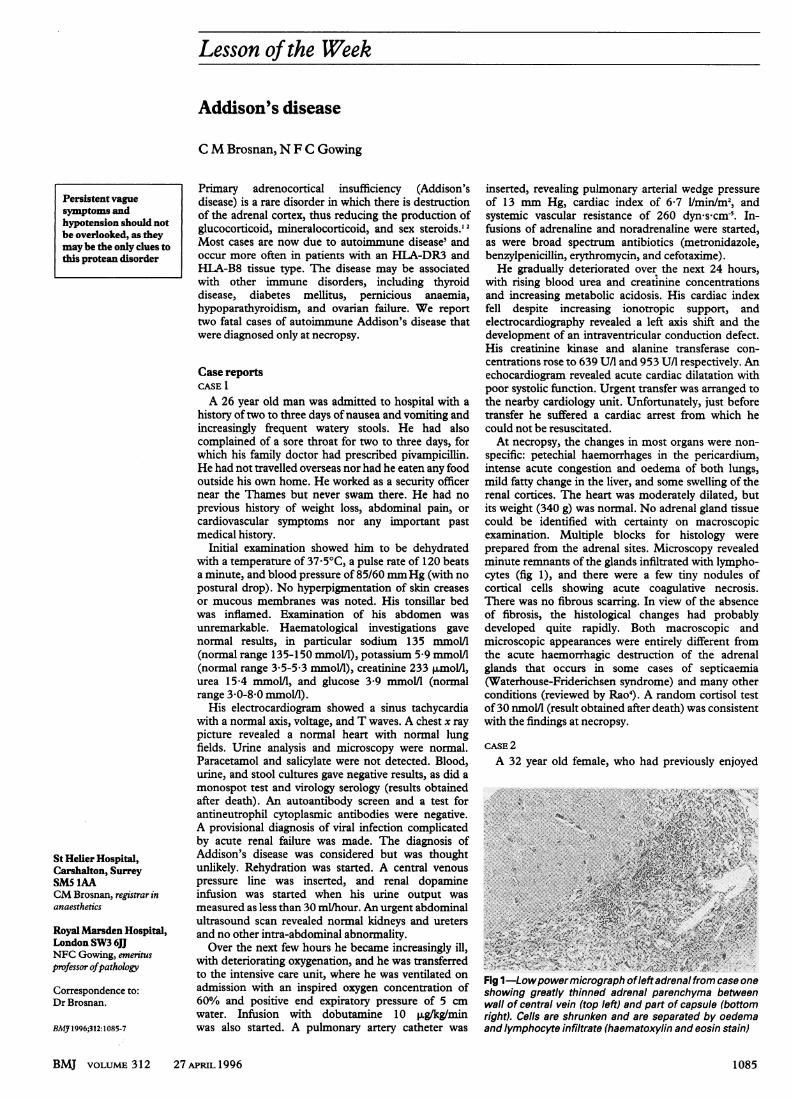
specific: petechial haemorrhages in the pericardium,intense acute congestion and oedema of both lungs,mild fatty change in the liver, and some swelling of therenal cortices. The heart was moderately dilated, butits weight (340 g) was normal. No adrenal gland tissuecould be identified with certainty on macroscopicexamination. Multiple blocks for histology wereprepared from the adrenal sites. Microscopy revealedminute remnants of the glands infiltrated with lympho-cytes (fig 1), and there were a few tiny nodules ofcortical cells showing acute coagulative necrosis.There was no fibrous scarring. In view of the absenceof fibrosis, the histological changes had probablydeveloped quite rapidly. Both macroscopic andmicroscopic appearances were entirely different fromthe acute haemorrhagic destruction of the adrenalglands that occurs in some cases of septicaemia(Waterhouse-Friderichsen syndrome) and many otherconditions (reviewed by Rao4). A random cortisol testof30 nmol/l (result obtained after death) was consistentwith the findings at necropsy.
CASE 2
A 32 year old female, who had previously enjoyed
Fig 1-Lowpowermicrograph ofleftadrenal from case oneshowing greatly thinned adrenal parenchyma betweenwall of central vein (top left) and part of capsule (bottomright). Cells are shrunken and are separated by oedemaand lymphocyte infiltrate (haematoxylin and eosin stain)
BMJ VOLUME 312 27APRIL1996
Persistent vaguesymptoms andhypotension should notbe overlooked, as theymay be the only clues tothis protean disorder
St Helier Hospital,Carshalton, SurreySM5 IAACM Brosnan, registrar inanaesthetics
Royal Marsden Hospital,London SW3 6JNFC Gowing, emeritusprofessor ofpathology
Correspondence to:Dr Brosnan.
BMY 1996;312:1085-7
1085

good health, suffered from a prolonged flu-like illnessfor six weeks and subsequently began to feel generallyunwell. She developed generalised arthralgia andattended her local hospital over a period of ninemonths, and a diagnosis of inflammatory polyarthritiswas made. She subsequently suffered from chest pains,severe headaches, and increasing general malaise.During this period she was not found to be hypo-tensive, no hyperpigmentation was ever noted, and nobiochemical or electrolyte disturbances were everdetected.Two days before her death she felt increasingly ill
and developed diarrhoea and vomiting. She vomitedcopious amounts during the night. The family doctorwas called the next morning, and he prescribedco-phenotrope after diagnosing a viral infection. Thevomiting subsided and her symptoms seemed to settle,but she died suddenly at home early next day.At necropsy, most changes were non-specific:
petechial haemorrhages in the visceral pleural mem-branes, some acute oedema of the cerebrum withflattening of the convolutions, and multiple smallhaemorrhages in the gastric mucosa. Each adrenalgland was reduced to a thickness of about 2 mm. Thethyroid gland was normal in size but extremely pale,especially its right lobe. Microscopy showed almostcomplete destruction of the adrenocortical tissue-theresidual parenchymal cells were irregular in shape witheosinophilic cytoplasm and distorted nuclei, and therewas a moderate lymphocyte infiltrate (fig 2). Thehistological changes were interpreted as autoimmuneadrenalitis (primary adrenal atrophy).
Sections of the thyroid gland showed infiltrationwith plasma cells and lymphocytes, lymphoid aggre-gates with reaction centres, reduction in the amount ofcolloid, and formation of Hurthle cells from thefollicular cells. The histological features were those ofHashimoto's thyroiditis.
DiscussionOur two case reports represent two contrasting
presentations of the same disease; the first was an acutesevere illness in a previously well man, while the otherwas a two year history of vague symptoms in a youngwoman. However, both cases ended in addisoniancrises, probably precipitated by viral infections.
Because the adrenal glands have a large reserve,more than 90% of the gland has to be destroyed beforesymptoms appear.5 Addison's disease may presentwith a wide range of symptoms and signs.6 Theseinclude weakness; anorexia and malaise7; hyper-pigmentation (particularly of the gingival mucosa,scars, and skin creases) and vitiligo8; postural hypo-tension; abdominal pain; nausea, vomiting, diar-rhoea, and constipation9; myalgia and arthralgia.Neuropsychiatric manifestations have also beendescribed, including confusion, psychosis, and de-pression.'0 Adrenal crisis is characterised by therapid onset of hypotension, tachycardia, fever, andhypoglycaemia and a progressive deterioration inmental status."
Biochemical abnormalities occur in 92% of patients,6most notably hyponatraemia and hyperkalaemia withan increased (or normal) blood urea concentration.In a representative population of hospital inpatientsthese typical biochemical abnormalities commonlyoccurred.'2 Other abnormalities include hypoglycaemia(which may be symptomatic), hypercalcaemia,metabolic and respiratory acidosis, normocyticanaemia, and moderate neutropenia with relativeeosinophilia and lymphocytosis."3 Elevated concentra-tions of hepatic enzymes have also been described, anda report described three patients who were investigatedfor an isolated increase in serum aminotransferase
E~~~~~~~~~~~~~~* (
.5 .45
Fig 2-High power photomicrograph of left adrenal fromcase 2 showing small, dissociated clusters of cortical cellsseparated by oedema and lymphocyte infiltrate. Part ofcapsule is in bottom right corner (haematoxylin and eosinstain)
concentration and were found to have Addison'sdisease.'14The normal ranges of cortisol and adrenocortico-
trophic hormone concentrations are broad. Thus,random serum samples for cortisol are unhelpful, anddynamic tests of function should be used. It has beenshown that in patients with septic shock low cortisolconcentrations are not uncommon and can be ac-companied by loss of diurnal cortisol rhythm.'5 Thereare, however, problems in deciding what is a "normal"response to a synacthen test in a critically ill patient,which makes diagnosing Addison's disease in such apatient much more difficult. When performning asynacthen test, it may be useful to take a basal plasmasample to measure adrenocorticotrophic hormnoneconcentration: a plasma concentration ofover 44 pmol/lis diagnostic. 16 However, this may also be unreliable, asa study of adrenocortical function in acutely ill patientsin an intensive care unit and in non-acutely ill, stressedpatients found adrenocorticotrophic hormnone con-centrations of 9-72 pmol/l and 7-141 pmoUl respec-tively. 7These two cases provide several important lessons:
* Persistent vague symptoms should not be over-looked, especially in the presence of other autoimmunedisorders (though antibodies are detectable in only60-70% ofpatients with idiopathic disease"8)* Hyperpigmentation is not a universal feature,especially if the cause is autoimmunel (it is absent in upto 8% of patients and has led to delayed diagnoses inother cases"9* Hypotension is a common occurrence on theITU, but persistent hypotension despite increasingionotropic support in any patient with suspectedsepticaemia should raise the possibility of undiagnosedadditional pathology (lack of cortisol causes a decreasein myocardial contractility, and a decreased vascularresponse to catecholamineS20)*Addison's disease remains the unforgiving master
of non-specificity and disguise.
We thank Mr Paul Rose, coroner, for permission to publishthese cases. We also thank Drs M Bending and M Stockwellfor permission to publish case 1, and Dr A Said for permissionto publish case 2. We thank Dr Rosemary Millis for her helpin preparing the photomicrographs.
1ukeC.Adeoorialdsass n:Waheal J edrgamJG
Warrl A d.Ofr etoko eiie xod xodUiest
Fg2-HeigM hepoenorinphotomicorpofhefadrenalfrtxn cGeJ scomcaser2gshowin esmal dissocitd clustersofcahlg.Oxodxortiacnielsit
caslDoisc in bdottmriAGhthornryL (htaemG.Autoylinitand teoi
diAseaste.' nedciedsass4eetAvnesinornlg
c Ronentratossvbiaelare enotuncomonrhaneMd Cain bert Ac-copneby579 los ofdura7crislrhtm.9Thr
1086 BMJ VOLUME 312 27APRIL1996

5 Liddle GW. The adrenals. Text book of endorinology. 6th ed. Philadelphia: W BSaunders, 1981.
6 Nerup J. Addison's disease-clinical studies. A report of 108 cases. AcaaEndocrinologica 1974;76:127-41.
7 Ringstad J, Rodge S, Loland W, Rode L Rapidly fatal Addison's disease: 3case reports. J Intern Med 1991;230:465-7.
8 Mulligan TM, Sowers JR. Hyperpigmentation, vitiligo and Addison's disease.CGtus 1985;36:317-8.
9 Tobin MV, Aldridge SA, Morris Al, Belchetz PE, Gilmore IT. Gastrointestinalmanifestations ofAddison's disease. AmyGasroenterol 1989;84:1302-5.
10 Varadaraj R, Cooper AJ. Addison's presenting with psychiatric symptoms. AmJPsychiatty 1986;143:553-4.
11 Leshin M. Acute adrenal insufficiency: recognition, prevention and manage-ment. UrolChn NorthAm 1982;9:229-35.
12 Paterson JR, Neithercut WD, Spooner RJ. Delayed diagnosis of Addison'sdisease. Ann Clin Biochem 1990;27:378-81.
13 Burke CW. Adrenocortical insufficiency. Clinics in Endocrinoogy andMetabolism 1985;14(4):947-76.
14 Olsson RG, Lindgren A, Zettergren L Liver involvement in Addison'sdisease. Amj Gastroenterol 1989;84:1302-5.
15 Moran JI, Chapman MJ, O'Fathartaigh MS, Peisach AR, Pannall PR,Leppard P. Hypocortisolaemia and adrenocortical responsiveness at onset ofseptic shock. Intensive CareMed 1994;20:489-95.
16 Clayton RN. Diagnosis ofadrenal insufficiency. BMJ 1989;298:271-2.17 Drucker D, Shandling M. Variable adrenocortical function in acute medical
illness. Cni Care Med 1985;13:477-9.18 Orth DN, Kovacs WJ, DeBold CR The adrenal cortex. In: Wilson JD, Foster
DW, eds. Wiliams tatbook of endocrinology. 8th ed. Philadelphia: WBSaunders, 1992:489-619.
19 Runcie CJ, Semple CG, Slater SD. Addison's disease without pigmnentation.Scot MedY 1986;31:111-2.
20 Ramey ER, Goldstein MS. The adrenal cortex and the sympathetic nervoussystem. PhysiolRev 1957;37:155-95.
(Accepted 18 December 1995)
Grand Rounds- Hammersmith Hospital
Reactive (AA) systemic amyloidosis
A cause ofrefractory nephrotic syndrome
Department ofMedicine,Royal PostgraduateMedical School,Hammersmith Hospital,London W12 ONNCase presented by:A R Allen, registrar innephrology
Chairman:J Scott, professor ofmedicine
Discussion group:E Clutterbuck, senior lecturerin nephrologyM J Walport, professor ofrheumatologyK Davies, senior lecturer inrheumatologyJ Wilding, senior registrar inendocrinologyM Thompson, consultanthistopathologist
Series edited by:DrW A Lynn.
BMJ 1996;312:1087-9
Reactive systemic (AA) amyloidosis occurs as a con-sequence of a prolonged acute phase response with thedeposition of amyloid fibrils in various tissues. Renalinvolvement with the nephrotic syndrome is common,and reactive systemic amyloidosis should be con-sidered in all patients with the nephrotic syndrome.Chronic infective foci have long been recognised as acause of reactive systemic amyloidosis, and patientswith this condition should be evaluated for occultinfection. We describe such a case, in which anundetected subphrenic abscess, possibly of nine years'duration, presented with severe nephrotic syndrome.
Case historyA 49 year old woman first presented to her local
hospital in 1984 with abdominal pain and haema-temesis. A duodenal ulcer was diagnosed, and she wastreated with H2 antagonists. An abdominal ultrasoundscan showed no abnormalities. She presented again in1989 with severe left sided abdominal pain, and a chest
s-- i9ji........ ....................-f.5-.;Z.. ?~~~~~ ~ ~~ ~ ~ ~ ~~~~~~~~~~ ,...$|i | .
::,.''fS':'~ ~~~~~~~~~~~~~~~~~~~~~~~~~~~~ ..........................................
~~~~~...S~~~~~~~~~~~~~~.... ::::-" ' : .. ...W ...
Fig 1 Chestx ray film showing raised left hemidiaphragmwith patchy basal atelectasis
*..:~~~~~~~~~~~~~~........::;: .
:.:.::.:.-:::::::::.:::: :::::..:::::::.::: ::::.:.. :: ::::::.--:
Fig 2-Abdominal computed tomogram at level of livershowing large left subdiaphragmatic collection withassociated pleural effusion
x ray film showed bibasal consolidation in associationwith serological evidence of acute mycoplasmainfection. She was treated with erythromycin andrecovered. (All chest x ray films after this episodeshowed a raised left hemidiaphragm (fig 1).)
In September 1993 she was admitted with similarsevere left upper quadrant pamn but was nowoedematous, with serum albumin concentration 11 g/l,proteinuria 5 g/24 hours, a creatinmne clearance of66 mI/min, and an acute phase response (C reactiveprotein 60 mg/l (normal <10 mg/l)). Eventually, arectal biopsy showed the presence of amyloid, and shebriefly received steroid treatment, which was of nobenefit. She was transferred to this hospital in earlyDecember 1993 for further management.On arrival she was extremely unwell with massive
oedema, ascites, and bilateral pleural effusions. Shewas afebrile, had a blood pressure of 100/60 mm Hgwith a postural drop of 20 mmHg in systolic pressure,and was tender in the left upper quadrant. She had aserum creatinine concentration of 169 ,umol/l,creatinine clearance of 37 ml/min, and serum albuminconcentration of 3 g/l. She had 7.5 g protemunura per24 hours. She had received a blood transfusion tomaintain her haemoglobin concentration at around100 g/l and still had a C reactive protein concentrationof 117 mg/I and serum amyloid A concentration of45 mg/I (normal <10 mg/I). Blood cultures weresterile, and an abdominal computed tomogram showeda well defined left subphrenic collection of fluid (fig 2).
BMJ voLuME312 27APmL1996 1087