1 Anatomy & Physiology of Cells Chapters 3 & 4 Anatomy & Physiology.
Upload
puneeth-isloorCategory
view
183download
15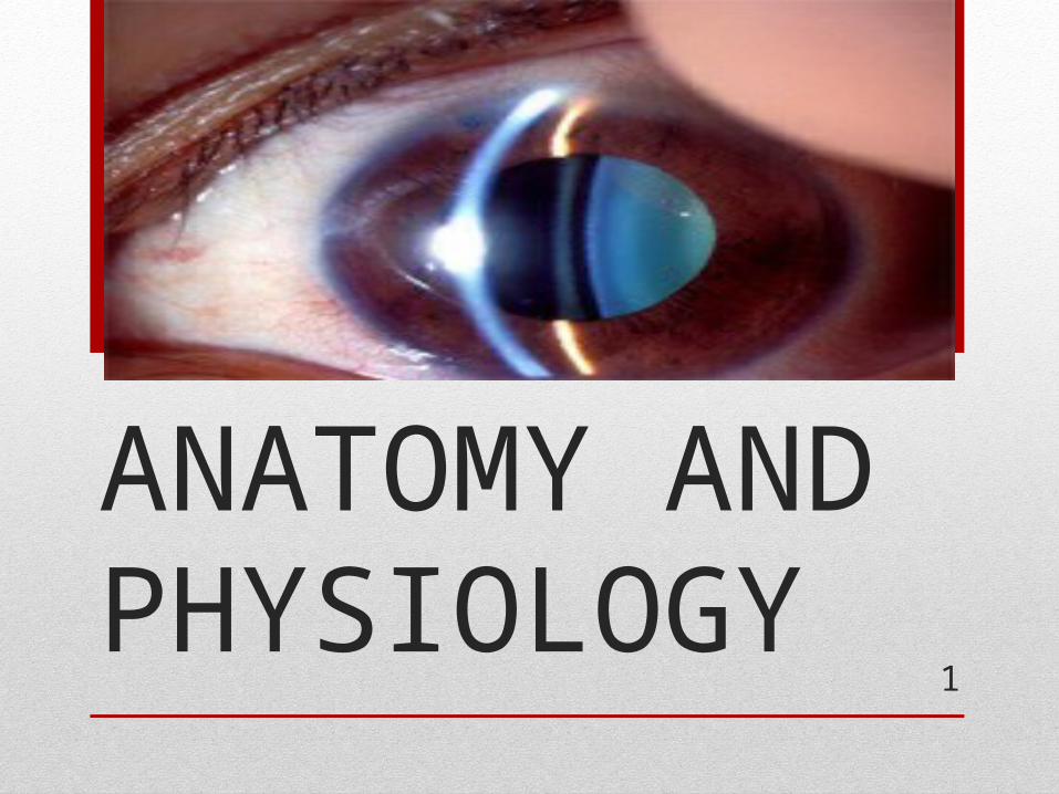
1
LENS – ANATOMY AND PHYSIOLOGY

2
EMBRYOLOGY
EYEBALL DEVELOPMENT• - Outgrowth from prosencephalon
called optic vesicle• - Specialised area of surface ectoderm
called lens placode

3
• Lens placode is identified by 27 days gestation• Lens placode invaginates sinus below
surface ectoderm to form lens vesicle- Lens vesicle – single layer of cells
covered by a basal lamina
- Primary lens fibres –
develop from posterior wall of vesicle- Secondary lens fibres –
develop from equatorial cells of ant.epithelium
- Tunica vasculosa lentis – surrounds lens by
9 weeks

4

5
EMBRYONIC NUCLEUS – 1 – 3 Months of GA
- Sutural pattern - 2 Y sutures
FETAL NUCLEUS – 3 months GA – Birth
INFANTILE NUCLEUS – Birth to puberty
ADULT NUCLEUS - Adults

6
• Lens – Trasparent , Biconvex , crystalline structure
Location – PATELLAR FOSSA – saucer shaped depression
Posterior capsule of the lens is attached to the vitreous with
WEIGERT ‘S LIGAMENT – A circular area of attachment
between the hyaloid face and the lens
capsule – potential space called the BERGER ‘S SPACE

7
DIMENSIONS
Equatorial diameter of lens – 6.5mm at birth ,increases to 9-10 mm in second decade
Thickness – 3.5 mm at birth ,increases to 5 mm with increase in age
Weight – 255 mg at 40 – 50 years of age
8
• SURFACES• - Anterior surface – Part of a sphere of radius 10 mm(8-14)
Centre – Anterior pole• - Posterior surface – More curved than anterior -6 mm
Centre – Posterior pole• - The two surfaces meet at the equator
- REFRACTIVE INDEX
Nucleus – 1.42
Cortex – 1.38
Refractive power – 16-17 dioptres
Accomodative power – 14-16 D at birth
- 7 – 8 D at 25 years of age
- 1 – 2 D at 50 years of age

9
• BIOMICROSCOPIC STRATIFICATION OF LENS
- Capsule
- Superficial cortex – 3 zones – C1α,C1β,C2
- Deep Cortex – C3 and C4
- Nucleus
SURGICAL ANATOMY
- Capsule
- Cortex
- Epinuclear plate
- Nucleus
10
STRUCTURE OF THE LENS
• LENS CAPSULE - Thickest BM in the body – 2- 20um thick
- Type 4 Collagen and GAG
-Thinnest at posterior pole
- Ultramicroscopy – Lamellar appearance – lamellae fine filaments

11
-Lens capsule is similar to other basement membranes in
the body – Glomeruli , Blood vessels, spleen ,lung
-Pseudo exfoliation of lens capsule – abnormal formation of capsule like material on the lens capsule,ciliary processes,posterior iris layer
- True exfoliation – Lamellar splitting of the capsule – found among glassblowers

12
ANTERIOR LENS EPITHELIUM
- 3 Zones
-Central zone – Cuboidal cells with round apically located nuclei
- Normally no mitosis occurs here
- Metaplasia – Myofibroblasts – Shield cataract
and glaucomflecken
-Intermediate zone – Smaller and more cylindrical cells –Mitosis+

13
-Germinative zone – Columnar peripheral cells – pre equatorial
- Actively divide to form new cells
- New cells migrate posteriorly – Lens fibres
- Dysplasia –PSCC – radiation etc

14
• Lens epithelium is Metabolically very active• Has a prominent cytoskeletal framework• Lateral membranes of the cells – have gap junctions-
Contains Na- K ATPases and Acid phosphatases• Apical membrane of Lens epithelium ---- interfaces ---
apical membrane of fiber cells – Epithelial Fiber cell interface

15
• LENS FIBRES
- Derived from anterior epithelium –hexagonal in cross section
- Newer fibres are laid upon older ones – high ribosomal content
- New fibres have nuclei temporarily and later disappears
- LENS BOW or NUCLEAR BOW
- The areas in which anterior and posterior fibres meet are called the
sutures – there are 2 Y shaped sutures in early stages of
human lenses
- Later on – irregular dendritic pattern forms.

16
17
• CILIARY ZONULES• Zonules of Zinn or suspensory ligaments of lens
• They run from ciliary body and fuse into lens capsule at equator
• Fibres are made up of glycoproteins and mucopolysaccharides• Microfibrils have a diameter of 8-40nm
• 3 types of zonular fibres
- First type fibres – THICK STRONG fibres
- Second type fibres – THIN FLAT
- Third type fibres – Very fine and run a circular course

18
• OLD CONCEPT OF ARRANGEMENT• Orbiculo- posterior capsular fibres – second type• Orbiculo – anterior capsular fibres – first type• Cilio –posterior capsular fibres – arise from the valleys• Cilio – equatorial fibres – third type fibres – abundant in
young people – decrease with age

19
• NEW CONCEPT OF ARRANGEMENT • Pars orbicularis –arise from posterior end of pars plana upto 1.5 mm
from the ora serrata• Zonular fibres – lie in the valley between ciliary processes• Zonular fork• Zonular limbs - Anterior limb – dense and insert at 1.5 mm from equator
- Equatorial limb
- Posterior limb

20
CANAL OF HANNOVER – Circumferential space between anterior and posterior zonular fibres around the equator of lens
• Zonules have 7% cysteine and failure to convert the amino acid homocysteine into cysteine results in broken zonules and dislocated lenses in homocystinuria

21
• LENS – Composed of water (65%) and proteins(34%)• Other constituents are Lipids ,Inorganic Ions ,Glucose(1%)
• WATER
- Lens is a relatively dehydrated organ
- Dehydration is maintained by the Active sodium pump
- Cortex is more hydrated
- No significant alteration in lens water with age
BIOCHEMICAL COMPOSITION

22
• LENS PROTEINS
• Lens proteins
SolubleCrystallins (85%)
InsolubleAlbuminoids(15%)
Alpha crystallins
Beta crystallins
Gamma crystallins

23
• Protein content of lens is higher than that of any organ in the body
• Other minor proteins in the lens are Phosphoproteins, Glycoproteins, Lipoproteins ,Flourescent proteins
• As lens ages , Soluble alpha crystallins get gradually converted to albuminoids
• Cortex – rich in Crystallins• Nucleus – rich in Albuminoid• Lens proteins – Immunologically privileged site
24
• Amino acids in the lens – All amino acids except Tryptophan , Cysteine and hydroxyproline
• Higher concentration of amino acids in lens compared to
Aqueous humor and vitreous humor
• Amino acids are actively transported into the lens - ensures that the protein synthesis is not hampered

25
• Amino acid concentration of lens is not affected by Ageing, fasting or starvation
• Glutathione is a chief constituent – it exists in the reduced
state (GSH) in the lens .Acts as an anti-oxidant in the lens

26
• CARBOHYDRATES
- Lens contains glucose, fructose and glycogen
- Glucose level of normal lens is 20-120mg%
- Source – aqueous humor
- Glucose in lens is 1/10th of aqueous
- Glycogen in mainly in the nucleus and replaces gamma
crystallins with age

27
- Sorbitol , Inositol , Gluconic acid ,Glucosamine are also
found- Ascorbic acid – high concentrations in the lens -
involved in oxidation – reduction systems in the lens

28
• LIPIDS
- Lipids represent 2% of the dry weight of lens
- Lipids – Cholesterol(50%)
- Phospholipids(45%) - mainly sphingomyelin
- Glycosphingolipids and ceramides(5%)
- They constitute major part of lens fibre membranes
- Decrease in their synthesis or impaired degradation ----
membrane damage and lens opacities
- Sphinomyelin and its precursor – ceramides – increase in
senile cataracts.

29
METABOLIC PATHWAYS
GLUCOSE METABOLISM • Occurs via 4 pathways • GLYCOLYSIS • KREB S CYCLE• HMP SHUNT PATHWAY• SORBITOL PATHWAY

30

31
PATHWAY INTERMEDIATES END PRODUCTS GLUCOSEMETABOLISED(%)
NO.OFATP
Glycolysis Glu-6-po4 , fru-1,6-dipo4, pyruvate
Lactate 80% 2
Kreb’s cycle Tricarboxylic acids CO2,H20, 5% 36
HMP shunt Pentoses Co2,NADPH 15% -
Sorbitolpathway
Sorbitol, Fructose
Lactate 2

32
• WATER AND ELECTROLYTE TRANSPORT
-Electrolyte and water content of lens resembles an intact
cell while that in the aqueous n vitreous resembles plasma
-Na+-K+ ATPase in the anterior epithelium uses ATP to extrude Na + and take up K+ into the cell by Active transport

33

34
- Cations – Na , K – 145 mEq/L and Anions – 50-60mEq/L. - The equilibrium is disrupted if concentration of osmotic substance
increases in lens – lens hydration –swelling
- Amino acids are actively transported into cells – there are 3 different pumps for acidic ,basic and neutral amino acids
- The transfer of cations and amino acids occurs only at anterior surface by active transport and also inositol – active transport
- Glucose transport occurs by simple and facilitated diffusion

35
• FACTORS MAINTAINING LENS TRANSPARENCY
-Regular arrangement of lens fibres – lamellar conformation
-Single layer of epithelial cells - Semipermeable lens capsule
- The relative dehydrated state of the lens- Avascularity of the lens
- High concentration of reduced glutathione in lens ---maintains lens proteins in reduced state -----ensures integrity of the cell membrane pump

36
• CATARACTOGENESIS
• Factors implicated in Senile cataract
- Exposure to UV radiation
-Dietary factors
-Severe Diarrhoea
-Diabetes
-Renal failure

37
• UV rays
• Absorbed by tryptophan
• Tryptophan N-Formyl kynurenine
• photosensitizatione and production of free radical oxygen
• Downregulation of Na/K ATPase
• Lens swelling and opacification

38
• INCREASING AGE
• Decrease in pump reduced oxidative
mechanism reactions
Reversal of Na/K ratio decreased amino acids
Hydration of lens fibres decreased protein
synthesis
Denaturation of lens proteins Opacification of
cortical lens fibres
39
• CHANGES IN CORTICAL CATARACT
- Progressive decrease in lens proteins and free amino acid
levels in lens
- Conversion of soluble proteins into insoluble proteins
- Decreased synthesis of lens proteins and increased protein
catabolism
- Decrease in the level of amino acids due to leakage
from disrupted membrane
-

40
Alteration in electrolyte and water content of lens- water
content increases from immature to hypermature cataract• Sodium and calcium content of the lens ---INCREASES• Potassium content – DECREASES

41
NUCLEAR CATARACT -Dehydration and compaction of nucleus - Increase in water insoluble proteins – Increases light scattering - Brown colour of lens – due to urochrome –absorbs light

42
• ROLE OF GLUTATHIONE• Glutathione is made of –Glycine,cysteine and glutamic acid
• Most of Glutathione in lens is in reduced form GSH
2GSH +1/2 02 GSSG + H20
• GSH levels in cortex is higher than nucleus
• HMP Shunt pathway generates NADPH which maintains
glutathione in reduced state

43
Lens proteins contain reduced sulfhydryl groups(PSH) and
oxidised disulfide groups (PSSP).
2PSH + GSSG 2GSH + PSSP
- Thus decreased GSH will result in PSH oxidation
alteration in protein linkages, transparency and their solubility
- Glutathione provides lens with a major detoxifying
mechanism via glutathione peroxidase

44
• DIABETIC CATARACTS• 2 theories – 1) Sorbitol pathway and 2) Glycosylation pathway
1) Excess glucose is directed into sorbitol pathway
aldose reductase
Glucose Sorbitol
NADPH NADP
Sorbitol accumulates in lens Osmolarity of lens increases
NADPH is depleted Water content increases
Water clefts and vacuolesReduced GSH is depleted

45
• Non enzymatic glycosylation of the lens proteins occurs
• Excessive glycosylation of lens capsule ------ increased fragility of lens capsule
• Structural changes in Enzymes,membrane proteins and crystallins in lens
GALACTOSEMIC CATARACT- Excessive Galactose Galactitol(Dulcitol)
in lens Aldose reductase- Galactitol accumulates and draws water
Water in lens fibres ruptures membranes and causes loss of K,Amino acids and Inositol

46
• CATARACT DUE TO UVEITIS /INTRAOCULAR SURGERY / RETINITIS PIGMENTOSA
- In uveitis ----influx of plasma components into aqueous
- Following vitreo-retinal surgery ---breakdown of blood-vitreous barrier ----posterior subcapsular cataracts
- In RP – breakdown of the blood vitreous barrier by destruction of pigment epithelium.
-

47
- Plasma phospholipids – Lysophosphatidyl choline and Lysophosphatidyl ethanolamine are implicated –they leak into the eye from ocular circulation.
- Two Unsaturated Fatty acids –Arachidonic acid and DHA are also implicated
- Macrophage oxidative enzymes can cause lytic effect by generation of peroxides

48
• RADIATION CATARACT• -Ionising radiations – X rays,Gamma rays,Beta rays-
induce lens vacuoles and lens opacities
• Initially vacuoles appear in the equator and posterior subcapsular areas
• 3 mechanisms of damage by radiation
- Increased leakage of K,Inositol and glutathione
-Decreased incorporation of amino acids into proteins
- Arrest of cells entering mitosis and epithelial damage

49
• CORTICOSTEROID INDUCED CATARACT
- Steroids induce abberant differentiation and migration of
epithelial cells
- Other mechanisms
- Elevation of glucose
- Inhibits Glucose -6-po4 dehydrogenase decreases NADH and
reduced glutathione
Inhibits Na/K ATPase
-Chemical modification of crystallins --- disulphide cross links between lens fibres----causes aggregation of lens fibres

50

51
• PHYSIOLOGY OF ACCOMODATION• Accomodation is the ability to focus the diverging rays coming from a near
object onto the retina
• Nearest point at which objects can be seen clearly – Punctum proximum
• Farthest point –Punctum remotum
• Distance between these two points is called range of accomodation• The difference in Dioptric power needed to focus at near point and far point is
called amplitude of accomodation

52
• MECHANSISMS OF ACCOMODATION• 3C s – Constriction of pupil , Convergence of the eyes
- Increase in Anterior curvature of lens
THEORIES OF ACCOMODATION
1.Relaxation theory(Hemhotlz theory/capsular theory)
- Most accepted theory worldwide .
-WHEN EYE AT REST WHEN EYE ACCOMODATES
Lens is compressed by
Capsule due to tension
of zonules
Due to relaxation of
the ciliary muscles
Contraction of ciliary muscle occurs
Causes ciliary ring to shorten and move the equator lens forward
Zonules are relaxed
Tension on capsule is relieved
Lens becomes more spherical

53

54
• Other theories
2) Schahar ‘s Theory – Accomodation occurs when ciliary muscle contraction tenses the zonules
Lens stretches equatorially (coronally)
Only Central part of the lens bulges and moves anteriorly
According to this theory ,Presbyopia occurs due to a growth in the equatorial diameter of the lens with age
This in turn reduces the perilenticular space and hence ciliary muscle contraction can no longer tense the zonules

55
• OCULAR CHANGES IN ACCOMODATION• -Slackening of zonules
• - Changes in anterior curvature of lens – radius of curvature decreases from 11 mm to 6mm
• - Anterior pole of lens moves forward
• Axial thickness of lens increases
• Constriction of pupil and convergence of eyes
• Choroid is stretched forwards and so is the ora serrata which moves forward by 0.05 mm
56
• PATHOPHYSIOLOGY OF PRESBYOPIA
Presbyopia occurs due to- Changes in the elastic property of lens capsule- Sclerosis or hardening of the lens- Weakening of the ciliary muscle
Near point changes with age - 7cm at the age of 10 years- 25 cm at the age of 40 years- 33 cm at the age of 45 years- 50 cm at the age of 50 years

57THANK YOU