Lecture 7 DNA REPLICATION - Rutgers Universitychem.rutgers.edu/~kyc/Teaching/Files/543-05/09 544-07...
34
Lecture 7 DNA REPLICATION 1. A double helix separate into two single strands and each strand serves as a template on which complementary strand is synthesized. 2. A mechanism is required to separate the strands locally for replication. 3. A mechanism is required to release the strain created by local unwinding. 4. A mechanism is required to account for the high fidelity of duplication, 1 per 10 10 nt. 5. A mechanism is required to account for the speed of replication; 2000 nt per sec in E.coli. 6. Direction of the replication? 7. How to replicate the ends of linear DNA?
Transcript of Lecture 7 DNA REPLICATION - Rutgers Universitychem.rutgers.edu/~kyc/Teaching/Files/543-05/09 544-07...

Lecture 7 DNA REPLICATION
1. A double helix separate into two single strands and each strand serves as a template on which complementary strand is synthesized.
2. A mechanism is required to separate the strands locally for replication.
3. A mechanism is required to release the strain created by local unwinding.
4. A mechanism is required to account for the high fidelity of duplication, 1 per 1010 nt.
5. A mechanism is required to account for the speed of replication; 2000 nt per sec in E.coli.
6. Direction of the replication?
7. How to replicate the ends of linear DNA?

DNA double helix structures

Meselson Stahl Experiment
Three replication models: conservative, semiconservative, and random disperse models
Semiconservative replication

Three models: continuous, semidiscontinuous, and discontinuous.
Replication fork

Reiji Okazaki's predictions and experiments Semidiscontinuous model
at least half of the newly synthesized DNA appears first as short pieces(1000-2000 nt);
Reiji Okazaki's experiments: at least half of the newly synthesized DNA appearsas short pieces(1000-2000 nt); if no ligation, short pieces will accumulate. Replication of T4 phage DNA in E. coli, wild type vs. ligase mutant, with 3H-thymidine pulse labeling (when ung- E. coli mutant was used, >50% of newly labeled DNA was still in short pieces).
wt mutant
Replication fork

Tuneko Okazaki's experimentPriming
Primer will be removed by a 5’ to 3’ exonuclease activity in DNA polymerase I.
DNA pol needs s short RNA primer
Use fragment from normal (d,h) and mutant cells (a-c, e-g) labeled the primer with 32P-GTP. Lanes a,e, no RNase H;lanes b,f no nuclease activity;lanes c, g no RNase H and nuclease;
lanes d, h wild type;
DNase before after

Three possible mechanisms
Direction of Replication
Predicted vs. data (JMB 32, 327, 1968); heavily labeled pulse followed by lightly labeled pulse.
Bidirectional replication in Bacillus subtilis DNA (John Cairns, JMB 58, ’73): lightly label followed by heavily label;

Regardless of organism, replication origins are unique DNA segments with multiple short repeats, recognized by multimeric origin-binding proteins, and usually contain an A-T rich stretch.oriC: origin of replication in E. coli: OriC 245 bp (3 13-nt and dnaA binding sites) in 4.8 m bp genome.
Origin of replication
The consensus sequence of oriC
Minimal bacterial replication origin: 13-mer and 9-mers

Construction of an oriC plasmid
Origin of replication

Priming at oriC
DnaA binds to ATP forming multimers which together with HU bind to the four 9-mers (dnaA boxes), inducing bend and destabilizes the 13-mer repeats and causes local melting, allowing DnaB binding (with DnaC help)

Helicase for unwinding
E. coli DnaB helicase: the enzyme will translocate along dsDNA from 3’ to 5’ direction.

Helicase assay
Lane 1, before reaction, circular and linear form of M13-fragment;
lane 2 after reaction, JBC 261, 4740, 1986)
substrate (labeled 1.06-kb HinCII fragment (red) annealed to M13
reaction

Bacterial helicase
Bacterial helicase (PcrA): A1 with P-loop NTPase fold, B1 similar to A1 without loop
Conserved regions: A1 and B1 interface and along ATP binding site

Helicase for unwinding
For DNA duplex to replicate, the two strands must be separated from each other,at least locally. Helicaseuses ATP energy to perform this strand separation job. Both A1 and B1 bind to ss DNA. ATP hydrolysis leads to cleft closure and sliding of ssDNA. Since A1 has a tighter grip of ssDNA, this causes a net translocation of the enzyme toward the dsDNAUnwinding mechanism: (i) Here the A1 and B1 domain bind to ssDNA.
(ii) Upon ATP binding the cleft between A1 and B1 closes and A1 slides along DNA;(iii) Upon hydrolysis cleft opens, pulling B1 to A1.
B1A1

Polymerization
Reaction catalyzed by DNA polymerases and the formation of phosphodiester bond, DNA polymerase: 5’-p(N)n-3’ + dNTP 5’-p(N)n+1-3’

E. coli DNA polymerase I
DNA pol I= 1 polypeptide (polymerase, 3’-->5’ exonuclease proofreading, 5’-->3’exonuclease); processivity 20, catalytic rate 10 nt/secDNA pol III = 10 polypeptides, 900 kD, processivity >5000, rate 1000 nt/sec
E. coli DNA polymerase I: 102 kD Klenow fragment (polymerase activity + 3' 5' exonuclease activity) and small domain (5' 3' exonuclease activity); all DNA pol have similar shape, thumb, palm and finger.

Crucial metals
Two metal ions (Mg or Mn) are crucial to the action of DNA polymerase. One metal coordinates with 3’-OH at the primer whereas the alpha phosphate group from dNTPbridges between two metals
DNA pol III holoenzyme
Proposed structure of DNA pol III holoenzyme (900 kD, 10 subunits, asymmetric dimer, one for leading, one for lagging strand (α is polymerase, ε is proofreading 3' 5' exonuclease, β2 and δ2 for processivity). The sliding clamp for processivity is done by β2. DNA pol I = 1 polypeptide, processivity 20, catalytic rate 10 nt/sec. DNA pol III = 10 polypeptide, processivity >5000, rate 1000 nt

Fidelity
Fidelity of polymerase: R and Q from polymerase serve as a ruler by forming H-bonding at the minor groove of base pair at the active site.

Shape selectivity
The binding of the correct dNTP induces a conformational change, generating a tight pocket
Proofreading
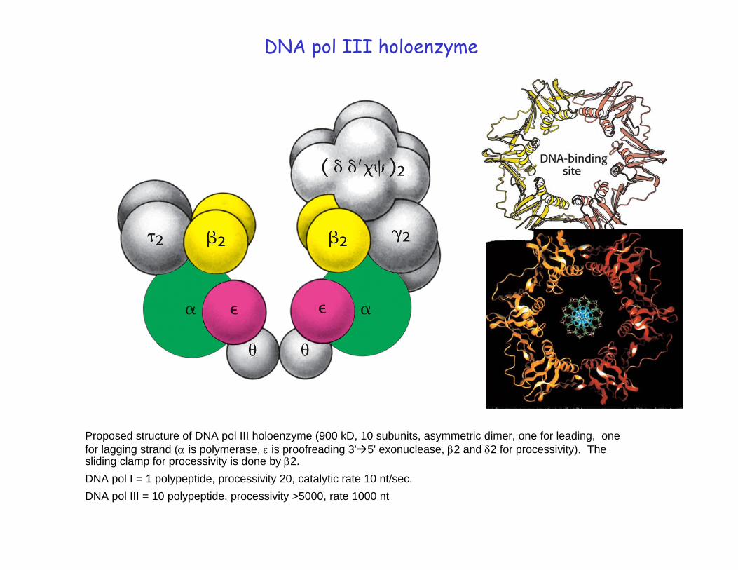
Mismatched base will cause pause or stall and give extra time to excise it. Mismatched base can leave polymerase site and swing into exonuclease site to be cleaved. The newly formed duplex in the polymerase site assumes A-form for extensive H-bonding at minor groove.

Cell 69, 427, 1992;84, 643, 1996
Processivity
DNA polymerase III has a dimeric structure. � is polymerase, β2 and δ2 confer the processivity. 1000 nt added per sec means a sliding of 100 turns of duplex through the central hole of β2 per sec

Ligase reaction

Overview of replication
Coordination between the leading and the lagging strands: looping of the template for the lagging strand enables a dimeric DNA pol III holoenzyme to synthesize both daughter strands

Detailed view of the E. coli fork
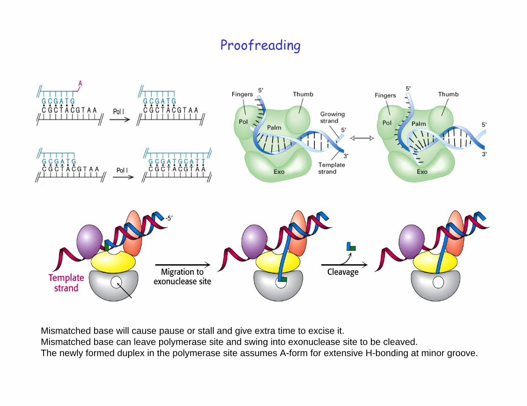
In vitro replication of SV40 DNA

Termination
Bacterial termination site: E. coli Ter sites (Ter E, D, A stop the ccw fork; Ter F,B,C stop the CW fork; Tus, terminus utilization
substance, binds to the terminator sites and helps arrest the moving forks).
EM of torus catenanes from replication of pBR322 in mutant S. typhimurium

add6 nt (GGGTTG), repeat many times
Termination
prokaryote
Eukaryote

Termination

Supercoil
Lk = Tw + Wr (linking number is the sum of twisting number and writhing number)

Supercoil
Separation of SV40 DNA topoisomers by gel electrophoresis. Lane 1, relaxed and maximally supercoiled DNA; lane 2 topo I for 3 min; lane 3, topo I for 30 min
EM picture of two topoisomers(molecules differ in linking numbers) showing relaxed circular and negatively supercoiled DNA
Nicking one strand relaxes supercoiled DNA

Two types of topoisomerases
Topo I binds and cleaves one stand. The broken strand will rotate around the other one and rejoin, which leads to partial or complete relaxation of a supercoil
Topo I action, from three negatives to 2 negatives
Human Topoisomerase I with DNA

Topo II cleaves both strands and create supercoiling
Action sof Topoisomerase II

1959 "for their discovery of the mechanisms in the biological synthesis of ribonucleic acid and deoxyribonucleic acid"
Severo Ochoa1905-1993
Arthur Kornberg1918-
1930-1975
2005
Okazaki