Lecture 5 Pharmacology -Hasani
8
1 Pharmacology 2020-2021 Lecture 5 Dr Noor Al-Hasani Antiarrhythmics The arrhythmias are dysfunctions cause abnormalities in impulse formation and conduction in the myocardium. Arrythmias is organised into groups according to the anatomic site of the abnormality: the atria, the AV node, or the ventricles. Physiology of Muscle Contraction The myocardium, like smooth and skeletal muscle, responds to stimulation by depolarisation of the membrane, which is followed by shortening of the contractile proteins and ends with relaxation and return to the resting state (repolarisation). Cardiac myocytes are interconnected in groups that respond to stimuli as a unit, contracting together whenever a single cell is stimulated. Action potential Cardiac myocytes are electrically excitable and have a spontaneous, intrinsic rhythm generated by specialised “pacemaker” cells located in the sinoatrial (SA) and atrioventricular (AV) nodes. Cardiac myocytes also have an unusually long action potential, which can be divided into five phases (0 to 4). Figure 1 illustrates the major ions contributing to depolarisation and repolarisation of cardiac myocytes. Figure 1: Action potential of a cardiac myocyte. ATPase = adenosine triphosphatase. Cardiac conduction system and the ECG (electrocardiogram) Analysis Cardiac conduction system is summarised and demonstrated in figure 2. Effective refractory period (ERP)
Transcript of Lecture 5 Pharmacology -Hasani
1
Pharmacology
2020-2021
Lecture 5
Dr Noor Al-Hasani
Antiarrhythmics
The arrhythmias are dysfunctions cause abnormalities in impulse formation and conduction in the myocardium. Arrythmias is organised into groups according to the anatomic site of the abnormality: the atria, the AV node, or the ventricles. Physiology of Muscle Contraction
The myocardium, like smooth and skeletal muscle, responds to stimulation by
depolarisation of the membrane, which is followed by shortening of the contractile
proteins and ends with relaxation and return to the resting state (repolarisation).
Cardiac myocytes are interconnected in groups that respond to stimuli as a unit,
contracting together whenever a single cell is stimulated.
Action potential
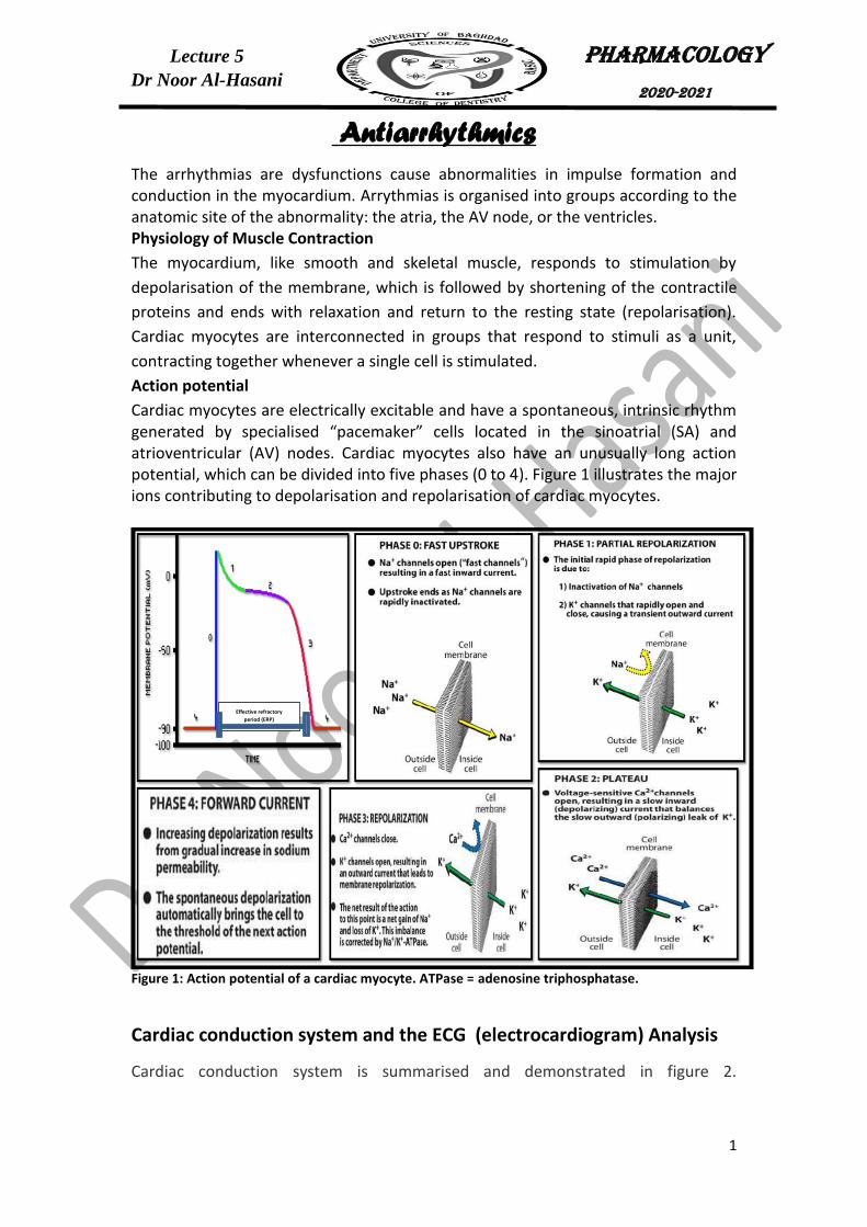
Cardiac myocytes are electrically excitable and have a spontaneous, intrinsic rhythm generated by specialised “pacemaker” cells located in the sinoatrial (SA) and atrioventricular (AV) nodes. Cardiac myocytes also have an unusually long action potential, which can be divided into five phases (0 to 4). Figure 1 illustrates the major ions contributing to depolarisation and repolarisation of cardiac myocytes.
Figure 1: Action potential of a cardiac myocyte. ATPase = adenosine triphosphatase.
Cardiac conduction system and the ECG (electrocardiogram) Analysis
Cardiac conduction system is summarised and demonstrated in figure 2.
Effective refractory
period (ERP)
2
Pharmacology
2020-2021
Lecture 5
Dr Noor Al-Hasani
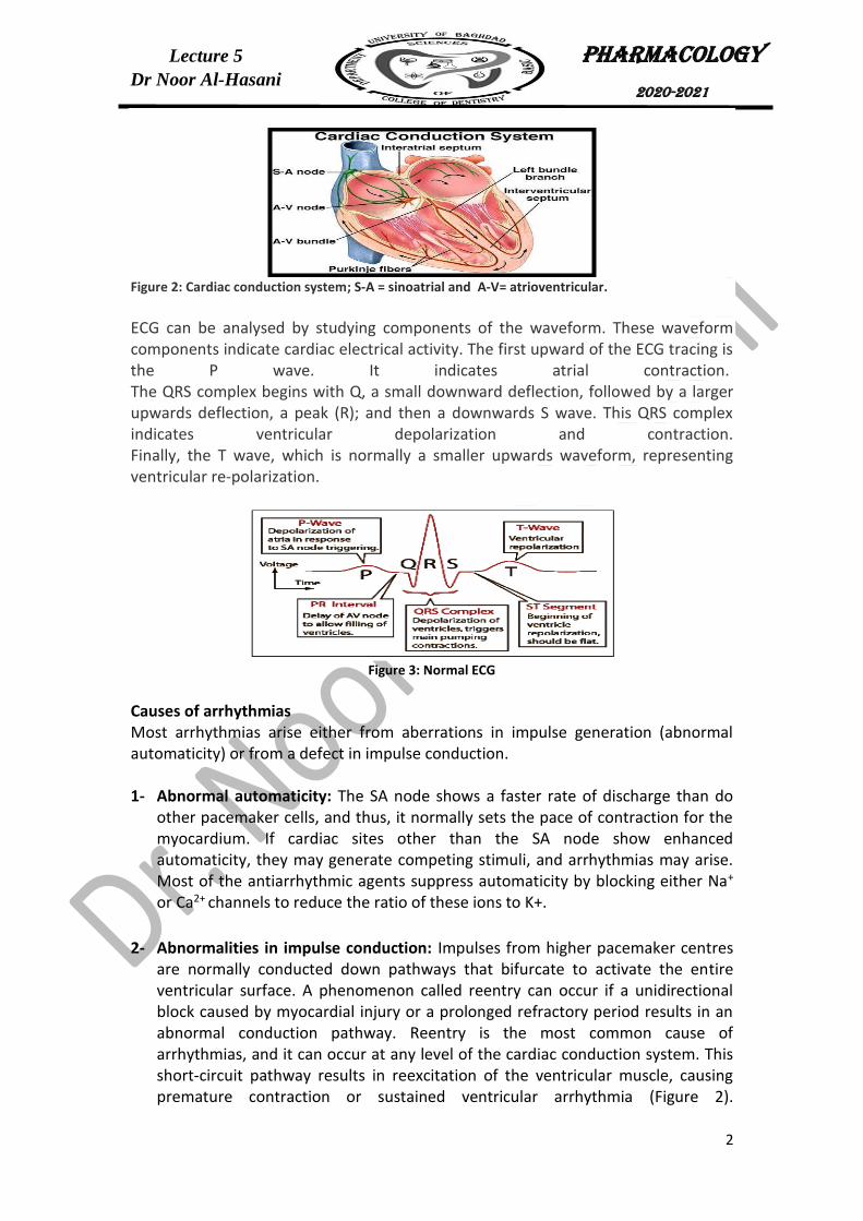
Figure 2: Cardiac conduction system; S-A = sinoatrial and A-V= atrioventricular.
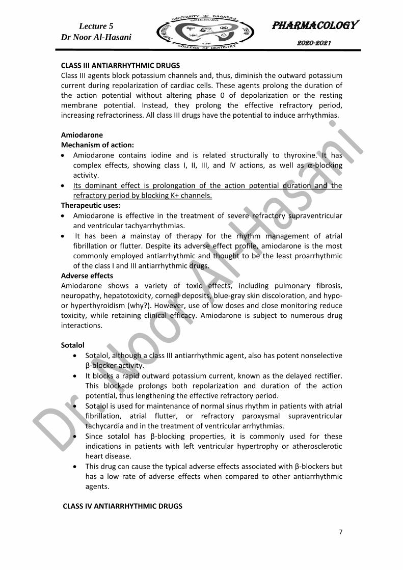
ECG can be analysed by studying components of the waveform. These waveform components indicate cardiac electrical activity. The first upward of the ECG tracing is the P wave. It indicates atrial contraction. The QRS complex begins with Q, a small downward deflection, followed by a larger upwards deflection, a peak (R); and then a downwards S wave. This QRS complex indicates ventricular depolarization and contraction. Finally, the T wave, which is normally a smaller upwards waveform, representing ventricular re-polarization.
Figure 3: Normal ECG
Causes of arrhythmias Most arrhythmias arise either from aberrations in impulse generation (abnormal automaticity) or from a defect in impulse conduction. 1- Abnormal automaticity: The SA node shows a faster rate of discharge than do
other pacemaker cells, and thus, it normally sets the pace of contraction for the myocardium. If cardiac sites other than the SA node show enhanced automaticity, they may generate competing stimuli, and arrhythmias may arise. Most of the antiarrhythmic agents suppress automaticity by blocking either Na+ or Ca2+ channels to reduce the ratio of these ions to K+.
2- Abnormalities in impulse conduction: Impulses from higher pacemaker centres are normally conducted down pathways that bifurcate to activate the entire ventricular surface. A phenomenon called reentry can occur if a unidirectional block caused by myocardial injury or a prolonged refractory period results in an abnormal conduction pathway. Reentry is the most common cause of arrhythmias, and it can occur at any level of the cardiac conduction system. This short-circuit pathway results in reexcitation of the ventricular muscle, causing premature contraction or sustained ventricular arrhythmia (Figure 2).

3
Pharmacology
2020-2021
Lecture 5
Dr Noor Al-Hasani
Antiarrhythmic agents prevent reentry by slowing conduction (class I drugs) and/or increasing the refractory period (class III drugs).
a- Acute ventricular tachycardia: This arrhythmia is a common cause of death in
patients who have had a MI. Cardiac output is impaired, and tachycardia may deteriorate into ventricular fibrillation. Therefore, ventricular tachycardia requires prompt management.
b- Ventricular fibrillation (V-fib): In V-fib, the lower heart chambers contract in a very rapid and uncoordinated manner. Ventricular fibrillation may also cause sudden cardiac arrest and lead to death if not treated immediately.
Antiarrhythmic drugs Antiarrhythmic drugs can modify impulse generation and conduction to prevent arrhythmias from occurring or to reduce symptoms associated with arrhythmias. Unfortunately, many of the antiarrhythmic agents are known to have dangerous proarrhythmic actions—that is, to cause arrhythmias. Inhibition of potassium (K+) channels (typically thought of as class III activity) widens the action potential and can, thus, prolong the QT interval. If prolongation is excessive, these drugs increase the risk of developing life-threatening ventricular tachyarrhythmias (torsades de pointes). The most common cause of QT prolongation is drug-induced, although other conditions (for example, ischemia and hypokalaemia) and genetic profiles may contribute. QT prolongation is not only seen with class III antiarrhythmics. Many drugs are known to prolong the QT interval, such as macrolide antibiotics and antipsychotics. Caution should be employed when combining drugs with additive
Main types of arrythmias 1- Atrial arrhythmias
a- Atrial Fibrillation (sometimes called "afib"): A disorganized rhythm in the atria and it is the most common arrhythmia. With atrial fibrillation, the heart's upper chambers beat irregularly, affecting blood flow to the heart muscle and to the rest of the body.
b- Atrial flutter: Similar to atrial fibrillation but has a more regular pattern.
2- Supraventricular tachycardia (SVT): Characterized by a rapid heart rate that ranges between 100 and 240 beats per minute, SVT usually begins and ends suddenly. SVT occurs when an electrical impulse 're-enters' the atrial muscles.
3- Ventricular tachycardia (VT): is a heart rhythm disorder (arrhythmia) caused by abnormal electrical signals in the lower chambers of the heart (ventricles). The most important types of VT are:
Figure 2: Schematic representation of reentry.

4
Pharmacology
2020-2021
Lecture 5
Dr Noor Al-Hasani
effects on the QT interval or when giving QT-prolonging antiarrhythmic drugs with drugs known to inhibit their metabolism. Antiarrhythmic drugs can be classified according to their predominant effects on the action potential. Although this classification is convenient, it is not entirely clear-cut, because many drugs have actions relating to more than one class or may have active metabolites with a different class of action. A summary about the antiarrhythmic drugs is demonstrated in table 1. Table 1: Actions of antiarrhythmic drugs. SA = sinoatrial; AV = atrioventricular.
1- CLASS I ANTIARRHYTHMIC DRUGS Class I antiarrhythmic drugs act by blocking voltage- sensitive sodium (Na+) channels. The use of sodium channel blockers has declined due to their proarrhythmic effects. The class I drugs have been subdivided into three groups according to their effect on the duration of the ventricular action potential (Table 1). Class IA antiarrhythmic drugs: Quinidine, procainamide, and disopyramide
Quinidine is the prototype class IA drug. Other agents in this class include procainamide and disopyramide. Because of their concomitant class III activity, they can precipitate arrhythmias that can progress to ventricular fibrillation. Mechanism of action:
Quinidine binds to open and inactivated sodium channels and prevents sodium influx, thus slowing the rapid upstroke during phase 0 (Figure 3).
It decreases the slope of phase 4 spontaneous depolarization, inhibits potassium channels, and blocks calcium channels. Because of these actions, it slows conduction velocity and increases refractoriness.
Procainamide and disopyramide have actions similar to those of quinidine. Therapeutic uses:
Quinidine is used in the treatment of a wide variety of arrhythmias, including atrial, AV junctional, and ventricular tachyarrhythmias.
Procainamide is available in an intravenous formulation only and may be used to treat acute atrial and ventricular arrhythmias. However, amiodarone has mostly replaced procainamide in clinical use.

5
Pharmacology
2020-2021
Lecture 5
Dr Noor Al-Hasani
Disopyramide is used in the treatment of ventricular arrhythmias as an alternative to procainamide or quinidine and may also be used for maintenance of sinus rhythm in atrial fibrillation.
Adverse effects:
Large doses of quinidine may induce the symptoms of cinchonism (for example, blurred vision, tinnitus, headache, disorientation, and psychosis).
Drug interactions are common with quinidine since it is an inhibitor of both CYP2D6 and P-glycoprotein.
Intravenous administration of procainamide may cause hypotension.
As disopyramide has anticholinergic action more than the other drugs in this class, it shows the most anticholinergic adverse effects of the class IA drugs (for example, dry mouth, urinary retention, blurred vision, and constipation).
Class IB antiarrhythmic drugs: Lidocaine
The class IB agents rapidly associate and dissociate from sodium channels. Thus, the actions of class IB agents are manifested when the cardiac cell is depolarized or firing rapidly.
The class IB drug lidocaine is useful in treating ventricular arrhythmias. Mechanism of action:
In addition to sodium channel blockade, lidocaine shortens phase 3 repolarization and decrease the duration of the action potential (Figure 3).
Therapeutic uses:
Although amiodarone has supplanted lidocaine for use in ventricular fibrillation or pulseless ventricular tachycardia (VT), lidocaine may be useful as an alternative.
Lidocaine may also be used in polymorphic VT or in combination with amiodarone for VT storm. The drug does not markedly slow conduction and, thus, has little effect on atrial or AV junction arrhythmias.
Pharmacokinetics
Lidocaine is given intravenously because of extensive first-pass transformation by the liver, which precludes oral administration.
Adverse effects:
Lidocaine has a fairly wide therapeutic index. It shows little impairment of left ventricular function and has no negative inotropic effect.
CNS effects include nystagmus (early indicator of toxicity), drowsiness, slurred speech, agitation, confusion, and convulsions, which often limit the duration of continuous infusions.
Class IC antiarrhythmic drugs: Flecainide and propafenone These drugs slowly dissociate from resting sodium channels and show prominent effects even at normal heart rates. Several studies have cast serious doubts on the safety of the class IC drugs, particularly in patients with structural heart disease. Mechanism of action:

6
Pharmacology
2020-2021
Lecture 5
Dr Noor Al-Hasani
Flecainide suppresses phase 0 upstroke in Purkinje and myocardial fibres. This causes marked slowing of conduction in all cardiac tissue, with a minor effect on the duration of the action potential and refractoriness.
Automaticity is reduced by an increase in the threshold potential, rather than a decrease in slope of phase 4 depolarization.
Flecainide also blocks potassium channels leading to increased action potential duration, even more so than propafenone.
Propafenone, like flecainide, slows conduction in all cardiac tissues but does not block potassium channels.
Therapeutic uses: Flecainide is useful in the maintenance of sinus rhythm in atrial flutter or fibrillation in patients without structural heart disease and in treating refractory ventricular arrhythmias. Use of propafenone is restricted mostly to atrial arrhythmias. A schematic summary about class I antiarrhythmic drugs effect on the action potential of a cardiac myocyte is summarised in figure 3.
Figure 3: Class I antiarrhythmic drug effects on the action potential of a cardiac myocyte are summarised in figure 3.
CLASS II ANTIARRHYTHMIC DRUGS
Class II agents are β-adrenergic antagonists, or β-blockers. These drugs diminish phase 4 depolarization and, thus, depress automaticity, prolong AV conduction, and decrease heart rate and contractility.
Class II agents are useful in treating tachyarrhythmias caused by increased sympathetic activity.
They are also used for atrial flutter and fibrillation and for AV nodal re-entrant tachycardia. In addition, β-blockers prevent life-threatening ventricular arrhythmias following a myocardial infarction.
Metoprolol is the β-blocker most widely used in the treatment of cardiac arrhythmias. Compared to nonselective β-blockers, such as propranolol, it reduces the risk of bronchospasm.
Esmolol is a very-short-acting β-blocker used for intravenous administration in acute arrhythmias that occur during surgery or emergency situations. It has a fast onset of action and a short half-life, making it ideal for acute situations and also limiting its adverse effect profile.
Esmolol is rapidly metabolized by esterases in red blood cells. As such, there are no pharmacokinetic drug interactions.
7
Pharmacology
2020-2021
Lecture 5
Dr Noor Al-Hasani
CLASS III ANTIARRHYTHMIC DRUGS Class III agents block potassium channels and, thus, diminish the outward potassium current during repolarization of cardiac cells. These agents prolong the duration of the action potential without altering phase 0 of depolarization or the resting membrane potential. Instead, they prolong the effective refractory period, increasing refractoriness. All class III drugs have the potential to induce arrhythmias. Amiodarone Mechanism of action:
Amiodarone contains iodine and is related structurally to thyroxine. It has complex effects, showing class I, II, III, and IV actions, as well as α-blocking activity.
Its dominant effect is prolongation of the action potential duration and the refractory period by blocking K+ channels.
Therapeutic uses:
Amiodarone is effective in the treatment of severe refractory supraventricular and ventricular tachyarrhythmias.
It has been a mainstay of therapy for the rhythm management of atrial fibrillation or flutter. Despite its adverse effect profile, amiodarone is the most commonly employed antiarrhythmic and thought to be the least proarrhythmic of the class I and III antiarrhythmic drugs.
Adverse effects Amiodarone shows a variety of toxic effects, including pulmonary fibrosis, neuropathy, hepatotoxicity, corneal deposits, blue-gray skin discoloration, and hypo- or hyperthyroidism (why?). However, use of low doses and close monitoring reduce toxicity, while retaining clinical efficacy. Amiodarone is subject to numerous drug interactions. Sotalol
Sotalol, although a class III antiarrhythmic agent, also has potent nonselective β-blocker activity.
It blocks a rapid outward potassium current, known as the delayed rectifier. This blockade prolongs both repolarization and duration of the action potential, thus lengthening the effective refractory period.
Sotalol is used for maintenance of normal sinus rhythm in patients with atrial fibrillation, atrial flutter, or refractory paroxysmal supraventricular tachycardia and in the treatment of ventricular arrhythmias.
Since sotalol has β-blocking properties, it is commonly used for these indications in patients with left ventricular hypertrophy or atherosclerotic heart disease.
This drug can cause the typical adverse effects associated with β-blockers but has a low rate of adverse effects when compared to other antiarrhythmic agents.
CLASS IV ANTIARRHYTHMIC DRUGS

8
Pharmacology
2020-2021
Lecture 5
Dr Noor Al-Hasani
Class IV drugs are the nondihydropyridine calcium channel blockers verapamil and diltiazem. Although voltage- sensitive calcium channels occur in many different tissues, the major effect of calcium channel blockers is on vascular smooth muscle and the heart.
Verapamil shows greater action on the heart than on vascular smooth muscle, and diltiazem is intermediate in its actions.
In the heart, verapamil and diltiazem bind to open depolarized voltage-sensitive channels, thus decreasing the inward current carried by calcium. They prevent repolarization until the drug dissociates from the channel, resulting in a decreased rate of phase 4 spontaneous depolarization.
They also slow conduction in tissues that are dependent on calcium currents, such as the AV and SA nodes. These agents are more effective against atrial than against ventricular arrhythmias.
They are useful in treating reentrant supraventricular tachycardia and in reducing the ventricular rate in atrial flutter and fibrillation.
Other antiarrhythmic drugs Digoxin Digoxin inhibits the Na+/K+-ATPase pump, ultimately shortening the refractory period in atrial and ventricular myocardial cells while prolonging the effective refractory period and diminishing conduction velocity in the AV node. Digoxin is used to control ventricular response rate in atrial fibrillation and flutter. At toxic concentrations, digoxin causes ectopic ventricular beats that may result in VT and fibrillation. Magnesium sulfate
Magnesium is necessary for the transport of sodium, calcium, and potassium across cell membranes.
It slows the rate of SA node impulse formation and prolongs conduction time along the myocardial tissue.
Intravenous magnesium sulfate is the salt used to treat arrhythmias, as oral magnesium is not effective in the setting of arrhythmia.
Most notably, magnesium is the drug of choice for treating the potentially fatal arrhythmia torsades de pointes and digoxin-induced arrhythmias.
References:
1- Katzung, B.G., 2018. Basic and clinical pharmacology. Mc Graw Hill. 2- Whalen, K., 2019. Lippincott illustrated reviews: pharmacology. Lippincott Williams
& Wilkins.