Lectuer 6 chromatofocusing, 2 de, ief
38
Analytical and Biochemical Techniques
-
Upload
sidra-shaffique -
Category
Education
-
view
97 -
download
1
Transcript of Lectuer 6 chromatofocusing, 2 de, ief

Analytical and Biochemical Techniques

Chromatofocusing is a protein-separation technique that allows
resolution of single and other ampholytes from a complex mixture
according to differences in their isoelectric point. Chromatofocusing
utilizes ion exchange resins and is typically performed on fast protein
liquid chromatography (FPLC) or similar equipment capable of
producing continuous buffer gradients though this is not a requirement.
In contrast to typical ion exchange chromatography, where bound
molecules are eluted from the resin by increasing the ionic strength of
the buffer environment, chromatofocusing elutes bound species by
altering the pH of the buffer. This changes the net surface charge of
bound molecules, altering their affinity for the resin. As the changing
pH of the buffer system traverses the pI of a given molecule, that
molecule will elute from the resin as it will no longer possess a net
surface charge (a requisite for molecular binding to ion exchange
resins).
Chromatofocusing

Chromatofocusing is a powerful purification technique with
respect to proteins as it can resolve very similar species only
differing by 0.02 pH units that may not separate well, or at all,
using traditional ion exchange strategies.
A major drawback to this technique is that some proteins will
aggregate when present at relatively high concentrations and
carry no net surface charge. This can cause blockage of the
resin, which is highly problematic when using sealed columns
of ion exchange resin on FPLC equipment, resulting in pressure
build up and possible equipment failure.
Apparent aggregation issues can sometimes be overcome by
limiting the sample concentration and use of buffer additives
that prevent aggregate formation.


ISOELECTRIC FOCUSING
Electrophoretic method that separates
proteins according to the iso-electric points
Is ideal for seperation of amphoteric
substances
Seperation is achieved by applying a potential
difference across a gel that contain a pH
gradient
Isoelectric focusing requires solid support
such as agarose gel and polyacrylamide gel

Isoelectric focusing gels contain synthetic
buffers called ampholytes that smooth the pH
gradients.
Ampholytes are complex mixtures of
synthetic polyamino-polycarboxylic acids
Commercially available ampholytes are-
BIO-LYTE
PHARMALYTE

It gives good separation with a high resolution compared to any other method
Resolution depends on
I. The pH gradient,
II. The thickness of the gel
III. Time of electrophoresis,
IV.The applied voltage,
V. Diffusion of the protein into the gel.


• At pH = pI, a protein will have no net charge stop moving– At any other pH in the gradient, the protein has either a positive
charge (pH<pI) or negative charge (pH>pI)
• Runs requires higher voltages and longer periods of time, but gives resolution up ±0.001 pH
Dr Gihan Gawish17

PREPARATION OF IEF GEL
Gel is polymerised
Second glass plate is placed on first
Mixture is poured over a glass plate which contain spacer
Carrier ampholytes (suitable pH) and riboflavin mixed with acrylamide solution

Ampholytes form a pH gradient between anode and cathode
Potential difference is applied
Electrode wicks are laid along the long length of each side of the gel
After the gel has set glass plates are prised apart
This takes 2-3 hr

Become stationary when they reaches isoelectric point
Proteins having positive charge will migrates towards the cathode. negatively charged protein will migrates towards anode
Voltage is again applied for 30 min
Samples applied by laying on gel filter paper soaked in the sample
The power is then turned off


Destained
Gel is stained with Coomasie Brilliant Blue
This precipitaes the proteins and allows smaller ampholytes to be washed out
The gel is washed with trichloroacetic acid

A TYPICAL ISOELECTRIC FOCUSING GEL

Two Dimensional Electrophoresis (2-DE)

Three properties of proteins
Size: molecular weight (utilized in 2-DE)
Charge: pI (utilized in 2-DE)
Hydrophobicity

What is 2-DE?
Digest to peptide fragment MS analysis
1. First dimension:denaturing isoelectric focusing separation according to the pI
2. Second dimension:SDS electrophoresis (SDS-PAGE)Separation according to the MW
Interested spot

Only “Proteomics” is the large-scale screening of the proteins of a cell, organism or biological fluid, a process which requires stringently controlled steps of sample preparation, 2-D electrophoresis, image detection and analysis, spot identification, and database searches.
The core technology of proteomics is 2-DE
At present, there is no other technique that is capable of simultaneously resolving thousands of proteins in one separation procedure.
Two dimensional electrophresis, 2-DE

Traditional IEF procedure:
IEF in run in thin polyacrylamide gel rods in glass or plastic tubes.
Gel rods containing: 1. urea, 2. detergent, 3. reductant, and 4. carrier ampholytes (form pH gradient).
Problem: 1. tedious. 2. not reproducible.
Evolution of 2-DE methodology
In the past

Problems with traditional 1st dimension IEF
Works well for native protein, not good for denaturing proteins, because:
Takes longer time to run.
Techniques are cumbersome. (the soft, thin, long gel rods needs excellent experiment technique)
Batch to batch variation of carrier ampholytes.
Patterns are not reproducible enough.
Lost of most basic proteins and some acidic protein.
Evolution of 2-DE methodology
OPERATOR DEPENDENT
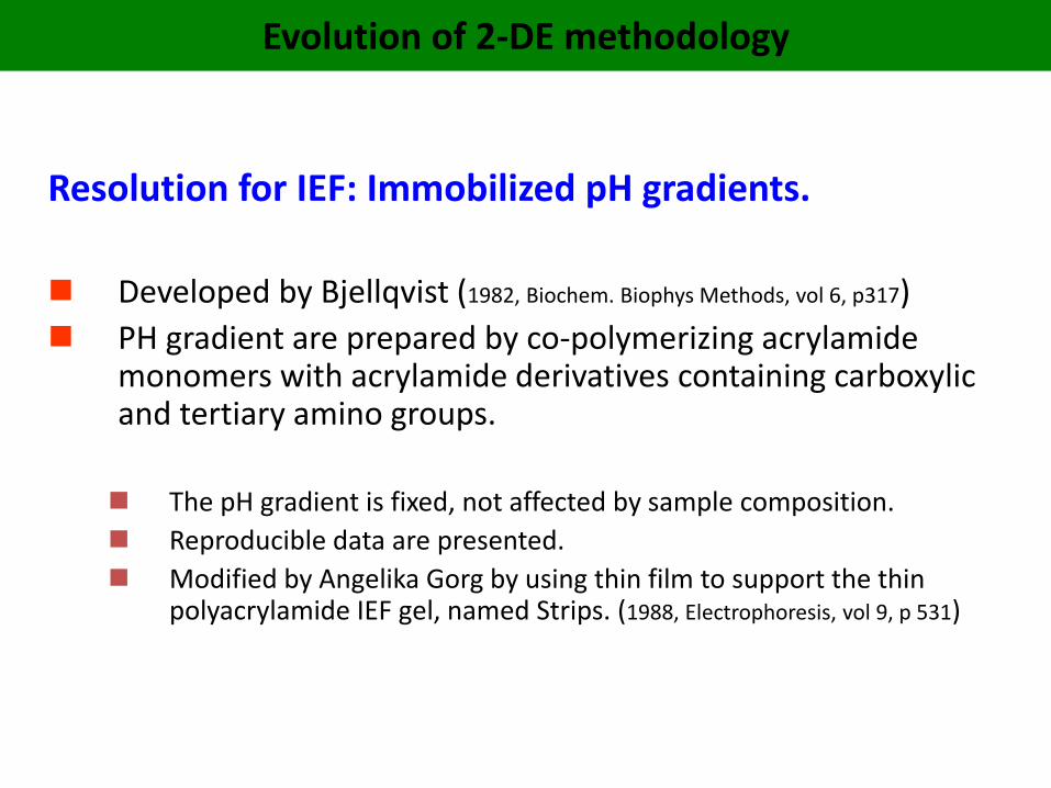
Resolution for IEF: Immobilized pH gradients.
Developed by Bjellqvist (1982, Biochem. Biophys Methods, vol 6, p317)
PH gradient are prepared by co-polymerizing acrylamide monomers with acrylamide derivatives containing carboxylic and tertiary amino groups.
The pH gradient is fixed, not affected by sample composition.
Reproducible data are presented.
Modified by Angelika Gorg by using thin film to support the thin polyacrylamide IEF gel, named Strips. (1988, Electrophoresis, vol 9, p 531)
Evolution of 2-DE methodology

Run 2-DE, a quick overview


Run 2-DE step by step

Run 2-DE step by step

Total E. coli Proteins - 2-Dimensional Gel


2-DE gel images of serum glycoprotein samples from the healthy and LC patients.
(A) Normal sample, (B) LC sample. The identified protein spots: (1) Anti TNFα antibody light chain (ATAL); (2) Chain L, structure of Fab D3h44 (D3h44); (3) Transthyretin (TTR); (4) AIM/CD69; (5) Alpha1-Antitrypsin (AAT); (6) Alpha2-HS-glycoprotein (AHSG) (7) Complement C3; (8) Zinc-alpha2-glycoprotein (ZAG); (9) Haptoglobin alpha2 chain (HpA2); (10) Ig heavy chain mu (BOT); (11) IGHM protein.