Lectin Magnetic Bead Array for Biomarker Discovery218575/UQ218575.pdf · Lectin Magnetic Bead Array...
5
Lectin Magnetic Bead Array for Biomarker Discovery Dorothy Loo, †,‡ Alun Jones, ‡ and Michelle M. Hill* ,†,‡ The University of Queensland Diamantina Institute, Princess Alexandra Hospital, and Institute for Molecular Bioscience, Brisbane, Australia Received May 15, 2010 Abstract: Alterations in protein glycosylation play an important role in patho-physiology, and much effort has been devoted to detecting glycoprotein biomarkers. In this manuscript, we describe the development of a novel method for monitoring alterations in protein glycosyla- tion. Lectins are used as individual affinity reagents and coupled to magnetic beads (Dynabeads) in a microplate array format for isolation of glycosylated proteins. Isolated glycoproteins are digested with trypsin in-solution fol- lowed by LC-MS/MS, allowing a liquid handler-assisted high throughput workflow. We demonstrate the specific and reproducible affinity-isolation of glycoproteins using the lectin Dynabead array technology. When used with serum, we achieved one-step purification of glycoproteins with minimal coisolation of abundant serum proteins including albumin. We further optimized the proteomics workflow to allow transfer to a liquid handler for automa- tion. In summary, we report the development of a high throughput platform to detect alterations in protein gly- cosylation which will be useful in glycoproteomics stud- ies, particularly clinical proteomics studies where large sample sizes are required to achieve statistical power. Keywords: biomarker discovery • serum proteomics • glycosylation • cancer • lectin • magnetic beads Introduction The study of glycans (glycomics) and glycosylated proteins (glycoproteomics) has become a leading area of biomarker discovery activities in recent years. 1-5 Lectins, naturally oc- curring proteins that preferentially bind specific glycan moi- eties, are well-used research tools in glycoprotein studies. As early as the 1980s, lectin-coupled beads were used to capture glycoproteins. 6 Lectin immunohistochemistry was used to demonstrate altered glycosylation in breast cancer, 7 and more recently, lectin-affinity chromatography has been used in combination with mass spectrometry to examine the glyco- proteome of pancreatic, breast, liver and colorectal cancer using serum, plasma or cells as sample source. 8-12 These studies demonstrate the utility and feasibility of lectin-affinity isolation- coupled proteomics as a means of identifying glycoproteins. These studies have used relatively small sample sizes, partly due to the high level of sample handling. To achieve statistical power for clinical proteomics studies, and to distinguish between different states of glycosylation, we set out to establish a high throughput platform using lectins as monoaffinity reagents. In this Technical Note, we report the development a proteomics workflow using lectin-coupled magnetic beads, in- solution digest and LC-MS/MS for isolation and identification of serum glycoproteins. This workflow achieved a one-step purification of serum glycoproteins, removing the need for depletion, separation or cleanup steps. Importantly, we show that protein binding to lectin-magnetic beads is lectin-selective and highly reproducible. Experimental Section Reagents. MyOne and M280 magnetic Dynabeads were from Life Technologies (Dynal). Lectins and lectin-coupled agarose beads were purchased from Vector Laboratories. Modified sequencing grade bovine trypsin was from Promega. All other reagents were from Sigma unless otherwise specified. Patient Sample. Blood samples were collected with consent and ethics approval by the Study of Digestive Health (Queen- sland Institute for Medical Research) and accessed with ap- proval by University of Queensland Human Ethics Committee. Sera were stored in aliquots at -80 °C. Preparation and Use of Lectin-Dynabeads. Lectin coupling to surface-activated Dynabeads was performed according to manufacturer’s protocol, and the beads were then blocked with 1 M Tris buffer pH 7.5 for 16 hours. Lectin-Dynabeads were washed 3 times with binding buffer containing 20 mM Tris/ HCl pH 7.4, 15 mM NaCl, 1 mM CaCl 2 , 1 mM MnCl 2 , (1% Triton X-100, 0.2% SDS, 1 mM DTT where indicated) and protease inhibitors (1 µg/µL Aprotinin, 1 µg/µL Antipain, 1 µg/µL Pepstatin, 1 µg/µL Leupeptin and 2.5 mM Benzamidine). Protein sample (2 µL of serum) was incubated with lectin- coupled beads diluted in binding buffer as specified and rotated at 4 °C for 30 min. After glycoprotein capture, beads were washed three times with 10 volumes of binding buffer. Protein elution methods depended on subsequent analysis. For 2D gel electrophoresis, proteins were eluted with 2D sample buffer (7 M urea, 2 M thiourea, 4% CHAPS, 100 mM DTT 0.5% ampholyte 3-10, trace bromophenol blue) for 30 min at room temperature prior to rehydrating pH 3-11, nonlinear, 7 cm Immobilzed pH gradient strips (GE healthcare). Isoelectric point focusing was performed using the OffGel Fractionator (Agilent) programmed for total of 8 kVh, limited at 5000 V, 50 µA, 200mW and 5 h. After equilibration with 2% DTT and then 2.5% IAA in equilibration buffer (6 M urea, 375 mM Tris/HCl pH 8.8, 2% SDS, 20% glycerol), strips were placed on top of * To whom correspondence should be addressed. Dr. Michelle M. Hill, e-mail: [email protected]. † University of Queensland Diamantina Institute. ‡ Institute for Molecular Bioscience. 5496 Journal of Proteome Research 2010, 9, 5496–5500 10.1021/pr100472z 2010 American Chemical Society Published on Web 08/23/2010
Transcript of Lectin Magnetic Bead Array for Biomarker Discovery218575/UQ218575.pdf · Lectin Magnetic Bead Array...

Lectin Magnetic Bead Array for Biomarker Discovery
Dorothy Loo,†,‡ Alun Jones,‡ and Michelle M. Hill*,†,‡
The University of Queensland Diamantina Institute, Princess Alexandra Hospital, and Institute for MolecularBioscience, Brisbane, Australia
Received May 15, 2010
Abstract: Alterations in protein glycosylation play animportant role in patho-physiology, and much effort hasbeen devoted to detecting glycoprotein biomarkers. In thismanuscript, we describe the development of a novelmethod for monitoring alterations in protein glycosyla-tion. Lectins are used as individual affinity reagents andcoupled to magnetic beads (Dynabeads) in a microplatearray format for isolation of glycosylated proteins. Isolatedglycoproteins are digested with trypsin in-solution fol-lowed by LC-MS/MS, allowing a liquid handler-assistedhigh throughput workflow. We demonstrate the specificand reproducible affinity-isolation of glycoproteins usingthe lectin Dynabead array technology. When used withserum, we achieved one-step purification of glycoproteinswith minimal coisolation of abundant serum proteinsincluding albumin. We further optimized the proteomicsworkflow to allow transfer to a liquid handler for automa-tion. In summary, we report the development of a highthroughput platform to detect alterations in protein gly-cosylation which will be useful in glycoproteomics stud-ies, particularly clinical proteomics studies where largesample sizes are required to achieve statistical power.
Keywords: biomarker discovery • serum proteomics •glycosylation • cancer • lectin • magnetic beads
Introduction
The study of glycans (glycomics) and glycosylated proteins(glycoproteomics) has become a leading area of biomarkerdiscovery activities in recent years.1-5 Lectins, naturally oc-curring proteins that preferentially bind specific glycan moi-eties, are well-used research tools in glycoprotein studies. Asearly as the 1980s, lectin-coupled beads were used to captureglycoproteins.6 Lectin immunohistochemistry was used todemonstrate altered glycosylation in breast cancer,7 and morerecently, lectin-affinity chromatography has been used incombination with mass spectrometry to examine the glyco-proteome of pancreatic, breast, liver and colorectal cancer usingserum, plasma or cells as sample source.8-12 These studiesdemonstrate the utility and feasibility of lectin-affinity isolation-coupled proteomics as a means of identifying glycoproteins.These studies have used relatively small sample sizes, partly
due to the high level of sample handling. To achieve statisticalpower for clinical proteomics studies, and to distinguishbetween different states of glycosylation, we set out to establisha high throughput platform using lectins as monoaffinityreagents. In this Technical Note, we report the development aproteomics workflow using lectin-coupled magnetic beads, in-solution digest and LC-MS/MS for isolation and identificationof serum glycoproteins. This workflow achieved a one-steppurification of serum glycoproteins, removing the need fordepletion, separation or cleanup steps. Importantly, we showthat protein binding to lectin-magnetic beads is lectin-selectiveand highly reproducible.
Experimental Section
Reagents. MyOne and M280 magnetic Dynabeads were fromLife Technologies (Dynal). Lectins and lectin-coupled agarosebeads were purchased from Vector Laboratories. Modifiedsequencing grade bovine trypsin was from Promega. All otherreagents were from Sigma unless otherwise specified.
Patient Sample. Blood samples were collected with consentand ethics approval by the Study of Digestive Health (Queen-sland Institute for Medical Research) and accessed with ap-proval by University of Queensland Human Ethics Committee.Sera were stored in aliquots at -80 °C.
Preparation and Use of Lectin-Dynabeads. Lectin couplingto surface-activated Dynabeads was performed according tomanufacturer’s protocol, and the beads were then blocked with1 M Tris buffer pH 7.5 for 16 hours. Lectin-Dynabeads werewashed 3 times with binding buffer containing 20 mM Tris/HCl pH 7.4, 15 mM NaCl, 1 mM CaCl2, 1 mM MnCl2, (1% TritonX-100, 0.2% SDS, 1 mM DTT where indicated) and proteaseinhibitors (1 µg/µL Aprotinin, 1 µg/µL Antipain, 1 µg/µLPepstatin, 1 µg/µL Leupeptin and 2.5 mM Benzamidine).Protein sample (2 µL of serum) was incubated with lectin-coupled beads diluted in binding buffer as specified and rotatedat 4 °C for 30 min. After glycoprotein capture, beads werewashed three times with 10 volumes of binding buffer. Proteinelution methods depended on subsequent analysis. For 2D gelelectrophoresis, proteins were eluted with 2D sample buffer(7 M urea, 2 M thiourea, 4% CHAPS, 100 mM DTT 0.5%ampholyte 3-10, trace bromophenol blue) for 30 min at roomtemperature prior to rehydrating pH 3-11, nonlinear, 7 cmImmobilzed pH gradient strips (GE healthcare). Isoelectricpoint focusing was performed using the OffGel Fractionator(Agilent) programmed for total of 8 kVh, limited at 5000 V, 50µA, 200mW and 5 h. After equilibration with 2% DTT and then2.5% IAA in equilibration buffer (6 M urea, 375 mM Tris/HClpH 8.8, 2% SDS, 20% glycerol), strips were placed on top of
* To whom correspondence should be addressed. Dr. Michelle M. Hill,e-mail: [email protected].
† University of Queensland Diamantina Institute.‡ Institute for Molecular Bioscience.
5496 Journal of Proteome Research 2010, 9, 5496–5500 10.1021/pr100472z 2010 American Chemical SocietyPublished on Web 08/23/2010

10% mini-SDS-PAGE gels and subjected to electrophoresis. Gelswere stained with colloidal coomassie.13 In-gel digest wasperformed as previously described.14
For direct in-solution digest and LC-MS/MS, beads wereadditionally washed twice with 200 mM Tris/HCl pH 7.4, twicewith 50 mM NH4HCO3, then the proteins were eluted with onevolume of 50% formic acid followed by one volume of 20%acetonitrile. The formic acid in the eluate was neutralized withtwo volumes of 50 mM NH4HCO3. The samples were dried ina speedy vac, resuspended in 20 µL of NH4HCO3 and digestedwith 0.5 µg trypsin (Promega sequencing grade modified bovinetrypsin) overnight at 37 °C. Trypsin was inactivated by acidi-fication to 5% formic acid. Samples were dried in a speedy vacand resuspended in 40 µL of 5% formic acid.
LC-MS/MS. Tryptic peptides corresponding to 5% of lectin-Dynabead pulldown (approximately 1 µg of protein from 2 µLof starting serum) were subjected to LC-MS/MS using anAgilent 6520 QTOF coupled with a Chip CUBE and 1200 HPLC.The nano pump was set at 0.3 µL/min and the capillary pumpat 4 µL/min. The HPLC-chip used contained a 40nl C18trapping column, and a 150 mm C18 resolving column (Agi-lent). Buffer A was 0.1% formic acid and buffer B was 0.1%formic acid in 90% acetonitrile. For in-solution digest, thegradient went from 10% to 50% Buffer B in 45 min, and then95% Buffer B at 46 min, returning to 10% buffer B at 50 min.For 2D gel spots, the gradient went from 10% to 50% Buffer Bin 20 min, and then 95% Buffer B at 20.10 min, returning to10% buffer B at 22.10 min. The mass spectrometer wasprogrammed to acquire 8 precursor MS1 spectra per minuteand 4 MS/MS spectra per minute. Dynamic exclusion wasapplied after 2 MS/MS within 0.25 min. Results were searchedagainst IPI human v3.64 database using Spectrum Mill (AgilentA03.03). The following parameters were used for search: 2maximum missed cleavage, precursor mass tolerance of (20and product mass tolerance of (50. For 2D spots and whentesting reduction/alkylation in solution, carbamidomethylationwas selected as fixed modification.
Safety Considerations. Some lectins can be mitogenic orcytotoxic, material safety datasheet should be consulted foreach lectin and appropriate personnel protective equipmentused.
Results and Discussion
In order to achieve sample throughput for clinical proteom-ics studies, we wished to establish a high throughput autom-atable procedure that links protein isolation/separation to massspectrometry. Magnetic beads are easily adaptable to roboticliquid handlers and thus represent the support of choice whenconsidering high throughput affinity isolation procedures. Sinceour affinity reagents, lectins, are proteins, we examined thefunctionalized Dynabeads that were recommended for directprotein coupling, namely epoxy, tosyl and carboxylic acidactivated Dynabeads (LifeTechnologies, Dynal). Preliminaryresults suggest that epoxy and tosyl-activated beads are betterthan carboxylic beads at capturing wheat germ agglutinin (datanot shown), therefore we further compared the couplingefficiency of tosyl and epoxy activated beads with commercialagarose beads (Vector Laboratories) for Concanavalin A (ConA),wheat germ agglutinin (WGA) and Jacalin (JAC), with visualiza-tion by colloidal Coomassie staining. Tosyl-activated beadscaptured the highest level of lectin, as judged by coomassiestaining (Figure 1). We selected 1 µm diameter MyOne tosyl-activated beads (Life Technologies Dynal) for use in further
experiments because the small diameter of the beads offersmore surface area for protein capture.
Initial experiments comparing the ability of lectin magneticbeads to isolate glycoproteins from serum with commerciallectin agarose beads showed comparable results (data notshown), suggesting that lectin magnetic beads will be suitablefor glycoprotein isolation. One important consideration in lectinpull down experiments is the possibility of isolating proteincomplexes, leading to the erranous assignment of nonglyco-sylated proteins, or glycoprotein with other glycan moieties.Thus the use of binding buffer should be carefully consideredto reduce the presence of protein complexes, while retaininglectin-glycan interactions. To this end, we compared pull downusing ConA-tosyl-activated Dynabeads under four differentbinding buffers. Condition 1 used a lectin binding buffer fromYang and Hancock15 that contained 20 mM Tris/HCl pH 7.4,150 mM NaCl, 1 mM CaCl2, 1 mM MnCl2, with additional 0.05%Triton X-100, and protease inhibitors as described in Experi-mental Section. This represents a mild buffer condition withminimal salt and detergent. This buffer was used duringbinding and washing steps. In condition 2, we included 0.5 Murea as denaturing agent in the base buffer in the binding andwashing steps to disrupt protein complexes. In condition 3,additional 1 mM DTT, 0.2% SDS, and 1% Triton-X100 wasincluded in the wash buffer to increase the stringency of thewashing step. In the most stringent condition, the additionalDTT, SDS and Triton-X100 was included in both the bindingand wash buffers (condition 4). Since this condition containedreducing agent and strong detergent during the entire pulldown, it was possible that lectin binding efficiency will bereduced. After washing in binding buffer, bound proteins wereeluted in 2D sample buffer and analyzed by 2D gel electro-phoresis with 2D standards loaded with each sample (Figure2). Compared to input serum, the ConA-Dynabead pulldownshowed no visible albumin spot, demonstrating the selectivityof the pulldown. Similar number of spots was observed in allfour conditions, with the numbers being 60, 57, 46, 50 spotsfor conditions 1-4, respectively. While the inclusion of reducingagent and strong detergent in binding and washing steps(condition 4) resulted in ∼20% loss of protein binding, we still
Figure 1. Comparison of binding efficiency of Concanavalin A(ConA), Jacalin (JAC) and wheat germ agglutinin (WGA) toagarose and Dynabeads. Each lectin was bound to epoxy andtosyl activated magnetic beads to the same concentration ascommercial agarose beads (ConA 6 mg/mL, Jac 4 mg/mL, WGA7 mg/mL). Lectins were eluted from 2 µL of beads and examinedon 17% SDS-PAGE and colloidal coomassie staining.
Lectin Magnetic Bead Arrays technical notes
Journal of Proteome Research • Vol. 9, No. 10, 2010 5497
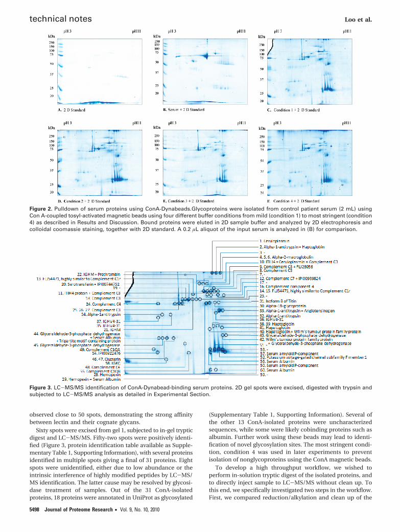
observed close to 50 spots, demonstrating the strong affinitybetween lectin and their cognate glycans.
Sixty spots were excised from gel 1, subjected to in-gel trypticdigest and LC-MS/MS. Fifty-two spots were positively identi-fied (Figure 3, protein identification table available as Supple-mentary Table 1, Supporting Information), with several proteinsidentified in multiple spots giving a final of 31 proteins. Eightspots were unidentified, either due to low abundance or theintrinsic interference of highly modified peptides by LC-MS/MS identification. The latter cause may be resolved by glycosi-dase treatment of samples. Out of the 31 ConA-isolatedproteins, 18 proteins were annotated in UniProt as glycosylated
(Supplementary Table 1, Supporting Information). Several ofthe other 13 ConA-isolated proteins were uncharacterizedsequences, while some were likely cobinding proteins such asalbumin. Further work using these beads may lead to identi-fication of novel glycosylation sites. The most stringent condi-tion, condition 4 was used in later experiments to preventisolation of nonglycoproteins using the ConA magnetic beads.
To develop a high throughput workflow, we wished toperform in-solution tryptic digest of the isolated proteins, andto directly inject sample to LC-MS/MS without clean up. Tothis end, we specifically investigated two steps in the workflow.First, we compared reduction/alkylation and clean up of the
Figure 2. Pulldown of serum proteins using ConA-Dynabeads.Glycoproteins were isolated from control patient serum (2 mL) usingCon A-coupled tosyl-activated magnetic beads using four different buffer conditions from mild (condition 1) to most stringent (condition4) as described in Results and Discussion. Bound proteins were eluted in 2D sample buffer and analyzed by 2D electrophoresis andcolloidal coomassie staining, together with 2D standard. A 0.2 µL aliquot of the input serum is analyzed in (B) for comparison.
Figure 3. LC-MS/MS identification of ConA-Dynabead-binding serum proteins. 2D gel spots were excised, digested with trypsin andsubjected to LC-MS/MS analysis as detailed in Experimental Section.
technical notes Loo et al.
5498 Journal of Proteome Research • Vol. 9, No. 10, 2010

in-solution digest (the “standard” nonhigh throughput work-flow) with a shortened protocol that leaves out these steps. Afterwashing the pulldown in binding buffer, and further washingin 50 mM NH4HCO3, bound proteins were sequentially elutedwith 50% formic acid and 20% acetonitrile. After drying in aspeedvac, the samples were divided into two workflows. Forone set, proteins were reduced, alkylated, digested with trypsinand then cleaned up with a C18 STAGE-tip.16 For the secondset, proteins were directly digested without reduction alkylation.This experiment was designed to test if a shortened in-solutiondigest procedure without reduction/alkylation produces ac-ceptable number of protein identifications. Peptides wereanalyzed by LC-MS/MS and database searching. Three techni-cal replicates were performed. A protein identity was consid-ered safe if it was identified in 2 out of the 3 replicates(Supplementary Table 2, Supporting Information). Based onthese criteria, 50 proteins were identified from ConA-Dynabead
pulldowns using in-solution digest without reduction/alkylationand clean up, in contrast to only 24 identified when reduction/alkylation and STAGE-tip clean up was performed (Figure 4,Supplementary Table 2, Supporting Information). The reducednumber of protein identified in the second protocol may bedue to significant sample loss during the clean up step. Analysisby of similar pulldown 2D gel and in-gel digest had identified31 proteins (Figure 3, Supplementary Table 1, SupportingInformation). When the overlap between the 3 protein iden-tification procedures were compared, we found that 14 proteinswere identified by all 3 methods, while 22 proteins wereidentified only using solution digest without reduction/alky-lation. The 2D gel protocol identified 9 proteins that were notfound using solution digest. However, the entire pulldown wasloaded onto a single 2D gel, while only 5% of the sample wasloaded on the LC-MS/MS from the solution digest procedures,suggesting a much reduced rate of protein identification with2D gel separation. Based on these results, we selected in-solution digest without reduction/alkylation as the proteinprocessing method prior to LC-MS/MS.
As our aim is to develop a liquid handler-assisted highthroughput workflow, we next examined if the entire procedureperforms as efficiently in 96 well microplates. For this experi-ment, we divided ConA-Dynabeads into 6 aliquots, one set in3 separate eppendorf tubes and one set in 3 wells of a 96 wellmicroplate. Pulldown was performed from aliquots of a singleserum sample, using a hand-held 96 well microplate barmagnet (Life Technologies) for the microplate samples. Allsamples were processed in parallel using in-solution digestwithout reduction/alkylation, and analyzed by LC-MS/MS.Protein identities were confirmed if 2 out of the 3 technicalreplicates identified the protein. Surprisingly, we found thatperforming the procedure in 96 well microplates actuallyidentified slightly more proteins, and the scores for proteinidentification were also better with microplates (Figure 5,Supplementary Table 3, Supporting Information). The majorityproteins were identified in both vessel formats, supporting theconsistency of the lectin-Dynabead pulldown workflow. Weanalyzed the reproducibility between the 3 technical replicatesfor the tube and microplate workflows. Protein identities werehighly reproducible in both microplate and tube workflows,with the majority of proteins identified in all 3 technicalreplicates (Figure 6, Supplemental Table 3, Supporting Infor-mation). These results suggest that transferring the lectin-Dynabead pulldown workflow to microplates will not reduceefficiency but may in fact enhance the sensitivity and confi-dence of protein identifications.
Figure 4. Number of proteins identified from ConA-Dynabeadspulldown from serum. Overlap between serum proteins isolatedusing ConA-Dynabeads were subjected to in-solution digest withor without reduction, alkylation and STAGE-tip clean up, oranalyzed by 2D gel and in-gel digest of spots.
Figure 5. Comparison of tube versus microplate performace forlectin-Dynabead pulldown coupled to in-solution digest andLC-MS/MS.
Table 1. Selectivity of Lectin Magnetic Bead Pulldowna
known target ConA Jac SBA SNA STA UEA I WGA
R-Man, R-Glc, and R-GlcNAc ConA 53 16 5 38 19 8 20GalR1-6GalNAc and Gal�1-3GalNAc Jac 49 6 17 8 5 9GalNAcR1-3Gal SBA 8 4 5 10 4Neu5AcR2-6Gal and Neu5AcR2-6GalNAc SNA 53 22 10 20GlcNAc�1-4GlcNAc oligomers STA 36 10 21FucR1-2Gal�1-4GlcNAc UEA I 13 9Neu5Ac and GlcNAc�1-4GlcNAc WGA 31
a An array of seven lectin-coupled tosyl-activated beads was prepared and used to isolate serum glycoproteins from a single patient serum. Proteinswere eluted, digested in-solution and analysed by LC-MS/MS. The table shows the known glycan target of each lectin, and the number of overlappingproteins identified between pairs of lectin pulldowns. Protein identification table is available as Supplementary Table 4 (Supporting Information).Abbreviations: Fuc, fucose; Gal, galactose; GalNAc, N-Acetylgalactosamine; Glc, glucose; GlcNAc, N-Acetylglucosamine; Man, mannose; Neu5Ac,N-acetylneuraminic acid (also called sialic acid); ConA, concanavalin A; Jac, Jacalin; SBA, Soy Bean Agglutinin (Glycine max lectin); SNA, Sambucus NigraAgglutinin; STA, Solanum Tuberosum Agglutinin; UEA I, Ulex Europaeus agglutinin I; WGA, wheat Germ agglutinin.
Lectin Magnetic Bead Arrays technical notes
Journal of Proteome Research • Vol. 9, No. 10, 2010 5499

To demonstrate the utility and selectivity of lectin-Dynabeadarray, we prepared a panel of 7 lectin-Dynabeads and used thissmall array in a pulldown experiment. The lectins were selectedbased on their reported glycan affinities, covering both N- andO-glycosylation as well as sialic acid modification (Table 1).When aliquots of a single serum sample were subjected topulldown using this panel of lectin-Dynabeads, we observedselective binding of proteins to the lectin-Dynabeads (Table 1,Supplementary Table 4, Supporting Information). Most identi-fied proteins bound to several lectins, reflecting the fact thatglycoproteins are often modified with multiple types and sitesof glycosylation. Table 1 shows the number of proteins isolatedby each lectin, and the number that is identified in pulldownfrom pairs of lectin-Dynabeads. ConA and SNA (Sambucusnigra Agglutinin) bound the highest number of proteins, withmore than 50% of the proteins in common. These may indicatethat ConA and SNA have similar binding specificities, or thetarget glycans preferentially occur together. Glycomic analysisof the isolated proteins will be required to further characterizethe glycan modifications. In contrast to ConA and SNA, whichhave broad target specificity, SBA (Soy Bean Agglutinin) has anarrow glycan selectivity, and only 8 proteins were isolated bySBA-Dynabeads (Table 1). Thus, by combining lectin-Dynabeadarray with bioinformatic analysis of the protein (and peptide/glycan) identifications, this workflow has the potential toidentify alterations in glycosylation profiles due to disease.
Conclusions
We have developed a novel high throughput technique forbiomarker discovery and glycoproteomics studies. This methodis based on the use of lectin-Dynabeadsto isolate glycoproteinsin a glycan-specific manner. The increased throughput meansthat many lectins can be tested per sample, and alterations inlectin binding ability can be determined for many biologicalsamples. The utility of lectin magnetic beads as a one-stepserum glycoprotein isolation procedure will greatly assistidentification of serum biomarkers. Development of glycomicanalysis workflows combined with bioinformatic and biostatis-tic analysis algorithms will be required to fully utilize thistechnology for biomarker discovery.
Acknowledgment. This work was supported byUniversity of Queensland Early Career Researcher Grant andFirst Link Fund to M.M.H. M.M.H. is supported by Career
Development Award No. 569512 from the National Healthand Medical Research Council of Australia, and aDiamantina Institute Fellowship. We thank ErlendRagnhildstveit (Life Technologies) for preliminary data andhelpful discussions.
Supporting Information Available: SupplementaryTables 1-4. This material is available free of charge via theInternet at http://pubs.acs.org.
References(1) Dayarathna, M. K.; Hancock, W. S.; Hincapie, M. A two step
fractionation approach for plasma proteomics using immun-odepletion of abundant proteins and multi-lectin affinity chro-matography: Application to the analysis of obesity, diabetes, andhypertension diseases. J. Sep. Sci. 2008, 31, 1156–1166.
(2) Mechref, Y.; Madera, M.; Novotny, M. V. Glycoprotein enrichmentthrough lectin affinity techniques. Methods Mol. Biol. 2008, 424,373–396.
(3) Li, C.; Simeone, D. M.; Brenner, D. E.; Anderson, M. A.; Shedden,K. A.; Ruffin, M. T.; Lubman, D. M. Pancreatic cancer serumdetection using a lectin/glyco-antibody array method. J. ProteomeRes. 2009, 8, 483–492.
(4) El-Boubbou, K.; Zhu, D. C.; Vasileiou, C.; Borhan, B.; Prosperi, D.;Li, W.; Huang, X. Magnetic Glyco-Nanoparticles: A Tool To Detect,Differentiate, and Unlock the Glyco-Codes of Cancer via MagneticResonance Imaging. J. Am. Chem. Soc. 2010, 132 (12), 4490–4499.
(5) Narimatsu, H. A strategy for discovery of cancer glyco-biomarkersin serum using newly developed technologies for glycoproteomics.FEBS J. 2010, 277, 95–105.
(6) Cummings, R. D.; Kornfeld, S. Fractionation of asparagine-linkedoligosaccharides by serial lectin-Agarose affinity chromatography.A rapid, sensitive, and specific technique. J. Biol. Chem. 1982, 257,11235–11240.
(7) Brooks, S. A.; Leathem, A. J. Prediction of lymph node involvementin breast cancer by detection of altered glycosylation in theprimary tumour. Lancet 1991, 338, 71–74.
(8) Zhao, J.; Simeone, D. M.; Heidt, D.; Anderson, M. A.; Lubman,D. M. Comparative serum glycoproteomics using lectin selectedsialic acid glycoproteins with mass spectrometric analysis: ap-plication to pancreatic cancer serum. J. Proteome Res. 2006, 5,1792–1802.
(9) Yang, Z.; Harris, L. E.; Palmer-Toy, D. E.; Hancock, W. S. Multilectinaffinity chromatography for characterization of multiple glyco-protein biomarker candidates in serum from breast cancer pa-tients. Clin. Chem. 2006, 52, 1897–1905, doi:clinchem.2005.065862[pii]10.1373/clinchem.2005.065862.
(10) Xu, Z.; Zhou, X.; Lu, H.; Wu, N.; Zhao, H.; Zhang, L.; Zhang, W.;Liang, Y. L.; Wang, L.; Liu, Y.; Yang, P.; Zha, X. Comparativeglycoproteomics based on lectins affinity capture of N-linkedglycoproteins from human Chang liver cells and MHCC97-H cells.Proteomics 2007, 7, 2358–2370.
(11) Qiu, Y.; Patwa, T. H.; Xu, L.; Shedden, K.; Misek, D. E.; Tuck, M.;Jin, G.; Ruffin, M. T.; Turgeon, D. K.; Synal, S.; Bresalier, R.; Marcon,N.; Brenner, D. E.; Lubman, D. M. Plasma glycoprotein profilingfor colorectal cancer biomarker identification by lectin glycoarrayand lectin blot. J. Proteome Res. 2008, 7, 1693–1703.
(12) Jung, K.; Cho, W.; Regnier, F. E. Glycoproteomics of plasma basedon narrow selectivity lectin affinity chromatography. J. ProteomeRes. 2009, 8, 643–650.
(13) Candiano, G.; Bruschi, M.; Musante, L.; Santucci, L.; Ghiggeri,G. M.; Carnemolla, B.; Orecchia, P.; Zardi, L.; Righetti, P. G. Bluesilver: a very sensitive colloidal Coomassie G-250 staining forproteome analysis. Electrophoresis 2004, 25, 1327–1333.
(14) Hill, M. M.; Adrain, C.; Duriez, P. J.; Creagh, E. M.; Martin, S. J.Analysis of the composition, assembly kinetics and activity ofnative Apaf-1 apoptosomes. EMBO J. 2004, 23, 2134–2145.
(15) Yang, Z.; Hancock, W. S. Monitoring glycosylation pattern changesof glycoproteins using multi-lectin affinity chromatography. J. Chro-matogr., A 2005, 1070, 57–64.
(16) Rappsilber, J.; Ishihama, Y.; Mann, M. Stop and go extraction tipsfor matrix-assisted laser desorption/ionization, nanoelectrospray,and LC/MS sample pretreatment in proteomics. Anal. Chem. 2003,75, 663–670.
PR100472Z
Figure 6. Reproducibility of lectin Dynabead pulldown. ConA-coupled magnetic beads was used to isolate serum glycopro-teins, which were eluted, digested in-solution and identified byLC-MS/MS. The Venn diagram shows the reproducibility of theprocedure over 3 independent technical repeats when used with(A) Eppendorf tubes and (B) 96-well microplates.
technical notes Loo et al.
5500 Journal of Proteome Research • Vol. 9, No. 10, 2010