Leber Congenital Amaurosis: Clinical Profiling and Genetic ...
193
Leber Congenital Amaurosis: Clinical Profiling and Genetic Analysis Using High Throughput Resequencing in an Indian Cohort THESIS Submitted in partial fulfilment of the requirements for the degree of DOCTOR OF PHILOSOPHY by SRIKRUPA. N. N 2011PHXF0100H Under the Supervision of Dr. N. SOUMITTRA & Under the Co-Supervision of Prof. SUMAN KAPUR BIRLA INSTITUTE OF TECHNOLOGY AND SCIENCE, HYDERABAD 2017
Transcript of Leber Congenital Amaurosis: Clinical Profiling and Genetic ...

Leber Congenital Amaurosis: Clinical Profiling and Genetic
Analysis Using High Throughput Resequencing in an Indian Cohort
THESIS
Submitted in partial fulfilment
of the requirements for the degree of
DOCTOR OF PHILOSOPHY
by
SRIKRUPA. N. N
2011PHXF0100H
Under the Supervision of
Dr. N. SOUMITTRA
&
Under the Co-Supervision of
Prof. SUMAN KAPUR
BIRLA INSTITUTE OF TECHNOLOGY AND SCIENCE,
HYDERABAD
2017

BIRLA INSTITUTE OF TECHNOLOGY AND SCIENCE,
HYDERABAD
CERTIFICATE
This is to certify that the thesis entitled “Leber Congenital Amaurosis: Clinical
Profiling and Genetic Analysis Using High Throughput Resequencing in an
Indian Cohort” submitted by Ms. Srikrupa. N. N ID No 2011PHXF0100H for the
award of Ph.D. of the Institute embodies original work done by her under my
supervision.
Signature in full of the Supervisor: Signature in full of the Co-Supervisor:
Prof. Suman Kapur
Dean, Research and Consultancy Division Department of Biological Sciences, BITS-Pilani, Hyderabad campus Hyderabad -500078
Date:
Dr. N. Soumittra
Principal Scientist & Associate Professor SNONGC Department of and Molecular Biology Vision Research Foundation Chennai- 600006
Date:
Prof. Suman Kapur
Dean, International Programmes and Collaborations Senior Professor Department of Biological Sciences, BITS-Pilani, Hyderabad campus Hyderabad -500078
Date:

.......Dedicated to my Family
(For their strong support, encouragement and understanding)

ACKNOWLEDGEMENTS
"Being a graduate student is like becoming all the Seven Dwarves of the fairy tale
“Snow white and the Seven Dwarves”. In the beginning you're Dopey (mute) and
Bashful (shy). In the middle, you are usually sick (Sneezy), tired (Sleepy), and
irritable (Grumpy). But at the end, they call you Doc, and then you're Happy."
- Ronald T. Azuma
It is a delight to acknowledge those who have supported me throughout my PhD.
Firstly, my mentor, Dr. N. Soumittra, who has been there throughout - motivating
and inspiring me with constant guidance, cooperation and support which kept me
going ahead. Her tremendous support during NGS analysis, where she taught and
learned alongside, encouraging several thought provoking discussions and her
guidance in comprehensive understanding of informatics pipelines helped me sail
through the initial fumbling to reach this pinnacle. Her friendly nature, constant
advises and elderly supports have shaped me both in my career and personal front. I
owe a lot of gratitude to her and feel privileged to be mentored by her.
My sincere thanks for the support and guidance provided by my co-supervisor Prof.
Suman kapur, Department of Biological sciences, BITS-Pilani Hyderabad campus.
A caring, inspirational teacher and an enthusiastic researcher, her continuous
support and suggestions during assessments, teaching practices, paper and thesis
corrections were tremendous. It was because of her and her team of students, my
BITS travels were convenient and a good learning experience.
My heartfelt gratitude also goes to Dr. S. Sripriya, Associate professor, for always
being motivating, helpful and kind. Her observations of the data, correlating ideas
and converting them to new possibilities have always inspired me. Her scientific
inputs, personal suggestions have always made me feel at ease with her and I
believe I can always count on her support during my course of PhD and even later.
I extend my sense of gratitude to Dr. S. Mathavan, Head of the department,
SNONGC department of Genetics and Molecular biology and former Heads Dr. A.J
Pandian and Dr. J. Madhavan for their support and encouragement.

I am very grateful that my doctoral programme is affiliated to two esteemed
institutions, Vision research foundation (VRF), Sankara Nethralaya and BITS-Pilani. I
sincerely thank and seek the blessings of Padmabhushan Dr. S.S. Badrinath,
Chairman Emeritus, Sankara Nethralaya for my doctoral thesis in such a well
established research and academic platform. I thank Prof. Souvik Bhattacharyya,
Vice chancellor, BITS Pilani for providing me an opportunity to be a part of their
revered institute.
I extend my gratitude to Dr. Lingam Gopal, President, VRF, Dr. S. Bhaskaran,
Honorary secretary, VRF, Dr. Ronnie George, Director - Research, VRF, Dr. Rama
Rajaram, Ex-Advisor, VRF, Mr. S. Narayanan, Manager, VRF, Dr. H.N. Madhavan,
Director of Microbiology, VRF, Dr. S. Meenakshi, Director of Academics, Medical
Research Foundation (MRF) for their support.
Also, I sincerely acknowledge Prof. S.K. Verma, Dean, research and consultancy
Division, Pilani, Prof. G. Sundar, Director, BITS Pilani, Hyderabad campus,
Hyderabad. My heartfelt thanks to Dr. P.R. Deepa, BITS co-ordinator for being the
bridge between us and BITS, extending her support, ideas and co-ordinating
between the institutes.
My sincere thanks to my Doctoral committee members (DAC) Dr. Sridev
Mohapatra, Prof. P. Sankar Ganesh and other faculties of biological sciences
department, BITS Pilani, Hyderabad campus, Prof. Vidya Rajesh, Prof.
Ramakrishna Vadrevu, Prof. Kumar Pranav Narayan, Prof. Naga Mohan
Kommu, Dr. Jayati Ray dutta, Prof. S. Swaminathan and Dr. Debashree
Bandyopadhyay for their continuous evaluations and constructive criticisms to
improve my thesis.
I thank Dr. Parveen Sen, Dr. S. Meenakshi and Dr. Muna Bhende for their clinical
support, phenotype documentation and correlation. My extended gratitude to
Vidyasagar Institute of Biomedical Sciences (VIBS), Dr. K. Lily Therese, Dr.
K.N. Sulochana, Dr. N. Angayarkanni, Dr. Dorien Gracious, Dr. S.
Krishnakumar, Dr. J. Malathi, Dr. B. Mahalakshmi, Dr. K. Coral, Dr. Bharathi
devi, Dr. J. Subbulakshmi, Dr. Ananth Badrinath, Dr. V. Umashankar, Dr.
Nivedita Chatterjee for evaluating my practice lectures.

My heartfelt thanks to my friends or I would rather say my stress busters K. Sudha,
Malaichamy, Bhavna S Rao, D. Sudha, Divya Rao, Sathyapriya, Ferdina Marie
Sharmila, and Kavitha who were always there as a moral support, making me feel
so important, bearing with me during all the good and stressful days, extending their
helping hands without fail. My heartfelt thanks to my PG mates Shylaja, Dharanija,
Revathi, Aarthi and Prathibha who helped shape up my focus towards this goal.
My special thanks to Shabna and Srilekha ma’m who shared this journey with me
through all hurdles. Our travel to BITS Hyderabad every semester, our stay,
evaluations and assignments will be an unerasable memory in this journey of mine.
I also owe my thanks to Jeevajothi mam, Karthiyayini mam, Sumathi mam,
Sacikala mam, Ms. Porkodi, Ms. Suganya, Ms. Jothi, Mr. Jayaprakash,
Mr.Venkatesan, Mr. Manoharan, Mr. Babu for being friendly, cooperative and
supportive. I would also like to acknowledge my school and college friends
Saraniya.S, Saranya. D, Sunitha ma’m, Anbarasan, my cousins and my friends
across my department Mr. Naresh, Ms. Vimalin Jayalatha, Mr. Bhuvanasundar,
Ms. Janani, Ms. Dhanurekha, Ms. Foujana Jenofer, Ms. Abirami who extended
timely suggestions and help throughout this journey. A special mention to Mr. T.
Arokiasamy, social worker, for helping in patient recruitment. A wonderful person,
elderly figure, who would go to all extent to help the patients, be it for their
academics, rehabilitation or financial support expecting nothing in return. I feel
blessed to be associated with him during this venture. I extend my acknowledgement
to all patients and their family for their co-operation.
I acknowledge the support extended by my friends in Genomics lab, BITS Pilani,
Hyderabad campus, Ms. SaiChinmayi, Ms. Yenmandra Padma, Ms. Sruthi Varier,
Ms. Shivani Guptha, Ms. Anuradha pal, Mr. Pavan, Mr. Rupak, Ms. Minal, Dr.
Blesson, Ms. Kriti and Pooja mam. The journey wouldn‟t have been so comfortable
without their warm attitude and friendliness. A special thanks to Chinmayi, who have
gone out of way to help me with several official matters at critical times.
I would like to acknowledge the people, who mean the world to me. My
parents Natarajan (Appa), Sampoornam (Amma), my brother Srivatsan and sister
in law Sandyasree for giving me the liberty to choose what I desired, showing faith
in me, and standing behind me with their unconditional love and support forever.

Special mention to Srivatsan for his unconditional trust and timely encouragement!! I
was lucky to have the best parents in law Raghu (appa) and Rajeshwari (amma)
who continued to carry the torch of faith in me and supported me throughout to
achieve this dream of mine. I extend my love and respect to Kanthan peripa and
Nagi perima for being so proud of me on my zeal which encouraged me to achieve
it.
I appreciate the love and support extended by my husband Mr. Chandrasekar. He
had been a great companion, nurtured my learning, and supported my dreams
encouraging, entertaining and boosting me up during the stressful days helping me
get through this period in the most positive way. Thanks to the little ones Maanav
and Thejhas, their smiles always cheers me up in all situations and their love makes
me feel out of the world.
Above all, I owe it all to Almighty for giving me the strength, patience, wisdom and
health to undertake this research and enabling me take to its completion.
Srikrupa.N.N

ABSTRACT
BACKGROUND
Retinal blinding disorders together have a prevalence of 1 in 2000 worldwide.
Mutations in genes that are expressed either in retinal pigment epithelium (RPE)
cells, photoreceptors or bipolar cells can cause varying forms of degenerative or
stationary retinal disorders, as the encoded proteins are crucial for normal function,
maintenance and synaptic interaction. Depending on the type of photoreceptor
affected, the retinal degenerative diseases (RDDs) are categorised as rod
dominated, cone dominated, and generalised retinal degenerations involving both
rods and cones. Leber congenital amaurosis (LCA) is an inherited blindness, caused
due to degenerations of rods and cones, occurring within the age of one year,
characterized by an extinguished electroretinogram (ERG) and manifesting
nystagmus, photophobia, hyperopia. Genetically it is heterogeneous, with twenty-
nine candidate genes identified so far (AIPL1, ALMS, CABP4, CCT2, CEP290,
CNGA3, CLUAP1, CRB1, CRX, DTHD1, GDF6, GUCY2D,
IQCB1, IMPDH1, IFT140, KCNJ13, LCA5, LRAT, MERTK, MYO7A, NMNAT1,
OTX2, PRPH2, RD3, RDH12, RPE65, RPGRIP1, SPATA7 and TULP1) and most of
them are inherited in an autosomal recessive manner except CRX, IMPDH1 and
OTX2 which are associated with autosomal dominant inheritance pattern.The
conventional method of disease gene discovery in Mendelian disorders includes
those based on linkage analysis as well as homozygosity mapping. Recent
developments in high-throughput sequence capture methods have made next-
generation sequencing technologies more feasible and cost effective. In LCA, the
prevalence of mutation in the candidate genes varies in different populations and
comprehensive mutation study focused on individual ethnicities in large cohorts are
few. The mutation studies conducted on Indian LCA cohorts either had a very small
sample size with fewer genes screened or included Indians as a part of multicentre
studies or case report. A comprehensive candidate gene study or data on the
prevalence of mutations in LCA candidate genes from a larger Indian cohort is still
unavailable. This report, for the first time, presents a comprehensive data on the

prevalence of mutations from an Indian cohort of 92 LCA cases using targeted re-
sequencing on NGS platform for 20 candidate genes.
AIM
To perform a comprehensive candidate genes screening for LCA from a larger
Indian cohort by targeted next generation sequencing technology (NGS) and study
genotype-phenotype correlation.
OBJECTIVES
• Clinical examination, complete phenotype documentation and recruitment of
around 100 patients with Leber Congenital Amaurosis (LCA).
• Perform high throughput re-sequencing for the candidate genes on NGS platform
• To validate the results obtained by targeted re-sequencing using Sanger
sequencing, perform segregation analysis in the families and control screening for
the identified mutations to confirm the pathogenicity.
• Analyse the pathogenicity of the identified mutations using various bioinformatics
tools.
• To perform genotype-phenotype correlation.
METHODOLOGY:
Subjects were recruited after complete ophthalmic examination and written informed
consent was obtained. Apart from ERG, they were examined for finer fundus details
with posterior pole fundus photograph, fundus autoflourescence (FAF), retinal optical
coherence tomography (OCT) measurements (if possible) for performing Genotype-
phenotype correlation. DNA was extracted from the Acid Citrate Dextrose (ACD)
anticoagulated blood samples using NucleoSpin® Blood XL kit. Targeted re-
sequencing was performed using custom target enrichment probes designed by
Agilent Sure Design software. Target enrichment was done using Agilent HaloPlex
target enrichment assay and subjected to NGS in IlluminaMiSeq platform. The
filtered reads were aligned to the reference genome (hg19) using Burrow-Wheeler
Aligner (BWA) program and further called and annotated using Samtools and
Genome Analysis Toolkit (GATK), respectively. The identified pathogenic and likely
pathogenic variants were validated by Sanger sequencing, segregation analysis

within the family and control screening too was performed. Online bioinformatics
tools were used to predict the pathogenicity of the identified mutations.
RESULTS:
The NGS assay generated 75 million paired end reads, 9.7 GB of data with 83% of
the data having >Q30 quality. An average of 99.03% sequence coverage across the
20 candidate genes with an average depth of 134X was obtained. The coding
regions of all the twenty genes alone had 126 single nucleotide variations (SNVs)
and 13 Indels. Data analysis using the standard bioinformatics pipeline and further
Sanger validation identified pathogenic or likely pathogenic mutation in 61% (56/92)
of the cohort, with 39% (21/53) mutation being novel. These mutations are
distributed among 14/20 candidate genes. We also observed digenic and triallelic
variantsthat may contribute to the phenotype of the disease.
Two cases of syndromic LCA were also observed in the study. Senior-Loken
Syndrome in LRS 92 characterized by combination of LCA and kidney disease was
found to be caused due to IQCB1 mutation and, thiamine responsive megaloblastic
anaemia (TRMA) in LRS 73 characterized by triad of megaloblastic anemia, diabetes
mellitus (non-type 1) and sensorineural deafness along with LCA was found to be
due to SLC19A2 mutation.
CONCLUSION:
To our knowledge, this study presents the first comprehensive mutation spectrum of
LCA in a large Indian cohort of 92 unrelated index cases diagnosed with LCA. Also,
this study for the first time reported association of TRMA with LCA as the retinal
disease component. Distinct fundus phenotype to genotype was also observed
which might aid the clinicians to prognosticate the progression of the disease. This
study has aided in patient care through genetic counselling by offering carrier
testing, prenatal testing and disease management. In this cohort 38% have no
mutation in the twenty known candidate genes thus providing a scope for finding
novel candidate gene(s). Molecular diagnosis may also help to offer potential gene
therapy trials to patients in future and thus treatment of the disease.
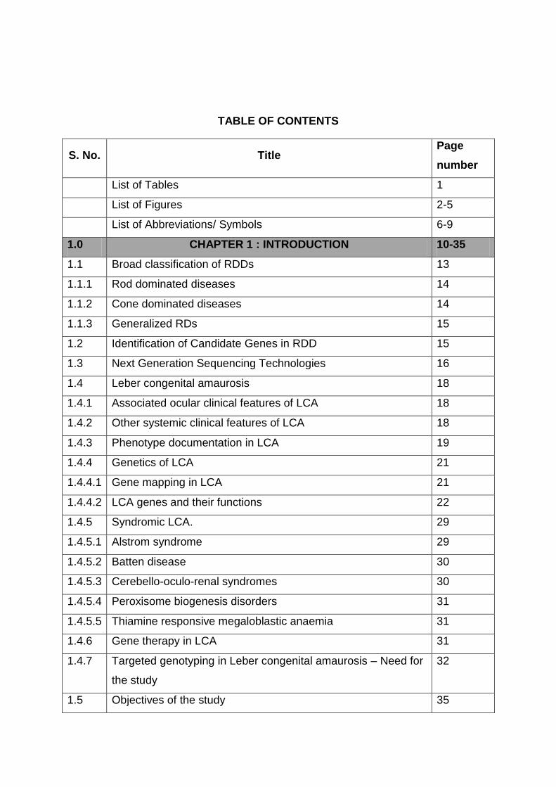
TABLE OF CONTENTS
S. No. Title Page
number
List of Tables 1
List of Figures 2-5
List of Abbreviations/ Symbols 6-9
1.0 CHAPTER 1 : INTRODUCTION 10-35
1.1 Broad classification of RDDs 13
1.1.1 Rod dominated diseases 14
1.1.2 Cone dominated diseases 14
1.1.3 Generalized RDs 15
1.2 Identification of Candidate Genes in RDD 15
1.3 Next Generation Sequencing Technologies 16
1.4 Leber congenital amaurosis 18
1.4.1 Associated ocular clinical features of LCA 18
1.4.2 Other systemic clinical features of LCA 18
1.4.3 Phenotype documentation in LCA 19
1.4.4 Genetics of LCA 21
1.4.4.1 Gene mapping in LCA 21
1.4.4.2 LCA genes and their functions 22
1.4.5 Syndromic LCA. 29
1.4.5.1 Alstrom syndrome 29
1.4.5.2 Batten disease 30
1.4.5.3 Cerebello-oculo-renal syndromes 30
1.4.5.4 Peroxisome biogenesis disorders 31
1.4.5.5 Thiamine responsive megaloblastic anaemia 31
1.4.6 Gene therapy in LCA 31
1.4.7 Targeted genotyping in Leber congenital amaurosis – Need for
the study
32
1.5 Objectives of the study 35

2.0 CHAPTER 2 – METHODOLOGY 36-57
Overview of the methodology 36
2.1 Patients recruitment 37
2.1.1 Clinical documentation 37
2.1.2 Sample collection 39
2.2 DNA extraction 40
2.3 DNA Quantification and Quality check 40
2.4 Targeted re-sequencing using Illumina MiSeq platform 41
2.4.1 Designing target enrichment probes: 41
2.4.2 Target enrichment and Sequencing 43
2.4.3 Analysis pipeline 46
2.5 Validation of the identified pathogenic and likely pathogenic
mutations
48
2.6 Insilico Predictions 55
2.7 Screening of SLC19A2 in a patient diagnosed with Thiamine
Responsive Megaloblastic Anaemia (TRMA) with LCA as
ocular feature.
55
3.0 CHAPTER 3 – RESULT 58-106
3.1 Targeted re-sequencing 58
3.1.1 Data analysis 58
3.1.2 Validation of the identified pathogenic and likely pathogenic
mutations
61
3.1.3 Other reported or novel variations and SNPs in coding and
non–coding regions
75
3.2 cDNA analysis 79
3.3 Bioinformatics Analyses 81
3.4 Genotype- Phenotype Correlation 84
3.5 Screening of SLC19A2 in a patient diagnosed with Thiamine
Responsive Megaloblastic Anaemia (TRMA) with LCA as
ocular phenotype.
106
4.0 CHAPTER 4 – DISCUSSION 108-117
4.1 Targeted resequencing in Indian LCA cohort 108
4.1.2 Possible functional impact of mutant proteins 112

4.2 Observed syndromic LCA 115
4.2.1 Senior-Loken syndrome (SLSN) 115
4.2.2 Thiamine responsive megaloblastic anaemia 116
CONCLUSION 119
SPECIFIC CONTRIBUTIONS 120-121
LIMITATIONS 122
FUTURE SCOPE OF THE STUDY 122
References 123-147
Appendices 148-171
List of Publications and Presentations 172-174
Brief Biography of the Candidate 175
Brief Biography of the Supervisor 176
Brief Biography of the Co- Supervisor 177-178

LIST OF TABLES
Table number
Title Page number
1. A partial list of syndromic and non-syndromic retinal
degenerative disease with monogenic inheritance
11
2. List of novel genes identified in retinal and macular
degeneration using next-generation sequencing approaches
17
3.
4.
Reaction protocol for polymerase chain reaction.
List of regions analysed by PCR based direct sequencing
with annealing temperature and product size
48
49
5.
6.
7.
8.
ExoSAP protocol
Reaction protocol for cycle sequencing.
Reaction protocol for cDNA Synthesis.
cDNA region analysed by PCR based direct sequencing with
annealing temperature and product size
51
52
53
54
9. List of regions in SLC19A2 analysed by PCR based direct
sequencing with annealing temperature and product size.
ASPCR- Allele specific PCR for the identified mutation in
exon 2.
57
10. Raw read summary 58
11. Average coverage and depth for each of 20 genes 59
12. Shows the list of positive control samples and the variants 60
13. Lists of the pathogenic variants identified in fifty-six cases. 62
14.
15.
Lists the heterozygous variants observed in mutation positive
cases possibly contributing to triallelism
Bioinformatic analyses of missense variants
76
82
16. Shows the clinical features of mutation positive index cases
affected by LCA
89

2
LIST OF FIGURES
Figure
Number Title
Page
number
1. Genes expressed in different layers of retina and RPE, and
the spectrum of diseases they cause when mutated
10
2. Overlapping genotypes in non-syndromic monogenic retinal
and vitreoretinal degenerative diseases.
13
3. Diagrammatic representation of retinal layers and the retinal
cells associated with specific ERG wave pattern
19
4. A-D OCT image of normal subject and CEP290 mutation positive
patient with defined photoreceptor inner/ outer segment
junction.
20
5. Representative picture of retinal colour fundus photograph
with known genotypes.
21
6. A. Pie chart showing distribution of LCA candidate genes
mutations in North American cohort from 30 published studies
34
B. Distribution of LCA candidate genes mutations in Indian
patients from 8 published studies.
7. Diagrammatic representation of methodology followed. 36
8. A. Shows the ERG of LCA patient showing extinguished rod and
cone response as a flat wave pattern
37-38
B. OCT of patient having mutation in RDH12 gene showing
foveal thinning with RPE atrophy
C. Fundus photo
D. Fundus autoflourescence of a patient with mutation in AIPL1
gene showing macular changes
9. A-D Representative pedigrees of cases recruited. 39
10. Shows distribution of consanguinity among the north and
south Indian cases in the cohort of 92 LCA families.
40
11. Agarose gel image of DNA samples to check their quality.
41
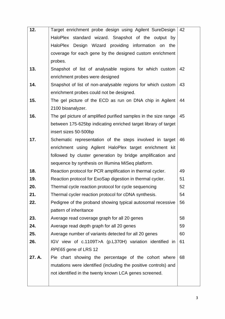
3
12. Target enrichment probe design using Agilent SureDesign
HaloPlex standard wizard. Snapshot of the output by
HaloPlex Design Wizard providing information on the
coverage for each gene by the designed custom enrichment
probes.
42
13. Snapshot of list of analysable regions for which custom
enrichment probes were designed
42
14. Snapshot of list of non-analysable regions for which custom
enrichment probes could not be designed.
43
15. The gel picture of the ECD as run on DNA chip in Agilent
2100 bioanalyzer.
44
16. The gel picture of amplified purified samples in the size range
between 175-625bp indicating enriched target library of target
insert sizes 50-500bp
45
17.
Schematic representation of the steps involved in target
enrichment using Agilent HaloPlex target enrichment kit
followed by cluster generation by bridge amplification and
sequence by synthesis on Illumina MiSeq platform.
46
18.
19.
20.
21.
22.
Reaction protocol for PCR amplification in thermal cycler.
Reaction protocol for ExoSap digestion in thermal cycler.
Thermal cycle reaction protocol for cycle sequencing
Thermal cycler reaction protocol for cDNA synthesis.
Pedigree of the proband showing typical autosomal recessive
pattern of inheritance
49
51
52
54
56
23. Average read coverage graph for all 20 genes 58
24. Average read depth graph for all 20 genes 59
25. Average number of variants detected for all 20 genes 60
26. IGV view of c.1109T>A (p.L370H) variation identified in
RPE65 gene of LRS 12
61
27. A. Pie chart showing the percentage of the cohort where
mutations were identified (including the positive controls) and
not identified in the twenty known LCA genes screened.
68

4
B. Pie chart showing the percentage distribution of different
types of mutations among the mutation positive cases.
C. Bar diagram showing the distribution of reported and novel
mutations among the different types of identified mutations.
28. Pie chart showing frequency of mutations in the twenty LCA
candidate genes in the Indian cohort studied.
69
29. Electrophoretogram of the identified mutations (marked by
arrows) in the LCA probands.
70-71
30. Representative pedigree of the family LCARS-81, segregating
a homozygous nonsense mutation, c.910G>T p. (E340X) in
AIPL1 gene.
72
31. Putative protein structure showing domains and mutations
(marked by arrows) in proteins of LCA candidate genes
identified by targeted resequencing
73
32. Pedigree of LRS 90 showing digenic inheritance segregating
in the family. Proband and affected sib are heterozygous for
both AIPL1 c.834G>A; p.(W278X) and KCNJ13 c.485G>A;
p.(R162Q) mutations. The father and the mother are
heterozygous for KCNJ13 and AIPL1 mutations, respectively.
74
33. In family LRS 67 79
A. 0.7% agarose gel picture showing RNA
B. 2% agarose gel picture showing cDNA amplified products of
525bp size targeting SPATA7- c.913-2A>G optimized at 63-
56 (-0.5°C) touchdown protocol.
34. In family LRS 29 80
A. 0.7% agarose gel picture showing RNA
B. 2% agarose gel picture showing GAPDH amplification of the
corresponding cDNA
C. cDNA amplified products of 596bp size targeting SPATA7-
c.1215+5C>A optimized at 65-58 (-0.5°C) touchdown
protocol.
35. Electrophoretogram of cDNA analysis for splice variations in
SPATA7 gene.
80

5
A. Wild type cDNA sequence (forward) in control showing exon 7
and exon 8
B. Homozygous indel (insertion of GT and first 7 bases deletion
in exon 8) in LRS 67 due to splice mutation c. 913-2A>G
C. Heterozygous indel in parent of LRS 67.
36. SPATA7- c.1215+5C>A electrophoretogram showing
representative reverse primer sequence of
81
A. Wild type control cDNA
B. Homozygous deletion of exon 10 in proband
C. Heterozygous deletion of exon 10 in parent.
37. Fundus photograph showing Genotype- phenotype
correlation in LCA patients with mutation in genes
87
38. 2% Agarose gel picture of the amplified products of the six
exons of SLC19A2 gene
106
39. A-D
40
Sequence showing a novel point mutation in exon 2, a
c.314G>A transition resulting in a missense mutation
p.(G105E).
Pie charts of cohort studies showing frequency of LCA
candidate gene mutation in the population. A. Italy B. Japan
C. China D. Australia E. Brazil F. Western cohort G. Current
study
107
109

6
LIST OF ABBREVIATIONS/ SYMBOLS
ABCA4 ATP-Binding Cassette, Subfamily A, Member 4
ABHD12 Abhydrolase Domain-Containing Protein 12
ACD Acid Citrate Dextrose
AIPL1 Aryl hydrocarbon-Interacting Receptor Protein-Like 1
ALMS1 Alstrom syndrome protein 1
AMD Age related Macular Degeneration
APEX Arrayed primer extension
BBS Bardet Biedl Syndrome
bp Base pair
CACNA1F 1 Calcium Channel, Voltage-Dependent, Alpha-1F1 Subunit
CCDS consensus coding sequence
CD Cone Dystrophy
CNGA3 Cyclic nucleotide gated channel alpha 3
CNGB3 Cyclic nucleotide gated channel beta 3
CRB1 Crumbs, Drosophila, Homolog Of, 1
CRD Cone Rod Dystrophy
CSNB Congenital Stationary Night Blindness
DHDDS 1 Dehydrodolichyl Diphosphate Synthase
DNA Deoxyribonucleic acid

7
EDTA Ethylenediaminetetraacetic acid
ELOVL4 Elongation of Very Long Chain Fatty Acids-Like 4
ERG Electroretinography
ExAC The Exome Aggregation Consortium
ExoSap Exonuclease and Shrimp alkaline phosphatase
GA Genome Analyzer
GAPDH Glyceraldehyde 3-phosphate dehydrogenase
GATK Genome Analysis Toolkit
Gb Giga base
GNAT2 Guanine Nucleotide-Binding Protein, Alpha-Transducing 2
GTPase Guanosine triphosphate hydrolase
GUCY2D Guanylate Cyclase 2D
HK1 Hexokinase 1
hRPE65v2 Recombinant adeno-associated virus retinal pigment epithelium gene
vector
hTERT Human Telomerase reverse transcriptase
IGV Integrated genome viewer
Indel Insertion deletion
LCA Leber Congenital Amaurosis
LRIT3 Leucine-Rich Repeat, Immunoglobulin-like and Transmembrane
Domains-Containing Protein 3
LRS LCA resequencing
MAF Minimum Allele Frequency
MYO7A Myosin VIIA

8
NAD Nicotinamide adenine dinucleotide
NaOH Sodium hydroxide
NC Negative control
NGS Next Generation Sequencing
NMNAT1 Nicotinamide Nucleotide Adenylyltransferase 1
OCT Optical coherence tomography
OD (Latin oculus dexter) indicates the right eye.
OMIM Online Mendelian Inheritance in Man
ONL Outer nuclear layer
OS (Latin oculus sinister) indicates the left eye.
PCR Polymerase Chain Reaction
PDE6C Phosphodiesterase 6C
PDE6H Phosphodiesterase 6H
Q30 Quality score 30
RAB28 RAS-Associated Protein 28
RD Retinal Degeneration
RDH12 Retinol Dehydrogenase 12
RNA Ribonucleic acid
RP Retinitis Pigmentosa
RPE Retinal Pigment Epithelium
RPE65 Retinal Pigment Epithelium-Specific Protein, 65- Kilo Dalton
RPGR Retinitis Pigmentosa GTPase Regulator
RPGRIP1 Retinitis Pigmentosa GTPase Regulator-Interacting Protein

9
RT-PCR Reverse transcriptase PCR
SLC19A2 Solute Carrier Family 19 (Thiamine Transporter), Member 2
SNP Single Nucleotide Polymorphism
TCP T-complex polypeptide 1
UTR untranslated region
VUS Variant of unknown significance
WES Whole Exome Sequencing

10
CHAPTER 1
INTRODUCTION
The completion of the human genome project has paved way in providing new
avenues and advances in medicine. The knowledge of human genome and
significant development in genomic research has substantially improved the
understanding of genetic basis of many diseases including Mendelian,
mitochondrial and complex disorders, their classification and management.
Retinal degeneration are heterogeneous group of inherited diseases which are
currently untreatable and share common pathological features affecting the
photoreceptor and retinal pigment epithelial cells of retina causing varying degrees
of irreversible vision loss1. Retinal degenerative diseases (RDD) together have a
worldwide prevalence of 1 in 2000. Photoreceptors, retinal ganglion cells and other
second order neurons encode proteins that are vital for normal function,
maintenance, synaptic interaction, and signalling (Figure 1).

11
Figure 1: Genes expressed in different layers of retina and RPE, and the spectrum
of diseases they cause when mutated
Mutations in these genes cause various types of either degenerative or non-
progressive retinal diseases2.
Most RDDs follow Mendelian (monogenic) pattern of inheritance and may be non-
syndromic or syndromic forms (Table 1) with clinically distinguishable findings.
These affect photoreceptor and/or RPE development and function3 as seen in
retinitis pigmentosa, Leber congenital amaurosis etc. Retinal degenerative diseases
can also follow multifactorial inheritance like in age related macular degeneration
(ARMD) and glaucoma. Visual image is formed by the interaction of many proteins
synthesised by different cells of the retina. So far more than 200 different genes
have been identified to cause monogenic retinal degenerative diseases4. The
severity of the disease/s depends on the type of mutation within the gene, degree of
damage caused by different mutations that may result in either total absence or
presence of a non-functional or potentially toxic protein.
Table 1: A partial list of syndromic and non-syndromic retinal degenerative disease
with monogenic inheritance.4, 5
Diseases Affected cell type
Mode of inheritance
Genes associated
Non - syndromic monogenic
CSNB
Rods more than cones
Dominant GNAT1, PDE6B, RHO
Recessive
CABP4, GNAT1, GNB3, GPR179, GRK1,GRM6, LRIT3, RDH5, SAG, SLC24A1, TRPM1
X-linked CACNA1F, NYX
LCA Rods and cones
Dominant CRX, IMPDH1, OTX2
Recessive
AIPL1, ALMS1, CABP4, CCT2, CEP290, CNGA3, CRB1, CRX, CLUAP1, DTHD1, GUCY2D, GDF6, IQCB1, IMPDH1, IFT140, KCNJ13, LCA5, LRAT,MERTK, MYO7A, NMNAT1, OTX2, PRPH2, RD3, RDH12, RPE65, RPGRIP1, SPATA7, TULP1

12
RP Rods, Cones and RPE
Dominant
ARL3, BEST1, CA4, CRX, FSCN2, GUCA1B,HK1, IMPDH1, KLHL7, NR2E3, NRL, PRPF3,PRPF4, PRPF6, PRPF8, PRPF31, PRPH2,RDH12, RHO, ROM1, RP1, RP9, RPE65,SEMA4A, SNRNP200, SPP2, TOPORS
Recessive
ABCA4, AGBL5, ARL6, ARL2BP, BBS1,BBS2, BEST1, C2orf71, C8orf37, CERKL,CLRN1, CNGA1, CNGB1, CRB1, CYP4V2,DHDDS, DHX38, EMC1, EYS, FAM161A,GPR125, HGSNAT, IDH3B, IFT140, IFT172,IMPG2, KIAA1549, KIZ, LRAT, MAK,MERTK, MVK, NEK2, NEUROD1, NR2E3,NRL, PDE6A, PDE6B, PDE6G, POMGNT1,PRCD, PROM1, RBP3, RGR, RHO, RLBP1,RP1, RP1L1, RPE65, SAG, SLC7A14, SPATA7,TRNT1, TTC8, TULP1, USH2A, ZNF408,ZNF513
X-linked OFD1, RP2, RPGR
CD or CRD Cones more than rods
Dominant AIPL1, CRX, GUCA1A, GUCY2D, PITPNM3,PROM1, PRPH2, RIMS1, SEMA4A, UNC119
Recessive
ABCA4, ADAM9, ATF6, C21orf2, C8orf37,CACNA2D4, CDHR1, CERKL, CNGA3,CNGB3, CNNM4, GNAT2, KCNV2, PDE6C,PDE6H, POC1B, RAB28, RAX2, RDH5,RPGRIP1, TTLL5
X-linked CACNA1F, RPGR
Macular degeneration
Rods and cones
Dominant BEST1, C1QTNF5, CTNNA1, EFEMP1,ELOVL4, FSCN2, GUCA1B, HMCN1, IMPG1,OTX2, PRDM13, PROM1, PRPH2, RP1L1,TIMP3
Recessive ABCA4, CFH, DRAM2, IMPG1, MFSD8
X-linked RPGR
Syndromic
BBS Rods and cones
Recessive
ADIPOR1, ARL6, BBIP1, BBS1, BBS2, BBS4,BBS5, BBS7, BBS9, BBS10, BBS12, C8orf37,CEP290, IFT172, IFT27, INPP5E, KCNJ13,LZTFL1, MKKS, MKS1, NPHP1, SDCCAG8,TRIM32, TTC8
Joubert syndrome
Rods and cones
Recessive
INPP5E, TMEM216, AHI1, NPHP1, CEP290 (NPHP6), TMEM67 (MKS3), RPGRIP1L, ARL13B, CC2D2A, TTC21B, KIF7, TCTN1, TCTN2, TMEM237, CEP41, TMEM138, C5orf42, TCTN3, TMEM231, CSPP1, PDE6D
Dominant ZNF423
X-linked OFD1
Senior- loken syndrome
Rods and cones
Recessive CEP164, CEP290, INVS, IQCB1, NPHP1, NPHP3, NPHP4, SDCCAG8, TRAF3IP1, WDR19
Usher syndrome
Rods and cones
Recessive ABHD12, ADGRV1, CDH23, CEP250, CIB2,CLRN1, DFNB31, HARS, MYO7A, PCDH15,USH1C, USH1G, USH2A

13
Mutations in the same gene cause a range of clinical phenotypes defined as allelic
heterogeneity, a common feature of monogenic RDD. For example, mutation in
transcription factor, CRX (cone-rod homeobox), that plays a vital role in
photoreceptor development and homeostasis6 (Figure 2) can cause either Leber
congenital amaurosis (LCA) where there is congenital blindness or progressive
cone-rod dystrophy (CRD)/ retinitis pigmentosa (RP), where the disease progresses
over time. Hereditary retinal degenerations are probably the most genetically
heterogeneous group of diseases in humans i.e. they demonstrate locus
heterogeneity where a single disease phenotype can be caused by mutations in
different genes. Therefore, providing a molecular diagnosis in RDD becomes
challenging along with variable expressivity, incomplete penetrance, and frequent
clinical and genetic overlap7.
Figure 2: Overlapping genotypes in non-syndromic monogenic retinal and
vitreoretinal degenerative diseases.
1.1. Broad classification of RDDs:
Depending on the type of photoreceptor affected, the RDDs are categorised as rod
dominated diseases, cone dominated diseases, and generalised retinal
degenerations involving both rods and cones7.
14
1.1.1. Rod dominated diseases
In these diseases, the rod photoreceptors are primarily affected followed by cones,
like in retinitis pigmentosa (RP). Retinitis pigmentosa is considered the most
common form of RD with a frequency of 1 in 3000-7000 individuals8. It is a
progressive disease and has been associated with many syndromic RDs like
Bardet- Biedl syndrome (BBS), Usher syndrome etc. The disease presents with
progressive deterioration in the ability to see in dim light causing night blindness,
followed by loss of peripheral vision that slowly moves towards the centre resulting
in tunnel vision. So far 82 genes and 7 loci have been associated with autosomal
dominant, autosomal recessive, and X-linked RP.
1.1.2. Cone dominated diseases
Cone dystrophies have a prevalence of 1/40,000and are caused due to
degeneration of the cone cells of the retina. Cone cells are present throughout
retina with maximum concentration clustered in the macula. Degeneration of cones
leads to reduced visual acuity, central vision loss, reduced ability to see colours and
photophobia. Cone dystrophy (CD) can be autosomal dominant, recessive or X-
linked and can be stationary or progressive. Till now, 33 genes have been
associated with the disease. Achromatopsia is a stationary, congenital, autosomal
recessive inherited disorder characterized by reduced visual acuity, pendular
nystagmus, increased sensitivity to light (photophobia) and reduced or complete
loss of colour discrimination. Mutations in CNGA3, CNGB3, GNAT2, PDE6C or
PDE6H are shown to cause achromatopsia9.
In the progressive forms, complete blindness may occur in the later stages because
the rod photoreceptors also undergo degeneration. Stargardt disease is a juvenile
macular degeneration characterised by central vision loss where cone cells are
more concentrated. It is associated with mutation in ABCA4 (autosomal recessive)
and ELOVL4 (autosomal dominant) gene. X-linked cone dystrophy (XLCOD) is a
progressive disorder where the affected males have decreased visual acuity,
myopia, cone ERG disturbance and colour vision defects. Mutation in CACNA1F
and RPGR genes are shown to cause XLCOD10.

15
1.1.3. Generalized RDs
This involves the simultaneous degeneration of both rod and cone photoreceptor
functions and is present with progressive, often severe, deterioration of vision.
Leber congenital amaurosis is a generalised RD, which is congenital and is
considered the most severe. LCA accounts for 5% of inherited retinal degenerative
disorders. The reported worldwide prevalence of LCA is 1 in 30,000 to 1 in 81,000
11.
The only X-linked form of non-syndromic generalised RD is choroideremia (CHM).
Affected males develop night blindness in their second decade, followed by
progressive loss of peripheral vision and blindness. The incidence of CHM is
estimated as 1 in 100,00012.
Congenital stationary night blindness (CSNB) is a non-progressive retinal disorder
characterized by impaired night vision, decreased visual acuity, nystagmus,
myopia, and strabismus. Thirteen genes have been associated with the disease so
far and are inherited either as autosomal dominant or recessive or X- linked. Based
on the ERG wave form CSNB is classified as Riggs and Schubert-Bornschein. The
later is further classified as complete form (type 1 CSNB), characterized by the
complete absence of rod pathway function, whereas the incomplete form (type 2
CSNB) is due to impaired rod and cone pathway function. Oguchi
disease and fundus albipunctatus are forms of CSNB with abnormal fundi. In
Oguchi disease, the fundus displays a yellow sheen after exposure to light; this
sheen disappears following dark adaptation. In fundus albipunctatus, the retina
develops yellow-white dots. The mutated genes code for proteins that are involved
either in phototransduction cascade or signalling from photoreceptors to second
order neurons13, 14.
1.2. Identification of Candidate Genes in RDD
Identification of candidate genes and the causal mutation/ variant for monogenic
diseases has been mainly through linkage analysis. The segregation of the genetic
markers in affected individuals indicate the plausible genomic region of the
candidate gene15. The distance between the genetic markers (θ=frequency of
recombination) helps identify the disease loci. There are two types of linkage

16
mapping – parametric, where the exact mode of inheritance is a pre requisite and
often performed for Mendelian disorders and non-parametric that is independent of
inheritance patterns and analysed based on: identical by descent (IBD) or identical
by state (IBS). Linkage mapping requires large multigenerational families with many
affected and unaffected and it is used to identify candidate genes for all modes of
inheritance i.e. autosomal dominant or recessive and X-linked recessive.
Homozygosity mapping is a very efficient approach to study recessive disorders in
both consanguineous and non-consanguineous families where the information of
IBD is used. Here the markers (alleles) are homozygous by descent and are shared
between the affected individuals thus indicating possible disease loci16, 17. Animal
studies and knowledge of allelic heterogeneity have also lead to identification of
candidate genes in various forms of RDD.
Over the years, numerous candidate genes have been identified. Sanger
sequencing is considered 'gold standard' in terms of both read length and
sequencing accuracy. But screening all the genes by Sanger sequencing is both
time consuming and expensive. In order to simplify molecular diagnostics in
diseases that are genetically heterogeneous, APEX genotyping microarrays
(ASPER Ophthalmics, Estonia)18 were developed in which the PCR products of
each amplimer targeting the mutation regions are combined and hybridized to
oligonucleotide primers arrayed on the chip. A template-dependent single-
nucleotide extension reaction with fluorescently labelled dye terminator nucleotides
helps in the detection of variants for both sense and antisense strand. The limitation
of this technology is that it allows the detection of only known mutations and novel
mutations are missed. These limitations have been overcome by advancement in
genotyping technology, i.e. the advent of massively parallel sequencing using NGS.
1.3. Next Generation Sequencing Technologies
Several next generation sequencing (NGS) technology platforms have emerged
which includes Roche 454, Illumina GA, Ion torrent, and ABI SOLiD that are based
on massive parallel sequencing and generate large data. They are considerably
less expensive especially in screening genetically heterogeneous diseases.
NGS and its wide range of applications include chromatin immunoprecipitation
coupled to sequencing (ChIP-seq), Bisulphite sequencing, RNA sequencing (RNA-

17
seq), whole genome sequencing, whole exome sequencing, targeted re-sequencing
denovo assembling, and re-assembling of genome19. The broadest application of
NGS is the re-sequencing of human genomes to understand the genetic differences
in health and disease20.
NGS has helped in comprehensive understanding of the genetic architecture of
RDDs. NGS was first employed in retinal disease to study an Ashkenazi Jewish
family, where whole exome sequencing of three affected siblings revealed a
mutation in a novel gene, DHDDS, as a cause of RP 21. Linkage analysis followed
by whole exome sequencing (WES) or targeted sequencing has identified HK1 as a
novel causative gene for adRP, RAB28 gene in a German family with arCRD22,
ABHD12 gene in a family clinically diagnosed with Usher syndrome type 323, LRIT3
gene in CSNB24, NMNAT1 in Leber congenital amaurosis25.
Table 2: List of novel genes identified in retinal and macular degeneration using
next-generation sequencing approaches26.
Method Diseases Gene(s) identified
Whole -exome sequencing Retinitis pigmentosa (RP)
DHDDS, MAK, GNPTG, EMC1, GPR125, KIAA1549, ARL2BP
Leber congenital amaurosis ( LCA)
NMNAT1, KCNJ13, DTHD1, CCT2, CLUAP1, ALMS1, CNGA3 and MYO7A
Congenital stationary night blindness LRIT3, GPR179,
Ciliopathy with skeleton abnormality WDR19
High myopia ZNF644
Bardet- Biedl syndrome (BBS) LZTFL1
Nephronophthisis with retinal degeneration ZNF423, CEP164
Usher syndrome HARS
Cone-rod dystrophy RAB28, ACBDS, C21orf2
Targeted sequencing RP, Cone -rod dystrophy C8orf37
AMD CFH, CF1
LCA IFT140, PRPH2,
Usher syndrome ABHD12
CSNB GPR179
X-Linked RP OFD1
Joubert syndrome TMEM237
Familial exudative vitreoretinopathy TSPAN12

18
Targeted NGS or WES has identified the genetic cause in 50-80% of the RDD in
various cohorts studied. It has proven to be a robust and cost effective technology
for identifying both novel and known mutations in candidate genes by targeted
capture 27 or involvement of known or novel genes by exome or whole genome
capture 25 (Table 2).
1.4. Leber congenital amaurosis
Leber congenital amaurosis is a severe form of retinal degenerative disease
diagnosed in children earlier than one year of age. This disease was described
initially by Theodore Leber in 1869 as a congenital form of retinitis pigmentosa28.
The clinically distinguishing features of LCA include severe visual impairment
present at birth or shortly thereafter; extinguished or non-recordable ERG, pendular
or searching nystagmus, photophobia, and digito-ocular sign (Franceschetti- Leber
phenomenon), with progressive retinal degeneration29.
1.4.1. Associated ocular clinical features of LCA:
Visual acuity (VA) in LCA range widely from 20/200 to little or no light perception.
LCA patients often have high refractive errors (from hyperopia to myopia) but most
patients are high hyperopes30. The oculo- digital sign of Franceschetti is an
important feature of LCA. The sign consists of a repetitive, deep pushing of the
knuckle or finger into the eye and socket. The oculo- digital phenomenon due to
persistent pushing, causes orbital fat atrophy, deep set eyes (enophthalmos) and
keratoconus, (the thinning of the cornea)30. Cataracts and keratoconus are often
associated with LCA31 as is nyctalopia (night blindness)32. Certain features are
specific to genotypes such as patients with RPE65 mutations show mild
improvements in their visual function in their second decade, but it is then shown to
decline after aperiod of stability33. Patients with CRX and GUCY2D mutations
appear to have a very significant loss of vision, but the loss remains stable34.
1.4.2. Other systemic clinical features of LCA
Apart from these, about 19.8% LCA patients are shown to suffer from mental
retardation or seizures or autism30, 35.

19
1.4.3. Phenotype documentation in LCA:
Electroretinogram (ERG)
ERG documentation is the primary diagnostic tool used to confirm LCA. It
evaluates the visual function by measuring the electrical response of the
entire retina i.e. first order neuron function (a-wave from photoreceptors),
and second-order neuron function (b-wave from ON- bipolar and Muller
cells). ERG helps in differential diagnosis among retinal dystrophies. In LCA,
ERG responses are non-detectable or extinguished within one year of life
indicating the severity of the rod and cone degeneration (Figure 3)36.
Figure 3: A. Diagrammatic representation of retinal layers and the retinal
cells associated with specific ERG wave pattern. B. Shows ERG of Leber
congenital amaurosis compared with a normal control.
Optical coherence tomography Optical coherence tomography (OCT), is a non-invasive imaging technique
used to obtain high resolution cross-sectional images of the retinal
architecture. OCT imaging is considered an important phenotypic
documentation as it helps in staging the severity of retinal degenerative
diseases by calculating the average photoreceptor layer thickness across a
wide retinal area (Figure 4). Measurable photoreceptor thickness in patients
increases the prospect for clinical trials where retrospective or prospective
staging could be related to treatment efficacy37.
20
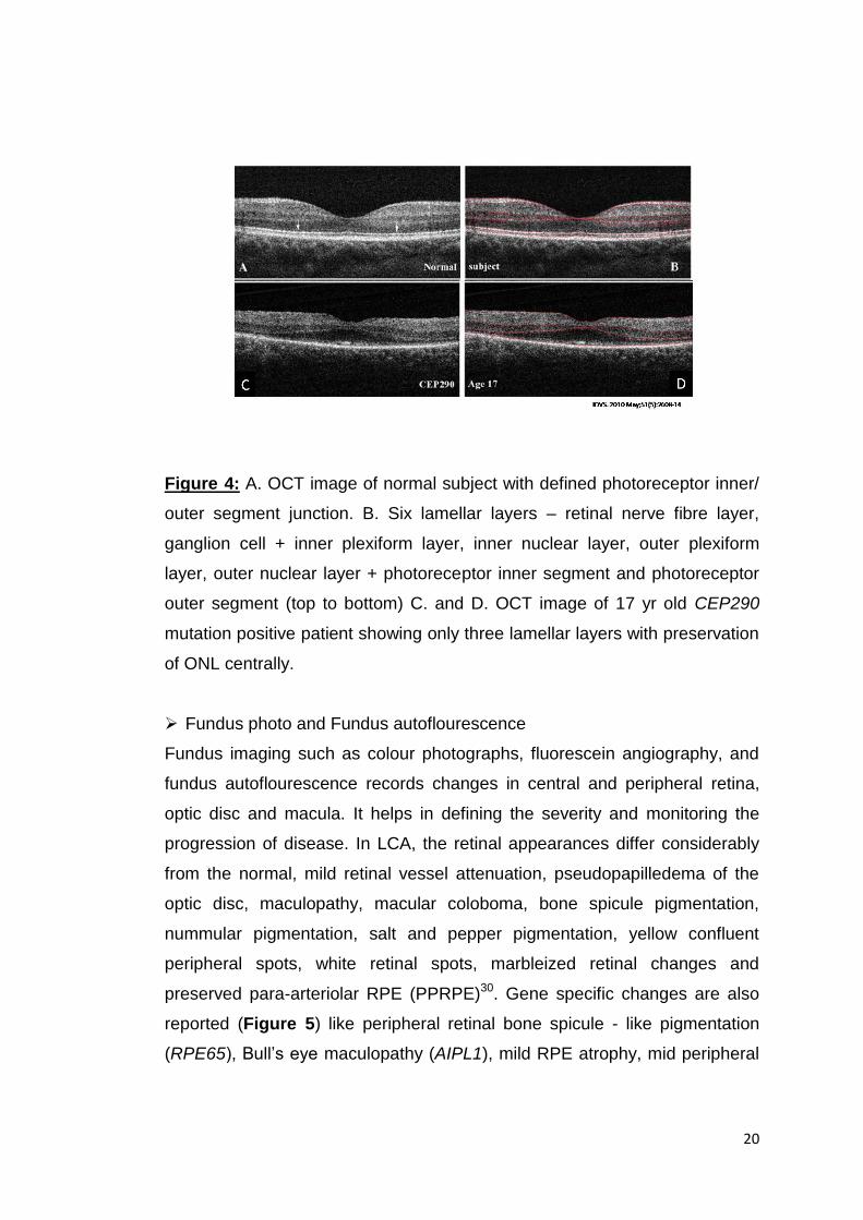
Figure 4: A. OCT image of normal subject with defined photoreceptor inner/
outer segment junction. B. Six lamellar layers – retinal nerve fibre layer,
ganglion cell + inner plexiform layer, inner nuclear layer, outer plexiform
layer, outer nuclear layer + photoreceptor inner segment and photoreceptor
outer segment (top to bottom) C. and D. OCT image of 17 yr old CEP290
mutation positive patient showing only three lamellar layers with preservation
of ONL centrally.
Fundus photo and Fundus autoflourescence
Fundus imaging such as colour photographs, fluorescein angiography, and
fundus autoflourescence records changes in central and peripheral retina,
optic disc and macula. It helps in defining the severity and monitoring the
progression of disease. In LCA, the retinal appearances differ considerably
from the normal, mild retinal vessel attenuation, pseudopapilledema of the
optic disc, maculopathy, macular coloboma, bone spicule pigmentation,
nummular pigmentation, salt and pepper pigmentation, yellow confluent
peripheral spots, white retinal spots, marbleized retinal changes and
preserved para-arteriolar RPE (PPRPE)30. Gene specific changes are also
reported (Figure 5) like peripheral retinal bone spicule - like pigmentation
(RPE65), Bull‟s eye maculopathy (AIPL1), mild RPE atrophy, mid peripheral

21
hyperpigmentation (RDH12), nummular pigmentation (CRB1), tapetal reflex
(CEP290) and mild foveal atrophy to macular coloboma (NMNAT1) 38.
Figure 5: Representative picture of retinal colour fundus photograph with
known genotypes. Fundus of A. normal individual B. LCA patient with RDH12
mutation C. LCA patient with RPGRIP1 mutation.
1.4.4. Genetics of LCA:
LCA is both clinically and genetically heterogeneous. So far, twenty-nine candidate
genes (AIPL1 (MIM*604392), ALMS1 (MIM*606844), CABP4 (MIM *608965), CCT2
(MIM*605139), CEP290 (MIM *610142), CNGA3 (MIM* 600053), CLUAP1 (MIM*
616787), CRB1 (MIM *604210), CRX (MIM *602225), DTHD1 (MIM* 616979),
GDF6 (MIM* 601147), GUCY2D (MIM *600179), IQCB1 (MIM *609237), IMPDH1
(MIM *146690), IFT140 (MIM* 614620), KCNJ13 (MIM *603208), LCA5 (MIM
*611408), LRAT (MIM *604863), MERTK (MIM *604705), MYO7A (MIM* 276903),
NMNAT1 (MIM *608700), OTX2 (MIM* 600037), PRPH2 (MIM* 179605), RD3 (MIM
*180040), RDH12 (MIM *608830), RPE65 (MIM *180069), RPGRIP1 (MIM
*605446), SPATA7 (MIM *609868) and TULP1 (MIM 602280)) have been identified
and most of them are inherited in an autosomal recessive manner except CRX,
IMPDH1 and OTX2 which are associated with autosomal dominant inheritance
pattern4, 11.
1.4.4.1. Gene mapping in LCA
Various methods like classical linkage analysis, homozygosity mapping, candidate
gene approach based on tissue specific expression or animal models, next
generation sequencing and/ or combinations of one or more approaches have been
used to identify candidate genes for LCA. Genes such as RDH12, TULP1, CRB1,
CRX, GDF6, IMPDH1, OTX2, RD3, RPE65 and RPGRIP1 were reported to be

22
associated with retinal functions or other retinal diseases. These were screened
and identified to be associated with LCA too39,40,41,42,43,44,45,46,47,48. Using whole
genome linkage analysis CEP29049 was identified. AIPL1 was identified as
candidate gene using linkage analysis coupled with insitu hybridization50.
Homozygosity mapping identified CABP451, GUCY2D52, IQCB153, LCA554 and
SPATA755. LRAT and MERTK were recognized as candidate genes for LCA while
screening many retinal genes in oligonucleotides SNP arrays56. With the advent of
next generation sequencing technology, targeted re-sequencing of retinal and
ciliopathy genes identified IFT14057 and PRPH258 while whole exome sequencing
alone or with homozygosity mapping identified CCT2, CLUAP1, ALMS1, CNGA3,
MYO7A, DTHD1, KCNJ13, and NMNAT129, 59-62.
1.4.4.2. LCA genes and their functions
The candidate genes code for proteins that belong to different families and perform
varied functions. Some of these genes code for ciliary proteins – as photoreceptors
are modified cilia, they are involved in photoreceptor morphogenesis and
stabilization, those that code for signalling molecules and ion channels- as ions are
required for synapses, those that are essential in phototransduction pathways and
visual cycle and those that perform other functions like phagocytosis, protein
trafficking, as molecular chaperones and signalling peptides.
Ciliary genes:
ALMS1
ALMS1, located on chromosome 2p13 codes for a centrosome and basal body
associated protein. The protein localizes to centrosomes and basal bodies of
ciliated cells. ALMS1 protein is involved in microtubule organization, cell cycle
regulation, intraciliary transport63, cell migration and extracellular matrix
production64. As ciliary bodies are present in multiple systems, defect in the protein
results in syndromic disease like Alstrom syndrome involving cone-rod retinal
dystrophy, hearing loss, childhood obesity, type 2 diabetes mellitus,
cardiomyopathy, fibrosis and multiple organ failure65. ALMS1 is also shown to be
associated with non-syndromic LCA29.

23
CEP290
Centrosomal protein, 290-KD (also known as NPHP6) is involved in ciliary
assembly and trafficking. CEP290 is located on 12q21.32 and mutation in this gene
is shown to be associated with many diseases, ranging from isolated blindness to
various syndromes like Senior-Loken syndrome (SLS), nephronophthisis (NPHP),
Joubert syndrome (related disorders) (JS[RD]), Bardet-Biedl syndrome (BBS), to
the lethal Meckel-Gruber syndrome (MKS). Knockdown experiments in zebra fish
using morpholinos revealed Cep290 deficiency caused defects involving retinal,
cerebellar, and optic cavity developmental abnormalities as well as pronephric cyst
formation, ectopic brain tissue in the fourth ventricle and an abnormal mid to-
hindbrain region associated with hydrocephalus66.
CLUAP1
Clusterin-associated protein 1 (CLUAP1) appears to be involved in assembly and
turnaround of intraflagellar transport (IFT) particles at the base and tip of the cilium.
Located at 16p13.3 CLUAP1 transcript is expressed abundantly in testis, thyroid,
trachea, eye (connecting cilium of photoreceptor cells), and moderately in spinal
cord and adrenal gland 67. CLUAP1 has been shown to be a tumor associated
antigen68 and recently is identified as a candidate gene for non-syndromic LCA60
IQCB1
Located on 3q13.33, it encodes an IQ motif-containing protein B1 (also known as
NPHP5), these proteins localize to connecting cilia of photoreceptors. Present in
almost all tissues, NPHP5 and CEP290 plays important role in controlling integrity
and ciliary trafficking of the BBSome. As a transition zone protein NPHP5 contains
two separate BBS-binding sites and interacts with the BBSome to mediate its
integrity69. Mutations in IQCB1 are reported in Senior-Loken syndrome, Bardet-
Biedl syndrome and non-syndromic LCA53.
IFT140
Intraflagellar transport 140 (IFT140) located on 16p13.3, encodes a subunit of
intraflagellar transport complex A (IFTA), which is involved in retrograde ciliary
transport. It is shown to be highly expressed in kidney, moderately in ovary, testis,
prostate, and lung, and low expression in thymus, brain, heart, placenta, and
skeletal muscle. In the eye, IFT proteins are expressed during all stages of

24
ciliogenesis in photoreceptor cells and are found within the differentiating
photoreceptor ciliary shaft during early stages of development70. Mutations in
IFT140 were previously reported in skeletal ciliopathies and recently in non-
syndromic LCA71.
LCA5
Located in 6q14.1, encodes Lebercilin (LCA5) protein that localizes to the ciliary
axoneme, with increased amounts at the base of the primary cilia. It is a
ubiquitously expressed protein and in the eye it is found to be present in the
connecting cilium and the basal bodies of photoreceptor cells and involved in ciliary
microtubule dynamics54.
MYO7A
Located in 11q13.5 encodes a protein belonging to myosin family. These are motor
molecules with structurally conserved heads that move along actin filaments thus
enabling them to transport cargo72, MYO7A expression is seen in human liver,
kidney, retinal pigment epithelium, and photoreceptor cells of the retina as well as in
cochlear and vestibular neuroepithelia. Mutations in MYO7A were previously found
to be associated with deafness and Usher syndrome type 1B73.
RPGRIP1
Located on 14q11.2, encodes Retinitis pigmentosa GTPase regulator-interacting
protein which is expressed in retina and testis. It localizes specifically in the
photoreceptor connecting cilium. RPGRIP anchors RPGR in the connecting cilium
and regulates protein transport.74. Mutations in RPGRIP1 also cause cone-rod
dystrophy75.
SPATA7
Spermatogenesis-associated protein 7, a ciliary protein, is expressed only in testis
and retina. The gene is located on 14q31.3. In P21 mice, Spata7 protein is
observed in multiple layers of the retina, including the ganglion cell, inner nuclear
layers, and inner segments of photoreceptors. SPATA7 localizes at the primary
cilium and connecting cilium (CC) of photoreceptor cells in hTERT RPE-1
cells.SPATA7 directly interacts with the retinitis pigmentosa GTPase regulator
interacting protein 1 (RPGRIP1), a key connecting cilium protein that has also

25
known to cause LCA. This complex plays an important role in protein trafficking
across the CC to the outer segments and a malfunction of this result in rhodopsin
accumulation in the inner segments and around the nucleus of photoreceptors. This
accumulation then likely triggers the apoptosis of rod photoreceptors 76. Apart from
LCA, mutation in SPATA7 is also associated with autosomal recessive retinitis
pigmentosa (RP).
Photoreceptor morphogenesis and maintenance genes:
CRB1
Crumbs, drosophila, homolog of, 1 (CRB1) localize to subdomains of the
photoreceptor apical plasma membrane i.e. the inner segment of mammalian
photoreceptors. The subdomains support the morphogenesis and orientation of the
photosensitive membrane organelles. Located on 1q31.3, it is expressed
exclusively in eye and the central nervous system77. Mutations in CRB1 is also
associated with pigmented paravenous chorioretinal atrophy and autosomal
recessive retinitis pigmentosa78.
CRX
Cone-rod homeobox-containing gene is located on 19q13.33 and is also associated
with cone-rod dystrophy79. CRX is expressed specifically in the photoreceptor cells
of developing and adult retina. It is a photoreceptor-specific transcription factor
which plays a role in the differentiation of these cells80.
CABP4
Located in 11q13.2 encodes a calcium-binding protein 4 (CABP4) that localizes to
the synaptic terminals of photoreceptors. It belongs to a subfamily of at least eight
calmodulin (CaM)-like neuronal Ca2+ binding proteins. It co-localizes and interacts
with Cav1.4 voltage-dependent Ca2+ channels (VDCCs) and plays an important role
in the development and/ or maintenance of the photoreceptor output synapse,
probably through modulation of photoreceptor VDCCs and transmitter release81.
CABP4 is also a candidate gene for congenital stationary night blindness82.
GDF6
Growth/ differentiation factor 6 is a member of the transforming growth factor-beta
superfamily. Located on 8q22.1, it is expressed in ganglion cell layer, inner
plexiform layer, and retinal pigment epithelia in the eye. It has been hypothesised

26
that GDF6 may regulate ectoderm patterning and control eye morphogenesis by
regulating neural and vascular development. GDF6 mutations have been
associated with eye phenotypes such as microphthalmia, anophthalmia83 and
Klippel–Feil syndrome84.
KCNJ13
Potassium channel, inwardly rectifying, subfamily J, member 13, is expressed in
wide variety of cells with the common role of maintaining resting membrane
potential near the potassium equilibrium potential. Located in 2q37.1, KCNJ13 is
expressed in apical RPE and help to maintain potassium homeostasis around the
photoreceptor outer segments85. KCNJ13 mutations were first reported in
Snowflake vitreoretinal degeneration86
OTX2
Located on 14q22.3, Orthodenticle, drosophila homolog of 2, is a transcription
factor and plays a role in brain, craniofacial and sensory organ development. OTX2
is also required for the development and maintenance of the neural retina. It is
found in bipolar and in ganglion cells, while in the outer nuclear layer it regulates
the expression of the closely related CRX gene that controls the expression of a
suite of photoreceptor function genes, including opsins87. Mutations in this OTX2
also cause syndromic microphthalmia 5 (MCOPS5) and combined pituitary
hormone deficiency 6 (CPHD6)88.
PRPH2
Peripherin-2 is tetra spanning membrane proteins localized along the rim of rod and
cone photoreceptor outer segment discs. Peripherin-2 is critical to the formation
and stabilization of photoreceptor outer segments. It is located in 6p21.1 and
mutations in this gene is reported in retinitis pigmentosa and macular dystrophy89,
90.
RD3
Located on 1q32.3, retinal degeneration 3 (RD3) is expressed only in retina. The
protein is localised in outer nuclear layer, inner nuclear layer, and the ganglion cell
layer. RD3 mediates the export of GC1 (Guanylatecyclase 1) from the endoplasmic
reticulum to endosomal vesicles. RD3 co localizes, binds to GC1 and GC2 to play a

27
crucial role in their stable expression and membrane trafficking in rod and cone
photoreceptors91.
Molecular chaperones:
AIPL1
Located in 17p13.1, AIPL1 encodes human aryl hydrocarbon receptor-interacting
protein-like 1 (AIPL1) and is similar to (49% identity, 69% positive) human aryl
hydrocarbon receptor-interacting protein (AIP), a member of the FK506-binding
protein (FKBP) family. This protein contains three tetratricopeptide (TPR) motifs,
consistent with nuclear transport or chaperone activity. It is expressed in
photoreceptor and pineal-gland50. Mutation in AIPL1 is also reported in cone-rod
dystrophy and retinitis pigmentosa92.
CCT2
Chaperonin containing TCP1, subunit 2, located on 12q15, encodes the molecular
chaperone protein, CCTβ. Although expressed ubiquitously, in retina it is expressed
in retinal ganglion cell layer and near the connecting cilium in the photoreceptor
cells. CCTβ interacts with chaperonin type BBSs, including BBSs 6, 10, and
12 which are ciliary proteins. CCT2 is also shown to bind to G protein subunit Gβ1
which is involved in transducin- associated phototransduction59
NMNAT1
Nicotinamide nucleotide adenylyltransferase 1, located on 1p36.22, is a central
enzyme in NAD biosynthesis, catalysing the condensation of nicotinamide
mononucleotide (NMN) or nicotinic acid mononucleotide (NaMN) with the AMP
moiety of ATP to form NAD or NaAD. It is expressed in skeletal muscle, heart, liver,
and kidney. In the eye, NMNAT act as a stress-response protein that works as a
chaperone for neuronal maintenance and protection93.
Phototransduction:
CNGA3
Cyclic nucleotide-gated channel, alpha-3 (CNGA3), an achromatopsia-2 gene, is
located on 2q11.2. It is found to be expressed in cone photoreceptors, testis,
kidney, and heart94. The CNGA3 encodes one of a family of alpha subunits that
form CNG ion channels required for sensory transduction in rod photoreceptors and

28
in olfactory neurons. Localized to the photoreceptor plasma membrane, is essential
for the generation of light- evoked electrical responses in the red, green, and blue-
sensitive cones.
GUCY2D
Guanylatecyclase 2D, in 17p13.1 region encodes a retina-specific Guanylate
cyclase (RetGC), which is a member of the membrane guanylylcyclase family.
RetGC mediates Ca2+ feedback on cGMP metabolism during photoreceptor light
adaptation and/ or recovery following photo excitation95. Mutations in this gene are
also reported in dominant cone-rod dystrophy96.
LRAT
Lecithin retinol acyltransferase catalyses the esterification of all-trans-retinol into all-
trans-retinyl ester. This reaction is an important step in vitamin A metabolism in the
visual system. Located in 4q32.1, it is expressed in several fetal and adult human
tissues, including the retinal pigment epithelium (RPE) and liver. Mutation in LRAT
was also observed in early-onset severe retinal dystrophy97.
RDH12
Located on 14q24.1, retinol dehydrogenase 12 belongs to a family of dual-
specificity retinol dehydrogenases that metabolize both all-trans- and cis-retinols.
RDH12 is expressed predominantly in eye and also in kidney, brain, skeletal
muscle and stomach. In eye, it is expressed at the base of photoreceptor inner
segments and during visual cycle catalyses the reduction of all-trans-retinal and its
9-cis-, 11-cis-, and 13-cis-retinal isomers in the presence of NADPH98.
RPE65
Located on 1p31.3 encodes the retinal pigment epithelium- specific protein 65KD, ,
which catalyses the conversion of all-trans retinyl ester to 11-cis retinol in the
RPE99. This RPE specific protein acts as the receptor for retinol-binding protein on
the surface of the retinal pigment epithelium. Other than LCA, mutations in RPE65
are also implicated in retinitis pigmentosa.
IMPDH1
Inosine-5-prime-monophosphate dehydrogenase, type I is an enzyme that
catalyses the synthesis of xanthine monophosphate (XMP) from inosine-5'-mono-

29
phosphate (IMP). This is the rate-limiting step in the de novo synthesis of guanine
nucleotides. Located on 7q32.1, it is also a candidate gene for autosomal dominant
retinitis pigmentosa and is ubiquitously expressed but has a retina specific isoform
derived from alternative splicing. In the retina, it is localized to the inner segment
and synaptic terminals of photoreceptors100 and is found to be associated with
polyribosomes containing rhodopsin mRNA. Mutations in IMPDH1 are reported in
autosomal dominant LCA as well.
Signalling pathway, Phagocytosis and protein trafficking
DTHD1
Death domain-containing protein 1 in 4p14 is predicted to function in signalling
pathways, formation of signalling complexes, as well as in apoptosis pathway.
Localization of these signalling proteins in the eye are yet unknown.
MERTK
Mer tyrosine kinase protooncogene located on 2q13 is expressed in normal
peripheral blood monocytes, bone marrow, in tissues of epithelial and reproductive
origin. Mer receptor tyrosine kinase is critical for the engulfment and efficient
clearance of apoptotic cells. In retina, MERTK plays an important role in
phagocytosis of outer segments by RPE cells101. Mutations in MERTK were first
implicated in RP101.
TULP1
Located on 6p21.31, Tubby-like protein 1 is expressed exclusively in retina. TULP1
is localized to the photoreceptor inner segment and synapse. TULP1 is shown to be
involved in protein trafficking from inner segment to outer segment where they bind
to several cytoskeletal scaffold proteins, as well as members of the
phototransduction cascade and in vesicle cycling at the synaptic terminal102.
Mutations in TULP1 are also implicated in RP103.
1.4.5. Syndromic LCA
„LCA like ocular phenotype‟ is observed as one of the characteristic features in few
syndromes. These syndromes initially present as LCA, as it is diagnosed earlier to
one year of age and later presents systemic features, which involve various other
organs.

30
1.4.5.1. Alstrom syndrome
Alstrom syndrome is an autosomal recessive disorder characterised by LCA
appearing first in infancy and then progressive development of multi-organ
pathology which include hearing loss, childhood truncal obesity, insulin resistance
and hyperinsulinemia, type 2 diabetes, hypertriglyceridemia, short stature in
adulthood, cardiomyopathy, and progressive pulmonary, hepatic, and renal
dysfunction. Alstrom syndrome is caused by mutations in ALMS gene. ALMS1
protein is found in centrosomes, basal bodies, and cytosol of all tissues affected by
the disease104.
1.4.5.2. Batten disease
Also known as the neuronal ceroid lipofuscinoses (NCL; CLN), is clinically and
genetically heterogeneous group of neurodegenerative disorders caused due to
intracellular accumulation of autofluorescent lipopigment storage material.
Clinically, the phenotypes are categorized according to the age of onset and order
of appearance of clinical features; as infantile, late-infantile, juvenile, adult, Northern
epilepsy. The genes associated among these phenotypes are also different. The
disease is characterized by progressive intellectual and motor deterioration,
seizures, and early death. Visual loss is a feature of most form and LCA is reported
in the infantile form105.
1.4.5.3. Cerebello-oculo-renal syndromes
Syndromes involving brain, eye and kidney abnormalities come under Cerebello-
oculo-renal syndromes.
Senior-Loken syndrome
Senior-Loken syndrome is a ciliopathic autosomal recessive disease with main
features being nephronophthisis (an autosomal recessive cystic kidney disease)
and progressive eye disease106. Both Leber congenital amaurosis and retinitis
pigmentosa are reported as ocular components. Six genes and one locus have so
far been identified and IQCB1 (SLSN5) is reported as a candidate gene in non-
syndromic LCA as well. Some of the LCA patients with IQCB1 mutation develop
renal abnormality and are thus re-diagnosed as SLSN53.

31
Joubert syndrome
Joubert syndrome is an autosomal recessive disease involving defects in
cerebellum and common features include developmental delays, ataxia (lack of
muscle control), hyperpnoea (abnormal breathing patterns), sleep apnea, abnormal
eye and tongue movements, and hypotonia in early childhood. Features of retinal
dystrophy like RP or LCA are also reported. Twenty-six genes have been reported
so far in Joubert syndrome including CEP290 which is one of the candidate genes
for LCA107.
1.4.5.4. Peroxisome biogenesis disorders
Peroxisomal disorders are a group of genetically heterogeneous metabolic
diseases that share dysfunction of peroxisomes, an organelle important for specific
metabolic pathways, such as beta-oxidation of very-long-chain fatty acids (VLCFA),
detoxification of hydrogen peroxide and production of cholesterol, bile acids, and
plasmalogens. Some common clinical characteristic of Zellweger spectrum of
peroxisomal biogenesis defects include neurologic, eye abnormalities (Leber
congenital amaurosis, retinitis pigmentosa, optic atrophy, cataracts), hepatorenal
defects, rhizomelic limb shortening, and calcific stippling of the patella108.
1.4.5.5. Thiamine responsive megaloblastic anaemia
Thiamine responsive megaloblastic anaemia (TRMA) is an autosomal recessive
disease caused by mutations in SLC19A2 and is characterized by a triad of
megaloblastic anaemia, non-type 1 diabetes mellitus and sensorineural deafness.
Ocular abnormalities which include cone rod dystrophy, retinitis pigmentosa and
optic atrophy are also reported in few cases. Thiamine transporter protein THTR1,
localized in red blood cells, inner hair cells of the cochlea, pancreas, and small
intestine is encoded by SLC19A2 gene. This protein helps in the absorption of
thiamine inside the cells. 109.
1.4.6. Gene therapy in LCA
Gene identification, proof-of-concept and safety studies of gene therapy in
animals, and detailed human studies on photoreceptor layer integrity have led to
treatment trials in the molecular form of LCA caused by mutations in RPE65, the
gene encoding retinal pigment epithelium-specific protein, 65 kDa110. In this gene
therapy trial, the researchers packaged a normal version of RPE65 gene inside a

32
genetically engineered vector, the adeno-associated virus (AAV). This was injected
subretinally to RPE65 mutated LCA patients. The vector delivers the gene to cells
in the retina, where the gene produces the functional enzyme and completes the
visual cycle which otherwise is not functional due to mutated RPE65.
The results of the phase 1 gene replacement therapy trial conducted at Moorefield‟s
eye hospital, University College London eye gene therapy study group showed
sustained improvement in three young adults who received gene therapy and
remained healthy maintaining the visual gains one year after treatment with AAV2-
hRPE65v2111, 112. Early intervention was associated with better results. The
immunological effect of antibodies after administration of AAV2-hRPE65v2 viral
vector in both eyes in canine and non-human primate animal models suggested
that simultaneous treatment of both eyes does not induce an immune response
complicating the treatment113. Jacobson et al in 2012 reported the safety and
efficacy in 15 children and adults followed up to 3 years where visual function
improved in all patients to different degrees and improvements were localized to
treated areas. Cone and rod sensitivities increased significantly in the study eyes
but not in the control eyes. Minor acuity improvements were recorded in many study
and control eyes114. Issues such as pseudo fovea formation, continued
degeneration of photoreceptors, increase in dosage level are now being
addressed115, 116. Other than the RPE65 human gene therapy trials, animal models
for gene therapy for LCA genes, AIPL1117, SPATA7118, GUCY2D119 and CEP290120
are being conducted and they also show promising results.
Another strategy that is being developed to treat RD is by targeting mutated gene
using antisense oligonucleotides (AONs). AONs are small stretch of
oligonucleotides that binds complementarily to the target sequence thereby
hindering either transcription or translation. By binding to the target sequence, they
can mask pre-mRNA splicing or trigger mRNA degradation as a DNA oligo can
activate RNAse H. In CEP290 associated LCA, AONs are used for splice correction
where the intronic CEP290 mutation c.2991+1655A>G creates cryptic splice-site
leading to pseudoexon formation and thereby aberrant protein product. By AON-
based therapy, the cryptic site is masked and the pseudoexon is skipped leading to
normal CEP290 splicing and wild-type CEP290 protein levels. Invitro and invivo

33
studies on mice models are promising 121. Thus, the AON treatment addressing
altered splicing has a therapeutic potential for many genetic diseases.
1.4.7. Targeted genotyping in Leber congenital amaurosis – Need for the study
LCA accounts for 3-5% of childhood blindness and has a prevalence of 1/50,000 -
1/33,000 live births (http://www.orpha.net -last updated July 2015). The genes
associated with LCA have so far accounted for approximately 75% of all LCA
cases. The frequency of mutations in LCA genes varies in different populations and
comprehensive mutation study focused on individual ethnicities in large cohorts are
few. Also, the gene that contributes to the highest mutation rate differs among the
populations.
The clinical and genetic heterogeneity of LCA hampers its routine molecular
diagnosis. Mutation- specific assays that are designed based on data available
from LCA patients from one ethnic population are not the most efficient approach
for studying patients from different populations and ethnic backgrounds. The
mutation studies conducted on Indian LCA cohorts either had a very small sample
size with fewer genes screened or included Indians as a part of multicentre studies
or case report. Screening RPE65 in 60 unrelated cases from Asian Indian cohort
contributed to only 1.7%122 whereas it contributes to 6% of LCA among various
other populations. Thirty-eight LCA patients from Southern India were tested for the
presence of 104 previously reported mutations in eight LCA genes from a Northern
American cohort data. Only one proband was found to carry one of these 104
mutations123.
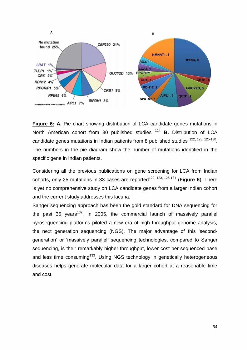
34
Figure 6: A. Pie chart showing distribution of LCA candidate genes mutations in
North American cohort from 30 published studies 124 B. Distribution of LCA
candidate genes mutations in Indian patients from 8 published studies 122, 123, 125-130.
The numbers in the pie diagram show the number of mutations identified in the
specific gene in Indian patients.
Considering all the previous publications on gene screening for LCA from Indian
cohorts, only 25 mutations in 33 cases are reported122, 123, 125-131 (Figure 6). There
is yet no comprehensive study on LCA candidate genes from a larger Indian cohort
and the current study addresses this lacuna.
Sanger sequencing approach has been the gold standard for DNA sequencing for
the past 35 years132. In 2005, the commercial launch of massively parallel
pyrosequencing platforms piloted a new era of high throughput genome analysis,
the next generation sequencing (NGS). The major advantage of this „second-
generation‟ or „massively parallel‟ sequencing technologies, compared to Sanger
sequencing, is their remarkably higher throughput, lower cost per sequenced base
and less time consuming133. Using NGS technology in genetically heterogeneous
diseases helps generate molecular data for a larger cohort at a reasonable time
and cost.

35
1.5. Objectives of the study
Objective 1:
• Clinical examination, complete phenotype documentation and recruitment of 100
patients with Leber congenital amaurosis (LCA).
Objective 2:
• To design a customized probe to capture all exons and 20bp flanking sequences
(exon - intron boundary) of the LCA candidate genes.
• Perform high throughput re-sequencing for the candidate genes on NGS
platform.
.Objective 3:
• To validate the results obtained by targeted re-sequencing using Sanger
sequencing, to perform segregation analysis in the families and control screening
for the identified mutations to confirm the pathogenicity.
• To perform appropriate bioinformatics analyses for the identified mutations.
Objective 4:
• To perform genotype-phenotype correlation.

36
CHAPTER 2: MATERIALS AND METHODS
Overview of the methodology
Figure 7: Diagrammatic representation of methodology.
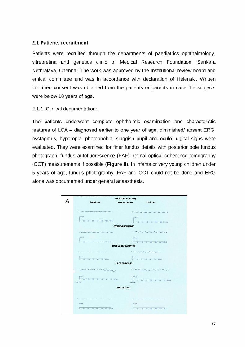
37
2.1 Patients recruitment
Patients were recruited through the departments of paediatrics ophthalmology,
vitreoretina and genetics clinic of Medical Research Foundation, Sankara
Nethralaya, Chennai. The work was approved by the Institutional review board and
ethical committee and was in accordance with declaration of Helenski. Written
Informed consent was obtained from the patients or parents in case the subjects
were below 18 years of age.
2.1.1. Clinical documentation:
The patients underwent complete ophthalmic examination and characteristic
features of LCA – diagnosed earlier to one year of age, diminished/ absent ERG,
nystagmus, hyperopia, photophobia, sluggish pupil and oculo- digital signs were
evaluated. They were examined for finer fundus details with posterior pole fundus
photograph, fundus autofluorescence (FAF), retinal optical coherence tomography
(OCT) measurements if possible (Figure 8). In infants or very young children under
5 years of age, fundus photography, FAF and OCT could not be done and ERG
alone was documented under general anaesthesia.
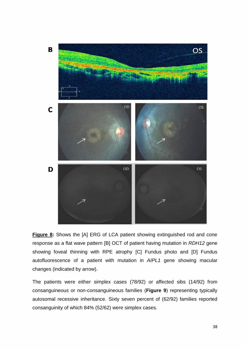
38
Figure 8: Shows the [A] ERG of LCA patient showing extinguished rod and cone
response as a flat wave pattern [B] OCT of patient having mutation in RDH12 gene
showing foveal thinning with RPE atrophy [C] Fundus photo and [D] Fundus
autofluorescence of a patient with mutation in AIPL1 gene showing macular
changes (indicated by arrow).
The patients were either simplex cases (78/92) or affected sibs (14/92) from
consanguineous or non-consanguineous families (Figure 9) representing typically
autosomal recessive inheritance. Sixty seven percent of (62/92) families reported
consanguinity of which 84% (52/62) were simplex cases.

39
Figure 9: Representative pedigrees of cases recruited. Pedigrees of [A] isolated
proband from a consanguineous family [B] affected sibs from non-consanguineous
family [C] isolated proband from non-consanguineous family [D] affected sibs from
consanguineous family.
2.1.2. Sample collection
Ninety-two LCA patients were recruited in the study from across India. Seventy-
three patients are from south India while 19 were from north India. Of these 79%
(58/73) and 16% (3/19) are from consanguineous south and north Indian families,
respectively (Figure 10). Venous blood (8-10 ml) was collected with ACD
anticoagulant (BD Vacutainer, BD, New Jersey, USA) from the probands, affected
or unaffected sibs (if any) and parents after obtaining their consent.

40
Figure 10: Shows distribution of consanguinity among the north and south Indian
cases in the cohort of 92 LCA families.
Control samples were taken from previous epidemiological studies conducted by
Medical Research Foundation and they were ruled out for any inherited retinal
dystrophies134, 135
2.2. DNA extraction
Genomic DNA was extracted from ACD blood samples using NucleoSpin® Blood XL
kit (Macherey-Nagel, GmbH, Duren, Germany). [Procedure as in appendix 1]
2.3. DNA Quantification and Quality check
The DNA was quantified using Nano drop Spectrophotometer ND1000 (Thermo
Fisher Scientific, Wilmington, USA).The integrity of the DNA was checked by
running on 1% agarose gel (Figure 11). [Procedure as in appendix 2]

41
Figure 11: Agarose gel image of DNA samples to check their quality (Lane 1-20).
2.4. Targeted re-sequencing using Illumina MiSeq platform
2.4.1. Designing target enrichment probes:
Target enrichment probes were designed using Agilent SureDesign HaloPlex
standard wizard covering 20 LCA candidate genes AIPL1, CABP4, CEP290,
CRB1, CRX, GUCY2D, IQCB1, IMPDH1, KCNJ13, LCA5, LRAT, MERTK,
NMNAT1, OTX2, RD3, RDH12, RPE65, RPGRIP1, SPATA7 and TULP1
including the exon-intron boundaries, padding each exon with 20 bp
sequences upstream and downstream.
For the probe design the CCDS reference sequence databases were selected
for the coding transcripts of all the genes except CEP290 for which the
RefSeq database was used. UTR regions were not included in the designing.
Two-hundred thirty-nine regions of 50062 bp covering all the coding regions
with splice junctions were targeted and probe design was automatically
generated by HaloPlex Design Wizard software with 99.8% coverage i.e.
49,958 bp of region of interest (Figure 12).

42
Figure 12: Target enrichment probe design using Agilent SureDesign HaloPlex
standard wizard. Snapshot of the output by HaloPlex Design Wizard providing
information on the coverage for each gene by the designed custom enrichment
probes.
The region of interest, list of analysable (Figure 13) and unanalysable amplicons
that were generated by the software was checked manually with the reference
sequences to confirm whether all exonic regions of candidate genes were
covered or not.
Figure 13: Above is the snapshot of list of analysable regions for which custom
enrichment probes were designed.

43
Of the 239 targets, 9 were not covered completely. These include few bases in
some exons of genes CEP290, LCA5, RPE65 and SPATA7 (Figure 14). Bases
that were not covered by the custom enrichment probes were genotyped by
direct sequencing.
Figure 14: Above is the snapshot of list of non-analysable regions for which custom
enrichment probes could not be designed.
The custom enrichment probes were submitted to the manufacturer for the
synthesis of target enrichment kit.
2.4.2. Target enrichment and Sequencing
Target enriched and indexed library was prepared for each sample to be
sequenced using HaloPlex PCR target enrichment and library preparation kit.
This was performed in Scigenom labs Pvt Ltd, Kochi, India
Restriction digestion:
The gDNA was digested with 16 restriction enzymes (RE) in eight different
reactions each containing 2 RE. This resulted in fragment library which include
both target and non-target gDNA regions.
During each batch of library preparation, an Enrichment Control DNA (ECD) was
included to validate the restriction digestion reaction.

44
The digested ECD was run on Agilent 2100 Bioanalyzer (Figure 15).
Figure 15: The gel picture of the ECD as run on DNA chip in Agilent 2100
bioanalyzer. „L‟ represents 50bp ladder, lane 1-8 represents ECD digested with
specific pair of RE and the pattern of digested fragments, lane 9 and 10 represents
undigested DNA.
Enrichment:
The gDNA restricted fragments were then hybridized to HaloPlex probe capture
library that were designed to the target regions.
Each of the eight restriction digested reactions of a sample were pooled and
added to the probe capture library.
o The biotinylated probes are designed along with unique indexes and Illumina
paired end sequencing motifs in the centre, thus facilitating indexing of the
enriched sequences. The unique indexes help tag the samples during
multiplexing for bridge amplification and sequencing on the NGS platform.
The presence of target sequence on ends of the probe directs circularization
of the target regions during hybridization.
The circularized target DNA-HaloPlex probe hybrids, containing biotin, were
captured on streptavidin beads and the non-target regions were removed.

45
This was followed by ligation of the nicks in the circularized target DNA in the
HaloPlex probe-target DNA hybrids using DNA ligase.
The captured, fully circularized targets were eluted from the bead using 50mM
NaOH and were PCR amplified.
o Universal primers complementary to Illumina paired end sequencing
motifs were used during PCR amplification.
Bead based purification of the amplified product was performed which was
followed by validating and quantifying the enriched target samples by Agilent
2100 Bioanalyzer prior to sample pooling and sequencing preparation (Figure
16).
Figure 16: The gel picture of amplified purified samples in the size range between
175-625bp indicating enriched target libraries of target insert sizes 50-500bp. Green
line- lower marker; purple line – upper marker.
Sequencing:
Equimolar amount of each sample was pooled prior to cluster generation by
bridge amplification and sequencing on Illumina MiSeq (Figure 17). 10nmoles/
10ul of each sample were pooled and 7 pmoles of this pool was loaded in the

46
Illumina MiSeq flow cell for cluster generation followed by paired end parallel
sequencing.
Figure 17: Schematic representation of the steps involved in target enrichment
using Agilent HaloPlex target enrichment kit followed by cluster generation by
bridge amplification and sequence by synthesis on Illumina MiSeq platform.
2.4.3. Analysis pipeline:
The steps followed to analyse the datasets obtained from next-generation
sequencing are briefly described below.
I. Quality Check and Filter: First the low quality bases from the reads were
removed. Average Q30 score was used as a cut-off to remove low quality
bases. Then the adapter sequences were trimmed from both 5‟ and 3‟ end of
the read sequence. The base quality score distributions, average base content
per read and GC distribution in the reads were considered for quality checking.

47
II. Alignment: The filtered reads were aligned to the reference genome (hg19)
using BWA program. After alignment only reads with mapping quality ≥20 were
used for further analysis. The coding region of each gene was identified for
taking into consideration all protein-coding transcripts of the gene in Ensembl
68 database.
III. Variant Calling: The variants in the samples were predicted using Samtools
toolkit. The variants with quality score ≥ 50 and read depth of at least 5 were
taken further for annotation.
IV. Annotation: The variants were annotated using GATK and compared with
various databases including OMIM, ClinVar, SNPedia, Ensemble (GRCh37)
variation table, 1000 genome project data, Exomeserver (ESP5400), ExAC
databasev.0.3.1, dbSNP (build 150) and Human genome mutation database
for identifying clinically relevant variants.
The initial analysis using the above pipeline was done in Scigenom labs Pvt.
Ltd, Kochi, India. The same pipeline was constructed in CLC Cancer Genomics
workbench (v.8.0.1) and the samples were re-analysed.
The single nucleotide variations (SNVs) and indels were further analysed
manually as defined below
i. As LCA is an autosomal recessive disease, we short listed all the homozygous
variants first.
ii. Scrutinized all variants and indels in the coding regions
iii. The canonical splice-sites, regions 10bp upstream and downstream of splice
sites (-10 to +10) and variants in the last codon of the exons were identified for
further validation and bioinformatics analyses.
iv. The heterozygous variants (MAF< 0.01) were shortlisted to identify possible
compound heterozygous mutations.
v. The homozygous and heterozygous SNVs and indels were checked if they are
novel or reported in the databases like OMIM 136, ClinVar137, SNPedia138,
Ensemble variation table 139, Exome server 140, 1000 genome project data 141,
ExAC database 142, dbSNP143 and Human genome mutation database 144.
vi. All other reported and novel, synonymous and intronic variants were noted.

48
2.5. Validation of the identified pathogenic and likely pathogenic mutations:
The pathogenic or likely pathogenic mutations shortlisted using the above
criteria was validated by PCR and bidirectional direct Sanger sequencing of
the respective regions. The segregation analysis was performed in parents
and available family members. Variants that were validated and segregating
within the families were screened in an average of 120 unrelated controls
either by custom designed allele specific PCR (ASPCR) or Sanger
sequencing.
I. Polymerase Chain Reaction (PCR):
Primers were designed using Primer3 (Version 0.4.0). The primers were
designed to amplify the exons in which the pathogenic or likely pathogenic
mutations were identified. The primer sequences used for the PCR are
provided in appendix 3.
Table 3: Reaction protocol for Polymerase chain reaction.
Reagents Stock
concentration
Volume
in μl
Genomic DNA 50 ng 1
Forward Primer (10pmol/ µl) (Xcelris
Genomics, Ahmadabad, India)/ Sigma-
Aldrich, Missouri, USA/IDT, Iowa, USA)
5 pmol
0.5
Reverse Primer (10pmol/ µl) (Xcelris
Genomics, Ahmadabad, India)/ Sigma-
Aldrich, Missouri, USA/ IDT, Iowa, USA)
5 pmol
0.5
Deoxyribo nucleotide phosphates
[dNTPs] (Applied Biosystems, Foster
City, USA)
250uM
0.125
Taq DNA polymerase (3U/µl) (Thermo
Fisher Scientific, Massachusetts, USA)
0.3U 0.1
Taq Buffer (Thermo Fisher Scientific,
Massachusetts, USA)
1X 1.25
Betaine (Sigma-Aldrich, Missouri, USA) 5M 1
Sterile water 8.025
Total 12.5

49
The thermal cycler profile is as given below.
Figure 18: Reaction protocol for PCR amplification in thermal cycler.
The standardized annealing temperature and the product size of the exons
that were PCR amplified for validation of identified pathogenic and likely
pathogenic mutations from the targeted re-sequencing data and for exons
that were incompletely covered by target enrichment probes are given below
in the table 4. The regions covered by the primers are shown in appendix 6.
Table 4: List of regions analysed by PCR based direct sequencing with
annealing temperature and product size
S.No Gene Exon Tm (c°) Product size (bp)
1 AIPL1 6 63 500
2 AIPL1 5 60 279
3 AIPL1 2 60 316
4 CRB1 5 60 317
5 CRB1 11 60 283
6 CRB1 12A 62 482
7 CRX 2 67.8-60.8(-0.5) 314
8 CRX 3b 63 294
9 GUCY2D 14 69-62(-0.5) 306
10 GUCY2D 15 69-62(-0.5) 260
11 GUCY2D 17 69-62(-0.5) 168
12 GUCY2D 10 69-62(-0.5) 235
13 GUCY2D 2b 59 551
14 GUCY2D 9 69-62(-0.5) 295
15 GUCY2D 3 69-62(-0.5) 381
16 GUCY2D 16 69-62(-0.5) 200

50
17 GUCY2D 11 69-62(-0.5) 220
18 GUCY2D 2a 64 472
19 IQCB1 14 60 351
20 IQCB1 11 60 383
21 IQCB1 13 60 400
22 KCNJ13 3A 56 471
23 LCA5 7a 57 340
24 LCA5 3 65-58(-0.5) 365
25 LCA5 5 56 353
26 LCA5 6 56 237
27 NMNAT1 2 62 425
28 RD3 1 60 480
29 RDH12 2 60 359
30 RDH12 6 60 492
31 RPE65 4 54 501
32 RPE65 2 60 360
33 RPE65 14 53 453
34 RPE65 9 56 292
35 RPE65 13 42 199
36 RPE65 10 56 226
37 RPGRIP1 21 58 287
38 RPGRIP1 14 52 554
39 RPGRIP1 6 52 282
40 RPGRIP1 24 56 205
41 SLC19A2 2 63-56(-0.5) 700
42 SPATA7 10 60 243
43 SPATA7 1 60 144
44 SPATA7 7 60 477
45 TULP1 11 59 247
Unanalysable regions
46 CEP290 Intronic 63 294
47 CEP290 40 58 592
48 CEP290 18 62 495
49 RPE65 12 60 214
50 SPATA7 3 65 312
51 SPATA7 6 60 250
52 SPATA7 11c 63 396
II. ExoSap digestion of PCR product.
The amplified products were digested by ExoSap prior to cycle sequencing.
This is an enzymatic procedure for purifying the PCR product where the
unused primers and dNTPs are hydrolysed.

51
Table 5: ExoSAP protocol
Reagents Stock
concentration
Volume
(µl)
Exonuclease I (Thermoscientific, Massachusetts,
USA)
20U/μl 1
Exonuclease buffer (Thermoscientific,
Massachusetts, USA)
10X 4
FastAP thermosensitive alkaline phosphatase
(Thermoscientific, Massachusetts, USA)
1U/μl 2
FastAP buffer (Thermoscientific, Massachusetts,
USA)
10X 3
Procedure
1.5 μl of the above cocktail and 5 μl of amplified PCR product were mixed
well, microfuged and reaction proceeded.
Figure 19: Reaction protocol for ExoSap digestion in thermal cycler.
III. Cycle Sequencing:
Cycle sequencing was done using big dye terminator v3.1 kit (Applied
Biosystems, Foster City, USA). Sequencing of all amplified products was carried
out using the either forward primer/ reverse primer. A 10 μl reaction was
prepared as given in the protocol below, mixed and microfuged.

52
Table 6: Reaction protocol for cycle sequencing.
Reagents Volume in μl
5X sequencing Buffer (Applied Biosystems, Foster
City, USA) 1.5
Primer (100pmol/ µl) 1
RR mix (Applied Biosystems, Foster City, USA) 0.5
Exo Sap product 1
Sterile water 6
Total 10
The reaction was set in a thermal cycler as given in the conditions below.
Figure 20: Thermal cycle reaction protocol for cycle sequencing
The samples were purified by ethanol/ EDTA/ Sodium Acetate precipitation
method. The precipitated product was resuspended in Hi-DiTM Formamide
(Applied Biosystems, Foster City, USA) and was loaded on to ABI 3100 Avant or
3730 Genetic analyzer (Applied Biosystems, Foster City, USA). [Procedure as in
appendices 4 & 5]
IV. Reverse transcriptase PCR
1. RNA extraction
RNA extraction was performed from whole blood (two families) of the
probands, parents and affected/ unaffected sib using Trizol method.
Procedure
2 ml Trizol (TRI Reagent® (Sigma Aldrich, Missouri, USA)) was added to
1ml whole ACD/ EDTA blood, mixed, incubated for 10 minutes at room
temperature

53
It was centrifuged at 12000 rpm for 15 minutes at 4°C.
To the supernatant 2ml Trizol and 400µl chloroform (Merck, New
Jersey, USA) was added, mixed, incubated for 10 minutes at room
temperature and centrifuged at 12000 rpm for 20 minutes at 4°C.
The aqueous phase was separated and equal volume of isopropanol
(Merck, New Jersey, USA) was added, mixed well, incubated for 10
minutes at room temperature and centrifuged at 12000 rpm for 10
minutes at 4°C.
To the pellet, 2ml of 75% ethanol (Hayman Limited, Witham, England)
was added and washed twice by centrifuging at 8400 rpm for 5 minutes
at 4°C.
The pellet was air dried and dissolved in RNAse free water.
The RNA was quantitated using Nano drop Spectrophotometer ND1000
and electrophoresed in 1% agarose gel to check the quality.
2. cDNA conversion:
cDNA conversion was performed using Verso cDNA Synthesis Kit, (Thermo
Scientific, Massachusetts, USA).
Procedure
A 20 μl reaction was prepared as given in the protocol below, mixed and
microfuged.
Table 7: Reaction protocol for cDNA Synthesis.
Reagents Volume in μl
Template RNA (~1µg) 1
Verso enzyme mix 1
RT enhancer 1
RNA primer mix
(Random hexamer: oligodt = 3:1) 1
dNTP mix 2
5X cDNA synthesis buffer 4
Nuclease free Water 10
Total 20

54
The reaction was set in a thermal cycler as given in the condition below.
Figure 21: Thermal cycler reaction protocol for cDNA synthesis.
3. Reverse transcriptase PCR:
Reverse transcriptase PCR was performed on cDNA using primers
encompassing exons 7-10 and 9-11 covering the mutations c.913-2A>G and
c.1215+5C>A of the SPATA7 gene to identify the altered splicing. PCR was
performed using the reaction protocol as in table 3 and thermal profile as in
figure 18. The standardized annealing temperature and product size is
provided in table 8. The regions covered by the primers are shown in
appendix 6.
Table 8: cDNA region analysed by PCR based direct sequencing with
annealing temperature and product size
S.No Region Target exon Tm (c°)
Product size (bp)
1 SPATA7RT1 7-10 63-56(-0.5) 575
2 SPATA7RT2 9-11 61 418
V. Gel extraction
As there were two different transcripts of SPATA7 gene expressed in blood,
gel extraction followed by sequencing was performed to identify them. Gel
extraction was done using QIAquick Gel Extraction Kit (Qiagen, Hilden,
Germany)
Procedure
The specific amplified band was excised from agarose gel with a clean
scalpel and weighed.
Buffer QG (provided in kit) was added to the gel slice such that for
100mg gel slice 100µl buffer QG was added.

55
The tube was incubated at 50°C until the gel slice dissolved completely.
It was also vortexed intermittently.
Equal volume of isopropanol was added to the sample and mixed.
The sample was added to QIAquick column (provided in kit) and
centrifuged at 13000 rpm for 1 min.
The flow through was discarded and 500 µl of buffer QG was added to
the column and centrifuged at 13000 rpm for 1 min.
To the column, 750 µl of wash buffer PE (provided in kit) was added,
incubated for 5 minutes and centrifuged at 13000 rpm for 1 minute. This
step was repeated twice.
The flow through was discarded and the column was centrifuged at
13000 rpm for 1 minute to remove residual buffer PE.
The column was transferred to 1.5 ml microfuge tube and 15 µl elution
buffer EB (provided in kit) was added, incubated for 4 minutes and
centrifuged at 13000 rpm for 1 minute.
The eluted products (2.0 ul each) were directly used for bidirectional
Sanger sequencing.
2.6. Insilico Predictions:
Online bioinformatics prediction tools like SIFT145, PolyPhen146, PMut147,
Mutationaccessor (http://mutationassessor.org/v1), Mutpred148 and Variant
effect predictor149 were used to predict the effect of the missense mutations.
Mutation taster150, Netgene2151, NNsplice152, Fsplice
(http://www.softberry.com/berry.phtml?topic=fsplice&group=programs&subgr
oup=gfind) and Splice finder153 predicted the effect of splice-site mutations.
CADD analysis was performed for all identified variants.
2.7. Screening of SLC19A2 in a patient diagnosed with Thiamine Responsive
Megaloblastic Anaemia (TRMA) with LCA as ocular feature.
A patient, reported to our hospital at 5 months of age and showed typical
characteristics of LCA, later developed megaloblastic anaemia, non-type 2
diabetes mellitus and sensory neural deafness. The proband (Figure 22)
underwent complete clinical, hematological, ophthalmic and auditory
examination and presented the triad features - anaemia, diabetes and

56
deafness which are typical characteristics of syndrome called „Thiamine
responsive megaloblastic anaemia, an autosomal recessive disorder154. The
only known causative gene for the syndrome SLC19A2, encodes for
thiamine transporter protein, THTR1155. Blood samples were collected from
the patient and her family members after an informed consent.
Figure 22: Pedigree of the proband showing typical autosomal recessive
pattern of inheritance
The PCR primers for all 6 exons of the SLC19A2 gene were designed using
Primer3 software (Version 0.4.0). PCR was standardized as in table 3.
Standardization of PCR for all six exons of SLC19A2 gene was done (Table
9). The regions covered by the primers are shown in appendix 12.
The Exo-SAP treated PCR products were genotyped by direct sequencing
using 3100-Avant genetic analyzer. SIFT and PolyPhen analyses were
performed to predict the possible impact of amino acid substitution on the
structure and function of protein.

57
Table 9: List of regions in SLC19A2 analysed by PCR based direct
sequencing with annealing temperature and product size. ASPCR- Allele
specific PCR for the identified mutation in exon 2.
S.No Region Tm (c°) Product size (bp)
1 SLC19A2- Exon 1 61 589
2 SLC19A2- Exon 2 63-56
(-1ºC) 700
3 SLC19A2- Exon 3 61 391
4 SLC19A2- Exon 4 61 429
5 SLC19A2- Exon 5 61 300
6 SLC19A2- Exon 6 61 295
7 SLC19A2-Exon 2-
ASPCR
56 186

58
CHAPTER 3 – RESULTS
3.1. Targeted resequencing
3.1.1. Data analysis
I. Quality Check and Filter:
a. Raw read summary
The reads/ data obtained after sequencing was analysed using specific analysis
pipeline. This involves trimming of reads depending on their quality, removal of
the adapter sequence, and then aligning to the reference sequences before the
variants are called and annotated. Below (Table 10) is the summary of raw
fastq files obtained from NGS for the processed 92 samples.
Table 10: Raw read summary
All samples
# of paired-end reads 75,006,077
# of bases (Gb) 9.74
% of data >= Q30 83.2
Raw read length (bp) 151 x 2
II. Alignment:
a. Coverage:
The reads covering the coding region of all genes is represented as coverage.
For all 20 genes that were studied we obtained an average coverage of
99.03%. The coverage of each gene is provided in figure 23 and table 11
Figure 23: Average read coverage graph for all 20 genes

59
Table 11: Average coverage and depth for each 20 genes studied.
Gene Average coverage
(%) Average depth
(X)
AIPL1 100 224.783653
CEP290 93.45964192 37.08527504
CRB1 99.69204983 93.72696533
CRX 100 103.4220974
GUCY2D 98.51097797 147.0135295
IMPDH1 99.97926786 269.9190111
IQCB1 98.09025253 69.26971055
KCNJ13 99.87620773 80.8880666
LRAT 99.85664776 98.76046058
LCA5 99.11889464 39.42222961
MERTK 99.60507028 173.1452721
RDH12 99.43473675 208.0075136
RD3 99.93682646 173.8373646
RPE65 99.99320227 156.7695652
RPGRIP1 98.63304982 100.09021
SPATA7 95.10451291 78.28896321
TULP1 99.91919031 233.9596462
NMNAT1 99.43671388 96.9768017
CABP4 100 167.9272444
OTX2 99.97560142 139.8586469
b. Depth:
The average number of reads covering the coding region is represented as
depth. An average depth of 134X was obtained for all 20 genes. The depth for
each gene is provided in table 11 and figure 24.
Figure 24: Average read depth graph for all 20 genes

60
III. Variant calling
The variants were predicted using Samtools program. An average of 78variants
was identified in each sample for the 20 selected genes. The average number
of total variants identified for each gene over all samples is shown in figure 25.
Highest number of variants was observed for MERTK gene. Overall, 508 (378
SNPs, 130 Indels) different variants were identified for 20 genes in 92 samples.
Figure 25: Average number of variants detected for all 20 genes
IV. Variant Annotation:
The variants were annotated and compared with various databases including
OMIM, ClinVar and SNPedia, Ensemble variation table, Exome server, 1000
genome project data and Human genome mutation database for identifying
clinically relevant variants. In total, we identified 126 SNVs and 13 Indels
present in coding region of the selected 20 genes in the 92 samples screened.
Four out of all 92 samples were positive controls with known mutations (Table
12).
Table 12: Shows the list of positive control sample and variants
Sample ID Gene Variants
Sample 2 RPE65 c.1409C>T (p.Pro470Leu)
Sample 12 RPE65 c.1109T>A (p.Leu370His)
Sample 58 RPE65 c.362insT (p.Ser121Phefs1290)
Sample 60 LCA5 c.955G>A (activates cryptic splice-site)

61
Figure 26: [A] IGV view of c.1109T>A (p.L370H) variation identified in RPE65
gene of LRS 12 in IGV viewer156 [B] IGV view of c.3065T>A (p.L1022X)
variation identified in GUCY2D gene of LRS 1 from CLC genomics workbench
(v.8.0.1).
3.1.2. Validation of the identified pathogenic and likely pathogenic mutations:
The single nucleotide variations (SNVs) and indels were further analysed
manually as defined in materials and methods (C.IV). The analysis by the
mentioned criteria identified fifty-four cases with homozygous pathogenic / likely
pathogenic variants including double mutations (2 homozygous mutations in
same gene) in two cases. Compound heterozygous pathogenic variants in one
case and digenic likely pathogenic variants in another were also identified.
(Table 13).
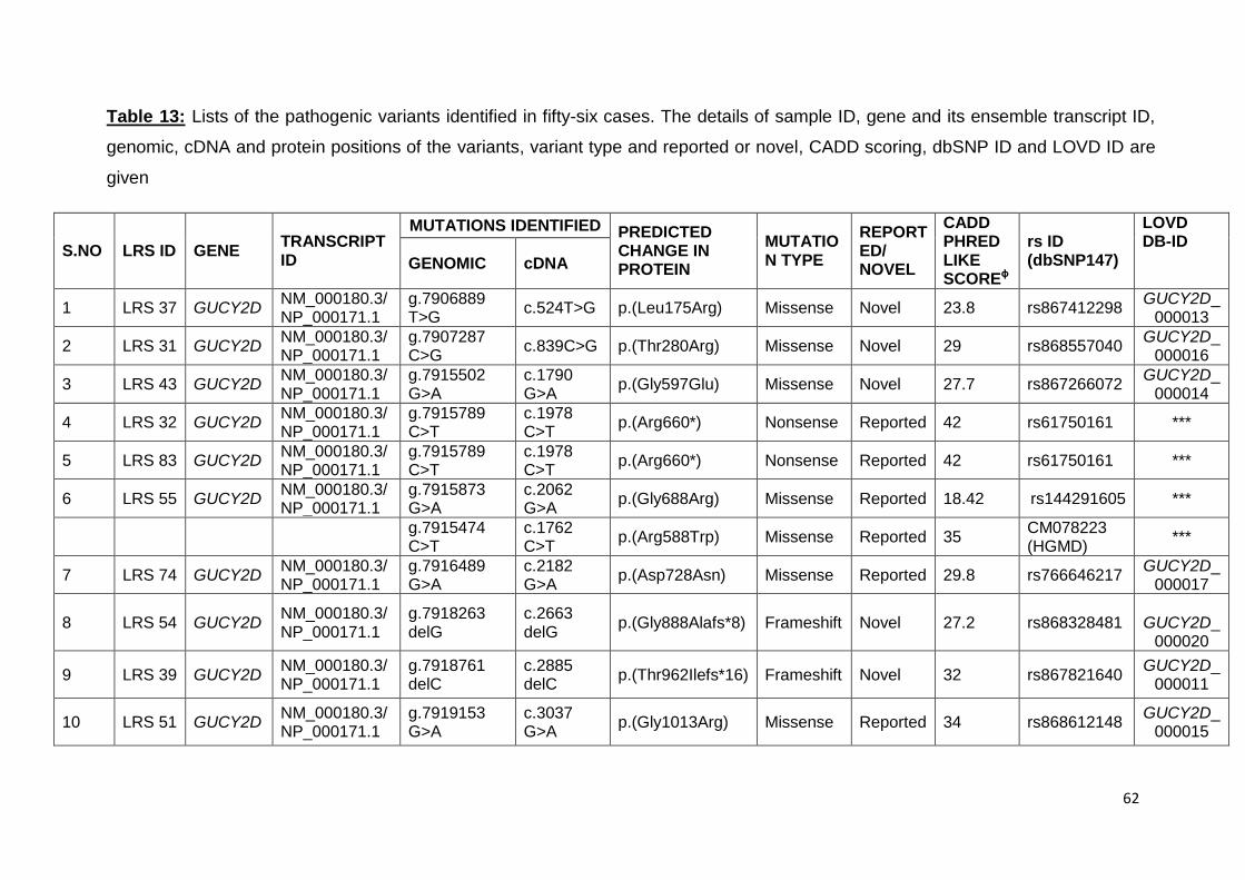
62
Table 13: Lists of the pathogenic variants identified in fifty-six cases. The details of sample ID, gene and its ensemble transcript ID,
genomic, cDNA and protein positions of the variants, variant type and reported or novel, CADD scoring, dbSNP ID and LOVD ID are
given
S.NO LRS ID GENE TRANSCRIPT ID
MUTATIONS IDENTIFIED PREDICTED CHANGE IN PROTEIN
MUTATION TYPE
REPORTED/ NOVEL
CADD PHRED LIKE SCOREɸ
rs ID (dbSNP147)
LOVD DB-ID
GENOMIC cDNA
1 LRS 37 GUCY2D NM_000180.3/ NP_000171.1
g.7906889 T>G
c.524T>G p.(Leu175Arg) Missense Novel 23.8 rs867412298 GUCY2D_
000013
2 LRS 31 GUCY2D NM_000180.3/ NP_000171.1
g.7907287 C>G
c.839C>G p.(Thr280Arg) Missense Novel 29 rs868557040 GUCY2D_
000016
3 LRS 43 GUCY2D NM_000180.3/ NP_000171.1
g.7915502 G>A
c.1790 G>A
p.(Gly597Glu) Missense Novel 27.7 rs867266072 GUCY2D_
000014
4 LRS 32 GUCY2D NM_000180.3/ NP_000171.1
g.7915789 C>T
c.1978 C>T
p.(Arg660*) Nonsense Reported 42 rs61750161 ***
5 LRS 83 GUCY2D NM_000180.3/ NP_000171.1
g.7915789 C>T
c.1978 C>T
p.(Arg660*) Nonsense Reported 42 rs61750161 ***
6 LRS 55 GUCY2D NM_000180.3/ NP_000171.1
g.7915873 G>A
c.2062 G>A
p.(Gly688Arg) Missense Reported 18.42 rs144291605 ***
g.7915474 C>T
c.1762 C>T
p.(Arg588Trp) Missense Reported 35 CM078223 (HGMD)
***
7 LRS 74 GUCY2D NM_000180.3/ NP_000171.1
g.7916489 G>A
c.2182 G>A
p.(Asp728Asn) Missense Reported 29.8 rs766646217 GUCY2D_
000017
8 LRS 54 GUCY2D NM_000180.3/ NP_000171.1
g.7918263 delG
c.2663 delG
p.(Gly888Alafs*8) Frameshift Novel 27.2 rs868328481
GUCY2D_ 000020
9 LRS 39 GUCY2D NM_000180.3/ NP_000171.1
g.7918761 delC
c.2885 delC
p.(Thr962Ilefs*16) Frameshift Novel 32 rs867821640 GUCY2D_
000011
10 LRS 51 GUCY2D NM_000180.3/ NP_000171.1
g.7919153 G>A
c.3037 G>A
p.(Gly1013Arg) Missense Reported 34 rs868612148 GUCY2D_
000015

63
11 LRS 1 GUCY2D NM_000180.3/ NP_000171.1
g.7919266 T>A
c.3065 T>A
p.(Leu1022*) Nonsense Novel 36 rs866511152 GUCY2D_
000012
12 LRS 14#
GUCY2D NM_000180.3/ NP_000171.1
g.7919319 C>G
c.3118 C>G
p.(Arg1040Gly) Missense Reported 29.9 rs61750194 ***
g.7906524 delG
c.159delG p.(Phe54Ser fs*31)
Frameshift Novel 26.1 rs868091683 GUCY2D_
000018
13 LRS 48 AIPL1 NM_014336.4/ NP_055151.3
g.6337268 G>A
c.247G>A p.(Glu83Lys) Missense Reported 31 rs267605009 AIPL1_ 000014
14 LRS 4 AIPL1 NM_014336.4/ NP_055151.3
g.6330030 A>G
c.689A>G p.(Asn230Ser) Missense Novel 12.96 rs866980715 AIPL1_ 000008
15 LRS 9 AIPL1 NM_014336.4/ NP_055151.3
g.6329101 G>A
c.834G>A p.(Trp278*) Nonsense Reported 38 rs62637014 AIPL1_ 000010
16 LRS 71 AIPL1 NM_014336.4/ NP_055151.3
g.6329101 G>A
c.834G>A p.(Trp278*) Nonsense Reported 38 rs62637014 AIPL1_ 000010
17 LRS 13 AIPL1 NM_014336.4/ NP_055151.3
g.6329091 G>T
c.844G>T p.(Glu282*) Nonsense Novel 39 rs779454542 AIPL1_ 000009
18 LRS 22 AIPL1 NM_014336.4/ NP_055151.3
g.6329091 G>T
c.844G>T p.(Glu282*) Nonsense Novel 39 rs779454542 AIPL1_ 000009
19 LRS 81 AIPL1 NM_014336.4/ NP_055151.3
g.6329025 G>T
c.910G>T p.(Glu304*) Nonsense Reported 42 rs746116735 AIPL1_ 000006
20 LRS 25 RPE65 NM_000329.2/ NP_000320.1
g.68914352_68914355 delTTTG
c.46_49 delTTTG
p.(Phe16Lys fs*14)
Frameshift Novel 35 rs866809120 RPE65_ 000009
21 LRS 28 RPE65 NM_000329.2/ NP_000320.1
g.68910458 G>T
c.353+1 G>T
r.spl? Splice-site Reported 26.4 rs61752876 ***
22 LRS 23 RPE65 NM_000329.2/ NP_000320.1
g.68910347 insA
c.361dupT p.(Ser121Phe fs*10)
Frameshift Reported 35 rs121918844 RPE65_ 000073
23 LRS 58 RPE65 NM_000329.2/ NP_000320.1
g.68910347 insA
c.361dupT p.(Ser121Phe fs*10)
Frameshift Reported 35 rs121918844 RPE65_ 000073

64
24 LRS 12 RPE65 NM_000329.2/ NP_000320.1
g.68903889 T>A
c.1109 T>A
p.(Leu370His) Missense Reported 29.6 rs776250699 ***
25 LRS 2 RPE65 NM_000329.2/ NP_000320.1
g.68896789 C>T
c.1409 C>T
p.(Pro470Leu) Missense Reported 26.2 *** RPE65_ 000128
26 LRS 86 RPE65 NM_000329.2/ NP_000320.1
g.68895547 T>G
c.1514 T>G
p.(Leu505Arg) Missense Novel 29.8 rs868473289 RPE65_ 000012
g.68904660 T>G
c.963T>G p.(Asn321Lys) Missense Reported 4.096 rs149916178 RPE65_ 000013
27 LRS 47 RPGRIP1 NM_020366.3/ NP_065099.3
g.21775984 G>T
c.895G>T p.( Glu299*) Nonsense Novel 39 *** ***
28 LRS 65 RPGRIP1 NM_020366.3/ NP_065099.3
g.21793055 C>T
c.2041 C>T
p.(Gln681*) Nonsense Novel 37 rs868548381 RPGRIP1
_ 000010
29 LRS 45 RPGRIP1 NM_020366.3/ NP_065099.3
g.21793055 C>T
c.2041 C>T
p.(Gln681*) Nonsense Novel 37 rs868548381 RPGRIP1
_ 000010
30 LRS 15 RPGRIP1 NM_020366.3/ NP_065099.3
g.21811289 delA
c.3434 delA
p.(Glu1145Gly fs*18)
Frameshift Novel 34 rs866944524 RPGRIP1
_ 000009
31 LRS 26 RPGRIP1 NM_020366.3/ NP_065099.3
g.21819302 T>C
c.3788 T>C
p.(Leu1263Pro) Missense Novel 25.7 rs866154508 RPGRIP1
_ 000011
32 LRS 87 LCA5 NM_181714.3/ NP_859065.2
g.80203350 C>T
c.838C>T p.(Arg280*) Nonsense Reported 35 rs866395428 LCA5_ 000041
33 LRS 60 LCA5 NM_181714.3/ NP_859065.2
g.80202268 G>A
c.955G>A p.(Ala319Thr) Missense Reported 23.3 CS081931 LCA5_ 000008
34 LRS 20 LCA5 NM_181714.3/ NP_859065.2
g.80201335_80201341 delGTTTTCG
c.1062_ 1068 delCGAAAAC
p.(Tyr354*) Frameshift Novel 35 *** ***

65
35 LRS 30 LCA5 NM_181714.3/ NP_859065.2
g.80201335_80201341 delGTTTTCG
c.1062_ 1068 delCGAAAAC
p.(Tyr354*) Frameshift Novel 35 *** ***
36 LRS 21 LCA5 NM_181714.3/ NP_859065.2
g.80197393 delT
c.1422 delT
p.(Ile474Met fs*11)
Frameshift Novel 23.2 rs866833585 LCA5_ 000040
37 LRS 10 IQCB1
NM_001023570.3/ NP_001018864.2
g.121509055G>A
c.994C>T p.(Arg332*) Nonsense Reported 36 CM050645
(HGMD) ***
38 LRS 92 IQCB1
NM_001023570.3/ NP_001018864.2
g.121500667G>A
c.1333 C>T
p.(Arg445*) Nonsense Novel 38 rs867772426 IQCB1_ 000004
39 LRS 77 IQCB1
NM_001023570.3/ NP_001018864.2
g.121500637G>A
c.1363 C>T
p.(Arg455*) Nonsense Reported 37 rs866982675 ***
40 LRS 79 IQCB1
NM_001023570.3/ NP_001018864.2
g.121491413G>A
c.1558 C>T
p.(Gln520*) Nonsense Reported 40 rs779858591 IQCB1_ 000006
41 LRS 88 CRB1 NM_201253.2/ NP_957705.1
g.197326045_1973260 insTGAG
c.1073ins TGAG
p.(Ser359Glu fs*20)
Frameshift Novel 26.6 rs867681420 CRB1_ 000040
42 LRS 56 CRB1 NM_201253.2/ NP_957705.1
g.197411423G>A
c.4005+1 G>A
r.spl? Splice site Reported 28.6 *** CRB1_ 000092
43 LRS 7 CRB1 NM_201253.2/ NP_957705.1
g.197446956 C>T
c.4168 C>T
p.(Arg1390*) Nonsense Reported 42 rs763324776 CRB1_ 000045
44 LRS 78 CRB1 NM_201253.2/ NP_957705.1
g.197446956 C>T
c.4168 C>T
p.(Arg1390*) Nonsense Reported 42 rs763324776 CRB1_ 000045
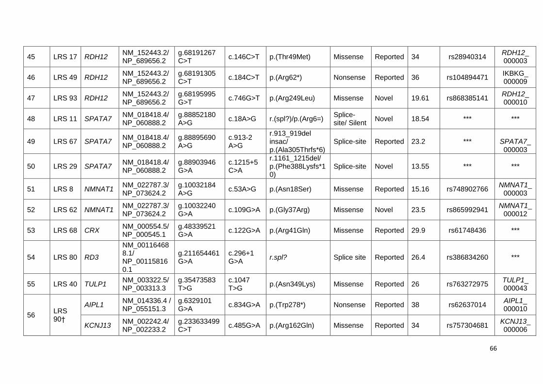
66
45 LRS 17 RDH12 NM_152443.2/ NP_689656.2
g.68191267 C>T
c.146C>T p.(Thr49Met) Missense Reported 34 rs28940314 RDH12_ 000003
46 LRS 49 RDH12 NM_152443.2/ NP_689656.2
g.68191305 C>T
c.184C>T p.(Arg62*) Nonsense Reported 36 rs104894471 IKBKG_ 000009
47 LRS 93 RDH12 NM_152443.2/ NP_689656.2
g.68195995 G>T
c.746G>T p.(Arg249Leu) Missense Novel 19.61 rs868385141 RDH12_ 000010
48 LRS 11 SPATA7 NM_018418.4/ NP_060888.2
g.88852180 A>G
c.18A>G r.(spl?)/p.(Arg6=) Splice-site/ Silent
Novel 18.54 *** ***
49 LRS 67 SPATA7 NM_018418.4/ NP_060888.2
g.88895690 A>G
c.913-2 A>G
r.913_919del insac/ p.(Ala305Thrfs*6)
Splice-site Reported 23.2 ***
SPATA7_ 000003
50 LRS 29 SPATA7 NM_018418.4/ NP_060888.2
g.88903946 G>A
c.1215+5 C>A
r.1161_1215del/ p.(Phe388Lysfs*10)
Splice-site Novel 13.55 *** ***
51 LRS 8 NMNAT1 NM_022787.3/ NP_073624.2
g.10032184 A>G
c.53A>G p.(Asn18Ser) Missense Reported 15.16 rs748902766 NMNAT1_
000003
52 LRS 62 NMNAT1 NM_022787.3/ NP_073624.2
g.10032240 G>A
c.109G>A p.(Gly37Arg) Missense Novel 23.5 rs865992941 NMNAT1_
000012
53 LRS 68 CRX NM_000554.5/ NP_000545.1
g.48339521 G>A
c.122G>A p.(Arg41Gln) Missense Reported 29.9 rs61748436 ***
54 LRS 80 RD3
NM_001164688.1/ NP_001158160.1
g.211654461 G>A
c.296+1 G>A
r.spl? Splice site Reported 26.4 rs386834260 ***
55 LRS 40 TULP1 NM_003322.5/ NP_003313.3
g.35473583 T>G
c.1047 T>G
p.(Asn349Lys) Missense Reported 26 rs763272975 TULP1_ 000043
56 LRS 90†
AIPL1 NM_014336.4 / NP_055151.3
g.6329101 G>A
c.834G>A p.(Trp278*) Nonsense Reported 38 rs62637014 AIPL1_ 000010
KCNJ13 NM_002242.4/ NP_002233.2
g.233633499C>T
c.485G>A p.(Arg162Gln) Missense Reported 34 rs757304681 KCNJ13_ 000006

67
†Indicates digenic inheritance involving AIPL1 and KCNJ13 gene #Indicates compound heterozygous mutation in GUCY2D
gene. All variations were observed in homozygous state except AIPL1 c.834G>A, KCNJ13 c.485G>A in LRS 90, GUCY2D
c.3118C>G and GUCY2D c.159delG in LRS 14.
***Indicates ID not available.
ɸ Indicates Phred like CADD scoring – Score ≥ 10 is considered deleterious. The higher the CADD score, the more likely that the
variant has deleterious effects.

68
The 53 pathogenic/ likely pathogenic variants include 9 frameshift, 14 nonsense,
24 missense and 6 splice-site mutations (Figure 27b). Further 21 of these were
novel (8 frameshift, 4 nonsense, 7 missense and 2 splice-site mutations) (Figure
27c). These disease-causing variants were observed in fourteen of the twenty
candidate genes screened (Figure 28).
Figure 27: A: Pie chart showing the percentage of the cohort where mutations
were identified (including the positive controls) and not identified in the twenty
known LCA genes screened. B: Pie chart showing the percentage distribution of
different types of mutations among the mutation positive cases. C: Bar diagram
showing the distribution of reported and novel mutations among the different
types of identified mutations.

69
Figure 28: Pie chart showing frequency of mutations in the twenty LCA candidate
genes in the Indian cohort studied.
Screening of the unanalysable regions (regions not covered by probes in target
enrichment) and CEP290 intronic variant, c.2991+1655A>G by PCR direct
sequencing in the negative cases revealed no disease-causing variants in these
regions.
Validation of the identified mutations:
All identified pathogenic and likely pathogenic variants were validated (Figure 29)
and segregated in the families.

70

71
Figure 29: Electrophoretogram of the identified mutations (marked by arrows) in
the LCA probands.
The parents were heterozygous carriers and the unaffected sibs were either
heterozygous carriers or homozygous wild type (Figure 30). Of the 92 samples,
ten probands; LRS 7, LRS 12, LRS 20, LRS 28, LRS 29, LRS 30, LRS 31, LRS
32, LRS 40 and LRS 90 had affected sibs segregating the same homozygous
pathogenic variant. Control screening of 240 chromosomes by either direct
sequencing or ASPCR for the identified pathogenic and likely
pathogenicvariantsrevealed wild type sequence for all. The identified novel
variants were submitted to LOVD (http://www.lovd.nl/3.0/home) and dbSNP147.

72
Figure 30: Representative pedigree of the family LCARS-81, segregating a
homozygous nonsense mutation, c.910G>T p. (E340X) in AIPL1 gene.

73
Figure 31: Putative protein structure showing domains and mutations (marked by
arrows) in proteins of LCA candidate genes identified by targeted resequencing.

74
Possible Digenic Inheritance
We observed heterozygous mutations in two different genes in one of our case,
LRS 90. The proband had heterozygous variant in AIPL1, c.834G>A; p.(Trp278*)
and KCNJ13, c.485G>A; p.(Arg162Gln). The affected sib also inherited the same
variants. On segregation, the mother was found to be heterozygous for AIPL1
c.834G>A; p. (Trp278*) and the father for KCNJ13 c.485G>A; p.(Arg162Gln). The
parents were found to be wild type for the other alternate allele thus confirming the
digenic status (Figure 32). Both variants were absent in the controls screened.
The AIPL1 c.834G>A; p.(Trp278*) (rs62637014) variant has been shown to be
pathogenic in many study population157.
Figure 32: Pedigree of LRS 90 showing digenic inheritance segregating in the
family. Proband and affected sib are heterozygous for both AIPL1 c.834G>A;
p.(Trp278*) and KCNJ13 c.485G>A; p.(Arg162Gln) mutations. The father and the
mother are heterozygous for KCNJ13 and AIPL1 mutations, respectively.

75
3.1.3. Other reported or novel variants and SNPs in coding and non–coding
regions
Triallelic variants
Among mutation positive cases, 12 cases with identified pathogenic mutations in
the coding regions also had a reported heterozygous variant in another gene with
MAF ≤ 0.005. Seven different variants were observed and three of these
heterozygous variants are reported as mutations either in homozygous or
compound heterozygous state. Segregation of above variants showed
heterozygosity in either or both parents. These could contribute to triallelism,
modifying the disease severity (Table 14).
Novel heterozygous variants:
Novel heterozygous variants with no other mutation/ variant were observed in
three cases in this study. The variants are MERTK- c.1627T>C, p. (Ser543Pro) in
LRS51; RPE65- c.814C>T, p. (Gly272Arg) in LRS24; RPGRIP1- c.1480delA, p.
(Asn495Thrfs*11) in LRS89. The possibility of these novel variants being
pathogenic and hence representing a carrier state in respective individuals or
being a novel SNP can be defined only when the population frequency of these
variants are known. The frequency of these variants in Indian cohort is
unavailable. However, the regulatory regions of the genes have not been
screened and possibility of any variant in these regions cannot be ruled out.
We have also observed one novel homozygous and thirteen novel heterozygous
synonymous variants; thirty-five novel homozygous and ninety novel
heterozygous intronic variants in this cohort.

76
Table 14: Lists the heterozygous variants observed in mutation positive cases possibly contributing to triallelism
S.NO
LRS ID
IDENTIFIED PATHOGENIC VARIANTS
OBSERVED HETEROZYGOUS VARIANTS
GENE PATHOGENIC
VARIANTS
GENE VARIANTS NOVEL /
REPORTED CADD
MAF (ExAc)
1 LRS 21
LCA5
g.80197392delT
AIPL1
g.6331702 A>T
Reported (rs16955851)
24.4 0.004 c.1422delA c.401A>T
p.(lle474Metfs*11) p.(Tyr134Phe)
(rs866833585)
2 LRS 58
RPE65
g.68910347insT
CABP4
g.67223001 C>T
Reported (rs766418692)
8.996 0.00003808 c.362dupT c.107C>T
p.(Ser121Phefs*10) p.(Pro36Leu)
(rs121918844)
3 LRS 28
RPE65
g.68910458G>T
MERTK
g.112786031G>T
Reported (rs557004700)
10.59 0.0004366 c.353+1G>T c.2590G>T
r.(spl?) p.(Val864Phe)
(rs61752876)
4 LRS 15
RPGRIP1
g.21811289delA
MERTK
g.112786031G>T
Reported (rs557004700)
10.59 0.0004366
c.3434delA c.2590G>T
p.(Glu1145Glyfs*18) p.(Val864Phe)
(rs866944524)

77
5 LRS 68
CRX
g.48339521G>A
MERTK
g.112786031G>T
Reported (rs557004700)
10.59 0.0004366 c.122G>A c.2590G>T
p.(Arg41Gln) p.(Val864Phe)
(rs61748436)
6 LRS 40
TULP1
g.35473583T>G
MERTK
g.112777002C>A
Reported (rs761295704)
28.1 0.00006589 c.1047T>G c.2092C>A
p.(Asn349Lys) p.(Gln698Lys)
(rs763272975)
7 LRS 78
CRB1
g.197446956C>T
RPE65
g.68904660 T>G
Reported (rs149916178)
4.096 0.005362
c.4168C>T c.963T>G
p.(Arg1390*)
p.(Asn321Lys)
(rs763324776)
8 LRS 80
RD3
g.211654461G>A
RPE65
g.68904660 T>G
Reported (rs149916178)
4.096 0.005362
c.296+1G>A c.963T>G
r.(spl?) p.(Asn321Lys)
(rs386834260)
9 LRS 88
CRB1
g.197326045insTGAG
RPE65
g.68904660 T>G
Reported (rs149916178)
4.096 0.005362
c.1073_1074insTGAG c.963T>G
p.(Ser359Glufs*20) p.(Asn321Lys)
(rs867681420)

78
10 LRS 53
AIPL1
g.6337268G>A
MERTK
g.112732891A>G
Reported (rs34943572)
22.2 0.0002553 c.247C>T c.986A>G
p.(Glu83Lys) p.(Asn329Ser)
(rs267605009)
11 LRS 8 NMNAT1
g.10032184A>G
RPE65
g.68904660 T>G
Reported (rs149916178)
4.096 0.005362 c.53A>G c.963T>G
p.(Asn18Ser) p.(Asn321Lys)
( rs748902766)
12
LRS 14
GUCY2D
g.7919319C>G
CRB1
g.197326047
T>G c.1075T>G
p.(Ser359Ala)
Reported (rs533227950)
3.021 0.000832
c.3118C>G
p.(Arg1040Gly)
(rs61750194)
g.7906524delG
c.159delG
p.(Phe54Serfs*31)
(rs868091683)

79
3.2. cDNA analysis
RT-PCR using cDNA was standardized using ARPE-19 cell lines as positive
control for SPATA7 gene expression. The same RT-PCR reactions were
performed using cDNA obtained from peripheral blood of probands and their
families, LRS67 (Figure 33) and LRS29 (Figure 34) to check the effect on
splicing for the mutations, c.1215+5C>A and c.913-2A>G, respectively. cDNA
primers targeting GAPDH was used as an internal control. Two transcripts of
SPATA7 gene were observed on 3% agarose gel electrophoresis. Gel extraction
was performed followed by sequencing of the extracted product.
Figure 33: In family LRS 67 [A] 0.7% agarose gel picture showing RNA in Lane 1-
4: 1. Proband 2.Sib. 3.Father 4.Mother [B] 2% agarose gel picture showing cDNA
amplified products of 525bp size targeting SPATA7- c.913-2A>G optimized at 63-
56(-0.5°C) touchdown protocol Lane 5-8: 5.Proband 6.Father 7.Sib 8. NC. Lane 9-
11: GAPDH amplification of the corresponding cDNA 9. Proband 10. Father
11. Sib.
The bidirectional Sanger sequencing in family LRS 67 revealed an indel, where
the first 7 bases of exon 8 were deleted and 2 bases inserted resulting in
predicted truncated protein p.(Ala305Thrfs*6) (Figure 35). The parents were
heterozygous for the deletions. This confirms the c.913-2A>G mutation in the
canonical splice-site in intron 7 affects splicing. In family LRS 29, the presence of
variant c.1215+5C>A in the intron 10 resulted in the deletion of exon 10, leading

80
to predicted truncated protein p. (Phe388Lysfs*10) (Figure 36). The parents were
heterozygous for the deletion.
Figure 34: In family LRS 29 [A] 0.7% agarose gel picture showing RNA in Lane 1-
4: 1. Proband 2.Father 3.Mother 4.Sib [B] 2% agarose gel picture showing
GAPDH amplification of the corresponding cDNA Lane 5-8: 5. Proband 6.Father
7.Mother 8.Sib [C] cDNA amplified products of 596bp size targeting SPATA7-
c.1215+5C>A optimized at 65-58(-0.5°C) touchdown protocol. Lane 9-13: 9.NC
10.Proband 11.Father 12.Mother 13.Sib
Figure 35: Electrophoretogram of cDNA analysis for splice variants in SPATA7
gene. [A] Wild type cDNA sequence (forward) in control showing exon 7 and exon
8 [B] homozygous indel (insertion of GT and first 7 bases deletion in exon 8) in
LRS 67 due to splice mutation c. 913-2A>G [C] heterozygous indel in parent of
LRS 67 .

81
Figure 36: SPATA7- c.1215+5C>A electrophoretogram showing representative
reverse primer sequence of [A] Wild type control cDNA [B] Homozygous deletion
of exon 10 in proband [C] Heterozygous deletion of exon 10 in parent.
The cDNA analyses for the synonymous variantc.18A>G; p.(Arg6Arg), in the
penultimate base of exon 1 in SPATA7, predicted to alter splicing, identified in
family LRS 11, couldn‟t be performed as the family was unavailable for
recollection and analysis.
3.3. Bioinformatics Analyses
Apart from CADD scoring, six online bioinformatics tools were used for predicting
the effect of the identified missense variants. Of the 24 missense variants, 12
were predicted to be damaging by 5/6 tools and 9 by more than 3/6 tools (Table
15). Table 13 and table 14 include Phred-like CADD scores for all the identified
variants.
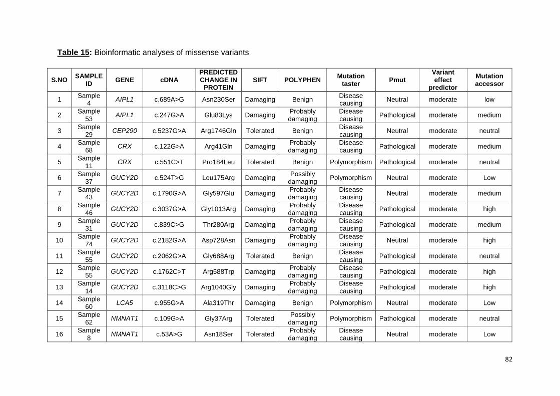
82
Table 15: Bioinformatic analyses of missense variants
S.NO SAMPLE
ID GENE cDNA
PREDICTED CHANGE IN
PROTEIN SIFT POLYPHEN
Mutation taster
Pmut Variant effect
predictor
Mutation accessor
1 Sample
4 AIPL1 c.689A>G Asn230Ser Damaging Benign
Disease causing
Neutral moderate low
2 Sample
53 AIPL1 c.247G>A Glu83Lys Damaging
Probably damaging
Disease causing
Pathological moderate medium
3 Sample
29 CEP290 c.5237G>A Arg1746Gln Tolerated Benign
Disease causing
Neutral moderate neutral
4 Sample
68 CRX c.122G>A Arg41Gln Damaging
Probably damaging
Disease causing
Pathological moderate medium
5 Sample
11 CRX c.551C>T Pro184Leu Tolerated Benign Polymorphism Pathological moderate neutral
6 Sample
37 GUCY2D c.524T>G Leu175Arg Damaging
Possibly damaging
Polymorphism Neutral moderate Low
7 Sample
43 GUCY2D c.1790G>A Gly597Glu Damaging
Probably damaging
Disease causing
Neutral moderate medium
8 Sample
46 GUCY2D c.3037G>A Gly1013Arg Damaging
Probably damaging
Disease causing
Pathological moderate high
9 Sample
31 GUCY2D c.839C>G Thr280Arg Damaging
Probably damaging
Disease causing
Pathological moderate medium
10 Sample
74 GUCY2D c.2182G>A Asp728Asn Damaging
Probably damaging
Disease causing
Neutral moderate high
11 Sample
55 GUCY2D c.2062G>A Gly688Arg Tolerated Benign
Disease causing
Pathological moderate neutral
12 Sample
55 GUCY2D c.1762C>T Arg588Trp Damaging
Probably damaging
Disease causing
Pathological moderate high
13 Sample
14 GUCY2D c.3118C>G Arg1040Gly Damaging
Probably damaging
Disease causing
Pathological moderate high
14 Sample
60 LCA5 c.955G>A Ala319Thr Damaging Benign Polymorphism Neutral moderate Low
15 Sample
62 NMNAT1 c.109G>A Gly37Arg Tolerated
Possibly damaging
Polymorphism Pathological moderate neutral
16 Sample
8 NMNAT1 c.53A>G Asn18Ser Tolerated
Probably damaging
Disease causing
Neutral moderate Low

83
S.NO SAMPLE
ID GENE CDNA MUTATION SIFT POLYPHEN
Mutation taster
Pmut Variant effect
predictor
Mutation accessor
17 Sample
17 RDH12 c.146C>T Thr49Met Damaging
Probably damaging
Disease causing
Neutral moderate medium
18 Sample
93 RDH12 c.746G>T Arg249Leu Tolerated Benign Polymorphism Neutral moderate neutral
19 Sample
86 RPE65 c.1514T>G Leu505Arg Damaging
Probably damaging
Disease causing
Pathological moderate medium
20 Sample
86 RPE65 c.963T>G Asn321Lys Tolerated Benign Polymorphism Neutral moderate low
21 Sample
2 RPE65 c.1409C>T Pro470Leu Damaging
Probably damaging
Disease causing
Pathological moderate medium
22 Sample
12 RPE65 c.1109T>A Leu370His Damaging
Probably damaging
Disease causing
Neutral moderate medium
23 Sample
26 RPGRIP1 c.3788T>C Leu1263Pro Damaging
Probably damaging
Disease causing
Pathological moderate medium
24 Sample
40 TULP1 c.1047T>G Asn349Lys Damaging
Probably damaging
Disease causing
Neutral moderate medium
84
All the identified splice-site variants, canonical and in region within 10bp of exon-
intron junction were predicted to alter donor or acceptor sites probably affecting
splicing. In LRS 11, a variant in the penultimate base of exon 1 of SPATA7 gene
translates to a synonymous change, p.(Arg6Arg). However, the splice prediction
tools predicted this variant to cause alteration of the donor splice-site affecting
splicing.
3.4. Genotype- phenotype Correlation
Genotype and clinical phenotype correlation (Figure 37) was performed for
subjects with identified pathogenic and likely pathogenic variants. All these cases
had profound loss of vision with only 3/56 patients having a best corrected visual
acuity better than 6/60. Five patients (LRS 12, LRS 15, LRS 17, LRS 39, and LRS
40) were myopic and the rest were hyperopic. Extinguished rod and cone
response in ERG within first year of life and pendular nystagmus were observed in
all cases. Among the mutation positive cases, keratoconus in 7% (4/56), delayed
milestone in ~4% (2/56), astigmatism in 16% (9/56), cleft palate, mental
retardation, autism, hearing deficit and cataract in ~2% each (1/56) were
observed. The clinical data of the mutations positive cases are detailed (Table 16)
AIPL1:
Patients with AIPL1 mutations had optic disc pallor with attenuated vessels.
Macular atrophy and yellowish spots in the macular region which corresponded to
dark spots in FAF were noted. RPE granularity, metallic sheen and coarse
roundish pigments were seen in the periphery.
CRB1:
Patients with CRB1 mutations presented with mild optic disc pallor, attenuated
vessels with para arteriolar sparing of the retinal pigment epithelium (PPRPE)
typical of CRB1 mutation. Macula shows atrophy with surrounding pigmentation.
Nummular (roundish coin shaped) pigmentation was observed more in posterior
pole than in the periphery.

85
CRX:
Posterior pole of the patient with CRX mutation had pale disc with attenuated
vessels. Macular atrophy and pinhead size round shaped pigments at the
posterior pole were observed.
GUCY2D:
Phenotype of patients with GUCY2D mutations showed mild to nil disc pallor and
arterial attenuation. The predominant feature observed was a greyish tapetal
reflex and minimal peripheral RPE granularity.
IQCB1:
Patients with IQCB1 mutations presented with minimal to nil disc pallor and
arteriolar attenuation. Mild peripheral granularity and metallic sheen in the fundus
was observed.
LCA5:
All patients with LCA5 mutation showed mild optic disc pallor and mild arteriolar
attenuation. The typical changes observed in phenotype were macular atrophy,
pinhead sized round pigments and peripheral white spots beyond the arcade
which were seen as dark spots on FAF.
NMNAT1:
Patients with NMNAT1 mutation showed mild optic disc pallor, arteriolar
attenuation, macular atrophy with coarse pigment more in the posterior pole and
few bony spicules in the periphery.
RD3:
Patient had mild optic disc pallor, arteriolar attenuation and macular atrophy with
yellowish pigment. Pinhead sized round pigments more evident as dark spots in
FAF were observed with widespread RPE damage.
RDH12:
The fundus phenotype of RDH12 patients showed optic disc pallor and arteriolar
attenuation, macular coloboma and bony spicules like pigmentation seen in both
posterior pole and periphery.

86
RPE65:
These patients showed disc pallor and arterial attenuation. Varied presentations
were seen among the patients, with white spots in posterior pole in three patients,
metallic sheen in three and macular atrophy in two. LRS 12 (13 yrs) showed
yellow macular scaring and coarse pigmentation.
RPGRIP1:
All patients showed mild disc pallor with attenuated vessels. Macular atrophy with
coarse coin shaped early bony spicules and RPE mottling was observed in all the
patients.
SPATA7:
Patients with mutation in SPATA7 showed minimal to nil disc pallor and
attenuated vessels. Tapetal reflex was observed wide spread on peripheral
fundus. One patient also showed presence of peripheral pigment.
TULP1:
Fundus showed optic disc pallor with marked attenuated arterioles. Perifoveal
yellow annular ring with macular atrophy and mild peripheral bone spicules
pigments were observed.

87
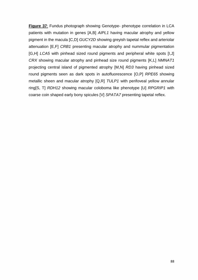
88
Figure 37: Fundus photograph showing Genotype- phenotype correlation in LCA
patients with mutation in genes [A,B] AIPL1 having macular atrophy and yellow
pigment in the macula [C,D] GUCY2D showing greyish tapetal reflex and arteriolar
attenuation [E,F] CRB1 presenting macular atrophy and nummular pigmentation
[G,H] LCA5 with pinhead sized round pigments and peripheral white spots [I,J]
CRX showing macular atrophy and pinhead size round pigments [K,L] NMNAT1
projecting central island of pigmented atrophy [M,N] RD3 having pinhead sized
round pigments seen as dark spots in autofluorescence [O,P] RPE65 showing
metallic sheen and macular atrophy [Q,R] TULP1 with perifoveal yellow annular
ring[S, T] RDH12 showing macular coloboma like phenotype [U] RPGRIP1 with
coarse coin shaped early bony spicules [V] SPATA7 presenting tapetal reflex.
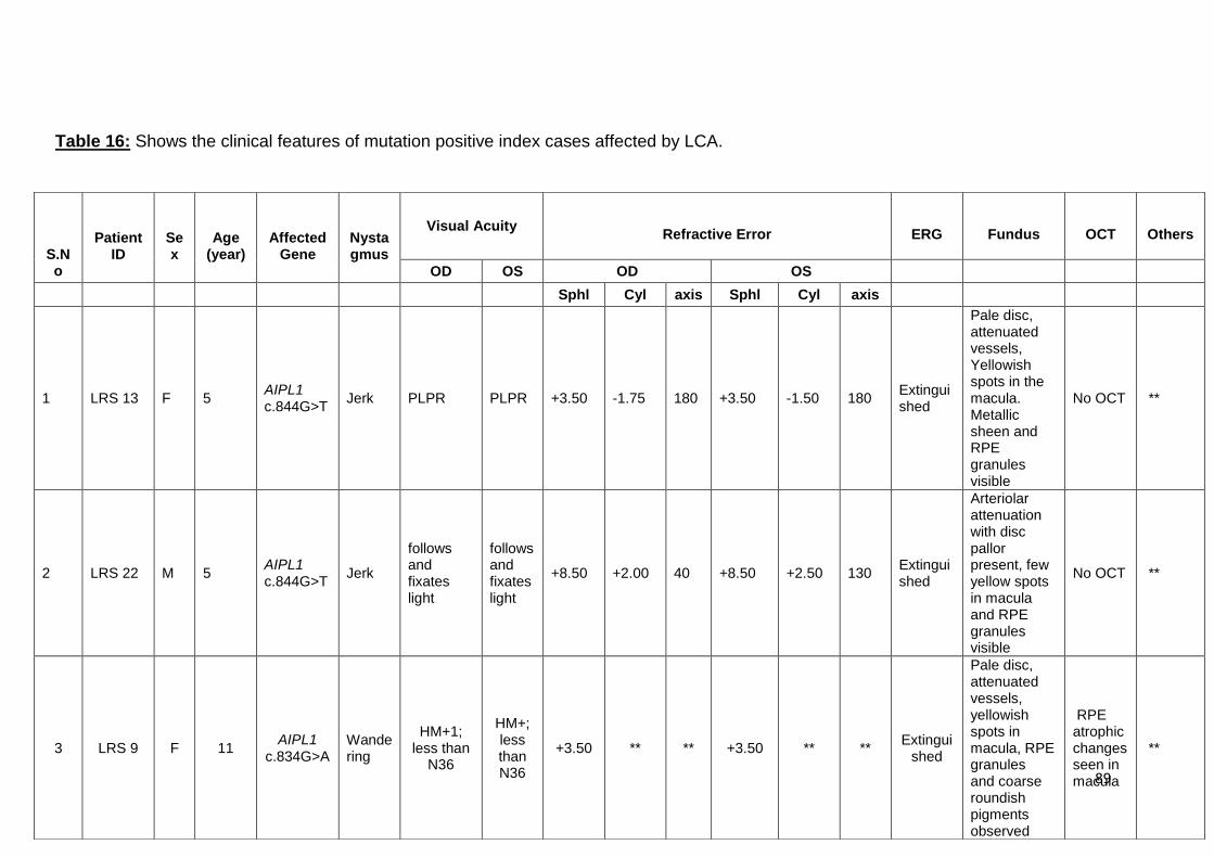
89
S.No
Patient
ID
Sex
Age
(year)
Affected
Gene
Nystagmus
Visual Acuity
Refractive Error
ERG
Fundus
OCT
Others
OD OS OD OS
Sphl Cyl axis Sphl Cyl axis
1 LRS 13 F 5 AIPL1 c.844G>T
Jerk PLPR PLPR +3.50 -1.75 180 +3.50 -1.50 180 Extinguished
Pale disc, attenuated vessels, Yellowish spots in the macula. Metallic sheen and RPE granules visible
No OCT **
2 LRS 22 M 5 AIPL1 c.844G>T
Jerk
follows and fixates light
follows and fixates light
+8.50 +2.00 40 +8.50 +2.50 130 Extinguished
Arteriolar attenuation with disc pallor present, few yellow spots in macula and RPE granules visible
No OCT **
3 LRS 9 F 11 AIPL1
c.834G>A Wandering
HM+1; less than
N36
HM+; less than N36
+3.50 ** ** +3.50 ** ** Extingui
shed
Pale disc, attenuated vessels, yellowish spots in macula, RPE granules and coarse roundish pigments observed
RPE atrophic changes seen in macula
**
Table 16: Shows the clinical features of mutation positive index cases affected by LCA.

90
4 LRS 71 M 3 AIPL1 c.834G>A
Pendular
follows and fixates light
follows and fixates light
+5.50 ** ** +5.00 ** ** Extinguished
Disc pallor, mild arteriolar attenuation, Yellowish spots in the macular region and metallic sheen in macula seen
OD-Foveal thinning with thinning of IS-OS junction noted at macula
**
5 LRS 4 M 5 AIPL1 c.689A>G
Jerk PLPR; less than N36
PLPR; less than N36
+9.00 -0.50 180 +9.00 -0.50 180 Extinguished
Pale disc, attenuated vessels, yellow spots, and coarse roundish pigments observed in periphery
No OCT
Delayed milestone
6 LRS 81 M 17 AIPL1 c.910G>T
Jerk PLPR; less than N36
PLPR; less than N36
+0.75 -2.75 40 +1.00 -2.00 160 Extinguished
Pale disc, attenuated vessels, atrophic yellowish disc shaped macula with coarse pigment granules and metallic sheen
No OCT OS-keratoconus

91
7 LRS 48 F 5 AIPL1 c.247G>A
Wandering
follows and fixates light
follows and fixates light
+5.50 -1.00 180 +6.00 -1.25 180 Extinguished
Mild arteriolar attenuation, disc pallor, yellowish spots, RPE granules and metallic sheen observed.
No OCT
Astigmatism, cleft palate
8 LRS 88 M 3
CRB1 c.1073_ 1074 insTGAG
Jerk
Eccentric fixation, follows light
Eccentric fixation, follows light
+6.00 ** ** +2.00 -0.75 180 Extinguished
Pale disc, minimal attenuation of vessels, greyish reflex in the macula, para arteriolar sparing and RPE loss in the periphery
No OCT Astigmatism
9 LRS 78 F 10 CRB1 c.4168 C>T
Jerk PLPR; less than N36
PLPR; less than N36
+12.50 -1.00 180 +13.00 -1.00 180 Extinguished
Pale disc, minimal attenuation of vessels, greyish reflex in the macula and roundish coin shaped pigmentary clumps are seen
Foveal thinning (OD>OS) with RPE atrophy noted. Thin ERM seen
Astigmatism

92
10 LRS 56 M 13 CRB1 c.4005+1 G>A
Jerk PLPR; less than N36
PLPR; less than N36
+8.00 -1.5 180 +8.50 -1.50 180 Extinguished
Pale disc, attenuated vessels, minimal para arteriolar sparing, coarser pigment and atrophic patches in the macula and calcareous yellowish deposits observed
No OCT **
11 LRS 7 F 2 CRB1 c.4168 C>T
Jerk
follows and fixates light
follows and fixates light
+6.50 -2.25 155 +7.00 -3.00 180 Extinguished
Pale disc, minimal attenuation of vessels, para arteriolar sparing greyish reflex in the macula and roundish coin shaped pigmentary clumps are seen
Altered foveal contour seen Thinning of IS-OS junction noted, RPE alterations seen
Keratoconus
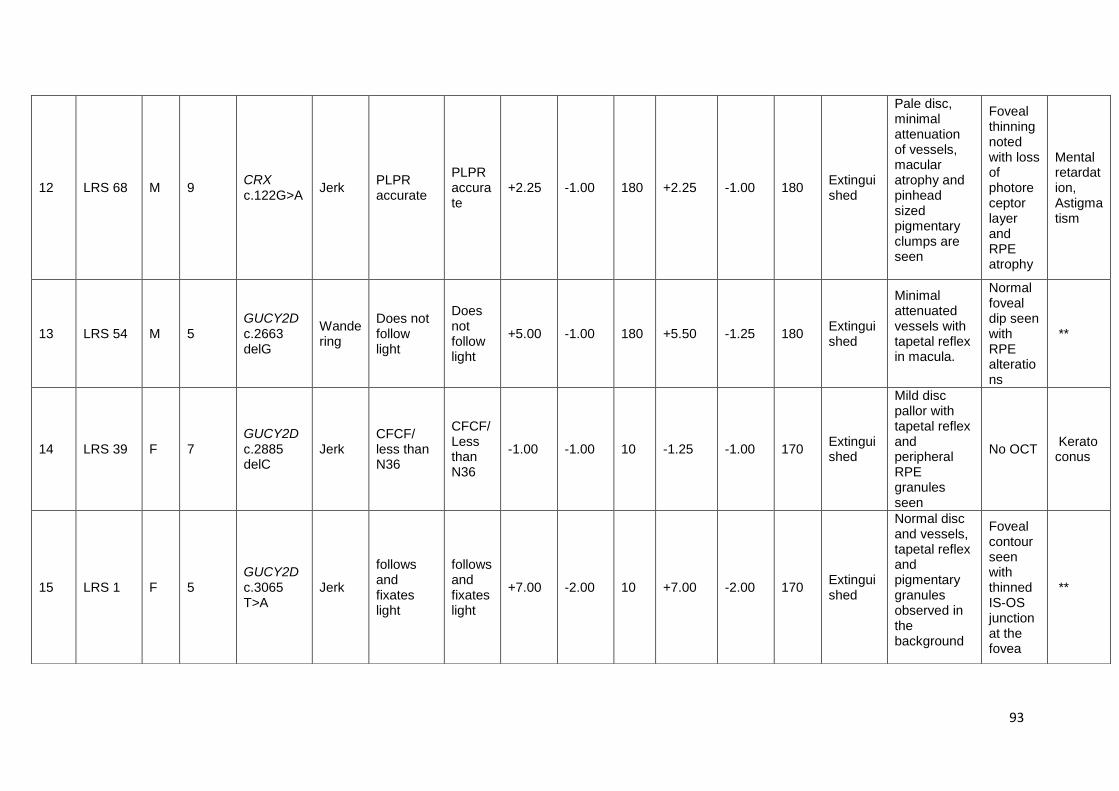
93
12 LRS 68 M 9 CRX c.122G>A
Jerk PLPR accurate
PLPR accurate
+2.25 -1.00 180 +2.25 -1.00 180 Extinguished
Pale disc, minimal attenuation of vessels, macular atrophy and pinhead sized pigmentary clumps are seen
Foveal thinning noted with loss of photoreceptor layer and RPE atrophy
Mental retardation, Astigmatism
13 LRS 54 M 5 GUCY2D c.2663 delG
Wandering
Does not follow light
Does not follow light
+5.00 -1.00 180 +5.50 -1.25 180 Extinguished
Minimal attenuated vessels with tapetal reflex in macula.
Normal foveal dip seen with RPE alterations
**
14 LRS 39 F 7 GUCY2D c.2885 delC
Jerk CFCF/ less than N36
CFCF/Less than N36
-1.00 -1.00 10 -1.25 -1.00 170 Extinguished
Mild disc pallor with tapetal reflex and peripheral RPE granules seen
No OCT Keratoconus
15 LRS 1 F 5 GUCY2D c.3065 T>A
Jerk
follows and fixates light
follows and fixates light
+7.00 -2.00 10 +7.00 -2.00 170 Extinguished
Normal disc and vessels, tapetal reflex and pigmentary granules observed in the background
Foveal contour seen with thinned IS-OS junction at the fovea
**

94
16 LRS 83 F 6 GUCY2D c.1978 C>T
Rotatory
PLPR PLPR +6.50 ** ** +7.00 ** ** Extinguished
Minimal attenuation of vessels, tapetal reflex in the macula.
No OCT **
17 LRS 32 M 14 GUCY2D c.1978 C>T
Jerk 2/60 3/60 +6.50 ** ** +6.00 ** ** Reduced
Tapetal reflex observed with peripheral RPE granules.
No OCT **
18 LRS 37 F 11 GUCY2D c.524T>G
Wandering
CFCF/ Less than N36
CFCF/Less than N36
+3.50 ** ** +3.75 ** ** Reduced
Tapetal reflex and peripheral RPE granules observed
No OCT **
19 LRS 43 M 3 GUCY2D c.1790 G>A
Wandering
follows light
follows light
+5.50 ** ** +5.50 ** ** Extinguished
peripheral granularity observed
No OCT Autism
20 LRS 51 F 7 GUCY2D c.3037 G>A
Jerk 6/60; N36 6/60;N36
+3.50 -1.25 180 +3.50 -1.25 180 Extinguished
Tapetal reflex observed in both eyes
No OCT **
21 LRS 31 F 13 GUCY2D c.839C>G
Pendular
1/60 less than N36
1/60 close to N36
+5.00 -0.75 180 +5.50 -1.00 180 Extinguished
Yellowish spots in macula and coarse RPE granules in periphery observed
No OCT **

95
22 LRS 74 F 3 GUCY2D c.2182 G>A
Wandering
follows and fixates light
follows and fixates light
+3.50 -1.00 180 +3.50 -1.00 180 Extinguished
Minimal attenuation of vessels, tapetal reflex in the macula.
No OCT **
23 LRS 55 M 6
GUCY2D c.2062 G>A c.1762 C>T
Rotatory
follows and fixates light
follows and fixates light
+5.00 -1.5 90 +5.00 -1.50 90 Extinguished
Tapetal reflex observed in both eyes
No OCT **
24 LRS 14 M 10
GUCY2D c.3118 C>G c.159delG
Wandering
occasionally follows light
occasionally follows light
+0.00 ** ** +3.50 ** ** Extinguished
Mild disc pallor and arteriolar attenuation present, tapetal reflex and RPE granules visible
No OCT **
25 LRS 79 F 9 IQCB1 c.1558 C>T
Jerk HM+1; less than N36
HM+1;less than N36
+7.50 ** ** +8.50 -1.00 100 Extinguished
Minimal attenuation of vessels and metallic sheet in the fundus seen
Normal foveal dip seen. Thinning of IS-OS junction noted
**
26 LRS 77 M 4 IQCB1 c.1363 C>T
Jerk
PLPR inaccurate; less than N36
PLPR inaccurate; less than N36
+6.50 ** ** +6.50 -0.50 180 Extinguished
Minimal attenuation of vessels and metallic sheet in the fundus seen
No OCT **

96
27 LRS 92 M 5 IQCB1 c.1333 C>T
Wandering
Does not follow light
Does not follow light
+0.00 ** ** +1.00 ** ** Extinguished
Metallic sheet in the fundus and RPE granules visible.
No OCT
Enlarged kidney on scan at 5 months
28 LRS 10 F 11 IQCB1 c.994C>T
Pendular
HM+1; less than N36
HM+1; less than N36
+7.50 -1.50 180 +7.50 -1.50 180 Extinguished
Mild disc pallor, Minimal attenuation of vessels and metallic sheet in the fundus seen
No OCT Myopic, Astigmatism
29 LRS 87 M 3 LCA5 c.838C>T
Wandering
follows and fixates light
follows and fixates light
+6.50 -1.50 180 +6.00 -1.50 180 Extinguished
Mild disc pallor, Arteriolar attenuation present, shiny reflex and pinhead sized round shaped pigment observed.
No OCT
Hearing problem, delayed milestone
30 LRS 21 F 8 LCA5 c.1422 delT
Jerk
follows and fixates light
follows and fixates light
+4.00 -1.50 180 +4.00 -1.50 180 Extinguished
Pale disc, mild attenuated vessels, macular atrophy observed
No OCT **

97
31 LRS 60 F 6 LCA5 c.955G>A
Wandering
Does not fix light
Does not fix light
+6.50 ** ** +5.50 ** ** Extinguished
Pale disc, mild attenuated vessels, macular atrophy and white spots observed in fundus
No OCT Astigmatism
32 LRS 20 F 11
LCA5 c.1062_ 1068 delCGAAAAC
Jerk
follows and fixates light
follows and fixates light
+4.50 -1.25 150 +5.00 -1.25 150 Extinguished
Pale disc, mild attenuated vessels, macular atrophy and peripheral white spots observed
No OCT **
Affected sib of LRS 20
F 3
LCA5 c.1062_ 1068 delCGAAAAC
Jerk
follows and fixates light
follows and fixates light
+3.00 -1.00 180 +3.00 -1.00 180 Extinguished
Pale disc, mild attenuated vessels, macular atrophy seen
No OCT **
33 LRS 30 M 14
LCA5 c.1062_ 1068 delCGAAAAC
Wandering
follows and fixates light
follows and fixates light
+1.00 -1.75 10 +1.00 -1.00 10 Extinguished
Pale disc, mild attenuated vessels, macular atrophy, shiny reflex seen and white dots in RPE observed.
No OCT **

98
34 LRS 62 F 9 months
NMNAT1 c.109G>A
Pendular
follows and fixates light
follows and fixates light
+6.00 -0.75 180 +5.50 -0.75 180 Extinguished
Pale disc, mild attenuated vessels, coarse pigment with macular atrophy observed
No OCT **
35 LRS 8 M 5 NMNAT1 c.53A>G
Jerk PLPR accurate
PLPR accurate
+6.00 ** ** +6.50 -1.00 70 Extinguished
Pale disc, mild attenuated vessels, coarse pigment with macular atrophy observed, typical bony spicules in the periphery
Foveal thinning with RPE atrophy noted at fovea. Pigmentation causing shadowing noted.
**
36 LRS 80 M 8 RD3 c.296+1 G>A
Jerk HM+;1 less than N36
CFCF/Less than N36
+3.50 -2.50 20 +3.00 -1.75 160 Extinguished
Pale disc, mild attenuated vessels and early macular scarring seen. Pinhead sized round pigment observed
No OCT **

99
37 LRS 49 F 3 RDH12 c.184C>T
Jerk 6/24 6/24 +8.00 -0.75 180 +8.00 -0.75 180 Extinguished
Pale disc, attenuated vessels and macular coloboma with bony spicules and pigmentary clumps observed
No OCT **
38 LRS 17 M 29 RDH12 c.146C>T
Nil CF at 50;N36
CF at 50;N36
-6.00 ** ** -5.00 ** ** Extinguished
Pale disc, marked attenuated vessels and large area of macular coloboma with bony spicules observed
No OCT **
39 LRS 93 F 17 RDH12 c.746G>T
Jerk 3/60 1/60 +3.00 ** ** +3.50 -1.00 180 Reduced
Pale disc, marked attenuated vessels and macular coloboma with bony spicules observed
Foveal thinning with RPE atrophy and pigmentation causing shadowing noted.ERM noted with IPVD.OS: IPVD
**
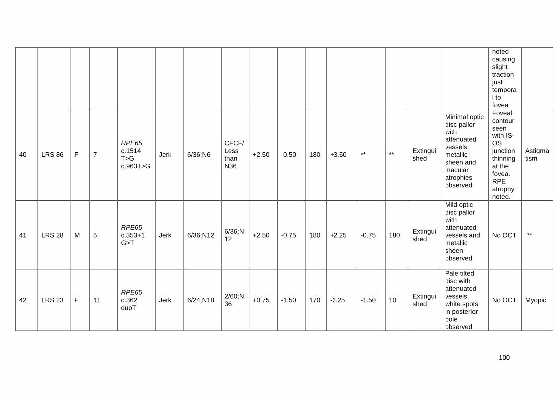
100
noted causing slight traction just temporal to fovea
40 LRS 86 F 7
RPE65 c.1514 T>G c.963T>G
Jerk 6/36;N6
CFCF/Less than N36
+2.50 -0.50 180 +3.50 ** ** Extinguished
Minimal optic disc pallor with attenuated vessels, metallic sheen and macular atrophies observed
Foveal contour seen with IS-OS junction thinning at the fovea. RPE atrophy noted.
Astigmatism
41 LRS 28 M 5 RPE65 c.353+1 G>T
Jerk 6/36;N12 6/36;N12
+2.50 -0.75 180 +2.25 -0.75 180 Extinguished
Mild optic disc pallor with attenuated vessels and metallic sheen observed
No OCT **
42 LRS 23 F 11 RPE65 c.362 dupT
Jerk 6/24;N18 2/60;N36
+0.75 -1.50 170 -2.25 -1.50 10 Extinguished
Pale tilted disc with attenuated vessels, white spots in posterior pole observed
No OCT Myopic

101
43 LRS 25 F 7
RPE65 c.46_49 del TTTG
Jerk 6/18;N6 6/18;N6
+1.75 ** ** +1.75 ** ** Extinguished
Optic disc pallor with attenuated vessels, white spots in posterior pole and metallic sheen observed
Foveal thinning noted. RPE alteration noted
**
44 LRS 2 F 9 RPE65 c.1409 C>T
Jerk 6/18;N6 6/60;N18
+2.50 -1.00 20 +4.00 -1.50 160 Extinguished
Optic disc pallor with attenuated vessels, with white spots observed
Minimal foveal thinning noted with thinning of IS-OS junction.
**
45 LRS 12 M 12 RPE65 c.1109 T>A
Pendular
HM+;1less than N36
CF at 50;less than N36
-4.00 -2.00 180 -16.00 -4.00 170 Extinguished
Pale disc, attenuated vessels, yellowish scarring seen in macula. coarse pigmentary clumps more in posterior pole observed
No OCT
Myopic, astigmatism, keratoconus
46 LRS 58 M 36 RPE65 c.362 dupT
Jerk CF at 50;less than N36
CF at 1m;less than N36
+3.50 ** ** +4.00 ** ** Extinguished
Optic disc pallor with attenuated vessels and peripheral spicule pigmentation observed
No OCT **

102
47 LRS 15 M 34 RPGRIP1 c.3434 delA
Jerk
PLPR accurate; less than N36
PLPR inaccurate; less than N36
-3.00 ** ** -5.50 ** ** Extinguished
Pale disc, attenuated vessels, macular atrophy with coarse coin shaped early bony spicules.
No OCT Myopic
48 LRS 65 M 4 RPGRIP1 c.2041 C>T
Rotatory
Does not follow light
Does not follow light
+10.50 -2.00 180 +9.50 -1.00 180 Extinguished
Minimal attenuation of vessels with RPE mottling seen.
No OCT Astigmatism
49 LRS 45 M 6 RPGRIP1
c.2041 C>T
Wandering
CFCF HM+ ** ** ** ** ** ** Extingui
shed
Mild optic disc pallor, attenuation of vessels with RPE mottling seen.
OU: Foveal contour seen with thinning of IS-OS junction noted. FT- OD-170 micron, OS- 180 micron
**
50 LRS 26 M 22 RPGRIP1 c.3788 T>C
Jerk PLPR accurate
PLPR accurate
+6.50 -3.00 180 +6.50 -3.00 180 Extinguished
Mild optic disc pallor, attenuation of vessels, extensive
No OCT **

103
RPE mottling with early bony spicule pigmentation seen. Calcified deposits observed
51 LRS 47 F 27 RPGRIP1 c.895G>T
Wandering
PLPR accurate
PLPR accurate
+5.50 -1.00 30 +5.00 -1.00 130 Extingui
shed
Mild optic disc pallor, attenuation of vessels, with bony spicule pigmentation seen.
Foveal thinning noted with RPE atrophy at the fovea, thinning of IS-OS layer seen.FT-OD-45 microns, OS-63 microns
**
52 LRS 40 M 22 TULP1 c.1047 T>G
Jerk 1/60 1/60 -5.00 -0.75 90 -3.50 -0.75 60 Extinguished
Pale disc, marked attenuated vessels, Peri foveal yellow annular ring with macular atrophy and mild peripheral bone spicules like pigmentation seen
Foveal thinning noted with RPE atrophy seen sparring fovea. Thin ERM noted with ILM wrinkling seen.
Myopic, cataract

104
53 LRS 29 F 22 SPATA7 c.1215+5C>A
wandering
PLPR accurate; less than N36
PLPR accurate; less than N36
+7.50 ** ** +8.00 ** ** Extinguished
Pale disc, attenuated vessels, tapetal reflex seen in periphery
NO OCT
**
Affected sib of LRS 29
M 14 SPATA7 c.1215+5C>A
wandering
PLPR accurate; less than N36
PLPR accurate; less thanN36
+6.00 -0.5 180 +6.00 ** ** Extinguished
Pale disc, attenuated vessels, RPE atrophy
NO OCT
**
54
LRS 67 M 15 SPATA7 c.913-2A>G
Jerk PLPR accurate
PLPR accurate
+1.00 -1.00 170 +1.00 -2.25 170 Extinguished
Pale disc, attenuated vessels with RPE atrophy seen.
Normal foveal dip seen with thinning of IS-OS junction noted. RPE alterations seen
**
55 LRS 11 F 11 SPATA7 c.18A>G
Jerk 3/60 HM+ +2.00 -1.50 20 +2.00 -3.00 120 Extinguished
Pale disc, attenuated vessels with tapetal reflex and peripheral pigmentary changes seen.
NO OCT
**

105
Table 11: Shows the clinical features of mutation positive index cases affected by LCA. IS-OS = Inner segment-outer segment;
ERM = Epiretinal membrane; RPE = Retinal pigment epithelium; ILM = Internal limiting membrane; PLPR= Perception and
projection of light; HM = Hand movements; CFCF = Counting finger close to face.

106
3.5. Screening of SLC19A2 in a patient diagnosed with Thiamine Responsive
Megaloblastic Anaemia (TRMA) with LCA as ocular phenotype.
Figure 38: 2% Agarose gel picture of the amplified products of the six exons of
SLC19A2 gene. Lane 1: Exon 1- 589 bp, Lane 2: Exon 2- 700 bp, Lane 3: Exon 3-
391 bp, Lane 4: Exon 4- 429 bp, Lane 5: Exon 5- 300 bp, Lane 6: Exon 6 - 295 bp.
Screening identified a novel point mutation in exon 2, a c.314G>A transition resulting
in a missense mutation p.(Gly105Glu) (Figure 39). SIFT and PolyPhen analysis
predicted the mutation to be “damaging” (score-0.02) and “probably damaging”
(score-0.994), with glycine residue conserved across vertebrates and human SLC19
family. Parents were also screened and were found to be heterozygous for the
mutation and the unaffected sib homozygous for wild type.

107
Figure 39: Sequence showing a novel point mutation in exon 2, a c.314G>A
transition resulting in a missense mutation p.(Gly105Glu). [A] Heterozygous
mutation in mother [B] Homozygous mutation in proband [C] Homozygous wild
type in sib [D] Heterozygous mutation in father.
108
CHAPTER-4
DISCUSSION
Next generation sequencing has transformed genomic research from a technically
laborious, time consuming and an expensive experimentation to a fast, cost
effective and efficient tool in investigating genetic basis of many diseases. Clinical
implementations of NGS are still evolving; however, it has increased the
understanding of genetics in everyday medicine. The massively parallel sequencing
on NGS platforms has made possible the simultaneous sequencing of multiple
genes in a single reaction thus increasing the knowledge on genetic basis of
various Mendelian diseases especially those that are genetically heterogeneous.
Leber congenital amaurosis is characterised by both clinical variability and genetic
heterogeneity. The autosomal dominant and recessive LCA are shown to be
caused by 29 different genes. Therefore, next generation sequencing technologies
is currently the most promising approach to identify mutations in candidate genes of
LCA.
4.1. Targeted resequencing in Indian LCA cohort:
In this study, after complete ophthalmic examination, phenotype documentation and
pre-test genetic counselling, genotyping by NGS based targeted re-sequencing of
twenty LCA candidate genes was performed to identify the disease-causing variant
in ninety-two LCA families from an Indian cohort. Except the four positive
controls122, 127 (LRS 2, LRS 12, LRS 58, LRS 60) the index cases were not
screened for mutations by any other methodologies.
The frequency of mutations in LCA genes varies in different populations. In a
Chinese cohort of 145 patients screening 163 genes by retinal gene panel, 76.6%
were mutation positive with GUCY2D mutations being more common 27 while in an
Italian cohort of 95 patients, screening 8 LCA genes identified mutations in 28%
with RPE65 being most frequent158. Screening 13 genes in 37 Saudi Arabian
families showed 24% mutation positivity with TULP1 being common159. In a Belgium
cohort of 91 cases, 69% were positive with CEP290 mutations being frequent 160. In
a Spanish cohort eight genes screened in 84 patients revealed 23.8% mutation
identification with highest mutation rate in CRB1161. Screening of nine genes in 64

109
Figure 40: Pie charts of cohort studies showing frequency of LCA candidate gene mutation in the population. A. Italy158 B. Japan162
C. China27 D. Australia163 E. India (Current study) F. Brazil164 G. Western cohort124

110
Danish patients identified 49% mutation positivity with high contribution of RPE65
gene mutations165.
In the current study, after the analysis, validation and segregation of the annotated
data, we found 61% (56/92) of recruited LCA cases to be mutation positive in the
twenty known candidate genes. The mutation spectrum was found to be as follows -
GUCY2D gene contributing the highest (13%) followed by AIPL1 and RPE65 (8%),
RPGRIP1 and LCA5 (6%), IQCB1 and CRB1 (4%), SPATA7 and RDH12 (3%),
NMNAT1 (2%), KCNJ13, CRX, RD3 and TULP1 contributing to 1%. We also
observed same mutation in two or more unrelated individuals in our cohort.
No mutations were observed in CABP4, CEP290, LRAT, MERTK, OTX2 and
IMPDH1 in this cohort. According to world-wide reports, CEP290 is the most
frequently mutated gene11 followed by GUCY2D. However, we did not observe any
mutations in CEP290 gene in the cohort studied.
In LRS 11 (with consanguineous parentage), we observed a homozygous silent
variant, c.18A>G; p.(Arg6Arg) in the penultimate base of exon 1 in SPATA7. The
mother was heterozygous for the variant but the father was unavailable for analysis.
This variant was not seen in controls screened. Although a synonymous change,
computational predictions showed that it affects the wild type donor splice-site
probably affecting splicing. This could either lead to a truncated protein or result in
non-sense mediated decay166. The patient and the family were unavailable for
cDNA analysis to confirm the exact splicing defect. However, reports on silent
variants in the last codon of exon leading to disease phenotype suggest this variant
to be likely pathogenic167. The same patient had a heterozygous missense variant
c.551C>T in CRX. Bioinformatics tools predicted the variant to be damaging. The
mother was wild type and the father was unavailable for gene testing. Hence, this
CRX variant is defined as variant of unknown significance.
We observed specific bilateral changes in fundus features of our patients. Distinct
phenotypes as found in earlier reports were observed in our study too; macular
atrophy and yellowish spots in the macular region in AIPL1 mutation 168,
paraarteriolar sparing of RPE and nummular pigmentation in CRB1169. Greyish
tapetal reflex was significantly observed in many patients with GUCY2D mutation in
our cohort. LCA5 mutation positive patients had hypopigmented whitish spots 127,

111
while a typical macular colobomatous atrophy and bony spicules were evident in
RDH12 170 and RPGRIP1 mutation, respectively. Typical fundus features of these
various mutations are almost like the footprints of the disease that can be easily
evaluated on retinal examination. These allow clinicians to consider few possible
underlying mutated gene(s) and to some extent prognosticate the progression of
the disease without subjecting all patients to a detailed genetic evaluation.
Among the 8 AIPL1 mutation positive individuals, we observed the most frequent
mutation p.(Trp278*) in three patients however the characteristic occurrence of
keratoconus associated with AIPL1 mutation was observed in only one. In RPE65
affected individuals, we observed deterioration of vision with increase in age which
correlates with the absence of macular involvement in early years and development
of atrophy in later ages.
As reported, the visual acuities in our subjects varied from no light perception to
20/100 in many171 and cycloplegic refractions revealed predominantly hyperopic
and few myopic LCA patients172. Other ocular phenotypes like astigmatism,
keratoconus and cataract that were reported in other studies were also observed in
this cohort, astigmatism in a SPATA7 mutated patient173, keratoconus in AIPL1,
CRB1, RPGRIP1, LCA5, RPE65 and TULP1 mutated LCA patients172, 174. Cataract
was seen in some patients with AIPL1, TULP1, RPGRIP1, LCA5 and CRB1
mutated patients174. Other anomalies that are reported to be associated with LCA,
like cleft palate171, mental retardation, autism, delayed milestone175 and hearing
deficit were observed in few of our patients which signifies the importance of a
thorough systemic evaluation in these subjects.
Double mutations i.e. two homozygous pathogenic variants in same gene were
observed in two of our patients. LRS 86, with homozygous RPE65 mutations,
(p.(Asn321Lys) and p.(Leu505Arg)), had a severe macular phenotype with multiple
small atrophic patches which is different from other RPE65 cases. While LRS 55
with GUCY2D mutations (p.(Gly688Arg) and p.(Arg588Trp)), showed no significant
difference compared to other GUCY2D mutation positive cases.
In LRS 90 family, the proband (3 yrs) and the affected sib (6 yrs) with digenic
variants in KCNJ13 and AIPL1 showed diffused RPE mottling. The characteristic
fundus phenotypes may be observed at a later age.
112
Among the mutation positive cases, there were twelve possible triallelic cases,
where a heterozygous variant was observed along with disease causing pathogenic
homozygous variant. The role of the third heterozygous allele remains unclear as
we did not observe any significant difference in the fundus phenotype due to this
allele. Although all the third heterozygous allele segregated in the families, the low
CADD scores of CABP4 and CRB1 variants excludes the possibility of triallelism in
LRS 58 and LRS 14 respectively. The pathogenicity of heterozygous RPE65,
p.(Asn321Lys) variant in LRS 8, LRS 78, LRS 80 and LRS 88 as a disease allele
remains unclear because of relatively high MAF and low CADD scoring, though the
variant has been defined as disease causing in many studies176, 177. Further
molecular characterization of the variants might clarify the functional consequences,
if any.
4.1.2. Possible functional impact of mutations on their protein:
Mutations in specific domains of protein are shown to have a major impact on their
structure and functions. In our study, about 80% of mutations are in the functional
domain and 44% of the mutations are either nonsense or frameshift which are
present on or before the functional domain and predicted to result in truncated
protein (Figure 31).
AIPL1:
The PPIase domain binds to farnesylated PDE6 protein178 and mutation in this
domain disrupts its binding. Glu83Lys mutation identified in this study is present in
this domain and was predicted to alter the structure as the amino acid change is
from acidic to a basic residue. The tetratricopeptide repeat (TPR) domain in AIPL1
is essential for the protein to function as chaperone heterocomplex for assisting in
retinal protein maturation. Mutations in TPR domain or deletion of the domain are
shown to disrupt this interaction between AIPL1 and its co-chaperone Hsp70179.
Three of the identified mutations p.(Asn230Ser), p.(Trp278*) and p.(Glu282*) are
present in this domain. The p.(Trp278*) mutation is shown to result incomplete loss
of protein180.
CRB1:
The CRB1 protein has a signal peptide, EGF-like domains, laminin AG-like domains
and cytoplasmic domains. We identified frameshift mutation p.(Ser359Glufs*20) in

113
the 5th epidermal growth factor-like (EGF) domain and p.(R1390X) in the 3rd laminin
aglobular like domain leading to premature truncation. Mutations in EGF domain are
said to affect interdomain packing181. The laminin G-like domains are conserved
and are predicted to affect calcium binding, protein folding and interactions182. The
functional impact of the identified canonical splice variant c.4005+1G>A is unknown
but is predicted to be damaging by insilico analysis.
CRX
The CRX protein contains an otd/ Otx-like paired homeodomain (HD) in the N-
terminus that are required for DNA-binding and nuclear localization and a
transcription activation domain at the C-terminal183. The mutation identified in this
study p.(Arg41Gln) is present in HD domain while p.(Pro184Leu) is present in
transcription activation domain. p.(Arg41Gln) has already been characterized by co-
transfection assays and was shown to significantly reduce DNA-binding activity184.
GUCY2D
GUCY2D protein has N-terminal extracellular domain (ECD), a single membrane-
spanning domain, an intracellular kinase homology domain (KHD), and a catalytic
domain (CD). In our study, p.(Phe54Serfs*31) in ECD, p.(Arg660*) in KHD,
p.(Gly888Alafs*8), p.(Thr962Ilefs*16), p.(Leu1022*) in CD all lead to premature
protein truncation causing the disruption in protein activity. Studies have shown that
the protein activity is compromised even with point mutations in KHD and CD
domains 185. All other missense variants in our study are distributed in all three
domains and are predicted to be disease causing by bioinformatics tools.
RDH12
RDH12 has a NAD(P) binding domain that is essential for its catalytic activity
possessing cofactor or substrate binding sites186. The nonsense variant p.(Arg62*)
leads to protein truncation while one of the missense mutations identified,
p.(Thr49Met), was well characterized and had shown to be rapidly degraded
leading to decreased cellular availability, thus causing the disease187
LCA5
LCA5 has 4 coiled coil domains that aid in the interaction with its other partners like
OFD1 188, NlpisoB etc. All mutations identified in LCA5 in this study are frameshift
p.(Ile474Metfs*11), and nonsense p.(Tyr354*), p.(Arg280*) leading to truncation of
114
the protein. The c.955G>A lead to change in the last base of exon 6 leads to amino
acid change from alanine to threonine. This mutation has been characterized by
cDNA analysis leading to a 5 bp insertion of intronic sequence, causing a frameshift
and premature truncation of protein189.
RPE65
RPE65 has a catalytic domain that is responsible for its isomerohydrolase
activities190. The 2 frameshift mutants observed in the study, p.(Phe16Lysfs*14) and
p.(Ser121Phefs*10) are before and in the beginning of the catalytic domain,
respectively. Hence there would be disruption of whole domain resulting possibly in a
non-functional protein and causing disease. Although studies on the functional
consequence of the other mutants p.(Leu505Arg), p.(Asn321Lys), p.(Pro470Leu)
and p.(Leu370His) are unavailable, we find that all these mutants lie on the catalytic
domain and are predicted to have change in amino acid charge and polarity except
for p.(Asn321Lys).
NMNAT1
NMNAT1 has substrate binding domain, isoform-specific targeting and interaction
domains that are essential for neuroprotective function of the protein191. We
identified 2 missense variants that are not characterized before. By Insilico
analysis192, the p.(Asn18Ser) mutant is predicted to affect correct protein folding as
the hydrophobicity of the wild-type and mutant residue differs. In p.(Gly37Arg)
mutant, the glycine to arginine change introduces a charge (neutral to positive) in
this position which might result in a repulsion between the altered residue and the
adjacent amino acid residues. Also, glycine with its neutral charge is flexible for any
change in protein conformation thereby helping in protein function. Its alteration to
arginine might decrease the flexibility thus affecting protein function.
IQCB1
IQCB1 possess IQ calmodulin-binding and coiled coil domains 193, 194.The functional
domain is essential for its interaction with RPGR and calmodulin during
ciliogenesis29. All IQCB1 mutants identified in the study are nonsense mutants
(Gln520*, Arg455*, Arg445*, Arg332*), and might have a functional impact on
protein.

115
RPGRIP1
RPGRIP1 contains a C-terminal RPGR interacting domain (RID) and a coiled-coil
(CC) domain. These domains are homologous among the proteins that are involved
in vesicular trafficking195. The mutants identified in our study- p.(Glu1145Glyfs*18),
p.(Gln681*) and p.(Glu299*) result in proteins that lack RPGR interacting domain
causing disease.
TULP1
TULP1 has carboxy-terminal tubby domain which acts as DNA binding domain and
the N-terminal regions helps in transcription activation196. The p.(Asn349Lys)
mutation is found to be in the tubby domain. The mutation segregated with the
disease phenotype in the family with the affected sib harbouring the same mutation
and is predicted to be damaging by insilico tools. The functional impact of this
possibly pathogenic variant is yet to be studied.
SPATA7
The N-terminal domain of SPATA7 functions as binding region for other ciliary
proteins to perform ciliary trafficking 76. The mutants from this study c.913-2A>G,
c.1215+5C>A and c.18A>G are splice mutants. The two splice variants c.913-2A>G
and c.1215+5C>A were characterized by RT-PCR and cDNA sequencing that
confirm the altered splicing which would lead to premature truncation of protein. The
cDNA mutant is predicted by bioinformatic analysis to alter splicing as well.
4.2. Observed syndromic LCA
4.2.1. Senior-Loken syndrome (SLSN)
SLSN is an autosomal recessive syndrome. It is a nephronophthisis-associated
disorder, where the patient presents cystic kidney disease and retinal dystrophy.
Retinal abnormalities like retinitis pigmentosa or Leber congenital amaurosis is
reported so far. Prognosis with respect to vision is usually poor and treatment is still
unavailable. But for the kidney, renal transplantation is recommended for the end
stage renal failure197. Mutations in IQCB1 have been shown to cause Senior-Loken
syndrome (SLSN). In the study by Estrada Cuzcano et. al, LCA patients with IQCB1
mutations were recalled and assessed for renal abnormalities and those positive
were subsequently re-diagnosed as SLSN198. Patients with IQCB1 mutations are
116
shown to be at a high risk of renal failure during or after second decade of life.In our
study, we observed pathogenic variants in IQCB1 gene in four patients – LRS 10,
LRS 77, LRS 79 and LRS 92. Following the results obtained from our targeted re-
sequencing assay, we re-called the IQCB1 mutation positive patients, re-counselled
and advised for comprehensive renal evaluation at regular intervals. The case LRS
92 first reported to us at seven months of age and was diagnosed with LCA.
Following gene testing and re-counselling at 5 years of age, the patient‟s family
revealed history of bilateral marginal nephromegaly diagnosed at 9 months. At 7yrs,
he developed chronic kidney disease stage II and presented with behavioural
issues both characteristic of IQCB1 mutation and thus re-diagnosed as Senior-
Loken syndrome. The age ranges of other IQCB1 mutation positive patients are
between 4 and 11 years and no renal abnormality has been observed yet.
4.2.2. Thiamine responsive megaloblastic anaemia:
TRMA Syndrome was first reported in 1969 by Rogers et al. 199. It is autosomal
recessive and the onset of the syndrome is in infancy or in early childhood with
main features being megaloblastic anemia, diabetes mellitus (non-type 1) and
sensorineural deafness, responding in varying degrees to thiamine treatment200, 201.
Other associated findings described are congenital cardiac malformations,
cardiomyopathy andretinal abnormalities.
In a case of cone-rod dystrophy with TRMA syndrome, deafness was the first
feature to be noticed at 1 year of age followed by diabetes mellitus. At 7 years of
age, the patient developed pancytopenia, cardiomyopathy and vision loss, where
the ERG was consistent with cone-rod dystrophy. By 15 years of age, vision in both
eyes deteriorated and fundi showed bull‟s eye maculopathy with peripheral
pigmentary changes202. Contrarily, in the current study, the patient complained
severe vision loss with photophobia, nystagmus and hyperopia consistent with LCA
manifesting within one year of age. ERG was extinguished. This was followed by
sensorineural deafness at one year and diabetes mellitus later at three and half
years. The manifestation of all features of TRMA was much early in our patient.
Homozygosity mapping and subsequent gene screening identified on chromosome
1 (1q23.2-23.3), SLC19A2, a member of solute carrier family 19, encoding a
thiamine transporter, THTR1 as the candidate gene, for TRMA155, 203. Many
mutations in SLC19A2 gene in TRMA patients have been reported204-206. Gene

117
screening aids in the differential diagnosis of neonatal diabetes as SLC19A2 is not
mutated in patients with permanent neonatal diabetes mellitus (PNDM) in which
diabetes is presented in the first 6 months of life207.
A higher expression of hTHTR-1 and moderate expression of hTHTR-2 were found
to be present in ARPE-19 cells as well as in native human retinal tissue. By
confocal imaging, ARPE-19 cells expressing TRMA associated hTHTR-1 mutants
(Gly172Asp, Ser143Phe and Asp93His) have shown impaired cell surface
expression or function of the transporter in this cell. However, it is still not known as
to why only some TRMA patients present with retinal degeneration and why there is
a difference in the clinical presentation of the same208.
In our patient, we report a novel missense mutation in exon 2, a c.314G>A
transition resulting in p.(Gly105Glu), converting nonpolar hydrophobic glycine to
negatively charged hydrophilic glutamic acid. The retinal degenerative (RD)
phenotypes so far described as part of TRMA are cone rod dystrophy202, retinitis
pigmentosa209 and optic atrophy210. Here for first time, we report Leber‟s congenital
amaurosis (LCA) as a component of RD in our patient.
In the last decade, tremendous advances in genotyping and genomic research have
improved molecular diagnosis aiding in genetic counselling and development of
therapeutic strategies for the treatment of various diseases like cancer, genetic
defects, infectious diseases or autoimmune disorders. Even when significant
progress has been made in recent years in gene therapy, most genetic diseases do
not have therapies, and it will be important to continue developing treatment
strategies for individual genetic diseases even if they occur only rarely211.
Advancements in understanding the genetic and molecular basis of a variety of
retinal diseases has led to the development of gene therapy for these and the
original hope that gene therapy could cure otherwise untreatable, inherited genetic
diseases has been rekindled by gene therapy clinical trial for LCA. The difficulties in
designing gene therapy are due to the choice of suitable gene transfer vehicle, the
efficiency of gene transfer and problems related to immune reactions of the treated
patient against the vector carrying the therapeutic gene. AAV2-mediated gene
transfer to the human retina does not elicit the cytotoxic T-lymphocyte responses to
AAV capsids unlike that were observed in muscle and central nervous system, thus
supporting the use of AAV-mediated gene augmentation therapy for treatment of

118
few inherited retinal diseases. Molecular testing aids in the diagnosis of LCA and
knowledge of the disease gene in LCA patients are important in counselling
patient‟s family and in determining their suitability for gene-specific therapies for
very few now and for many subjects in future.
In conclusion, this study provides an insight on the genetic profile and mutation
spectrum of candidate genes of LCA from a larger Indian cohort. The molecular
findings have also helped in re-diagnosis, medical management, carrier testing,
prenatal testing and genetic counseling.
„

119
CONCLUSION
In conclusion, this is the first comprehensive study on the genetic profile and
mutation spectrum of candidate genes of LCA from a larger Indian cohort. Also, this
study for the first time reported association of TRMA with LCA as the retinal disease
component. In this cohort, mutation was found in 62% (57/92 - 56 by targeted
resequencing and 1 TRMA) of patients recruited. Among the mutations found 39%
(21/53) were novel. The mutation spectrum and the presence of large number of
novel mutations suggests that using custom made chips designed for targeting
specific mutations for genetic testing would not be an efficient method of genetic
diagnosis, especially in genetically heterogeneous diseases.
The current study also reports a possible digenic inheritance which proves the
advantage of using NGS over other screening methodologies, as a large set of
genes are screened and analysed for possible disease pathogenicity. The triallelic
inheritance reported in the current study provides an insight on the modifier alleles
that could probably make a difference in the disease severity among. However, we
did not observe any alteration in the disease severity in our cohort with triallelic
variants. Mutation was not identified in 35/92 cases (38%) where screening of
mutations in unscreened deep intronic and regulatory regions might contribute to a
small percentage of disease etiology in these patients. These negative cases also
represent cohort where other retinal genes or novel genes could be involved. Also,
large structural rearrangements such as duplications are not identified by targeted
re-sequencing. Whole exome or genome sequencing would identify all these
including novel genes.

120
SPECIFIC CONTRIBUTIONS
First report on mutation spectrum in LCA from a large Indian cohort
First report on association of TRMA with LCA as an ocular disease phenotype.
Distinct phenotype to genotype correlation in a large number of LCA patients.
21 novel pathogenic variants submitted to public databases.
Genetic testing data of 57 patients positive for mutation in candidate genes
screened aided in
Genetic counselling
Genetic diagnostics has strengthened genetic counselling and management
of genetic diseases. Although gene therapies for ocular genetic diseases are
still underway, gene testing confirms the clinical diagnosis and has helped
the patient and their families in rehabilitation and disease management.
Carrier testing
Inthe current study, after identifying the mutation in the probands we
extended carrier testing to other members of the families. As a part of
segregation analysis and carrier testing the siblings were checked for carrier
status. This had aided in genetic counselling for the carrier sibling by
providing the risk of disease for the next generation, especially if they decide
on consanguineous marriages.
Prenatal testing
Molecular genetic test, prenatally, are usually carried out in the fetal DNA
from chorionic villi sample. In two of our patients, LRS 43 with p.(Gly597Glu)
and LRS 74 with p.(Asp728Asn) mutations in GUCY2D, following the genetic
test in proband, the results were discussed with the family and the proband‟s
parents opted for prenatal testing for the successive pregnancy. Both the
foetuses were found to be heterozygous carriers and had normal vision after
birth.

121
Management of genetic disease
The patients with IQCB1 mutations are shown to be at a high risk of renal
failure during or after second decade of life. We identified IQCB1 pathogenic
variants in four patients and all were re-called, re-counselled and advised for
comprehensive renal evaluation at regular intervals. LRS92 with IQCB1
mutation, at 7yrs, developed chronic kidney disease stage II and presented
with behavioural issues both characteristic of IQCB1 mutation. Monitoring the
renal function of patients with IQCB1 mutation periodically help in avoiding
end stage complication and proper management of disease.
Screening of SLC19A2 in the TRMA patient helped in confirming the
diagnosis and thereby management. The megaloblastic anaemia was
corrected using a pharmacological dose of thiamine (25-75 mg /day) and for
diabetes; the patient is under 18U and 10U of insulin at morning and night,
respectively.

122
LIMITATIONS
Large structural rearrangements such as duplications were not analyzed in this
study. Deep intronic and regulatory regions of the twenty candidate genes screened
were not sequenced in the current study. This might contribute to a small percentage
of mutations in the negative cases. Three cases, LRS 46, LRS 24 and LRS 89 with
novel heterozygous variants MERTK, c.1627T>C, p.(Ser543Pro); RPE65, c.814C>T,
p.(Gly272Arg) and RPGRIP1, c.1480delA, p.(Asn495Thrfs*11) could harbour deep
intronic variants. Recently, more candidate genes; ALMS1, CCT2, CNGA3,
CLUAP1, DTHD1, GDF6, IFT140, MYO7A and PRPH2 have been reported which
were not screened in the current study. Although variants in each of these genes
were reported in only one case, it might also contribute to the mutation spectrum in
our cohort.
FUTURE SCOPE OF THE STUDY
Whole exome or genome sequencing might help in identifying the causative
variant involved in the mutation negative cases, either variants in deep intronic
and regulatory regions of known candidate genes or novel genes.
The phenotype and genotype correlation understood by the current study can be
extrapolated to design phenotype based diagnostic testing to reduce the cost of
gene testing for the patients.
The available data on the mutation spectrum of the patients would help in the
application of gene replacement therapy in the future.
Identifying novel genes and understanding their function and disease
mechanism may pave way for new therapeutic strategies.

123
REFERENCES
1. HT Gabriel: HUMAN GENETICS ‟98: APOPTOSIS: Mechanisms of Cell
Death in the Inherited Retinal Degenerations. Am J Hum Genet 1998, 62:
503–508.
2. Lorenz B Preising M, Stieger K.: Retinal blinding disorders and gene therapy-
molecular and clinical aspects. . Curr Gene Ther 2010, 10 ((5)): 350-370.
3. Shobi V Csilla HL, Chang,, Paul AS, Eyal B, S. Anand: Biology and therapy of
inherited retinal degenerative disease: insights from mouse models. Dis
Model Mech 2015, 8 (2): 109–129.
4. Daiger SP, Rossiter BJF, Greenberg J, Christoffels A, W. Hide: Data services
and software for identifying genes and mutations causing retinal
degeneration. Invest OphthalmolVis Sci 1998, 39: S295.
5. Veleri S Lazar CH, Chang B, Sieving PA, Banin E, Swaroop A.: Biology and
therapy of inherited retinal degenerative disease: insights from mouse
models. Dis Model Mech 2015, 8 (2): 109-129.
6. Swaroop A Kim D, Forrest D: Transcriptional regulation of photoreceptor
development and homeostasis in the mammalian retina. . Nat Rev Neurosci
2010, 11: 563-576.
7. Benjamin MN Dale CW, John RG, Bruce B, Robyn VJ.: Retinal dystrophies,
genomic applications in diagnosis and prospects for therapy. Transl Pediatr
2015, 4 (2): 139-163.
8. Parmeggiani F, Sorrentino FS, Ponzin D, Barbaro V, S Ferrari, E. Di Iorio:
Retinitis pigmentosa: genes and disease mechanisms. Curr Genomics 2011,
12: 238-249.
9. Kohl S, Varsanyi B, Antunes GA, Baumann B, Hoyng CB, Jägle H, Rosenberg
T, Kellner U, Lorenz B, Salati R et al: CNGB3 mutations account for 50% of all
cases with autosomal recessive achromatopsia. Eur J Hum Genet 2005, 13
(3): 302-308.
10. Gardner JC Webb TR, Kanuga N, Robson AG, Holder GE, Stockman A,
Ripamonti C, Ebenezer ND, Ogun O, Devery S, Wright GA, Maher ER,
Cheetham ME, Moore AT, Michaelides M, Hardcastle AJ.: X-linked cone
124
dystrophy caused by mutation of the red and green cone opsins. Am J Hum
Genet 2010, 87 (1): 26-39.
11. Rachna S Chitra K, Subhadra J.: Genetics of Leber congenital amaurosis:
an update. In: Expert Review of Ophthalmology. vol. 7; 2012: 141-151.
12. Garcia-Hoyos M, Lorda-Sanchez , Gómez-Garre P, Villaverde C,
Cantalapiedra D, Bustamante A, Diego-Alvarez D, Vallespin E, Gallego-Merlo
J, Trujillo MJ et al: New type of mutations in three spanish families with
choroideremia. Invest Ophthalmol Vis Sci 2008, 49 (4): 1315-1321.
13. Hiroyuki Y András S, Ulf E, Eddie H, Eliot LB, Thaddeus PD: Mutations in the
gene encoding 11-cis retinol dehydrogenase cause delayed dark adaptation
and fundus albipunctatus. Nature Genetics 1999, 22: 188 - 191
14. Katsanis N Shroyer NF, Lewis RA, Cavender JC, AA Rajhi AI, Jabak M,
Lupski JR.: Fundus albipunctatus and retinitis punctata albescens in a
pedigree with an R150Q mutation in RLBP1. Clin Genet 2001, 59: 424–429.
15. Ku CS, Naidoo N, Y. Pawitan: Revisiting Mendelian disorders through exome
sequencing. Hum Genet 2011, 129: 351-370.
16. Harville HM Held S, Diaz-Font A, Davis EE, Diplas BH, Lewis RA,
Borochowitz ZU, Zhou W, Chaki M, MacDonald J, Kayserili H, Beales PL,
Katsanis N, Otto E, Hildebrandt F. : Identification of 11 novel mutations in
eight BBS genes by high-resolution homozygosity mapping. J Med Genet
2010, 47: 262-267.
17. Collin RW Safieh C, Littink KW, Shalev SA, Garzozi HJ, Rizel L, Abbasi AH,
Cremers FP, den Hollander AI, Klevering BJ, Ben-Yosef T.: Mutations in
C2ORF71 cause autosomal-recessive retinitis pigmentosa. Am J Hum Genet
2010, 86: 783-788.
18. Jaakson K Zernant J, Kulm M, Hutchinson A, Tonisson N, Glavac D, Ravnik-
Glavac M, Hawlina M, Meltzer MR, Caruso RC, Testa F, Maugeri A, Hoyng
CB, Gouras P, Simonelli F, Lewis RA, Lupski JR, Cremers FP, Allikmets R.:
Genotyping microarray (gene chip) for the ABCR (ABCA4) gene. Hum Mutat
2003, 22: 395-403.
19. Chandra SP Rafal S, Andrze JT. : Sequencing Technologies and Genome
Sequencing. J Appl Genetics 2011, 52: 413-435.
20. ML Metzker: Sequencing technologies - the next generation. Nat Rev Genet
2010, 11 (1): 31-46.

125
21. Züchner S Dallman J, Wen R, Beecham G, Naj A, Farooq A, Kohli MA,
Whitehead PL, Hulme W, Konidari I, Edwards YJ, Cai G, Peter I, Seo D,
Buxbaum JD, Haines JL, Blanton S, Young J, Alfonso E, Vance JM, Lam BL,
Peričak-Vance MA.: Whole-exome sequencing links a variant in DHDDS to
retinitis pigmentosa. Am J Hum Genet 2011, 88 (2): 201-206.
22. Roosing S Rohrschneider K, Beryozkin A, Sharon D, Weisschuh N, Staller J,
Kohl S, Zelinger L, Peters TA, Neveling K, Strom TM; European Retinal
Disease Consortium, van den Born LI, Hoyng CB, Klaver CC, Roepman R,
Wissinger B, Banin E, Cremers FP, den Hollander AI.: Mutations in RAB28,
encoding a farnesylated small GTPase, are associated with autosomal-
recessive cone-rod dystrophy. Am J Hum Genet 2013, 93 (1): 110-117.
23. Eisenberger T Slim R, Mansour A, Nauck M, Nürnberg G, Nürnberg P, Decker
C, Dafinger C, Ebermann I, Bergmann C, Bolz HJ.: Targeted next-generation
sequencing identifies a homozygous nonsense mutation in ABHD12, the gene
underlying PHARC, in a family clinically diagnosed with Usher syndrome type
3. Orphanet J Rare Dis 2012, 7 (59).
24. Christina Z Samuel GJ, Christian PH, Kinga B, Marion N, Elise O, Xavier Z,
Marie EL, Christelle M, Sharon BS, Beatrice B, Congenital Stationary Night
Blindness Consortium, Aline A, Claire A, Melanie L, Jean PS, Tien DL, Florian
S, Hoan N,Olivier P, Helene D, Odile L, Susanne K, Jose AS, Shomi SB,
Isabelle A.: Whole-Exome Sequencing Identifies LRIT3 Mutations as a Cause
of Autosomal-Recessive Complete Congenital Stationary Night Blindness. Am
J Hum Genet 2013, 92 (1): 67-75.
25. Koenekoop RK Wang H, Majewski J, Wang X, Lopez I, Ren H, Chen Y, Li Y,
Fishman GA, Genead M, Schwartzentruber J, Solanki N, Traboulsi EI, Cheng
J, Logan CV, McKibbin M, Hayward BE, Parry DA, Johnson CA, Nageeb M,
Poulter JA, Mohamed MD, Jafri H, Rashid Y, Taylor GR, Keser V, Mardon G,
Xu H, Inglehearn CF, Fu Q, Toomes C, Chen R.: Mutations in NMNAT1 cause
Leber congenital amaurosis and identify a new disease pathway for retinal
degeneration. Nat Genet 2012, 44 (9): 1035-1039.
26. Ratnapriya R Swaroop A.: Genetic architecture of retinal and macular
degenerative diseases: the promise and challenges of next-generation
sequencing. Genome Med 2013 5(10): 84.

126
27. Wang H, Wang X, Zou X, Xu S, Li H, Soens ZT, Wang K, Li Y, Dong F, al
Chen R et: Comprehensive Molecular Diagnosis of a Large Chinese Leber
Congenital Amaurosis Cohort. Invest Ophthalmol Vis Sci 2015, 56 (6): 3642-
3655.
28. T. Leber: Uber retinitis pigmentosa und angeborene amaurose. Albrecht Von
Graefes Arch Ophthalmol 1869, 15: 1-25.
29. Wang X Wang H, Cao M, Li Z, Chen X, Patenia C, Gore A, Abboud EB, Al-
Rajhi AA, Lewis RA, Lupski JR, Mardon G, Zhang K, Muzny D, Gibbs RA,
Chen R.: Whole-exome sequencing identifies ALMS1, IQCB1, CNGA3, and
MYO7A mutations in patients with Leber congenital amaurosis. Hum Mutat
2011, 32 (12): 1450-1459.
30. den Hollander AI Roepman R, Koenekoop RK, Cremers FP.: Leber congenital
amaurosis: genes, proteins and disease mechanisms. Prog Retin Eye Res
2008, 27 (4): 391-419.
31. Heher KL Traboulsi EI, Maumenee IH. : The natural history of Leber‟s
congenital amaurosis. Age-related findings in 35 patients. Ophthalmology
1992, 99: 241-245.
32. Lorenz B Gyürüs P, Preising M, Bremser D, Gu S, Andrassi M, Gerth C, Gal
A.: Early-onset severe rod-cone dystrophy in young children with RPE65
mutations. invest Ophthalmol Vis Sci 2000, 41 (9): 2735-2742.
33. Yzer S van den BLI, Schuil J, Kroes HY, van GMM, Boonstra N, van den HB,
Brunner HG, Koenekoop RK, Cremers FPM.: A Tyr368His RPE65 founder
mutation is associated with early-onset retinal dystrophy with variable
expression and progression in 10 families of a genetically isolated population.
. J Med Genet 40 2003, 40: 709-713.
34. Dharmaraj S Silva E, Pina AL, Li YY, Yang JM, Carter RC, Loyer M, El-Hilali
H, Traboulsi E, Sundin O, Zhu D, Koenekoop RK, Maumenee IH.: Mutational
analysis and clinical correlation in Leber congenital amaurosis. Ophthal Genet
2000, 21: 135-150.
35. Schuil J Meire FM, Delleman JW.: Mental retardation in amaurosis congenita
of Leber. Neuropediatrics 1998, 29 (6): 294-297.
36. Kurent A Stirn-Kranjc B, Brecelj J.: Electroretinographic characteristics in
children with infantile nystagmus syndrome and early-onset retinal
dystrophies. Eur J Ophthalmol 2015, 25 (1): 33-42.

127
37. Samuel GJ Artur VC, Gustavo DA, Alejandro JR, Alexander S, William WH,
Krzysztof P.: Improvement in vision: a new goal for treatment of hereditary
retinal degenerations. Expert Opinion on Orphan Drugs 2015, 3 (5): 563-575.
38. Chacon-Camacho OF Zenteno JC.: Review and update on the molecular
basis of Leber congenital amaurosis. World J Clin Cases 2015, 3 (2): 112-
124.
39. Perrault I Hanein S, Gerber S, Barbet F, Ducroq D, Dollfus H, Hamel C, Dufier
JL, Munnich A, Kaplan J, Rozet JM.: Retinal dehydrogenase 12 (RDH12)
mutations in leber congenital amaurosis. Am J Hum Genet 2004, 75 (4): 639-
646.
40. Hanein S Perrault I, Gerber S, Tanguy G, Barbet F, Ducroq D, Calvas P,
Dollfus H, Hamel C, Lopponen T, Munier F, Santos L, Shalev S, Zafeiriou D,
Dufier JL, Munnich A, Rozet JM, Kaplan J: Leber congenital amaurosis:
comprehensive survey of the genetic heterogeneity, refinement of the clinical
definition, and genotype-phenotype correlations as a strategy for molecular
diagnosis. Hum Mutat 2004, 23 (4): 306-317.
41. Lotery AJ Jacobson SG, Fishman GA, Weleber RG, Fulton AB,
Namperumalsamy P, Héon E, Levin AV, Grover S, Rosenow JR, Kopp KK,
Sheffield VC, Stone EM.: Mutations in the CRB1 gene cause Leber congenital
amaurosis. Arch Ophthalmol 2001, 119 (3): 415-420.
42. Freund CL Wang QL, Chen S, Muskat BL, Wiles CD, Sheffield VC, Jacobson
SG, McInnes RR, Zack DJ, Stone EM.: De novo mutations in the CRX
homeobox gene associated with Leber congenital amaurosis. Nat Genet
1998, 18 (4): 311-312.
43. Asai-Coakwell M March L, Dai XH, Duval M, Lopez I, French CR, Famulski J,
De Baere E, Francis PJ, Sundaresan P, Sauvé Y, Koenekoop RK, Berry FB,
Allison WT, Waskiewicz AJ, Lehmann OJ.: Contribution of growth
differentiation factor 6-dependent cell survival to early-onset retinal
dystrophies. Hum Mol Genet 2013, 22 (7): 1432-1442.
44. Bowne SJ Sullivan LS, Mortimer SE, Hedstrom L, Zhu J, Spellicy CJ, Gire AI,
Hughbanks-Wheaton D, Birch DG, Lewis RA, Heckenlively JR, Daiger SP.:
Spectrum and frequency of mutations in IMPDH1 associated with autosomal
dominant retinitis pigmentosa and leber congenital amaurosis. Invest
Ophthalmol Vis Sci 2006, 47 (1): 34-42.
128
45. Ragge NK Brown AG, Poloschek CM, Lorenz B, Henderson RA, Clarke MP,
Russell-Eggitt I, Fielder A, Gerrelli D, Martinez-Barbera JP, Ruddle P, Hurst J,
Collin JR, Salt A, Cooper ST, Thompson PJ, Sisodiya SM, Williamson KA,
Fitzpatrick DR, van Heyningen V, Hanson IM.: Heterozygous mutations of
OTX2 cause severe ocular malformations. Am J Hum Genet 2005, 77 (2):
334.
46. Friedman JS Chang B, Kannabiran C, Chakarova C, Singh HP, Jalali S,
Hawes NL, Branham K, Othman M, Filippova E, Thompson DA, Webster AR,
Andréasson S, Jacobson SG, Bhattacharya SS, Heckenlively JR, Swaroop A.:
Premature truncation of a novel protein, RD3, exhibiting subnuclear
localization is associated with retinal degeneration. Am J Hum Genet 2006, 80
(2): 388.
47. Morimura H Fishman GA, Grover SA, Fulton AB, Berson EL, Dryja TP.:
Mutations in the RPE65 gene in patients with autosomal recessive retinitis
pigmentosa or leber congenital amaurosis. Proc Natl Acad Sci U S A 1998, 95
(6): 3088-3093.
48. Dryja TP Adams SM, Grimsby JL, McGee TL, Hong DH, Li T, Andréasson S,
Berson EL.: Null RPGRIP1 alleles in patients with Leber congenital
amaurosis. Am J Hum Genet 2001, 68 (5): 1295-1298.
49. den Hollander AI Koenekoop RK, Yzer S, Lopez I, Arends ML, Voesenek KE,
Zonneveld MN, Strom TM, Meitinger T, Brunner HG, Hoyng CB, van den Born
LI, Rohrschneider K, Cremers FP.: Mutations in the CEP290 (NPHP6) gene
are a frequent cause of Leber congenital amaurosis. Am J Hum Genet 2006,
79 (3): 556-561.
50. Sohocki MM Bowne SJ, Sullivan LS, Blackshaw S, Cepko CL, Payne AM,
Bhattacharya SS, Khaliq S, Qasim Mehdi S, Birch DG, Harrison WR, Elder
FF, Heckenlively JR, Daiger SP.: Mutations in a new photoreceptor-pineal
gene on 17p cause Leber congenital amaurosis. Nat Genet 2000, 24 (1): 79-
83.
51. Aldahmesh MA1 Al-Owain M, Alqahtani F, Hazzaa S, Alkuraya FS.: A null
mutation in CABP4 causes Leber's congenital amaurosis-like phenotype. Mol
Vis 2010, 16: 207-212.
52. Perrault I Rozet JM, Calvas P, Gerber S, Camuzat A, Dollfus H, Châtelin S,
Souied E, Ghazi I, Leowski C, Bonnemaison M, Le Paslier D, Frézal J, Dufier

129
JL, Pittler S, Munnich A, Kaplan J.: Retinal-specific guanylate cyclase gene
mutations in Leber's congenital amaurosis. Nat Genet 1996, 14 (4): 461-464.
53. Estrada-Cuzcano A Koenekoop RK, Coppieters F, Kohl S, Lopez I, Collin RW,
De Baere EB, Roeleveld D, Marek J, Bernd A, Rohrschneider K, van den
Born LI, Meire F, Maumenee IH, Jacobson SG, Hoyng CB, Zrenner E,
Cremers FP, den Hollander AI.: IQCB1 mutations in patients with leber
congenital amaurosis. invest Ophthalmol Vis Sci 2011, 52 (2): 834-839.
54. den Hollander AI Koenekoop RK, Mohamed MD, Arts HH, Boldt K, Towns KV,
Sedmak T, Beer M, Nagel-Wolfrum K, McKibbin M, Dharmaraj S, Lopez I,
Ivings L, Williams GA, Springell K, Woods CG, Jafri H, Rashid Y, Strom TM,
van der Zwaag B, Gosens I, Kersten FF, van Wijk E, Veltman JA, Zonneveld
MN, van Beersum SE, Maumenee IH, Wolfrum U, Cheetham ME, Ueffing M,
Cremers FP, Inglehearn CF, Roepman R.: Mutations in LCA5, encoding the
ciliary protein lebercilin, cause Leber congenital amaurosis. Nat Genet 2007,
39 (7): 889-895.
55. Wang H den Hollander AI, Moayedi Y, Abulimiti A, Li Y, Collin RW, Hoyng CB,
Lopez I, Abboud EB, Al-Rajhi AA, Bray M, Lewis RA, Lupski JR, Mardon G,
Koenekoop RK, Chen R.: Mutations in SPATA7 cause Leber congenital
amaurosis and juvenile retinitis pigmentosa. Am J Hum Genet 2010, 86 (2):
293.
56. Zernant J Külm M, Dharmaraj S, den Hollander AI, Perrault I, Preising MN,
Lorenz B, Kaplan J, Cremers FP, Maumenee I, Koenekoop RK, Allikmets R.:
Genotyping microarray (disease chip) for Leber congenital amaurosis:
detection of modifier alleles. invest Ophthalmol Vis Sci 2005, 46 (9): 3052-
3059.
57. Khan AO Bolz HJ, Bergmann C.: Early-onset severe retinal dystrophy as the
initial presentation of IFT140-related skeletal ciliopathy. J AAPOS 2014, 18
(2): 203-205.
58. Wang X Wang H, Sun V, Tuan HF, Keser V, Wang K, Ren H, Lopez I,
Zaneveld JE, Siddiqui S, Bowles S, Khan A, Salvo J, Jacobson SG,
Iannaccone A, Wang F, Birch D, Heckenlively JR, Fishman GA, Traboulsi EI,
Li Y, Wheaton D, Koenekoop RK, Chen R.: Comprehensive molecular
diagnosis of 179 Leber congenital amaurosis and juvenile retinitis pigmentosa

130
patients by targeted next generation sequencing. J Med Genet 2013, 50 (10):
674-688.
59. Minegishi Y Sheng X, Yoshitake K, Sergeev Y, Iejima D, Shibagaki Y, Monma
N, Ikeo K, Furuno M, Zhuang W, Liu Y, Rong W, Hattori S, Iwata T.: CCT2
Mutations Evoke Leber Congenital Amaurosis due to Chaperone Complex
Instability. Sci Rep 2016, 6: 33742.
60. Soens ZT Li Y, Zhao L, Eblimit A, Dharmat R, Li Y, Chen Y, Naqeeb M,
Fajardo N, Lopez I, Sun Z, Koenekoop RK, Chen R.: Hypomorphic mutations
identified in the candidate Leber congenital amaurosis gene CLUAP1. Genet
Med 2016, 18 (10): 1044-1051.
61. Abu-Safieh L Alrashed M, Anazi S, Alkuraya H, Khan AO, Al-Owain M, Al-
Zahrani J, Al-Abdi L, Hashem M, Al-Tarimi S, Sebai MA, Shamia A, Ray-Zack
MD, Nassan M, Al-Hassnan ZN, Rahbeeni Z, Waheeb S, Alkharashi A,
Abboud E, Al-Hazzaa SA, Alkuraya FS: Autozygome-guided exome
sequencing in retinal dystrophy patients reveals pathogenetic mutations and
novel candidate disease genes. Genome Res 2013, 23 (2): 236-247.
62. Sergouniotis PI Davidson AE, Mackay DS, Li Z, Yang X, Plagnol V, Moore
AT, Webster AR.: Recessive mutations in KCNJ13, encoding an inwardly
rectifying potassium channel subunit, cause leber congenital amaurosis. Am J
Hum Genet 2011, 89 (1): 183-190.
63. Hearn T Spalluto C, Phillips VJ, Renforth GL, Copin N, Hanley NA, Wilson DI:
Subcellular localization of ALMS1 supports involvement of centrosome and
basal body dysfunction in the pathogenesis of obesity, insulin resistance, and
type 2 diabetes. Diabetes 2005, 54 ((5)): 1581-1587.
64. Zulato E Favaretto F, Veronese C, Campanaro S, Marshall JD, Romano S,
Cabrelle A, Collin GB, Zavan B, Belloni AS, Rampazzo E, Naggert JK,
Abatangelo G, Sicolo N, Maffei P, Milan G, Vettor R: ALMS1-deficient
fibroblasts over-express extra-cellular matrix components, display cell cycle
delay and are resistant to apoptosis. Plos one 2011 6 ((4)): e19081.
65. Marshall JD Muller J, Collin GB, Milan G, Kingsmore SF, Dinwiddie D, Farrow
EG, Miller NA, Favaretto F, Maffei P, Dollfus H, Vettor R, Naggert JK.: Alström
Syndrome: Mutation Spectrum of ALMS1. Hum Mutat 2015, 36(7): 660-668.
66. Sayer JA Otto EA, O‟Toole JF, Nurnberg G, Kennedy MA, Becker C, Hennies
HC,Helou J, Attanasio M, Fausett BV, Utsch B, Khanna H, Liu Y, Drummond

131
I,Kawakami I, Kusakabe T, Tsuda M, Ma L, Lee H, Larson RG, Allen
SJ,Wilkinson CJ, Nigg EA, Shou C, Lillo C, Williams DS, Hoppe B, Kemper
MJ,Neuhaus T, Parisi MA, Glass IA, Petry M, Kispert A, Gloy J, Ganner A,
Walz G,, Zhu X Goldman D, Nurnberg P, Swaroop A, Leroux MR, Hildebrandt
F. : The centrosomal protein nephrocystin-6 is mutated in Joubert syndrome
and activates transcription factor ATF4. Nat Genet 2006., 38: 674–681.
67. Meiko T Yu ML, Yusuke N, Yoichi F: Isolation and characterization of a novel
gene CLUAP1 whose expression is frequently upregulated in colon cancer.
Oncogene 2004, 23: 9289–9294.
68. Ishikura H Ikeda H, Abe H, Ohkuri T, Hiraga H, Isu K, Tsukahara T, Sato N,
Kitamura H, Iwasaki N, Takeda N, Minami A, Nishimura T. : Identification of
CLUAP1 as a human osteosarcoma tumor-associated antigen recognized by
the humoral immune system. Int J Oncol 2007, 30: 461-467.
69. Barbelanne M Hossain D, Chan DP, Peränen J, Tsang WY.: Nephrocystin
proteins NPHP5 and Cep290 regulate BBSome integrity, ciliary trafficking and
cargo delivery. Hum Mol Genet 2015, 24 (8): 2185-2200.
70. W Tina S and Uwe: Intraflagellar transport proteins in ciliogenesis of
photoreceptor cells. Biol Cell 2011, 103: 449–466.
71. Schmidts M Frank V, Eisenberger T, Al Turki S, Bizet AA, Antony D, Rix S,
Decker C, Bachmann N, Bald M, Vinke T, Toenshoff B, Di Donato N,
Neuhann T, Hartley JL, Maher ER, Bogdanović R, Peco-Antić A, Mache C,
Hurles ME, Joksić I, Guć-Šćekić M, Dobricic J, Brankovic-Magic M, Bolz HJ,
Pazour GJ, Beales PL, Scambler PJ, Saunier S, Mitchison HM, Bergmann C.:
Combined NGS approaches identify mutations in the intraflagellar transport
gene IFT140 in skeletal ciliopathies with early progressive kidney disease.
Hum Mutat 2013, 34 (5): 714-724.
72. Weil D Blanchard S, Kaplan J, Guilford P, Gibson F, Walsh J, Mburu P,
Varela A, Levilliers J, Weston MD, Kelley PM, Kimberling WJ, Wagenaar M,
Levi-Acobas F, Larget-Piet D, Munnich A, Steel KP, Brown SDM, Petit C.:
Defective myosin VIIA gene responsible for Usher syndrome type 1B. . Nature
1995, 374: 60-61.
73. Weil D., Blanchard, S., Kaplan, J., Guilford, P., Gibson, F., Walsh, J., Mburu,
P., Varela, A., Levilliers, J., Weston, M. D., Kelley, P. M., Kimberling, W. J.,
Wagenaar, M., Levi-Acobas, F., Larget-Piet, D., Munnich, A., Steel, K. P.,
132
Brown, S. D. M., Petit, C. : Defective myosin VIIA gene responsible for Usher
syndrome type 1B. Nature Genet 1995, 374: 60-61.
74. Dong HH Guohua Y, Michael A, Tiansen L.: Retinitis Pigmentosa GTPase
Regulator (RPGR)-interacting Protein Is Stably Associated with the
Photoreceptor Ciliary Axoneme and Anchors RPGR to the Connecting Cilium.
The journal of biological chemistry 2001, 276: 12091-12099.
75. Hameed A Abid A, Aziz A, Ismail M, Mehdi SQ, Khaliq S. : Evidence of
RPGRIP1 gene mutations associated with recessive cone-rod dystrophy. . J
Med Genet 2003, 40: 616-619.
76. Eblimit A Nguyen TM, Chen Y, Esteve-Rudd J, Zhong H, Letteboer S, Van
Reeuwijk J, Simons DL, Ding Q, Wu KM, Li Y, Van Beersum S, Moayedi Y,
Xu H, Pickard P, Wang K, Gan L, Wu SM, Williams DS, Mardon G, Roepman
R, Chen R.: Spata7 is a retinal ciliopathy gene critical for correct RPGRIP1
localization and protein trafficking in the retina. Hum Mol Genet 2015, 24 (6):
1584-1601.
77. den Hollander AI Ghiani M, de Kok YJ, Wijnholds J, Ballabio A, Cremers FP,
Broccoli V.: Isolation of Crb1, a mouse homologue of Drosophila crumbs, and
analysis of its expression pattern in eye and brain. Mech Dev 2002, 110 (1-2):
203-207.
78. den Hollander A ten Brink JB, de Kok YJM, van Soest S, van den Born LI, van
Driel MA, van de Pol DJR, Payne AM, Bhattacharya SS, Kellner U, Hoyng
CB, Westerveld A, Brunner HG, Bleeker-Wagemakers EM, Deutman AF,
Heckenlively JR, Cremers FPM, Bergen AAB. : Mutations in a human
homologue of Drosophila crumbs cause retinitis pigmentosa (RP12) Nature
Genet 1999, 23: 217-221.
79. Freund CL, Gregory Evans CY, Furukawa T, Papaioannou M, Looser J,
Ploder L, Bellingham J, Ng D, Herbrick JA, Duncan A, Scherer SW, Tsui LC,
Loutradis AA, Jacobson SG, Cepko CL, Bhattacharya SS, McInnes RR. :
Cone-rod dystrophy due to mutations in a novel photoreceptor-specific
homeobox gene (CRX) essential for maintenance of the photoreceptor. . Cell
1997, 91: 543-553.
80. Takahisa F Eric MM, Constance LC: Crx, a Novel otx-like Homeobox Gene,
Shows Photoreceptor-Specific Expression and Regulates Photoreceptor
Differentiation. Cell 1997, 91: 531–541.

133
81. Haeseleer F Imanishi Y, Maeda T, Possin DE, Maeda A, Lee A, Rieke F,
Palczewski K.: Essential role of Ca2+-binding protein 4, a Cav1.4 channel
regulator, in photoreceptor synaptic function. . Nat Neurosci 2004, 7: 1079-
1087.
82. Zeitz C Kloeckener Gruissem B, Forster U, Kohl S, Magyar I, Wissinger B,
Matyas G, Borruat FX, Schorderet DF, Zrenner E, Munier FL, Berger W. :
Mutations in CABP4, the gene encoding the Ca(2+)-binding protein 4, cause
autosomal recessive night blindness. . Am J Hum Genet 2006, 79: 657-667.
83. Asai-Coakwell M French CR, Berry KM, Ye M, Koss R, Somerville M, Mueller
R, van Heyningen V, Waskiewicz AJ, Lehmann OJ.: GDF6, a novel locus for a
spectrum of ocular developmental anomalies. Am J Hum Genet 2007, 80 (2):
306-315.
84. Tassabehji M Fang ZM, Hilton EN, McGaughran J, Zhao Z, de Bock CE,
Howard E, Malass M, Donnai D, Diwan A, Manson FDC, Murrell D, Clarke
RA. : Mutations in GDF6 are associated with vertebral segmentation defects
in Klippel-Feil syndrome. . Hum Mutat 2008, 29: 1017-1027
85. Bikash RP Matti PA, Ryan S, De-Ann MP. : Genetic defects in the hotspot of
inwardly rectifying K+ (Kir) channels and their metabolic consequences: A
review. Molecular Genetics and Metabolism 2012, 105: 64-72.
86. Hejtmancik JF Jiao X, Li A, Sergeev YV, Ding X, Sharma AK, Chan CC,
Medina I, Edwards AO. : Mutations in KCNJ13 cause autosomal-dominant
snowflake vitreoretinal degeneration. . Am J Hum Genet 2008. , 82 174-180.
87. Henderson RH Williamson KA, Kennedy JS, Webster AR, Holder GE, Robson
AG, FitzPatrick DR, van Heyningen V, Moore AT.: A rare de novo nonsense
mutation in OTX2 causes early onset retinal dystrophy and pituitary
dysfunction. Mol Vis 2009, 15: 2442–2447.
88. Shimada A Takagi M, Nagashima Y, Miyai K, Hasegawa Y.: A Novel Mutation
in OTX2 Causes Combined Pituitary Hormone Deficiency, Bilateral
Microphthalmia, and Agenesis of the Left Internal Carotid Artery. Horm Res
Paediatr 2016, 86 (1): 62-69.
89. Wells J Wroblewski J, Keen J, Inglehearn C, Jubb C, Eckstein A, Jay M,
Arden G, Bhattacharya S, Fitzke F, Bird A. : Mutations in the human retinal
degeneration slow (RDS) gene can cause either retinitis pigmentosa or
macular dystrophy. . Nature Genet 1993., 3: 213-218.

134
90. Christopher JRL Orson LM, Robert SM.: Molecular Characterization of
Peripherin-2 and Rom-1 Mutants Responsible for Digenic Retinitis
Pigmentosa. The journal of biological chemistry 2001, 276 (25): 22388–
22396.
91. Seifollah A Laurie L, Robert SM: RD3, the protein associated with Leber
congenital amaurosis type 12, is required for guanylate cyclase trafficking in
photoreceptor cells. PNAS 2010, 107 (49): 21158–21163 |
92. Sohocki MM Perrault I, Leroy BP, Payne AM, Dharmaraj S, Bhattacharya SS,
Kaplan J, Maumenee IH, Koenekoop R, Meire FM, Birch DG, Heckenlively
JR, Daiger SP. : Prevalence of AIPL1 mutations in inherited retinal
degenerative disease. . Molec Genet Metab 2000, 70: 142-150.
93. MacDonald JM Beach MG, Porpiglia E, Sheehan AE, Watts RJ, Freeman MR.
: The Drosophila cell corpse engulfment receptor draper mediates glial
clearance of severed axons. Neuron 2006, 50: 869-881.
94. Zagotta WN Siegelbaum SA.: Structure and function of the cyclic
nucleotidegated channels. . Annu Rev Neurosci 1996, 19: 235–263.
95. Alexander MD David GL, Elena VO, Richard PL, James BH.: The human
photoreceptor membrane guanylyl cyclase, RetGC, is present in outer
segments and is regulated by calcium and a soluble activator. Neuron 1994,
12: 1345-1352.
96. Kelsell RE Gregory-Evans K, Payne AM, Perrault I, Kaplan J, Yang RB,
Garbers DL, Bird AC, Moore AT, Hunt DM. : Mutations in the retinal guanylate
cyclase (RETGC-1) gene in dominant cone-rod dystrophy. Hum Molec Genet
1998, 7: 1179-1184.
97. Thompson DA Li Y, McHenry CL, Carlson TJ, Ding X, Sieving PA, Apfelstedt-
Sylla E, Gal A. : Mutations in the gene encoding lecithin retinol
acyltransferase are associated with early-onset severe retinal dystrophy. .
Nature Genet 2001., 28: 123-124.
98. Francoise H Geeng FJ, Yoshikazu I, Carola AGG, Driessen, Masazumi M,
Peter SN, Krzysztof P.: Dual-substrate Specificity Short Chain Retinol
Dehydrogenases from the Vertebrate Retina. The journal of biological
chemistry 2002, 277 (47): 45537–45546.

135
99. Gennadiy M Ying C, Yusuke T, Bill XW, Jian XM.: RPE65 is the
isomerohydrolase in the retinoid visual cycle. PNAS 2005, 102 (35): 12413–
12418.
100. Sara JB Qin L, Lori SS, Jingya Z, Catherine JS, Catherine BR, Eric AP,
Stephen PD: Why Do Mutations in the Ubiquitously Expressed Housekeeping
Gene IMPDH1 Cause Retina-Specific Photoreceptor Degeneration? Invest
Ophthalmol Vis Sci 2006, 47 (9): 3754–3765.
101. D'Cruz PM Yasumura D, Weir J, Matthes MT, Abderrahim H, LaVail MM,
Vollrath D.: Mutation of the receptor tyrosine kinase gene MERTK, in the
retinal dystrophic RCS rat. Hum Mol Genet 2000, 9 (4): 645-651.
102. Ebke LA Pauer GJ, Willard B, Hagstrom SA.: A Novel Approach to Identify
Photoreceptor Compartment-Specific Tulp1 Binding Partners. Adv Exp Med
Biol 2016, 854: 605-611.
103. Hagstrom SA North MA, Nishina PM, Berson EL, Dryja TP. : Recessive
mutations in the gene encoding the tubby-like protein TULP1 in patients with
retinitis pigmentosa. . Nature Genet 1998., 18: 174-176.
104. Marshall JD Maffei P, Collin GB, Naggert JK.: Alström syndrome: genetics
and clinical overview. Curr Genomics 2011, 12 (3): 225-235.
105. Vaizey MJ Sanders MD, Wybar KC, Wilson J. : Neurological abnormalities in
congenital amaurosis of Leber. Review of 30 cases. . Archives of Disease in
Childhood 1977, 52: 399-402.
106. Caridi G Murer L, Bellantuono R, Sorino P, Caringella DA, Gusmano R,
Ghiggeri GM.: Renal-retinal syndromes: association of retinal anomalies and
recessive nephronophthisis in patients with homozygous deletion of the NPH1
locus. Am J Kidney Dis 1998, 32 (6): 1059-1062.
107. Parisi MA Doherty D, Chance PF, Glass IA. : Joubert syndrome (and related
disorders) (OMIM 213300) European Journal of Human Genetics 2007, 15
(5): 511-521.
108. Michelakakis HM Zafeiriou DI, Moraitou MS, Gootjes J, Wanders RJ.: PEX1
deficiency presenting as Leber congenital amaurosis. Pediatr Neurol 2004, 31
(2): 146-149.
109. Srikrupa NN Meenakshi S, Arokiasamy T, Murali K, Soumittra N.: Leber's
congenital amaurosis as the retinal degenerative phenotype in thiamine

136
responsive megaloblastic anemia: a case report. Ophthalmic Genet 2014, 35
(2): 119-124.
110. Cideciyan AV Aleman TS, Boye SL, Schwartz SB, Kaushal S, Roman AJ,
Pang JJ, Sumaroka A, Windsor EAM, Wilson JM, Flotte TR, Fishman GA,
Heon E, Stone EM, Byrne BJ, Jacobson SG, Hauswirth WW. : Human gene
therapy for RPE65 isomerase deficiency activates the retinoid cycle of vision
but with slow rod kinetics. PNAS 2008, 105: 15112–15117.
111. Maguire AM High KA, Auricchio A, Wright JF, Pierce EA, Testa F, Mingozzi F,
Bennicelli JL, Ying GS, Rossi S, Fulton A, Marshall KA, Banfi S, Chung DC,
Morgan JI, Hauck B, Zelenaia O, Zhu X, Raffini L, Coppieters F, De Baere E,
Shindler KS, Volpe NJ, Surace EM, Acerra C, Lyubarsky A, Redmond TM,
Stone E, Sun J, McDonnell JW, Leroy BP, Simonelli F, Bennett J. : Age-
dependent effects of RPE65 gene therapy for Leber's congenital amaurosis: a
phase 1 dose-escalation trial. The Lancet 2009, 374: 1597-1605. .
112. Bainbridge JWB Smith AJ, Barker SS, Robbie S, Henderson R, Balaggan K,
Viswanathan A, Holder GE, Stockman A, Tyler N, Jones SP, Bhattacharya
SS, Thrasher AJ, Fitzke FW, Carter BJ, Rubin GS, Moor AT, Ali RR. : Effect
of Gene Therapy on Visual Function in Leber's Congenital Amaurosis. N Engl
J Med 2008, 358: 2231-2239.
113. Amado D Mingozzi F, Hui D, Bennicelli JL, Wei Z, Chen Y, Bote E, Grant RL,
Golden JA, Narfstrom K, Syed NA, Orlin SE, High KA, Maguire AM, Bennett J.
: Safety and Efficacy of Subretinal Readministration of a Viral Vector in Large
Animals to Treat Congenital Blindness. Science Translational Medicine 2010,
2 (21): 21ra16.
114. Jacobson SG Cideciyan AV, Ratnakaram R, Heon E, Schwartz SB, Roman
AJ, Peden MC, Aleman TS, Boye SL, Sumaroka A, Conlon TJ, Calcedo R,
Pang JJ, Erger KE, Olivares MB, Mullins CL, Swider M, Kaushal S, Feuer WJ,
Iannaccone A, Fishman GA, Stone EM, Byrne BJ, Hauswirth WW. : Gene
Therapy for Leber Congenital Amaurosis Caused by RPE65 Mutations.
"Safety and Efficacy in 15 Children and Adults Followed Up to 3 Years. Arch
Ophthalmol 2012, 130: 9-24.
115. Bainbridge JW Mehat MS, Sundaram V, Robbie SJ, Barker SE, Ripamonti C,
Georgiadis A, Mowat FM, Beattie SG, Gardner PJ, Feathers KL, Luong VA,
Yzer S, Balaggan K, Viswanathan A, de Ravel TJ, Casteels I, Holder GE,

137
Tyler N, Fitzke FW, Weleber RG, Nardini M, Moore AT, Thompson DA,
Petersen-Jones SM, Michaelides M, van den Born LI, Stockman A, Smith AJ,
Rubin G, Ali RR.: Long-term effect of gene therapy on Leber's congenital
amaurosis. N Engl J Med 2015, 372 (20): 1887-1897.
116. Cideciyan AV Jacobson SG, Beltran WA, Sumaroka A, Swider M, Iwabe S,
Roman AJ, Olivares MB, Schwartz SB, Komáromy AM, Hauswirth WW,
Aguirre GD.: Human retinal gene therapy for Leber congenital amaurosis
shows advancing retinal degeneration despite enduring visual improvement.
Proc Natl Acad Sci U S A 2013, 110 (6): E517-525.
117. Aboshiha J Dubis AM, Van der Spuy J, Nishiguchi KM, Cheeseman EW,
Ayuso C, Ehrenberg M, Simonelli F, Bainbridge JW, Michaelides M.:
Preserved outer retina in AIPL1 Leber's congenital amaurosis: implications for
gene therapy. Ophthalmology 2015, 122 (4): 862-864.
118. Zhong H Eblimit A, Moayedi Y, Boye SL, Chiodo VA, Chen Y, Li Y, Nichols
RM, Hauswirth WW, Chen R, Mardon G.: AAV8(Y733F)-mediated gene
therapy in a Spata7 knockout mouse model of Leber congenital amaurosis
and retinitis pigmentosa. Gene Ther 2015.
119. SE. Boye: Insights gained from gene therapy in animal models of retGC1
deficiency. Front Mol Neurosci 2014, 7: 43.
120. Burnight ER Wiley LA, Drack AV, Braun TA, Anfinson KR, Kaalberg EE,
Halder JA, Affatigato LM, Mullins RF, Stone EM, Tucker BA.: CEP290 gene
transfer rescues Leber congenital amaurosis cellular phenotype. Gene Ther
2014, 21 (7): 662-672.
121. Garanto A Chung DC, Duijkers L, Corral-Serrano JC, Messchaert M, Xiao R,
Bennett J, Vandenberghe LH, Collin RW.: In vitro and in vivo rescue of
aberrant splicing in CEP290-associated LCA by antisense oligonucleotide
delivery. Hum Mol Genet 2016, 25 (12): 2552-2563.
122. Mamatha G Srilekha S, Meenakshi S, Kumaramanickavel G.: Screening of
the RPE65 gene in the Asian Indian patients with leber congenital amaurosis.
Ophthalmic Genet 2008, 29 (2): 73-78.
123. Sundaresan P Vijayalakshmi P, Thompson S, Ko AC, Fingert JH, Stone EM.:
Mutations that are a common cause of Leber congenital amaurosis in
northern America are rare in southern India. Mol Vis 2009, 15: 1781-1787.

138
124. Sweeney MO, McGee TL, Berson EL, TP. Dryja: Low prevalence of lecithin
retinol acyltransferase mutations in patients with Leber congenital amaurosis
and autosomal recessive retinitis pigmentosa. Mol Vis 2007, 13: 588-593.
125. Srilekha S Arokiasamy T, Srikrupa NN, Umashankar V, Meenakshi S, Sen P,
Kapur S, Soumittra N.: Homozygosity Mapping in Leber Congenital
Amaurosis and Autosomal Recessive Retinitis Pigmentosa in South Indian
Families. PLoS One 2015, 10 (7): e0131679.
126. Verma A Perumalsamy V, Shetty S, Kulm M, Sundaresan P.: Mutational
screening of LCA genes emphasizing RPE65 in South Indian cohort of
patients. PLoS One 2013, 8 (9): e73172.
127. Ramprasad VL Soumittra N, Nancarrow D, Sen P, McKibbin M, Williams GA,
Arokiasamy T, Lakshmipathy P, Inglehearn CF, Kumaramanickavel G.:
Identification of a novel splice-site mutation in the Lebercilin (LCA5) gene
causing Leber congenital amaurosis. Mol Vis 2008, 14: 481-486.
128. Friedman JS Chang B, Kannabiran C, Chakarova C, Singh HP, Jalali S,
Hawes NL, Branham K, Othman M, Filippova E, Thompson DA, Webster AR,
Andreasson S, Jacobson SG, Bhattacharya SS, Heckenlively JR, Swaroop A.:
Premature truncation of a novel protein, RD3, exhibiting subnuclear
localization is associated with retinal degeneration. Am J Hum Genet 2006, 79
(6): 1059-1070.
129. Falk MJ Zhang Q, Nakamaru OE, Kannabiran C, Fonseca K Z, Chakarova C,
Audo I, Mackay DS, Zeitz C, Borman AD, Staniszewska M, Shukla R,
Palavalli L, Mohand SS, Waseem NH, Jalali S, Perin JC, Place E, Ostrovsky
J, Xiao R, Bhattacharya SS, Consugar M, Webster AR, Sahel JA, Moore AT,
Berson EL, Liu Q, Gai X, Pierce EA.: NMNAT1 mutations cause Leber
congenital amaurosis. Nat Genet 2012, 44 (9): 1040-1045.
130. Lotery AJ Namperumalsamy P, Jacobson SG, Weleber RG, Fishman GA,
Musarella MA, Hoyt CS, Heon E, Levin A, Jan J, Lam B, Carr RE, Franklin A,
Radha S, Andorf JL, Sheffield VC, Stone EM.: Mutation analysis of 3 genes in
patients with Leber congenital amaurosis. Arch Ophthalmol 2000, 118 (4):
538-543.
131. Sundaramurthy S Swaminathan M, Sen P, Arokiasamy T, Deshpande S, John
N, Gadkari RA, Mannan AU, Soumittra N.: Homozygosity mapping guided
139
next generation sequencing to identify the causative genetic variation in
inherited retinal degenerative diseases. J Hum Genet 2016, 61 (11): 951-958.
132. Cliff M Maria AD, Richard WT. : Next-Generation Sequencing for Cancer
Diagnostics: a Practical Perspective Clin Biochem Rev 2011, 32.
133. Eva CB Anna K, Ann CS. : Next-Generation Sequencing Technologies and
Applications for Human Genetic History and Forensics. Investigative Genetics
2011, 2 (2).
134. Saumya PS Raman R, Ganesan S, Sahu C, Sharma T.: Sankara Nethralaya
Diabetic Retinopathy Epidemiology and Molecular Genetic Study (SN--
DREAMS III): study design and research methodology. BMC Ophthalmol
2011, 11: 7.
135. George R Arvind H, Baskaran M, Ramesh SV, Raju P, Vijaya L.: The Chennai
glaucoma study: prevalence and risk factors for glaucoma in cataract
operated eyes in urban Chennai. Indian J Ophthalmol 2010, 58 (3): 243-245.
136. Online Mendelian Inheritance in Man, OMIM (TM). 1996.
137. Landrum MJ Lee JM, Riley GR, Jang W, Rubinstein WS, Church DM, Maglott
DR.: ClinVar: public archive of relationships among sequence variation and
human phenotype. Nucleic Acids Res 2014, 42 (Database issue): D980-985.
138. Cariaso M Lennon G.: SNPedia: a wiki supporting personal genome
annotation, interpretation and analysis. Nucleic Acids Res 2012, 40 (Database
issue): D1308-1312.
139. Cunningham F Amode MR, Barrell D, Beal K, Billis K, Brent S, Carvalho-Silva
D, Clapham P, Coates G, Fitzgerald S, Gil L, Giron CG, Gordon L, Hourlier T,
Hunt SE, Janacek SH, Johnson N, Juettemann T, Kahari AK, Keenan S,
Martin FJ, Maurel T, McLaren W, Murphy DN, Nag R, Overduin B, Parker A,
Patricio M, Perry E, Pignatelli M, Riat HS, Sheppard D, Taylor K, Thormann A,
Vullo A, Wilder SP, Zadissa A, Aken BL, Birney E, Harrow J, Kinsella R,
Muffato M, Ruffier M, Searle SM, Spudich G, Trevanion SJ, Yates A, Zerbino
DR, Flicek P.: Ensembl 2015. Nucleic Acids Res 2015, 43 (Database issue):
D662-669.
140. Exome Variant Server, NHLBI GO Exome Sequencing Project (ESP). July
2015.

140
141. Abecasis GR Auton A, Brooks LD, DePristo MA, Durbin RM, Handsaker RE,
Kang HM, Marth GT, McVean GA.: An integrated map of genetic variation
from 1,092 human genomes. Nature 2012, 491 (7422): 56-65.
142. Exome Aggregation Consortium (ExAC). July 2015.
143. Sherry ST Ward MH, Kholodov M, Baker J, Phan L, Smigielski EM, Sirotkin
K.: dbSNP: the NCBI database of genetic variation. Nucleic Acids Res 2001,
29 (1): 308-311.
144. Stenson PD Mort M, Ball EV, Shaw K, Phillips A, Cooper DN.: The Human
Gene Mutation Database: building a comprehensive mutation repository for
clinical and molecular genetics, diagnostic testing and personalized genomic
medicine. Hum Genet 2014, 133 (1): 1-9.
145. Kumar P, Henikoff S, PC. Ng: Predicting the effects of coding non-
synonymous variants on protein function using the SIFT algorithm. Nat Protoc
2009, 4 ((7)): 1073-1081.
146. Adzhubei IA Schmidt S, Peshkin L, Ramensky VE, Gerasimova A, Bork P,
Kondrashov AS, Sunyaev SR.: A method and server for predicting damaging
missense mutations. Nat Methods 2010, 7 (4): 248-249.
147. Ferrer CC Orozco M, de la CX. : Sequence-based prediction of pathological
mutations. Proteins 2004, 57: 811-819.
148. Li B Krishnan VG, Mort ME, Xin F, Kamati KK, Cooper DN, Mooney SD,
Radivojac P. : Automated inference of molecular mechanisms of disease
from amino acid substitutions. Bioinformatics 2009, 25 ((21) ): 2744-2750.
149. McLaren W, Pritchard B, Rios D, Chen Y, Flicek P, F. Cunningham: Deriving
the consequences of genomic variants with the Ensembl API and SNP Effect
Predictor. Bioinformatics 2010, 26 ((16)): 2069-2070.
150. Schwarz JM, Cooper DN, Schuelke M, D. Seelow: MutationTaster2: mutation
prediction for the deep-sequencing age Nat Methods 2014, 11 ((4)): 361-
362.
151. Hebsgaard SM Korning PG, Tolstrup N, Engelbrecht J, Rouze P, Brunak S.:
Splice site prediction in Arabidopsis thaliana DNA by combining local and
global sequence information. Nucleic Acids Research 1996, 24 (17): 3439-
3452.
152. Reese MG Eeckman, FH, Kulp, D, Haussler, D: Improved Splice Site
Detection in Genie. J Comp Biol 1997, 4 (3): 311-323.

141
153. FO Desmet, Hamroun D, Lalande M, Collod-Beroud G, Claustres M, C.
Beroud: Human Splicing Finder: an online bioinformatics tool to predict
splicing signals. Nucleic Acid Research 2009.
154. Rogers LE Porter FS, Sidbury JBJ. : Thiamine-responsive megaloblastic
anemia. J Pediatr 1969, 74: 494–504.
155. Diaz GA Banikazemi M, Oishi K, Desnick RJ, Gelb BD. : Mutations in a new
gene encoding a thiamine transporter cause thiamine-responsive
megaloblastic anemia syndrome. . Nat Genet 1999, 22 309–312.
156. Helga T James TR, Jill PM. : Integrative Genomics Viewer (IGV): high-
performance genomics data visualization and exploration Briefings in
Bioinformatics 2013, 14: 178-192.
157. Landrum MJ Lee JM, Benson M, Brown G, Chao C, Chitipiralla S, Gu B, Hart
J, Hoffman D, Hoover J, Jang W, Katz K, Ovetsky M, Riley G, Sethi A, Tully
R, Villamarin-Salomon R, Rubinstein W, Maglott DR. : ClinVar: public archive
of interpretations of clinically relevant variants. . Nucleic Acids Res 2016, 44
(D1): D862-868.
158. Simonelli F, Ziviello C, Testa F, Rossi S, Fazzi E, Bianchi PE, Fossarello M,
Signorini S, Bertone C, Galantuomo S et al: Clinical and Molecular Genetics
of Leber‟s Congenital Amaurosis: A Multicenter Study of Italian Patients.
Invest Ophthalmol Vis Sci 2007, 48 (9): 4284-4290.
159. Li Y, Wang H, Peng J, Gibbs RA, Lewis RA, Lupski JR, Mardon G, R Chen:
Mutation survey of known LCA genes and loci in the Saudi Arabian population
Invest Ophthalmol Vis Sci 2009, 50 (3): 1336-1343.
160. Coppieters F, Casteels I, Meire F, De Jaegere S, Hooghe S, van Regemorter
N, Van Esch H, Matuleviciene A, Nunes L, Meersschaut V et al: Genetic
screening of LCA in Belgium: predominance of CEP290 and identification of
potential modifier alleles in AHI1 of CEP290-related phenotypes. Hum Mutat
2010, 31 (10): E1709-1766.
161. Vallespin E Cantalapiedra D, Riveiro-Alvarez R, Wilke R, Aguirre-Lamban J,
Avila-Fernandez A, Lopez-Martinez MA, Gimenez A, Trujillo-Tiebas MJ,
Ramos C et al: Mutation screening of 299 Spanish families with retinal
dystrophies by Leber congenital amaurosis genotyping microarray. Invest
Ophthalmol Vis Sci 2007, 48 (12): 5653-5661.

142
162. Hosono K Nishina S, Yokoi T, Katagiri S, Saitsu H, Kurata K, Miyamichi D,
Hikoya A, Mizobuchi K, Nakano T, Minoshima S, Fukami M, Kondo H, Sato
M, Hayashi T, Azuma N, Hotta Y.: Molecular Diagnosis of 34 Japanese
Families with Leber Congenital Amaurosis Using Targeted Next Generation
Sequencing. Sci Rep 2018, 8 (1): 8279.
163. Thompson JA De Roach JN, McLaren TL, Montgomery HE, Hoffmann LH,
Campbell IR, Chen FK, Mackey DA, Lamey TM.: The genetic profile of Leber
congenital amaurosis in an Australian cohort. Mol Genet Genomic Med 2017,
5 (6): 652-667.
164. Porto FBO Jones EM, Branch J, Soens ZT, Maia IM, Sena IF, Sampaio SAM,
Simoes RT, Chen R.: Molecular Screening of 43 Brazilian Families Diagnosed
with Leber Congenital Amaurosis or Early-Onset Severe Retinal Dystrophy.
Genes (Basel) 2017, 8 (12): E355.
165. Astuti GD Bertelsen M, Preising MN, Ajmal M, Lorenz B, Faradz SM, Qamar
R, Collin RW, Rosenberg T, Cremers FP.: Comprehensive genotyping reveals
RPE65 as the most frequently mutated gene in Leber congenital amaurosis in
Denmark. Eur J Hum Genet 2016, 24 (7): 1071-1079.
166. Lewis BP Green RE, Brenner SE.: Evidence for the widespread coupling of
alternative splicing and nonsense-mediated mRNA decay in humans. Proc
Natl Acad Sci U S A 2003, 100 (1): 189-192.
167. Settara CC Francis PL, Danielle CK, Aparna K, Jamie KT, Frank XD,
Elizabeth F, Shurjo KS, Supawat T, Erica S, Agata S, Arleen DA, Elaine AO,
NISC Comparative Sequencing Program: Massively parallel sequencing,
aCGH, and RNA-Seq technologies provide a comprehensive molecular
diagnosis of Fanconi anemia. BLOOD 2013, 121 (22): e138-e148.
168. Dharmaraj S, Leroy BP, Sohocki MM, Koenekoop RK, Perrault I, Anwar K,
Khaliq S, Devi RS, Birch DG, De Pool E et al: The Phenotype of Leber
Congenital Amaurosis in Patients With AIPL1 Mutations. Arch Ophthalmol
2004, 122 (7): 1029-1037.
169. Lotery AJ, Jacobson SG, Fishman GA, Weleber RG, Fulton AB,
Namperumalsamy P, Héon E, Levin AV, Grover S, Rosenow JR et al:
Mutations in the CRB1 gene cause Leber congenital amaurosis. Arch
Ophthalmol 2001, 119 (3): 415-420.

143
170. Schuster A, Janecke AR, Wilke R, Schmid E, Thompson DA, Utermann G,
Wissinger B, Zrenner E, A. Gal: The phenotype of early-onset retinal
degeneration in persons with RDH12 mutations. Invest Ophthalmol Vis Sci
2007, 48 (4): 1824-1831.
171. Katrinka LH Elias IT, Irene HM: The Natural History of Leber's Congenital
Amaurosis- Age related Findings in 35 Patients. Ophthalmology 1992, 99: :
241-245.
172. Dharmaraj S Leroy BP, Sohocki MM, Koenekoop RK, Perrault I, Anwar K,
Khaliq S, Devi RS, Birch DG, De Pool E, Izquierdo N, Van Maldergem L,
Ismail M, Payne AM, Holder GE, Bhattacharya SS, Bird AC, Kaplan J,
Maumenee IH.: The phenotype of Leber congenital amaurosis in patients with
AIPL1 mutations. Arch Ophthalmol 2004, 122 (7): 1029-1037.
173. Wang H den Hollander AI, Moayedi Y, Abulimiti A, Li Y, Collin RW, Hoyng CB,
Lopez I, Abboud EB, Al-Rajhi AA, Bray M, Lewis RA, Lupski JR, Mardon G,
Koenekoop RK, Chen R.: Mutations in SPATA7 Cause Leber Congenital
Amaurosis and Juvenile Retinitis Pigmentosa. Am J Hum Genet 2010, 86 (2):
293.
174. McKibbin M Ali M, Mohamed MD, Booth AP, Bishop F, Pal B, Springell K,
Raashid Y, Jafri H, Inglehearn CF.: Genotype-phenotype correlation for leber
congenital amaurosis in Northern Pakistan. Arch Ophthalmol 2010, 128 (1):
107-113.
175. Coppieters F Casteels I, Meire F, De Jaegere S, Hooghe S, van Regemorter
N, Van Esch H, Matuleviciene A, Nunes L, Meersschaut V, Walraedt S,
Standaert L, Coucke P, Hoeben H, Kroes HY, Vande Walle J, de Ravel T,
Leroy BP, De Baere E.: Genetic Screening of LCA in Belgium: Predominance
of CEP290 and Identification of Potential Modifier Alleles in AHI1 of CEP290-
related Phenotypes. Hum Mutat 2010, 31 (10): E1709-1766.
176. Lotery AJ Namperumalsamy P, Jacobson SG, Weleber RG, Fishman GA,
Musarella MA, Hoyt CS, Heon E, Levin A, Jan J, Lam B, Carr RE, Franklin A,
Radha S, Andorf JL, Sheffield VC, Stone EM Mutation analysis of 3 genes in
patients with Leber congenital amaurosis. . Arch Ophthalmol 2000, 118: : 538-
543.

144
177. Simovich MJ Miller B, Ezzeldin H, Kirkland BT, McLeod G, Fulmer C, Nathans
J, Jacobson SG, Pittler SJ.: Four novel mutations in the RPE65 gene in
patients with Leber congenital amaurosis. Hum Mutat 2001, 18 (2): 164.
178. Anurima M Kota NG, Pallavi C, Lokesh G, Nikolai OA.: Interaction of Aryl
Hydrocarbon Receptor-interacting Protein-like 1 with the Farnesyl Moiety. The
Journal of Biological Chemistry 2013, 288 (29): 21320 –21328.
179. Hidalgo-de-QJ Evans RJ, Cheetham ME, van der Spuy J.: The Leber
congenital amaurosis protein AIPL1 functions as part of a chaperone
heterocomplex. Invest Ophthalmol Vis Sci 2008, 49 (7): 2878-2887.
180. Visvanathan R Melanie R, Focco VDA, Gregory N, T. A. Reh, James BH:
AIPL1, a protein implicated in Leber's congenital amaurosis, interacts with and
aids in processing of farnesylated proteins. Proc Natl Acad Sci U S A 2003,
100 (22): 12630–12635.
181. Davis JA Handford PA, Redfield C.: The N1317H substitution associated with
Leber congenital amaurosis results in impaired interdomain packing in human
CRB1 epidermal growth factor-like (EGF) domains. J Biol Chem 2007,
282(39): (39): 28807-28814.
182. Yin Y Yeming Y, Lulin H, Yaru Z, Jie L, Zhilin J, Bo G, Hao F, Ramasamy K,
Zhenglin Y, Periasamy S, Xianjun Z, Yu Z: Whole exome sequencing
identified novel CRB1 mutations in Chinese and Indian populations with
autosomal recessive retinitis pigmentosa. Scientific Reports 2016, 6 (Article
number: 33681).
183. Chau KY Chen S, Zack DJ, Ono SJ.: Functional domains of the cone-rod
homeobox (CRX) transcription factor. J Biol Chem 2000, 275 (47): 37264-
37270.
184. Shiming C Qing LW, Siqun X, Ivy L, Lili YL, Yufang W, Donald JZ: Functional
analysis of cone–rod homeobox (CRX) mutations associated with retinal
dystrophy. Human Molecular Genetics 2002, 11 (8): 873–884.
185. Duda T Venkataraman V, Goraczniak R, Lange C, Koch KW, Sharma RK. :
Functional consequences of a rod outer segment membrane guanylate
cyclase (ROS-GC1) gene mutation linked with Leber's congenital amaurosis
Biochemistry 1999, 38: 509-515.
186. Hofmann L Tsybovsky Y, Alexander NS, Babino D, Leung NY, Montell C,
Banerjee S, von Lintig J, Palczewski K.: Structural Insights into the Drosophila

145
melanogaster Retinol Dehydrogenase, a Member of the Short-Chain
Dehydrogenase/Reductase Family. Biochemistry 2016, 55 (47): 6545-6557.
187. Seung AL Olga VB, Natalia YK: Disease-Associated Variants of Microsomal
Retinol Dehydrogenase 12 (RDH12) are degraded at Mutant-Specific Rates.
FEBS Lett 2010, 584 (3): 507–510.
188. Coene KLM Roepman R, Doherty D, Afroze B, Kroes HY, Letteboer SJF, Ngu
LH, Budny B, van Wijk E, Gorden NT, Azhimi M, Thauvin-Robinet C, Veltman
JA, Boink M, Kleefstra T, Cremers FPM, van BH, de BAPM. : OFD1 is
mutated in X-Linked Joubert Syndrome and interacts with LCA5-encoded
lebercilin. Am J Hum Genet 2009, 85 465-481.
189. Ramprasad VL Soumittra N, Nancarrow D, Parveen S, Martin M, Grange AW,
Tharigopala A, Praveena L, Chris FI, Govindasamy K.: Identification of a
novel splice-site mutation in the Lebercilin (LCA5) gene causing Leber
congenital amaurosis. Molecular Vision 2008, 14: 481-486.
190. Yusuke T Ying C, Gennadiy M, Jian XA: Two Point Mutations of RPE65 from
Patients with Retinal Dystrophies Decrease the Stability of RPE65 Protein and
Abolish Its Isomerohydrolase Activity. The Journal of Biological Chemistry
2006, 281: 21820-21826.
191. Lau C Dolle C, Gossmann TI, Agledal L, Niere M, Ziegler M: Isoform-specific
targeting and interaction domains in human nicotinamide mononucleotide
adenylyltransferases. J Biol Chem 2010, 285 (24): 18868-18876. .
192. Venselaar H Te Beek TA, Kuipers RK, Hekkelman ML, Vriend G.: Protein
structure analysis of mutations causing inheritable diseases. An e-Science
approach with life scientist friendly interfaces. BMC Bioinformatics 2010, 11:
548.
193. Otto EA Loeys B, Khanna H, Hellemans J, Sudbrak R, Fan S, Muerb U,
O'Toole JF, Helou J, Attanasio M, Utsch B, Sayer JA, Lillo C, Jimeno D,
Coucke P, De Paepe A, Reinhardt R, Klages S, Tsuda M, Kawakami I,
Kusakabe T, Omran H, Imm A, Tippens M, Raymond PA, Hill J, Beales P, He
S, Kispert A, Margolis B, Williams DS, Swaroop A, Hildebrandt F.:
Nephrocystin-5, a ciliary IQ domain protein, is mutated in Senior-Loken
syndrome and interacts with RPGR and calmodulin. Nat Genet 2005, 37 (3):
282-288.

146
194. Schafer T Putz M, Lienkamp S, Ganner A, Bergbreiter A, Ramachandran H,
Gieloff V, Gerner M, Mattonet C, Czarnecki PG, Sayer JA, Otto EA,
Hildebrandt F, Kramer Zucker A, Walz G.: Genetic and physical interaction
between the NPHP5 and NPHP6 gene products. . Hum Molec Genet 2008,
17: 3655-3662.
195. KK. Robert: RPGRIP1 is Mutated in Leber Congenital Amaurosis: A Mini-
Review. Ophthalmic Genetics 2005, 26 (4).
196. Boggon TJ Shan WS, Santagata S, Myers SC, Shapiro L.: Implication of
tubby proteins as transcription factors by structure-based functional analysis.
Science 1999, 286 (5447): 2119-2125.
197. Makia JM Fahd AM. : Senior-Loken syndrome: A novel NPHP5 gene mutation
in a family from Kuwait. Egyptian Journal of Medical Human Genetics 2014,
15 (2): 203-207.
198. Estrada-Cuzcano A Koenekoop RK, Coppieters F, Kohl S, Lopez I, Collin RW,
De Baere EB, Roeleveld D, Marek J, Bernd A, Rohrschneider K, van den
Born LI, Meire F, Maumenee IH, Jacobson SG, Hoyng CB, Zrenner E,
Cremers FP, den Hollander AI.: IQCB1 mutations in patients with leber
congenital amaurosis. Invest Ophthalmol Vis Sci 2011, 52 (2): 834-839.
199. Rogers LE Porter FS, Sidbury JB, Jr.: Thiamine-responsive megaloblastic
anemia. J Pediat 1969. , 74 494-504.
200. Viana MB Carvalho RI. : Thiamine-responsive megaloblastic anemia,
sensorineural deafness, and diabetes mellitus: a new syndrome? . J Pediat
1978, 93: 235-238.
201. Mandel H Berant M., Hazani A, Naveh Y. : Thiamine-dependent beriberi in the
'thiamine-responsive anemia syndrome. New Eng J Med 1984, 311: 836-838.
202. Meire FM Van Genderen MM, Lemmens K, Ens-Dokkum MH.: Thiamine-
responsive megaloblastic anemia syndrome (TRMA) with cone-rod dystrophy
Ophthalmic Genet 2000 21: 243-250.
203. Neufeld EJ Mandel H, Raz T, Szargel R, Yandava CN, Stagg A, Faure S,
Barrett T, Buist N, Cohen N. : Localization of the gene for thiamine-responsive
megaloblastic anemia syndrome, on the long arm of chromosome 1, by
homozygosity mapping Am J Hum Genet 1997, 61: 1335–1341.
204. Labay V Raz T, Baron D, Mandel H, Williams H, Barrett T, Szargel R,
McDonald L, Shalata A, Nosaka K, Gregory S, Cohen N. : Mutations in

147
SLC19A2 cause thiamineresponsive megaloblastic anemia associated with
diabetes mellitus and deafness. Nat Genet 1999, 22: 300–304.
205. Yilmaz AS Aycan Z, Bas VN, Peltek KHN, Onder A. : Thiamine responsive
megaloblastic anemia syndrome: a novel mutation. . Genet Couns 2012, 23:
149-156.
206. Katipoglu N Karapinar TH, Demir K, Aydin Koker S, Nalbantoglu O, Ay Y,
Korkmaz HA, Oymak Y, Yıldız M, Tunç S, Hazan F, Vergin C, Ozkan B.:
Infantile-onset thiamine responsive megaloblastic anemia syndrome with
SLC19A2 mutation: a case report. Arch Argent Pediatr 2017 115 (3): e153-
e156. .
207. Shaw-Smith C Flanagan SE, Patch AM, Grulich-Henn J, Habeb AM, Hussain
K, Pomahacova R, Matyka K, Abdullah M, Hattersley AT, Ellard S. :
Recessive SLC19A2 mutations are a cause of neonatal diabetes mellitus in
thiamine-responsive megaloblastic anaemia. . Pediatr Diabetes 2012, 13:
314-321.
208. Subramanian VS Mohammed ZM, Molina A, Marchant JS, Vaziri ND, Said
HM. : Vitamin B1 (thiamine) uptake by human retinal pigment epithelial
(ARPE-19) cells: mechanism and regulation. . J Physiol 2007 582. (1): 73–85.
209. Lagarde WH Underwood LE, Moats-Staats BM, Calikoglu AS. : Novel
mutation in the SLC19A2 gene in an African-American female with thiamine-
responsive megaloblastic anemia syndrome. . Am J Med Genet 2004, 125A:
299-305.
210. Borgna-Pignatti C Marradi P, Pinelli L, Monetti N, Patrini C. : Thiamine
responsive anemia in DIDMOAD syndrome J Pediatr 1989 114: 405-410.
211. HW. John: Gene Therapy in Large Animal Models of Human Genetic
Diseases. . ILAR J 2009, 50 (2): 107-111.

148
Appendix
1. DNA extraction:
Requirements:
NucleoSpin® Blood XL kit (Macherey-Nagel, GmbH, Duren, Germany)
50 ml Collection tubes
50 ml Collection tubes with Nucleospin® Blood XL column*
REMI R-23 Centrifuge
Water Bath with Shaker
Pasteur pipettes
Reagents:
Proteinase K [Lyophilized] *– 126 mg of proteinase K [lyophilized form] was
dissolved in 5.75 ml of proteinase buffer. It was stored at 4ºC
Proteinase Buffer*
Sterile Dulbecco's Phosphate Buffered Saline - pH - 7.4 (HiMedia, Mumbai,
India)
Lysis Buffer BQ1*
Absolute Alcohol (Hayman Limited,Witham, England)
Wash Buffer concentrate BQ2*
Elution Buffer BE*
* Provided in the kit.
Procedure:
Five hundred µl of proteinase K was added to 50ml collection tube.
The blood sample was added along the sides of the labeled tubes. If the
volume of blood was ≤ 10 ml, it is made up to 10 ml with phosphate buffer
saline.
Ten ml of lysis buffer was added to the blood sample and the tube was
shaken vigorously for 5 minutes.
The tube was then placed in 56ºC shaker water bath for 15 minutes.
The lysate was cooled at room temperature.
Ten ml of chilled absolute alcohol was added to the lysate and shaken
vigorously for 5 minutes.
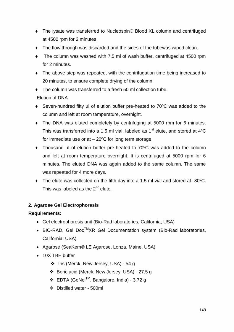
149
The lysate was transferred to Nucleospin® Blood XL column and centrifuged
at 4500 rpm for 2 minutes.
The flow through was discarded and the sides of the tubewas wiped clean.
The column was washed with 7.5 ml of wash buffer, centrifuged at 4500 rpm
for 2 minutes.
The above step was repeated, with the centrifugation time being increased to
20 minutes, to ensure complete drying of the column.
The column was transferred to a fresh 50 ml collection tube.
Elution of DNA
Seven-hundred fifty µl of elution buffer pre-heated to 70ºC was added to the
column and left at room temperature, overnight.
The DNA was eluted completely by centrifuging at 5000 rpm for 6 minutes.
This was transferred into a 1.5 ml vial, labeled as 1st elute, and stored at 4ºC
for immediate use or at – 20ºC for long term storage.
Thousand µl of elution buffer pre-heated to 70ºC was added to the column
and left at room temperature overnight. It is centrifuged at 5000 rpm for 6
minutes. The eluted DNA was again added to the same column. The same
was repeated for 4 more days.
The elute was collected on the fifth day into a 1.5 ml vial and stored at -80ºC.
This was labeled as the 2nd elute.
2. Agarose Gel Electrophoresis
Requirements:
Gel electrophoresis unit (Bio-Rad laboratories, California, USA)
BIO-RAD, Gel DocTMXR Gel Documentation system (Bio-Rad laboratories,
California, USA)
Agarose (SeaKem® LE Agarose, Lonza, Maine, USA)
10X TBE buffer
Tris (Merck, New Jersey, USA) - 54 g
Boric acid (Merck, New Jersey, USA) - 27.5 g
EDTA (GeNeiTM, Bangalore, India) - 3.72 g
Distilled water - 500ml

150
Ethidium bromide (Sigma-Aldrich, Missouri, USA)- 2mg/ml – dissolved in
distilled water
Tracking dye- Bromophenol blue (BPB)
Bromophenol blue (HiMedia, Mumbai, India) - 250 mg dissolved in
50ml 1X TBE buffer.
Sucrose (Merck, New Jersey, USA) - 40 g dissolved in 50ml Milli Q
water.
The above contents are mixed and stored in brown bottle.
Procedure
The gel trough was cleaned with ethanol and the ends were sealed with
cellophane tape. Combs were placed to form wells.
Agarose was weighed and dissolved in 100 ml of 1X TBE buffer (1% for DNA,
2% for PCR products, 3% for ASPCR products and gel extraction)
The agarose was melted in microwave oven and 20 μl of ethidium bromide
was added to the molten agarose.
This was mixed and poured on to the sealed trough and allowed to set.
After the gel solidified, the cellophane tapes and combs were removed and
the trough was placed in electrophoresis tank containing 1X TBE buffer.
4ul of sample and 4ul of tracking dye were mixed and loaded on to the wells.
1ul of molecular weight maker (100bp ladder) and 2ul of tracking dye were
mixed and loaded on to the well.
The electrophoresis was run at 120V for 20 to 30 minutes.
The gel was docked in BioRad Gel Documentation system for visualization
using Quantity one (v.4.6.2) software.
3. Polymerase Chain Reaction (PCR):
3.1. Primer Design:
For Direct sequencing:
The primers were designed using Primer 3 input (v.0.4.0) software. The primers
were designed in such a way that they flank the complete exon sequence
including 60-100bp intronic sequences. The properties like GC content, self-
annealing, dimer formation, hairpin loop formation was checked for both forward
and reverse primers using the Oligocalc software.

151
The table below lists the forward and reverse primer sequences for amplification
of specific exons of the candidate genes.
S.
No Gene Exon Primer (5' - 3')
Forward Primer Reverse Primer
1 AIPL1 2 GGGCCTTGAACAGTGTGTCT GCTGCTGTGTTTCGGGAAA
2 AIPL1 5 GCAGCTGCCTGAGGTCATG GTGGGGTGGAAAGAAAAG
3 AIPL1 6 CTGGGAAGGGAGCTGTAG AAAAGTGACACCACGATCC
4 CEP290 Intronic ACAGGCACCCACCATCAT TTCTTGTGGCAGTAAGGAGGA
5 CEP290 40 GCCAAAATGAAACGTGCATA TGAAAGTCAGTTTTAACAAATACCAT
6 CEP290 18 TGGAGGGATTTTGGAAACAA CCGGCTAAAACGGTGAAAC
7 CRB1 5 CCTCCTTTTAGGCAAATGCTC AAAGCCATGGTCTGCCATAA
8 CRB1 11 TTCCCATTTCACAACCAATGT GCTCGTCATTCATACGCAAA
9 CRB1 12A GCTTGCTCTGGTTGGTCTTC GCGGAACCACTGTGAAAGTT
10 CRX 2 GGATGGAATTCTTGGCATCCCAC CTCTTTGTTCCGGGCAGGCCTC
11 CRX 3b CCTCCACAGATGTGTGTCCAGAC TGGGAGAAAGGTAGGGGTCTAGG
12 GUCY2D 3 GGACGGCGCCGCGAGCCAAGC TCCCCTCTCCCTTGCCTTCTT
13 GUCY2D 9 CCCACATTGCCCTGGGCAGAA CCTGCCCCCAGGACGTCACCC
14 GUCY2D 10 AGCAGGCTGAGGCTGCCTCTT TCCCGGTGGATCCTCGTCTGC
15 GUCY2D 11 GCGGCGCCTCAGCCCCTTCCC CCCTGTGGGCCGAACTCCACG
16 GUCY2D 14 CAGCTGGAGCCCAGCCAGGTA CTGGTGAAGCTGAATTGAAGG
17 GUCY2D 15 GTGGAGACAATAGGGGACGCC CTAAAGAGGGAGATGGGCTGG
18 GUCY2D 16 CCCCGAGGCCCTACCTAGGTG ACCTCCCCGTCTTGTCCCCGT
19 GUCY2D 17 GGTGAGTCCCGAGCTCACGGC CGGGGCCTCCGGGAGGGGTTGG
20 GUCY2D 2a GGGTTACTCGGGCTTGGA GTTCACCGGACCCACGAG
21 GUCY2D 2b GTCCCCGCTTCGAGGTAGCGC CCGAGTGCATCACCATGATCAC
22 IQCB1 11 CACAACAGCAGCAGATGACA TCATCACGTAGCTAGAAAAGTTGG
23 IQCB1 13 TCCCCTCCTTATACACACTCAGA CAATGCATTACCTTATACCAGCA
24 IQCB1 14 CCTCTGCTAAGTGGTTGGGTA TTTCCTGAGGTTAGGGGATGA
25 KCNJ13 3a TGGCACCAAGAAATGTGTAAA CCTGCATTGCTGAAAGGAA
26 LCA5 3 CACTGCCATGTGTTATGTTTCA CCAACAAACCTTTTCTAAGTGC
27 LCA5 5 CATGCCAGCTGAATTCCATA CCCTTCAAAAAGGCTAGTCG
28 LCA5 6 CCCCCTTTTCTATAATTTGTGA TTCTTTCTCAAGGGATGCTGA
29 LCA5 7a AAATATGGTGGTTTTATGAAAGTT AGGTAACAATGGCAAAACAGG
30 NMNAT1 2 GGTGGCAGAGCAAGACCTTA ACAGGCACAGTGAATTTTCG
31 RD3 1 CTCTCTCCCCCTTGGGTTC CTCTAGTCCTGGTGGCCTCA
32 RDH12 2 ACCCTTCTTTGAGGCTGGAT TTGAATCCCAGGTTCCTTGA
33 RDH12 6 TTGCTGCAGGAGATAAGCTG GAAAAGCGGCACACGAGTAT

152
34 RPE65 2 CCCACCCAGCTGAGACTAGA TAAAAAGCCCAAACCACCTG
35 RPE65 4 CTGTACGGATTGCTCCTGTC TTAGAATCATACATTCGCAGCATG
36 RPE65 9 GTACACTTTTTTCCTTTTTAAATG
CATC
GTTTTAGATGTGATTCAGATTGAGTG
37 RPE65 10 TTGTCATTGCCTGTGCTCATG TGAGAGAGATGAAACATTCTGG
38 RPE65 13 GCATATTGACTGATTGCTTG GCAGTAAGAAGAGTATTCAG
39 RPE65 14 AGTCAGAAAAAGAAGTCAGGTC ATTGCTTGCTCAACTCAGTGC
40 RPE65 12 CACACGGGAGTGAACAAATG GCATATACTCAAAGCACTGTTCAAA
41 RPGRIP1 6 AGGGCATAGTCAAGGAGAA CTGAATTGTGGCTTCTCATA
42 RPGRIP1 14 GAAAGAGCTCCCTACCCTT GGAAATTCTGCATTGGTGC
43 RPGRIP1 21 CTTGGAGCCTCACTAACC TTCATCAGACTTCCTCACC
44 RPGRIP1 24 AGTGTTCAACTGAGTGATGC ACTATGGTTCCCTCAGAGAC
45 SLC19A2 2 CCAGGTCCTTTCATCACTAATGT TGTAATTGCTACTATGGGGGC
46 SPATA7 1 GTCGGCTCCTCTTTTCCAG CCCTGACAGCTGCCCTTTAC
47 SPATA7 3 ACAGCTGCAAGGTCTGGAAC ACCTCCAGAACTCTCTTTATACTGC
48 SPATA7 6 TTCTAGCCAGTAAACCTTGTTACC CCACCAACAGATTATTCTTC
49 SPATA7 7 AAAAAGTGCTGGATGGATAGAA CATTTCAACTTTTACTAAGCACTTCA
50 SPATA7 10 CAACCTTTGTAGTTTCAGTGTTACG GCACTTGCTTTTAATGTATTGTTTG
51 SPATA7 11c AGAACGAGATATTCCCTTCACCA TGAGTTACTGGCCATTTGAGG
52 SPATA7RT1 cDNA
exon
7-10
CCAGTGGGGATCTTTTGGAT GTCGCTCGAACAGTCGTTCT
53 SPATA7RT2 cDNA
exon
9-11
CCAAGGGCAATGTGTCAGTA CATCCTTTGGTGCCGACAAT
54 TULP1 11 TTCCCATCCTCTCACCTGTC ACTGTGGTGGGTGCTCTACC
55 SLC19A2 1 CAATGGAAGAGCAGGCAAGT CGCTTTTCTCGGTCCTCTCT
56 SLC19A2 2 CCAGGTCCTTTCATCACTAATGT GCCCCCATAGTAGCAATTACA
57 SLC19A2 3 TGGGCCTGTAAATTGCTTTC CAAATTTGGGAGGGGTGAAT
58 SLC19A2 4 GCAACAGCATTTGTGTAGCA TGGGAATAAACAAAACTTGCCTA
59 SLC19A2 5 GTTGGAAAGGCAATTGACAG TTCGATGCCAAAGGAGAGAT
60 SLC19A2 6 GGCACGTGGTGTAAGTATGC TGCTGTGCAGAGTTCTTGCT
For ASPCR:
Primer designing for ASPCR is similar to that as above where either the forward
or reverse primer is specific to the wild and mutant type alleles at 3‟ end while
the other primer in the pair is designed complementary to the wild type. Two
PCR reactions, one with wild type and other with mutant primer were set for

153
each mutation to be screened. ASPCR was performed to screen control samples
for certain identified mutations.
S.No PRIMER SEQUENCE 5'-3'
1 AIPL1-X5-N230S-FP-WT AAGCTGGAGAAGATGTTCAA
AIPL1-X5-N230S-FP-MT AAGCTGGAGAAGATGTTCAG
2 AIPL1-X2-E83K-RP-WT ACCAGAACTCGGCCTCCTC
AIPL1-X2-E83K-RP-MT ACCAGAACTCGGCCTCCTT
3 IQCB1-R332X-RP-WT TCTCCAGCAACATCTTTCATCG
IQCB1-R332X-RP-MT TCTCCAGCAACATCTTTCATCA
4 RPGRIP1-L1263P-FP-WT CTACCCCAATAGGATGGCT
RPGRIP1-L1263P-FP-MT CTACCCCAATAGGATGGCC
5 TULP1-N349K-FP-WT AACGGAGCAAGACAGGCAAT
TULP1-N349K-FP-MT AACGGAGCAAGACAGGCAAG
6 IQCB1-Q520X-FP-WT GATCAGCACCAACGTGGAAC
IQCB1-Q520X-FP-MT GATCAGCACCAACGTGGAAT
7 RPE65-N321K-FP-WT ACATCAACACCTATGAAGTCAAT
RPE65-N321K-FP-MT ACATCAACACCTATGAAGTCAAG
8 RPE65-L505R-FP-WT AGCCTGCTTATCTCCTGTTTCT
RPE65-L505R-FP-MT AGCCTGCTTATCTCCTGTTTCG
9 CRB1-R1390X-RP-WT ATCAAGTTCCACATTTCCTCTCG
CRB1-R1390X-RP-MT ATCAAGTTCCACATTTCCTCTCA
10 GUCY2D-G1013R-FP-WT GGATGCACTTAACAAGGCT
GUCY2D-G1013R-RP-WT GTCACACTCACGCAGCAC
GUCY2D-G1013R-RP-MT GTCACACTCACGCAGCAT
11 LCA5-FP-R280X-WT TTCTTCAAAAGGAGGTTCAGC
LCA5-FP-R280X-MT TTCTTCAAAAGGAGGTTCAGT
12 RDH12-R249L-RP-WT GACAAAGGGGGAGAATAGCC
RDH12-R249L-RP-MT GACAAAGGGGGAGAATAGCA
13 RPGRIP1-Q40X-FP-WT CCTCTATGACTTCACGTCCC
RPGRIP1-Q40X-FP-MT CCTCTATGACTTCACGTCCT
14 GUCY2D-L175R-FP-WT GGATGCCCTCTACCCCCT
GUCY2D-L175R-FP-MT GGATGCCCTCTACCCCCG

154
15 SLC19A2-Exon 2-FP CCAGGTCCTTTCATCACTAATGT
SLC19A2-Exon 2 –WT-RP CCATGTAACAATAAGGCTGAGAC
SLC19A2-Exon 2 –MT-RP CCATGTAACAATAAGGCTGAGAT
4. Purification of cycle sequenced extension products
Requirements:
Microfuge
Cycle sequenced product.
0.5M EthyleneDiamineTetraacetic Acid (EDTA) (GeNeiTM, Bangalore, India)
[pH 8.0] - 1 in 4 dilution of above stock to obtain 0.125M EDTA- Freshly
prepared
3M Sodium Acetate (GeNeiTM, Bangalore, India) [pH 4.6]
100% absolute alcohol (Hayman Limited, Witham, England)
70% absolute alcohol- 70ml of 100% absolute alcohol made up to 100ml with
distilled water. Freshly prepared.
Sterile Water
Procedure
Ten μl of sterile water, 2 μl of 0.125M EDTA, 2 μl of 3M sodium acetate (pH
4.6) and 50μl of chilled absolute ethanol were taken in a 0.5ml vial.
To this mixture 10 μl of the cycle sequenced product was added, vortexed and
incubated at room temperature for 15 minutes.
It was microfuged at 13000 rpm for 20 min.
The supernatant was discarded without disturbing the pellet.
Two-hundred μl of chilled 70% ethanol added, vortexed and microfuged for 10
minutes at 13000 rpm.
The supernatant was discarded and the precipitate was once again washed
with chilled 70% ethanol, centrifuged at 13000rpm for 10 minutes.
The pellet was air dried completely before loading to the sequencer.
5. Sequencing in genetic analyzer
Reagent:
Hi-DiTM Formamide (Applied Biosystems, Foster City, USA)

155
Procedure
The cycle sequenced, purified sample was re-suspended in 12μl of Hi-DiTM
formamide.
The sample was mixed well and microfuged. It was then loaded onto 96 well
optical plate.
The sample was then denatured in the thermal cycler at 95°C for 3 minutes.
After denaturation, the plate was loaded and linked to the sequencer ABI
3100 Avant or 3730 Genetic analyzer (Applied Biosystems, Foster City, USA)
for sequencing by capillary electrophoresis.
The data obtained was viewed by ABI PRISM DNA Sequencing Analysis
Software v. 5.1.1 and also analyzed by BioEdit sequence alignment editor
v.7.2.5.
6. Target sequences
AIPL1– Exon 2
GGGCCTTGAACAGTGTGTCTAGAGCAGAGTGCACCGTCTCGGTGACTAGG
TGATCTTTCATTTCCGCACCATGAAATGTGATGAGGAGCGGACAGTCATTG
ACGACAGTCGGCAGGTGGGCCAGCCCATGCACATCATCATCGGAAACATG
TTCAAGCTCGAGGTCTGGGAGATCCTGCTTACCTCCATGCGGGTGCACGA
GGTGGCCGAGTTCTGGTGCGACACCATCGTAAGTAGGCCCTGCGCGCCTG
TCTCCTGGGACTAGTCTTTTCTGGGCTCACCCACCCGCTTTGCGGGGCTGC
TGTGTTTCGGGAAA
AIPL1– Exon 5
GCAGCTGCCTGAGGTCATGGCTGACCTTCTCCCTGGGCAGGAGAAGCCAT
GGGAGGTGCAGTGGCTGAAGCTGGAGAAGATGATCAATACTCTGATCCTCA
ACTACTGCCAGTGCCTGCTGAAGAAGGAGGAGTACTATGAGGTGCTGGAG
CACACCAGTGATATTCTCCGGCACCACCCAGGTGCGCGGGGCTGCAGGGG
CGGACAGTGAGGGGGCGCCCAGCCCAGGGCCACGGAGACACCTGCCATA
GCCTTCCTGGACTTTTCTTTCCACCCCAC
AIPL1 – Exon 6
CTGGGAAGGGAGCTGTAGCTGGATGCTCCCTGCTCCCCACAGGCATCGTG
AAGGCCTACTACGTGCGTGCCCGGGCTCACGCAGAGGTGTGGAATGAGGC
CGAGGCCAAGGCGGACCTCCAGAAAGTGCTGGAGCTGGAGCCGTCCATG

156
CAGAAGGCGGTGCGCAGGGAGCTGAGGCTGCTGGAGAACCGCATGGCGG
AGAAGCAGGAGGAGGAGCGGCTGCGCTGCCGGAACATGCTGAGCCAGGG
TGCCACGCAGCCTCCCGCAGAGCCACCCACAGAGCCACCCGCACAGTCAT
CCACAGAGCCACCTGCAGAGCCACCCACAGCACCATCTGCAGAGCTGTCC
GCAGGGCCCCCTGCAGAGCCAGCCACAGAGCCACCCCCGTCCCCAGGGC
ACTCGCTGCAGCACTGAGCCCCCTGAGGCCCACAGCCACCCAGGCAGGG
AGCAAGTGGCCTGGTCACTTCTGGTTCGATTGACCAGGATCGTGGTGTCAC
TTTT
CEP290 - Intronic- c.2991_1655A/G
ACAGGCACCCACCATCATGCCCGGCTAATTTTTTGTATTTTTAGTAGAGATG
GGGTTTCACCTTGTTAGCCAGGATGGTGTCGATCTCCTGAACTCGTGATCC
ACCCGCCTCGGCCTCCTAAAGTGCTGGGATTACAGATGTGAGCCACCGCA
CCTGGCCCCAGTTGTAATTGTGAATATCTCATACCTATCCCTATTGGCAGTG
TCTTAGTTTTATTTTTTATTATCTTTATTGTGGCAGCCATTATTCCTGTCTCTA
TCTCCAGTCTTACATCCTCCTTACTGCCACAAGAA
CEP290 –Exon 40
GCCAAAATGAAACGTGCATATTTGATAAAATTTTGTTTGTGGGTGTTTGTTG
AAGATGAATTGCTTTACACTAGTTTTTTTTTTTTTTCTCAAAGTCGACTTTTTT
CCTCAAGGTAGACTTGACATGAATATGGAAAAATATATGTAGTTTGTGGTTA
TTTTTTTTCTCTTGTGTACTTAAAAATTCAGACTGAATTTTTCTTATAATGGTA
TATTTTCTGTTTTATGTTCCTTTTATCATTGATACTTCTTGAAGAGTCATGAAT
AATACCTTTCTTTTTCTCTTATTAGACACAAGTTGAAGATTTAAATGAAAATCT
TTTAAAATTGAAAGAAGCACTTAAAACAAGTAAAAACAGAGAAAACTCACTA
ACTGATAATTTGAATGACTTAAATAATGAACTGCAAAAGAAACAAAAAGCCT
ATAATAAAATACTTAGAGAGAAAGAGGAAATTGATCAAGAGAATGATGAACT
GAAAAGGCAAATTAAAAGACTAACCAGTGGATTACAGGTAATTTTATATTTAA
CTCTGATAATGTCTGATTTACAATATAGAGGTAGTAGTTTATTTCTACTTTAT
CATTTTATCTATGGTATTTGTTAAAACTGACTTTCA
CEP290 –Exon 18
TGGAGGGATTTTGGAAACAATTATTCTACCTTTGCATTAAAACTTGATTGTA
GGTTTTAAGAATTAAAGTGTTGGAATAGTAGGAGGGTTATTTTAATGTTTTTA
GTTTGTTAATTCTCTTATATATAGGATTAACCACTGAGGACCTGAACCTAACT

157
GAAAACATTTCTCAAGGAGATAGAATAAGTGAAAGAAAATTGGATTTATTGA
GCCTCAAAAATATGAGTGAAGCACAATCAAAGGTAATAGTAAAGTATTGCAA
AGAGAGTAAAGGAAAATATTTTTTTTTTTTTTTTTTTTTGAGACGGAGTCTCG
CTCTGTCTCCCAGGCTGGAGTGCAGTGGCGCGATCTCGGCTCACTGCAAG
CTCCGCCTCCCGGGTTCATGCCATTCTCCTGCCTCAGCCTCCCAAGTAGCT
GGGACTACAGGCGCCCGCCACCACGCCCGGCTAATTTTTTGTATTTTTAGT
AGAGACGGGGTTTCACCGTTTTAGCCGG
CRB1 – Exon 5
CCTCCTTTTAGGCAAATGCTCTATAATTCAACACCTTTGACTTAGCAGCTTC
TCTGAATTTTCATCATGCAGGATACACAGGTGCCCAGTGTGAGATCGACCT
CAATGAATGCAATAGTAACCCCTGCCAGTCCAATGGGGAATGTGTGGAGCT
GTCCTCAGAGAAACAATATGGACGCATCACTGGACTGCCTTCTTCTTTCAG
CTACCATGAAGCCTCAGGTTATGTCTGTATCTGTCAGCCTGGATTCACAGGT
GAGGCCAAGGAGATGGGATATGACTTGACTTTCTGGTATTTTATGGCAGAC
CATGGCTTT
CRB1 – Exon 11
TTCCCATTTCACAACCAATGTATTCAACAGGGACCTGGGTTTCTGCTGTTCT
GTTTATTTTGAAGGTGTGAAAAGGACATTGATGAGTGTGCCTCTGATCCGTG
TGTCAATGGAGGTCTGTGCCAGGACTTACTCAACAAATTCCAGTGCCTCTG
TGATGTTGCCTTTGCTGGCGAGCGCTGCGAGGTGGACGTAAGCAGCCTCT
CCTTTTATGTCTCTCTCTTATTCTGGCAGAATCTTTTTCAGCTTCTTTCTTAC
CTCATTTTGCGTATGAATGACGAGC
CRB1 – Exon 12a
GCTTGCTCTGGTTGGTCTTCATTCCTGAGTAGTTCCATTGTCCTGAATATTT
ATTTGCCTTTGCTATAGAATTCGCATCCCAATGATTTCAATCTTTCCAGTTGG
CAGATGACTTGATCTCCGACATTTTCACCACTATTGGCTCAGTGACTGTCGC
CTTGTTACTGATCCTCTTGCTGGCCATTGTTGCTTCTGTTGTCACCTCCAAC
AAAAGGGCAACTCAGGGAACCTACAGCCCCAGCCGTCAGGAGAAGGAGGG
CTCCCGAGTGGAAATGTGGAACTTGATGCCACCCCCTGCAATGGAGAGACT
GATTTAGGAGCATTGTGTCCCTTCGAGATGGGGATCCACACACTGTGAATG
TGATGACTGTACTTCAGGTATCTCTGACATACCTGACAATGTTAATCTGCAA
158
CTGGGATTACACTGGAACTACAGGAATGATTCCTTTGACCACCTTAAAAACT
TTCACAGTGGTTCCGC
CRX – Exon 2
GGATGGAATTCTTGGCATCCCACCCAGCCTCAGGGCCTCACACCAGCCCA
TGTGGATGACCTGAGGGTCCTGTTTCCCATCCCACCCCAGGCGCCCCCAG
GAAGCAGCGGCGGGAGCGCACCACCTTCACCCGGAGCCAACTGGAGGAG
CTGGAGGCACTGTTTGCCAAGACCCAGTACCCAGACGTCTATGCCCGTGA
GGAGGTGGCTCTGAAGATCAATCTGCCTGAGTCCAGGGTTCAGGTGGGGT
GGTGGGTCCCTGGACCCCTCCCGACACTTCCTGTGATCTCAGGAGGCCTG
CCCGGAACAAAGAG
CRX – Exon 3b
CCTCCACAGATGTGTGTCCAGACCCTCTGGGCATCTCAGATTCCTACAGTC
CCCCTCTGCCCGGCCCCTCAGGCTCCCCAACCACGGCAGTGGCCACTGTG
TCCATCTGGAGCCCAGCCTCAGAGTCCCCTTTGCCTGAGGCGCAGCGGGC
TGGGCTGGTGGCCTCAGGGCCGTCTCTGACCTCCGCCCCCTATGCCATGA
CCTACGCCCCGGCCTCCGCTTTCTGCTCTTCCCCCTCCGCCTATGGGTCTC
CGAGCTCCTATTTCAGCGGCCTAGACCCCTACCTTTCTCCCA
GUCY2D – Exon 2a
GGGTTACTCGGGCTTGGAGAAACTCGGGGTTACGGGGAGAACCCTAGGG
GAGGCCGGGGTCTCAGTCGCTCAGCCTGCTCCGTCTGTGTTCGCAGAAGC
CGGCAATGACCGCCTGCGCCCGCCGAGCGGGTGGGCTTCCGGACCCCGG
GCTCTGCGGTCCCGCGTGGTGGGCTCCGTCCCTGCCCCGCCTCCCCCGG
GCCCTGCCCCGGCTCCCGCTCCTGCTGCTCCTGCTTCTGCTGCAGCCCCC
CGCCCTCTCCGCCGTGTTCACGGTGGGGGTCCTGGGCCCCTGGGCTTGC
GACCCCATCTTCTCTCGGGCTCGCCCGGACCTGGCCGCCCGCCTGGCCGC
CGCCCGCCTGAACCGCGACCCCGGCCTGGCAGGCGGTCCCCGCTTCGAG
GTAGCGCTGCTGCCCGAGCCTTGCCGGACGCCGGGCTCGCTGGGGGCCG
TGTCCTCCGCGCTGGCCCGCGTGTCGGGCCTCGTGGGTCCGGTGAAC
GUCY2D – Exon 2b
GTCCCCGCTTCGAGGTAGCGCTGCTGCCCGAGCCTTGCCGGACGCCGGG
CTCGCTGGGGGCCGTGTCCTCCGCGCTGGCCCGCGTGTCGGGCCTCGTG
GGTCCGGTGAACCCTGCGGCCTGCCGGCCAGCCGAGCTGCTCGCCGAAG

159
AAGCCGGGATCGCGCTGGTGCCCTGGGGCTGCCCCTGGACGCAGGCGGA
GGGCACCACGGCCCCTGCCGTGACCCCCGCCGCGGATGCCCTCTACGCC
CTGCTTCGCGCATTCGGCTGGGCGCGCGTGGCCCTGGTCACCGCCCCCCA
GGACCTGTGGGTGGAGGCGGGACGCTCACTGTCCACGGCACTCAGGGCC
CGGGGCCTGCCTGTCGCCTCCGTGACTTCCATGGAGCCCTTGGACCTGTC
TGGAGCCCGGGAGGCCCTGAGGAAGGTTCGGGACGGGCCCAGGGTCACA
GGTAGGCTCCCTTGCAGGGTGCGAGGAGGTCGGCTGGTCCTGCCGGCAG
CCGGACGGCGCCGCGAGCCAAGCCTCTGTCCGCAGCAGTGATCATGGTG
ATGCACTCGG
GUCY2D – Exon 3
GGACGGCGCCGCGAGCCAAGCCTCTGTCCGCAGCAGTGATCATGGTGAT
GCACTCGGTGCTGCTGGGTGGCGAGGAGCAGCGCTACCTCCTGGAGGCC
GCAGAGGAGCTGGGCCTGACCGATGGCTCCCTGGTCTTCCTGCCCTTCGA
CACGATCCACTACGCCTTGTCCCCAGGCCCGGAGGCCTTGGCCGCACTCG
CCAACAGCTCCCAGCTTCGCAGGGCCCACGATGCCGTGCTCACCCTCACG
CGCCACTGTCCCTCTGAAGGCAGCGTGCTGGACAGCCTGCGCAGGGCTCA
AGAGCGCCGCGAGCTGCCCTCTGACCTCAATCTGCAGCAGGTAGACGGTC
CCGGGAGGAGGGAAGAAGGCAAGGGAGAGGGGA
GUCY2D – Exon 9
CCCACATTGCCCTGGGCAGAAAATGCAAGTCAACTCTCCCCCTCTCAGCTC
CAGGAGCTCCGGCATGAGAACGTGGCCCTCTACCTGGGGCTTTTCCTGGC
TCGGGGAGCAGAAGGCCCTGCGGCCCTCTGGGAGGGCAACCTGGCTGTG
GTCTCAGAGCACTGCACGCGGGGCTCTCTTCAGGACCTCCTCGCTCAGAG
AGAAATAAAGCTGGACTGGATGTTCAAGTCCTCCCTCCTGCTGGACCTTAT
CAAGGTGTGTGTCTGGGGGTGGTGGGGTGACGTCCTGGGGGCAGG
GUCY2D – Exon 10
AGCAGGCTGAGGCTGCCTCTTACCCTACCCATTCCAAGGGAATAAGGTATC
TGCACCATCGAGGCGTGGCTCATGGGCGGCTGAAGTCACGGAACTGCATA
GTGGATGGCAGATTCGTACTCAAGATCACTGACCACGGCCACGGGAGACT
GCTGGAAGCACAGAAGGTGCTACCGGAGCCTCCCAGAGCGGAGGGTAAG
AG TCCCCTGTGCAGACGAGGATCCACCGGGA
160
GUCY2D – Exon 11
GCGGCGCCTCAGCCCCTTCCCCATCCCCAGACCAGCTGTGGACAGCCCCG
GAGCTGCTTAGGGACCCAGCCCTGGAGCGCCGGGGAACGCTGGCCGGCG
ACGTCTTTAGCTTGGCCATCATCATGCAAGAAGTAGTGTGCCGCAGTGCCC
CTTATGCCATGCTGGAGCTCACTCCCGAGGGTAAGGCTGCCCTGTGCGTG
GAGTTCGGCCCACAGGG
GUCY2D – Exon 14
CAGCTGGAGCCCAGCCAGGTAGAGTGGCCCCCAGGTGACCTCACTGCCT
GCCATCCCTAGGTCTGTGGCTGAGGCCTTGAAGACGGGGACACCAGTGGA
GCCCGAGTACTTTGAGCAAGTGACACTGTACTTTAGTGACATTGTGGGCTT
CACCACCATCTCTGCCATGAGTGAGCCCATTGAGGTTGTGGACCTGCTCAA
CGATCTCTACACACTCTTTGATGCCATCATTGGTTCCCACGATGTCTACAAG
GTGCAGTGTGTAGGGGACAAGCCCTCCTGACCTTCAATTCAGCTTCACCA
G
GUCY2D – Exon 15
GTGGAGACAATAGGGGACGCCTATATGGTGGCCTCGGGGCTGCCCCAGC
GGAATGGGCAGCGACACGCGGCAGAGATCGCCAACATGTCACTGGACATC
CTCAGTGCCGTGGGCACTTTCCGCATGCGCCATATGCCTGAGGTTCCCGT
GCGCATCCGCATAGGCCTGCACTCGGGTAACTCCCGGGTCTTCCCAGGCT
CCAGCCCATCTCCCTCTTTAG
GUCY2D – Exon 16
CCCCGAGGCCCTACCTAGGTGCAGCCCAGGGCCGGCCCTGCTAGCCCCG
CCGACCCCCAGCATCTCCACAGGTCCATGCGTGGCAGGCGTGGTGGGCCT
CACCATGCCGCGGTACTGCCTGTTTGGGGACACGGTCAACACCGCCTCGC
GCATGGAGTCCACCGGGCTGCGTGAGTGTGACGGGGACAAGACGGGGAG
GT
GUCY2D – Exon 17
GGTGAGTCCCGAGCTCACGGCGTCCCCCACCGCCACAGCTTACCGCATCC
ACGTGAACTTGAGCACTGTGGGGATTCTCCGTGCTCTGGACTCGGGCTACC
AGGTGGAGCTGCGAGGCCGCACGGAGCTGAAGGTGAGGCAGGGCCCCAA
CCCCTCCCGGAGGCCCCG

161
IQCB1 – Exon 11
CACAACAGCAGCAGATGACAAGCTATTTTATCATACCTTATTAACACTAGAA
ATTATCTTAAAATTTTTTCTGTTTACATTTATTTGATTACTAGTGAGGTTATGC
ATTTTTTCATATGATTTATGAAAATTATGTATAACACACTGTTTCCATCACAGA
TCCAAACGATCAAAGATGTTGCTGGAGATAAATAGGCAGAAGGAAGAAGAG
GACCTCAAATTACAATTGCAACTTCAAAGACAGAGAGCCATGAGACTTTCCC
GAGAATTGCAGCTGAGTATGCTCGAAATAGTTCATCCAGGTGAGTGGCAGT
ATCAACTTTATCAATATAAGAATATTTAGGTTATCTTTTAACAAACCAACTTTT
CTAGCTACGTGATGA
IQCB1 – Exon 13
TCCCCTCCTTATACACACTCAGAGTTAATCATTATTGCAATGTGATCACTAC
CATATGTTGTAGTGAGAAAATAGTTCTATTAGTTAGGGGAGAAAAATCACAA
ACCTTCAATTTTTTTTCTTCCAGGCGCTTAAATTCCTAGCGAAGTGCCGTAA
GAAAAAGAAACTATTTGCTCCTTGGCGAGGACTCCAAGAACTCACTGATGC
ACGCCGAGTTGAACTGAAGAAACGAGTGGATGACTATGTCAGAAGACATTT
GGTATGAAGCTTCTGTACCATATCAGTATTACATTATTCCAATGCATGATATT
GATAACTGTACACACACACACATATATTGTGTGTGTGTGTGTGTGTGTGTGT
GTGTGTACATTTATTGCTGGTATAAGGTAATGCATTG
IQCB1 – Exon 14
CCTCTGCTAAGTGGTTGGGTACTTTGATACCTATGTCCTCTCTGTGACAGG
TTTTTCTGTCCTGTGTTTCCAGGGCTCTCCAATGTCAGATGTGGTCAGTAGG
GAGCTCCATGCCCAAGCTCAAGAACGACTGCAACACTACTTTATGGGCAGG
GCCCTAGAAGAGCGAGCCCAGCAGCACAGAGAAGCTCTGATAGCACAGAT
CAGCACCAACGTTGAACAGCTAATGAGTATGTGCTCATTTTACTTTCGTTCT
CTAAAAGGAACAACCTATGCGAGGTTTGTGACATCCAAAACCTTTAGAAGG
AAACCATTACTAAGCATCTTTAGCTCATCCCCTAACCTCAGGAAA
KCNJ13- Exon 3a
TGGCACCAAGAAATGTGTAAATATTTGATATACACTTGTCATTCAGAAAAAG
TATTGATATTTAAAGTCTTCATTCTAAAAAAAAATCAATAATTTCTTATTTAGG
TGCTTTTGTGGCGAAGATTGCCCGGCCAAAAAATCGAGCTTTTTCAATTCGC
TTTACTGACACAGCAGTAGTAGCTCACATGGATGGCAAACCTAATCTTATCT
TCCAAGTGGCCAACACCCGACCTAGCCCTCTAACCAGTGTCCGGGTCTCA

162
GCTGTACTCTATCAGGAAAGAGAAAATGGCAAACTCTACCAGACCAGTGTG
GATTTCCACCTTGATGGCATCAGTTCTGACGAATGTCCATTCTTCATCTTTC
CACTAACGTACTATCACTCCATTACACCATCAAGTCCTCTGGCTACTCTGCT
CCAGCATGAAAATCCTTCTCACTTTGAATTAGTTGTATTCCTTTCAGCAATG
CAGG
LCA5 – Exon 3
CACTGCCATGTGTTATGTTTCATTGTTGGAATTTTTTTAAAGGACTGCTTTTT
ATGTATTTATACATATAGGAGCTATCGAAAAACCTTGAACTGAGTACTAACA
GTTTCCAACGACAGTTGCTTGCTGAAAGGAAAAGGGCATATGAGGCTCATG
ATGAAAATAAAGTTCTTCAAAAGGAGGTACAGCGACTATATCACAAATTAAA
GGTAAGTATTTTAAATTGCATTGTTTTCCATAATGTGTATTAGACGAAAATTA
AATATTTTAAAACACTAAATGTTACTATTCATATTTGTAAGTTGGTACAATATA
ATTTACAAAATTCTGATATTATCTTTAGCACTTAGAAAAGGTTTGTTGG
LCA5 – Exon 5
CATGCCAGCTGAATTCCATATATTTTAGCATTATTCATTGTAATAATTTTTTA
AAAATAATTTTCACTGTAGCTGCATGCCAGAGTGATTTTGCAGACCTGTGTA
CAAAAGGAGTACAAACCATGGAAGACTTCAAGCCAGAAGAATATCCTTTAAC
TCCAGAAACAATTATGTGTTACGAAAACAAATGGGAAGAACCAGGACATCTT
ACTTTGGTGAGTTTAGCAGTATTGTTATGTACCAAACTCGGTCTGAAGTTAT
TCCTAAAAACACAATATCAGACATTTTAAAATTTCAGTAATGAAGCTAGTACT
ATATCTATATGAATATGCGACTAGCCTTTTTGAAGGG
LCA5 – Exon 6
CCCCCTTTTCTATAATTTGTGAACTTGCATTTTTTTTAACGTATAATAGGACT
TGCAATCTCAAAAGCAAGACAGGCATGGAGAAGCAGGGATTCTAAACCCAA
TTATGGAAAGAGAAGAAAAATTTGTTACAGATGAAGAACTCCATGTCGTAAA
ACAGGAGGTTGAAAAGCTGGAGGATGGTAAGAAAAAGAGTTTGTTTAAGCA
TGTGACAAGTCAGCATCCCTTGAGAAAGAA
LCA5 – Exon 7a
AAATATGGTGGTTTTATGAAAGTTGATAGTTTTCCTATTTAAAAATATTGACT
GTTACAAAATCCCATTTAAAAAATTTGATTTCATACAGAATGGGAAAGAGAA
GAACTTGATAAAAAGCAAAAAGAAAAGGCATCTTTACTGGAAAGAGAAGAAA

163
AGCCAGAGTGGGAAACTGGAAGGTACCAACTAGGAATGTATCCAATTCAGA
ATATGGATAAATTGCAAGGAGAGGAAGAAGAAAGACTGAAGAGAGAAATGC
TACTTGCTAAACTGAATGAAATTGACAGAGAACTCCAAGATTCTCGAAATCT
AAAATACCCTGTTTTGCCATTGTTACCT
NMNAT1 – Exon 2
GGTGGCAGAGCAAGACCTTATCTTAGGGAAAAAAAAATGCATAACTGAATT
TATTTTCTTTTTCCTTTGTAGACAACAAGGGAGGTGTCACAGTTTTCCATTTA
GATCAACAACTTCAAGTTCTTACCATGGAAAATTCCGAGAAGACTGAAGTGG
TTCTCCTTGTGGTTCATTCAATCCCATCACCAACATGCACCTCAGGTTGTTT
GAGCTGGCCAAGGACTACATGAATGGAACAGGTAGGAGCAGTAACCAAAA
GTGGCTTAAGACTAGAGAACCAGCCGGGTGCAGTGGCTCACACCTGCAAT
CCCAGCATTTTGGGAGGCTGAGACGGGCAGATCACAAGGTCAAGAGATCG
AGACCATCCTAGCCAACATGGTAAAACCCTGTCTCTACTAAAAAATACGAAA
ATTCACTGTGCCTGT
RD3 – Exon 1
CTCTCTCCCCCTTGGGTTCCCAGGTTCCCCACTCTGACTAACAGAGGGGCC
CAGGAAAGATGCACATCTGTCCTGTCTCAGCAGGGCCAGGGGCTATGTCT
CTCATCTCATGGCTTCGGTGGAACGAGGCCCCATCCCGGCTGTCCACCAG
GAGCCCTGCTGAGATGGTGCTGGAGACGCTTATGATGGAGCTGACGGGGC
AGATGCGAGAGGCTGAGAGGCAGCAGCGGGAGCGCAGCAATGCGGTCAG
AAAGGTCTGCACCGGTGTGGACTACAGCTGGCTGGCCAGCACACCCCGGT
CCACCTATGACCTCAGCCCCATTGAGCGGTTGCAGCTGGAAGATGTCTGC
GTTAAGATCCACCCATCCTATTGTGGGCCTGCTATCCTCAGGTGAGCACTG
GGATGGGGACCCTTGCTGGGCTGCAGGCTCCAGGAAAGGTGGCTGCAGT
GGCAGGGGCCTGAGGCCACCAGGACTAGAG
RDH12 – Exon 2
ACCCTTCTTTGAGGCTGGATAGAGTTTTTTTTTTTTTTTTTAACGTATCTTAG
TGTGAGCTCGTGAAGGATGGTACGTGATGCTCTTGTTTCCCTTGCCGATAG
GAAGTTCTTTGCTGGTGGAGTGTGTAGAACAAATGTGCAGCTTCCTGGCAA
GGTAGTGGTGATCACTGGCGCCAACACGGGCATTGGCAAGGAGACGGCCA
GAGAGCTCGCTAGCCGAGGTAAGTGTTTCCCCTTTAGTCTCCAAAGGGCCA
TGCCTCCCACCCTTCTTCCCACTGGGGCCTCTGTCCATATTGCTTTGTGTTT

164
CCTCCTAGGCTTGGGGGCTCTGACTAGAAATTCAAGGAACCTGGGATTCA
A
RDH12 – Exon 6
TTGCTGCAGGAGATAAGCTGTTTTCCTGGGCTCAGAGTGTGTCCCTGATCT
AATTGTGCCCTCTTTGTCCCAGGCACCGGGGTCACCACCTACGCAGTGCAC
CCAGGCGTCGTCCGCTCTGAGCTGGTCCGGCACTCCTCCCTGCTCTGCCT
GCTCTGGCGGCTCTTCTCCCCCTTTGTCAAGACGGCACGGGAGGGGGCGC
AGACCAGCCTGCACTGCGCCCTGGCTGAGGGCCTGGAGCCCCTGAGTGG
CAAGTACTTCAGGTGTGTGAAGGCAATGCGGTTCTCTCCACCACCTGTGTG
CATGGGAGGTGCCGGACTCGCTGGGCTGTTCATCCTGAGAAGCTGAGTTT
GTGCCTGATGATGCAATCCAGGTTTGGGTTGGGCCTGCAAACAGAATGCC
GTTGCTTTGTTAAGGAAACTTACAGTACAAACTTATGTGTTGGGAAGAGTTG
CTTTTCTGGCTTTATTTTATACTCGTGTGCCGCTTTTC
RPE65 – Exon 2
CCCACCCAGCTGAGACTAGACCGGCAGGAGTGAACAGGCTTTGAGCCAG
CCCTAGAGTGCCTTCTCTCCTGCAGCTCTGCCTCTATCTCTGCGGACTTTG
AGCATCAACATGGGCTTCTTCCTTATTCTTCCACCATTTCAGGGTTGAGCAT
CCTGCTGGTGGTTACAAGAAACTGTTTGAAACTGTGGAGGAACTGTCCTCG
CCGCTCACAGCTCATGTAACAGGTTGGTCTCGCCCATCTTGAAGCCATCCT
CTTTTATGTCAGTCTCTCTTCTCTGGCTTCCTATTCCTTGGCGTCTCTCAGT
GCAGACTCCCCCTGTGGGTCACAGGGAGAGGGATCAGGTGGTTTGGGCTT
TTTA
RPE65 – Exon 4
CTGTACGGATTGCTCCTGTCTATACTCTTCCCTATGTTTCAATGTCCTTCAG
GTTCATCCGCACTGATGCTTACGTACGGGCAATGACTGAGAAAAGGATCGT
CATAACAGAATTTGGCACCTGTGCTTTCCCAGATCCCTGCAAGAATATATTT
TCCAGGTTACTGAACCCAAACTGAATGTTACTCAAGACATTTTATATTAGCC
CTTTTTCTCTCATGGCTTGAAAATTACTGGACTGAAAAATTCATTTGTTTCTA
CAGGTTTTTTTTCTTACTTTCGAGGAGTAGAGGTTACTGACAATGCCCTTGT
TAATGTCTACCCAGTGGGGGAAGATTACTACGCTTGCACAGAGACCAACTT
TATTACAAAGATTAATCCAGAGACCTTGGAGACAATTAAGCAGGTGGGACA

165
CAGTGCTAGGTGATGTTCAGGAATTTAGAATTTGGAACTTAAAATTAATTCA
ACATAAATTATTCATGCTGCGAATGTATGATTCTAA
RPE65 – Exon 9
GTACACTTTTTTCCTTTTTAAATGCATCAAAATATTTTCCTCATTTTTCAAGG
TTTGGCTTCATATTGCTGACAAAAAAAGGAAAAAGTACCTCAATAATAAATAC
AGAACTTCTCCTTTCAACCTCTTCCATCACATCAACACCTATGAAGACAATG
GGTTTCTGATTGTGGATCTCTGCTGCTGGAAAGGGTAAGAAAGGACACTGG
ACAAATGGGACACCTCCCATTGTTCCTGGAAATTACGGGGTTTTTACAGAG
CTGCTGCACTCAATCTGAATCACATCTAAAAC
RPE65 – Exon 10
TTGTCATTGCCTGTGCTCATGTTTGACTTTTTATTTTTGCAGATTTGAGTTTG
TTTATAATTACTTATATTTAGCCAATTTACGTGAGAACTGGGAAGAGGTGAA
AAAAAATGCCAGAAAGGCTCCCCAACCTGAAGTTAGGAGATATGTACTTCC
TTTGAATATTGACAAGGTAACCTGCTTCTCTGTAGATTTCAGATTTAACCAG
AATGTTTCATCTCTCTCA
RPE65- EXON 12
CACACGGGAGTGAACAAATGTTTCTTTCAAAGAGATTAAGAGTTTTCCTAA
GCATGTGCTCTATTTCGTAGCATTTGAGTTTCCTCAAATCAATTACCAGAAG
TATTGTGGGAAACCTTACACATATGCGTATGGACTTGGCTTGAATCACTTTG
TTCCAGATAGGGTAATTAATCCTTCTTACTAATATTTGAACAGTGCTTTGAGT
ATATGC
RPE65 – Exon 13
GCATATTGACTGATTGCTTGATTGATTTTTCTTTCTCACAAACAGCTCTGTAA
GCTGAATGTCAAAACTAAAGAAACTTGGGTTTGGCAAGAGCCTGATTCATAC
CCATCAGAACCCATCTTTGTTTCTCACCCAGATGCCTTGGAAGAAGATGATG
GTAATGAAAGCAATTGTTGTGTCTGAATACTCTTCTTACTGC
RPE65 – Exon 14
AGTCAGAAAAAGAAGTCAGGTCATATGGTTTTCTATATTTGTCAATGTAATA
CCTCCTATATTATTTCAATGACATTCAATCTATAGCTTGGGCTTTTAAAAACT
CAATATTGCCTAATTTACTTCTGATAAACAGGTGTAGTTCTGAGTGTGGTGG
TGAGCCCAGGAGCAGGACAAAAGCCTGCTTATCTCCTGATTCTGAATGCCA
AGGACTTAAGTGAAGTTGCCCGGGCTGAAGTGGAGATTAACATCCCTGTCA

166
CCTTTCATGGACTGTTCAAAAAATCTTGAGCATACTCCAGCAAGATATGTTT
TTGGTAGCAAAACTGAGAAAATCAGCTTCAGGTCTGCAATCAAATTCTGTTC
AATTTTAGCCTGCTATATGTCATGGTTTTAACTTGCAGATGCGCACAATTTTG
CAATGTTTTACAGAAAGCACTGAGTTGAGCAAGCAAT
RPGRIP1 – Exon 6
AGGGCATAGTCAAGGAGAAAATGTCTTTAATTCTATCCATGTTCAGACAGA
ATAATTTAGCGCCTTTCTCTGCAGAGCTTCCATTAAAGAGAAGGTAGAGCTG
ATTCGACTTAAGAAGCTCTTACATGAAAGAAATGCTTCATTGGTTATGACAA
AAGCACAATTAACAGAAGTTCAAGAGGTGAGTTGCCATCATCAGCTGTGCT
TTCTTGGTGGGGGGAAACCCCAATTAAGAGATTCATATTATTTTCTCCATTTT
AAATATGAGAAGCCACAATTCAG
RPGRIP1 – Exon 14
GAAAGAGCTCCCTACCCTTTAACGGATAGGCAGCTTTCTTTCCCCTCTAGA
ACAGCTCAAAGATGTTGCTTATGGCACCCGACCGTTGTCGTTATGTTTGGAA
ACACTGCCAGCCCATGGAGATGAGGATAAAGTGGATATTTCTCTGCTGCAT
CAGGGTGAGAATCTTTTTGAACTGCACATCCACCAGGCCTTCCTGACATCT
GCCGCCCTAGCTCAGGCTGGAGATACCCAACCTACCACTTTCTGCACCTAT
TCCTTCTATGACTTTGAAACCCACTGTACCCCATTATCTGTGGGGCCACAGC
CCCTCTATGACTTCACCTCCCAGTATGTGATGGAGACAGATTCGCTTTTCTT
ACACTACCTTCAAGAGGCTTCAGCCCGGCTTGACATACACCAGGCCATGGC
CAGTGAACACAGCACTCTTGCTGCAGGATGGATTTGCTTTGACAGGGTGCT
AGAGACTGTGGAGAAAGTCCATGGCTTGGCCACACTGATTGGTAAGTGCC
GTTGGCTTCCTGCGGCTCCTAAGCACCAATGCAGAATTTCC
RPGRIP1 – Exon 21
CTTGGAGCCTCACTAACCTTTAGGAACTAAATAAACATTTTCCTTATCAGGA
TTCAGAGAAGATGTGCATTGAAATTGTCTCCCTGGCCTTCTACCCAGAGGC
AGAAGTGATGTCTGATGAGAACATAAAACAGGTGTATGTGGAGTACAAATTC
TACGACCTACCCTTGTCGGAGACAGAGACTCCAGTGTCCCTAAGGAAGCCT
AGGGCAGGAGAAGAAATCCACTTTCACTTTAGCAAGGGTGAGGCATCCTGT
GTGGTTACTGGGGTGAGGAAGTCTGATGAA
167
RPGRIP1 – Exon 24
AGTGTTCAACTGAGTGATGCTGTTTTTTTCCCTTTCCCAACAGTTGTTAGCC
CTGAAGATCTGGCTACCCCAATAGGAAGGCTGAAGGTTTCCCTTCAAGCAG
CTGCTGTCCTCCATGCTATTTACAAGGAGATGACTGAAGATTTGTTTTCATG
AAGGAACAAGTGCTATTCCAATCTAAAAGTCTCTGAGGGAACCATAGT
SLC19A2 – Exon 2
CCAGGTCCTTTCATCACTAATGTTTTTTGTTTTTTTTTTTTCTCTTTCTACCAG
GTCTTCAATGAAATTTATCCAGTATGGACTTACTCTTACCTGGTGCTACTGTT
TCCTGTGTTCCTTGCCACAGACTACCTCCGTTATAAACCTGTTGTTCTACTG
CAGGGGCTCAGCCTTATTGTTACATGGTTTATGCTGCTCTATGCCCAGGGA
CTGCTGGCCATTCAATTTCTAGAATTTTTTTATGGCATCGCCACAGCCACTG
AAATTGCCTATTACTCTTATATCTACAGTGTGGTGGACCTGGGCATGTACCA
GAAAGTCACAAGTTACTGTCGAAGTGCCACTTTGGTGGGCTTTACAGTGGG
CTCTGTCCTAGGGCAAATCCTTGTCTCAGTGGCAGGCTGGTCGCTGTTCAG
CCTGAATGTCATCTCTCTTACCTGTGTTTCAGTGGCTTTTGCTGTGGCCTGG
TTTTTACCTATGCCACAGAAGAGCCTCTTCTTTCACCACATTCCTTCTACCTG
CCAGAGAGTGAATGGCATCAAGGTACAAAATGGTGGCATTGTTACTGACAC
CCCAGCTTCTAACCACCTTCCTGGCTGGGAGGACATTGAGTCAAAAATCCC
TCTAAATATGGAGGAGCCTCCCGTGGAGGAACCGGTAAGCTCAGCCTTAAA
TATCTTGTAATTGCTACTATGGGGGC
SPATA7– Exon 1
GTCGGCTCCTCTTTTCCAGTCCTCCACTGCCGGGGCTGGGCCCGGCCGCG
GGAAGGACCGAAGGGGATACAGCGTGTCCCTGCGGCGGCTGCAAGAGGA
CTAAGCATGGATGGCAGCCGGAGAGGTAAAGGGCAGCTGTCAGGG
SPATA7 – Exon 3
ACAGCTGCAAGGTCTGGAACATTTTGTGATTTCCACATTTGGTATTTGTCAT
TTATAAATACTGACAATATTATTAAACTATTACCTTTCTCTTTCAGCTGCAGTA
GACTGCTCGGTTCCAGTAAGCGTGAGTACCAGCATAAAGTGTAAGTAATTTT
TGGACATTATTACCTTTTTAAAAAAAAATTAAGGTAAATATACATAACATAAAA
TTTACTGTCTTAACCATTTTTAAGTGTCCAGTTCAATATTAAGTACATTAATAT
TGTGGTACAATGATAACTACTACCTCCAGAACTCTCTTTATACTGC
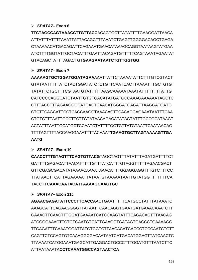
168
SPATA7– Exon 6
TTCTAGCCAGTAAACCTTGTTACCACAGTGCTTATATTTTGAAGGATTAACA
ATTATTTATTTTAAATTATTACAGCTTTAAATCTGAGTTGGGGACAGCTGAGA
CTAAAAACATGACAGATTCAGAAATGAACATAAAGCAGGTAATAAGTATGAA
ATCTTTTGGTATTGCTACATTTGAATTACAGATGTTTTTCAGTAAATAGAATAT
GTACAGCTATTTAGACTGTGAAGAATAATCTGTTGGTGG
SPATA7– Exon 7
AAAAAGTGCTGGATGGATAGAAAAATTATTCTAAAATATTCTTTGTCGTACT
GTATAATTTTTATCTACTGGATATCTCTGTTCAATCACTTAAAATTTGCTGTGT
TATATTCTGCTTTCGTAATGTATTTTTAAGCAAAAATAAATATTTTTTTTATTG
CATCCCCAGGCATCTAATTGTGTGACATATGATGCCAAAGAAAAAATAGCTC
CTTTACCTTTAGAAGGGCATGACTCAACATGGGATGAGATTAAGGATGATG
CTCTTCAGCATTCCTCACCAAGGTAAACAGTTCACAGGAGAAATAATTTCAA
CTGTCTTTAATTGCCTTCTTGTATAACAGACATATAGTATTTGCCGCATAAGT
ACTATTTAATTGCATGCTCCAATCTATTTTGGTGTTATGTAATTCAATAACAG
TTTTAGTTTTACCAAGGAAATTTTACAAATTGAAGTGCTTAGTAAAAGTTGA
AATG
SPATA7– Exon 10
CAACCTTTGTAGTTTCAGTGTTACGTAGCTAGTTTATATTTAGATGATTTTCT
GATTTTGAGACATTAACATTTTTGTTTATCATTTGTAGGTTTTTAGAACGACT
GTTCGAGCGACATATAAAACAAAATAAACATTTGGAGGAGGTTTGTCTTTCC
TTATAACTTCATTAGAAAAATTATAATGTAAAAATAATTGTATGGTTTTTTTCA
TACCTTCAAACAATACATTAAAAGCAAGTGC
SPATA7– Exon 11c
AGAACGAGATATTCCCTTCACCAACTGAATTTTTCATGCCTATTTATAAATC
AAAGCATTCAGAAGGGGTTATAATTCAACAGGTGAATGATGAAACAAATCTT
GAAACTTCAACTTTGGATGAAAATCATCCAAGTATTTCAGACAGTTTAACAG
ATCGGGAAACTTCTGTGAATGTCATTGAAGGTGATAGTGACCCTGAAAAGG
TTGAGATTTCAAATGGATTATGTGGTCTTAACACATCACCCTCCCAATCTGTT
CAGTTCTCCAGTGTCAAAGGCGACAATAATCATGACATGGAGTTATCAACTC
TTAAAATCATGGAAATGAGCATTGAGGACTGCCCTTTGGATGTTTAATCTTC
ATTAATAAATACCTCAAATGGCCAGTAACTCA

169
TULP1– Exon 11
TTCCCATCCTCTCACCTGTCTCCCCTTTTCCCCAGGTGTTCCTCTTGGCTGG
CAGGAAACGAAAACGGAGCAAGACAGCCAATTACCTCATCTCCATCGACCC
TACCAATCTGTCCCGAGGAGGGGAGAATTTCATCGGGAAGCTGAGGTGGG
GCTGGGCTTCCTGGGGCTGGGGGGACTTGGGTCAGCAAAAGGCCATAGC
CCGTCATCTCTGTCCTGCTGTGCCTGGTAGAGCACCCACCACAGT
SPATA7RT1 – Exon 7-10
CCAGTGGGGATCTTTTGGATAAACATTCTGAACTCTTTTCTAACAAACAATT
GCCATTCACTCCTCGCACTTTAAAAACAGAAGCAAAATCTTTCCTGTCACAG
TATCGCTATTATACACCTGCCAAAAGAAAAAAGGATTTTACAGATCAACGGA
TAGAAGCTGAAACCCAGACTGAATTAAGCTTTAAATCTGAGTTGGGGACAG
CTGAGACTAAAAACATGACAGATTCAGAAATGAACATAAAGCAGGCATCTAA
TTGTGTGACATATGATGCCAAAGAAAAAATAGCTCCTTTACCTTTAGAAGGG
CATGACTCAACATGGGATGAGATTAAGGATGATGCTCTTCAGCATTCCTCAC
CAAGGGCAATGTGTCAGTATTCCCTGAAGCCCCCTTCAACTCGTAAAATCTA
CTCTGATGAAGAAGAACTGTTGTATCTGAGTTTCATTGAAGATGTAACAGAT
GAAATTTTGAAACTTGGTTTATTTTCAAACAGGTTTTTAGAACGACTGTTCG
AGCGAC
SPATA7RT2 – Exon 9-11
CCAAGGGCAATGTGTCAGTATTCCCTGAAGCCCCCTTCAACTCGTAAAATC
TACTCTGATGAAGAAGAACTGTTGTATCTGAGTTTCATTGAAGATGTAACAG
ATGAAATTTTGAAACTTGGTTTATTTTCAAACAGGTTTTTAGAACGACTGTTC
GAGCGACATATAAAACAAAATAAACATTTGGAGGAGGAAAAAATGCGCCAC
CTGCTGCATGTCCTGAAAGTAGACTTAGGCTGCACATCGGAGGAAAACTCG
GTAAAGCAAAATGATGTTGATATGTTGAATGTATTTGATTTTGAAAAGGCTG
GGAATTCAGAACCAAATGAATTAAAAAATGAAAGTGAAGTAACAATTCAGCA
GGAACGTCAACAATACCAAAAGGCTTTGGATATGTTATTGTCGGCACCAAA
GGATG
SLC19A2–EXON1
CAATGGAAGAGCAGGCAAGTATTCCCGGCGTCCGCTGTGATTGGTTCCCG
GAGTGGAGGCGGTGGCAGAGGGTGGGCCTTAGGACGGGTCTCCCTTAAA
CTGGGCGATCAGGCAGCGACCCTAGAGGCGTCTGTAGGGTAAAGCTGGG

170
GGTTCTGTAGCCGGAGGCGGCGGCGAGTCCAGAACGTCCTGGCCTTACAG
GGAGAAGGCGTCACTCGCGGTTACAAGTGCCTGACCCTCACTCCAGTTGG
CGGAGGAGGAGAAGGAAGGGGCCGGGCCGGGTCCCCTCCCCTCGCGCC
CCGGATGGATGTGCCCGGCCCGGTGTCTCGGCGGGCGGCGGCGGCGGC
GGCCACTGTGCTCCTGCGGACCGCTCGGGTCCGTCGCGAATGCTGGTTCT
TGCCGACCGCGCTGCTCTGCGCCTACGGCtTTCTTCGCCAGCCTCAGGCCG
TCCGAGCCCTTCCTGACCCCGTACCTGCTGGGGCCGGACAAGAACCTGAC
CGAGAGGGAGGTACGCGCGGCGGACGCGGGGCGCGGGAAGGGCGGGCG
ACCGGCCTGCGGGTGGGCAGCGGAGGAAGAGAGGACCGAGAAAAGCG
SLC19A2- EXON 2
CCAGGTCCTTTCATCACTAATGTTTTTTGTTTTTTTTTTTTCTCTTTCTACCAG
GTCTTCAATGAAATTTATCCAGTATGGACTTACTCTTACCTGGTGCTACTGTT
TCCTGTGTTCCTTGCCACAGACTACCTCCGTTATAAACCTGTTGTTCTACTG
CAGGGGCTCAGCCTTATTGTTACATGGTTTATGCTGCTCTATGCCCAGGGA
CTGCTGGCCATTCAATTTCTAGAATTTTTTTATGGCATCGCCACAGCCACTG
AAATTGCCTATTACTCTTATATCTACAGTGTGGTGGACCTGGGCATGTACCA
GAAAGTCACAAGTTACTGTCGAAGTGCCACTTTGGTGGGCTTTACAGTGGG
CTCTGTCCTAGGGCAAATCCTTGTCTCAGTGGCAGGCTGGTCGCTGTTCAG
CCTGAATGTCATCTCTCTTACCTGTGTTTCAGTGGCTTTTGCTGTGGCCTGG
TTTTTACCTATGCCACAGAAGAGCCTCTTCTTTCACCACATTCCTTCTACCTG
CCAGAGAGTGAATGGCATCAAGGTACAAAATGGTGGCATTGTTACTGACAC
CCCAGCTTCTAACCACCTTCCTGGCTGGGAGGACATTGAGTCAAAAATCCC
TCTAAATATGGAGGAGCCTCCCGTGGAGGAACCGGTAAGCTCAGCCTTAAA
TATCTTGTAATTGCTACTATGGGGGC
SLC19A2 - EXON 3
TGGGCCTGTAAATTGCTTTCTATATAATTGCCTTCATCAGAGTTTGAGCATT
GTTCCCTTTTGCTATGTAGGAACCCAAGCCAGACCGTCTCCTGTATTGAAAG
TACTATGGAATGATTTCCTGATGTGCTACTCCTCTCGCCCTCTTCTCTGCTG
GTCTGTGTGGTGGGCCCTCTCTACCTGTGGCTATTTTCAAGTTGTGAACTAC
ACACAGGGCCTGTGGGAGAAAGTGATGCCTTCTCGCTATGCTGCTATCTAT
AATGGTGGCGTGGAGGCCGTTTCAACCTTACTGGGTAAGCAATGCAGGATA
ATAGAGGGTCGTGGTGGGGGAGGGATTCCTCTGGTAGTTAAGCCAGCCAT
AATCA

171
SLC19A2 - EXON 4
GCAACAGCATTTGTGTAGCAAAATAATTGGATTTTTTTTGTTTGTTCTGTAG
GTGCTGTTGCTGTGTTTGCAGTTGGTTATATAAAAATATCCTGGTCAACTTG
GGGAGAAATGACATTATCTCTCTTTTCTCTCCTGATTGCTGCTGCAGTGTAT
ATCATGGACACTGTGGGTAACATTTGGGTGTGCTATGCATCCTATGTTGTCT
TCAGAATCATCTACATGTTACTCATCACGATAGCAACGTATGTATTTTGGCT
CATAGAAGCCTTAGGAAAAATTGTAACAGGATAAATTTTTTGGTTTCTTTACA
AACCAAAATATTCAAGTTTCTAGTTAAATGAGGCAAATGGGAGGAAGCATTG
TATTATATGTGCATTATCAGTTTTTCTCCTGAGGAAGCATTTAGGCAAGTTTT
GTTTATTCCCA
SLC19A2 - EXON 5
GTTGGAAAGGCAATTGACAGTAGAAAGAAGGACTCTTCATGTTTAAAGATA
AATGTTTACTTTTATTTAGAATAAATATTATAAGTACTCCAGAAGTAATTCTTA
TTTATTCACTCTTTTATTTTTTTTTAAGTTTTCAAATTGCTGCAAACCTCAGCA
TGGAACGCTATGCCCTAGTATTTGGTGTAAATACCTTCATTGCCCTGGCACT
GCAGACGCTGCTCACTCTAATTGTGGTAGATGCCAGTGGCCTTGGATTAGA
AATTACCACTCAGGTAAGATCTCTCCTTTGGCATCGAA
SLC19A2 - EXON 6
GGCACGTGGTGTAAGTATGCAATATTTTTTGAGGCTTTATGGACAAATTCTGA
ATATACTTTATTATAGAGTAATCCTCTTGTCATTGATAATTCAATAAACTGCTTT
TCCCCCTTCTAGTTTTTGATCTATGCCAGTTATTTTGCACTCATCGCTGTGGT
TTTCCTGGCCAGTGGTGCAGTCAGTGTTATGAAGAAATGTAGAAAGCTGGAA
GATCCACAATCAAGTTCTCAAGTAACCACTTCATAATATACTGCTGAAGGGCT
TCTTCTTATAGCAAGAACTCTGCACAGCA
The sequence in bold indicate the forward and reverse primer sequences.

172
LIST OF PUBLICATIONS AND PRESENTATIONS
PUBLICATIONS
Natarajan N. Srikrupa, Sundaramurthy Srilekha, Parveen Sen, Tharigopala
Arokiasamy, Swaminathan Meenakshi, Muna Bhende, Suman Kapur and
Nagasamy Soumittra. Genetic Profile and Mutation Spectrum of Leber
Congenital Amaurosis in a Larger Indian Cohort using High Throughput
Targeted Re-sequencing. Clin Genet. 2017 Oct 25. doi: 10.1111/cge.13159.
[Epub ahead of print].
Srikrupa NN, Meenakshi S, Arokiasamy T, Murali K, Soumittra N. Leber's
congenital amaurosis as the retinal degenerative phenotype in thiamine
responsive megaloblastic anaemia: a case report. Ophthalmic Genet. 2014
Jun; 35(2):119-24. doi: 10.3109/13816810.2013.793363. PMID: 23638917.
Srilekha S, Arokiasamy T, Srikrupa NN, Umashankar V, Meenakshi S, Sen
P, Kapur S, Soumittra N. Homozygosity Mapping in Leber Congenital
Amaurosis and Autosomal Recessive Retinitis Pigmentosa in South Indian
Families. PLoS One. 2015; 10(7):e0131679. doi:
10.1371/journal.pone.0131679. PMID: 26147992.
Bende P, Natarajan K, Marudhamuthu T, Madhavan J. Severity of familial
isolated retinitis pigmentosa across different inheritance patterns among an
Asian Indian cohort. J Pediatr Ophthalmol Strabismus, 2013; 50(1):34-6.
PMID: 23463886.
Dhandayuthapani Sudha, Srividya Neriyanuri, Srikrupa Natarajan, Mamatha
Gandra, Arokiasamy Tharigopala, SathyaBharathi Ravichandran, Al Ameen
Muhammed, Umashankar Vetrivel, Lingam Gopal, VikasKetan, Rajiv Raman,
Subbulakshmi Chidambaram and Jayamurugapandian Arunachalam. Variable
disease severity among XLRS patients with similar mutant RS1 secretion
profile: An insight on the localization of mutant RS1. Experimental Eye
Research. (Under review)

173
PRESENTATIONS
Natarajan N. Srikrupa, Srilekha Sundar, Swaminathan Meenakshi, Parveen
Sen, Muna Bende, Tharigopala Arokiasamy, Suman Kapur, Nagasamy
Soumittra. High throughput gene screening of patients with Leber Congenital
Amaurosis in an Indian Cohort. NextGen Genomics, Biology, Bioinformatics and
Technologies (NGBT) Conference 2016, Cochin. Awarded Travel fellowship.
Natarajan N. Srikrupa, Srilekha Sundar, Swaminathan Meenakshi, Parveen
Sen, Muna Bende, Tharigopala Arokiasamy, Suman Kapur, Nagasamy
Soumittra. Swarnalatha Punshi Award presentation 2015. Vision research
foundation, Chennai. Awarded Swarnalatha Punshi Award for Best
Research in Basic Science.
Natarajan Srikrupa N, Swaminathan Meenakshi, Bhende Muna, Tharigopala
Arokiasamy, Nagasamy Soumittra.Screening of LCA9 locus gene, NMNAT1, in
LCA cases of Indian cohort. ASIA ARVO 2013, New Delhi. Awarded Second
Best poster.
Srikrupa Natarajan, Meenakshi S, Kaushik Murali, Nagasamy Soumittra. A
novel mutation in SLC19A2 gene in a south Indian patient with thiamine-
responsive megaloblastic anaemia (TRMA). Indian Society of Human Genetics
2012, Chandigarh.
Srikrupa Natarajan, Manoharan Aarthi, Ronnie George, Manmath Kumar Das,
VL Ramprasad, G Kumaramanickavel, Lingam Vijaya, Sripriya S, Parveen Sen.
Association between the indel variant in the LOC387715/ ARMS2 gene and
Age-related Macular Degeneration in South Indian population. Indian Eye
Research Group 2010, Hyderabad. Awarded Travel fellowship.
D Sudha, Srikrupa Natarajan, T Karthiyayini, S Muthukumaran, V
Umashankar, L. Gopal, VikasKhetan, A Jayamuruga Pandian. Molecular
characterization of RS1 gene mutants implicated in retinoschisis. Indian Society
of Human Genetics 2012, Varanasi.

174
Sudha. D, Srividya. N, Srikrupa. N. N, Dharanija. M, Mamatha. G, Karthiyayini.
T, Arokiasamy. T, SathyaBharathi. R, Muthukumaran. S, Al Ameen. M,
Umashankar. V, Lingam. G, Vikas. K, Rajiv. R, Subbulakshmi. J,
JayamurugaPandian. A. Nonsecreted mutant retinoschisin localizes to the
plasma membrane. Indian Society of Human Genetics 2016, Chennai.

175
BRIEF BIOGRAPHY OF THE CANDIDATE
Ms. Srikrupa N N completed her bachelor‟s degree in Biochemistry at Dr. MGR
Janaki College of arts and science for women affiliated to University of Madras,
Chennai in the year 2006. She joined Medical Research Foundation, Sankara
Nethralaya for her post-graduation degree in MS(MLT) (2006-2009) affiliated to
BITS Pilani, Following her post-graduation, she joined the SNONGC department
of Genetics and Molecular biology as a research fellow in a DBT funded project on
age related macular degeneration (AMD) for a year (2009-2010). She was
awarded a travel fellowship to present this work in IERG (2010) conference. She
was then deputed in a 3 year DBT funded project on “Molecular diagnostics of
LCA - a chip based resequencing approach”. During this fellowship tenure she
registered for PhD in BITS Pilani, under the guidance of Dr. N. Soumittra from
Vision Research Foundation, Chennai and Prof. Suman Kapur from BITS Pilani,
Hyderabad campus as co-supervisor. She had attended national and international
conferences. She was awarded travel fellowship to attend and present her work in
Indian Eye Research Group (IERG) 2010 and NextGen Genomics, Biology,
Bioinformatics and Technologies (NGBT) Conference 2016. She is a recipient of
the Swarnalatha Punshi Award for Best Research in Basic Science (2015) by
Vision Research Foundation and awarded best poster in ASIA ARVO, the
association for research in vision and ophthalmology (2013). She has 3 published
research papers, and two manuscripts under revision. She has hands on
experience in performing molecular biology techniques like PCR and its variants,
Sanger sequencing, analysis and interpretation of next generation sequencing
(NGS) data, cloning, transformation and tissue culture techniques. Her area of
research is on genetics of retinal degenerative disease specifically Leber
congenital amaurosis, mutation screening by NGS, phenotype genotype
correlation and assisting in genetic counselling.

176
BRIEF BIOGRAPHY OF THE SUPERVISOR
Dr. N.Soumittra, Associate Professor and Principal Scientist, joined as Senior
Scientist in the department of Genetics and Molecular Biology, Vision Research
Foundation, Chennai in 2006. She completed her PhD from University of Madras
and her doctoral work was on genetics of hereditary cancers. She is a recipient of
Best outgoing student in biochemistry and overall Best outgoing student in the
post graduate programme, MSMLT. She is UGC-CSIR lectureship qualified and
was awarded “Best Paper award” by the Indian Society of Oncology Conference,
2004, “Young Scientist award” for the year 2004by the Indian Society of Human
Genetics and “Best free paper” in Genetics at Asia ARVO 2013 conference. Her
current research interests are genetics of retinal diseases and corneal
dystrophies. Her technical areas of expertise include medium and high through
put genotyping and expression analyses studies like linkage analyses,
homozygosity mapping, next generation sequencing analyses (targeted re-
sequencing and whole exome sequencing) and whole transcriptome expression
using microarray. She has received grants from DBT and ICMR, Govt. of India, as
principal and co-investigator and has two current and four completed grants to her
credit including an ICMR-INSERM collaborative project. She has published
twenty-two research articles in international journals with two publications in
Nature Genetics and two book chapters. She is involved in teaching genetics and
molecular biology courses to post graduate students and is also a recognized PhD
guide by SASTRA University, Thanjavore and University of Madras. Currently, she
has two PhD students under her supervision and one candidate who has
completed her PhD. Dr. N. Soumittra is a genetic counselor and is involved in
molecular diagnostics of inherited ocular diseases.
177
BRIEF BIOGRAPHY OF THE CO-SUPERVISOR
Dr. Suman Kapur joined BITS Pilani on 17th July 2004 as Professor in the centre
for Biotechnology, department of Biological Sciences. She is presently working as
the Dean, International Programs and Collaboration Division since June 2012.
She has earlier served in several senior administrative positions like Dean,
Research & Consultancy at the Hyderabad Campus and Chief of Community
Welfare and International Relations Unit at BITS-Pilani.
Dr. Kapur is a popular teacher at BITS, Pilani and has been instrumental in
introducing several new courses, namely MPH G513, MPH G522, MPH G692,
MPH G539, MPH G521, MPH G681,BIO G515 and has developed the curriculum
for a new degree program “Master of Public Health”, incorporating learning
through field visits and interdisciplinary teaching. Several of her students have
gone to make excellent careers for themselves as CEO‟s of start up companies
and faculty at some of the best institutions in USA. She has published more than
100 research articles in International and national journals.
Dr. Kapur‟s research interests lie in identifying biomarkers for unravelling the
genetic basis of human diseases such as psychiatric disorders like depression,
schizophrenia, addiction and Alzheimer‟s disease and metabolic disorders such
as diabetes (T2DM), obesity, cataract and metabolic syndrome. Early and specific
diagnosis is the backbone of effective treatment and reduction of both disease
associated morbidity and mortality. Ours is the first group to show that in the
Indian population a mutation in the mu opiate receptor is linked to risk for
addiction to opiates, a mutation in the Ob (leptin) gene may be linked to
hypertension in depressed individuals and similarly mutations in CRYGA& B,
SPHK and SPAG 16 genes are linked to Cataract.
Modern day integration of electronics and biological possibilities on an integrated
chip can be successfully used to develop POC devices, especially suited for low-
cost settings and our group has already developed two such devices for bacterial
antibiotic susceptibility and blood glucose monitoring. She has founded a
biotechnology start up company for commercialization of the same.

178
Revival of research on Traditional Medicine/Herbal Remedies with a locally
relevant evidence-based, disease-oriented approach is particularly relevant for
India. Her group is also involved in developing clonal variants of Indian Medicinal
plants and screening natural products for anti-diabetic, anti-inflammatory and anti-
obesity activities in specific animal models for these diseases. Several industry
sponsored projects are also in progress for bio-conversion, remediation & effluent
treatment using consortia of microbial populations.

179

180