Leaf Anatomical Studies on Some Mangrove Plantsjairjp.com/MARCH 2014/13 POOMPOZHI-01.pdf ·...
7
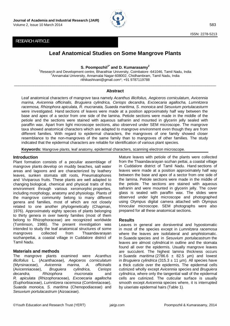
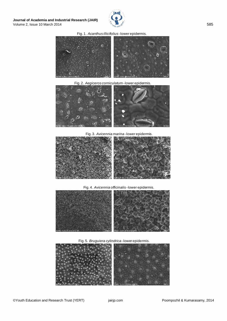
Journal of Academia and Industrial Research (JAIR) Volume 2, Issue 10 March 2014 583 ©Youth Education and Research Trust (YERT) jairjp.com Poompozhil & Kumarasamy, 2014 ISSN: 2278-5213 Leaf Anatomical Studies on Some Mangrove Plants S. Poompozhil 1* and D. Kumarasamy 2 1 Research and Development centre, Bharathiar University, Coimbatore -641046, Tamil Nadu, India 2 Annamalai University, Annamalai Nagar-608002, Chidhambram, Tamil Nadu, India [email protected]*; +91 9787119788 ______________________________________________________________________________________________ Abstract Leaf anatomical characters of mangrove taxa namely Acanthus illicifolius, Aegiceros corniculatum, Avicennia marina, Avicennia officinalis, Bruguiera cylindrica, Ceriops decandra, Excoecaria agallocha, Lumnitzera racemosa, Rhizophora apiculata, R. mucranata, Suaeda maritima, S. monoica and Sesuvium portulacasturm were investigated. Hand sections of leaves were made at a position approximately half way between the base and apex of a sector from one side of the lamina. Petiole sections were made in the middle of the petiole and the sections were stained with aqueous safranin and mounted in glycerin jelly sealed with paraffin wax. Apart from light microscope sections, also observed under SEM microscope. The mangrove taxa showed anatomical characters which are adapted to mangrove environment even though they are from different families. With regard to epidermal characters, the mangroves of one family showed closer resemblance to the non-mangroves of the same family than to mangroves of other families. The study indicated that the epidermal characters are reliable for identification of various plant species. Keywords: Mangrove plants, leaf anatomy, epidermal characters, scanning electron microscope. Introduction Plant formation consists of a peculiar assemblage of mangrove plants develop on muddy beaches, salt water areas and lagoons and are characterized by leathery leaves, sunken stomata stilt roots, Pneumatophores and Viviparous fruits. These plants are well adapted to changing biological, chemical and physical traits of this environment through various xeromorphic properties, including morphology, anatomy and physiology. Plants of the mangrove community belong to many different genera and families, most of which are not closely related to one another phylogenetically (Chapman, 1976). Approximately eighty species of plants belonging to thirty genera in over twenty families (most of them belong to Rhizophoraceae) are recognized worldwide (Tomlinson, 1986). The present investigation was intended to study the leaf anatomical structures of some mangroves collected from Thaandavarayan sozhanpettai, a coastal village in Cuddalore district of Tamil Nadu. Materials and methods The mangrove plants examined were Acanthus illicifolius L. (Acanthaceae), Aegiceros corniculatum (Myrsinaceae), Avicennia marina, A. officinalis (Avicenniaceae), Bruguiera cylindrica, Ceriops decandra, Rhizophora mucronata and R. apiculata (Rhizophoraceae), Excoecaria agallocha (Euphorbiaceae), Lumnitzera racemosa (Combretaceae), Suaeda monoica, S. maritima (Chenopodiaceae) and Sesuvium portulacastrum (Aizoaceae). Mature leaves with petiole of the plants were collected from the Thaandavarayan sozhan pettai, a coastal village in Cuddalore district of Tamil Nadu. Hand sections of leaves were made at a position approximately half way between the base and apex of a sector from one side of the lamina. Petiole sections were made in the middle of the petiole. The sections are stained with aqueous safranin and were mounted in glycerin jelly. The cover glass is sealed with paraffin wax. The slides were observed under light microscope and photographed using Olympus digital camera attached with Olympus trinocular microscope. SEM photographs were also prepared for all these anatomical sections. Results Leaves in general are dorsiventral and hypostomatic in most of the species except in Lumnitzera racemosa where the leaves are isobilateral and amphistomatic. In Suaeda species and in Sesuvium portulacastrum the leaves are almost cylindrical in outline and the stomata found all over the epidermis. Usually mangrove leaves are succulent. The highest lamina thickness occurs in Suaeda maritima (2786.6 ± 82.5 μm) and lowest in Bruguiera cylindrica (315.3 ± 11 μm). All species have a thick cuticle over the epidermis. The epidermal cells cutinized wholly except Avicennia species and Bruguiera cylindrica, where only the tangential wall of the epidermal cells are cutinized. The cuticular surface is usually smooth except Avicennia species where, it is interrupted by uiseriate epidermal hairs (Table 1). RESEARCH ARTICLE
Transcript of Leaf Anatomical Studies on Some Mangrove Plantsjairjp.com/MARCH 2014/13 POOMPOZHI-01.pdf ·...
Journal of Academia and Industrial Research (JAIR) Volume 2, Issue 10 March 2014 583
©Youth Education and Research Trust (YERT) jairjp.com Poompozhil & Kumarasamy, 2014
ISSN: 2278-5213
Leaf Anatomical Studies on Some Mangrove Plants
S. Poompozhil1* and D. Kumarasamy2 1Research and Development centre, Bharathiar University, Coimbatore -641046, Tamil Nadu, India
2Annamalai University, Annamalai Nagar-608002, Chidhambram, Tamil Nadu, India [email protected]*; +91 9787119788
______________________________________________________________________________________________
Abstract Leaf anatomical characters of mangrove taxa namely Acanthus illicifolius, Aegiceros corniculatum, Avicennia marina, Avicennia officinalis, Bruguiera cylindrica, Ceriops decandra, Excoecaria agallocha, Lumnitzera racemosa, Rhizophora apiculata, R. mucranata, Suaeda maritima, S. monoica and Sesuvium portulacasturm were investigated. Hand sections of leaves were made at a position approximately half way between the base and apex of a sector from one side of the lamina. Petiole sections were made in the middle of the petiole and the sections were stained with aqueous safranin and mounted in glycerin jelly sealed with paraffin wax. Apart from light microscope sections, also observed under SEM microscope. The mangrove taxa showed anatomical characters which are adapted to mangrove environment even though they are from different families. With regard to epidermal characters, the mangroves of one family showed closer resemblance to the non-mangroves of the same family than to mangroves of other families. The study indicated that the epidermal characters are reliable for identification of various plant species.
Keywords: Mangrove plants, leaf anatomy, epidermal characters, scanning electron microscope.
Introduction Plant formation consists of a peculiar assemblage of mangrove plants develop on muddy beaches, salt water areas and lagoons and are characterized by leathery leaves, sunken stomata stilt roots, Pneumatophores and Viviparous fruits. These plants are well adapted to changing biological, chemical and physical traits of this environment through various xeromorphic properties, including morphology, anatomy and physiology. Plants of the mangrove community belong to many different genera and families, most of which are not closely related to one another phylogenetically (Chapman, 1976). Approximately eighty species of plants belonging to thirty genera in over twenty families (most of them belong to Rhizophoraceae) are recognized worldwide (Tomlinson, 1986). The present investigation was intended to study the leaf anatomical structures of some mangroves collected from Thaandavarayan sozhanpettai, a coastal village in Cuddalore district of Tamil Nadu. Materials and methods The mangrove plants examined were Acanthus illicifolius L. (Acanthaceae), Aegiceros corniculatum (Myrsinaceae), Avicennia marina, A. officinalis (Avicenniaceae), Bruguiera cylindrica, Ceriops decandra, Rhizophora mucronata and R. apiculata (Rhizophoraceae), Excoecaria agallocha (Euphorbiaceae), Lumnitzera racemosa (Combretaceae), Suaeda monoica, S. maritima (Chenopodiaceae) and Sesuvium portulacastrum (Aizoaceae).
Mature leaves with petiole of the plants were collected from the Thaandavarayan sozhan pettai, a coastal village in Cuddalore district of Tamil Nadu. Hand sections of leaves were made at a position approximately half way between the base and apex of a sector from one side of the lamina. Petiole sections were made in the middle of the petiole. The sections are stained with aqueous safranin and were mounted in glycerin jelly. The cover glass is sealed with paraffin wax. The slides were observed under light microscope and photographed using Olympus digital camera attached with Olympus trinocular microscope. SEM photographs were also prepared for all these anatomical sections. Results Leaves in general are dorsiventral and hypostomatic in most of the species except in Lumnitzera racemosa where the leaves are isobilateral and amphistomatic. In Suaeda species and in Sesuvium portulacastrum the leaves are almost cylindrical in outline and the stomata found all over the epidermis. Usually mangrove leaves are succulent. The highest lamina thickness occurs in Suaeda maritima (2786.6 ± 82.5 µm) and lowest in Bruguiera cylindrica (315.3 ± 11 µm). All species have a thick cuticle over the epidermis. The epidermal cells cutinized wholly except Avicennia species and Bruguiera cylindrica, where only the tangential wall of the epidermal cells are cutinized. The cuticular surface is usually smooth except Avicennia species where, it is interrupted by uiseriate epidermal hairs (Table 1).
RESEARCH ARTICLE

Journal of Academia and Industrial Research (JAIR) Volume 2, Issue 10 March 2014 584
©Youth Education and Research Trust (YERT) jairjp.com Poompozhil & Kumarasamy, 2014
In most of the species, both the adaxial and the abaxial epidermal cells are more or less similar in size except in Avicennia officinalis, Ceriops decandra and Acanthus illicifolius where the adaxial epidermal cells are larger in size compare to those of abaxial cells. The epidermal cells are polygonal in outline with more or less straight walls in all species studied except Ceriops decandra and Excoecaria agallocha. In Ceriops decandra, the wall of the epidermal cells is sinuous, where as in Excoecaria agallocha it is wavy. The stomata are of anomocytic type in Aegiceros corniculatum, diacytic in Acanthus illicifolius, Cyclocytic in Ceriops decandra, in all other it is paracytic. Cuticular striations radiating from the stomata towards the walls of the subsidiary cell are recorded in Excoecaria agallocha. In Rhizophora apiculata, the subsidiary cells having papillary projections. Both glandular and non-glandular trichomes were observed in leaves of Avicennia species. Each glandular trichome or salt gland having a stalk cell and a head comprising of variable number of cells found on both adaxial and abaxial surfaces of leaves, whereas non-glandular trichomes are multicellular with a stalk of 2 to 3 cells with an awl shaped terminal cell observed abundantly all over the lower epidermis. Glandular trichomes similar to that of Avecennia is also observed in Aegiceros corniculatum (Figs. 1-14).
The thickness of the upper epidermis ranging from 86.1 ± 2.7 µm in Suaeda maritima to 8.4 ± 0.55 µm in Aegiceros corniculatum and thickness of lower epidermis ranging from 22 ± 0 µm in lumnitzera racemosa to 9.0 ± 0.55 µm in Aegiceros corniculatum. In all species studied, the mesophyll differentiated into palisade and spongy tissues except Lumnitzera racemosa, where only palisade layer is present on both sides. The palisade tissue below the upper epidermis is two layers in thickness in Ceriops decandra, Acanthus illicifolius and Suaeda species and one layer in thickness in Bruguiera cylindrica and more than two layers in thickness in the rest. Water storing tissues of varying proportions has been observed in all species. In most of the species, the water storing tissue found as a hypodermis except Lumnitzera racemosa, where it is centrally located in between the upper and lower palisade layer. The water storing tissue is uniseriate in Bruguiera cylindrica, biseriate in Ceriops decandra and Acanthus illicifolius, multiseriate in other species observed. In Lumnitzera racemosa, the palisade tissue found in two layers on both sides of the epidermis. The palisade and spongy ratio was highest in Excoecaria agallocha (1.51 ± 0.02 µm) and lowest in Ceriops decandra (0.24 ± 0.03 µm).
Table 1. Laminar characters of some mangroves.
Species Thickness of the Lamina
(in µm)
Thickness of the UE (in µm )
Thickness of the LE (in µm)
Thickness of the
Palisade layer
(in µm)
Thickness of the
Spongy layer (in µm)
P/S Ratio (in µm)
Thickness of the WST
(in µm)
Percent of WST to Total
Lamina Thickness
(in µm) Aegiceros
corniculatum 359.3±5.5 8.4±0.55 9.0±0.55 113.6±8.2 141.1±8.2 0.7±0.0.5 176±11 48.9±3.8
Acanthus illicifolius 601.3±11 38.5±5.5 22±0.0 157.6±1 6.5 300.6±5.5 0.52±0.06 82.5±5.5 13.7±1.1
Avicennia marina 539±11 22±0.0 15±1.6 148.5±5.5 129.8±9.9 1.13±0.1 223.6±5.5 41.4±0.6
Avicennia officinalis 528±16.5 20.1±2.7 11±0.0 165±11.0 115.5±8.2 1.36±0.2 216.3±16.5 40.9±1.8
Bruguiera cylindrica 315.3±11 22±0.0 22±0.0 49.5±8.2 185.1±2.7 0.26±0.045 36.6±2.7 11.6±0.9
Ceriops decandra 682±11 30. 4±2.7 20.1±27.0 110±11 440±11 0.24±0.03 81.4±5.5 11.9±0.7
Excoecaria agallocha 517±11 20.1±2.7 17.9±1.65 270.2±5.5 177.4±3.3 1.51±0.02 31.1±2.7 6.0±0.6
Rhizophora apiculata 535.3±5.5 22±0.0 14.6±2.7 165±11 150.3±8.2 1.0±0.05 183.3±16.5 34.2±3.0
Rhizophora Mucronata 627±11 22±0.0 14.6±1.65 176±11 165±11 1.0±0.0 256.6±22 40.9±2.8
Lumnitzera racemosa 1279.6±16.5 22±0.0 22±0.0 132±11 - - 430.8±2.7 33.6±0.6
Suaeda maritima 2786.6±82.5 86.1±2.7 - 110±11 - - 990±22 35.5±0.7
Suaeda monoica 1210±22 53.1±2.75 - 152.1±13.7 - - 315.3±5.5 26±0.9
UE-Upper epidermis, LE-Lower epidermis, WST-Water storage tissue, P/S ratio-Palisade spongy ratio.
Journal of Academia and Industrial Research (JAIR) Volume 2, Issue 10 March 2014 585
©Youth Education and Research Trust (YERT) jairjp.com Poompozhil & Kumarasamy, 2014
Fig. 1. Acanthus illicifoliuslower epidermis.
Fig. 2. Aegiceros corniculatumlower epidermis.
Fig. 3. Avicennia marinalower epidermis.
Fig. 4. Avicennia officinalislower epidermis.
Fig. 5. Bruguiera cylindricalower epidermis.

Journal of Academia and Industrial Research (JAIR) Volume 2, Issue 10 March 2014 586
©Youth Education and Research Trust (YERT) jairjp.com Poompozhil & Kumarasamy, 2014
Fig. 6. Ceriops decandralower epidermis.
Fig. 7. Excoecaria agallochalower epidermis.
Fig. 8. Lumnitzera racemosaupper epidermis.
Fig. 9. Rhizophora apiculatalower epidermis.
Fig. 10. Rhizophora mucranatalower epidermis.Fig.10. Rhizophora mucronatalower epidermis.
Fig. 8. Lumnitzera racemosaupper and lower epidermis.

Journal of Academia and Industrial Research (JAIR) Volume 2, Issue 10 March 2014 587
©Youth Education and Research Trust (YERT) jairjp.com Poompozhil & Kumarasamy, 2014
Idioblasts and stellate sclerieds are observed in the mesophyll of Bruguiera cylindrica, Lumnitzera racemosa and Rhizophora mucronata. Tannin containing cells are also observed in Bruguiera cylindrica. The outline of the petiole observed in various shapes in different species. Thick cuticle covering the epidermis. The hypodermis consists of 2 to 3 layers of collenchyma in all species observed. Ground tissue consists of loosely arranged parenchyma cells, where as in Rhizophora mucronata, it is aerenchymatous. Stellate idioblasts are also observed in the ground tissue of Rhizophora mucronata.
The vascular bundle is medullated in Rhizophora mucronata, Rhizophora apiculata, Avicennia marina and Bruguiera cylindrica or the vascular bundles are in the form of deep arc or flat arc in Avicennia officinalis, A. marina, Aegiceros corniculatum and Lumnitzera racemosa and is represented by 3 or more discrete vascular bundles in Excoecaria agallocha, Acanthus illicifolius and Sesuvium portulacastrum. Discussion In the family Rhizophoraceae, the leaf anatomy of four taxa was studied in the present investigation and they are Rhizophora mucronata, R. apiculata, Ceriops decandra and Bruguiera cylindrica.
Fig. 11. Sesuvium portulacastrumupper epidermis.
Fig. 12. Suaeda maritimalower epidermis.
Fig. 13. Suaeda maritimaupper epidermis.
Fig. 14. Suaeda monoicaupper epidermis.
Journal of Academia and Industrial Research (JAIR) Volume 2, Issue 10 March 2014 588
©Youth Education and Research Trust (YERT) jairjp.com Poompozhil & Kumarasamy, 2014
The epidermal cells in all these species are having slightly curved walls. Stomata are sunken and cyclocytic. In Rhizophora apiculata, the stomatal ledge is clear. But in Rhizophora mucronata, it is not clear and also in Bruguiera cylindrica and Ceriops decandra. In Poga of this family, the stomata are anomocytic and paracytic in Anisophyllea and Combretocarpus and showing various intermediate types in Gynotroches, Crossostylis, Cassipourea and Anopyxis (Tobe and Raven, 1988). In Lumnitzera, there are two species reported so far, they are Lumnitzera racemosa and L. littorea. These two species are not readily distinguished using epidermal characters except the size of the stomata and epidermal characters. There are two species in Ceriops, namely Ceriops decandra and C. tagal. In Ceriops tagal the subsidiary slightly overarching the guard cells than in Ceriops decandra. Except this character, there is no character was found to distinguish these two species. In Bruguiera, there are five species reported, they are Bruguiera cylindrica, B. gymnorrhiza, B. hainesic, B. sexangula, B. parviflora and B. exaxistata. These species differ only in their size of the epidermal cells and stomata. In Bruguiera cylindrica and B. gymnorrhiza, the stomata are small compared with those of other Rhizophoraceae mangroves. The epidermal cells are also smaller in size in these two species compared to other species of Bruguiera. In Rhizophora, there are eight species reported so far; they are Rhizophora apiculata, R. annamalayana, R. mucronata, R. lamarckii, R. mangle, R. harrisonii, R. racemosa and R. stylosa (Das and Ghose, 1996). Among these two species studied in the present work, in Rhizophora apiculata, the lateral subsidiary cells overarching the guard cells, where as in Rhizophora mucronata there is no such overlapping. Among the other species studied, in Rhizophora mangle, R. harrisonii, R. racemosa and R. annamalayana there is no overarching of subsdiary cells over the guard cells. But it is seen in Rhizophora stylosa and R. lamarckii. Apart from this character, there is no other character is there to separate or distinguish the various species. In most of the genera, the trichomes are absent including the mangrove genera and wherever it is present it is only sparse except Macarisia sp. where they may be dense and clustered. Among the Combretaceae taxa, three genera are mangrove plants and they are Lumnitzera, Laguncularia and Conocarpus (Stace, 1965). Normally leaves in Combretaceae members are dorsiventral in these three genera and in Macropteranthes the leaves are isobilateral. In dorsiventral leaves, in these three genera, the mesophyll is differentiated into palisade and spongy tissues. The palisade tissue consists of one layer of cells below the upper epidermis and a broad spongy zone. In Lumnitzera the leaves are isobilateral, similarly in other two mangrove genera also the leaves are isobilateral apart from the non-mangrove genus Macropteranthes.
The stomata are anomocytic in most of the members of Combretaceae but it is paracytic in strephonema and cyclocytic in Lumnitzera and Langucularia. Glandular hairs are present in most of the genera of Combretaceae including the mangrove genus Languncularia but it is absent in other two mangrove genera. In Avicennia there are twelve taxa reported so far, they are Avicennia marina var marina var. resinifera, var. acutissima, var. rumphiana, A. bicolor, A. alba var alba, A. eucalyptifolia, A. schaceriana, A. tonduzii, A. germinans, A. Africana and A. officinalis. Among these, two species were studied namely Avicennia officinalis, Avicennia marina var. marina. In Avicennia officinalis, the glandular trichomes are 20-25 per mm whereas in Avicennia marina var. marina there are more than 50 glandular hairs per mm. Among the other species studied, Avicennia marina, var. resinifera, var. rumpriana, var. acutissima, Avicennia bicolor, Avicennia alba, var. resinifera, A. eucalyptifolia, the number of glandular trichomes was less than 25 per sq. mm where as in the remaining taxa, it is more than 25 per mm. In the family Chenopodiaceae, two taxa were studied namely Suaeda maritima and S. monoica. These two taxa showed cylindrical leaves. These cylindrical leaves not only found in these taxa and most of the other members of Chenopodiaceae. The stomata are paracytic and the epidermal cells are polygonal. In Aegiceros corniculatum, the stomata are anomocytic and they are irregularly arranged. In Acanthus illicifolius, glandular hairs are present on the upper epidermis and the stomatal ledge clearly seen over the guard cells. The stomatal ledge also seen in A. corniculatum. Another similarity between these two genera is the stomata which is not sunken unlike other mangroves. In Excoecaria agallocha, many cuticular striations radiating from the stomata towards the walls of the subsidiary cells. These may be the character of this species. This is also noticed by Seshavatharam and Srivalli (1989) in their study. Conclusion From this study it is concluded that certain characters like thick cuticle, water storage tissue, extensive palisade, salt glands and trichome exhibited by most of the mangrove species appears to ecophysiological adaptations. But as suggested by Stace (1966), that in their anatomical characters, the mangrove species exhibit closer affinities to non-mangrove species of their family than to mangrove of other families. This character includes isobilateral nature of leaves in some plants like lumnitzera and cylindrical nature of leaves in Chenopodiaceae members and the type of stomata and epidermal cells. References 1. Chapman, V.J. 1976. Mangrove vegetation. J Cramer,
Valduz, Germany.

Journal of Academia and Industrial Research (JAIR) Volume 2, Issue 10 March 2014 589
©Youth Education and Research Trust (YERT) jairjp.com Poompozhil & Kumarasamy, 2014
2. Das, S. and Ghose, M. 1996. Anatomy of leaves of some mangroves and their associates from Sunderbans (West Bengal). Phytomorphol. 46: 139-150.
3. Seshavatharam, V. and Srivalli, M. 1989. Systematic leaf anatomy of some Indian mangroves. Proc. Ind. Acad. Sci. (Pl. Sci.). 99: 557-565.
4. Stace, C.A. 1965. The significance of the leaf epidermis in the taxonomy of the Combretaceae. I. A general review of tribal, generic and specific characters. J. Linn. Soc. (Bot.). 59: 229.
5. Stace, C.A. 1966. The use of epidermal characters in phylogenetic considerations. New Phytol. 65: 304-318.
6. Tobe, H. and Raven, P.H. 1988. Floral morphology and evolution in Anisophylleaceae. Bot. J. Linn. Soc. 98: 1-25.
7. Tomlinson, P.B. 1986. The Botany of Mangroves. Cambridge University Press, Cambridge, New York. p.413.