Laura Eva Hartman Winterthur/University of...
41
Laura Eva Hartman Winterthur/University of Delaware Revealing Layers: Asher B. Durand’s Painting Practice Studied Through the Examination of a Portrait and Two Paint Palettes
Transcript of Laura Eva Hartman Winterthur/University of...
Laura Eva Hartman Winterthur/University of Delaware
Revealing Layers: Asher B. Durand’s Painting Practice Studied Through the Examination of a Portrait and Two Paint Palettes
Hartman, ANAGPIC 2012,
2
Abstract A portrait by Asher B. Durand (1796-1886) and two of the artist’s later paint palettes owned by the New-York Historical Society were examined to obtain information about the pigments, binders, fillers, and additive materials. Technical analysis of the materials present in the painting and palettes revealed evidence of Durand’s evolving working practices as a painter. Durand was primarily self-taught, developing his skill with the guidance of colleagues such as Thomas Cole. Cole and Durand later pioneered the Hudson River School of American landscape painting. Materials and techniques identified in the painting and palettes were compared to those mentioned in correspondence between these artists as well as with other painters and available technical sources. These communications can be used to place Durand’s work in context with that of other artists of his time and with the evolution of 19th-century painting materials. Information acquired through the study also informed the conservation treatment of the examined portrait. Analytical techniques used include: energy dispersive x-ray fluorescence spectroscopy, scanning electron microscopy with energy dispersive spectroscopy and in the backscattered electron mode, Raman spectroscopy, Fourier transform infrared spectroscopy, and gas-chromatography mass spectrometry. Other examination techniques included: X-radiography, ultraviolet light illumination, infrared photography, polarized light microscopy, and cross-sectional microscopy with fluorochrome staining. Durand apparently used interlayers of organic media and applications of a toned glaze during his early painting process. Similar techniques have been documented in written manuals and identified on paintings by influential painters of the time. A continued use of interlayers or the addition of a resin containing drying-oil medium was found on the palettes, visible as transparent puddles that were probably incorporated into the artist’s paint as he worked; this technique has been documented in the working practices of other Hudson River School painters. The palettes contained an expanded range of pigments compared to the earlier portrait. 1. INTRODUCTION:
1.1 Historical Context
Asher Brown Durand (1796-1886) along with contemporary artists such as Thomas Cole
(1801-1848), are predominately known for their landscape paintings and drawings of the
Catskill, Adirondack, and White Mountains and for pioneering the Hudson River School
of American landscape painting. Durand began his artistic career as an engraver and
eventually became an influential leader in the development of an American practice of
landscape painting. (Ferber 2007) Discourse with established artists served as the primary
source of technical education for many emerging artists of the 19th century including
Durand, who eagerly sought mentorship from fellow artists such as Cole. (Ferber 2007,
131) Durand’s dynamic artistic career spans much of the 19th century, providing a unique
Hartman, ANAGPIC 2012,
3
perspective into developing practices and the foundation of an American painting
tradition.
Durand arrived in New York City from his hometown of Maplewood, New Jersey, in
1820, at the age of 24, and worked as an engraver. He established himself as an
influential force within the art world in the span of only five years. The eminent painter
John Trumbull (1756-1843) was instrumental in fostering Durand’s painting career and
became both a friend and colleague. (Durand 1894, 26) In 1820 Trumbull commissioned
Durand to execute an engraving of his painting The Declaration of Independence
(completed in 1823). The success of the engraving enhanced Durand’s reputation and he
was then invited to join various social clubs and organizations. Throughout the 19th
century artists in America turned away from formal academies and joined sketch clubs
and other social associations. They worked together in painting and drawing, developing
their artistic practices. These organizations reflected the dissatisfaction felt by young
artists towards the academies of fine art, especially with regard to technical information.
(Lawall 1978, 6) (Ferber 2007, 53)
Durand’s invitation to the novelist James Fennimore Cooper’s (1789-1851) Bread and
Cheese Club in 1825 was especially important to Durand’s social and artistic
development, and placed him in contact with future Hudson River School members.
From associations formed within such organizations Durand would become a leader in
the formation of the New-York Drawing Association (Marchwardt, 1935, 397). In 1826
this organization evolved to become the National Academy of Design (NAD) over which
Durand presided from 1845-1861. (Lawall 1978, 6) In a memorial speech for Durand
given in 1887 Daniel Huntington elaborated on Durand’s early interest in painting, as
well as the intentions behind the formation of the NAD, stating:
Although so much occupied with engraving, Durand took the time to paint an
occasional portrait or group of figures. In 1825 he was a ringleader of that band of
rebel students of the Old American Academy of Fine Arts, who, disgusted with
the harsh response to their request for better opportunities for drawing from the
Hartman, ANAGPIC 2012,
4
antique, united in a society for evening study, which soon resulted in the
foundation of the National Academy of Design. (Huntington 887, 19-20)
These groups served to develop relationships between artists and encouraged open
communication in an effort to fill a void many felt had been left by the art academies of
the time, particularly in regard to technical information. (Lawall 1978, 6) (Ferber 2007,
53) An account bill from 1825 documents the purchase of three Thomas Cole paintings
by Asher B. Durand, John Trumbull, and William Dunlap from William Coleman’s
frame shop. Considering that at the time Cole was still an unknown painter, this purchase
further emphasizes the formation of close personal networks that thrived on mutual
support and the open exchange of ideas. (Katlan 1992, 498) (Ferber 2007, 87) This type
of exchange and discourse within social groups would prove integral to Durand’s
development as a painter.
Durand’s career has been described as having three distinct periods: between 1812-1830
he was dedicated to being an engraver; between 1830-1838 Durand focused on portrait
painting, and between 1839-1879 he turned his attentions solely to landscape paintings.
(Durand 1894) Even during the earliest period of his career Durand was already
beginning to shift his interest from engraving to painting. In the Life and Times of A. B.
Durand, John Durand III, A. B. Durand’s son and editor of The Crayon, further details
his father’s transformation into a painter:
[A.B. Durand] found that… engraving afforded only a limited field for the
exercise of his artistic aptitudes. He began to paint as a pastime. The engravings
of pictures by famous painters, the knowledge of art and of celebrated artists he
picked up in a desultory manner from books and conversation, prompted him very
early to try experiments with the brush as he had before done with the graver.
Without instruction, as in his first attempts at engraving and when he made his
own tools, he bought a canvas, ground his colours, set his palette, and began to
paint. His initiatory efforts—as usual with impatient novices—seem to have been
‘high art’. (Durand 1894, 98)
Hartman, ANAGPIC 2012,
5
As his son expresses, during these early years Durand was primarily a portrait painter and
was experimenting in the prevalent trends passed down from Europe including the old
master aesthetic, or the ‘high art’ as it was referenced in the passage above. (Durand
1894, 147) During the early 1800’s this was the style that sold and was attractive to
patrons, something that likely contributed to Durand’s interest in this style. (Mayer and
Myers 2011, 181) Henry T. Tuckerman in his 1870 publication Book of the Artists
devotes a chapter to Durand’s landscapes and of Durand’s early portraits exclaimed, “In
his late visit to Europe, this unpretending and skillful artist has communed with the old
masters, to good effect. Observe the Girl with the Parrot. Every detail is finished with a
marvelous exactitude. It is perfectly Titian-like!” (Tuckerman 1870, 193) This passage
highlights not only Durand’s interest in the old master aesthetic, but also his developing
skill.
In the Life and Times John Durand implies a connection between his father’s emerging
practice and those evident in the works of established painters such as Benjamin West
(1748-1820), Washington Allston (1779-1843), Thomas Sully (1783-1872), and John
Trumbull (1756-1843); artists known for their devotion to rediscover the secrets of the
old master’s painting techniques. (Mayer and Myers 2011) (Durand 1894,147) Many of
these artists allowed open entry into their studios to students and fellow artists and were
open to provide instruction to the novice painter. (Mayer and Myers 2011) (Durand 1894)
Documentation suggests that Durand was actively visiting various artists’ studios during
his development as a painter. A portrait of Durand painted by Trumbull in 1826 further
evidences that in these early years of Durand’s painting practice he had already witnessed
some of these early figures at work.
John Durand also specifically mentions his father’s use of books and conversation as
primary guidance in the previously quoted passage. Throughout 1855 Durand published a
series of letters in The Crayon expressing advice to students of landscape painting and
lending insight into his own philosophies and ideas in regards to painting and the
depiction of nature. Durand directly advocated this same type of education to novice
painters in one of his “Letters on Landscape Painting,” in which he wrote: “You need not
a period of pupilage in an artist’s studio to learn to paint; books and the casual intercourse
Hartman, ANAGPIC 2012,
6
with artists, accessible to every respectable young student, will furnish you with all the
essential mechanism of the art.” (Durand, 1855 letter 1) This statement, and John’s
description of his father’s own education in painting, suggests that Durand was actively
seeking literature and discourse in his development as a painter, and encourages
connections to be drawn between Durand’s practices to his colleagues’ and those found
in the available technical literature.
A commission to produce a series of portraits and genre paintings in 1834 from Lumen
Reed, already a generous patron to painters such as Thomas Cole, definitively marks the
transition in Durand’s profession from engraver to painter. (Lawall 1978, 4-5) Durand
initially pursued a career as a portrait painter, experimenting with the inclusion of figures
within landscape settings. His son details his father’s early practice of au plein air
painting, describing how A.B. Durand would complete full compositions outdoors early
in his career. (Durand 1894, 81) After a sketching trip with Thomas Cole in 1837, there is
a notable shift in Durand’s focus from portraiture to landscape painting that was realized
after his 1840 European sojourn to study the works of the old masters. (Lawall 1978, 4-5)
During this trip Durand made copies of paintings by the old masters and his admiration
for the works of the great Venetian painters and the deep tones achieved by artists like
Rembrandt was recorded in his journal. (Ferber 2007, 110) Scholars have hypothesized
that Durand learned to paint from the old masters but that European contemporaries such
as John Constable may have ultimately influenced his matured style. (Ferber 2007, 123)
These observations are based on a notable shift in Durand’s practice in landscape
painting after 1840, which tended toward the naturalistic and “true” representation of
nature with a focus on capturing subtle atmospheric effects. (Ferber 2007, 123) In one of
Durand’s 1855 “Letters on Landscape Painting” he expresses the complexities of
atmospheric perspective and the textures and gradients experienced in nature. He states:
“That is a fine picture which at once takes possession of you, you traverse it—breath its
atmosphere—feel its sunshine, and you repose in its shade without thinking of its design
or execution, effect or color.” (Durand 1855, letter 3) This commitment to truthful
representation would ultimately be realized through experimentation with various
materials and techniques leading to the establishment of a painting practice that was truly
Hartman, ANAGPIC 2012,
7
unique to the young American landscape painters. (Ferber 2007, 123) Cole and Durand,
along with an emerging population of painters devoted to the same purpose, in their
pursuits, forged a new tradition in American painting.
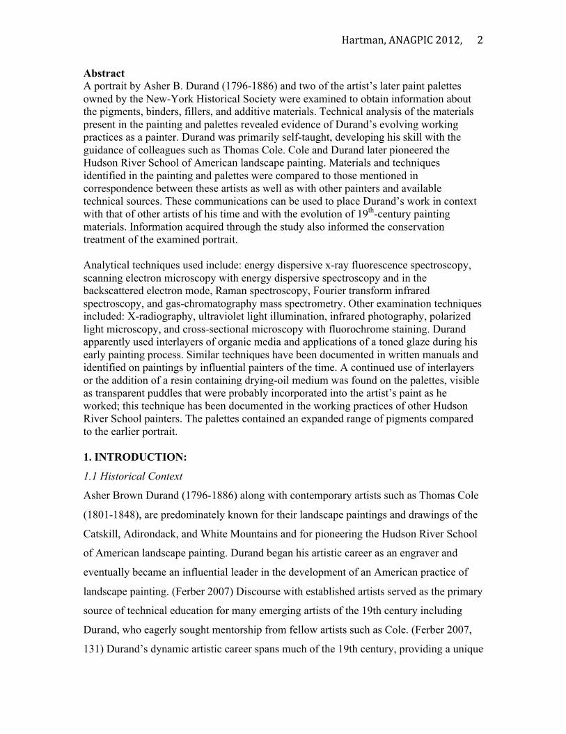
1.2 Construction, condition, and previous treatment of examined items
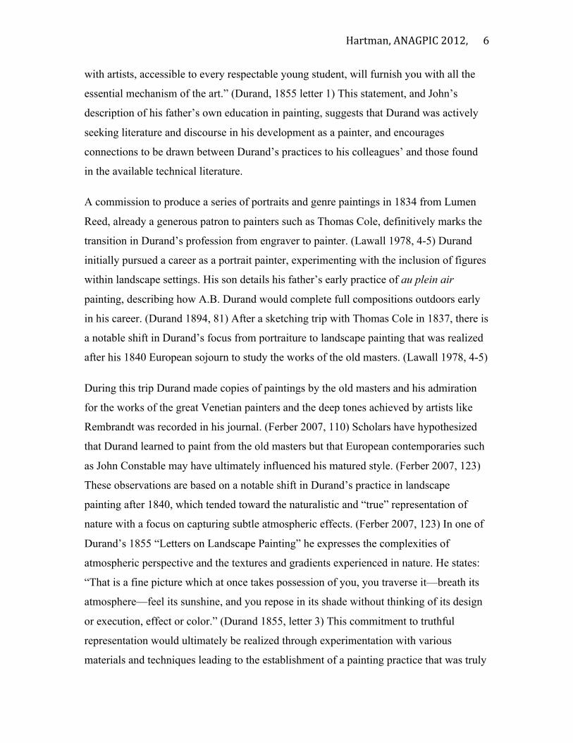
This study includes the examination of an early painting by
A. B. Durand, c. 1825, as well as two paint palettes from
within the artist’s tabouret of materials, most likely dating
to the end of his painting career around 1879. The paper
label on the verso of the upper stretcher bar member of the
painting identifies the sitter as John Durand III and the
artist as his father, Asher B. Durand. This label also
identifies the donor of the work, to the New-York
Historical Society (NYHS), as Miss Nora Durand
Woodman, John Durand’s niece. Records from the NYHS
identify the same niece as the donor of the palettes and
tabouret as a whole. (NYHS archives, accessed 2012)
In the portrait, figure 1, a boy is depicted sitting in a garden
with a little white, longhaired dog. The boy wears a red
jumper suit with white ruffles at the sleeves and neckline.
The front of the red jumper has a decorative floral pattern.
The boy’s right hand rests on the dogs back while his left
hand is held coyly at chest height. His head is cocked to his
right as he gazes at the viewer.
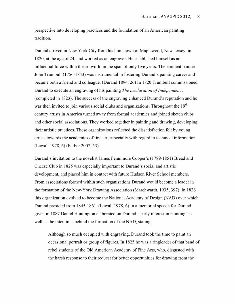
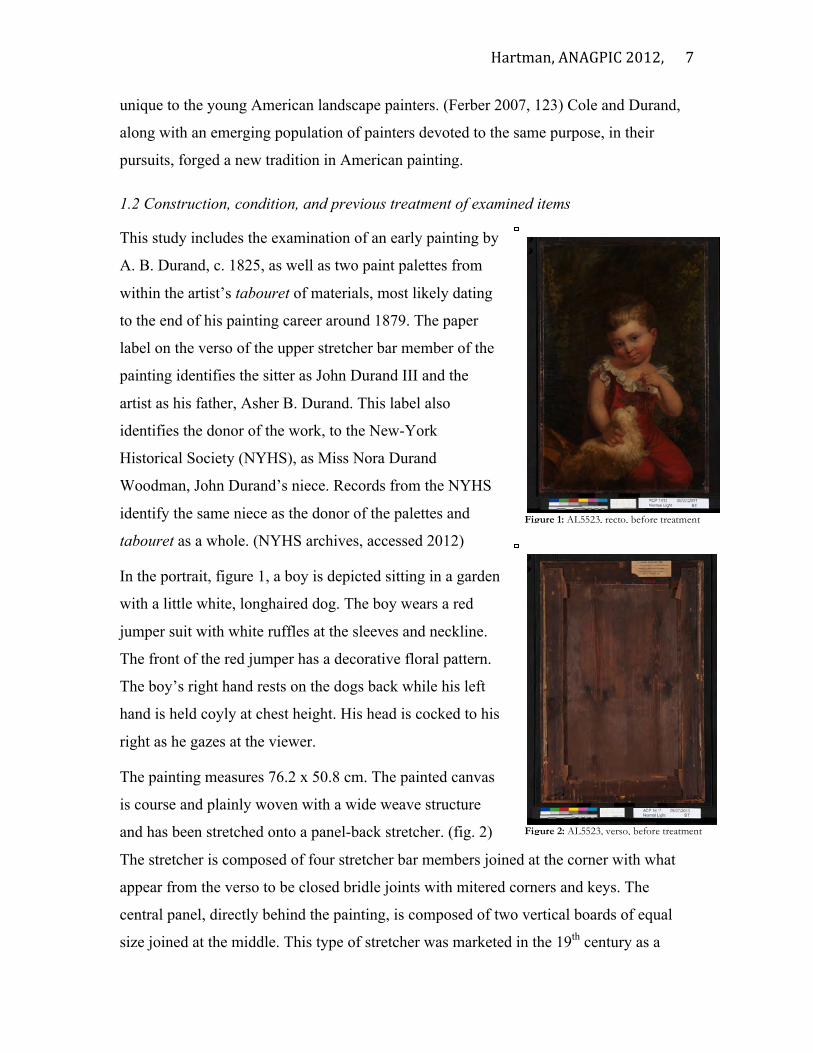
The painting measures 76.2 x 50.8 cm. The painted canvas
is course and plainly woven with a wide weave structure
and has been stretched onto a panel-back stretcher. (fig. 2)
The stretcher is composed of four stretcher bar members joined at the corner with what
appear from the verso to be closed bridle joints with mitered corners and keys. The
central panel, directly behind the painting, is composed of two vertical boards of equal
size joined at the middle. This type of stretcher was marketed in the 19th century as a
Figure 1: AL5523, recto, before treatment
Figure 2: AL5523, verso, before treatment t
Hartman, ANAGPIC 2012,
8
technical innovation, providing better support and lending long-term stability to
paintings. (Katlan 1992) This type of stretcher has also been documented as having been
used by contemporaries of Durand such as Thomas Cole, Frederick Church, Albert
Bierstadt, and John Trumbull. (Katlan 1992) (Mayer and Myers 2011, 150) There is a
white ground on the canvas support that extends to the two cut side edges of the canvas,
however the top and bottom side edges remain bare suggesting an artist preparation as
opposed to a commercial one.
Durand primarily applied his paint directly throughout the composition. The background
is created by the application of thin transparent layers of paint, with a limited palette of
greens and blues reserving lighter and more intense color such as yellow and red for areas
of highlight. More intense hues of color including reds, oranges, and browns are also
visible below areas of more transparently applied paint, particularly in the background.
The figure is painted using wet into wet paint applications to create the illusion of folds in
the textiles and mass in the figure.
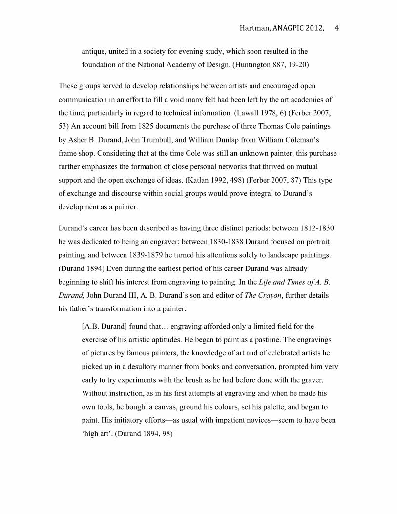
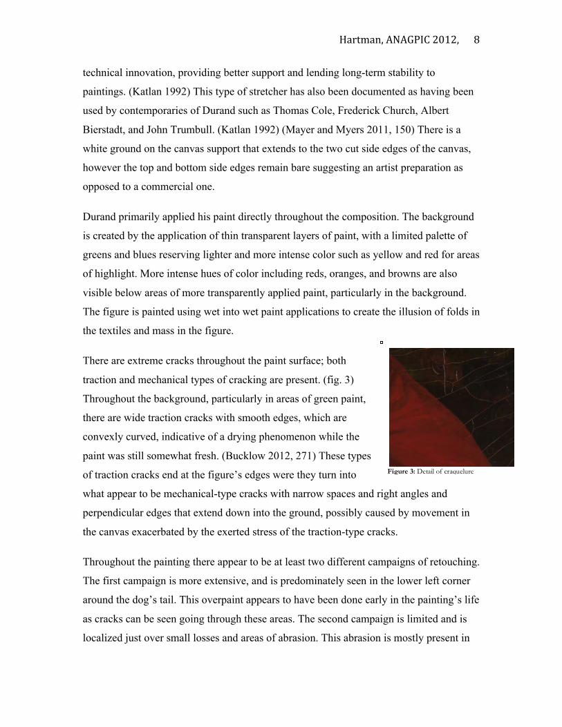
There are extreme cracks throughout the paint surface; both
traction and mechanical types of cracking are present. (fig. 3)
Throughout the background, particularly in areas of green paint,
there are wide traction cracks with smooth edges, which are
convexly curved, indicative of a drying phenomenon while the
paint was still somewhat fresh. (Bucklow 2012, 271) These types
of traction cracks end at the figure’s edges were they turn into
what appear to be mechanical-type cracks with narrow spaces and right angles and
perpendicular edges that extend down into the ground, possibly caused by movement in
the canvas exacerbated by the exerted stress of the traction-type cracks.
Throughout the painting there appear to be at least two different campaigns of retouching.
The first campaign is more extensive, and is predominately seen in the lower left corner
around the dog’s tail. This overpaint appears to have been done early in the painting’s life
as cracks can be seen going through these areas. The second campaign is limited and is
localized just over small losses and areas of abrasion. This abrasion is mostly present in
Figure 3: Detail of craquelure
Hartman, ANAGPIC 2012,
9
the upper glazes of the sitter’s flesh and is especially evident in IR reflectograms taken
pre-treatment. (appendix II) There is an extremely glossy and slick varnish coating on the
painting. This coating appears to have been applied when the cracks had already formed
and is pooled at the cupped edges of the cracks. (appendix II) Conservation records from
1942 at the NYHS describe that the first campaign of retouching, around the dog’s tail,
was already present upon acquisition of the painting by the historical society. During a
treatment at the NYHS in 1942, a cleaning attempt to remove a very tough coating
proved unsuccessful (following cleaning attempts using pure acetone, alcohol, and
turpentine), and a coat of dammar varnish was applied over the painting. Small losses and
various dark spots throughout the painting were also retouched with varnish and pigments
during this 1942 restoration campaign. (NYHS archives, accessed 2012)
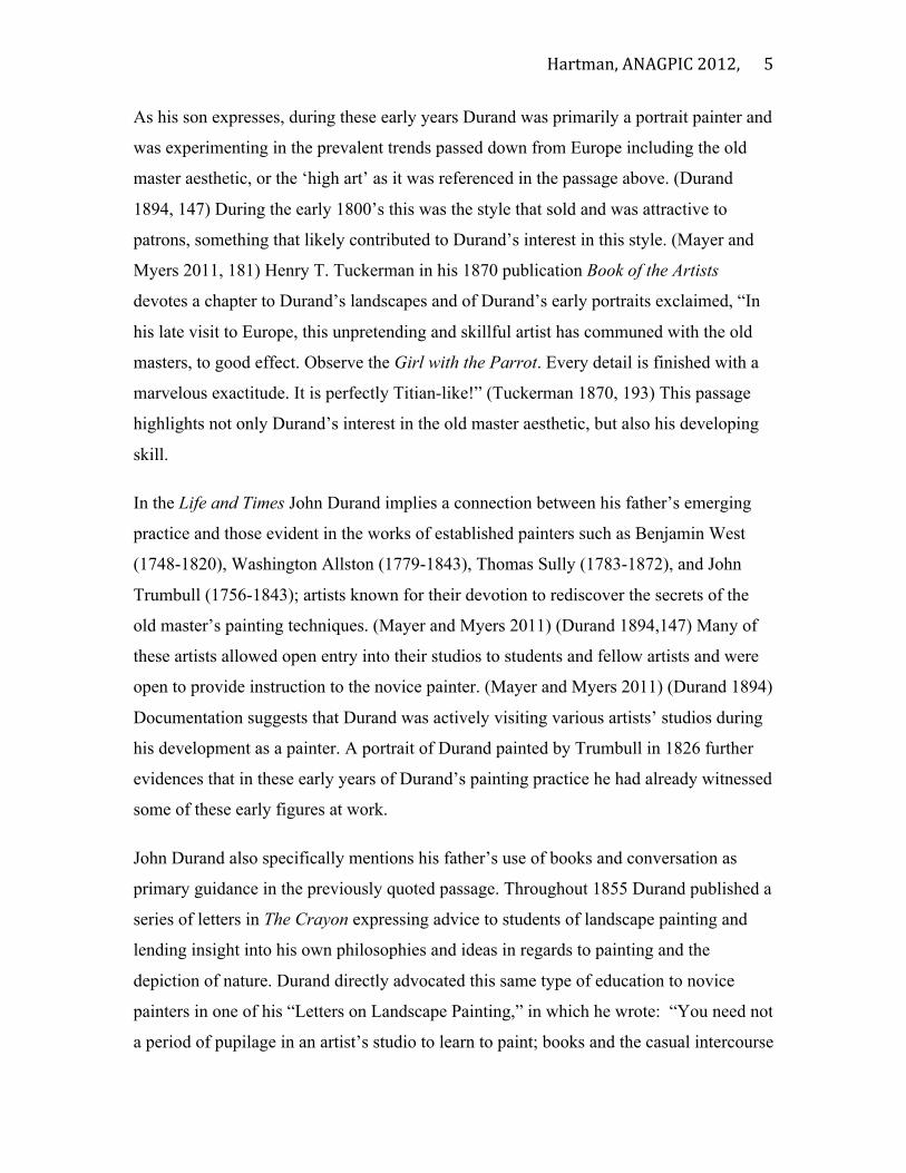
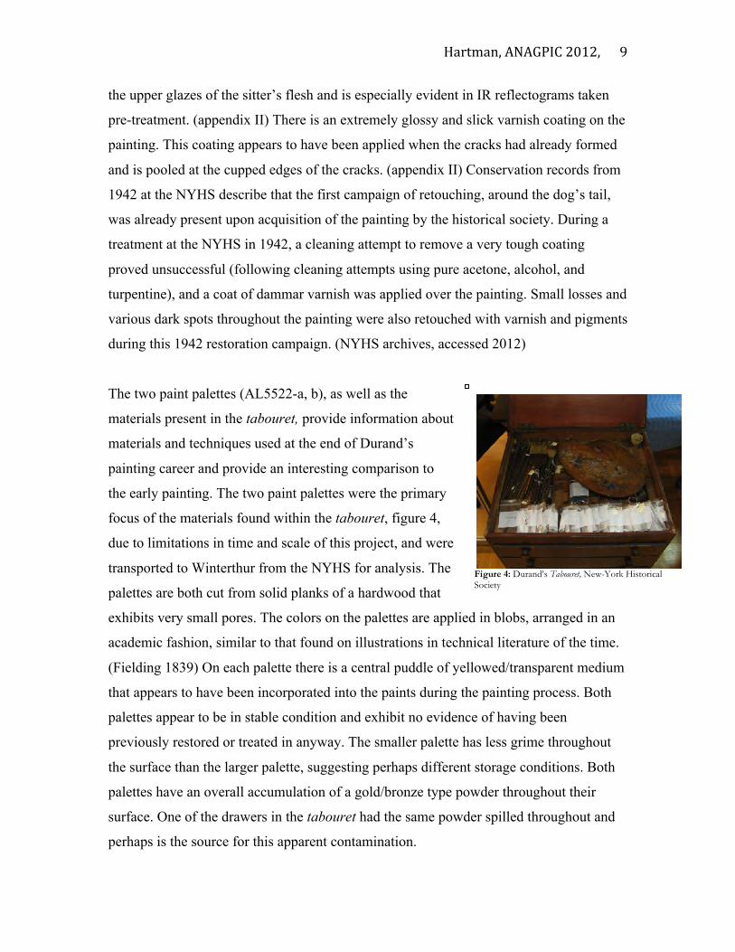
The two paint palettes (AL5522-a, b), as well as the
materials present in the tabouret, provide information about
materials and techniques used at the end of Durand’s
painting career and provide an interesting comparison to
the early painting. The two paint palettes were the primary
focus of the materials found within the tabouret, figure 4,
due to limitations in time and scale of this project, and were
transported to Winterthur from the NYHS for analysis. The
palettes are both cut from solid planks of a hardwood that
exhibits very small pores. The colors on the palettes are applied in blobs, arranged in an
academic fashion, similar to that found on illustrations in technical literature of the time.
(Fielding 1839) On each palette there is a central puddle of yellowed/transparent medium
that appears to have been incorporated into the paints during the painting process. Both
palettes appear to be in stable condition and exhibit no evidence of having been
previously restored or treated in anyway. The smaller palette has less grime throughout
the surface than the larger palette, suggesting perhaps different storage conditions. Both
palettes have an overall accumulation of a gold/bronze type powder throughout their
surface. One of the drawers in the tabouret had the same powder spilled throughout and
perhaps is the source for this apparent contamination.
Figure 4: Durand’s Tabouret, New-York Historical Society

Hartman, ANAGPIC 2012,
10
1.3 Literature Review Research was performed in order to assess the materials and techniques that would have
been available and actively used by Durand’s contemporaries. This research provided a
comparative base in which the materials found within Durand’s painting and palettes
could be interpreted, and in the process would draw relationships between these and the
practices documented by contemporary artists.
Lance Mayer and Gay Myers’ book American Painters on Technique: The Colonial
Period to 1860 chronicles the development of painting practices in America and
emphasizes the transfer of ideas from Europe to America, particularly the traditions
established by English painters throughout the 18th and 19th centuries. This book
elaborates on the lineage of painters spearheaded by Benjamin West and including John
Trumbull, Washington Allston, and Thomas Sully known for their pursuits of the secrets
of the old master’s painting techniques. One of the secrets was believed to be a long lost
paint vehicle. This prompted experimentation with glazing techniques, the addition of
various mediums, and the development of unique and often bizarre recipes to alter their
paint’s working properties. (Mayer and Myers 2011, 36) These men are now recognized
as modern masters of painting, and innovators, their pursuits eventually established an
experimental tradition in painting. Influenced by this, trial and error was continued by the
emerging artists of the day, including Durand and his colleagues such as Thomas Cole.
Scholars have emphasized the peer-to-peer aspect of material experimentation during the
early 19th century. (Zucker 1999, 3) (Mayer and Meyer 2011, 15) An example of this type
of communication is witnessed in a conversation recorded by John Trumbull in 1784
where Benjamin West describes his theory of glazing with complementary colors using
megilp- a mixture of a drying oil and mastic resin-- or other medium to enhance the
working properties of the paint. (Mayer and Myers 2011, 14-15) In another conversation
West suggests to Sully that if you need to “bring out” a part of a panting you can do so
using a mixture of drying oil and turpentine, a technique known at the time as “oiling
out.” (Mayer and Myers 2011, 19) Toned glazes were often employed to tone down areas
of the painting, or the painting as a whole, and affect the “old master” aesthetic. Allston
was known to apply these glazes to his paintings, referring to them as “Titian’s dirt”, a
Hartman, ANAGPIC 2012,
11
mixture composed of asphaltum, red, and blue pigments in a megilp medium. (Stoner
1990, 1) This glazing technique was opposed to Trumbull who instead would often just
tone down the paints on his palettes before painting. (Mayer and Myers 2011, 75)
Many of these experiments were initially problematic. Early response from critics and
recorded observations by artists themselves noted early deterioration of paintings shortly
after completion. (Mayer and Mayer 2011) Conservators who have treated paintings by
West, Trumbull, and others following these practices have also identified the common
occurrence of wide drying type cracks, likely due to the excessive addition of oil and
other materials. (Mayer and Myers 2011, 19) Trumbull is described as being one of the
last in the lineage of painters truly interested in finding these secrets. (Mayer and Myers
2011, 37) Sully describes how Trumbull himself even admitted that those of his paintings
created with simpler materials lasted longer and ultimately were better. (Mayer and
Myers 2011, 38) Past restoration treatments have also resulted in the misinterpretation
and removal of toned glazes from these paintings, altering the intended appearance of the
paintings overall. (Stoner 1990, 9)
In the middle of the 19th century commercial colourman became more prevalent in the
United States. (Mayer and Meyer 2011) (Katlan 1999) (Carlyle 2001) As a result, a wider
variety of artists’ materials were now readily accessible and with them even more
emerging experimental techniques. Technological innovations also led to the
development and distribution of new materials, particularly synthetic pigments and new
resins and paint additives. (Mayer and Myers 2011, 143) Another significant change was
the invention of the collapsible paint tube, doing away with the traditional pig bladder
and therefore making paint more accessible and versatile in its use. (Carlyle 2001)
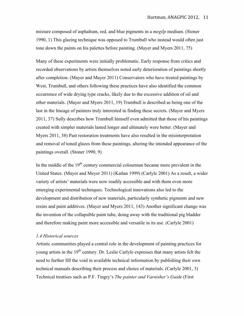
1.4 Historical sources Artistic communities played a central role in the development of painting practices for
young artists in the 19th century. Dr. Leslie Carlyle expresses that many artists felt the
need to further fill the void in available technical information by publishing their own
technical manuals describing their process and choice of materials. (Carlyle 2001, 3)
Technical treatises such as P.F. Tingry’s The painter and Varnisher’s Guide (First
Hartman, ANAGPIC 2012,
12
English edition 1804), J Bulkley’s Treatise on Landscape Painting in Oil (1821), and
John Cawse’s Introduction to the Art of Painting in Oil Colours, (1822) would have been
available to the emerging artist early in the century. The treatises supplemented
established reference manuals such as the colourmaker George Field’s Chromatography;
or, A treatise on Colours and dogments, and of their Powers in Painting & c., a text
which has been documented in the library collection of Durand’s contemporaries
including Frederic Church and Thomas Cole. (Carlyle 2001, 16) (Mayer and Myers 2011)
In an early recorded conversation between Washington Allston and Thomas Cole, Allston
conveyed to Cole: “I have been frequently told by friends of yours, sir, that they were
afraid you were running after the old masters. Now if that frightens them, I would make
every hair on their heads stand on end!”(Mayer and Myers 2011, 66) This statement
reiterates the active pursuit for the old master aesthetic even by emerging artists early in
the century. Cole copied directly from William Oram’s Preceopts and Observations on
the Art of Colouring in Landscape Painting (published in 1810 but likely written around
the 1760s), into his journal, a text linking him further to earlier painters such as Joshua
Reynolds and Benjamin West. (Mayer and Myers 2011, 162) William Dunlap in his
History of the rise of the Arts of Design in the United States copies from a letter by Cole
describing his early career in painting:
About the year 1820, Mr. Stein, a portrait painter, came to Steubenville. He lent
me an English work on painting… it was illustrated with engravings, and treated
of design, composition, and color. This book was my companion day and night,
nothing could separate us… painting was all in all to me… my love for the art
exceeded all love—my ambition grew, and in my imagination I pictured the glory
of being a great painter. The names of Stuart and Sully came to my ears like titles
of great conquerors, and the great masters were hallowed above all earthly things.
(Dunlap 1918, 140)
Although Cole does not explicitly name Oram’s text, this passage indicates that he was
not only learning directly from a book, but also from the works of Sully and Stuart.
Oram’s text outlines specific techniques and materials to use in order to paint landscapes
Hartman, ANAGPIC 2012,
13
and many techniques described within this source have been identified in the work of
Thomas Cole including his use of bright and varied preparatory layers and glazing
techniques. (Mayer and Myers 2011, 182) This type of interaction and exchange
illustrates the kinds of sources available for itinerate painters of the time who were
seeking mentorship.
Artists’ manuals of the time were largely concerned with the addition of materials into
paint to change their working properties. They also included advice on topics such as
setting one’s palette and arranging one’s studio, preparation of the canvas and ground,
application of paint, addition of various additives, and varnishing the painting. Dr. Lesley
Carlyle in her book The Artist’s Assistant; Oil Painting Manuals and Handbooks in
Britain 1800-1900, further describes how these written sources also document the
establishment of a system of painting in the 18th century that continued to the 19th
century. Carlyle describes Thomas Bardwell’s three-stage system of painting published in
his 1756 text on oil painting The Practice of Painting and Perspective Made Easy.
Bardwell’s influence continued to the 19th century as a standard method and basis from
which variations evolved. (Carlyle 2001, 197) Thomas Sully details a similar system of
painting in his 1851 publication Hints to Young Painters. While his system includes at
least 6 stages, they bear a pronounced similarity to Bardwell’s system overall.
1.5 Previous technical study of Hudson River School painters
Available technical examination of paintings by Durand is limited, especially of his early
work. Published studies of Hudson River School paintings largely consist of conservation
documentation including microscopic and ultraviolet illumination examination and
solubility testing. This documentation provides information regarding the techniques and
materials utilized by these artists in addition to condition issues and conservation
considerations. Studies carried out on the work of Hudson River School painters overall
confirm the common use of glazing techniques and layering systems, and resultantly
solubility concerns from the addition of excess oil and resin into their paints. (Zucker and
Boon 2007) (Mayer and Myers 2007) (Bradley 2009)
Hartman, ANAGPIC 2012,
14
Published technical studies of Thomas Cole’s paintings reveal that he predominately
painted in thin layers, allowing for warmer underlayers to show through. Further they
reveal that he also utilized loose applications of transparent paints and wet into wet
blending techniques. (Mayer and Myers 2007, 63) Cole also made use of varied
imprimatura colors generally ranging from a light buff to deep terracotta. (Zucker 1999)
Previous solubility tests and examination in these studies with ultra violet illumination
have indicated that Cole likely mixed large amounts of varnish in with his dark paint,
likely to increase transparency and enhance workability of the paint. (Mayer and Myers
2007, 63) The work of Frederic Church, Cole’s only true pupil, has also shown many
similarities to Cole’s technique including the application of thin paint over pinkish
imprimatura layers, wet into wet paint applications, and the use of resinous interlayers.
(Zucker and Boon 2007)
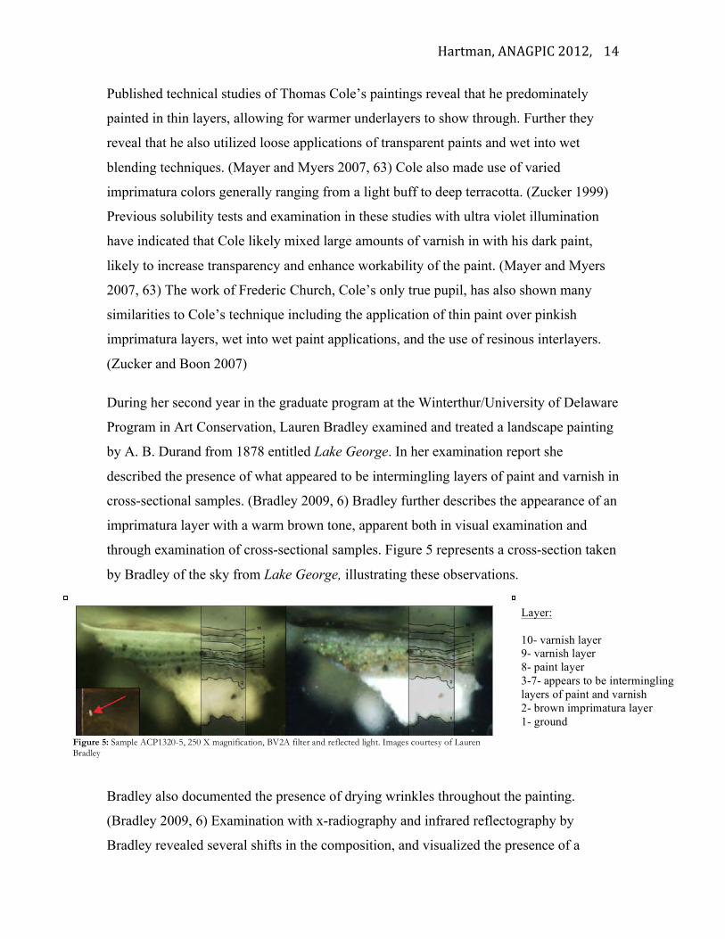
During her second year in the graduate program at the Winterthur/University of Delaware
Program in Art Conservation, Lauren Bradley examined and treated a landscape painting
by A. B. Durand from 1878 entitled Lake George. In her examination report she
described the presence of what appeared to be intermingling layers of paint and varnish in
cross-sectional samples. (Bradley 2009, 6) Bradley further describes the appearance of an
imprimatura layer with a warm brown tone, apparent both in visual examination and
through examination of cross-sectional samples. Figure 5 represents a cross-section taken
by Bradley of the sky from Lake George, illustrating these observations.
Bradley also documented the presence of drying wrinkles throughout the painting.
(Bradley 2009, 6) Examination with x-radiography and infrared reflectography by
Bradley revealed several shifts in the composition, and visualized the presence of a
Figure 5: Sample ACP1320-5, 250 X magnification, BV2A filter and reflected light. Images courtesy of Lauren Bradley
Layer: 10- varnish layer 9- varnish layer 8- paint layer 3-7- appears to be intermingling layers of paint and varnish 2- brown imprimatura layer 1- ground
Hartman, ANAGPIC 2012,
15
carbon-based underdrawing. Bradley also took disperse pigment samples from a range of
areas identifying the following pigments: chrome yellow, ultramarine blue, lead white,
red earth, earth pigments, and possibly viridian. (Bradley 2009, 16)
Concerns about solubility issues in cleaning paintings of this time are increasingly
appearing in the conservation literature. (Zucker and Boon 2007, 18) (Mayer and Myers
2007) The appearance of metallic soaps in Hudson River School paintings is also
increasingly mentioned. (Zucker and Boon 2007, 3) (Noble, van Loon, and Boon, 2005)
These soaps have been observed to protrude through paint film surfaces, as well as
enhance the transparency of paintings, which has led to misinterpretation of these works.
(Zucker and Boon 2007, 3) (Noble, van Loon, and Boon, 2005)
1.6 Objectives
The primary focus of this technical study is to develop an understanding of Durand’s
evolving working practices as a painter through technical examination of the materials
present on the palettes and painting. Evidence of Durand’s working practices where
revealed through the examination. These were compared to materials and techniques
mentioned in available technical sources of the time in the pursuit of placing his work
within the context of the 19th century. This study also provided an interesting insight into
the changing market of artist materials in the 19th century, while providing an opportunity
to document Durand’s own practices. As identified through the literature review, there is
limited information regarding technical examination of Durand’s paintings and therefore
this study intends to augment the available literature. This study also served to inform the
conservation treatment of the examined painting.
2. EXPERIMENTAL PROCEDURES 2.1 Experimental Design:
Various techniques were utilized in order to examine the materials used by Durand on the
Portrait of John Durand III, as well as those found on the paint palettes. Both non-
destructive and minimally destructive techniques were used in order to derive
information about the materials present overall, application methods, and condition. Non-
destructive techniques were employed initially to inform sampling sites, ensuring a
Hartman, ANAGPIC 2012,
16
minimum of samples taken. Analysis techniques included: UV radiation, IR
reflectography, X-radiography, XRF, CSM with fluorochrome staining, FTIR, SEM-
EDX, Raman spectroscopy, and GC-MS.
2.2 Visual examination
- Initial examination of the painting and two palettes was performed under low power
magnification using a stereobinocular microscope with a 6x-200x range. Sampling sites
were identified and samples were also taken using the same microscope.
- Observation under long wave ultraviolet radiation was done using a Mineralight UGVL-
58 Handheld Lamp (115v. 60 Hz. 0.16 Amps.), Long Wave: 365 nm to preliminarily
characterize surface coatings and identify areas of restoration.
- Infrared reflectographic examination was performed with a National instruments
Labview Frame Grabber using Indigo Systems Corp. IRvista 2.51 software and an
ALPHA NIR Infrared Camera with an Indium Gallium Arsenide (InGaAs) detector with
a spectral response of 0.9 to 1.7 microns and an array format of 320 x 256.
- X-radiographs were acquired using an X-ray tube - Pantek Seifert Eresco 65 MF2 with
digital control, 3 mm focal spot. Images were captured on phosphor imaging plates (IPS)
read with GE CR50P Phosphor Scanner. Scans were collected with GE Rhythm Acquire
software and processed with GE Rhythm Review software.
2.1 Sampling
- Polarized Light Microscopy (PLM)
Dispersed pigment samples, approximately 3 µg in size, were collected on a glass slide
using a #10 stainless steel scalpel. These were mounted with Cargille melt mount having
a refractive index of 1.66. Samples were examined using a Nikon Polarizing Microscope,
Eclipse E600 POL and images were captured with Spot RT software V3.5 and processed
with Spot Advanced software.
- Cross-Sectional Microscopy
Milligram size samples for cross-sectional analysis were collected from areas of flaking
paint or areas adjacent to pre-existing losses in order to examine the stratigraphy of the
paint layers and coatings. These samples were obtained with a #10 stainless steel scalpel
Hartman, ANAGPIC 2012,
17
and were transferred to a previously pored and set half cube of Extec Polyester Clear
Resin (polyester/methacrylate blend). The same Extec resin, mixed 10 mL of the resin to
8 drops of methyl ethyl ketone peroxide catalyst, was then pored to cover the samples and
cured under a tungsten halogen bulb. The samples were allowed to sit overnight before
polishing. Sanding of the sample resin cubes was first done with a rotary sander and then
further polished dry using Micro-Mesh papers with grits ranging from 1,500-12,000.
Mounted samples were also reused for SEM and RAMAN analysis. (further description
of staining procedure in appendix II)
2.2 Instrumental analysis
- X-Ray Fluorescence spectroscopy (XRF) An ArtTAX Micro X-Ray Fluorescence
Spectrometer (Mo anode, 50kV voltage, 700 µA current, 100 seconds live time,
atmosphere air, no filter) was used and data was collected and interpreted with artTAX
Basic Software (Version 5.3.21.0).
- Scanning Electron Microscopy in the Backscattered Electron Mode (SEM-BSE) and
with Energy Dispersive X-ray Spectroscopy (SEM-EDX) was performed on the same
samples used for cross-sectional microscopy. The clear mounting resin was thinned to
approximately 3 mm and the samples were then mounted on an SPI-Supplies pure carbon
mounting stub using double sided carbon sticky tape and the mounting resin support was
further coated with a layer of SPI-Chem carbon suspension particles. A Topcon ABT-60
scanning electron microscope was used and data was collected and processed using a
Bruker x-flash detector Quantax model 200 with Esprit 1.8 software. An accelerating
voltage of 20kV was used with a working distance of 21-30mm and a sample tilt of 20°
in a variable pressure atmosphere.
-Fourier Transform Infrared Spectroscopy (FTIR) was performed using a Nicolet 6700
FT-IR instrument (cooled with liquid Nitrogen) equipped with a Nicolet Continuµm FT-
IR microscope set to the transmission mode. 128 scans taken over a spectral range of
650-4000 cm-1 data was collected and analyzed using OMNIC 8.0 software with a variety
of reference spectra from various sources such as Infrared and Raman User’s Group
(IRUG) and other commercial reference libraries. Small samples were collected,
Hartman, ANAGPIC 2012,
18
approximately 3µg, and placed on diamond cell with a #11 steel scalpel blade and
flattened with a steel roller. All tools were cleaned with methanol in-between sample
preparation.
- Raman Spectroscopy was performed on both loose samples and samples mounted for
cross-sectional microscopy and used for SEM analysis. A Renishaw Invia Raman
Spectrometer was used for analysis (4000-150cm-1, sample area size 1 x 20µm, 1200
lines/mm diffraction grating, 50x objective, 3cm-1 spectral resolution) with either a red
diode laser (excitation at 785nm) or a green argon ion laser (excitation at 514nm) with
0.1% - 5% laser power. Spectra were collected and interpreted using WiRE 2.0 software
and RRUFF, University College Lincoln, and Burgio and Clark’s (2000) reference
libraries.
- Gas Chromatography-Mass Spectrometry (GC-MS) The samples were treated with
MethPrep II reagent that converts the carboxylic acids and esters (often components in
these types of classes of compounds) to their methyl ester derivatives and reduce the
molecular weight of the components and increase their volatility. 100µL of 1:2 MethPrep
II reagant (Alltech) in benzene was added to the sample containing vile. The vile was
then warmed for an hour at 60°C in a heating block and allowed to cool. Analysis was
carried out using the Hewlett-Packart 6890 gas chromatograph equipped with 5937 mass
selective detector (MSD) and 7683 automatic liquid injector. The Winterthur
RTLMPREP method was used with conditions as follows: inlet temperature was 300°C
and transfer line temperature to the MSD (SCAN mode) was 300°C. A sample volume
(splitless) of 1µL was injected onto a 30mx0.25µmx0.25µm film thickness HP-5MS
column (5% phenyl methyl siloxane at a flow rate of 2.3mL/minute). The oven
temperature was held at 55°C for two minutes, then programmed to increase at
10°C/minute to 325°C where it was held for 10.5 minutes for a total run time of 40
minutes.
3. RESULTS 3.1 Painting
Hartman, ANAGPIC 2012,
19
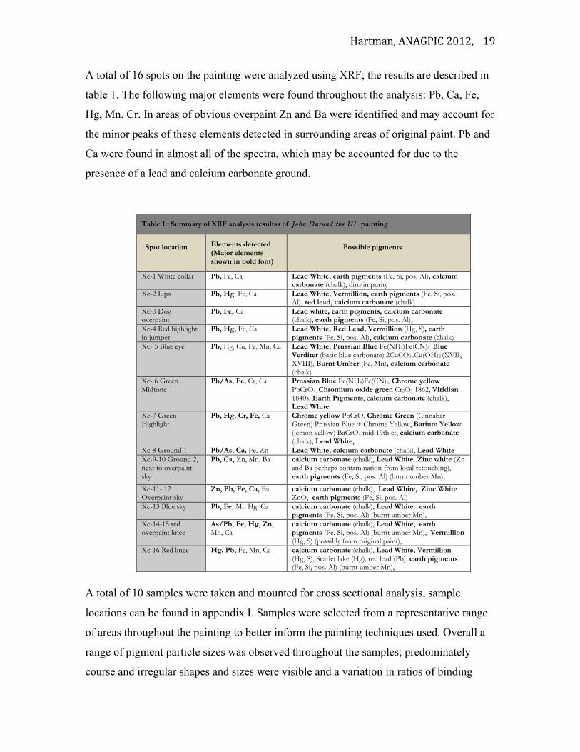
A total of 16 spots on the painting were analyzed using XRF; the results are described in
table 1. The following major elements were found throughout the analysis: Pb, Ca, Fe,
Hg, Mn. Cr. In areas of obvious overpaint Zn and Ba were identified and may account for
the minor peaks of these elements detected in surrounding areas of original paint. Pb and
Ca were found in almost all of the spectra, which may be accounted for due to the
presence of a lead and calcium carbonate ground.
A total of 10 samples were taken and mounted for cross sectional analysis, sample
locations can be found in appendix I. Samples were selected from a representative range
of areas throughout the painting to better inform the painting techniques used. Overall a
range of pigment particle sizes was observed throughout the samples; predominately
course and irregular shapes and sizes were visible and a variation in ratios of binding
Table 1: Summary of XRF analysis resultss of John Durand the III painting
Spot location Elements detected
(Major elements shown in bold font)
Possible pigments
Xc-1 White collar Pb, Fe, Ca Lead White, earth pigments (Fe, Si, pos. Al), calcium carbonate (chalk), dirt/impurity
Xc-2 Lips Pb, Hg, Fe, Ca Lead White, Vermillion, earth pigments (Fe, Si, pos. Al), red lead, calcium carbonate (chalk)
Xc-3 Dog overpaint
Pb, Fe, Ca Lead white, earth pigments, calcium carbonate (chalk), earth pigments (Fe, Si, pos. Al),
Xc-4 Red highlight in jumper
Pb, Hg, Fe, Ca Lead White, Red Lead, Vermillion (Hg, S), earth pigments (Fe, Si, pos. Al), calcium carbonate (chalk)
Xc- 5 Blue eye Pb, Hg, Cu, Fe, Mn, Ca Lead White, Prussian Blue Fe(NH4)Fe(CN)6, Blue Verditer (basic blue carbonate) 2CuCO3 .Cu(OH)2 (XVII, XVIII), Burnt Umber (Fe, Mn), calcium carbonate (chalk)
Xc- 6 Green Midtone
Pb/As, Fe, Cr, Ca Prussian Blue Fe(NH4)Fe(CN)6, Chrome yellow PbCrO4, Chromium oxide green Cr2O3 1862, Viridian 1840s, Earth Pigments, calcium carbonate (chalk), Lead White
Xc-7 Green Highlight
Pb, Hg, Cr, Fe, Ca Chrome yellow PbCrO, Chrome Green (Cinnabar Green) Prussian Blue + Chrome Yellow, Barium Yellow (lemon yellow) BaCrO4 mid 19th ct, calcium carbonate (chalk), Lead White,
Xc-8 Ground 1 Pb/As, Ca, Fe, Zn Lead White, calcium carbonate (chalk), Lead White Xc-9-10 Ground 2, next to overpaint sky
Pb, Ca, Zn, Mn, Ba calcium carbonate (chalk), Lead White. Zinc white (Zn and Ba perhaps contamination from local retouching), earth pigments (Fe, Si, pos. Al) (burnt umber Mn),
Xc-11- 12 Overpaint sky
Zn, Pb, Fe, Ca, Ba calcium carbonate (chalk), Lead White, Zinc White ZnO, earth pigments (Fe, Si, pos. Al)
Xc-13 Blue sky Pb, Fe, Mn Hg, Ca calcium carbonate (chalk), Lead White. earth pigments (Fe, Si, pos. Al) (burnt umber Mn),
Xc-14-15 red overpaint knee
As/Pb, Fe, Hg, Zn, Mn, Ca
calcium carbonate (chalk), Lead White, earth pigments (Fe, Si, pos. Al) (burnt umber Mn), Vermillion (Hg, S) (possibly from original paint),
Xc-16 Red knee Hg, Pb, Fe, Mn, Ca calcium carbonate (chalk), Lead White, Vermillion (Hg, S), Scarlet lake (Hg), red lead (Pb), earth pigments (Fe, Si, pos. Al) (burnt umber Mn),
Hartman, ANAGPIC 2012,
20
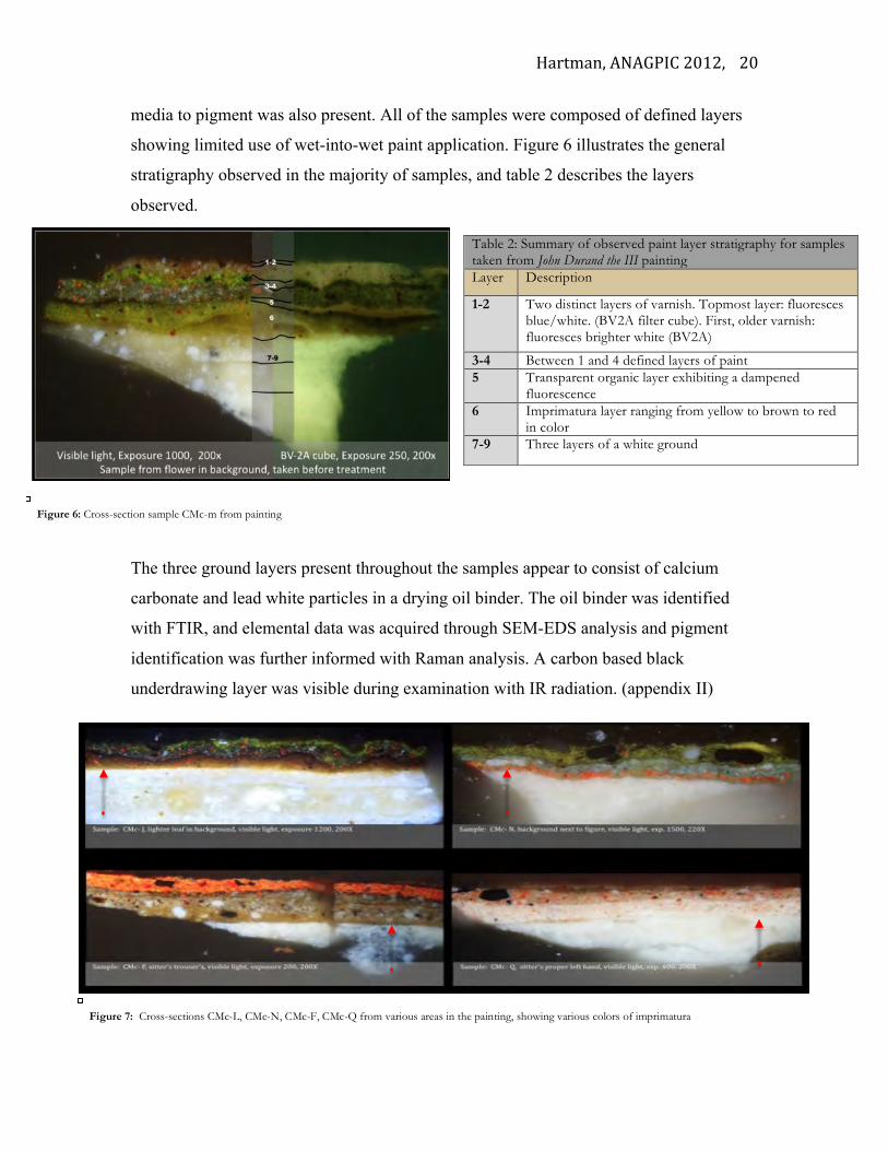
media to pigment was also present. All of the samples were composed of defined layers
showing limited use of wet-into-wet paint application. Figure 6 illustrates the general
stratigraphy observed in the majority of samples, and table 2 describes the layers
observed.
The three ground layers present throughout the samples appear to consist of calcium
carbonate and lead white particles in a drying oil binder. The oil binder was identified
with FTIR, and elemental data was acquired through SEM-EDS analysis and pigment
identification was further informed with Raman analysis. A carbon based black
underdrawing layer was visible during examination with IR radiation. (appendix II)
Table 2: Summary of observed paint layer stratigraphy for samples taken from John Durand the III painting Layer Description
1-2 Two distinct layers of varnish. Topmost layer: fluoresces blue/white. (BV2A filter cube). First, older varnish: fluoresces brighter white (BV2A)
3-4 Between 1 and 4 defined layers of paint 5 Transparent organic layer exhibiting a dampened
fluorescence 6 Imprimatura layer ranging from yellow to brown to red
in color 7-9 Three layers of a white ground
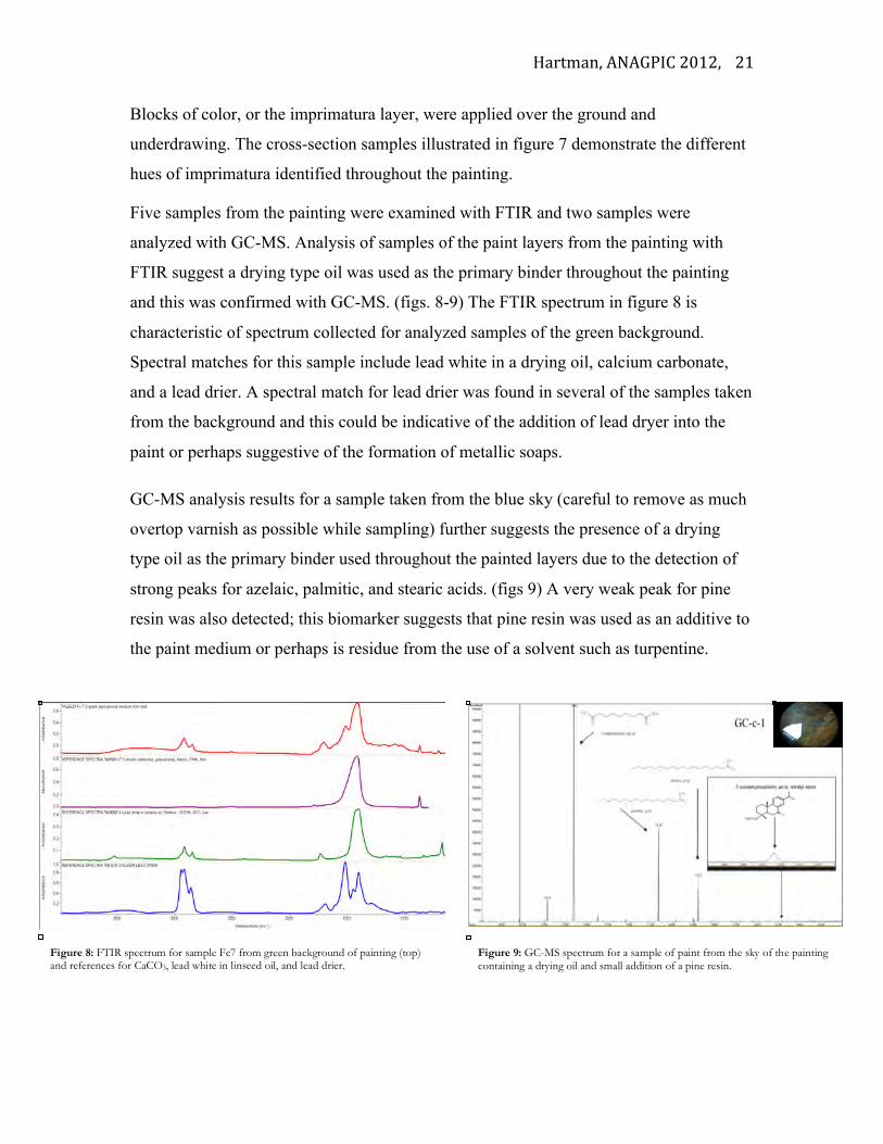
Figure 7: Cross-sections CMc-L, CMc-N, CMc-F, CMc-Q from various areas in the painting, showing various colors of imprimatura
Figure 6: Cross-section sample CMc-m from painting
Hartman, ANAGPIC 2012,
21
Blocks of color, or the imprimatura layer, were applied over the ground and
underdrawing. The cross-section samples illustrated in figure 7 demonstrate the different
hues of imprimatura identified throughout the painting.
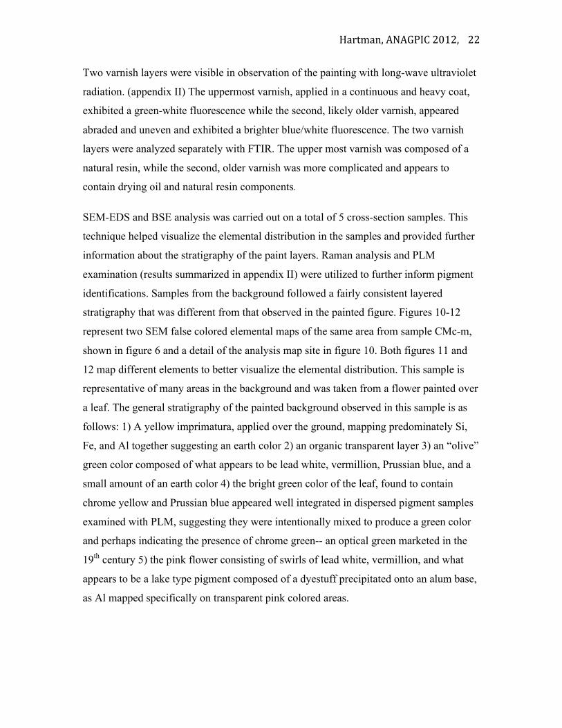
Five samples from the painting were examined with FTIR and two samples were
analyzed with GC-MS. Analysis of samples of the paint layers from the painting with
FTIR suggest a drying type oil was used as the primary binder throughout the painting
and this was confirmed with GC-MS. (figs. 8-9) The FTIR spectrum in figure 8 is
characteristic of spectrum collected for analyzed samples of the green background.
Spectral matches for this sample include lead white in a drying oil, calcium carbonate,
and a lead drier. A spectral match for lead drier was found in several of the samples taken
from the background and this could be indicative of the addition of lead dryer into the
paint or perhaps suggestive of the formation of metallic soaps.
GC-MS analysis results for a sample taken from the blue sky (careful to remove as much
overtop varnish as possible while sampling) further suggests the presence of a drying
type oil as the primary binder used throughout the painted layers due to the detection of
strong peaks for azelaic, palmitic, and stearic acids. (figs 9) A very weak peak for pine
resin was also detected; this biomarker suggests that pine resin was used as an additive to
the paint medium or perhaps is residue from the use of a solvent such as turpentine.
Figure 8: FTIR spectrum for sample Fc7 from green background of painting (top) and references for CaCO3, lead white in linseed oil, and lead drier.
Figure 9: GC-MS spectrum for a sample of paint from the sky of the painting containing a drying oil and small addition of a pine resin.
Hartman, ANAGPIC 2012,
22
Two varnish layers were visible in observation of the painting with long-wave ultraviolet
radiation. (appendix II) The uppermost varnish, applied in a continuous and heavy coat,
exhibited a green-white fluorescence while the second, likely older varnish, appeared
abraded and uneven and exhibited a brighter blue/white fluorescence. The two varnish
layers were analyzed separately with FTIR. The upper most varnish was composed of a
natural resin, while the second, older varnish was more complicated and appears to
contain drying oil and natural resin components.
SEM-EDS and BSE analysis was carried out on a total of 5 cross-section samples. This
technique helped visualize the elemental distribution in the samples and provided further
information about the stratigraphy of the paint layers. Raman analysis and PLM
examination (results summarized in appendix II) were utilized to further inform pigment
identifications. Samples from the background followed a fairly consistent layered
stratigraphy that was different from that observed in the painted figure. Figures 10-12
represent two SEM false colored elemental maps of the same area from sample CMc-m,
shown in figure 6 and a detail of the analysis map site in figure 10. Both figures 11 and
12 map different elements to better visualize the elemental distribution. This sample is
representative of many areas in the background and was taken from a flower painted over
a leaf. The general stratigraphy of the painted background observed in this sample is as
follows: 1) A yellow imprimatura, applied over the ground, mapping predominately Si,
Fe, and Al together suggesting an earth color 2) an organic transparent layer 3) an “olive”
green color composed of what appears to be lead white, vermillion, Prussian blue, and a
small amount of an earth color 4) the bright green color of the leaf, found to contain
chrome yellow and Prussian blue appeared well integrated in dispersed pigment samples
examined with PLM, suggesting they were intentionally mixed to produce a green color
and perhaps indicating the presence of chrome green-- an optical green marketed in the
19th century 5) the pink flower consisting of swirls of lead white, vermillion, and what
appears to be a lake type pigment composed of a dyestuff precipitated onto an alum base,
as Al mapped specifically on transparent pink colored areas.

Hartman, ANAGPIC 2012,
23
Sample CMc-q (shown in fig. 7 and a detail of the analysis map site in figure 13) was
also analyzed with SEM-EDS and BSE. Pigment identification was further informed with
Raman spectroscopy and PLM (see appendix II). This sample is representative of the
figure’s flesh and was taken from the sitter’s hand. The sitter’s flesh was painted in
defined layers like the background. The following stratigraphy was observed: 1) a toned
pink color was applied over the underdrawing and ground. This first layer appears to
contain predominately lead white, small amounts of vermillion, and an earth color. 2)
This bright pink layer was then toned with a medium rich, darker glaze layer exhibiting a
Figure 10: Detail of map area from sample CMc-m of upper paint layers including yellow imprimatura, an olive green layer composed of red, white, and blue particles, a bright green layer, and a pink layer.
Figure 11: SEM elemental mapping of a detail from cross-section sample CMc-m showing Al, Si, Fe, Hg, and Pb elemental distribution.
Figure 12: SEM elemental mapping of the same detail from cross-section sample CMc-m showing Al, Fe, Pb, Cr elemental distribution.
5 4 3 2 1
Figure 13: Detail of analysis map area from cross-section sample CMc-q showing toned paint layer, upper darker tinted glaze, two varnish layers, retouching from previous restoration.
Figure 14: SEM-BSE image of detail from CMc-q. The lower paint appears whiter, suggesting pigments have a heavier atomic weight, upper original paint layer exhibits a darker appearance.
Figure 15: SEM elemental mapping of detail from cross-section CMc-q.
5 3-‐4 2 1
Hartman, ANAGPIC 2012,
24
darker appearance in the BSE image (fig. 14) and mapping fewer elements in the false
colored elemental map (fig. 15) suggesting it is medium rich.
This layer also exhibited a dampened fluoresence when observed with UV illumination,
further suggesting greater oil content. This glaze layer appears to contain vermillion, lead
white, an earth color, and black particles. The spectrum attained through SEM-EDS
analysis of a large black particle in this layer revealed the presence of both elemental Ca
and P, indicative of a bone black pigment. (fig. 15)
This uppermost glaze had been abraded in a previous restoration campaign and
retouching was present (layer 5) over the two transparent varnishes (layers 3-4). The
retouching layer mapped elemental Fe. An IR reflectogram taken pre-treatment illustrates
the abrasion of the original darker glaze layer; in areas were this carbon containing black
pigmented glaze had been abraded there is a white appearance in the IR reflectogram.
(appendix II)
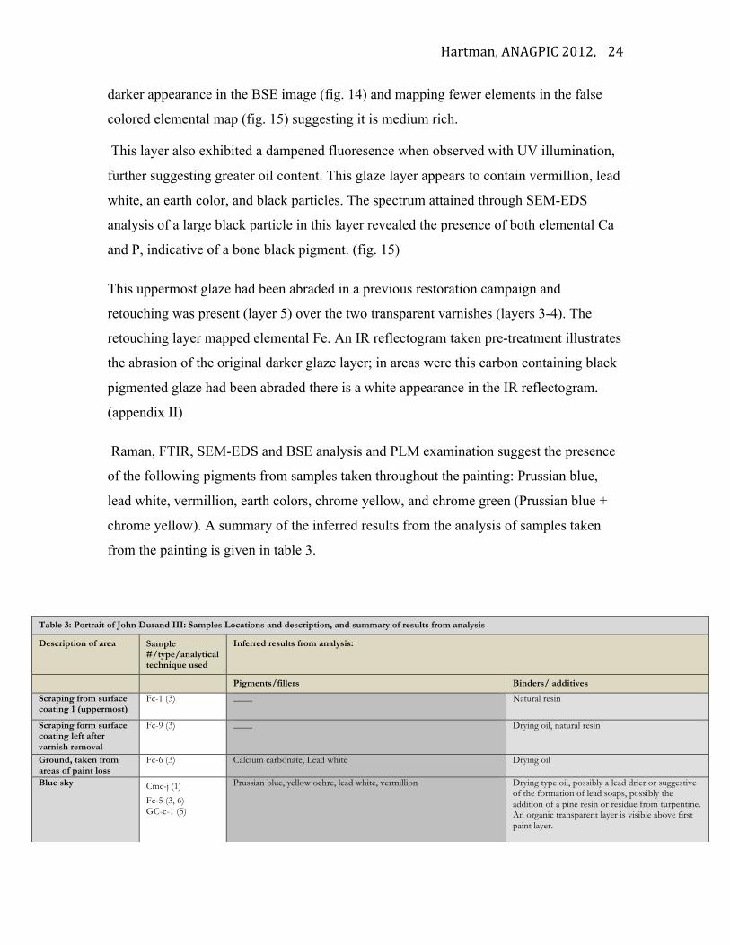
Raman, FTIR, SEM-EDS and BSE analysis and PLM examination suggest the presence
of the following pigments from samples taken throughout the painting: Prussian blue,
lead white, vermillion, earth colors, chrome yellow, and chrome green (Prussian blue +
chrome yellow). A summary of the inferred results from the analysis of samples taken
from the painting is given in table 3.
Table 3: Portrait of John Durand III: Samples Locations and description, and summary of results from analysis
Description of area Sample #/type/analytical technique used
Inferred results from analysis:
Pigments/fillers Binders/ additives
Scraping from surface coating 1 (uppermost)
Fc-1 (3) ____ Natural resin
Scraping form surface coating left after varnish removal
Fc-9 (3) ____ Drying oil, natural resin
Ground, taken from areas of paint loss
Fc-6 (3) Calcium carbonate, Lead white Drying oil
Blue sky Cmc-j (1)
Fc-5 (3, 6) GC-c-1 (5)
Prussian blue, yellow ochre, lead white, vermillion Drying type oil, possibly a lead drier or suggestive of the formation of lead soaps, possibly the addition of a pine resin or residue from turpentine. An organic transparent layer is visible above first paint layer.
Hartman, ANAGPIC 2012,
25
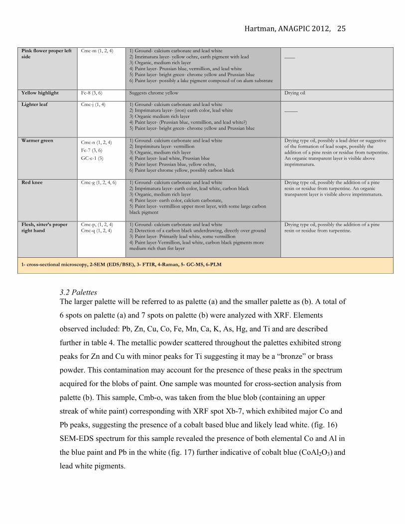
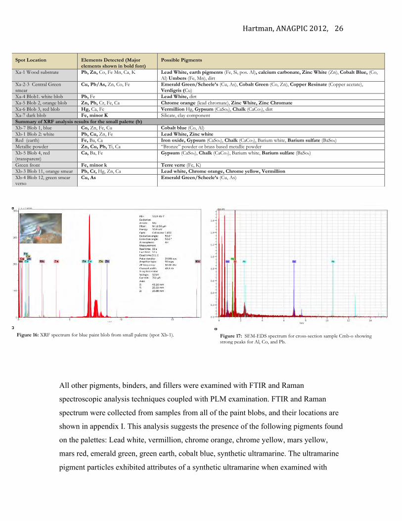
3.2 Palettes The larger palette will be referred to as palette (a) and the smaller palette as (b). A total of
6 spots on palette (a) and 7 spots on palette (b) were analyzed with XRF. Elements
observed included: Pb, Zn, Cu, Co, Fe, Mn, Ca, K, As, Hg, and Ti and are described
further in table 4. The metallic powder scattered throughout the palettes exhibited strong
peaks for Zn and Cu with minor peaks for Ti suggesting it may be a “bronze” or brass
powder. This contamination may account for the presence of these peaks in the spectrum
acquired for the blobs of paint. One sample was mounted for cross-section analysis from
palette (b). This sample, Cmb-o, was taken from the blue blob (containing an upper
streak of white paint) corresponding with XRF spot Xb-7, which exhibited major Co and
Pb peaks, suggesting the presence of a cobalt based blue and likely lead white. (fig. 16)
SEM-EDS spectrum for this sample revealed the presence of both elemental Co and Al in
the blue paint and Pb in the white (fig. 17) further indicative of cobalt blue (CoAl2O3) and
lead white pigments.
Pink flower proper left side
Cmc-m (1, 2, 4) 1) Ground- calcium carbonate and lead white 2) Imrimatura layer- yellow ochre, earth pigment with lead 3) Organic, medium rich layer 4) Paint layer- Prussian blue, vermillion, and lead white 5) Paint layer- bright green- chrome yellow and Prussian blue 6) Paint layer- possibly a lake pigment composed of on alum substrate
____
Yellow highlight Fc-8 (3, 6) Suggests chrome yellow Drying oil
Lighter leaf Cmc-j (1, 4) 1) Ground- calcium carbonate and lead white 2) Imprimatura layer- (iron) earth color, lead white 3) Organic medium rich layer 4) Paint layer- (Prussian blue, vermillion, and lead white?) 5) Paint layer- bright green- chrome yellow and Prussian blue
_____
Warmer green Cmc-n (1, 2, 4)
Fc-7 (3, 6)
GC-c-1 (5)
1) Ground- calcium carbonate and lead white 2) Imprimitura layer- vermillion 3) Organic, medium rich layer 4) Paint layer- lead white, Prussian blue 5) Paint layer: Prussian blue, yellow ochre, 6) Paint layer chrome yellow, possibly carbon black
Drying type oil, possibly a lead drier or suggestive of the formation of lead soaps, possibly the addition of a pine resin or residue from turpentine. An organic transparent layer is visible above imprimmatura.
Red knee Cmc-g (1, 2, 4, 6) 1) Ground- calcium carbonate and lead white 2) Imprimatura layer- earth color, lead white, carbon black 3) Organic, medium rich layer 4) Paint layer- earth color, calcium carbonate, 5) Paint layer- vermillion upper most layer, with some large carbon black pigment
Drying type oil, possibly the addition of a pine resin or residue from turpentine. An organic transparent layer is visible above imprimmatura.
Flesh, sitter’s proper right hand
Cmc-p, (1, 2, 4) Cmc-q (1, 2, 4)
1) Ground- calcium carbonate and lead white 2) Detection of a carbon black underdrawing, directly over ground 3) Paint layer- Primarily lead white, some vermillion 4) Paint layer-Vermillion, lead white, carbon black pigments more medium rich than fist layer
Drying type oil, possibly the addition of a pine resin or residue from turpentine.
1- cross-sectional microscopy, 2-SEM (EDS/BSE), 3- FTIR, 4-Raman, 5- GC-MS, 6-PLM
Hartman, ANAGPIC 2012,
26
All other pigments, binders, and fillers were examined with FTIR and Raman
spectroscopic analysis techniques coupled with PLM examination. FTIR and Raman
spectrum were collected from samples from all of the paint blobs, and their locations are
shown in appendix I. This analysis suggests the presence of the following pigments found
on the palettes: Lead white, vermillion, chrome orange, chrome yellow, mars yellow,
mars red, emerald green, green earth, cobalt blue, synthetic ultramarine. The ultramarine
pigment particles exhibited attributes of a synthetic ultramarine when examined with
Spot Location Elements Detected (Major elements shown in bold font)
Possible Pigments
Xa-1 Wood substrate Pb, Zn, Co, Fe Mn, Ca, K Lead White, earth pigments (Fe, Si, pos. Al), calcium carbonate, Zinc White (Zn), Cobalt Blue, (Co, Al) Umbers (Fe, Mn), dirt
Xa-2-3 Central Green smear
Cu, Pb/As, Zn, Co, Fe Emerald Green/Scheele’s (Cu, As), Cobalt Green (Co, Zn), Copper Resinate (Copper acetate), Verdigris (Cu)
Xa-4 Blob1. white blob Pb, Fe Lead White, dirt Xa-5 Blob 2, orange blob Zn, Pb, Cr, Fe, Ca Chrome orange (lead chromate), Zinc White, Zinc Chromate Xa-6 Blob 3, red blob Hg, Ca, Fe Vermillion Hg, Gypsum (CaSo4), Chalk (CaCo3), dirt Xa-7 dark blob Fe, minor K Silicate, clay component Summary of XRF analysis results for the small palette (b) Xb-7 Blob 1, blue Co, Zn, Fe, Cu Cobalt blue (Co, Al) Xb-1 Blob 2: white Pb, Cu, Zn, Fe Lead White, Zinc white Red (earth) Fe, Ba, Ca Iron oxide, Gypsum (CaSo4), Chalk (CaCo3), Barium white, Barium sulfate (BaSo4) Metallic powder Zn, Cu, Pb, Ti, Ca “Bronze” powder or brass based metallic powder Xb-5 Blob 4, red (transparent)
Ca, Ba, Fe Gypsum (CaSo4), Chalk (CaCo3), Barium white, Barium sulfate (BaSo4)
Green front Fe, minor k Terre verte (Fe, K) Xb-3 Blob 11, orange smear Pb, Cr, Hg, Zn, Ca Lead white, Chrome orange, Chrome yellow, Vermillion Xb-4 Blob 12, green smear verso
Cu, As
Emerald Green/Scheele’s (Cu, As)
Figure 16: XRF spectrum for blue paint blob from small palette (spot Xb-1). Figure 17: SEM-EDS spectrum for cross-section sample Cmb-o showing strong peaks for Al, Co, and Pb.
Hartman, ANAGPIC 2012,
27
PLM including overall purity, regular shapes, and very small sizes. All samples analyzed
with FTIR indicate they are bound in a drying oil. In both of the red blobs from palette
(b) possible filler materials were identified.
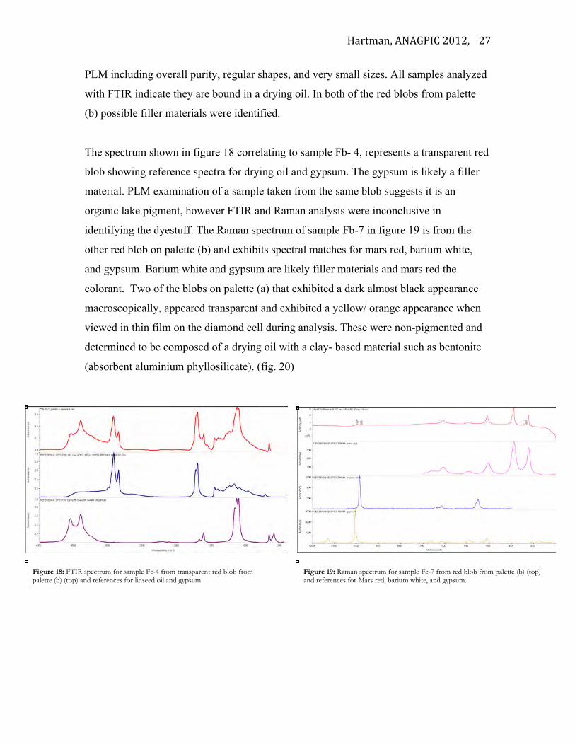
The spectrum shown in figure 18 correlating to sample Fb- 4, represents a transparent red
blob showing reference spectra for drying oil and gypsum. The gypsum is likely a filler
material. PLM examination of a sample taken from the same blob suggests it is an
organic lake pigment, however FTIR and Raman analysis were inconclusive in
identifying the dyestuff. The Raman spectrum of sample Fb-7 in figure 19 is from the
other red blob on palette (b) and exhibits spectral matches for mars red, barium white,
and gypsum. Barium white and gypsum are likely filler materials and mars red the
colorant. Two of the blobs on palette (a) that exhibited a dark almost black appearance
macroscopically, appeared transparent and exhibited a yellow/ orange appearance when
viewed in thin film on the diamond cell during analysis. These were non-pigmented and
determined to be composed of a drying oil with a clay- based material such as bentonite
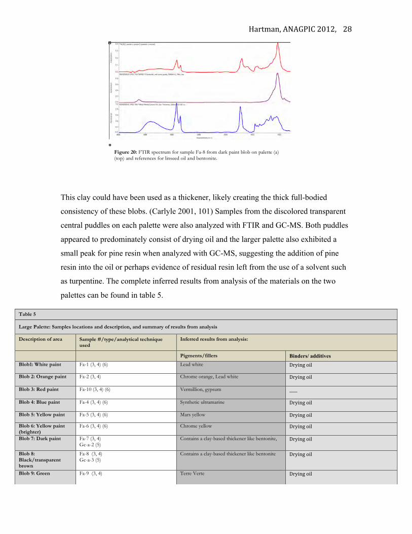
(absorbent aluminium phyllosilicate). (fig. 20)
Figure 18: FTIR spectrum for sample Fc-4 from transparent red blob from palette (b) (top) and references for linseed oil and gypsum.
Figure 19: Raman spectrum for sample Fc-7 from red blob from palette (b) (top) and references for Mars red, barium white, and gypsum.
Hartman, ANAGPIC 2012,
28
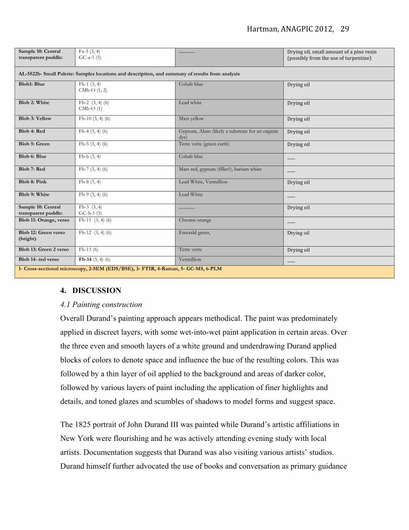
This clay could have been used as a thickener, likely creating the thick full-bodied
consistency of these blobs. (Carlyle 2001, 101) Samples from the discolored transparent
central puddles on each palette were also analyzed with FTIR and GC-MS. Both puddles
appeared to predominately consist of drying oil and the larger palette also exhibited a
small peak for pine resin when analyzed with GC-MS, suggesting the addition of pine
resin into the oil or perhaps evidence of residual resin left from the use of a solvent such
as turpentine. The complete inferred results from analysis of the materials on the two
palettes can be found in table 5.
Table 5
Large Palette: Samples locations and description, and summary of results from analysis
Description of area Sample #/type/analytical technique used
Inferred results from analysis:
Pigments/fillers Binders/ additives Blob1: White paint Fa-1 (3, 4) (6) Lead white Drying oil
Blob 2: Orange paint Fa-2 (3, 4)
Chrome orange, Lead white Drying oil
Blob 3: Red paint Fa-10 (3, 4) (6) Vermillion, gypsum ____
Blob 4: Blue paint Fa-4 (3, 4) (6) Synthetic ultramarine
Drying oil
Blob 5: Yellow paint Fa-5 (3, 4) (6) Mars yellow Drying oil
Blob 6: Yellow paint (brighter)
Fa-6 (3, 4) (6) Chrome yellow Drying oil
Blob 7: Dark paint Fa-7 (3, 4) Gc-a-2 (5)
Contains a clay-based thickener like bentonite, Drying oil
Blob 8: Black/transparent brown
Fa-8 (3, 4) Gc-a-3 (5)
Contains a clay-based thickener like bentonite Drying oil
Blob 9: Green Fa-9 (3, 4) Terre Verte Drying oil
Figure 20: FTIR spectrum for sample Fa-8 from dark paint blob on palette (a) (top) and references for linseed oil and bentonite.
Hartman, ANAGPIC 2012,
29
4. DISCUSSION
4.1 Painting construction
Overall Durand’s painting approach appears methodical. The paint was predominately
applied in discreet layers, with some wet-into-wet paint application in certain areas. Over
the three even and smooth layers of a white ground and underdrawing Durand applied
blocks of colors to denote space and influence the hue of the resulting colors. This was
followed by a thin layer of oil applied to the background and areas of darker color,
followed by various layers of paint including the application of finer highlights and
details, and toned glazes and scumbles of shadows to model forms and suggest space.
The 1825 portrait of John Durand III was painted while Durand’s artistic affiliations in
New York were flourishing and he was actively attending evening study with local
artists. Documentation suggests that Durand was also visiting various artists’ studios.
Durand himself further advocated the use of books and conversation as primary guidance
Sample 10: Central transparent puddle:
Fa-3 (3, 4) GC-a-1 (5)
______ Drying oil, small amount of a pine resin (possibly from the use of turpentine)
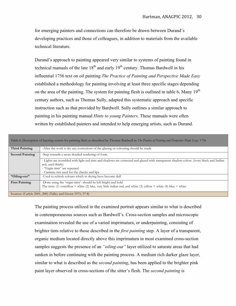
AL-5522b- Small Palette: Samples locations and description, and summary of results from analysis
Blob1: Blue Fb-1 (3, 4) CMb-O (1, 2)
Cobalt blue Drying oil
Blob 2: White Fb-2 (3, 4) (6) CMb-O (1)
Lead white Drying oil
Blob 3: Yellow Fb-10 (3, 4) (6) Mars yellow Drying oil
Blob 4: Red Fb-4 (3, 4) (6) Gypsum, Alum (likely a substrate for an organic dye)
Drying oil
Blob 5: Green Fb-5 (3, 4) (6) Terre verte (green earth) Drying oil
Blob 6: Blue Fb-6 (3, 4) Cobalt blue ____
Blob 7: Red Fb-7 (3, 4) (6) Mars red, gypsum (filler?), barium white ____
Blob 8: Pink Fb-8 (3, 4) Lead White, Vermillion Drying oil
Blob 9: White Fb-9 (3, 4) (6) Lead White ____
Sample 10: Central transparent puddle:
Fb-3 (3, 4)
GC-b-1 (5) ______ Drying oil
Blob 11: Orange, verso Fb-11 (3, 4) (6) Chrome orange ____
Blob 12: Green verso (bright)
Fb-12 (3, 4) (6) Emerald green,
Drying oil
Blob 13: Green 2 verso Fb-13 (6) Terre verte Drying oil
Blob 14- red verso Fb-14 (3, 4) (6) Vermillion ____ 1- Cross-sectional microscopy, 2-SEM (EDS/BSE), 3- FTIR, 4-Raman, 5- GC-MS, 6-PLM
Hartman, ANAGPIC 2012,
30
for emerging painters and connections can therefore be drawn between Durand’s
developing practices and those of colleagues, in addition to materials from the available
technical literature.
Durand’s approach to painting appeared very similar to systems of painting found in
technical manuals of the late 18th and early 19th century. Thomas Bardwell in his
influential 1756 text on oil painting The Practice of Painting and Perspective Made Easy
established a methodology for painting involving at least three specific stages depending
on the area of the painting. The system for painting flesh is outlined in table 6. Many 19th
century authors, such as Thomas Sully, adapted this systematic approach and specific
instruction such as that provided by Bardwell. Sully outlines a similar approach to
painting in his painting manual Hints to young Painters. These manuals were often
written by established painters and intended to help emerging artists, such as Durand.
The painting process utilized in the examined portrait appears similar to what is described
in contemporaneous sources such as Bardwell’s. Cross-section samples and microscopic
examination revealed the use of a varied imprimatura, or underpainting, consisting of
brighter tints relative to those described in the first painting step. A layer of a transparent,
organic medium located directly above this imprimatura in most examined cross-section
samples suggests the presence of an “oiling-out” layer utilized to saturate areas that had
sunken in before continuing with the painting process. A medium rich darker glaze layer,
similar to what is described as the second painting, has been applied to the brighter pink
paint layer observed in cross-sections of the sitter’s flesh. The second painting is
Table 6: Description of layering system for painting flesh as described by Thomas Bardwell in The Practice of Painting and Perspective Made Easy, 1756 Third Painting -After the work is dry any corrections of the glazing or colouring should be made
Second Painting -Step towards a more detailed rendering of form.
- Lights are scumbled with light red tints and shadows are corrected and glazed with transparent shadow-colors. (ivory black and Indian red, used thinly) - ‘Virgin tints” are repeated - Carmine tint used for the cheeks and lips
“Oiling-out” -Used to refresh colours which in drying have become dull
First Painting -Done using the ‘virgin tints’- should be left bright and bold The tints: (1) vermilion + white (2) lake, very little indian red, and white (3) yellow + white (4) blue + white
Sources: (Carlyle 2001, 200) (Talley and Groen 1975, 37-8)
Hartman, ANAGPIC 2012,
31
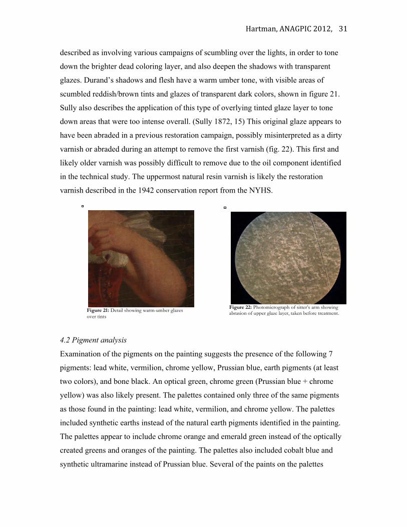
described as involving various campaigns of scumbling over the lights, in order to tone
down the brighter dead coloring layer, and also deepen the shadows with transparent
glazes. Durand’s shadows and flesh have a warm umber tone, with visible areas of
scumbled reddish/brown tints and glazes of transparent dark colors, shown in figure 21.
Sully also describes the application of this type of overlying tinted glaze layer to tone
down areas that were too intense overall. (Sully 1872, 15) This original glaze appears to
have been abraded in a previous restoration campaign, possibly misinterpreted as a dirty
varnish or abraded during an attempt to remove the first varnish (fig. 22). This first and
likely older varnish was possibly difficult to remove due to the oil component identified
in the technical study. The uppermost natural resin varnish is likely the restoration
varnish described in the 1942 conservation report from the NYHS.
4.2 Pigment analysis
Examination of the pigments on the painting suggests the presence of the following 7
pigments: lead white, vermilion, chrome yellow, Prussian blue, earth pigments (at least
two colors), and bone black. An optical green, chrome green (Prussian blue + chrome
yellow) was also likely present. The palettes contained only three of the same pigments
as those found in the painting: lead white, vermilion, and chrome yellow. The palettes
included synthetic earths instead of the natural earth pigments identified in the painting.
The palettes appear to include chrome orange and emerald green instead of the optically
created greens and oranges of the painting. The palettes also included cobalt blue and
synthetic ultramarine instead of Prussian blue. Several of the paints on the palettes
Figure 21: Detail showing warm-umber glazes over tints
Figure 22: Photomicrograph of sitter’s arm showing abrasion of upper glaze layer, taken before treatment.
Hartman, ANAGPIC 2012,
32
contained the addition of what appeared to be barium white and gypsum fillers.
Transparent blobs of drying oil with the addition of a clay filler, or bulking component,
were also present on the palettes. 19th century sources promote the use of various
mediums (such as oil and silica) that could alter the working property of the paint often
making it more brushable, while also increasing the transparency and allowing for the
improvement of shadows and application of glazes. (Carlyle 2012, 101) An even greater
variety of tubed paints were found throughout the tabouret including paints such as
chrome oxide green, asphaltum, yellow lake, Antwerp blue, and cappah brown.
In the middle of the 19th century, commercial colourman became more prevalent in the
United States. A wider variety of artists’ materials were now readily accessible, but
sometimes with unknown inclusions such as fillers and bulking agents. (Carlyle 2001,
153-155) A 1857 portrait of A.B. Durand by Daniel Huntington depicts Durand holding a
palette with a stunning range of colors, even though he is shown painting eu plein air.
This type of palette arrangement would have been difficult to accomplish for painting
outdoors earlier in the century– he would have had to hand grind his own pigments, store
them in pigs bladders and set up before hiking out to the Catskill mountains. This task
became more achievable following the invention of the collapsible paint tube and the
increased commercial sources. A somewhat similar distribution of paint is seen on the
two palettes.
4.3 Binding media/ additive materials
All of the pigments on the palettes and on the painting appear to be bound in drying oil
identified through molecular analysis with FTIR and GC-MS. Analysis of both “puddles”
of transparent media on the palettes also suggests a drying oil as the primary component.
A resinous component was also identified in the oil painting medium as well as the oil
containing puddle from the larger palette. This suggests that a resinous component, or
solvent like turpentine, was either added separately to the oil paint or perhaps, as is
visualized on the palettes, incorporated into the paints and oil medium during the painting
process.
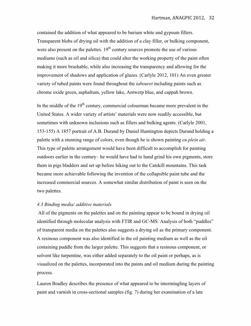
Lauren Bradley describes the presence of what appeared to be intermingling layers of
paint and varnish in cross-sectional samples (fig. 7) during her examination of a late
Hartman, ANAGPIC 2012,
33
Durand landscape painting dated to 1878, as previously discussed. Durand’s painting
method, and resulting intermingling of transparent media and paint, can be seen in his
painting palettes. Brushstrokes are evident in the transparent puddles extending into the
surrounding paint blobs, which are outlined with a black line in figure 23.
Durand’s use of oil in between paint layers and/or the addition of excess oil into his
paints probably contributed to the formation of the drying type cracks now visible in the
painting, despite the added precaution of the panel-back stretcher, and this topic would
benefit from further research. The possible addition of a metallic drier, or the presence of
metallic soaps in the paint also needs be explored further and the painting should be
monitored for what may be lead soap formation. Future research could also extend to a
more comprehensive examination of Durand’s ouvre in order to expand on his evolving
painting practices. Solvent sensitivity may well be an issue for conservators due to such
additions of resinous media to the paint, and documenting his process would further
inform conservation treatment of his work.
5. CONCLUSION Although the painting and palettes are separated by a span of approximately 54 years, and
represent only a small part of Durand’s work, the technical examination enabled
preliminary observations to be drawn about Durand’s evolving painting practices and
choices of materials. The investigation also helped to compare his work with the work of
his colleagues, manuals, and other technical sources. Durand’s approach to painting
Figure 23: Detail of puddles on both palettes (a) on left and (b) on right, outlined with black lines.
Hartman, ANAGPIC 2012,
34
utilized in the 1825 portrait of John Durand III overall appeared very similar to systems
of painting found in technical manuals of the late 18th and early 19th century. He utilized
glazing techniques such as those advocated in many technical sources and the
interlayering of organic media in what appears to be an “oiling out” process. The palettes
also exhibited transparent puddles composed of drying oil and possibly a resinous
component, or perhaps residual resin from the use of a solvent such as turpentine, that
appear to have been incorporated into the paints during the painting process.
In contrast to the pigments of the 1825 painting, the palettes of the 1870s incorporated a
more dynamic range of blues, greens, reds, and oranges as well as exhibit a continued use
of transparent glazes. Durand’s use of glazes and of these pigments would have
contributed to his depiction of the diaphanous effects he saw in nature. It seems as though
Durand benefited from the increased accessibility of new materials, which he combined
with earlier inherited techniques to create his complex mature landscape painting
technique.
The technical examination also informed conservation treatment decisions, based on a
better understanding of Durand’s artistic practices. Based on information attained through
the examination a cleaning system was designed in order to remove the upper restoration
natural resin varnish without damaging the remnants of the older, and possibly original
oil containing coating. Areas where the original tinted glaze had been abraded were also
locally retouched.
6. ACKNOWLEDGMENTS This project provided an incredible opportunity to work with specialists in the field as well as great mentors, along with the great pleasure of spending extended time with the materials of a magnificent painter. I would like to thank everyone that provided support and guidance throughout this project. From Winterthur’s Scientific Research and Analysis Laboratory (SRAL): my science advisor Dr. Joseph Weber, Dr. Jennifer L. Mass, and Catherine R. Matsen who all assisted with data collection and interpretation. Dr. Chris Petersen conducted the analysis and interpretation of samples examined with GC-MS. From the New-York Historical Society (NYHS): Linda Ferber, Scott Wixon, and Angela Nacol who all graciously allowed the palettes and painting to become part of the technical study, along with facilitating my visits to the society to spend time with the tabouret and providing advice and information on Durand.
Hartman, ANAGPIC 2012,
35
I would also like to thank Dr. Joyce Hill Stoner, Richard Wolbers, Mary McGinn, and Melanie Gifford for their expertise and advice, as well as guidance during the study and treatment of the painting, as well as the many other people who provided encouragement including Debbie Hess Norris, Jim Shneck, Jae Gutierrez, Kristin deGhetaldi, and the WUDPAC class of 2013. 7. REFERENCES Bucklow, S. 2012. The classification of craquelure patterns. The Conservation of Easel Paintings, ed. J.H. Stoner and R. Rushfield. Butterworth-Heinemann. Chap. 16. Carlyle, L. 2001. The artist’s assistant: Oil painting instruction manuals and handbooks in Britain 1800-1900, with reference to selected eighteenth century sources. London, England: Archetype Books. Dunlap, William. 1918. A History of the Rise and Progress of The Arts of Design In the United States. Boston: C.E Goodspeed and Co.
Durand, A. B. 1855. Letters on landscape painting. Originally published in The Crayon: A Journal Devoted to the Graphic Arts and the Literature Related to Them, ed. John Durand and William James Stillman, reprinted in Kindred Spirits: Asher B. Durand and the American Landscape, ed. Linda S. Ferber. London: D Giles Limited, 2007.
Durand, J. 1894. The Life and Times of A. B. Durand. New York: Charles Scribner’s
Sons.
Ferber, L. S. 2009. The Hudson River School: Nature and the American Vision. New York: The New-York Historical Society. ____. 2007. “Asher B. Durand, American Landscape Painter” In Kindred Spirits: Asher B. Durand and the American Landscape, ed. Linda S. Ferber. London: D Giles Limited. Field, G. 1869 Chromatography, salter’s edition, London: Winsor and Newton. Huntington, Daniel. 1887. Asher B. Durand: A Memorial Address. New York: Printed for the Century. Katlan, A.1999. “The American Artist’s Tools and Materials for On-Site Sketching.” Journal of the American Institute for conservation, Vol. 38, No. 1 pp 21-32 Lawall, D. B. 1977. Asher Brown Durand: His Art and Art Theory in Relation to His Times. New York and London: Garland Publishing Inc. Marckwardt. A. H. 1935. The chronology and personnel of the bread and cheese club.
Hartman, ANAGPIC 2012,
36
American Literature. 6(4): 389-399. Mayer, L. and G. Myers. 2011. American Painters on Technique: The Colonial Period to 1860. Los Angeles: J. Paul Getty Museum. ____. 1999. Bierstadt and other 19th-century American painters in context. Journal of the American Institute for Conservation. 38(1): 55-67. Stoner, J.H. 1990. Washington Allston: Poems, veils, and "Titian's dirt." Journal of the American Institute for Conservation. 29(1): 1-12. Sully, 1965 (Original 1872), Hints to Young Painters. New York: Reinhold Publishing Corporation. Talley, M. K. and K. Groen. 1975. Thomas Bardwell and his practice of painting: A comparative investigation between described and actual painting technique. Studies in Conservation. 20(2): 44-108. Zucker, J. and J. Boon. 2007. Opaque to transparent: paint film defects in the work of Frederic Church and the Hudson River School. Postprints (American Institute for Conservation of Historic and Artistic Works. Paintings Specialty Group) 19: 33- 41. Zucker, J. 1999. From the ground up: the ground in 19th-century American pictures. Journal of the American Institute for Conservation, 38(1): 3-20.
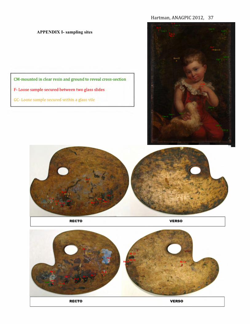
Hartman, ANAGPIC 2012,
37
APPENDIX I- sampling sites
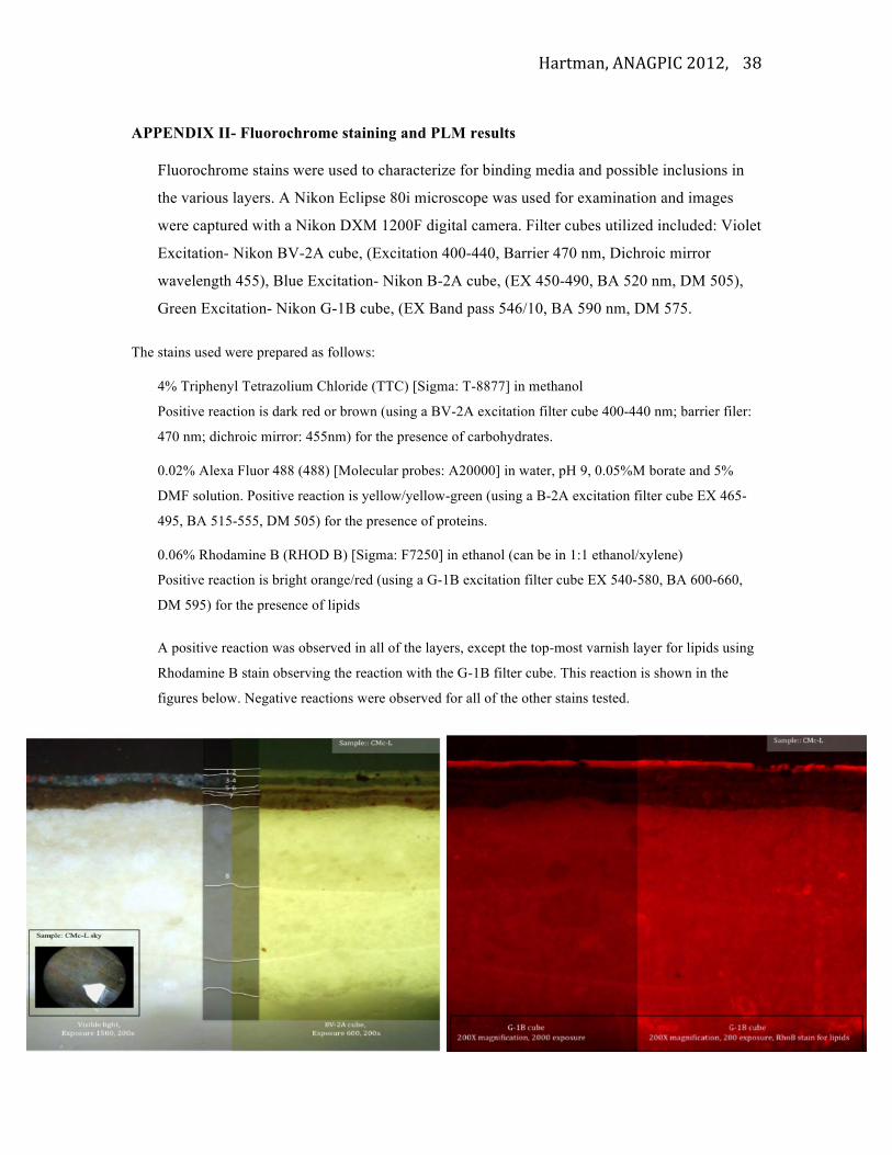
Hartman, ANAGPIC 2012,
38
APPENDIX II- Fluorochrome staining and PLM results
Fluorochrome stains were used to characterize for binding media and possible inclusions in
the various layers. A Nikon Eclipse 80i microscope was used for examination and images
were captured with a Nikon DXM 1200F digital camera. Filter cubes utilized included: Violet
Excitation- Nikon BV-2A cube, (Excitation 400-440, Barrier 470 nm, Dichroic mirror
wavelength 455), Blue Excitation- Nikon B-2A cube, (EX 450-490, BA 520 nm, DM 505),
Green Excitation- Nikon G-1B cube, (EX Band pass 546/10, BA 590 nm, DM 575.
The stains used were prepared as follows:
4% Triphenyl Tetrazolium Chloride (TTC) [Sigma: T-8877] in methanol
Positive reaction is dark red or brown (using a BV-2A excitation filter cube 400-440 nm; barrier filer:
470 nm; dichroic mirror: 455nm) for the presence of carbohydrates.
0.02% Alexa Fluor 488 (488) [Molecular probes: A20000] in water, pH 9, 0.05%M borate and 5%
DMF solution. Positive reaction is yellow/yellow-green (using a B-2A excitation filter cube EX 465-
495, BA 515-555, DM 505) for the presence of proteins.
0.06% Rhodamine B (RHOD B) [Sigma: F7250] in ethanol (can be in 1:1 ethanol/xylene)
Positive reaction is bright orange/red (using a G-1B excitation filter cube EX 540-580, BA 600-660,
DM 595) for the presence of lipids
A positive reaction was observed in all of the layers, except the top-most varnish layer for lipids using
Rhodamine B stain observing the reaction with the G-1B filter cube. This reaction is shown in the
figures below. Negative reactions were observed for all of the other stains tested.
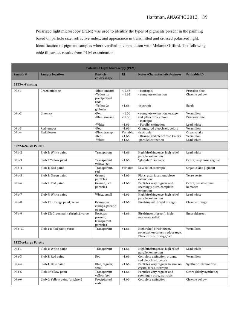
Hartman, ANAGPIC 2012,
39
Polarized light microscopy (PLM) was used to identify the types of pigments present in the painting
based on particle size, refractive index, and appearance in transmitted and crossed polarized light.
Identification of pigment samples where verified in consultation with Melanie Gifford. The following
table illustrates results from PLM examination.
Polarized Light Microscopy (PLM)
Sample # Sample location Particle color/shape
RI Notes/Characteristic features Probable ID
5523-‐c-‐Painting
DPc-‐1 Green midtone -‐Blue: smears -‐Yellow 1: precipitated, rods -‐Yellow 2: globular
< 1.66 > 1.66 >1.66
-‐ isotropic, -‐ complete extinction -‐isotropic
Prussian blue Chrome yellow Earth
DPc-‐2 Blue sky -‐Red: -‐Blue: smears -‐White:
> 1.66 < 1.66 >1.66
-‐ complete extinction, orange, red pleochroic colors -‐ Isotropic -‐ Parallel extinction
Vermillion Prussian blue Lead white
DPc-‐3 Red jumper -‐Red: >1.66 Orange, red pleochroic colors Vermillion DPc-‐4 Pink flower -‐Pink: transp.
-‐Red: -‐White:
Variable. >1.66 >1.66
-‐isotropic -‐ Orange, red pleochroic. Colors -‐parallel extinction
Organic lake Vermillion Lead white
5522-‐b-‐Small Palette
DPb-‐2 Blob 2: White paint Transparent >1.66 High birefringence, high relief, parallel extinction
Lead white
DPb-‐3 Blob 3:Yellow paint Transparent yellow ‘gel’
>1.66 “globular” isotropic Ochre, very pure, regular
DPb-‐4 Blob 4: Red paint Transparent, red
Variable Low relief, isotropic Organic lake pigment
DPb-‐5 Blob 5: Green paint Ground particles
<1.66 Flat crystal faces, undulose extinction
Terre verte
DPb-‐6 Blob 7: Red paint Ground, red particles
>1.66 Particles very regular and seemingly pure, complete extinction
Ochre, possible pure hematite
DPb-‐7 Blob 9: White paint White, small >1.66 High birefringence, high relief, parallel extinction
Lead white
DPb-‐8 Blob 11: Orange paint, verso Orange, in clumps, pseudo opaque
>1.66 Birefringent (bright orange) Chrome orange
DPb-‐9 Blob 12: Green paint (bright), verso Rosettes present, transparent particles
>1.66 Birefrincent (green), high-‐moderate relief
Emerald green
DPb-‐11 Blob 14: Red paint, verso Transparent >1.66 High relief, birefringent, polarization colors: red/orange, Pleochroism: orange/red
Vermillion
5522-‐a-‐Large Palette
DPa-‐1 Blob 1: White paint Transparent >1.66 High birefringence, high relief, parallel extinction
Lead white
DPa-‐3 Blob 3: Red paint Red >1.66 Complete extinction, orange, red pleochroic colors
Vermillion
DPa-‐4 Blob 4: Blue paint Blue, regular, small
<1.66 Particles very regular in size, no crystal faces, isotropic
Synthetic ultramarine
DPa-‐5 Blob 5:Yellow paint Transparent yellow ‘gel’
>1.66 Particles very regular and seemingly pure, isotropic
Ochre (likely synthetic)
DPa-‐6 Blob 6: Yellow paint (brighter) Precipitated, rods
>1.66 Complete extinction Chrome yellow
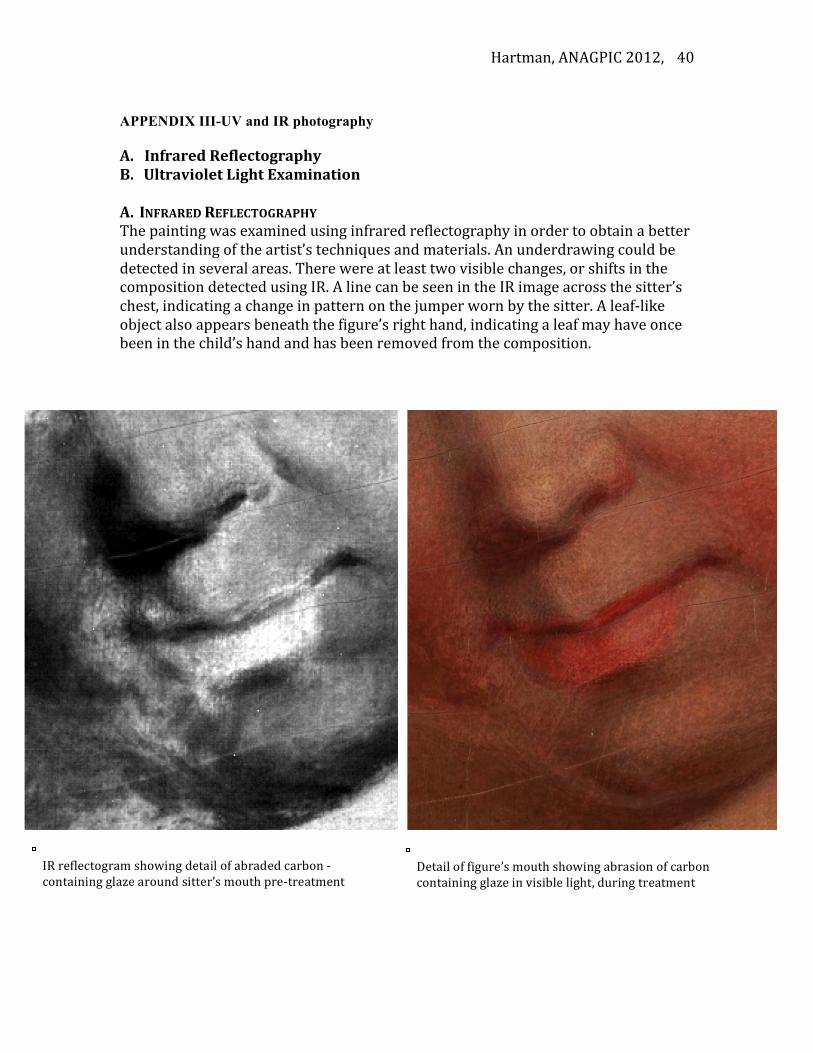
Hartman, ANAGPIC 2012,
40
APPENDIX III-UV and IR photography A. Infrared Reflectography B. Ultraviolet Light Examination A. INFRARED REFLECTOGRAPHY The painting was examined using infrared reflectography in order to obtain a better understanding of the artist’s techniques and materials. An underdrawing could be detected in several areas. There were at least two visible changes, or shifts in the composition detected using IR. A line can be seen in the IR image across the sitter’s chest, indicating a change in pattern on the jumper worn by the sitter. A leaf-‐like object also appears beneath the figure’s right hand, indicating a leaf may have once been in the child’s hand and has been removed from the composition.
IR reflectogram showing detail of abraded carbon -‐containing glaze around sitter’s mouth pre-‐treatment
Detail of figure’s mouth showing abrasion of carbon containing glaze in visible light, during treatment

Hartman, ANAGPIC 2012,
41
B. ULTRAVIOLET LIGHT EXAMINATION The painting was examined using long-‐wave ultraviolet radiation. The thick varnish coating exhibited a greenish-‐white fluorescence under this light source. This coating appears to have been applied loosely and in broad strokes, leaving many drips throughout the surface, seen in the detail below.
Older varnish, abraded throughout. Found to be composed of a drying oil and a natural resin.
Top varnish from previous restoration campaign, found to be composed of a natural resin and applied in a continuous, thick film.