Latest development in the synthesis of ursodeoxycholic ...€¦ · Latest development in the...
14
470 Latest development in the synthesis of ursodeoxycholic acid (UDCA): a critical review Fabio Tonin and Isabel W. C. E. Arends * Review Open Access Address: Department of Biotechnology, Delft University of Technology, Van der Maasweg 9, 2629 HZ Delft, The Netherlands Email: Isabel W. C. E. Arends * - [email protected] * Corresponding author Keywords: bile acids; biotransformation; hydroxysteroid dehydrogenases; production process; UDCA Beilstein J. Org. Chem. 2018, 14, 470–483. doi:10.3762/bjoc.14.33 Received: 30 October 2017 Accepted: 05 February 2018 Published: 20 February 2018 This article is part of the Thematic Series "Integrated multistep flow synthesis". Guest Editor: V. Hessel © 2018 Tonin and Arends; licensee Beilstein-Institut. License and terms: see end of document. Abstract Ursodeoxycholic acid (UDCA) is a pharmaceutical ingredient widely used in clinics. As bile acid it solubilizes cholesterol gall- stones and improves the liver function in case of cholestatic diseases. UDCA can be obtained from cholic acid (CA), which is the most abundant and least expensive bile acid available. The now available chemical routes for the obtainment of UDCA yield about 30% of final product. For these syntheses several protection and deprotection steps requiring toxic and dangerous reagents have to be performed, leading to the production of a series of waste products. In many cases the cholic acid itself first needs to be prepared from its taurinated and glycilated derivatives in the bile, thus adding to the complexity and multitude of steps involved of the syn- thetic process. For these reasons, several studies have been performed towards the development of microbial transformations or chemoenzymatic procedures for the synthesis of UDCA starting from CA or chenodeoxycholic acid (CDCA). This promising ap- proach led several research groups to focus their attention on the development of biotransformations with non-pathogenic, easy-to- manage microorganisms, and their enzymes. In particular, the enzymatic reactions involved are selective hydrolysis, epimerization of the hydroxy functions (by oxidation and subsequent reduction) and the specific hydroxylation and dehydroxylation of suitable positions in the steroid rings. In this minireview, we critically analyze the state of the art of the production of UDCA by several chemical, chemoenzymatic and enzymatic routes reported, highlighting the bottlenecks of each production step. Particular attention is placed on the precursors availability as well as the substrate loading in the process. Potential new routes and recent developments are discussed, in particular on the employment of flow-reactors. The latter technology allows to develop processes with shorter reaction times and lower costs for the chemical and enzymatic reactions involved. 470 Introduction Ursodeoxycholic acid (UDCA), is applied in the pharmaceuti- cal industry (Figure 1) [1]. As reported in several papers published in the 90’s, UDCA solubilizes cholesterol gallstones [2,3], it improves the liver function in cholestatic diseases [4-8] and it significantly decreases cholesterol saturation in the bile [8-10]. In terms of pharmacology, it is considered to be better
Transcript of Latest development in the synthesis of ursodeoxycholic ...€¦ · Latest development in the...
470
Latest development in the synthesis of ursodeoxycholic acid(UDCA): a critical reviewFabio Tonin and Isabel W. C. E. Arends*
Review Open Access
Address:Department of Biotechnology, Delft University of Technology, Van derMaasweg 9, 2629 HZ Delft, The Netherlands
Email:Isabel W. C. E. Arends* - [email protected]
* Corresponding author
Keywords:bile acids; biotransformation; hydroxysteroid dehydrogenases;production process; UDCA
Beilstein J. Org. Chem. 2018, 14, 470–483.doi:10.3762/bjoc.14.33
Received: 30 October 2017Accepted: 05 February 2018Published: 20 February 2018
This article is part of the Thematic Series "Integrated multistep flowsynthesis".
Guest Editor: V. Hessel
© 2018 Tonin and Arends; licensee Beilstein-Institut.License and terms: see end of document.
AbstractUrsodeoxycholic acid (UDCA) is a pharmaceutical ingredient widely used in clinics. As bile acid it solubilizes cholesterol gall-
stones and improves the liver function in case of cholestatic diseases. UDCA can be obtained from cholic acid (CA), which is the
most abundant and least expensive bile acid available. The now available chemical routes for the obtainment of UDCA yield about
30% of final product. For these syntheses several protection and deprotection steps requiring toxic and dangerous reagents have to
be performed, leading to the production of a series of waste products. In many cases the cholic acid itself first needs to be prepared
from its taurinated and glycilated derivatives in the bile, thus adding to the complexity and multitude of steps involved of the syn-
thetic process. For these reasons, several studies have been performed towards the development of microbial transformations or
chemoenzymatic procedures for the synthesis of UDCA starting from CA or chenodeoxycholic acid (CDCA). This promising ap-
proach led several research groups to focus their attention on the development of biotransformations with non-pathogenic, easy-to-
manage microorganisms, and their enzymes. In particular, the enzymatic reactions involved are selective hydrolysis, epimerization
of the hydroxy functions (by oxidation and subsequent reduction) and the specific hydroxylation and dehydroxylation of suitable
positions in the steroid rings. In this minireview, we critically analyze the state of the art of the production of UDCA by several
chemical, chemoenzymatic and enzymatic routes reported, highlighting the bottlenecks of each production step. Particular attention
is placed on the precursors availability as well as the substrate loading in the process. Potential new routes and recent developments
are discussed, in particular on the employment of flow-reactors. The latter technology allows to develop processes with shorter
reaction times and lower costs for the chemical and enzymatic reactions involved.
470
IntroductionUrsodeoxycholic acid (UDCA), is applied in the pharmaceuti-
cal industry (Figure 1) [1]. As reported in several papers
published in the 90’s, UDCA solubilizes cholesterol gallstones
[2,3], it improves the liver function in cholestatic diseases [4-8]
and it significantly decreases cholesterol saturation in the bile
[8-10]. In terms of pharmacology, it is considered to be better

Beilstein J. Org. Chem. 2018, 14, 470–483.
471
than chenodeoxycholic acid (CDCA) in the treatment against
biliary calculus, since it possesses high efficacy and total
absence of side effects [11].
Figure 1: Chemical structure of UDCA.
UDCA is commonly produced by transformation of cholic acid
(CA), which is the most abundant and least expensive bile acid
available. Because of the molecular complexity of bile acids,
the chemical modification requires several protection and
deprotection steps, resulting in an overall yield of about 30%
[12-15]. For that reason, research has been performed on the de-
velopment of more selective procedures which involve less
reaction steps. In particular, microbial transformations [16-19]
or chemoenzymatic procedures [20,21] employing CA, CDCA
or lithocholic acid (LCA) as starting material have been studied.
This minireview summarises different aspects to be addressed
and hurdles to be taken in the development of a selective and
sustainable process for the production of UDCA. Different
chemical, chemoenzymatic and enzymatic routes will be
considered. In addition, the precursors availability as well as the
substrate loading in the process and the requisites for potential
new routes will be discussed. Furthermore, the potential bene-
fits of a flow reactor setup for this multistep synthesis will be
discussed.
ReviewPrecursor availabilityBile acidsThe most important active ingredients of bile are the bile acids.
Together with their salts, they allow the emulsification of lipids,
a fundamental step for their absorption and digestion. Bile acids
are 24-carbon containing 5β-steroids. Their structure contains
multiple hydroxy substituents: the position and the stereochem-
istry of these OH groups influence the solubility and biochem-
ical properties of the compounds. In CA, for example, the
OH groups on the steroidal ring are all in α-position with
respect to the ring plane, defining a structure in which the mole-
cule has a polar and an apolar surface. For this reason, these
molecules and their derivatives are defined as amphipathic. Bile
acids are considered very important molecules for their ability
to form micelles in an aqueous environment [22].
Bile acids biosynthesis takes place in the liver starting from
cholesterol: 17 enzymes are involved in the production of these
molecules. The final products are the so-called primary
bile acids: CDCA and CA [23]. Subsequently, these bile
acids can be modified by intestinal bacteria to form the
secondary bile acids as, for example, deoxycholic acid
(DCA), LCA and UDCA. Secondary bile acids can be
subsequently resorbed and returned to the liver where
they are re-secreted in a process known as enterohepatic circu-
lation.
In mammals, bile acids are secreted as conjugated molecules
with glycin or taurine (Figure 2), forming the so called bile
salts, with slightly different properties (pKa, solubility) in com-
parison to the corresponding free acids [24,25]. These bile salts
also lead to an increased retention in the intestine.
Figure 2: Chemical structures of bile acids and salts.
The only economically viable resource of bile acids
is the bovine bile, which must be extracted at the time of
slaughter.
In slaughterhouses, the bovine gallbladder is recovered during
the processing of the meat and from a single cow, around
230 mL of bile can be obtained. The commercial prize of bile is
in the range of 0.1–0.4 $/L. Bile acids represent roughly
0.7% (w/w) of the bile [26].
In order to extract and purify the different bile acids, bile is
frozen and lyophilized: from 100 mL of bile 8 g of dry powder
can be obtained. From these about 6.9 g of 90% pure bile acids
can be obtained [27]. Cholesterol, cholesterol esters, triglyc-

Beilstein J. Org. Chem. 2018, 14, 470–483.
472
erides and free fatty acids are selectively extracted with organic
solvents from aqueous buffers at different pHs. Then, the bile
acids were separated from inorganic salts by extraction of the
dry residue with absolute ethanol. The major components of the
obtained mixture are the primary bile acids (CA and CDCA),
secondary bile acids (DCA and LCA) and bile salts like tauro-
cholic acid and glycocholic acid (derivatives of CA), tauro-
chenodeoxycholic acid and glycochenodeoxycholic acid (deriv-
atives of CDCA) and other conjugated salts of their 7-α-dehy-
droxylated derivatives [27].
CA3α,7α,12α-Trihydroxy-5β-cholan-24-oic acid, also named
cholic acid (CA, Figure 2), is one of the primary bile acids. It is
almost insoluble in water, but soluble in methanol, ethanol and
acetic acid. Differently from the acid, the sodium salt of CA is
much more soluble in water at pH > 8.0 [28,29]. Salts of CA are
called cholates. From the 3D structure of CA is it possible to
observe an hydrophilic and an hydrophobic face, giving to CA
its characteristic surfactant properties. CA is sold as a treatment
for children and adults affected by bile acids synthesis disor-
ders. Because of its abundance in bovine bile, CA is the precur-
sor for UDCA.
CDCA3α,7α-Dihydroxy-5β-cholan-24-oic acid, also known as CDCA
(Figure 2), is a primary bile acid in human, but represents only
2.7% (w/w) of total bovine bile salts [30]: this is the main
reason why it is not used as precursor for the preparation of
UDCA.
CDCA can be used to treat gallstones avoiding, unlike CA, the
downregulation of the cholesterol-7-α-hydroxylase, that repre-
sent the rate-limiting step in bile acid synthesis [31]. It can be
metabolized by bacteria in the colon to form the secondary bile
acid known as LCA [32].
LCA and other bile acidsLCA, also known as 3α-hydroxy-5β-cholan-24-oic acid
(Figure 2), is a secondary bile acid. It is produced by bacteria in
the colon from CDCA trough the dehydroxylation of the C7
functional group of the steroid framework. Low percentages of
other secondary bile acids and related keto derivatives can be
found in the bile. The solubility properties, interactions and
metabolisms are related to the position and stereochemistry of
the hydroxy groups attached to the steroid ring. A general struc-
ture with the names of several bile acids is reported in Figure 2.
DeconjugationFree bile acids can be obtained from the corresponding bile
amides (with glycine and taurine) through a deconjugation step.
Chemically, the reaction is an hydrolysis of the amide deriva-
tives, that can be carried out at high temperature in alkaline
environment. This reaction requires large amounts of sodium
hydroxide (30%) and high temperatures (120 °C) for extremely
long times (8–12 hours). Few enzymes (acylases, EC: 3.5.1)
have been reported to hydrolyse glycinates and taurinates to the
corresponding carboxylic acid. Recently, Pedrini et al. [33]
have isolated and characterized a cholylglycine hydrolase from
Xanthomonas maltophilia CBS 827.97: this enzyme completely
hydrolyses glycine and taurine conjugates in 20 minutes at
50 °C. Unfortunately the protein sequence of this enzyme is not
reported, making recombinant expression and its industrial use
impossible. A second enzyme, isolated in Lactobacillus plan-
tarum and recombinantly expressed in E. coli, was reported by
Christiaens et al. [34]: this enzyme shows almost the same
properties of the one described above but with lower activities
(the specific activities of X. maltophilia and L. plantarum
acylases on glycocholic acid as substrate are 100 U/mg and
3.42 U/mg, respectively).
From a biocatalytic point of view, other acylases and the well-
known lipases can be used to achieve the same reaction. Few
literature reports can be found on the promiscuous amidase ac-
tivity of wild-type or engineered lipases [35,36], but no one
have tested their activities on bile salts.
Conclusions on the precursor availabilityNatural UDCA is a very expensive pharmaceutical active ingre-
dient since, to date, it can only be obtained by isolation from
bear bile (practice used in traditional Chinese medicine) [37].
Alternatively, it can be produced by chemical transformation of
CA and CDCA from the cheaper bovine bile. Nowadays, the
transformation process is not yet optimized in terms of costs
and environmental impact [20]. CA is the main constituent of
bovine bile and is the main precursor for the synthesis of
UDCA.
One of the main problems regarding the availability of precur-
sors for the synthesis of UDCA is the direct relation with meat
industries. The major manufacturers of bovine meat are in
newly industrialised countries, in particular south America
(Brasil) and India. Reports on Ecuadorian slaughterhouses
[26,38-40], point out that in some cases there is a lack of
adequate technical conditions and hygienic protocols, leading to
environmental pollution and the need to include sanitary proce-
dures in the processing of bile acids.
Alternative sources of sterols can be found in eukaryotic micro-
organisms like yeast and algae [41]. However, technological
and scientific knowledge on these metabolic pathways are still
in an early stage, and will not be included in this review.

Beilstein J. Org. Chem. 2018, 14, 470–483.
473
C12 DehydroxylationChemical dehydroxylationUDCA can be obtained by a multistep chemical synthesis
starting from CA. Two main steps are involved: the dehydroxy-
lation at C12 and the epimerization of the 7-OH group.
In order to achieve chemical dehydroxylation, firstly CA has to
be oxidized in position C12 to the corresponding ketone, after
which Wolff–Kishner reduction can be applied. This whole se-
quence comprises 5 steps [13]. After the protection of the
carboxylic group by acid-catalyzed esterification (quantitative
yield), the 3- and 7-OH groups are protected selectively with
acetic anhydride and pyridine (yield 92%). The 12-OH group is
oxidized with CrO3 (yield 98%) and, after a deprotection step in
alkaline environment, the formed ketone group can be removed
by a Wolff–Kishner reaction yielding CDCA (yield 82%). The
overall yield of the dehydroxylation step is around 65%.
Wolff–Kishner reductionThe Wolff–Kishner reaction is widely used by chemists to
remove carbonyl moieties yielding unsubstituted alkyl chains.
The reaction requires two steps: the hydrazine first reacts with
the ketone forming a hydrazone; The addition of a strong base
and heat then promote a rearrangement with the elimination of
N2 yielding the desired alkyl chain (Figure 3). This reaction is
applied to the synthesis of UDCA in order to remove the car-
bonyl group at C12.
Figure 3: Comparison between Wolff–Kishner and Mozingo reduction.Notably the overall chemical reaction is the same for both cases.
The use of hydrazine is a disadvantage in terms of safety
(explosive risk) and economic costs (it should be used in
excess relative to substrate). Several modifications of the
Wolff–Kishner reaction can be found in literature: i.e., the
Huang–Minlon modification, that consists of removing the
unreacted hydrazine after the first step by distillation [42]. This
results in higher yield and partial recovery of the unreacted
hydrazine. In order to reduce the explosive risk, different
hydrazine derivatives have been discovered and tested (e.g.,
methyl hydrazinocarboxylate, [43]). Other reports have demon-
strated the feasibility of this reaction in a flow-system, avoiding
the large excess of hydrazine, giving high yields in a more rapid
reaction [44]. In this field, flow reactors offer benefits in terms
of mass and heat transfer, both enhanced by the geometry of the
reactor. Furthermore, the possibility to have a continuous pro-
duction and to easily perform multiple modular reactions, leads
to the improved scalability of these systems. In addition, a
microwave-assisted Wolff–Kishner reduction has been exam-
ined with good results in a 30 seconds reaction [45]. Another
option is the production of hydrazine in situ, using chemical
methods. Also enzymatic activities towards hydrazine have
been discovered [46], however, not enough optimized to be
applicable.
A Wolff–Kishner alternative: the Mozingo reductionThe Mozingo reduction effects the same reaction as
Wolff–Kishner, albeit under neutral conditions (Figure 3). It
involves two steps: firstly, the carbonyl compound is converted
into a dithioketal by adding a dithiol. The mechanism for this
step is analogous to the mechanism for ketal or acetal forma-
tion except sulphur replaces oxygen as the nucleophile attacking
the carbonyl group.
In a second step, the dithioketal is reduced to the corresponding
methylene compound by hydrogenolysis in presence of Raney
Nickel (actually used for the hydrogenation of fatty acids). In
comparison to the Wolff–Kishner reaction, the use of hydrazine
is replaced by the use of hydrogen gas, which can be seen as a
double-bladed knife. The reduction step can also be performed
with NaBH4 or other reductants. At the moment, a complete and
clear reaction mechanism has not been well identified yet.
There is one report that suggests the application of this reaction
for the synthesis of UDCA (yield 95%) [47]. The major prob-
lem related to this reaction is the very characteristic odor of
ethanedithiol which is compared by many people to rotten
cabbage. Ideally, there is the possibility of using other types of
less volatile compounds but no reports thereof have been found.
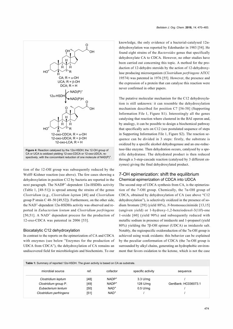
12α-Hydroxysteroid dehydrogenase12α-Hydroxysteroid dehydrogenases (12α-HSDH) are particu-
larly interesting for the selective oxidation of the 12-hydroxy
group of CA (Figure 4). These enzymes belong to the family of
oxidoreductases with NAD+ or NADP+ as electron acceptor.
This oxidation is a mandatory step for removing the OH func-
tionality at C12. In all the chemoenzymatic routes reported by
Eggert et al. [20], the carbonyl group resulting from the oxida-
Beilstein J. Org. Chem. 2018, 14, 470–483.
474
Table 1: Summary of reported 12α-HSDH. The given activity is based on CA as substrate.
microbial source ref. cofactor specific activity sequence
Clostridium leptum [48] NADP+ 3.3 U/mg /Clostridium group P. [49] NADP+ 128 U/mg GenBank: HC036073.1Eubacterium lentum [50] NAD+ 0.5 U/mg /
Clostridium perfringens [51] NAD+ / /
Figure 4: Reaction catalysed by the 12α-HSDH; the 12-OH group ofCA or UCA is oxidized yielding 12-oxo-CDCA or 12-oxo-UDCA, re-spectively, with the concomitant reduction of one molecule of NAD(P)+.
tion of the 12-OH group was subsequently reduced by the
Wolff–Kishner reaction (see above). The few cases showing a
dehydroxylation in position C12 by bacteria are reported in the
next paragraph. The NADP+-dependent 12α-HSDHs activity
(Table 1, [48-51]) is spread among the strains of the genus
Clostridium (e.g., Clostridium leptum [48] and Clostridium
group P strain C 48–50 [49,52]). Furthermore, on the other side,
the NAD+-dependent 12α-HSDHs activity was observed and re-
ported in Eubacterium lentum and Clostridium perfringens
[50,51]. A NAD+ dependent process for the production of
12-oxo-CDCA was patented in 2006 [53].
Biocatalytic C12 dehydroxylationIn contrast to the reports on the epimerization of CA and CDCA
with enzymes (see below ”Enzymes for the production of
UDCA from CDCA”), the dehydroxylation of CA remains an
undiscovered field for microbiologists and biochemists. To our
knowledge, the only evidence of a bacterial-catalysed 12α-
dehydroxylation was reported by Edenharder in 1983 [54]. He
found eight strains of the Bacteroides genus that specifically
dehydroxylate CA to CDCA. However, no other studies have
been carried out concerning this topic. A method for the pro-
duction of 12-dehydro steroids by the action of 12-dehydroxy-
lase producing microorganism (Clostridium perfringens ATCC
19574) was patented in 1976 [55]. However, the presence and
the expression of a protein that can catalyse this reaction were
never confirmed in other papers.
The putative molecular mechanism for the C12 dehydroxyla-
tion is still unknown: it can resemble the dehydroxylation
mechanism described for position C7 [56-58] (Supporting
Information File 1, Figure S1). Interestingly all the genes
catalysing that reaction where clustered in the BAI operon and,
by analogy, it can be possible to design a biochemical pathway
that specifically acts on C12 (see postulated sequence of steps
in Supporting Information File 1, Figure S2). The reaction se-
quence can be divided in 3 steps: firstly, the substrate is
oxidized by a specific alcohol dehydrogenase and an ene-reduc-
tase-like enzyme. Then dehydration occurs, catalysed by a spe-
cific dehydratase. The dehydrated product is then reduced
through a 3-step-cascade reaction (catalysed by 3 different en-
zymes) giving the final dehydroxylated product.
7-OH epimerization: shift the equilibriumChemical epimerization of CDCA into UDCAThe second step of UDCA synthesis from CA, is the epimeriza-
tion of the 7-OH group. Chemically, the 7α-OH group of
CDCA, obtained by dehydroxylation of CA (see above “C12
dehydroxylation”), is selectively oxidized in the presence of so-
dium bromate [59] (yield 88%), N-bromosuccinimide [13,15]
(ungiven yield) or 1-hydroxy-1,2-benziodoxol-3(1H)-one
1-oxide [60] (yield 90%) and subsequently reduced with
metallic sodium in presence of imidazole and 1-propanol (yield
80%) yielding the 7β-OH epimer (UDCA) as imidazole salt.
Notably, the regiospecific oxidoreduction of the 7α-OH group is
achieved using weak oxidants: this behavior can be explained
by the peculiar conformation of CDCA (the 7α-OH group is
surrounded by alkyl chains, generating an hydrophobic environ-
ment that favors oxidation to the ketone, which is not the case

Beilstein J. Org. Chem. 2018, 14, 470–483.
475
Table 2: Summary of reported 7α-HSDH. The given activity is on chenodeoxycholic acid as substrate.
microbial source ref. cofactor specific activity sequence
Clostridium sordelii [63] NADP+ 1.1 U/mg GenBank: AAA53556.1Eubacterium scindens [64] NADP+ 338 U/mg GenBank: AAB61151.1Clostridium absonum [65] NADP+ 59 U/mg GenBank: JN191345.1Clostridium difficile [66] NADP+ 8.5 U/mg Genbank: YP 001086529
Escherichia coli [67] NAD+ 190 U/mg GenBank: KXH01569.1Pseudomonas sp. B-0831 [68] NAD+ 941 U/mg GenBank: D50325.1
Bacteroides fragilis [69] NAD+ 351 U/mg GenBank: OGX95366.1Xanthomonas maltophilia [33] NAD+ 70 U/mg /Comamonas testosteroni [70] / / /
for the other epimer). These data are supported by a density
functional calculation or rather the differential change in elec-
tron density due to an infinitesimal change in the number of
electrons [61]. The overall yield of the epimerization step is
around 70% [12,15,62].
A further purification step is necessary for the preparation of
free UDCA: it can be easily obtained with sequential esterifica-
tion, extraction and hydrolysis (yield 91%). The theoretical
yield of the whole process fluctuates around 30 to 40%.
Enzymes for the production of UDCA from CDCAThe enzymatic transformation of CDCA into UDCA can be ob-
tained using different combinations of biocatalysts that act
specifically on the 7-OH group of hydroxysteroids. In order to
find the right combination of enzymes and the optimum reac-
tion conditions, different aspects (enzymatic activities, equilib-
rium of the reaction, inhibition of enzymes by substrate and
products and their stabilities) have to be assessed.
A list of enzymes that can be used for the transformation of
CDCA is presented in the next paragraphs.
7α-Hydroxysteroid dehydrogenases (7α-HSDH)These enzymes are able to oxidise specifically the α-hydroxy
group at C7 together with the concomitant reduction of NAD+
or NADP+ (Figure 5). All of them are part of the group of the
short chain dehydrogenases/reductases (SDR), showing a mo-
lecular weight around 30 kDa and a homodimeric or homote-
trameric quaternary structure.
Reported 7α-HSDHs were isolated from both aerobic and
anaerobic bacteria: the state of art, together with the cofactor
dependence and the specific activities, are summarized in
Table 2 [33,63-70]. Examples of processes for the selective oxi-
dation of bile acids, their salts or derivatives were patented
[71,72].
Figure 5: Epimerization reaction catalysed by the 7α-HSDH and7β-HSDH; the 7α-OH group of CA (R = OH) or CDCA (R = H) is firstlyoxidized by the 7α-HSDH yielding 7-oxo-DCA or 7-oxo-LCA, respec-tively. Subsequently, the keto group is reduced by the 7β-HSDH givingthe final products UCA or UDCA.
In addition to these reported biotransformations, many addition-
al 7α-HSDHs have been discovered and reported over the past
years. About 500 entries can be found in Reference sequence

Beilstein J. Org. Chem. 2018, 14, 470–483.
476
Table 3: Summary of reported 7β-HSDH. The given activity is based on 7-oxo-LCA as substrate.
microbial source ref. cofactor specific activity sequence
Clostridium absonum [65] NADP+ 65 U/mg GenBank: JN191345.1Eubacterium aereofaciens [74] NADP+ 30 U/mg GenBank: ZP0177306.1
Ruminococcus gnavus [75] NADP+ 23 U/mg GenBank: ZP02041813Collinsella aerofaciens [77] NADP+ 15 U/mg GenBank: WP006236005Collinsella aerofaciens [77] NADP+ 21 U/mg Engineereda
Ruminococcus torques [76] NADP+ 8.6 U/mg GenBank: WP015528793Ruminococcus torques [76] NADP+ 46.8 U/mg Engineeredb
Xanthomonas maltophilia [33] NAD+ 33 U/mg /aG39A variant of the 7β-HSDH from Collinsella aerofaciens; bT198V/V207M variant of the 7β-HSDH from Ruminococcus torques.
Table 4: Summary of reported whole-cell transformations with wild type microorganisms. Epimerization yields of CDCA to UDCA are given.
microorganism ref. yield (%)
Colinsiella aerofaciens [80] –Clostridium absonum [81] 75%
E. coli + Bacteroides fragilis [82] 25–30%Colinsiella aerofaciens + Bacteroides fragilis [82] 95%
mixed culture [83] –Clostridium limosum [84] 55–60% (75–80%a)
Stenotrophomonas maltophilia [85] 27% (80%b)aReported yield of epimerization of CA to ursocholic acid; breported yield of epimerization of 12-oxo-CDCA to 12-oxo-UDCA.
(RefSeq) database at NCBI [73] typing “7-alpha-hydroxy-
steroid dehydrogenase”.
7β-Hydroxysteroid dehydrogenases (7β-HSDH)Unlike their homologues, only few examples of bioconversion
with 7β-HSDH have been reported in literature (Table 3,
[33,65,74-77], Figure 5): one NADP+-dependent dehydroge-
nase from Clostridium absonum [65] was used in two different
processes for the production of UDCA [78,79]. The NADP+-
dependent enzyme from Eubacterium aerofaciens shows a
significantly lower specific activity [74] and another NADP+-
dependent enzyme was isolated from Ruminococcus gnavus
[75]. In order to increase the activity and stability of
7β-HSDHs, protein engineering studies were carried out, as
described in literature by Weuster-Botz et al. [77] and Zheng et
al. [76]. Up till now, Xanthomonas maltophilia 7β-HSDH
(33 U/mg) represents the only isolated NAD+-dependent en-
zyme [33]. Unfortunately its protein sequence has not been re-
ported.
Biocatalytic processesBoth microorganisms and purified enzymes have been applied
for the fully biocatalytic epimerization of the 7-OH group.
Several examples reported in literature are summarized in
Table 4 [80-85].
The use of whole-cell conversion offers both advantages
and disadvantages: wild-type microorganisms are normally
difficult to grow, especially if the enzyme expression is related
to anaerobic conditions. In addition the pathogenicity of
these microorganisms represents a problem for their use in
the pharmaceutical industry; additional steps of purification and
control of sterility are necessary to obtain a safe product for
the market. Otherwise, the circumvention of protein isolation
and production makes it cheaper than their free-enzyme ana-
logues.
In this way, the use of lyophilized whole-cell containing recom-
binant HSDHs can represent a solution in the reduction of cata-
lyst costs, maintaining a reasonable safety. This approach was
followed by Braun et al. and Sun et al. obtaining the 12-oxo-
UDCA with a yield of 99.5% using engineered E. coli cells
[86,87]. In comparison to the systems that employed purified
enzymes (see below), this approach allows higher substrate
loading (70–100 mM).
Several enzymatic systems have been proposed in the literature,
together with cofactor regeneration systems. As general rule,
the oxidative and reductive steps are coupled with a related
regeneration system. In this way, the equilibrium of the reac-
tion can be pushed to the production of UDCA. An overview of

Beilstein J. Org. Chem. 2018, 14, 470–483.
477
Table 5: Summary of reported chemoenzymatic transformations with purified enzymes.
reaction pathway ref. conversion yield (%) productivity (g L−1 d−1)
DHCA→12-oxo-UDCA
[12] 85% 7.0[86] 95% 9.6[87] 99% 40.1[88] 95% 55.6[89] 99% 7.3
CA→UDCA [90] 70% 1.8
CA→12-oxo-UDCA[78] 88% 8.0[91] 73% 24.1
CDCA→UDCA
[33] 82% 0.02[92] 100% 47.2[93] 100% 88.5[94] 63% 3.0
Figure 6: Overview of the chemoenzymatic process for the production of UDCA from CA: The oxidation, reduction and dehydroxylation reactions arehighlighted with a red, blue and green arrow, respectively. Names of compounds are indicated in the box. The general stereoinformation of steroidscaffold is shown in Figure 2. R = 4-pentanoic acid.
reported enzymatic and chemoenzymatic cascades is summa-
rized in Table 5 and Figure 6 [12,33,78,86-94].
From the comparison of the different routes, it can be observed
that higher productions are obtained when the Wolff–Kishner
reaction is carried out in the late stage. The typical substrate
loading in systems employing purified enzymes is in the range
of 10–15 mM. This disadvantage is partially compensated by
the reuse of the biocatalysts through the employment of mem-
brane reactors [12,78] or the immobilization of enzymes [93].

Beilstein J. Org. Chem. 2018, 14, 470–483.
478
The first system shows high stability (enzymes in the mem-
brane reactor have a half-life of 1–2 weeks) and the biocata-
lysts can be reused for eight cycles of conversions. On the other
hand, immobilized enzymes show a higher productivity
(88.5 vs 8 g L−1 d−1) despite the fact that the half-life (23 h) is
lower and the biocatalyst can be reused for only five cycles of
conversions.
In order to reduce the mechanical stress that might inactivate
the immobilized enzymes, the flow-system represents a valid
technology. The packed-bed reactor set up by Zheng et al.
partially solved this problem, achieving full conversion of
CDCA into UDCA for at least 12 hours. This represents an
improvement compared to the use of the same biocatalysts
under batch conditions. This particular flow-system consists of
two modular column reactors (Figure 7): firstly, CDCA is
oxidized to 7-oxo-LCA by an immobilized NAD+-dependent
7α-HSHS (first reactor column); afterwards, 7-oxo-LCA is
reduced to UDCA by an immobilized NADP+-dependent
7β-HSDH (second reactor column). The cofactors are individu-
ally regenerated in each column by the co-immobilized en-
zymes, lactate dehydrogenase (LDH) and glucose dehydroge-
nase (GDH), respectively.
The decoupling of the 2 reactions is an elegant way to spin the
equilibrium but, in every catalytic cycle, the co-substrates used
to regenerate the cofactor have to be added in great surplus,
leading to additional costs and additional problems in the down-
stream process. The most used enzymes for the cofactor regen-
eration are glucose dehydrogenase (glucose to glucuronic acid),
lactate dehydrogenase (pyruvate to lactate), glutamate dehydro-
genase (α-ketoglutarate to glutamate) and formate dehydroge-
nase (formate to CO2). In particular, the last enzyme is interest-
ing because formate is cheap and, because of the gaseous nature
of CO2 as product, the equilibrium of the reaction is entropical-
ly favoured.
Pedrini et al. in 2006 [33] reported the successful epimerization
of CDCA to UDCA using a redox-neutral cascade reaction,
with two NAD+-dependent dehydrogenases. In this way the
requirement of external systems for cofactor regeneration was
circumvented and UDCA was obtained with a final yield of
75%. Interestingly, the addition of 2-hexanol led to an increase
of NADH available for the reduction of 7-oxo-LCA and a final
yield of 82% was observed. According to the authors, the pres-
ence of another alcohol dehydrogenase in the partially-purified
enzyme preparation increases the amount of NADH for the
7-oxo-LCA reduction.
To conclude, it is difficult to denote the “best” route for the
7-OH epimerization. All the processes mentioned, demonstrate
Figure 7: Schematic representation of the flow reactor for the continu-ous conversion of CDCA to UDCA [93].
reasonable yield and high selectivity. A redox-neutral cascade
seems most elegant, but in order to fully understand and push
the equilibrium of the reaction, a full biochemical characteriza-
tion and a deep knowledge of the kinetics and stability of the
involved enzymes is required.
Other ways to obtain 7-OH epimerizationOther chemical routes for the production of UDCA have been
patented and published: for example, Dangate et al. [60] pro-

Beilstein J. Org. Chem. 2018, 14, 470–483.
479
posed a chemical route where the order of the two steps (C12
dehydroxylation and C7 epimerization) is reverted and the spe-
cific oxidation of 7- and 12-OH group can be achieved without
any protection step (yield 53%).
Another interesting chemoenzymatic way to obtain the epimeri-
zation of the 7-OH group consists is the removal of the func-
tionality and the subsequent rehydroxylation with a specific
final chiral configuration. Both steps can be performed by en-
zymes and/or microorganisms: Sawada et al. [95] reported that
a fungal strain (Fusarium equiseti M41) was able to introduce a
7β-hydroxy group into LCA by hydroxylation forming UDCA
directly.
Later, many other microorganisms with a 7β-hydroxylating
activity were discovered in strains of actinobacteria and
filamentous fungi [96,97]. The key-enzyme in that pathway is a
P450-like enzyme that catalyses the specific and irreversible
7β-hydroxylation. On this topic, a recent work by Kollerov et
al. [98] describes several DCA modifying filamentous fungi
strains (mostly ascomycetes and zygomycetes): the highest
7β-hydroxylase activity level was found in Fusarium meris-
moides VKM F-2310.
The possibility to access that kind of chemical and chemo-
enzymatic reactions pave the way for the design of other unex-
plored routes for the production of UDCA (example in
Figure 8).
In addition, other reported enzymes can eventually play a role
in the cascade reaction synthesis of UDCA. For example the
3α-HSDHs [51,99] catalyze the oxidoreduction of the
3α-OH groups to the corresponding ketones and the well-known
laccase-TEMPO system [100] can be used for the unselective
oxidation of CA to dehydrocholic acid (DHCA).
Solvent and substrate loadingconsiderations in processingFor an economically and environmentally sustainable process
volumetric productivities have to be considered. In other words
substrate loadings cannot be too low. While it does not repre-
sent a problem in chemical synthesis (UDCA, CDCA and CA
are pretty soluble in alcohols like methanol and ethanol), the
water-based environment required by enzymes is an obstacle in
the development of a biocatalytic process.
In comparison to CA, the solubility of CDCA and UDCA at
pH 8.0 (typically used for HSDHs) is lower (around 25 mM)
[101], and it could be increased when adding methanol or
ethanol as co-solvent. In addition, at higher concentrations than
the critical micelles concentration (CMC), bile acids tend to
Figure 8: Chemoenzymatic pathways for the formation of UDCA fromCA that profit by the C7 hydroxylation activity described by Sawada etal. [95]. CA can be transformed to 7,12-dioxo-LCA trough specific oxi-dation of 7α-OH and 12α-OH with a 7α-HSDH and 12α-HSDH, respec-tively. Alternatively, 7,12-dioxo-LCA can be obtained by chemicallyoxidizing (e.g., with CrO3) all the hydroxy groups, yielding DHCA andthen reducing the 3-oxo group to 3α-OH by a 3α-HSDH. LCA can beobtained from 7,12-dioxo-LCA through dehydroxylation byWolff–Kishner or Mozingo reduction. Finally, UDCA can be obtainedfrom LCA by 7β-hydroxylation. The general stereoinformation of thesteroid scaffold is shown in Figure 2.
form micelles: this phenomenon, due to the amphipathic struc-
ture of these molecules, is limiting the availability of free
hydroxysteroids in solution. The reported CMCs of bile acids

Beilstein J. Org. Chem. 2018, 14, 470–483.
480
are in the range of 5–15 mM [101-104]. Accordingly, the addi-
tion of co-solvents increases the CMC of bile acids and the
availability of monomers in solution. Notably, HSDHs are rela-
tively stable and active in 10–20% methanol. Moreover, the im-
mobilization of the enzyme can provide a higher stability to the
protein and makes the system work also at higher concentra-
tions of co-solvent [105]. However, working with a diluted
solution, produce a large amount of wastewater that had to be
treated.
Another option is represented by biphasic systems: In these
cases, the organic phase works as reservoir of reagents and
products. This methodology is widely used in biocatalysis to
solve solubility issues. Unfortunately, the solubility of hydroxy-
steroids in non-alcoholic organic solvents (e.g., ethers, alkanes,
dichloromethane, chloroform) is not very high (e.g., the re-
ported solubility values for CDCA and CA in chloroform are
7.6 and 14.4 mM, respectively [102]). Several attempts to carry
out hydroxysteroid transformations in biphasic systems were re-
ported [106-108]: good conversions and an increase of reaction
rates were observed for 7-OH and 3-OH epimerizations. In
these cases the reactions were carried out at final concentra-
tions of substrate in the range of 10–20 mM, the usual substrate
loading in monophasic systems.
Of no lesser importance, the increased amount of substrates and
products up to relevant concentrations for industrial application
can inhibit the enzymes used in the biocatalytic process. Several
examples are reported in literature about substrate or product
inhibition of HSDHs. Protein engineering could help to solve or
lowering the effect of these issues, leading to the optimization
of the biocatalyst for industrial applications. In addition, the use
of flow-reactors can be beneficial to diminish substrate and
product inhibition by controlling the contact time.
In conclusion, the increase of the substrate loading is one of the
main challenges in the development of an efficient biocatalytic
system for the production of UDCA form CA. More research is
needed to address this aspects.
ConclusionThe organic synthesis of CDCA and UDCA starting from tauri-
nated and glycinated cholic acid is a long process, complicated
and risky due to the nature and toxicity of the reagents used, the
costs of disposal of large amounts of sodium hydroxide,
chromium salts and organic solvents, and the purification pro-
cesses necessary to eliminate byproducts formed at each step of
reaction involved. All this extends the time, increases costs and
decreases production yields. Therefore, research nowadays is
geared towards more economical synthesis methods that are
waste-free and safe to operate.
An approach that bears great promise is the biotransformation
with non-pathogenic, easy-to-manage microorganisms, and
their enzymes. Several chemical, chemoenzymatic and
enzymatic routes have been proposed for the production
of UDCA. In view of sustainability, instead of pursuing a
step-wise approach, an integrated one-pot or one-flow
reaction, involving highly selective enzymatic steps would be
preferred.
When a multi-enzyme system is employed, the different en-
zyme activities, pH optima, cross reactions and inhibitions have
to be taken into account in order to reach high product yields
[109-111]. Furthermore, when a combination of chemical and
enzymatic steps is employed special attention has to be paid to
the compatibility: the combination of enzymatic and chemical
transformation steps is the main task to achieve in order to
obtain high yields of UDCA.
Nowadays, the most promising system for the biocatalytic pro-
duction of UDCA are flow-reactors. They can be used for the
setup of continuous working systems, lowering the quantity of
catalyst needed and the time of each reaction. This technology
was recently employed by Zheng et al. [93] leading to high
yields (99%) and productivity (88.5 g L−1 d−1) for the epimeri-
zation of CDCA to UDCA. However, the employed enzymes
have different cofactor specificities, leading to the consumption
of stoichiometric amounts of sacrificial substrates (pyruvate and
glucose). In addition, substrate loadings in the latter process are
still modest (10 mM). Therefore, there is much room for
improvement and further studies are needed to design a truly
sustainable integrated process for the production of UDCA.
AbbreviationsCA, cholic acid; CDCA, chenodeoxycholic acid; DCA, deoxy-
cholic acid; DHCA, dehydrocholic acid; LCA, lithocholic acid;
UCA, ursocholic acid; UDCA, ursodeoxycholic acid; HSDH,
hydroxysteroid dehydrogenase; LDH, lactate dehydrogenase;
GDH, glucose dehydrogenase.
Supporting InformationSupporting Information features Figure S1 relative to the
C7 dehydroxylation mechanism of hydroxysteroids and
Figure S2 relative to the postulated biochemical pathway
for the C12 dehydroxylation.
Supporting Information File 1Supporting Figures S1 and S2.
[https://www.beilstein-journals.org/bjoc/content/
supplementary/1860-5397-14-33-S1.pdf]

Beilstein J. Org. Chem. 2018, 14, 470–483.
481
AcknowledgementsThis work was done as part of the ONE-FLOW project (https://
one-flow.org) that has received funding from the European
Union's FET-Open research and innovation actions (proposal
737266 - ONE-FLOW). We thank Linda Otten, Duncan
McMillan and all the BOC group of TU Delft for the scientific
discussions.
References1. Ikegami, T.; Matsuzaki, Y. Hepatol. Res. 2008, 38, 123–131.
doi:10.1111/j.1872-034X.2007.00297.x2. Crosignani, A.; Battezzati, P. M.; Setchell, K. D. R.; Invernizzi, P.;
Covini, G.; Zuin, M.; Podda, M. Dig. Dis. Sci. 1996, 41, 809–815.doi:10.1007/BF02213140
3. Salen, G.; Colalillo, A.; Verga, D.; Bagan, E.; Tint, G.; Shefer, S.Gastroenterology 1980, 78, 1412–1418.
4. Colombo, C.; Setchell, K. D. R.; Podda, M.; Crosignani, A.; Roda, A.;Curcio, L.; Ronchi, M.; Giunta, A. J. Pediatr. 1990, 117, 482–489.doi:10.1016/S0022-3476(05)81103-5
5. Colombo, C.; Crosignani, A.; Assaisso, M.; Battezzati, P. M.;Podda, M.; Giunta, A.; Zimmer-Nechemias, L.; Setchell, K. D. R.Hepatology (Hoboken, NJ, U. S.) 1992, 16, 924–930.doi:10.1002/hep.1840160412
6. Colombo, C.; Battezzati, P. M.; Podda, M.; Bettinardi, N.; Giunta, A.Hepatology (Hoboken, NJ, U. S.) 1996, 23, 1484–1490.doi:10.1002/hep.510230627
7. Combes, B.; Carithers, R. L.; Maddrey, W. C.; Lin, D.;McDonald, M. F.; Wheeler, D. E.; Eigenbrodt, E. H.; Muñoz, S. J.;Rubin, R.; Garcia-Tsao, G.; Bonner, G. F.; West, A. B.; Boyer, J. L.;Luketic, V. A.; Shiffman, M. L.; Mills, A. S.; Peters, M. G.;White, H. M.; Zetterman, R. K.; Rossi, S. S.; Hofmann, A. F.;Markin, R. S. Hepatology (Hoboken, NJ, U. S.) 1995, 22, 759–766.doi:10.1002/hep.1840220311
8. Podda, M.; Zuin, M.; Battezzati, P. M.; Ghezzi, C.; De Fazio, C.;Dioguardi, M. L. Gastroenterology 1989, 96, 222–229.doi:10.1016/0016-5085(89)90784-1
9. Thistle, J. L. Semin. Liver Dis. 1983, 3, 146–156.doi:10.1055/s-2008-1040680
10. Ward, A.; Brogden, R. N.; Heel, R. C.; Speight, T. M.; Avery, G. S.Drugs 1984, 27, 95–131. doi:10.2165/00003495-198427020-00001
11. Roda, E.; Bazzoli, F.; Labate, A. M. M.; Mazzella, G.; Roda, A.;Sama, C.; Festi, D.; Aldini, R.; Taroni, F.; Barbara, L.Hepatology (Hoboken, NJ, U. S.) 1982, 2, 804–810.doi:10.1002/hep.1840020611
12. Carrea, G.; Pilotti, A.; Riva, S.; Canzi, E.; Ferrari, A. Biotechnol. Lett.1992, 14, 1131–1134. doi:10.1007/BF01027015
13. Hofmann, A. F. Acta Chem. Scand. 1963, 17, 173–186.doi:10.3891/acta.chem.scand.17-0173
14. Kanazawa, T.; Shimazaki, A.; Sato, T.; Hoshino, T.Nippon Kagaku Zasshi 1955, 76, 297–301.doi:10.1246/nikkashi1948.76.297
15. Fieser, L. F.; Rajagopalan, S. J. Am. Chem. Soc. 1950, 72,5530–5536. doi:10.1021/ja01168a046
16. Sutherland, J. D.; Macdonald, I. A.; Forrest, T. P. Prep. Biochem.1982, 12, 307–321. doi:10.1080/00327488208065679
17. Prabha, V.; Ohri, M. World J. Microbiol. Biotechnol. 2006, 22,191–196. doi:10.1007/s11274-005-9019-y
18. Midtvedt, T.; Norman, A. Acta Pathol. Microbiol. Scand. 1967, 71,629–638. doi:10.1111/j.1699-0463.1967.tb05183.x
19. Mahato, S. B.; Garai, S. Steroids 1997, 62, 332–345.doi:10.1016/S0039-128X(96)00251-6
20. Eggert, T.; Bakonyi, D.; Hummel, W. J. Biotechnol. 2014, 191, 11–21.doi:10.1016/j.jbiotec.2014.08.006
21. Bortolini, O.; Medici, A.; Poli, S. Steroids 1997, 62, 564–577.doi:10.1016/S0039-128X(97)00043-3
22. Mukhopadhyay, S.; Maitra, U. Curr. Sci. 2004, 87, 1666–1683.23. Hofmann, A. F. Arch. Intern. Med. 1999, 159, 2647–2658.
doi:10.1001/archinte.159.22.264724. Fini, A.; Roda, A. J. Lipid Res. 1987, 28, 755–759.25. Bonar-Law, R. P.; Davis, A. P. Tetrahedron 1993, 49, 9829–9844.
doi:10.1016/S0040-4020(01)80185-X26. Maldonado Rodríguez, M. E. Biotrasformazioni di Acidi Biliari. Ph.D.
Thesis, Università degli Studi di Ferrara, Italy, 2013.27. Ahrens, E. H.; Craig, L. C. J. Biol. Chem. 1952, 195, 763–778.28. Carey, M. C. Hepatology (Hoboken, NJ, U. S.) 1984, 4, 66S–71S.
doi:10.1002/hep.184004081229. Fossati, E.; Polentini, F.; Carrea, G.; Riva, S. Biotechnol. Bioeng.
2006, 93, 1216–1220. doi:10.1002/bit.2075330. Washizu, T.; Tomoda, I.; Kaneko, J. J. J. Vet. Med. Sci. 1991, 53,
81–86. doi:10.1292/jvms.53.8131. Iser, J. H.; Sali, A. Drugs 1981, 21, 90–119.
doi:10.2165/00003495-198121020-0000232. Carey, M. C. N. Engl. J. Med. 1975, 293, 1255–1257.
doi:10.1056/NEJM19751211293241233. Pedrini, P.; Andreotti, E.; Guerrini, A.; Dean, M.; Fantin, G.;
Giovannini, P. P. Steroids 2006, 71, 189–198.doi:10.1016/j.steroids.2005.10.002
34. Christiaens, H.; Leer, R.; Pouwels, P.; Verstraete, W.Appl. Environ. Microbiol. 1992, 58, 3792–3798.
35. Nakagawa, Y.; Hasegawa, A.; Hiratake, J.; Sakata, K.Protein Eng., Des. Sel. 2007, 20, 339–346.doi:10.1093/protein/gzm025
36. Torres-Gavilán, A.; Castillo, E.; López-Munguía, A.J. Mol. Catal. B: Enzym. 2006, 41, 136–140.doi:10.1016/j.molcatb.2006.06.001
37. Feng, Y.; Siu, K.; Wang, N.; Ng, K.-M.; Tsao, S.-W.; Nagamatsu, T.;Tong, Y. J. Ethnobiol. Ethnomed. 2009, 5, 2.doi:10.1186/1746-4269-5-2
38. Delgado, H.; Cedeño, C.; de Oca, N.; Villoch, A. Rev. Salud Anim.2015, 37, 1–9.
39. Delgado, D.; Roque, P.; Cedeño, P.; Villoch, C. Rev. Salud Anim.2015, 37, 69–78.
40. Delgado, H.; Cedeño, C.; Villoch, A.; Dueñas, R. Rev. Salud Anim.2015, 37, 198–202.
41. Volkman, J. Appl. Microbiol. Biotechnol. 2003, 60, 495–506.doi:10.1007/s00253-002-1172-8
42. Huang-Minlon, B. Y. J. Am. Chem. Soc. 1949, 71, 3301–3303.doi:10.1021/ja01178a008
43. Cranwell, P. B.; Russell, A. T.; Smith, C. D. Synlett 2016, 27,131–135. doi:10.1055/s-0035-1560805
44. Newman, S. G.; Gu, L.; Lesniak, C.; Victor, G.; Meschke, F.;Abahmane, L.; Jensen, K. F. Green Chem. 2014, 16, 176–180.doi:10.1039/C3GC41942H
45. Parquet, E.; Lin, Q. J. Chem. Educ. 1997, 74, 1225.doi:10.1021/ed074p1225

Beilstein J. Org. Chem. 2018, 14, 470–483.
482
46. Dietl, A.; Ferousi, C.; Maalcke, W. J.; Menzel, A.; de Vries, S.;Keltjens, J. T.; Jetten, M. S. M.; Kartal, B.; Barends, T. R. M. Nature2015, 527, 394–397. doi:10.1038/nature15517
47. Sato, Y.; Ikekawa, N. J. Org. Chem. 1959, 24, 1367–1368.doi:10.1021/jo01091a623
48. Harris, J. N.; Hylemon, P. B.Biochim. Biophys. Acta, Lipids Lipid Metab. 1978, 528, 148–157.doi:10.1016/0005-2760(78)90060-7
49. Braun, M.; Lünsdorf, H.; Bückmann, A. F. Eur. J. Biochem. 1991, 196,439–450. doi:10.1111/j.1432-1033.1991.tb15835.x
50. Macdonald, I. A.; Jellett, J. F.; Mahony, D. E.; Holdeman, L. V.Appl. Environ. Microbiol. 1979, 37, 992–1000.
51. Macdonald, I. A.; Meier, E. C.; Mahony, D. E.; Costain, G. A.Biochim. Biophys. Acta, Lipids Lipid Metab. 1976, 450, 142–153.doi:10.1016/0005-2760(76)90086-2
52. Aigner, A.; Gross, R.; Schmid, R. D.; Braun, M.; Mauer, S. Novel 12alpha-hydroxysteroid dehydrogenase, production and use thereof. USPatent 20110091921 A1, April 21, 2011.
53. Fossati, E.; Carrea, G.; Riva, S.; Polentini, F. Process for the selectiveoxydation of cholic acid. Eur. Patent EP 1731618 A1, Dec 13, 2006.
54. Edenharder, R. J. Steroid Biochem. 1984, 21, 413–420.doi:10.1016/0022-4731(84)90304-2
55. Saltzman, W. H. Synthesis of steroids. U.S. Patent US 3954562 A,May 4, 1976.
56. Wells, J. E.; Hylemon, P. B. Appl. Environ. Microbiol. 2000, 66,1107–1113. doi:10.1128/AEM.66.3.1107-1113.2000
57. Ridlon, J. M.; Kang, D.-J.; Hylemon, P. B. Anaerobe 2010, 16,137–146. doi:10.1016/j.anaerobe.2009.05.004
58. Ridlon, J. M.; Harris, S. C.; Bhowmik, S.; Kang, D.-J.; Hylemon, P. B.Gut Microbes 2016, 7, 22–39. doi:10.1080/19490976.2015.1127483
59. Ferrari, M.; Zinetti, F. Process for preparing high purityursodeoxycholic acid. WO Patent WO 2014020024 A1 06/02/2014.
60. Dangate, P. S.; Salunke, C. L.; Akamanchi, K. G. Steroids 2011, 76,1397–1399. doi:10.1016/j.steroids.2011.07.009
61. Parr, R. G.; Yang, W. J. Am. Chem. Soc. 1984, 106, 4049–4050.doi:10.1021/ja00326a036
62. Samuelsson, B. Acta Chem. Scand. 1960, 14, 17–20.doi:10.3891/acta.chem.scand.14-0017
63. Coleman, J. P.; Hudson, L. L.; Adams, M. J. J. Bacteriol. 1994, 176,4865–4874. doi:10.1128/jb.176.16.4865-4874.1994
64. Baron, S. F.; Franklund, C. V.; Hylemon, P. B. J. Bacteriol. 1991, 173,4558–4569. doi:10.1128/jb.173.15.4558-4569.1991
65. Ferrandi, E. E.; Bertolesi, G. M.; Polentini, F.; Negri, A.; Riva, S.;Monti, D. Appl. Microbiol. Biotechnol. 2012, 95, 1221–1233.doi:10.1007/s00253-011-3798-x
66. Bakonyi, D.; Hummel, W. Enzyme Microb. Technol. 2017, 99, 16–24.doi:10.1016/j.enzmictec.2016.12.006
67. Yoshimoto, T.; Higashi, H.; Kanatani, A.; Lin, X. S.; Nagai, H.;Oyama, H.; Kurazono, K.; Tsuru, D. J. Bacteriol. 1991, 173,2173–2179. doi:10.1128/jb.173.7.2173-2179.1991
68. Ueda, S.; Oda, M.; Imamura, S. Int. J. Biol. Macromol. 2004, 4, 33–38.69. Bennett, M. J.; McKnight, S. L.; Coleman, J. P. Curr. Microbiol. 2003,
47, 475–484. doi:10.1007/s00284-003-4079-470. Ji, W.; Chen, Y.; Zhang, H.; Zhang, X.; Li, Z.; Yu, Y. Microbiol. Res.
2014, 169, 148–154. doi:10.1016/j.micres.2013.07.00971. Monti, D.; Ferrandi, E. E.; Riva, S.; Polentini, F. New process for the
selective oxidation of bile acids, their salts or derivatives. WO PatentWO 2012131591 A1, Oct 4, 2012.
72. Gupta, A.; Tschentscher, A.; Bobkova, M. Process for theenantioselective reduction and oxidation, respectively, of steroids.U.S. Patent US 20090280525 A1, Nov 12, 2009.
73. O'Leary, N. A.; Wright, M. W.; Brister, J. R.; Ciufo, S.; Haddad, D.;McVeigh, R.; Rajput, B.; Robbertse, B.; Smith-White, B.;Astashyn, D. A.-A. A.; Batretdin, A.; Bao, Y.; Blinkova, O.; Brover, V.;Chetvernin, V.; Choi, J.; Cox, E.; Ermolaeva, O.; Farrell, C. M.;Goldfarb, T.; Gupta, T.; Haft, D.; Hatcher, E.; Hlavina, W.;Joardar, V. S.; Kodali, V. K.; Li, W.; Maglott, D.; Masterson, P.;McGarvey, K. M.; Murphy, M. R.; O'Neill, K.; Pujar, S.;Rangwala, S. H.; Rausch, D.; Riddick, L. D.; Schoch, C.; Shkeda, A.;Storz, S. S.; Sun, H.; Thiaud-Nissen, F.; Tolstoy, I.; Tully, R. E.;Vatsan, A. R.; Wallin, C.; Webb, D.; Wu, W.; Landrum, M. J.;Kimchi, A.; Tatusova, T.; DiCuccio, M.; Kitts, P.; Murphy, T. D.;Pruitt, K. D. Nucleic Acids Res. 2015, 44, D733–D745.doi:10.1093/nar/gkv1189
74. Liu, L.; Aigner, A.; Schmid, R. D. Appl. Microbiol. Biotechnol. 2011,90, 127–135. doi:10.1007/s00253-010-3052-y
75. Lee, J.-Y.; Arai, H.; Nakamura, Y.; Fukiya, S.; Wada, M.; Yokota, A.J. Lipid Res. 2013, 54, 3062–3069. doi:10.1194/jlr.M039834
76. Zheng, M.-M.; Chen, K.-C.; Wang, R.-F.; Li, H.; Li, C.-X.; Xu, J.-H.J. Agric. Food Chem. 2017, 65, 1178–1185.doi:10.1021/acs.jafc.6b05428
77. Hummel, W.; Bakonyi, D. 7-beta-hydroxysteroid dehydrogenasemutants and process for the preparation of ursodeoxycholic acid. WOPatent WO 2016016213 A1, Feb 4, 2016.; .
78. Bovara, R.; Carrea, G.; Riva, S.; Secundo, F. Biotechnol. Lett. 1996,18, 305–308. doi:10.1007/BF00142949
79. Monti, D.; Ottolina, G.; Carrea, G.; Riva, S. Chem. Rev. 2011, 111,4111–4140. doi:10.1021/cr100334x
80. Macdonald, I. A.; Hutchison, D. M. J. Steroid Biochem. 1982, 17,295–303. doi:10.1016/0022-4731(82)90203-5
81. Sutherland, J. D.; Macdonald, I. A. J. Lipid Res. 1982, 23, 726–732.82. Macdonald, I.; Rochon, Y.; Hutchison, D.; Holdeman, L.
Appl. Environ. Microbiol. 1982, 44, 1187–1195.83. Hirano, S.; Masuda, N. J. Lipid Res. 1982, 23, 1152–1158.84. Sutherland, J. D.; Holdeman, L. V.; Williams, C. N.; Macdonald, I. A.
J. Lipid Res. 1984, 25, 1084–1089.85. Medici, A.; Pedrini, P.; Bianchini, E.; Fantin, G.; Guerrini, A.;
Natalini, B.; Pellicciari, R. Steroids 2002, 67, 51–56.doi:10.1016/S0039-128X(01)00136-2
86. Braun, M.; Sun, B.; Anselment, B.; Weuster-Botz, D.Appl. Microbiol. Biotechnol. 2012, 95, 1457–1468.doi:10.1007/s00253-012-4072-6
87. Sun, B.; Kantzow, C.; Bresch, S.; Castiglione, K.; Weuster-Botz, D.Biotechnol. Bioeng. 2013, 110, 68–77. doi:10.1002/bit.24606
88. Bakonyi, D.; Wirtz, A.; Hummel, W. Z. Naturforsch., B: Chem. Sci.2012, 67, 1037–1044. doi:10.5560/ZNB.2012-0165
89. Liu, L.; Braun, M.; Gebhardt, G.; Weuster-Botz, D.; Gross, R.;Schmid, R. D. Appl. Microbiol. Biotechnol. 2013, 97, 633–639.doi:10.1007/s00253-012-4340-5
90. Giovannini, P. P.; Grandini, A.; Perrone, D.; Pedrini, P.; Fantin, G.;Fogagnolo, M. Steroids 2008, 73, 1385–1390.doi:10.1016/j.steroids.2008.06.013
91. Monti, D.; Ferrandi, E. E.; Zanellato, I.; Hua, L.; Polentini, F.;Carrea, G.; Riva, S. Adv. Synth. Catal. 2009, 351, 1303–1311.doi:10.1002/adsc.200800727

Beilstein J. Org. Chem. 2018, 14, 470–483.
483
92. Zheng, M.-M.; Wang, R.-F.; Li, C.-X.; Xu, J.-H.Process Biochem. (Oxford, U. K.) 2015, 50, 598–604.doi:10.1016/j.procbio.2014.12.026
93. Zheng, M.-M.; Chen, F.-F.; Li, H.; Li, C.-X.; Xu, J.-H. ChemBioChem2017, in press. doi:10.1002/cbic.201700415
94. Ji, Q.; Tan, J.; Zhu, L.; Lou, D.; Wang, B. Biochem. Eng. J. 2016, 105,1–9. doi:10.1016/j.bej.2015.08.005
95. Sawada, H.; Kulprecha, S.; Nilubol, N.; Yoshida, T.; Kinoshita, S.;Taguchi, H. Appl. Environ. Microbiol. 1982, 44, 1249–1252.
96. Carlström, K.; Kirk, D.; Sjövall, J. J. Lipid Res. 1981, 22, 1225–1234.97. Kollerov, V. V.; Monti, D.; Deshcherevskaya, N. O.; Lobastova, T. G.;
Ferrandi, E. E.; Larovere, A.; Gulevskaya, S. A.; Riva, S.;Donova, M. V. Steroids 2013, 78, 370–378.doi:10.1016/j.steroids.2012.12.010
98. Kollerov, V. V.; Lobastova, T. G.; Monti, D.; Deshcherevskaya, N. O.;Ferrandi, E. E.; Fronza, G.; Riva, S.; Donova, M. V. Steroids 2016,107, 20–29. doi:10.1016/j.steroids.2015.12.015
99. Möbus, E.; Maser, E. J. Biol. Chem. 1998, 273, 30888–30896.doi:10.1074/jbc.273.47.30888
100.Arends, I. W. C. E.; Li, Y.-X.; Ausan, R. A.; Sheldon, R. A.Tetrahedron 2006, 62, 6659–6665. doi:10.1016/j.tet.2005.12.076
101.Igimi, H.; Carey, M. C. J. Lipid Res. 1980, 21, 72–90.102.Cabral, D. J. In Physical chemistry of bile, Compr. Physiol.;
Small, D. M., Ed.; John Wiley & Sons, 2010.103.Carey, M. C.; Small, D. M. Arch. Intern. Med. 1972, 130, 506–527.
doi:10.1001/archinte.1972.03650040040005104.Hisadome, T.; Nakama, T.; Itoh, H.; Furusawa, T. J.
Gastroenterol. Jpn. 1980, 15, 257–263.105.Khmelnitsky, Y. L.; Levashov, A. V.; Klyachko, N. L.; Martinek, K.
Enzyme Microb. Technol. 1988, 10, 710–724.doi:10.1016/0141-0229(88)90115-9
106.Riva, S.; Bovara, R.; Zetta, L.; Pasta, P.; Ottolina, G.; Carrea, G.J. Org. Chem. 1988, 53, 88–92. doi:10.1021/jo00236a018
107.Bovara, R.; Canzi, E.; Carrea, G.; Pilotti, A.; Riva, S. J. Org. Chem.1993, 58, 499–501. doi:10.1021/jo00054a039
108.Monti, D.; Ferrandi, E. E.; Riva, S.; Polentini, F. Process for theselective reduction of bile acids, their salts or derivatives, in a biphasicsystem. WO Patent WO 2013179210 A2, Dec 5, 2013.
109.Ricca, E.; Brucher, B.; Schrittwieser, J. H. Adv. Synth. Catal. 2011,353, 2239–2262. doi:10.1002/adsc.201100256
110.Oroz-Guinea, I.; García-Junceda, E. Curr. Opin. Chem. Biol. 2013, 17,236–249. doi:10.1016/j.cbpa.2013.02.015
111.Riva, S.; Fessner, W.-D. Cascade Biocatalysis IntegratingStereoselective and Environmentally Friendly Reactions; John Wiley &Sons, 2014.
License and TermsThis is an Open Access article under the terms of the
Creative Commons Attribution License
(http://creativecommons.org/licenses/by/4.0), which
permits unrestricted use, distribution, and reproduction in
any medium, provided the original work is properly cited.
The license is subject to the Beilstein Journal of Organic
Chemistry terms and conditions:
(https://www.beilstein-journals.org/bjoc)
The definitive version of this article is the electronic one
which can be found at:
doi:10.3762/bjoc.14.33