Late Granule Cell Genesis in Quail...
17
Late Granule Cell Genesis in Quail Cerebellum ANTONIS STAMATAKIS, 1 HELEN BARBAS, 2 AND CATHERINE R. DERMON 1 * 1 Department of Biology, University of Crete, Heraklion 714 09, Crete, Greece 2 Department of Health Sciences, Boston University, Boston, Massachusetts 02215 ABSTRACT Proliferation of avian cerebellar neurons, including granule cells, is thought to be completed during embryonic life, and aspects of cell addition in cerebellar lobules in post- hatching life are unknown. The present study tested the hypothesis that cell genesis in late embryonic and posthatching stages of quail cerebellum occurs in parallel with the perfor- mance of motor programs. After exposure to bromodeoxyuridine, short (20 hours) and long survival time points were selected to investigate survival and migration of labeled cells. Quantitative analysis of the lobular distribution of labeled cells was performed with the stereological disector method. External granular layer (EGL) proliferation did not cease after hatching, indicating that there is an extended posthatching period, lasting until P20, when cells can be added into the internal granular layer, modifying the cerebellar circuitry and function. Indeed, long survival experiments suggested that EGL-labeled cells migrated into the internal granular layer and survived for a prolonged time, although many of the progen- itor cells remained in the EGL for days. Double-labeling experiments revealed that most of the late-generated granule cells were NeuN positive, but only few expressed nitric oxide synthase. In addition to granule cells, the white matter and a glutamic acid decarboxylase (GAD)-positive cell population in the molecular layer around Purkinje somata showed bro- modeoxyuridine labeling. Although all lobules showed significant posthatching proliferation, an anteroposterior gradient was evident. The index of granule cell production and survival supports a spatiotemporal pattern, in correlation with the functional division of cerebellum into anterior and posterior domains. J. Comp. Neurol. 474:173–189, 2004. © 2004 Wiley-Liss, Inc. Indexing terms: cerebellar development; external granular layer; posthatching proliferation; migration; BrdU immunocytochemistry; stereological disector The cerebellum, a highly conserved structure, is com- posed of anteroposterior and mediolateral modular com- partments where distinct components of motor behavior are presumably stored and modified when mastering mo- tor tasks or acquiring reflexes (Ito et al., 1982; De Zeeuw et al., 1994; Herrup and Kuemerle, 1997; Ito, 2000). Its laminar pattern and rectilinear geometry (Palay and Chan-Palay, 1974) have long been exploited to provide an important model for understanding the development of normal brain cytoarchitecture and function (Ramon y Ca- jal, 1911). The progeny of rhombic lip and external gran- ule layer (EGL) progenitors have been determined by ret- roviral lineage, gene expression, and chick-quail chimeric studies in early stages of avian embryos (Ryder and Cepko, 1994; Lin et al., 2001). Rostral metencephalic and caudal mesencephalic vesicles of embryonic brain partici- pate in the formation of the cerebellum in a topographic order (Hallonet et al., 1990; Hallonet and Le Douarin, 1993; Wassef and Joyner, 1997). Cerebellar cells are pro- duced in two distinct proliferation zones during two dis- tinct time frames: The ventricular zone of the IVth ven- tricle produces cells early, and the EGL on the surface of the developing cerebellum produces cells later and over an extended period. Cell types known to derive from the Grant sponsor: the European Union; Grant number: Q5RS-2000-31629; Grant sponsor: Greek General Secretariat Research and Technology (C.R.D.). In memory of Dr. R. Dermon. *Correspondence to: Catherine Dermon, Laboratory of Physiology/ Neurobiology, Department of Biology, University of Crete, Heraklion 714 09, Crete, Greece. E-mail: [email protected] Received 12 June 2003; Revised 27 October 2003; Accepted 18 December 2003 DOI 10.1002/cne.20066 Published online in Wiley InterScience (www.interscience.wiley.com). THE JOURNAL OF COMPARATIVE NEUROLOGY 474:173–189 (2004) © 2004 WILEY-LISS, INC.
Transcript of Late Granule Cell Genesis in Quail...

Late Granule Cell Genesisin Quail Cerebellum
ANTONIS STAMATAKIS,1 HELEN BARBAS,2AND CATHERINE R. DERMON1*
1Department of Biology, University of Crete, Heraklion 714 09, Crete, Greece2Department of Health Sciences, Boston University, Boston, Massachusetts 02215
ABSTRACTProliferation of avian cerebellar neurons, including granule cells, is thought to be
completed during embryonic life, and aspects of cell addition in cerebellar lobules in post-hatching life are unknown. The present study tested the hypothesis that cell genesis in lateembryonic and posthatching stages of quail cerebellum occurs in parallel with the perfor-mance of motor programs. After exposure to bromodeoxyuridine, short (20 hours) and longsurvival time points were selected to investigate survival and migration of labeled cells.Quantitative analysis of the lobular distribution of labeled cells was performed with thestereological disector method. External granular layer (EGL) proliferation did not cease afterhatching, indicating that there is an extended posthatching period, lasting until P20, whencells can be added into the internal granular layer, modifying the cerebellar circuitry andfunction. Indeed, long survival experiments suggested that EGL-labeled cells migrated intothe internal granular layer and survived for a prolonged time, although many of the progen-itor cells remained in the EGL for days. Double-labeling experiments revealed that most ofthe late-generated granule cells were NeuN positive, but only few expressed nitric oxidesynthase. In addition to granule cells, the white matter and a glutamic acid decarboxylase(GAD)-positive cell population in the molecular layer around Purkinje somata showed bro-modeoxyuridine labeling. Although all lobules showed significant posthatching proliferation,an anteroposterior gradient was evident. The index of granule cell production and survivalsupports a spatiotemporal pattern, in correlation with the functional division of cerebelluminto anterior and posterior domains. J. Comp. Neurol. 474:173–189, 2004.© 2004 Wiley-Liss, Inc.
Indexing terms: cerebellar development; external granular layer; posthatching proliferation;
migration; BrdU immunocytochemistry; stereological disector
The cerebellum, a highly conserved structure, is com-posed of anteroposterior and mediolateral modular com-partments where distinct components of motor behaviorare presumably stored and modified when mastering mo-tor tasks or acquiring reflexes (Ito et al., 1982; De Zeeuwet al., 1994; Herrup and Kuemerle, 1997; Ito, 2000). Itslaminar pattern and rectilinear geometry (Palay andChan-Palay, 1974) have long been exploited to provide animportant model for understanding the development ofnormal brain cytoarchitecture and function (Ramon y Ca-jal, 1911). The progeny of rhombic lip and external gran-ule layer (EGL) progenitors have been determined by ret-roviral lineage, gene expression, and chick-quail chimericstudies in early stages of avian embryos (Ryder andCepko, 1994; Lin et al., 2001). Rostral metencephalic andcaudal mesencephalic vesicles of embryonic brain partici-pate in the formation of the cerebellum in a topographicorder (Hallonet et al., 1990; Hallonet and Le Douarin,
1993; Wassef and Joyner, 1997). Cerebellar cells are pro-duced in two distinct proliferation zones during two dis-tinct time frames: The ventricular zone of the IVth ven-tricle produces cells early, and the EGL on the surface ofthe developing cerebellum produces cells later and over anextended period. Cell types known to derive from the
Grant sponsor: the European Union; Grant number: Q5RS-2000-31629;Grant sponsor: Greek General Secretariat Research and Technology(C.R.D.).
In memory of Dr. R. Dermon.*Correspondence to: Catherine Dermon, Laboratory of Physiology/
Neurobiology, Department of Biology, University of Crete, Heraklion 71409, Crete, Greece. E-mail: [email protected]
Received 12 June 2003; Revised 27 October 2003; Accepted 18 December2003
DOI 10.1002/cne.20066Published online in Wiley InterScience (www.interscience.wiley.com).
THE JOURNAL OF COMPARATIVE NEUROLOGY 474:173–189 (2004)
© 2004 WILEY-LISS, INC.
ventricular neuroepithelium include deep cerebellar nu-clei neurons, Purkinje cells, interneurons, and glia, asrevealed by chick-quail chimeric (Hallonet et al., 1990;Alvarez Otero et al., 1993) and autoradiographic (Han-away, 1968) studies during early and middle stages ofavian cerebellum ontogeny. EGL gives rise to granule cellsin birds (Hallonet et al., 1990; Alvarez Otero et al., 1993),as in mammals (Altman, 1972; Rakic, 1973; Gao and Hat-ten, 1994; Alder et al., 1996; Jankovski et al., 1996; Zhangand Goldman 1996). It is well established that avian EGLoriginates from a restricted area of the metencephalic alarplate, the rhombic lip, in a topographic manner (Hallonetand Le Douarin, 1993; Marin and Puelles, 1995). Theoriginal dorsal, lateral, and ventral portions of the meten-cephalic alar plate provide cells to the caudomedial, me-dial, and rostrolateral portions of the EGL. Early andmiddle phases of avian cerebellum ontogeny are charac-terized by significant regionalization events underlyingpatterns of granule cell migration in parasagittal lineararrays (Miale and Sidman, 1961; Feirabend, 1990; Ryderand Cepko, 1994; Marin and Puelles, 1995; Lin and Cepko,1998; Karam et al., 2001) and gene expression of Ephreceptors and ligands (Karam et al., 2000; Blanco et al.,2002). In contrast, cerebellar development patterns dur-ing late embryonic and early posthatching stages are notwell known for avian species.
The present study provides important, novel quantita-tive information on the lobular pattern of granule cellproduction that could be the basis of normal quail cerebel-lar development, widely used in developmental studies,particularly with chick-quail chimeras as a model (Hallo-net et al., 1990; Le Douarin, 1993). We focused on EGLproliferation during the late developmental stages andexplored the possibility of an extension of neurogenesisinto posthatching life. Different avian developmental pat-terns exist, and quail, as a precocial bird in terms ofdevelopment, is capable of moving on its own and behav-ing independently a few hours after hatching. In contrast,passerine birds (e.g., pigeon) or mammalian species (e.g.,rodents) are considered altricial because of their incapac-ity to move around on their own soon after birth. It isimportant to note that previous studies in another preco-cial avian species (chick) have suggested that granule cellgenesis is completed in late embryonic life (Hanaway,1967; Feirabend, 1990).
Previous studies have shown that addition of new cellswas related to an avoidance learning paradigm (Dermonet al., 2002) and song learning in birds (Goldman andNottebohm, 1983; Alvarez-Buylla et al., 1990a; Kirn et al.,1991) as well as sex change in hermaphrodite fish (Ziko-poulos et al., 2001). Here we sought to determine whetheractivity-dependent maturation processes observed inearly posthatching life coincide with the addition of newcells in the cerebellar circuits.
MATERIALS AND METHODS
To label cells undergoing cell division, we used the bro-modeoxyuridine (BrdU) immunocytochemical method(Miller and Nowakowski, 1988). This method is based onthe administration of BrdU, a thymidine analog; its per-manent incorporation into DNA of cells during S phase ofthe cell cycle; and its subsequent immunocytochemicaldetection. It should be noted that a single injection ofBrdU labels the nuclei of cells that are in S phase but not
the nuclei of proliferating cells that are in other phases ofthe cell cycle.
Experimental animals
Experiments were conducted according to the NIHGuide for the Care and Use of Laboratory Animals (NIHpublication 86-23, revised 1987) and have been approvedby the relevant authorities of Crete University. Quail(Coturnix japonica) embryos at embryonic days 15 and 16(E15, E16; n � 10 per stage) and animals at posthatchingdays 0 (P0; n � 4), 1 (P1; n � 6), 5 (P5; n � 10), 10 (P10;n � 7), 20 (P20; n � 5), and 50 (P50; n � 3) were used forthe determination of proliferation patterns in the devel-oping cerebellum. All animals were obtained from our owncolony by incubation of fertilized eggs (purchased from alocal dealer) in a humidified incubator at 38.3°C. Animalshatched after 17 days of incubation. The first 24 hoursafter hatching were assigned as posthatching day 0 (P0).Hatchlings were kept on an 18L:6D light cycle at 30°C andhad access to food and water ad libidum, except for aperiod of 3 hours following BrdU administration, whenfood was withheld to maximize BrdU incorporation intoDNA.
BrdU administration
To label cells in the S phase of posthatching quail, apulse of intraperitoneal injection of BrdU (100 mg/kg bodyweight in 200 �l sterile saline) was administered at P0,P1, P5, P10, P20, and P50, and animals were allowed tosurvive for 20 hours (short survival). To study the migra-tion and survival of the newborn cells, animals were al-lowed to survive until P20 (long survival). At selectedstages, animals were allowed to survive until P50. Prelim-inary experiments were also performed with 3- and 6-hoursurvival times to ensure the site of cell birth and themaximal labeling of newborn cells.
For the two embryonic stages studied (E15, E16), eggswere windowed, and BrdU solution (0.1 mg/g) was appliedon top of the air sac blood vessels, a site that permits rapidentrance into the blood stream. This is supported by thefact that, 3 hours post-BrdU application, labeled cellswere found in the EGL. This type of BrdU applicationdiffers significantly in terms of BrdU availability fromprevious methods used for avian eggs, in which injectionswere made into the yolk sac, where 3H-thymidine remainsavailable for 48 hours (Yurkewicz et al., 1981). Ourmethod could be considered a modification of direct appli-cation into the circulatory system (Striedter and Keefer,2000), as pulse labeling of proliferating cells with no morethan 24 hours of duration.
Early/intermediate embryonic neuroblasts of chick mes-encephalon and spinal cord have a cell cycle of 15–16hours, with S phase lasting 5–6 hours (Fujita, 1962; Wil-son, 1973), whereas early postnatal mammalian EGL cellshave a longer cell cycle lasting for 16–19 hours and Sphase 9–11 hours (Schultze and Korr, 1981). We assumed,based on evidence that cell cycle duration increases withprogression in embryonic age and that most of the in-crease is due to an increase in G1 phase, whereas S phaseseems fairly constant (Caviness et al., 1995; Nowakowskiet al., 2002), a cell cycle duration of at least 20 hours forthe developmental stages examined. Therefore, in theshort-term survival time (20 hours), the BrdU injectedwas likely available for only one cell cycle until the animalwas killed, although it is unclear whether BrdU was avail-
174 A. STAMATAKIS ET AL.
able for the entire 20-hour survival period or in only alimited time window. In mammals, BrdU incorporationinto DNA continues for approximately 5–6 hours afteradministration (Hayes and Nowakowski, 2000), and infish for only 2–4 hours (Zupanc and Horschke, 1995), butno data are available for avian species. In case BrdU wasavailable for a shorter time, BrdU� cells would be a frac-tion of the total number of cells born during the entiresurvival period. This would lead to underestimation of cellnumbers, although the developmental comparisons wouldnot necessarily be affected if the BrdU bioavailability wassimilar during the developmental periods compared. Forthe long-term survival groups, however, proliferating cellslabeled at 20 hours will produce labeled daughter cells ifthey continue to divide, as shown in mammals (Hayes andNowakowski, 2002). In this case, the number of cells de-tected as labeled at the longer survival times would begreater than the number detected as labeled at the shortersurvival times.
We note that BrdU may have an adverse effect on em-bryonic development, including embryonic malformations,such as cleft palate, and neural tube defects, and mayinterfere with the normal cell cycle and naturally occur-ring cell death (Yu, 1977; Bannigan and Langman, 1979).However, BrdU doses used in those studies were five tonine times higher than those used here. Moreover, theinjections were made in early embryonic stages in previ-ous studies, instead of in the late embryonic stages andhatchlings used here. We observed no morphological orapparent motor or behavioral malfunction in our quailhatchlings injected with BrdU.
BrdU immunocytochemistry
Animals were deeply anesthetized with chloroform andtranscardially perfused with ice-cold fixative solution [4%paraformaldehyde in 0.01 M phosphate-buffered saline(PBS), pH 7.4, 25°C]. The brains were removed from theskull, and the cerebellum was dissected and postfixed at4°C overnight in the same fixative, frozen in isopentane at–40°C, and stored at –80°C until immunocytochemistrywas performed.
The cerebellum was cut on a cryostat (Leica CM 1500)into transverse and sagittal sections at 50 �m. One sectionevery 150 �m was kept in phosphate buffer (PB; 0.1 M, pH7.4). To denature DNA, sections were treated with 2 NHCl for 30 minutes at room temperature; thoroughlywashed (6 � 5 minutes) in 0.01 M PBS, pH 7.4; immersedin blocking solution (1.5% normal horse serum, 0.2% Tri-ton X-100 in PBS); and incubated overnight at 8°C with amouse anti-BrdU monoclonal antibody (Becton Dickinson,San Jose, CA; diluted 1:100 in 0.01 M PBS). Sections werethen rinsed in PBS (3 � 5 minutes); incubated in biotin-ylated anti-mouse IgG antibody (Vector, Burlingame, CA;diluted 1:200 in 0.01 M PBS) for 2 hours at room temper-ature; rinsed in a solution containing 0.2% Triton X-100 in0.01 M PBS, pH 7.4; and incubated in an avidin-biotin-peroxidase solution (Vector ABC Elite kit, diluted 1/50 Aand 1/50 B in 0.2% Triton X-100 in 0.01 M PBS, pH 7.4) for1 hour in the dark at room temperature. The incubationwas followed by consecutive rinsing in 0.01 M PBS, pH7.4, and 25 mM Tris-buffered saline (TBS), pH 7.5. BrdU�
cells were visualized by the brown precipitate ofperoxidase-catalyzed polymerization of 1.5 mg/ml diami-nobenzidine (DAB) in the presence of 0.01% H2O2 in TBS.Reaction was terminated by repeated washes in ice-cold
TBS, pH 7.5. Sections were mounted on chrome alum/gelatin-coated slides, dehydrated, and coverslipped withEntellan (Merck, Darmstadt, Germany).
To detect nonspecific labeling, adjacent sections wereincubated in the absence of the primary or the secondaryantibody or the ABC solution, and in each case there wasno detectable labeling. Additional control animals with noBrdU injection were prepared, perfused, and BrdU immu-noassayed with primary and secondary antibodies, as pre-viously described, to exclude the possibility of false-positive labeling. No labeling was present in these controlexperiments. For identification of cytoarchitectonicboundaries and the type of BrdU-labeled cells, sectionswere counterstained with either methyl green (1% inddH2O) or cresyl violet (0.5% in ddH2O).
Quantitative analysis
BrdU-labeled cells were viewed under a microscope us-ing brightfield illumination (Nikon Optiphot-2). Each cer-ebellar lobule was analyzed separately. For each brain inthe short-term survival group, we determined the pres-ence of labeled cells in cerebellar layers. For all animalsstudied, every other section was analyzed (9–12 sections)and the average percentage of BrdU� cells within eachlayer of each lobule was calculated. The identification oflaminar boarders was based on the Nissl staining. For theEGL, we employed the stereological disector method (Ste-rio, 1984; Gundersen et al., 1988) to estimate the numberof cells labeled at each developmental stage studied. Weused a modification of the physical disector; instead ofusing two adjacent sections as reference and lookup, thetwo sides of one section, i.e., front vs. back, served as such.The more widely used optical disector could not be appliedhere, because labeling did not completely penetrate thesections. In a pilot study, we determined the optimal sizeof the counting frame (1,150 �m2) as well as the samplingdistance. Sampling distance depended on the size of thecerebellum, which, in turn, depended on the developmen-tal stage of the animal. We estimated lobule volumesaccording to the Cavalieri (1966) method. The same ste-reological analysis was performed for EGL and internalgranular layer (IGL) of animals in the long-term survivalgroups. All immunoreactive cells were considered BrdU�,regardless the intensity of labeling.
Double-labeling experiments
To investigate the type of BrdU� cells, we performeddouble-labeling experiments in selected developmentalstages of the long survival groups. To test whether BrdU-labeled cells were neurons or glia, we used double immu-nolabeling for BrdU and NeuN, a useful neuronal markerfor most neuronal types (Mullen et al., 1992). To definemore specifically the phenotype of the BrdU-labeled cells,we performed double immunolabeling for BrdU and glu-tamic acid decarboxylase (GAD), characterizing neuronssynthesizing �-aminobutyric acid (GABA; Spiro et al.,1999; Benagiano et al., 2000). NeuN or GAD immunola-beling was followed by BrdU immunocytochemistry, withthe following modifications. Sections were immersed inblocking solution for 1 hour, incubated for 24 hours at 8°Cwith the monoclonal anti-NeuN (Chemicon, Temecula,CA; 1:500 in 0.01 M PBS with 0.1% Triton X) or rabbitpolyclonal anti-GAD (Sigma, St. Louis, MO; �-GAD1:1,000 in PBS with 1% Triton X and 0.15% HNS), andrinsed (2 � 5 minutes) in 0.01 M PBS. For NeuN and GAD
175PATTERN IN LATE CEREBELLAR GRANULE CELL GENESIS

detection, sections were incubated with secondary fluores-cent antibody for 2 hours (�-mouse Alexa Fluor 568, 1:200in PBS, and anti-rabbit antibody; Jackson Immunore-search, West Grove, PA; Cy3, 1:1,000 in PBS with 1%Triton X, both labeling cells red, respectively). After NeuNand GAD immunolabeling, BrdU immunocytochemistrywas performed as described previously, by using a fluo-rescent primary antibody [Beckton-Dickinson, San Jose,CA; �-BrdU, fluorescein isothiocyanate (FITC), 1:100 inPBS, overnight, 8°C, labeling cells green]. Sections wereobserved under a fluorescence microscope (Nikon Eclipse800).
To investigate whether some newborn cells were neu-rons containing nitric oxide synthase (NOS), we per-formed NADPH-diaphorase histochemistry prior toBrdU immunolabeling, according to a modified method(Dermon and Stamatakis, 1994; Dombrowski and Bar-bas, 1996). Cerebellum was processed and cut as forsingle BrdU immunocytochemistry. Free-floating sec-tions were washed (3 � 10 minutes, 37°C) in 0.1 M TrisHCl (pH 7.4, 25°C) and then reacted with 0.8 mMNADPH (Sigma), 0.9 mM nitroblue tetrazolium(Sigma), 10 mM malic acid (Sigma), and a drop of TritonX, with gentle agitation for 15–20 minutes at 45°C inthe dark. The reaction was terminated by repeated rins-ing with ice-cold 0.1 M Tris HCl. After NADPH-diaphorase, BrdU immunocytochemistry was performedas described for single BrdU-labeling experiments.
Photomicrograph production
High-resolution microscopic images were digitally cap-tured using a color 3CCD Sony DXC-950P camera on aNikon Eclipse E800 microscope connected to a PC via aScion CG-7 frame grabber (Scion Corp.). Composite pho-tomicrographs were prepared with the Adobe Photoshop6.0 (Adobe Systems, Mountain View, CA). The schematicdrawing in Figure 2 was prepared electronically: A digi-tally captured image of cerebellum was transferred toCorelDraw 11, and the outline of lobules was drawn.
RESULTS
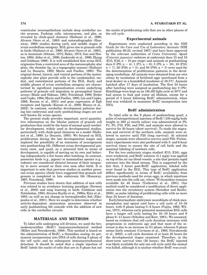
The cerebellar volume increased during the develop-mental period studied from 20 mm3 at E15 to 80 mm3 atP50, with significant growth occurring between P10 andP50 days (Fig. 1A). The volume change of each lobule inthe developmental stages studied is shown in Figure 1B,Cfor the anterior and posterior cerebellum, respectively.However, the volume percentage of each lobule with re-spect to the total cerebellar volume remained constantthroughout the developmental stages (Fig. 2), suggestinga preserved relationship among lobules. Lobules I and Xrepresented the smallest (2% and 4%, respectively) andlobules VII, VIII, and IX the highest (11%, 13%, and 11%,respectively) proportion of the cerebellum, whereas deepnuclei and white matter occupied about 19% of total cer-ebellum volume (Fig. 2). Cerebellar volume growth is duemainly to the addition of new parallel fibers (Rakic, 1973),which, in turn, depends on the addition of new granulecells produced in the EGL. Not surprisingly, the number ofnewborn cells in the EGL of cerebellar lobules at eachstage correlated well with the volume of the lobule (Pear-son R � 0.85–0.7, P � .01).
Short-term survival: labeled cells 20 hourspost-BrdU application
Twenty hours post-BrdU application, the vast majorityof BrdU� cells in the quail cerebellum was found in theEGL, but some were also noted in the molecular layer andthe IGL as well as the white matter, at all ages studied(E15, E16, P0, P1, P5, P10, P20, P50). It is noteworthlythat proliferation persisted at significant levels at post-
Fig. 1. A: Quail total cerebellar volume (mm3) at late embryonic(E15, E16) and posthatching (P1, P5, P10, P20, P50) ages. Significantchanges in the volume occur between P5–P10 and P20–P50. Diagram-matic presentation of regional volume changes with age is shown inB for anterior lobules and in C for posterior lobules.
176 A. STAMATAKIS ET AL.
hatching days, and a few labeled cells were found in theEGL even at P20 and P50.
EGL. The EGL was present as a germinal zone cover-ing the cerebellar lobules at all stages studied. Micro-scopic observation of Nissl-stained sections showed thatits width gradually reduced with age from about seven toten cell rows at E15 to one or two cell rows at P20. At E15and E16, high densities of BrdU� cells were observed inthe EGL (Fig. 3A,B), whereas, at P20 and P50, relativelyfewer cells could be found (Fig. 3G,H). At P5 and P10, thelabeled cells covered most but not all of the EGL surface,and, at P20 and P50, individual BrdU� cells were foundscattered in a superficial position. All cerebellar lobulesshowed steadily declining BrdU� labeling from late em-bryonic ages until P20 (Fig. 3). With the volume of eachlobule at each developmental stage taken into account, theproliferative activity was expressed as density (number ofcells/mm3) of labeled cells (Fig. 4A). At most developmen-tal stages, lobule I exhibited the highest density of labeledcells, amounting to approximately double that of all otherlobules. At E15, the highest density of BrdU� cells wasfound in lobules I, VII, VIb, and II, whereas, at E16, lobuleII exhibited the highest density of labeled cells, followedby lobules VII, VIb, V, and I. At posthatching stages, thedensity of newborn cells was similar in most lobules ex-cept for lobule I, ranging from 3,220 to 6,098 cells/mm3 atPO, 2,577–5,393 cells/mm3 at P1, and gradually decliningto 552–1,878 cells/mm3 at P20 (Fig. 4A).
The total number of labeled cells within each lobule ispresented in Figure 4B. Lobule VIII, the largest lobule inthe cerebellum, included the highest number of labeledcells at almost all stages studied. The time course of gen-eration of new cells within individual lobules varied, sothat lobules I, II, VII, VIII, and IX included maximalnumbers of labeled cells at E15–E16. Lobules V, VIb, VII,VIII, and IX showed the highest proliferation rates for thefirst days of life after hatching (P0–P1). Figure 5A illus-trates in a stack bar graph format the total number ofBrdU� cells determined for each lobule at a specific devel-
opmental stage, for the whole period studied. This nicelyillustrates the contribution (cell addition) of a specificstage for each lobule and emphasizes the differences in thecourse of maturation of the different lobules. Lobules thatformed early were distinguished from late-forming lobulesby a relatively low proportion of labeled cells in posthatch-ing ages and an earlier decline in the width of the EGL.For example, in lobules I, VII, and IX at E15 and E16, cellproliferation represented 49–52% of the total labeled cellsfor all stages studied, indicating their early formation(Fig. 5B). In contrast, lobules III and IV have a prolongedmaturation; i.e., the production of granule cells proceededin a rather constant manner until day P20 (representingat each stage 14–18% of the total labeled cells). A commoncharacteristic of the developmental profile of most lobuleswas a transient increase of neurogenesis at P10. At a laterstage (P20), lobules III and IV included significant propor-tions of labeled cells, which is important.
Molecular layer. Twenty hours post-BrdU, the molec-ular layer of all cerebellar lobules included only a fewlabeled cells during the developmental periods studied(Fig. 3). Their number gradually declined until day P50,when very few labeled cells could be detected. Some ofthese cells had the appearance of migrating granular neu-rons, with elongated cell somata, based on observation ofNissl-stained sections (Fig. 3A, E15 lobule I, arrow); thisfinding is consistent with the idea that they were cellsgenerated in the EGL within the previous 20 hours. Sec-tions treated to detect NADPH-diaphorase differentiatedthe molecular layer into an outer sublayer exhibitinglower diaphorase activity and an inner sublayer close tothe granular layer, noted from E16 throughout all devel-opmental stages studied (data not shown). The signifi-cance of this low expression of NOS, possibly by the later-forming parallel fibers (Rakic, 1973), remains to beexplored but could be related to the lack of NOS activity ofthe later-generated population of granule cells. At theseearly posthatch stages, confocal microscopy revealed ex-pansion of dendritic arborization of avian Purkinje cells(Mori and Matsushima 2002). This NADPH-diaphorasepattern did not relate to the BrdU� cells, which wereevenly distributed throughout both molecular sublayers.
IGL. As early as 20 hours post-BrdU injection, largenumbers of labeled cells were noted within the IGL, at allstages studied, but these decreased gradually with age.Labeled cells found in the IGL within 20 hours from BrdUapplication could represent granule cells that were pro-duced in EGL and completed their migration, or prolifer-ating population, glial cell progenitors. Specifically, thelabeled granule cells following BrdU injection at E15 re-sulted in high numbers of labeled cells in the granularlayer (Fig. 3A,B); labeled cells were topographically orga-nized and were distributed mainly in the superficial partof the IGL, close to the Purkinje cell layer (Fig. 3A,B). Thelikelihood that the labeled cells in this location representa proliferating population that may produce Bergmanglial cells is addressed in the genesis of other cell popula-tions in the Discussion.
NADPH-diaphorase activity within the granular layerwas lower in lobules I and II compared with the rest andgradually decreased in all lobules with age. At P50, weobserved two interlayer intensities, in lobules I–V andVIb, dividing the granular layer into superficial and deepzones, where the sublayer close to the Purkinje cell layerexhibited darker staining.
Fig. 2. Schematic presentation of parasagittal cerebellar view.Each lobule represents a volume percentage of the whole cerebellum.The volume percentage of each lobule is indicated as a number withinthe lobule and does not change as cerebellar size increases.
177PATTERN IN LATE CEREBELLAR GRANULE CELL GENESIS

White matter. In the cerebellar white matter, levels ofBrdU� cells were remarkably different between early andlate posthatching stages (Fig. 6). Whereas, at E15, E16,and P0, moderate numbers of BrdU� cells were present inthe white matter of all cerebellar lobules, the numbergradually decreased posthatching until P20, when almostno labeled cells could be detected in the white matter ofany lobule. An exception was seen for lobules VIa, VIb,where a few labeled cells were present at P10 and P20.Lobules I and X included only a thin layer of white matter,which could not be easily distinguished from the granularlayer.
Long-term survival: labeled cells at specificstages determined at P20 and P50
To investigate the fate of the EGL cells, animals wereinjected with BrdU at days E15, E16, P1, P5, and P10 andallowed to survive until day P20. In addition, a few ani-mals were allowed to survive until P50. The major focus ofinvestigation centered on cells born in EGL that migratedto the IGL (Fig. 7), because these were the most numer-ous, although some cells were found in other layers ofspecific lobules. Labeled cells within the IGL showed dif-ferent degrees in labeling (intensely to lightly labeled; Fig.7). Density measurements of labeled cells within the IGL(number of BrdU� cells vs. total number of granule cells)were performed in five adjacent counterstained sections ofeach animal to obtain an estimate of the contribution oflate-generated cells to the IGL at P20. Approximately 12%and 8% of the IGL cells were labeled at E16 and post-hatching stages, respectively. These percentages repre-sent a rough indication of survival, proliferation, and celldeath events, insofar as no stereological analysis was per-formed, and the cell cycle parameters and number of fur-ther divisions or apoptosis of the labeled cells are notknown.
However, not all labeled cells migrated away from theirplace of origin in the EGL. A few cells labeled at the stagesstudied were still found in the EGL at P20 day. Specifi-cally, labeled cells at embryonic stages E15 and E16 re-mained in EGL of lobules II, III, and VIb until at leastP20. In addition, a few cells labeled at early posthatchingstages were still present in the EGL at day P20 (labeled atP1 in lobule V; labeled at P5 in lobules I and VIa–VIII, X;labeled at P10 in lobules III, IV, VII, and VIII). However,by day P50, all labeled cell had moved away from the EGL.
Long-term intervals: BrdU labeling at E15 and E16.
Most of the cells of the EGL that were labeled at embry-onic stages E15 and E16 had migrated to the granularlayer by day P20 (Fig. 8A,B), but their distribution patternwithin IGL was still clearly heterogeneous, similarly totheir localization in short survival experiments. That is,we observed in most lobules an outside-inside gradient oflabeled cells in IGL, with the outer part of IGL (i.e., closer
Fig. 3. External granular layer mitotic activity at different devel-opmental stages of quail cerebellum 20 hours post-BrdU application.Photomicrographs of parasagittal sections showing the distribution ofBrdU� cells within cerebellar layers of representative lobules of archi(lobule I; A,C,E,G)- and neo (lobule VII; B,D,F,H)-cerebellum at daysE15 (A,B), P0 (C,D), P5 (E,F), and P20 (G,H). Arrow in A points tomigrating cells in a line. EGL, external granular layer; IGL, internalgranular layer; mol, molecular layer. Scale bar � 20 �m.
178 A. STAMATAKIS ET AL.

Fig. 4. EGL proliferation in individual lobules at E15–E16 and at P0–P20. Labeled cells determined 20 hours post-BrdU application wereestimated by using the disector method. Solid and open bars differentiate lobules of the anterior and posterior cerebellar lobe, respectively.A: Density of BrdU� cells (cell/mm3) within EGL of individual lobules. B: Total number of BrdU� cells within EGL of individual lobules. Notethat the ordinate varies among graphs, for optimal visualization of changes.

to the Purkinje cell layer) showing a higher density ofBrdU� cells (Fig. 9). In addition, in lobule VIa andmainly lobule VII, a clear rostrocaudal pattern wasnoted in the final position of the labeled cells at E15.Specifically, in the rostral compartment of lobules VIaand VII, the IGL was nearly devoid of BrdU� cells,whereas the caudal part included a high density ofpositive cells (Fig. 9B). This rostrocaudal gradient inthe final position of labeled granule cells did not char-acterize short-term experiments that determined theirprogenitor cells (that is, the distribution of labeled EGLcells 20 hours post-BrdU, in VIa and VII or any otherlobule). This lack of labeled granule cells in the IGL ofthe anterior pole of the above-mentioned lobules afterlong-term survival is interesting, particularly in thatthe overlying EGL of the rostral pole was densely la-beled 20 hours post-BrdU (Fig. 9A). Therefore, the pro-genitor cells labeled while in the EGL (their prolifera-tion site) were no longer present in the EGL (nosignificant labeling was found in the EGL at P20) buthad not move radially to the underlying IGL of therostral part of lobules VII and VIa (Fig. 9B). These cellslabeled at E15 did not contribute to the population ofgranule cells of rostral lobules VII and VIa, suggestingeither their tangential migration in a different parasag-
ittal domain or their apoptosis. Such nonribboned mi-gration with no respect to the PC compartmentalizationis suggested to occur late in development (Komuro andRakic, 1998). This speculation on possible tangentialmigration or apoptosis is based on the assumption thatthe specific part of the EGL of these lobules at this stageis producing postmitotic cells. However, it is possiblethat the specific parts of EGL are not yet producing anypostmitotic cells and that labeling is diluted and nolonger detectable because of continued proliferation,presumably with only symmetric nonterminal divisions(Hayes and Nowakowski, 2002). This observation sup-ports the idea that the onset of granule cell productiondiffers among lobules.
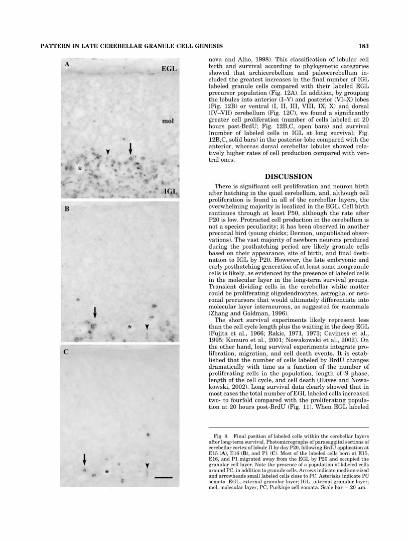
In addition to the granule cells, round cells labeled atE15 and E16 were localized in the molecular layer border-ing the Purkinje cell layer and within the Purkinje celllayer surrounding Purkinje cell somata (PC) by P20 (Fig.8A,B). These labeled cells could be classified into twogroups based on their size: 2–3�m in diameter (small celltype; arrowheads in Fig. 8) and 7–10 �m in diameter(medium-sized cell type; arrows in Fig. 8), evenly distrib-uted within and around the PC layer. It is important tonote that this labeled cell population did not exhibit anyrostrocaudal gradient, as was found for a subpopulation ofgranule cells in long-term survival. A few labeled cellswere also present in the molecular layer of most lobules(Fig. 8B), except for lobule VIII, as well as in the whitematter of lobules II–VIa and VII. Double-labeling experi-ments with GAD, NeuN, and BrdU immunocytochemistryrevealed that most of the BrdU� cells were NeuN� andthat those located next to PC in the molecular layer ex-pressed GAD (Fig. 9G–I).
Long term intervals: post-BrdU labeling at P1, P5,
and P10. By day P20, the majority of cells born in EGLat day P1 had migrated into the IGL (Fig. 7A), although afew BrdU� cells were also detected in the molecular layer(mainly in lobules II and III; Fig. 8C), but none was seenin the white matter. In double-labeling experiments, someof the BrdU� cells showed NADPH-diaphorase activity(Fig. 9C), suggesting that they are granule cells express-ing NOS. Similarly, cells born in the EGL at P5 and P10had migrated to IGL by day P20 (Fig. 7B,C). In addition,some labeled cells were found in the molecular layer oflobules I–IV, VIb, and VIII–IX as well as in the whitematter of lobules III–V. EGL cells proliferated also at dayP20 and migrated into the IGL by day P50. Double-labeling experiments with NeuN and BrdU immunocyto-chemistry revealed that most of the BrdU� cells wereNeuN� (Fig. 9D–F). No outside-inside gradient of the finalposition of BrdU-labeled cells in the IGL was found for theposthatching generated granule cells.
Figure 10A shows the specific addition of labeled gran-ule cells in the IGL of cerebellar lobules at P20, after longsurvival periods. The regional differences in granule cellproduction and survival in the stages investigated suggestthat lobules VIb, VIII, and X included a higher proportionof granule cells labeled at embryonic day 16, whereas, inlobules II, III, IV, V, VIa, VII, and IX, most late-generatedgranule cells were labeled after hatching (Fig. 10B).
Ratio of labeled cells in long/short intervals(index of survival vs. proliferation)
To investigate the survival of the EGL labeled cell pop-ulation (future granule cells), we compared the number of
Fig. 5. A: Age-specific EGL proliferation 20 hours post-BrdU ap-plication. The contribution of specific developmental stages to thetotal of EGL cell proliferationis shown as stacks in the bars, withearlier stages at the bottom of the bars. B: Lobular pattern of thepercentage of BrdU� cells determined at a specific stage vs. the totalBrdU� cells at all stages studied. Embryonic proliferation (E15, E16)accounted for about 50% of the cell production in lobules I, VII, and IX,whereas, for the other lobules, most cells determined from E15 to P20were born after hatching. The patterns indicating the ages are shownat right.
180 A. STAMATAKIS ET AL.

BrdU� cells, labeled at a specific developmental age, attwo survival points (short-term, 20 hours; long-term, P20).This comparison produced a labeling index of survival vs.proliferation, determined by the ratio of labeled cells atlong and short intervals. For the 20-hour survival time, we
chose to ignore the few BrdU� cells that were not in theEGL, because their number was very low compared withthe number of BrdU� cells present in the EGL, essentiallynot affecting the data. Note that, in posthatching experi-ments, 20 hours is less than the cell cycle length (plus the
Fig. 6. Labeled cells found within the cerebellar white matter 20hours post-BrdU injection. Photomicrographs of parasagittal cerebel-lar sections showing the distribution of labeled cells in the whitematter following BrdU application at E15 (A), P1 (B), P5 (C), and P20
(D). In most cases, cells encountered were elongated, indicating mi-gratory activity. IGL, internal granular layer; WH, white matter.Scale bar � 50 �m.
181PATTERN IN LATE CEREBELLAR GRANULE CELL GENESIS

waiting time in the deep EGL), as evidenced by our dataand from previous studies in mammals based on 3H-thymidine autoradiography (Fujita et al., 1966; Rakic,1971, 1973), cell cycle length (Caviness et al., 1995; Nowa-kowski et al., 2002), and real-time observation of the pro-spective granule cells after their last mitosis in living slicepreparations (Komuro et al., 2001). When labeled cells inthe EGL exited the cell cycle, they migrated to the IGL,where they were encountered at P20. However, some ofthese EGL cells, before migrating, reentered the cell cycleand produced daughter cells for the next three or fourcycles (Hayes and Nowakowski, 2002). With this evidencetaken into account, the number determined by the ratio oflabeled cells in long survival vs. short survival could bereferred to as the survival index. This survival index doesnot represent an absolute reference, in that cell cycleparameters differ among lobules and developmentalstage, but it does represent an integration of proliferativeand cell death events occurring during long-term survival.Most lobules showed two- to fourfold increases in thenumber of positive cells residing in the IGL by day P20compared with those labeled in the EGL 20 hours post-BrdU (Fig. 11). Specifically, major increases were notedfor cells born at E16 in lobules III–VIb and VIII (Fig. 11A);at P1 in lobules IV, VIa, and VIB (Fig. 11B); at P5 inlobules VIa and VII–IX (Fig. 11C); and at P10 in lobulesIII, VII, and X (Fig. 11D). However, in some lobules, thenumbers of BrdU� cells in the EGL 20 hours post-BrdUwere similar to those found in IGL approximately 20 dayslater (e.g., lobule I for all stages studied; VII and IX for dayE16; II and X for day P1; VIb and X for day P5; IV, VIa,VIb, VIII for day P10 BrdU application; Fig. 11A–D). Inlobule II, there was significant reduction in the number offuture granule cells labeled at E16 when animals wereallowed to survive until P20. That is, there were 50%fewer labeled cells determined in IGL at P20 than thosefound in EGL 20 hours post-BrdU (Fig. 11A). This evi-dence suggests that cells born in EGL may undergo apo-ptosis before entering the postmitotic zone, in agreementwith studies in the rat EGL (Tanaka and Marunouchi,1998), or labeled daughter cells may continue to prolifer-ate, diluting label so that it is not detected (Hayes andNowakowski 2002). These data clearly show that the sur-vival index differed both across lobules and as a functionof the stage examined, showing a need for further work onthe systematic variation of cell cycle parameters and celldeath events during lobule development.
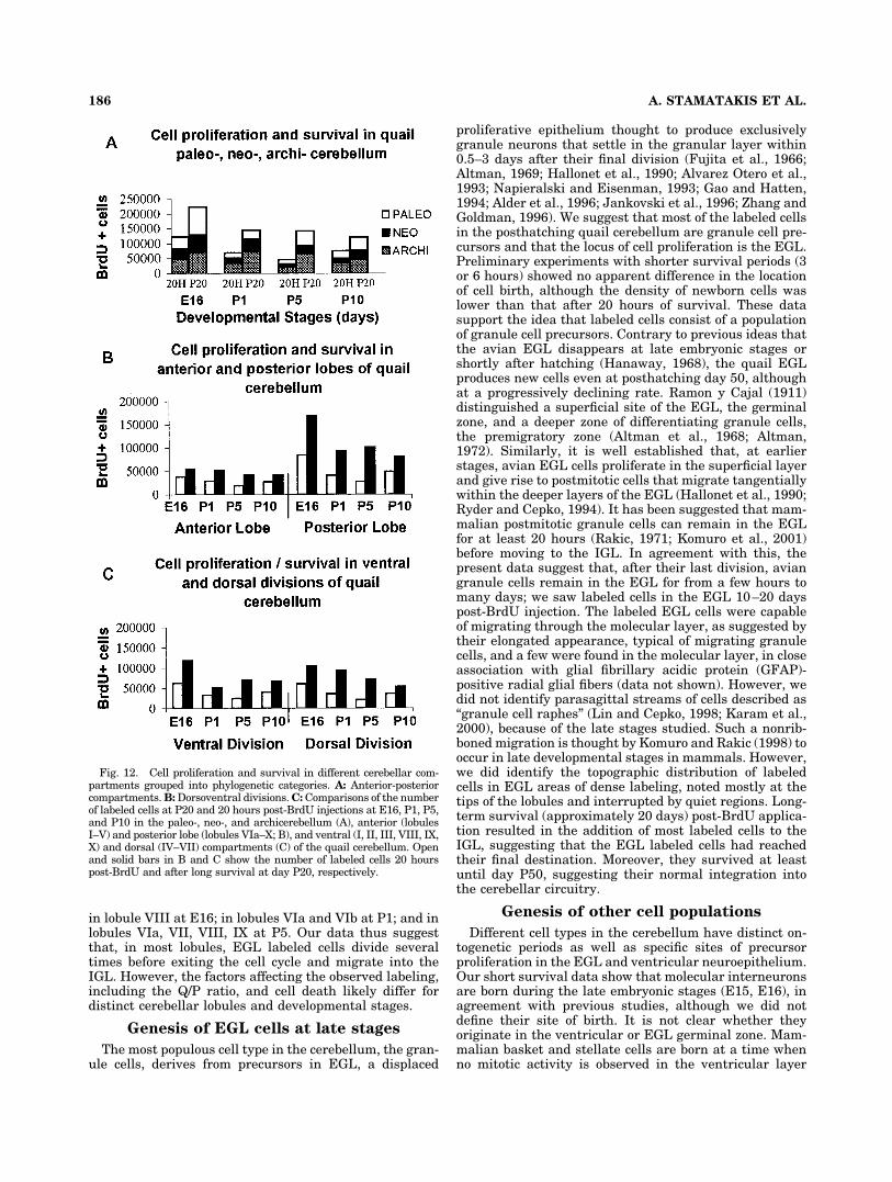
To reveal any trend in the proliferation and survivalpattern, we grouped the cerebellar lobules into archicere-bellum (lobules I–VIa), neocerebellum (lobules VIb–VII),and paleocerebellum (lobules VIII–X; vestibulocerebel-lum: lobules X and ventral IX), based on the proposedanatomical, functional, and developmental criteria (Alt-man, 1969; Shiga et al., 1983; Lau et al., 1998; Podklet-
Fig. 7. Final position of labeled cells within the cerebellar internalgranular layer after long-term survival. Photomicrographs of para-sagittal sections of cerebellar cortex of lobule VII by day P20, follow-ing BrdU application at days P1 (A), P5 (B), and P10 (C). The vastmajority of the labeled cells born at P1, P5 and P10 migrated awayfrom the EGL by P20 and occupied the IGL. Asterisks indicate PCsomata. IGL, internal granular layer; mol, molecular layer; PC, Pur-kinje cell somata. Scale bar � 20 �m.
182 A. STAMATAKIS ET AL.
nova and Alho, 1998). This classification of lobular cellbirth and survival according to phylogenetic categoriesshowed that archicerebellum and paleocerebellum in-cluded the greatest increases in the final number of IGLlabeled granule cells compared with their labeled EGLprecursor population (Fig. 12A). In addition, by groupingthe lobules into anterior (I–V) and posterior (VI–X) lobes(Fig. 12B) or ventral (I, II, III, VIII, IX, X) and dorsal(IV–VII) cerebellum (Fig. 12C), we found a significantlygreater cell proliferation (number of cells labeled at 20hours post-BrdU; Fig. 12B,C, open bars) and survival(number of labeled cells in IGL at long survival; Fig.12B,C, solid bars) in the posterior lobe compared with theanterior, whereas dorsal cerebellar lobules showed rela-tively higher rates of cell production compared with ven-tral ones.
DISCUSSION
There is significant cell proliferation and neuron birthafter hatching in the quail cerebellum, and, although cellproliferation is found in all of the cerebellar layers, theoverwhelming majority is localized in the EGL. Cell birthcontinues through at least P50, although the rate afterP20 is low. Protracted cell production in the cerebellum isnot a species peculiarity; it has been observed in anotherprecocial bird (young chicks; Dermon, unpublished obser-vations). The vast majority of newborn neurons producedduring the posthatching period are likely granule cellsbased on their appearance, site of birth, and final desti-nation to IGL by P20. However, the late embryonic andearly posthatching generation of at least some nongranulecells is likely, as evidenced by the presence of labeled cellsin the molecular layer in the long-term survival groups.Transient dividing cells in the cerebellar white mattercould be proliferating oligodendrocytes, astroglia, or neu-ronal precursors that would ultimately differentiate intomolecular layer interneurons, as suggested for mammals(Zhang and Goldman, 1996).
The short survival experiments likely represent lessthan the cell cycle length plus the waiting in the deep EGL(Fujita et al., 1966; Rakic, 1971, 1973; Caviness et al.,1995; Komuro et al., 2001; Nowakowski et al., 2002). Onthe other hand, long survival experiments integrate pro-liferation, migration, and cell death events. It is estab-lished that the number of cells labeled by BrdU changesdramatically with time as a function of the number ofproliferating cells in the population, length of S phase,length of the cell cycle, and cell death (Hayes and Nowa-kowski, 2002). Long survival data clearly showed that inmost cases the total number of EGL labeled cells increasedtwo- to fourfold compared with the proliferating popula-tion at 20 hours post-BrdU (Fig. 11). When EGL labeled
Fig. 8. Final position of labeled cells within the cerebellar layersafter long-term survival. Photomicrographs of parasaggital sections ofcerebellar cortex of lobule II by day P20, following BrdU application atE15 (A), E16 (B), and P1 (C). Most of the labeled cells born at E15,E16, and P1 migrated away from the EGL by P20 and occupied thegranular cell layer. Note the presence of a population of labeled cellsaround PC, in addition to granule cells. Arrows indicate medium-sizedand arrowheads small labeled cells close to PC. Asterisks indicate PCsomata. EGL, external granular layer; IGL, internal granular layer;mol, molecular layer; PC, Purkinje cell somata. Scale bar � 20 �m.
183PATTERN IN LATE CEREBELLAR GRANULE CELL GENESIS

Figure 9
184 A. STAMATAKIS ET AL.
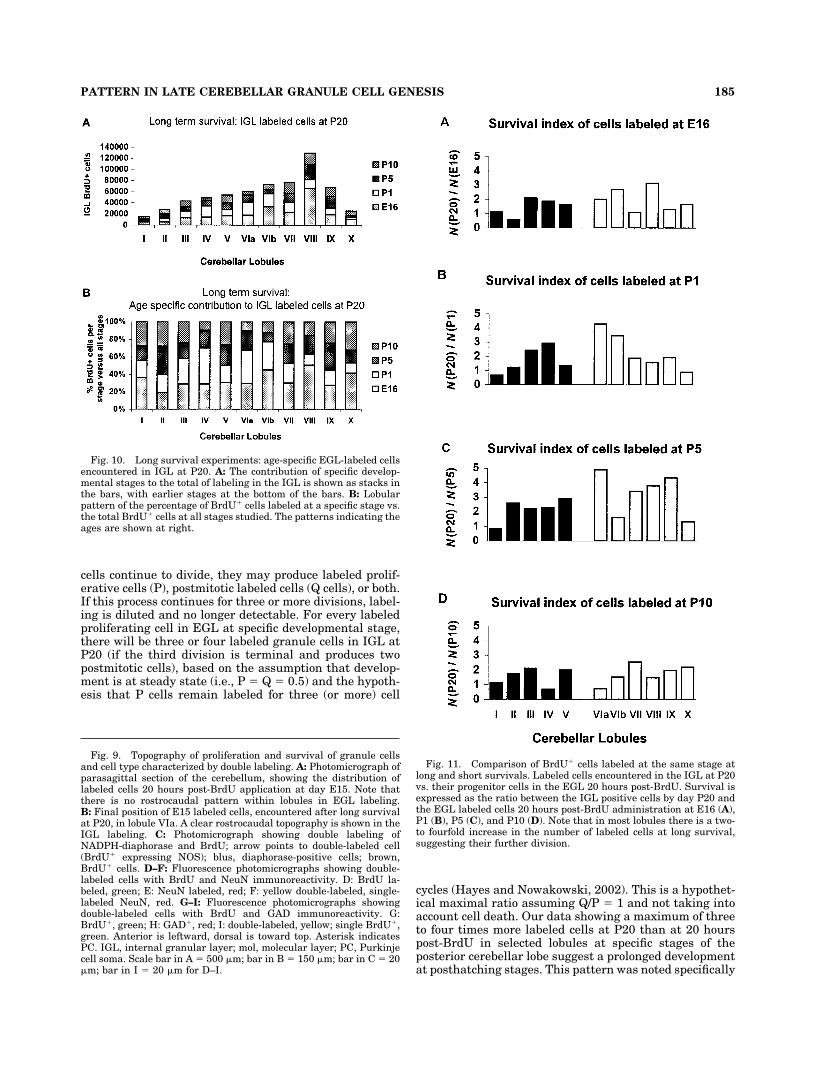
cells continue to divide, they may produce labeled prolif-erative cells (P), postmitotic labeled cells (Q cells), or both.If this process continues for three or more divisions, label-ing is diluted and no longer detectable. For every labeledproliferating cell in EGL at specific developmental stage,there will be three or four labeled granule cells in IGL atP20 (if the third division is terminal and produces twopostmitotic cells), based on the assumption that develop-ment is at steady state (i.e., P � Q � 0.5) and the hypoth-esis that P cells remain labeled for three (or more) cell
cycles (Hayes and Nowakowski, 2002). This is a hypothet-ical maximal ratio assuming Q/P � 1 and not taking intoaccount cell death. Our data showing a maximum of threeto four times more labeled cells at P20 than at 20 hourspost-BrdU in selected lobules at specific stages of theposterior cerebellar lobe suggest a prolonged developmentat posthatching stages. This pattern was noted specifically
Fig. 10. Long survival experiments: age-specific EGL-labeled cellsencountered in IGL at P20. A: The contribution of specific develop-mental stages to the total of labeling in the IGL is shown as stacks inthe bars, with earlier stages at the bottom of the bars. B: Lobularpattern of the percentage of BrdU� cells labeled at a specific stage vs.the total BrdU� cells at all stages studied. The patterns indicating theages are shown at right.
Fig. 11. Comparison of BrdU� cells labeled at the same stage atlong and short survivals. Labeled cells encountered in the IGL at P20vs. their progenitor cells in the EGL 20 hours post-BrdU. Survival isexpressed as the ratio between the IGL positive cells by day P20 andthe EGL labeled cells 20 hours post-BrdU administration at E16 (A),P1 (B), P5 (C), and P10 (D). Note that in most lobules there is a two-to fourfold increase in the number of labeled cells at long survival,suggesting their further division.
Fig. 9. Topography of proliferation and survival of granule cellsand cell type characterized by double labeling. A: Photomicrograph ofparasagittal section of the cerebellum, showing the distribution oflabeled cells 20 hours post-BrdU application at day E15. Note thatthere is no rostrocaudal pattern within lobules in EGL labeling.B: Final position of E15 labeled cells, encountered after long survivalat P20, in lobule VIa. A clear rostrocaudal topography is shown in theIGL labeling. C: Photomicrograph showing double labeling ofNADPH-diaphorase and BrdU; arrow points to double-labeled cell(BrdU� expressing NOS); blus, diaphorase-positive cells; brown,BrdU� cells. D–F: Fluorescence photomicrographs showing double-labeled cells with BrdU and NeuN immunoreactivity. D: BrdU la-beled, green; E: NeuN labeled, red; F: yellow double-labeled, single-labeled NeuN, red. G–I: Fluorescence photomicrographs showingdouble-labeled cells with BrdU and GAD immunoreactivity. G:BrdU�, green; H: GAD�, red; I: double-labeled, yellow; single BrdU�,green. Anterior is leftward, dorsal is toward top. Asterisk indicatesPC. IGL, internal granular layer; mol, molecular layer; PC, Purkinjecell soma. Scale bar in A � 500 �m; bar in B � 150 �m; bar in C � 20�m; bar in I � 20 �m for D–I.
185PATTERN IN LATE CEREBELLAR GRANULE CELL GENESIS
in lobule VIII at E16; in lobules VIa and VIb at P1; and inlobules VIa, VII, VIII, IX at P5. Our data thus suggestthat, in most lobules, EGL labeled cells divide severaltimes before exiting the cell cycle and migrate into theIGL. However, the factors affecting the observed labeling,including the Q/P ratio, and cell death likely differ fordistinct cerebellar lobules and developmental stages.
Genesis of EGL cells at late stages
The most populous cell type in the cerebellum, the gran-ule cells, derives from precursors in EGL, a displaced
proliferative epithelium thought to produce exclusivelygranule neurons that settle in the granular layer within0.5–3 days after their final division (Fujita et al., 1966;Altman, 1969; Hallonet et al., 1990; Alvarez Otero et al.,1993; Napieralski and Eisenman, 1993; Gao and Hatten,1994; Alder et al., 1996; Jankovski et al., 1996; Zhang andGoldman, 1996). We suggest that most of the labeled cellsin the posthatching quail cerebellum are granule cell pre-cursors and that the locus of cell proliferation is the EGL.Preliminary experiments with shorter survival periods (3or 6 hours) showed no apparent difference in the locationof cell birth, although the density of newborn cells waslower than that after 20 hours of survival. These datasupport the idea that labeled cells consist of a populationof granule cell precursors. Contrary to previous ideas thatthe avian EGL disappears at late embryonic stages orshortly after hatching (Hanaway, 1968), the quail EGLproduces new cells even at posthatching day 50, althoughat a progressively declining rate. Ramon y Cajal (1911)distinguished a superficial site of the EGL, the germinalzone, and a deeper zone of differentiating granule cells,the premigratory zone (Altman et al., 1968; Altman,1972). Similarly, it is well established that, at earlierstages, avian EGL cells proliferate in the superficial layerand give rise to postmitotic cells that migrate tangentiallywithin the deeper layers of the EGL (Hallonet et al., 1990;Ryder and Cepko, 1994). It has been suggested that mam-malian postmitotic granule cells can remain in the EGLfor at least 20 hours (Rakic, 1971; Komuro et al., 2001)before moving to the IGL. In agreement with this, thepresent data suggest that, after their last division, aviangranule cells remain in the EGL for from a few hours tomany days; we saw labeled cells in the EGL 10–20 dayspost-BrdU injection. The labeled EGL cells were capableof migrating through the molecular layer, as suggested bytheir elongated appearance, typical of migrating granulecells, and a few were found in the molecular layer, in closeassociation with glial fibrillary acidic protein (GFAP)-positive radial glial fibers (data not shown). However, wedid not identify parasagittal streams of cells described as“granule cell raphes” (Lin and Cepko, 1998; Karam et al.,2000), because of the late stages studied. Such a nonrib-boned migration is thought by Komuro and Rakic (1998) tooccur in late developmental stages in mammals. However,we did identify the topographic distribution of labeledcells in EGL areas of dense labeling, noted mostly at thetips of the lobules and interrupted by quiet regions. Long-term survival (approximately 20 days) post-BrdU applica-tion resulted in the addition of most labeled cells to theIGL, suggesting that the EGL labeled cells had reachedtheir final destination. Moreover, they survived at leastuntil day P50, suggesting their normal integration intothe cerebellar circuitry.
Genesis of other cell populations
Different cell types in the cerebellum have distinct on-togenetic periods as well as specific sites of precursorproliferation in the EGL and ventricular neuroepithelium.Our short survival data show that molecular interneuronsare born during the late embryonic stages (E15, E16), inagreement with previous studies, although we did notdefine their site of birth. It is not clear whether theyoriginate in the ventricular or EGL germinal zone. Mam-malian basket and stellate cells are born at a time whenno mitotic activity is observed in the ventricular layer
Fig. 12. Cell proliferation and survival in different cerebellar com-partments grouped into phylogenetic categories. A: Anterior-posteriorcompartments. B: Dorsoventral divisions. C: Comparisons of the numberof labeled cells at P20 and 20 hours post-BrdU injections at E16, P1, P5,and P10 in the paleo-, neo-, and archicerebellum (A), anterior (lobulesI–V) and posterior lobe (lobules VIa–X; B), and ventral (I, II, III, VIII, IX,X) and dorsal (IV–VII) compartments (C) of the quail cerebellum. Openand solid bars in B and C show the number of labeled cells 20 hourspost-BrdU and after long survival at day P20, respectively.
186 A. STAMATAKIS ET AL.

(Miale and Sidman, 1961). Therefore, it has been proposedthat their neuronal progenitors arise from the ventricularzone, migrate toward the molecular layer, and continue todivide in the cerebellar white matter, until settling intheir final position (Zhang and Goldman, 1996). Studiesemploying birth dating with 3H-thymidine and quail-chickchimeras also suggest that molecular layer interneuronsare likely produced by progenitors dividing in the ventric-ular epithelium and not in the EGL (Hanaway, 1968;Yurkewicz et al., 1981; Kanemitsu and Kobayashi, 1988;Hallonet et al., 1990; Hallonet and Le Douarin, 1992;Alvarez Otero et al., 1993; Napieralski and Eisenman,1993). In contrast, it has been suggested that Golgi neu-rons (Hausmann et al., 1985), cells of the isthmus androstral hindbrain (Lin et al., 2001), derive from EGL. Inthe present study, there was significant labeling in thewhite matter 20 hours post-BrdU injection at E15, coin-ciding with the appearance of a cell population in themolecular layer found around PC. After long survival (atP20), when cells labeled at E15 were no longer present inthe white matter, a BrdU� cell population about 10 �m indiameter, doubly labeled with GAD, was located aroundPC. This evidence suggests that cells proliferating at E15and settling at P20 around PC may be basket interneu-rons, based on their final location and GAD immunoreac-tivity. We could not identify any trend of cells from thewhite matter migrating toward the molecular layer butcannot exclude the possibility that these cells becamepostmitotic within the white matter, as has been sug-gested for the mammalian molecular layer interneurons.However, insofar as the more active proliferating zone atthese stages is EGL, it is also possible that at least part ofthese cell populations is derived from the EGL. Consistentwith this idea is our observation that generation of celltypes residing in the molecular layer continued at a lowerrate in most lobules until P10, although, at posthatchingdays P1–P10, we could see only a few labeled cells in thewhite matter.
In addition, it is known that glia develop at late stages(Shiga et al., 1983; Reynolds and Wilkin, 1991). Glioblastsare thought to originate from the ventricular epitheliumand to divide in situ, but their exact course of developmentin the avian cerebellum is not known (Hanaway, 1968;Altman, 1969). In the present study, we did detect smallBrdU� cells in the Purkinje cell layer, 2–3 �m in diame-ter. It has been suggested that small cells surroundingPurkinje cell bodies in chick cerebellum are formed by gliacell precursors dividing in the IGL (Hanaway, 1968), eventhough the glial nature of these cells has not yet beenconfirmed (Hallonet and Mallart, 1997). In addition, it isknown that some cells continue to proliferate within theIGL, although their final position and phenotype remainunclear (Fujita et al., 1966). Of great importance in thiscontext are recent studies providing evidence for the gen-eration of neurons from glial precursors (e.g., Malatesta etal., 2000; Seri et al., 2000; Noctor et al., 2001; Parnavelasand Nadarajah, 2001). Whether a population of the la-beled cells in the quail cerebellum could also be charac-terized as multipotent progenitors that can generate bothneurons and glia remains to be established.
Functional implications
The observed trend for late proliferation of granule cellprecursors in quail cerebellum followed, in part, the pre-viously suggested pattern for the rat, that is, ventral to
dorsal maturation, with the phylogenetically younger neo-cerebellum ending its ontogenesis last (Altman, 1969). Inaddition, our data suggest that, at late developmentalstages, a greater proportion of granule cells were born andsurvived in the caudal cerebellar cortex. This pattern oflate granule cells genesis relates well with the pattern oflater Purkinje cell generation in the posterior chick cere-bellum (Yurkewicz et al., 1981), in the context of themodular cerebellar organization in parasagittal territories(Karam et al., 2001; Blanco et al., 2002). In addition, thedifferent origins of anterior and posterior cerebellum, asshown from chick-quail chimeric embryos, could accountfor this difference in proliferation pattern. That is, part ofthe rostral cerebellum originates from the so-called mes-encephalic alar plate (Martinez and Alvarado-Mallart,1989), whereas caudal cerebellum originates from the met-encephalic alar plate (Hallonet et al., 1990). Moreover, thedivision into anterior and posterior domains is supportedby the complementary pattern of different Eph receptorsand ephrins (Karam et al., 2000; Blance et al., 2002).
The cerebellum is considered a major brain center formotor control, sensory association (Herrup and Kuemerle,1997), and high-order cognitive functions, such as learn-ing and memory (Thach, 1998; Hikosaka et al., 1998;Middleton and Strick, 2000). Our quantitative results sug-gest that granule cells born after hatching survive intoadulthood and become incorporated into the cerebellarnetwork. In the same early posthatch period, extensiveexpansion of dendritic arborization of PC occurs in quailcerebellum, and the formation of parallel fiber synapseson distal dendrites has been suggested (Mori and Matsu-shima, 2002). The period of rapid dendritic growth andactive synaptogenesis coincides with the observed lategranule cell genesis, supporting the hypothesis of theirincorporation into the cerebellar circuits. It has beenshown that, in meander tail mouse cerebellum, multipo-tent progenitors derived from the EGL could differentiateinto neurons and compensate for granule cell death (Ro-sario et al., 1997). This evidence suggests a developmentalmechanism with many implications, including motorlearning plasticity. In archicerebellar lobules III and IV,neurons were born in the EGL at a rate that deviated verylittle with age, suggesting a prolonged maturation periodand a possible extended developmental window for plasticresponses to internal or external changes. In the pigeon,there is a somatotopic representation of the wings in lob-ules III–V (Necker, 2001). If the same representation ex-ists in the quail cerebellum, the newborn cells could beintegrated in circuits for motor control of the wings. At thebehavioral level, cell birth in lobules III–V may be corre-lated with cerebellar participation in motor control, inthat, during this posthatching period, the quails startjumping and making their first flight attempts. It wouldbe most interesting to test whether interference with neu-rogenesis at this time would affect the training and exe-cution of such motor programs. Moreover, interferencewith the motor behavior of the animal could affect prolif-eration, migration, or survival. If substantiated throughfurther study, this would provide another example of therole of neurogenesis in modifying a motor circuit, as hasbeen shown for song learning in canaries (Goldman andNottebohm, 1983; Alvarez-Buylla et al., 1990a,b; Kirn andNottebohm, 1993).
187PATTERN IN LATE CEREBELLAR GRANULE CELL GENESIS

ACKNOWLEDGMENTS
The authors thank Prof. R. Nowakowski for most usefulcomments on the manuscript. We thank graduate stu-dents B. Zikopoulos, C. Ambatzis, and L. Panagis for ex-cellent technical assistance for the revised form of thearticle.
LITERATURE CITED
Alder J, Cho NK, Hatten ME. 1996. Embryonic precursor cells from therhombic lip are specified to a cerebellar granule neuron identity. Neu-ron 17:389–399.
Altman J. 1969. Autoradiographic and histological studies of postnatalneurogenesis. III. Dating the time of production and onset of differen-tiation of cerebellar microneurons in rats. J Comp Neurol 136:269–294.
Altman J. 1972. Postnatal development of the cerebellar cortex in the rat.I. The external germinal layer and the transitional molecular layer.J Comp Neurol 145:353–398.
Altman J, Anderson WJ, Wright KA. 1968. Differential radiosensitivity ofstationary and migratory primitive cells in the brains of infant rats.Exp Neurol 22:52–74.
Alvarez-Buylla A, Kirn JR, Nottebohm F. 1990a. Birth of projection neu-rons in adult avian brain may be related to perceptual or motor learn-ing. Science 249:1444–1446.
Alvarez-Buylla A, Theelen M, Nottebohm F. 1990b. Proliferation “hotspots” in adult avian ventricular zone reveal radial cell division. Neu-ron 5:101–109.
Alvarez Otero R, Sotelo C, Alvarado-Mallart RM. 1993. Chick-quail chime-ras with partial cerebellar grafts: an analysis of the origin and migra-tion of cerebellar cells. J Comp Neurol 333:597–615.
Bannigan J, Langman J. 1979. The cellular effect of 5-bromodeoxyuridineon the mammalian embryo. J Embryol Exp Morphol 50:123–135.
Benagiano V, Virgintino D, Rizzi A, Flace P, Troccoli V, Bormann J,Monaghan P, Robertson D, Roncali L, Ambrosi G. 2000. Glutamic aciddecarboxylase-positive neuronal cell bodies and terminals in the hu-man cerebellar cortex. Histochem J 32:557–564.
Blanco MJ, Pena-Melian A, Nieto MA. 2002. Expression of EphA receptorsand ligands during chick cerebellar development. Mech Dev 114:225–229.
Cavalieri B. 1966 [reprint]. Geometria degli indivisibili. Torino: UnioneTipografico-Editrice Torinese.
Caviness VS, Takahashi T, Nowakowski RS. 1995. Numbers, time andneocortical neuro-genesis: a general developmental and evolutionarymodel. Trends Neurosci 18:379–383.
De Zeeuw CI, Wylie DR, DiGiorgi PL, Simpson JI. 1994. Projections ofindividual Purkinje cells of identified zones in the flocculus to thevestibular and cerebellar nuclei in the rabbit. J Comp Neurol 349:428–448.
Dermon CR, Stamatakis A. 1994. Laminar pattern of NADPH-diaphoraseactivity in the developing avian cerebellum. Neuroreport 5:1941–1945.
Dermon CR, Zikopoulos B, Panagis L, Harrison E, Lancashire CL, Mil-eusnic R, Stewart MG. 2002. Passive avoidance training enhancesneuron proliferation in day old chicks. Eur J Neurosci 16:1267–1274.
Dombrowski S, Barbas H. 1996. Differential expression of NADPH diaph-orase in functionally distinct prefrontal cortices in the rhesus monkey.Neuroscience 72:49–62.
Feirabend HK. 1990. Development of longitudinal patterns in the cerebel-lum of the chicken (Gallus domesticus): a cytoarchitectural study of thegenesis of cerebellar modules. Eur J Morphol 28:169–223.
Fujita S. 1962. Kinetics of cellular proliferation. Exp Cell Res 28:52–60.Fujita S, Shimada M, Nakamura T. 1966. 3H-thymidine autoradiographic
studies on the cell proliferation and differentiation in the external andthe internal granular layers of the mouse cerebellum. J Comp Neurol128:191–208.
Gao W-Q, Hatten ME. 1994. Immortalizing oncogenes subvert the estab-lishment of granule cell identity in developing cerebellum. Develop-ment 120:1059–1070.
Goldman SA, Nottebohm F. 1983. Neuronal production, migration, anddifferentiation in a vocal control nucleus of the adult female canarybrain. Proc Natl Acad Sci U S A 80:2390–2394.
Gundersen HJ, Bagger P, Bendtsen TF, Evans SM, Korbo L, Marcussen N,Moller A, Nielsen K, Nyengaard JR, Pakkenberg B, Serensen FB,Vesterby A, West MJ. 1988. The new stereological tools: disector,
fractionator, nucleator and point sampled intercepts and their use inpathological research and diagnosis. Acta Pathol Microbiol ImmunolScand 96:857–881.
Hallonet M, Alvarado-Mallart RM. 1997. The chick/quail chimeric system:a model for early cerebellar development. Perspect Dev Neurobiol5:17–31.
Hallonet ME, Le Douarin NM. 1993. Tracing neuroepithelial cells of themesencephalic and metencephalic alar plates during cerebellar ontog-eny in quail-chick chimaeras. Eur J Neurosci 5:1145–1155.
Hallonet ME, Teillet M-A, Le Douarin NM. 1990. A new approach to thedevelopment of the cerebellum provided by the quail-chick markersystem. Development 108:19–31.
Hanaway J. 1967. Formation and differentiation of the external granularlayer of the chick cerebellum. J Comp Neurol 131:1–14.
Hanaway J. 1968. Origin of basket cells in the molecular layer of the chickcerebellum. Anat Rec 160:360.
Hausmann B, Mangold U, Sievers J, Berry M. 1985. Derivation of cerebel-lar Golgi neurons from external granular layer: evidence from explan-tation of external granule cells in vivo. J Comp Neurol 232:511–522.
Hayes NL, Nowakowski RS. 2000. Exploiting the dynamics of S-phasetracers in developing brain: interkinetic nuclear migration for cellsentering vs. leaving the S-phase. Dev Neurosci 22:44–45.
Herrup K, Kuemerle B. 1997. The compartmentalization we are keenlyaware. Annu Rev Neurosci 20:61–90.
Hikosaka O, Miyashita K, Miyachi S, Sakai K, Lu X. 1998. Differentialroles of the frontal cortex, basal ganglia, and cerebellum in visuomotorsequence learning. Neurobiol Learn Mem 70:137–149.
Ito M. 2000. Mechanisms of motor learning in the cerebellum. Brain Res886:237–245.
Ito M, Orlov I, Yamamoto M. 1982. Topographical representation ofvestibulo-ocular reflexes in rabbit cerebellar flocculus. Neuroscience7:1657–1664.
Jankovski A, Rossi F, Sotelo C. 1996. Neuronal precursors in the postnatalmouse cerebellum are fully committed cells: evidence from hetero-chronic transplantations. Eur J Neurosci 8:2308–2319.
Kanemitsu A, Kobayashi Y. 1988. Time of origin of Purkinje cells andneurons of the deep cerebellar nuclei of the chick embryo examined by3H-thymidine autoradiography. Anat Anz 165:167–175.
Karam SD, Burrows RC, Logan C, Koblar S, Pasquale EB, Bothwell M.2000. Eph receptors and ephrins in the developing chick cerebellum:relationship to sagittal patterning and granule cell migration. J Neu-rosci 20:6488–6500.
Karam SD, Kim YS, Bothwell M. 2001. Granule cells migrate withinraphes in the developing cerebellum: an evolutionary conserved mor-phogenetic event. J Comp Neurol 440:127–135.
Kirn JR, Nottebohm F. 1993. Direct evidence for loss and replacement ofprojection neurons in adult canary brain. J Neurosci 13:1654–1663.
Kirn JR, Alvarez-Buylla A, Nottebohm F. 1991. Production and survival ofprojection neurons in a forebrain vocal center of adult male canaries.J Neurosci 11:1756–1762.
Komuro H, Yacubova E, Yacubova E, Rakic P. 2001. Mode and tempo oftangential cell migration in the cerebellar external granular layer.J Neurosci 21:527–540.
Lau KL, Glover RG, Linkenhoker B, Wylie DR. 1998. Topographical orga-nization of inferior olive cells projecting to translation and rotationzone in the vestibulocerebellum of pigeon. Neuroscience 85:605–614.
Le Douarin NM. 1993. Embryonic neural chimeras in the study of braindevelopment. Trends Neurosci 16:64–72.
Lin JC, Cepko CL. 1998. Granule cell raphes and parasagittal domains ofPurkinje cells: complementary patterns in the developing chick cere-bellum. J Neurosci 18:9342–9353.
Lin JC, Cai L, Cepko CL. 2001. The external granule layer of the develop-ing chick cerebellum generates granule cells and cells of the isthmusand rostral hindbrain. J Neurosci 21:159–168.
Malatesta P, Hartfuss E, Gotz M. 2000. Isolation of radial glial cells byfluorescent-activated cell sorting reveals a neuronal lineage. Develop-ment 127:5253–5263.
Marin F, Puelles L. 1995. Morphological fate of rhombomeres in quail/chickchimeras: a segmental analysis of hindbrain nuclei. Eur J Neurosci7:1714–1738.
Martinez S, Alvarado-Mallard RM. 1989. Rostral cerebellum originatesfrom the caudal portion of the so-called ‘mesencephalic’ vescicle: astudy using chick/quail chimeras. Eur J Neurosci 1:549–560.
188 A. STAMATAKIS ET AL.

Miale IL, Sidman RL. 1961. An autoradiographic analysis of histogenesisin the mouse cerebellum. J Exp Neurol 4:277–296.
Middleton FA, Strick PL. 2000. Basal ganglia and cerebellar loops: motorand cognitive circuits. Brain Res Brain Res Rev 31:236–250.
Miller M, Nowakowski RS. 1988. Use of bromodeoxyuridine-immuno-histochemistry to examine the proliferation, migration and time oforigin of cells in the central nervous system. Brain Res 457:44–52.
Mori M, Matsushima T. 2002. Post-hatch development of dentritic ar-borization in cerebellar Purkinje neurons of quail chicks: a morphomet-ric study. Neurosci Lett 329:73–76.
Mullen RJ, Buck CR, Smith AM. 1992. NeuN, a neuronal specific nucleuarprotein in vertebrates. Development 116:201–211.
Napieralski JA, Eisenman LM. 1993. Developmental analysis of the exter-nal granular layer in the Meanter Tail mutant mouse: do cerebellarmicroneurons have independent progenitors? Dev Dyn 197:244–254.
Necker R. 2001. Spinocerebellar projections in the pigeon with specialreference to the neck region of the body. J Comp Neurol 429:403–418.
Noctor SC, Flint AC, Weissman TA, Dammerman RS, Kriegstein AR. 2001.Neurons derived from radial glial cells establish radial units in neo-cortex. Nature 409:714–720.
Nowakowski RS, Caviness VS Jr, Takahashi T, Hayes NL. 2002. Popula-tion dynamics during cell proliferation and neuronogenesis in the de-veloping murine neocortex. Results Probl Cell Differ 39:1–25.
Palay SL, Chan-Palay V. 1974. The cerebellar cortex. New York: Springer-Verlag.
Parnavelas JG, Nadarajah B. 2001. Radial glial cells: are they glia? Neuron31:881–884.
Podkletnova I, Alho H. 1998. Neonatal noradrenaline depletion preventsthe transition of Bergmann glia in the developing cerebellum. J ChemNeuroanat 14:167–173.
Rakic P. 1971. Neuro-glia relationship during granule cell migration indeveloping cerebellar cortex. J Comp Neurol 141:283–312.
Rakic P. 1973. Kinetics of proliferation and latency between final celldivision and onset of differentiation of cerebellar stellate and basketneurons. J Comp Neurol 147:523–546.
Ramon y Cajal S. 1911. Histologie du systeme nerveux de l’homme et desvertebras. Paris: A. Maloine.
Reynolds R, Wilkin GP. 1991. Oligodendroglial progenitor cells but notoligodendroglia divide during normal development of the rat cerebel-lum. J Neurocytol 20:216–224.
Rosario CM, Yandava BD, Kosaras B, Zurakowski D, Sidman RL, SnyderEY. 1997. Differentiation of engrafted multipotent neural progenitorstoward replacement of missing granule cell neurons in meander tailcerebellum may help determine the locus of mutant gene action. De-velopment 124:4213–4224.
Ryder EF, Cepko CL. 1994. Migration patterns of clonally related granulecells and their progenitors in the developing chick cerebellum. Neuron12:1011–1029.
Schultze B, Korr H. 1981. Cell kinetic studies of different cell types in thedeveloping and adult brain of the rat and the mouse: a review. CellTissue Kinet 14:309–325.
Seri B, Garcia-Verdugo JM, McEwen BS, Alvarez-Buylla A. 2001. Astro-cytes give rise to new neurons in the adult mammalian hippocampus.J Neurosci 21:7153–7160.
Shiga T, Ichikawa M, Hirata Y. 1983. Spatial and temporal pattern ofpostnatal proliferation of Bergmann glia cells in rat cerebellum: anautoradiographic study. Anat Embryol 167:203–211.
Spiro JE, Dalva MB, Mooney RJ. 1999. Long-range inhibition within thezebra finch song nucleus RA can coordinate the firing of multipleprojection neurons. Neurophysiology 81:3007–3020.
Sterio DC. 1984. The unbiased estimation of number and sizes of arbitraryparticles using the disector. J Microsc 134:127–136.
Striedter GF, Keefer BP. 2000. Cell migration and aggregation in thedeveloping telencephalon: pulse-labeling chick embryos with bromode-oxyuridine. J Neurosci 20:8021–8030.
Tanaka M, Marunouchi T. 1998. Immunohistochemical analysis of devel-opmental stage of external granular layer neurons which undergoapoptosis in postnatal rat cerebellum. Neurosci Lett 242:85–88.
Thach WT. 1998. A role for the cerebellum in learning movement coordi-nation. Neurobiol Learn Mem 70:177–188.
Wassef M, Joyner AL. 1997. Early mesencephalon/metencephalon pattern-ing and development of the cerebellum. Perspect Dev Neurobiol 5:3–16.
Wilson DB. 1973. Chronological changes in the cell cycle of chick neuroep-ithelial cells. J Embryol Exp Morphol 29:745–751.
Yu WHA. 1977. The effect of 5-bromodeoxyuridine on the postnatal devel-opment of the rat cerebellum: morphologic and radioautographic stud-ies. Am J Anat 150:89–108.
Yurkewicz L, Lauder JM, Marchi M, Giacobini E. 1981. 3H-thymidine longsurvival autoradiography as a method for dating the time of neuronalorigin in the chick embryo: the locus coeruleus and cerebellar Purkinjecells. J Comp Neurol 203:257–267.
Zhang L, Goldman JE. 1996. Generation of cerebellar interneurons fromdividing progenitors in white matter. Neuron 16:47–54.
Zikopoulos B, Kentouri M, Dermon CR. 2001. Cell genesis in the hypothal-amus is associated to the sexual phase of a hermaphrodite teleost.Neuroreport 12:2477–2481.
Zupanc GKH, Horscke I. 1995. Proliferation zones in the brain of adultGymnotiform fish: a quantitative mapping study. J Comp Neurol 353:213–233.
189PATTERN IN LATE CEREBELLAR GRANULE CELL GENESIS