
Lasker Basic Medical Research Awardcourses.biology.utah.edu/bastiani/3230/DB...
10
epithelioid (E) cell 4 . These C cells were the embryonal carcinoma (EC) stem cells from the tumor. Gail Martin joined my lab, and we were able to show that these spontaneously arising ‘E-cells’ could be replaced by mitotically inactivated chick or mouse fibroblasts, and that when these diminished or were withdrawn, extensive in vitro differentiation occurred. In every case, the differentiation proceeded through the produc- tion of a primary embryonic endoderm, and clumps of sus- pended cells formed recognizable embryoid bodies. Re-attachment of these to a solid surface gave rise to the most splendid and diverse differentiation, with beating cardiac mus- cle, nerve skin, cartilage and so on 5 . It was apparent, however, that they were undergoing the same first-step differentiation to an embryonic endoderm as did the inner cell mass (ICM) of a mouse embryo 6 . This likeness to the ICM was tested by experiments with Richard Gardner. I well remember transporting cells from University College London to Oxford, where he carefully intro- duced them into blastocysts. The chimeras we obtained demon- strated a dramatic result, with nearly every tissue of the derived mouse having contributions from the tissue culture cells 7 .These cells, however, were not normal. They were derived from seri- ally passaged tumors and had been cloned and cultured for some time. Karyotypically they were remarkably close to nor- mal for mouse tissue cells, but although they had an apparently normal chromosome number they only had one X chromosome and no Y chromosome. Many of the initially nor- mal mice later succumbed to somatic tumors (rhabdomyosarco- mas, fibrosarcomas and so on), presumably as a result of the passage-derived mutational load in these cells. We and our col- leagues in Oxford, as well as Francois Jacob’s laboratory in Paris, tried in vain to recover a euploid XY EC cell line to obtain a per- fect germline chimera, but this had to await the direct deriva- tion of the cells from embryos rather than from tumors. In 1978 I started work in the Department of Genetics at Cambridge University, and many investigations continued to show the close relationships between EC cells and early embryo epiblast. Together with Ten Fiezi, I was able to begin to deter- mine that the main cell surface antigens on the EC cells were carbohydrate epitopes of the glycohalix 8 , and Peter Stern, who had recently also moved from University College, London, to Cambridge to Sydney Brenner’s laboratory, produced a very useful monoclonal against a cell surface glycolipid: the Forsman antigen. The reaction of this monoclonal antibody with cells of the normal early mouse embryo allowed us to refine the apparent homology between EC cells and cells of the embryonic ectoderm before 6 days of development 9 . Robin Lovell-Badge, using what would now be called a proteomic approach—that is, two-dimensional gels of whole-protein REPRINTED FROM NATURE MEDICINE • VOLUME 7 • NUMBER 10 • OCTOBER 2001 iii COMMENTARY I was inspired by biology, particularly by my experience at Cambridge in Christ’s College, tutored by David Coombes and in Part II Biochemistry, where I remember in particular such luminaries as Malcolm Dixon and Don Northcote. In that year (1962–1963), a series of lectures at Cambridge by Jacques Monod burst open a new understanding for me and, together with a seminar series organized by Sidney Brenner in his rooms at King’s College, inspired me with the new concepts of control of genetic readout through mRNA. I resolved to work in either plant biochemistry or developmental biology. A bout of glan- dular fever prevented me from taking my final examinations for which I was so eagerly preparing, and resulted in my taking a research assistantship with Elizabeth Deuchar at University College London, on Xenopus development. My ambition was to isolate developmentally controlled mRNA, but at that time none of the cloning tools or probes on which we now rely were available. All I could study were double-reciprocally labeled ( 14 C and 3 H) profiles of polyribosomes and mRNA from dissected blastula and gastrula ectoderm by sucrose density gra- dient centrifugation and RNA by agarose electrophoresis. In modern terms, I was looking at animal cap development in cul- ture before induction and after commitment to either a neural or an epidermal ectoderm. At that time I saw two impediments to further progress: the difficulty of getting enough material for biochemical analysis, and the lack of any foreseeable genetics. I sought a more ‘tractable’ developmental system and, at the suggestion of Robin Weiss, looked to the possibility of establish- ing an in vitro system of mammalian cell differentiation from mouse teratocarcinomas. In 1967, Leroy Stevens 1 and Barry Pierce 2 both published reviews of their formative studies. Leroy Stevens had developed a strain of mice with a high incidence of spontaneous testicular teratomas (129Sv).These teratomas con- tain within them a complex mixture of tissue types; some (tera- tocarcinomas) grow progressively and are serially transplantable in the inbred mouse strain. Barry Pierce, who was interested in the relationship between the tumor-forming stem cells and their non-malignant differentiated products, led a series of ex- periments converting the tumors to an ascites state, in which they grew as embryoid bodies, and culturing mass populations of cells from these in vitro. A pivotal experiment by Kleinsmith and Pierce 3 showed that these tumors could be clonally derived from a single transplanted cell, thus proving that the diverse cell and tissue types arise by differentiation from a single pluripotential stem cell line. Leroy Stevens sent me breeding stock from his 129 inbred line and also several transplantable tumors that he had estab- lished. I established clonally derived tissue culture lines from these and demonstrated that the rounded cells (C “clump cells”) depended initially on co-culture with a more flattened Mouse gene targeting MARTIN J. EVANS, OLIVER SMITHIES & MARIO R. CAPECCHI The cultural mouse MARTIN J. EVANS Lasker Basic Medical Research Award
Transcript of Lasker Basic Medical Research Awardcourses.biology.utah.edu/bastiani/3230/DB...

epithelioid (E) cell4. These C cells werethe embryonal carcinoma (EC) stemcells from the tumor. Gail Martin joined
my lab, and we were able to show that these spontaneouslyarising ‘E-cells’ could be replaced by mitotically inactivatedchick or mouse fibroblasts, and that when these diminished orwere withdrawn, extensive in vitro differentiation occurred. Inevery case, the differentiation proceeded through the produc-tion of a primary embryonic endoderm, and clumps of sus-pended cells formed recognizable embryoid bodies.Re-attachment of these to a solid surface gave rise to the mostsplendid and diverse differentiation, with beating cardiac mus-cle, nerve skin, cartilage and so on5. It was apparent, however,that they were undergoing the same first-step differentiation toan embryonic endoderm as did the inner cell mass (ICM) of amouse embryo6.
This likeness to the ICM was tested by experiments withRichard Gardner. I well remember transporting cells fromUniversity College London to Oxford, where he carefully intro-duced them into blastocysts. The chimeras we obtained demon-strated a dramatic result, with nearly every tissue of the derivedmouse having contributions from the tissue culture cells7.Thesecells, however, were not normal. They were derived from seri-ally passaged tumors and had been cloned and cultured forsome time. Karyotypically they were remarkably close to nor-mal for mouse tissue cells, but although they had an apparently normal chromosome number they only had one Xchromosome and no Y chromosome. Many of the initially nor-mal mice later succumbed to somatic tumors (rhabdomyosarco-mas, fibrosarcomas and so on), presumably as a result of thepassage-derived mutational load in these cells. We and our col-leagues in Oxford, as well as Francois Jacob’s laboratory in Paris,tried in vain to recover a euploid XY EC cell line to obtain a per-fect germline chimera, but this had to await the direct deriva-tion of the cells from embryos rather than from tumors.
In 1978 I started work in the Department of Genetics atCambridge University, and many investigations continued toshow the close relationships between EC cells and early embryoepiblast. Together with Ten Fiezi, I was able to begin to deter-mine that the main cell surface antigens on the EC cells werecarbohydrate epitopes of the glycohalix8, and Peter Stern, whohad recently also moved from University College, London, toCambridge to Sydney Brenner’s laboratory, produced a veryuseful monoclonal against a cell surface glycolipid: theForsman antigen. The reaction of this monoclonal antibodywith cells of the normal early mouse embryo allowed us to refine the apparent homology between EC cells and cells of the embryonic ectoderm before 6 days of development9. RobinLovell-Badge, using what would now be called a proteomic approach—that is, two-dimensional gels of whole-protein
REPRINTED FROM NATURE MEDICINE • VOLUME 7 • NUMBER 10 • OCTOBER 2001 iii
COMMENTARY
I was inspired by biology, particularly bymy experience at Cambridge in Christ’sCollege, tutored by David Coombes andin Part II Biochemistry, where I remember in particular such luminaries as Malcolm Dixon and Don Northcote. In that year(1962–1963), a series of lectures at Cambridge by JacquesMonod burst open a new understanding for me and, togetherwith a seminar series organized by Sidney Brenner in his roomsat King’s College, inspired me with the new concepts of controlof genetic readout through mRNA. I resolved to work in eitherplant biochemistry or developmental biology. A bout of glan-dular fever prevented me from taking my final examinationsfor which I was so eagerly preparing, and resulted in my takinga research assistantship with Elizabeth Deuchar at UniversityCollege London, on Xenopus development. My ambition wasto isolate developmentally controlled mRNA, but at that timenone of the cloning tools or probes on which we now rely wereavailable. All I could study were double-reciprocally labeled(14C and 3H) profiles of polyribosomes and mRNA from dissected blastula and gastrula ectoderm by sucrose density gra-dient centrifugation and RNA by agarose electrophoresis. Inmodern terms, I was looking at animal cap development in cul-ture before induction and after commitment to either a neuralor an epidermal ectoderm. At that time I saw two impedimentsto further progress: the difficulty of getting enough material forbiochemical analysis, and the lack of any foreseeable genetics.
I sought a more ‘tractable’ developmental system and, at thesuggestion of Robin Weiss, looked to the possibility of establish-ing an in vitro system of mammalian cell differentiation frommouse teratocarcinomas. In 1967, Leroy Stevens1 and BarryPierce2 both published reviews of their formative studies. LeroyStevens had developed a strain of mice with a high incidence ofspontaneous testicular teratomas (129Sv).These teratomas con-tain within them a complex mixture of tissue types; some (tera-tocarcinomas) grow progressively and are serially transplantablein the inbred mouse strain. Barry Pierce, who was interested inthe relationship between the tumor-forming stem cells andtheir non-malignant differentiated products, led a series of ex-periments converting the tumors to an ascites state, in whichthey grew as embryoid bodies, and culturing mass populationsof cells from these in vitro. A pivotal experiment by Kleinsmithand Pierce3 showed that these tumors could be clonally derivedfrom a single transplanted cell, thus proving that the diversecell and tissue types arise by differentiation from a singlepluripotential stem cell line.
Leroy Stevens sent me breeding stock from his 129 inbredline and also several transplantable tumors that he had estab-lished. I established clonally derived tissue culture lines fromthese and demonstrated that the rounded cells (C “clumpcells”) depended initially on co-culture with a more flattened
Mouse gene targetingMARTIN J. EVANS, OLIVER SMITHIES & MARIO R. CAPECCHI
The cultural mouse
MARTIN J. EVANS
Lasker Basic Medical Research Award
iv REPRINTED FROM NATURE MEDICINE • VOLUME 7 • NUMBER 10 • OCTOBER 2001
COMMENTARY
extracts—showed a remarkably similar protein synthetic profilein EC cells and early embryo epiblast10. The stage was set11.
It was only when I met up with Matt Kaufman in 1980, how-ever, that the breakthrough could be made. I had remained con-vinced of the power of a genetic approach, but the somatic cellgenetic techniques we were able to use with EC cells at thattime—cell hybridization and selection together with explo-ration of variants in differentiative capacity—were ‘blunt instru-ments’. Matt Kaufman was making haploid mouse embryos,and I knew that I could grow cell lines from blastocysts (albeitnot pluripotential lines), so we hoped that we would be able toisolate an haploid cell culture from the embryos. (In retrospect,that never proved possible; the cells always doubled up to adiploid condition during isolation in culture.)
Haploid embryos are retarded in growth and have smallICMs, but Matt had a trick to allow them to catch up. Byputting them into implantation delay in vivo, the size of theICM could be allowed to increase before implantation. Weplanned to use such implantationally delayed, haploid-derivedembryos to attempt to establish a cell line, and Matt preparedsome normal diploid, but delayed, embryos as controls and forme to use for practice. When I cultured these blastocysts as ex-plants in tissue culture, using a medium that had been honedfor optimum cloning efficiency of both mouse and human ECcells, I immediately noted an outgrowth of EC-like cells. Thesewere clearly recognizable as the sought-after pluripotentialcells, and they passed every test: They formed teratomas in vivo,and they differentiated in vitro. They bore the cell surface anti-gens that we expected. They stained strongly positive for alka-line phosphatase, were karyotypically normal and, mostimportantly, made splendid chimeras. At first we called them‘ED’, for ‘embryo-derived’, and then ‘EK’, as a slight changefrom EC and as our initials (Evans–Kaufman). Gail Martin, whoderived similar but slightly abnormal cells a year later, coinedthe term ‘embryonic stem cells’ or ‘ES’, the name that has stuck.
Matt and I submitted our original derivation andcharacterization of the ES cells to Nature early in1981 and it was published in July12. Over the next 3 years we studied details of their establishmentand maintenance and ability to form chimeras. LizRobertson took up the challenge of determiningwhat happened in the derivation of the ES cellsfrom the haploid embryos, and demonstrated thatthe expected XX chromosome composition of thediploidized cell lines was very unstable, with eitherloss of one X chromosome producing XO cells or,more unexpectedly, partial deletion of one of thetwo X chromosomes. These deleted X chromo-somes helped Sohaila Rasten to identify the site ofX inactivation13. Allan Bradley joined me first as afinal-year-project student and subsequently as aPhD student. He and Liz were most instrumental inbringing the embryo injection technology to ourlab and the resulting proof of the germline capabil-ity of these cells, which we were able to report in1983–1984 (refs. 14,15).
Having proven the germline potential of thesecells, I sought to develop techniques for their mutagenesis. Richard Man, Richard Mulligan andDavid Baltimore published their seminal paper onpackaging retroviral vectors in 1983 (ref. 16), andin October 1985 I visited the Whitehead Institute
for a month of exclusive uninterrupted bench work inMulligan’s lab. We later used the techniques I had learnedthere to mutagenize hypoxanthine phosphoribosyltrans-ferase; this was our first specific ‘designer mutation’ in themouse17. When, during the stay, I received a call from OliverSmithies, I responded that only for him would I break mywork in the lab. His paper demonstrating gene targeting by homologous recombination into an endogenous locus intissue culture cells had just appeared18. I took samples of the ES cell cultures to him and spent a delightful weekend in Wisconsin.
Soon after I returned to my lab in Cambridge, Mario Capecchi came for a week’s visit to collect cells and
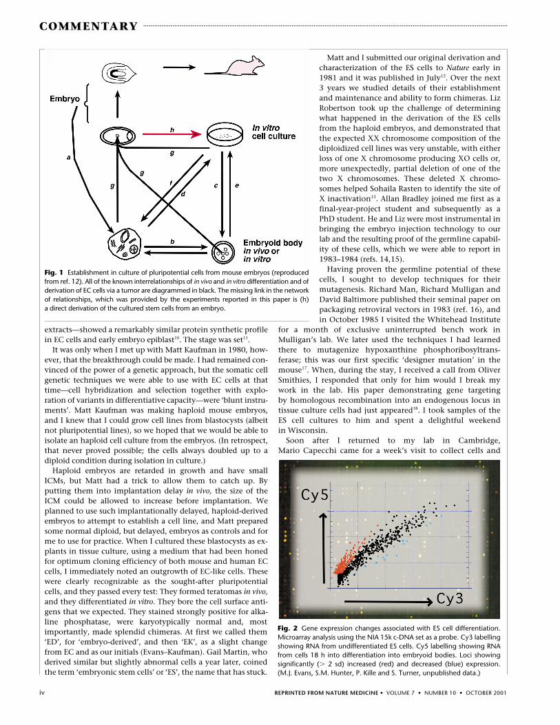
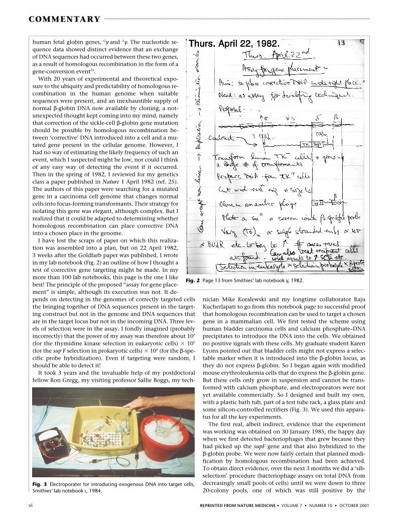
Fig. 2 Gene expression changes associated with ES cell differentiation.Microarray analysis using the NIA 15k c-DNA set as a probe. Cy3 labellingshowing RNA from undifferentiated ES cells. Cy5 labelling showing RNAfrom cells 18 h into differentiation into embryoid bodies. Loci showingsignificantly (� 2 sd) increased (red) and decreased (blue) expression. (M.J. Evans, S.M. Hunter, P. Kille and S. Turner, unpublished data.)
Fig. 1 Establishment in culture of pluripotential cells from mouse embryos (reproducedfrom ref. 12). All of the known interrelationships of in vivo and in vitro differentiation and ofderivation of EC cells via a tumor are diagrammed in black. The missing link in the networkof relationships, which was provided by the experiments reported in this paper is (h) a direct derivation of the cultured stem cells from an embryo.

Norma Ford Walker, who began my edu-cation as a geneticist, this was soonproven correct20. The field of normal
human protein polymorphic variants was seeded!The hereditary variations we had discovered proved to be in
the hemoglobin-binding serum protein haptoglobin, and theirdetails were worked out during a happy collaboration betweenGeorge E. Connell, Gordon H. Dixon and me in the early 1960s.The haptoglobin alleles Hp1F (fast) and Hp1S (slow) encodedpolypeptides differing by two amino acids, but the third allele,Hp2, seemed to be a tandem joining together of sequences fromHp1F with sequences from Hp1S. The then-chairman of my de-partment at the University of Wisconsin, James F. Crow, onbeing asked how the Hp2 allele might have arisen, directed meto the Bar locus in Drosophila with its fascinating history of re-peated ‘mutations’ resulting from unequal crossing over21. Thisled us to hypothesize that the Hp2 allele was formed by a uniquenon-homologous recombinational event that joined the end ofHp1F to the beginning of Hp1S (ref. 22). Hp2 therefore containeda small intragenic tandem duplication. The Bar gene inDrosophila is also a unique tandem duplication, but it is largeenough to be visible when the fly salivary chromosomes areunder the microscope. Yet the consequences of the tandemly re-peated sequences in Bar and in Hp are completely comparable.In both cases, subsequent predictable unequal homologouscrossing over events occur, which generate a new triplicate prod-uct and regenerate the singleton: B–B � B–B leads to B–B–B � B.
I found the predictabil-ity of homologous re-combination seductive,and enjoyed enormouslyhypothesizing that anti-body variability might beachieved by homologousrecombination betweentandemly arranged se-quences23. The hypothe-sis turned out to beincorrect in mammals,but was remarkably closeto being correct in chick-ens. Homologous recom-bination reappeared inmy experimental sciencein the early days ofcloning human geneswhen we were determin-ing the nucleotide se-quences of the two
REPRINTED FROM NATURE MEDICINE • VOLUME 7 • NUMBER 10 • OCTOBER 2001 v
COMMENTARY
Toolmakers—and I suspect that the threeof us being honored by the LaskerFoundation fit into this category—are for-tunate people. They see problems, invent tools to solve themand enjoy the solutions, which often demonstrate new princi-ples that were not part of the original thought. As a bonus, theyalso enjoy the vicarious pleasure of seeing other people use thesame tools to solve very different problems. Yet the invention ofan effective scientific tool is rarely an isolated event; there areoften many prior experiences that trigger the inventive thought,and there may be various unexpected additional problems tosolve before the toolmaker can bring a nascent idea into practice.
The chain of events leading to my contributions to the use ofhomologous recombination to modify genes in the mousegenome began over 40 years ago as an unplanned consequenceof my somewhat serendipitous invention in the 1950s of anearlier tool—high-resolution gel electrophoresis—to solve acompletely non-genetic problem. On 26 October 1954, duringfinal pre-publication tests of my starch-gel electrophoresis sys-tem (the immediate forerunner of one of molecular biologists’primary tools, polyacrylamide gel electrophoresis), I ran a sam-ple of serum from a female. My notebook (Fig. 1) has the entrythat the pattern was “Most odd—many extra components.” Forabout a week I enjoyed the misconception that I had discov-ered a new way of telling males from females. But this ‘sexy’hypothesis soon gave way to the idea that “hereditary factorsmay determine the serum groups”19 and, with the help of
Forty years with homologous recombination
OLIVER SMITHIES
learn the techniques. The rest of this story is better known.Many hundreds of specifically targeted mouse mutations havebeen made and the technique, although still not trivial, maynow merit no more than a few lines’ mention in experimentalgenetics papers. Almost any specific genetic change may nowbe generated, selected and verified in culture before beingconverted to the germ lines of mice, and this is the experi-mental genetics that is illuminating our understanding of the
mammalian genome physiology and human function inhealth and disease.
I set out to derive a ‘tractable’ system for following mRNAchanges coincident with embryonic cell differentiation. ES cellsnow provide the culture system and, at long last, methods forgenome-wide monitoring of mRNA have come of age in cDNAmicroarrays. I am now putting the two techniques together,and results are beginning to emerge from this work (Fig. 2).
Fig. 1 Pages 97 & 98 from Smithies’ lab notebook “Physical IV”, 1954.
vi REPRINTED FROM NATURE MEDICINE • VOLUME 7 • NUMBER 10 • OCTOBER 2001
COMMENTARY
human fetal globin genes, Gγ and Aγ. The nucleotide se-quence data showed distinct evidence that an exchangeof DNA sequences had occurred between these two genes,as a result of homologous recombination in the form of agene-conversion event24.
With 20 years of experimental and theoretical expo-sure to the ubiquity and predictability of homologous re-combination in the human genome when suitablesequences were present, and an inexhaustible supply ofnormal β-globin DNA now available by cloning, a not-unexpected thought kept coming into my mind, namelythat correction of the sickle-cell β-globin gene mutationshould be possible by homologous recombination be-tween ‘corrective’ DNA introduced into a cell and a mu-tated gene present in the cellular genome. However, Ihad no way of estimating the likely frequency of such anevent, which I suspected might be low, nor could I thinkof any easy way of detecting the event if it occurred.Then in the spring of 1982, I reviewed for my geneticsclass a paper published in Nature 1 April 1982 (ref. 25).The authors of this paper were searching for a mutatedgene in a carcinoma cell genome that changes normalcells into focus-forming transformants. Their strategy forisolating this gene was elegant, although complex. But Irealized that it could be adapted to determining whetherhomologous recombination can place corrective DNAinto a chosen place in the genome.
I have lost the scraps of paper on which this realiza-tion was assembled into a plan, but on 22 April 1982, 3 weeks after the Goldfarb paper was published, I wrotein my lab notebook (Fig. 2) an outline of how I thought atest of corrective gene targeting might be made. In mymore than 100 lab notebooks, this page is the one I likebest! The principle of the proposed “assay for gene place-ment” is simple, although its execution was not. It de-pends on detecting in the genomes of correctly targeted cellsthe bringing together of DNA sequences present in the target-ing construct but not in the genome and DNA sequences thatare in the target locus but not in the incoming DNA. Three lev-els of selection were in the assay. I fondly imagined (probablyincorrectly) that the power of my assay was therefore about 105
(for the thymidine kinase selection in eukaryotic cells) � 105
(for the sup F selection in prokaryotic cells) � 106 (for the β-spe-cific probe hybridization). Even if targeting were random, Ishould be able to detect it!
It took 3 years and the invaluable help of my postdoctoralfellow Ron Gregg, my visiting professor Sallie Boggs, my tech-
nician Mike Koralewski and my longtime collaborator RajuKucherlapati to go from this notebook page to successful proofthat homologous recombination can be used to target a chosengene in a mammalian cell. We first tested the scheme usinghuman bladder carcinoma cells and calcium phosphate–DNAprecipitates to introduce the DNA into the cells. We obtainedno positive signals with these cells. My graduate student KarenLyons pointed out that bladder cells might not express a selec-table marker when it is introduced into the β-globin locus, asthey do not express β-globin. So I began again with modifiedmouse erythroleukemia cells that do express the β-globin gene.But these cells only grow in suspension and cannot be trans-formed with calcium phosphate, and electroporators were notyet available commercially. So I designed and built my own,with a plastic bath tub, part of a test tube rack, a glass plate andsome silicon-controlled rectifiers (Fig. 3). We used this appara-tus for all the key experiments.
The first real, albeit indirect, evidence that the experimentwas working was obtained on 30 January 1985, the happy daywhen we first detected bacteriophages that grew because theyhad picked up the supF gene and that also hybridized to the β-globin probe. We were now fairly certain that planned modi-fication by homologous recombination had been achieved. To obtain direct evidence, over the next 3 months we did a ‘sib-selection’ procedure (bacteriophage assays on total DNA fromdecreasingly small pools of cells) until we were down to three20-colony pools, one of which was still positive by the
Fig. 2 Page 13 from Smithies’ lab notebook γ, 1982.
Fig. 3 Electroporater for introducing exogenous DNA into target cells,Smithies’ lab notebook ι , 1984.

REPRINTED FROM NATURE MEDICINE • VOLUME 7 • NUMBER 10 • OCTOBER 2001 vii
COMMENTARY
bacteriophage assay. Individual colonies from this pool weretested on 18 May 1985 by Southern blot analysis (a directassay). DNA from one of the colonies produced a hybridizingfragment of the correct size (Fig. 4), and we were ‘home’!
I presented the results of our work at a Gordon Conference in1985, and told the attendees the true story that, as I developedthe critical gel autoradiograph, which we knew would providethe first direct test of whether or not the target gene had beenmodified, I was thinking that we had been a long time (3 years)knowing that our experiment was working only by indirect ev-idence—much like being an airplane pilot on instruments inthe clouds. The autoradiograph was the moment of truth, com-parable to the moment when you descend below the cloudsand no longer depend on the indirect indications of your in-struments: The runway is either there or it is not! The thrill ofseeing it never pales. For the remainder of that meeting, otherinvestigators would say, as they pointed to a desired result,“And there is my runway!” We published our results in the 19 September 1985 issue of Nature18.
Nonetheless, our ‘runway’ was exceedingly difficult to find.In only about one in a million treated cells was homologous re-combination achieved. Such a low frequency of gene targetingwas not much use for gene therapy. And the assay, like my doc-toral-thesis method of measuring osmotic pressures26, was re-markably good at doing what it was designed to do, but bothmethods were impossibly laborious. No one, not even me, everused either again. So, what to do? The first order of businesswas to try to improve the method. For this we needed an easiertarget, preferably one whose targeting could be assessed di-rectly. The hypoxanthine phosphoribosyltransferase gene(HPRT) was an obvious choice, and so Ron Gregg began a seriesof attempts to correct a mutated HPRT or to inactivate a wild-type copy of the gene using homologous recombination.
We also needed to replace the bacteriophage recombinantfragment assay with something easier. Kary Mullis’ new PCRtool could in principle detect recombinants. We could choose
one primer specific to the incoming DNA and another primerspecific to the target gene. PCR amplification would then onlyoccur when the two primer sequences were juxtaposed by the desired homologous recombination. But, again, there wereno commercial PCR machines available. So we made our ownout of three old water-baths, home-made controllers and hot water valves used in domestic heating systems27. We stilluse it! Its six hoses look like octopus arms; for obvious reasonswe call it ‘hexapus’. The ease of this PCR-based recombinantfragment assay made screening for homologous recombinantsmuch less difficult.
Meanwhile, at a 1985 Gordon Conference, I heard ErwinWagner talk about Martin Evans’ embryonic stem cells12,which after injection into blastocysts can produce living prog-eny mice. Here was a more promising use of our one-in-a-mil-lion targeting skill. We could generate planned mutations orcorrect existing mutations in tissue culture, even if it took mil-lions of cells, and expect to transfer the alterations into livingmice. A visit to Erwin Wagner led to my contacting MartinEvans who, with typical generosity of spirit, personallybrought some of his EK CC-1 cells to us in November 1985. Myplan was “to use these to get HPRT– by recombination and getchimeras or germline by blastocyst route.” Martin also put mein touch with Tom Doetschman, an American postdoctoral fel-low wanting to return to the United States, who had personallyisolated embryonic stem cells (now called ES cells) while inRolf Kemmler’s laboratory. He joined our group in late 1986.
At this point, Nobuyo Maeda and I attended a conference in Scotland at which Martin Evans and Martin Hooper both reported that they had obtained HPRT– mutant ES cells in tissueculture experiments. Nobuyo recognized that, in the course ofhelping Ron Gregg, she had already made a construct that could correct either of their HPRT– mutant cells. We toldEvans and Hooper about this, and both immediately agreed tocollaborate with us: Martin Hooper sent his mutant cells (TG-2a) to us, and we sent our construct to Martin Evans. Tom
Doetschman triedNobuyo’s construct onthe TG-2a cells. The veryfirst experiment worked,and we published our re-sults in Nature 10December 1987 (ref. 28).But it still took 2 moreyears of valiant effort,spearheaded by my post-doctoral fellow BevKoller, to accomplishthe mouse blastocyst in-jections leading tochimeras, to obtainprogeny with the alteredgene and to report the“Germ-line transmissionof a planned alterationmade by homologous re-combination in embry-onic stem cells.”29
We next turned our attention to problemsrelated to human dis-eases, beginning byFig. 4 Pages 134 & 135 from Smithies’ lab notebook κ, 1985.
phate co-precipitation to introduce theDNA into cultured cells—was not effi-cient. With this method, incorporation
of functional copies of tk occurred in only one per million cellsexposed to the DNA–calcium phosphate co-precipitate. Usinga similar selection scheme, I sought to determine whether Icould introduce a functional tk into Tk– cells using very fineglass needles to inject DNA directly into nuclei34. This proce-dure proved extremely efficient. One cell in three that receivedthe DNA stably passed the functional tk to its daughter cells.The high efficiency of DNA transfer by microinjection made itpractical for investigators to generate transgenic mice contain-ing random insertions of exogenous DNA. This was accom-plished by injection of the desired DNA into nuclei of one-cellzygotes and allowing these embryos to come to term after sur-gical transfer to foster mothers35–39.
Efficient functional transfer of HSV-tk into cells required thatthe injected tk be linked to other short viral DNA sequences34. Itseemed plausible that highly evolved viral genomes might con-tain bits of DNA that enhance their ability to establish them-selves within mammalian cell genomes. I searched the genomeof the lytic simian virus SV40 for the presence of such se-quences and found one near the origin of viral DNA replication.When linked to HSV-tk, it increased the transforming capacityof the injected tk by 100-fold. I showed that the enhancementdid not seem to result from independent replication of the in-jected HSV-tk DNA as an extra-chromosomal plasmid, butrather that the efficiency-enhancing sequence was either
viii REPRINTED FROM NATURE MEDICINE • VOLUME 7 • NUMBER 10 • OCTOBER 2001
COMMENTARY
Mutational analysis is one of the mostinformative approaches available for thestudy of complex biological processes. Ithas been particularly successful in the analysis of the biologyof bacteria, yeast, the nematode worm Caenorhabditis elegansand the fruit fly Drosophila melanogaster. Extension of this ap-proach to the mouse, though informative, was far less success-ful relative to what has been achieved with these simplermodel organisms. This is because it is not numerically practicalin mice to use random mutagenesis to isolate mutations thataffect a specified biological process of interest. Nonetheless, bi-ological phenomena such as a sophisticated immune response,cancer, vascular disease or higher-order cognitive function, tomention just a few, must be analyzed in organisms that showsuch phenomena, and for this reason geneticists and other re-searchers have turned to the mouse. Gene targeting, the meansfor creating mice with designed mutations in almost anygene32, was developed as an alternative to the impractical useof random mutagenesis for pursuing genetic analysis in themouse. Now gene targeting has advanced the genomic manip-ulations possible in mice to a level that can be matched only infar simpler organisms such as bacteria and yeast.
The development of gene targeting in mice required the so-lution to two problems: How to produce a specific mutation ina chosen gene in cultured mammalian cells, and how to trans-fer this mutation to the mouse germ line. Oliver Smithies’ lab-oratory and mine worked independently on solutions to thefirst problem. Martin Evans’ laboratory provided the basis for asolution to the second problem.
Early experimentsOur entry into what became the field of gene targeting began
in 1977. At that time, I was attempting to improve the effi-ciency with which new genes could be introduced into mam-malian cells. It had just been demonstrated by Wigler and Axelthat cultured mammalian cells deficient in thymidine kinase(Tk–) could be transformed to Tk+ status by the introduction ofa functional copy of the herpes thymidine kinase gene (HSV-tk)33. Although an important advance for the field of so-matic cell genetics, their protocol—the use of calcium phos-
Generating mice with targeted mutations
MARIO R. CAPECCHI
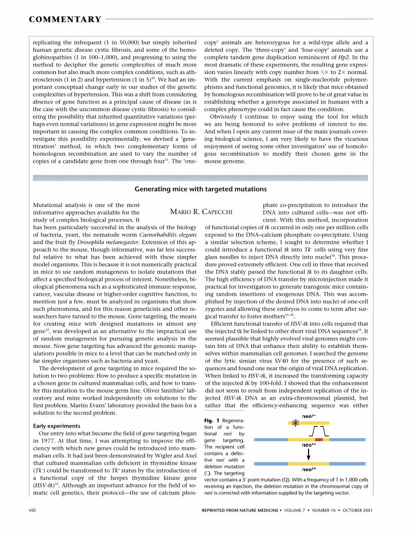
Fig. 1 Regenera-tion of a func-tional neor bygene targeting.The recipient cellcontains a defec-tive neor with adeletion mutation(∗ ). The targetingvector contains a 5’ point mutation (Q). With a frequency of 1 in 1,000 cellsreceiving an injection, the deletion mutation in the chromosomal copy ofneor is corrected with information supplied by the targeting vector.
replicating the infrequent (1 in 50,000) but simply inheritedhuman genetic disease cystic fibrosis, and some of the hemo-globinopathies (1 in 100–1,000), and progressing to using themethod to decipher the genetic complexities of much morecommon but also much more complex conditions, such as ath-erosclerosis (1 in 2) and hypertension (1 in 5)30. We had an im-portant conceptual change early in our studies of the geneticcomplexities of hypertension. This was a shift from consideringabsence of gene function as a principal cause of disease (as isthe case with the uncommon disease cystic fibrosis) to consid-ering the possibility that inherited quantitative variations (per-haps even normal variations) in gene expression might be moreimportant in causing the complex common conditions. To in-vestigate this possibility experimentally, we devised a ‘gene-titration’ method, in which two complementary forms ofhomologous recombination are used to vary the number ofcopies of a candidate gene from one through four31. The ‘one-
copy’ animals are heterozygous for a wild-type allele and adeleted copy. The ‘three-copy’ and ‘four-copy’ animals use acomplete tandem gene duplication reminiscent of Hp2. In themost dramatic of these experiments, the resulting gene expres-sion varies linearly with copy number from 1⁄2� to 2� normal.With the current emphasis on single-nucleotide polymor-phisms and functional genomics, it is likely that mice obtainedby homologous recombination will prove to be of great value inestablishing whether a genotype associated in humans with acomplex phenotype could in fact cause the condition.
Obviously I continue to enjoy using the tool for which we are being honored to solve problems of interest to me. And when I open any current issue of the main journals cover-ing biological science, I am very likely to have the vicarious enjoyment of seeing some other investigators’ use of homolo-gous recombination to modify their chosen gene in the mouse genome.

REPRINTED FROM NATURE MEDICINE • VOLUME 7 • NUMBER 10 • OCTOBER 2001 ix
COMMENTARY
increasing the frequency with which the exogenous DNA wasintegrated into the host genome, or increasing the probabilitythat tk, once integrated, was being expressed in the recipientcells. These experiments were completed before the idea of geneexpression ‘enhancers’ had emerged and contributed to the de-finition of these special DNA sequences40. The emerging idea ofenhancers profoundly influenced our contributions to the de-velopment of gene targeting by alerting us to the importance ofusing appropriate enhancers to mediate expression of newly in-troduced selectable genes regardless of the inherent expressioncharacteristics of the host site to which they were targeted.
Homologous recombinationThe observation I found most fascinating from these early
DNA microinjection experiments was that when many copiesof the tk plasmid were injected into cells, they were integratedin only one or two loci within any host cell’s chromosome, andthat multiple copies at those random sites were always presentas head-to-tail concatemers. We reasoned that such highly or-dered concatemers could only be generated either by replica-tion (for example, a rolling circle-type mechanism) or byhomologous recombination between plasmids. We proved thatthey were generated by homologous recombination41. Thisconclusion was very significant because it demonstrated thatmammalian somatic cells contained an efficient enzymaticmachinery for mediating homologous recombination. The effi-ciency of this machinery became evident from the observationthat when more than 100 tk plasmid molecules were injectedper cell, they were all incorporated into a single, ordered, head-to-tail concatemer. It was immediately apparent that if wecould harness this efficient machinery to accomplish homolo-gous recombination between a newly introduced DNA mole-cule of our choice and the same DNA sequence in a recipientcell’s genome, we would have the ability to mutate or modifyalmost any cellular gene in any chosen way.
Our next step in the quest for gene targeting required our becoming familiar with this machinery; specifically, with itssubstrate preferences and reaction products. By examining recombination between co-injected DNA molecules, welearned, among other things, that linear DNA molecules were the preferred substrate for homologous recombination;that recombination was cell cycle-dependent, showing a peak of activity in early S phase; and that although both recip-rocal and nonreciprocal exchanges occurred, there was a dis-tinct bias toward the latter42–44. These results contributedsubstantially to our choice of experimental design for the nextstage of this quest: the detection of homologous recombina-tion between newly introduced, exogenous DNA and its chro-mosome homolog.
In 1980, we submitted a grant proposal to the NationalInstitutes of Health to test the feasibility of gene targeting in mammalian cells; these experiments were rejected on the grounds that there was only a vanishingly small probabil-ity that the newly introduced DNA would find its matchingsequence within a host cell genome. Despite the rejection, I decided to continue this line of experimentation. Aware thatthe frequency of gene targeting was likely to be low, and thatthe far more common competitive reaction would be inser-tion of the targeting vector at various sites other than the tar-get locus, we proposed to use selection to eliminate cells notcontaining the desired homologous recombination products.The first test (Fig. 1) used artificially introduced chromosomal
target sites. The first step of this scheme required generationof cell lines containing random insertions of a defectiveneomycin-resistance gene (neor) containing either a deletionor a point mutation. In the second step, target vector DNAcarrying defective neor genes with different mutations was in-troduced into cells of those lines. Homologous recombinationbetween neor sequences in the targeting vector and recipientgenome could generate a functional neor from the two defec-tive parts, producing cells resistant to the drug G418, which islethal to cells without a functional neor.
In the first step, we generated recipient cell lines containingsingle copies of the defective neor, lines containing multiplecopies of the gene in head-to-tail concatemers and, by inhibit-ing concatemer formation, lines with multiple defective neor
targets, each located on separate chromosomes. These differentrecipient cell lines allowed us to evaluate how the number andlocation of targets within the recipient cell’s genome influ-enced the targeting frequency. By 1984 we had good evidencethat gene targeting in cultured mammalian cells was indeedpossible45. At this time I resubmitted our grant to the sameNational Institutes of Health study section that had rejectedour earlier grant proposal and their critique began with thephrase “We are glad that you didn’t follow our advice.”
To our delight, correction of the defective chromosomal neor
occurred at an absolute frequency of 1 per 1,000 cells receivingan injection. This frequency was not only higher than we ex-pected, but allowed us to accomplish multiple analyses of the experimental parameters that could influence the gene-targeting reaction44. An additional important lesson from theseexperiments was that all chromosomal target positions analyzed seemed to be equally accessible to the homologousrecombination machinery, indicating that a large fraction ofthe mouse genome could be modified by gene targeting.
At this time, Oliver Smithies and his colleagues reportedtheir classic experiment of targeted modification of the β-globin locus in cultured mammalian cells18. This elegant ex-periment demonstrated that it was feasible to disrupt an en-dogenous gene in cultured mammalian cells. Havingestablished that gene targeting could be achieved in culturedmammalian cells and having determined some of the parame-ters that influenced its frequency, we were ready to extend theapproach to the whole mouse. The low frequency of targeted
Fig. 2 Disruption of Hprt by gene targeting. The vector contains Hprt se-quences disrupted in the eighth exon by neor. After homologous pairingbetween the vector and genomic sequences, a homologous recombina-tion event replaces the genomic sequence with vector sequences contain-ing neor. These cells are able to grow in medium containing the drugsG418 and 6-TG.
x REPRINTED FROM NATURE MEDICINE • VOLUME 7 • NUMBER 10 • OCTOBER 2001
COMMENTARY
homologous recombination relative to random integration ofthe targeting vector into the recipient cell genome made it impractical to attempt gene targeting directly in one-cellmouse zygotes. Instead, it seemed our best option was to dogene targeting in cultured embryo-derived stem (ES) cells, fromwhich the relatively rare targeted recombinants would be se-lected and purified. These purified cells, when subsequently in-troduced into a preimplantation embryo and allowed tomature in a foster mother, would contribute to the formationof all tissues of the mouse, including the germ line.
Gene targeting in ES cellsAt a Gordon Conference in the summer of 1984, I heard a
discussion from a member of Martin Evans’ laboratory aboutES cells. They seemed much more promising in their potentialto contribute to the formation of the germ line than the previ-ously characterized embryonal carcinoma (EC) cells12,15. In thewinter of 1985, my wife and I spent a week in Martin Evans’laboratory learning how to derive, culture and generate mousechimeras from these cells.
In the beginning of 1986, our effort switched to doing gene targeting experiments in ES cells. We also decided to useelectroporation as the means of introducing our targeting vec-tors into ES cells. Although microinjection is orders of magni-tude more efficient than electroporation as a means forgenerating cells with targeted mutations, injections must bedone one cell at a time. With electroporation, we could intro-duce the targeting vector into 1 � 107 cells in a single experi-ment, easily producing large numbers of transformed cellseven with the lower efficiency.
To rigorously determine the quantitative efficiency of genetargeting in ES, we chose as our target locus the hypoxanthinephosphoribosyl transferase gene (Hprt). There were two mainreasons for this choice. As Hprt is located on the X chromo-some and the ES cell line that we were using was derived froma male mouse, only a single Hprt locus had to be disrupted toyield Hprt– cell lines. Moreover, a good protocol for selectingcells with disrupted Hprt genes existed, based on the drug 6-thioguanine (6-TG), which kills cells with a functional Hprt.The strategy we used was to generate a gene-targeting vector that contained an Hprt genomic sequence that was disrupted in an exon by insertion of neor (Fig. 2). Homologousrecombination between this targeting vector and the ES cellchromosomal Hprt would generate Hprt– cells that would be resistant to growth in medium containing both 6-TG (killingHprt+ cells) and G418 (killing cells lacking neor). All lines gener-ated from cells selected in this way lost Hprt function as a result of gene-targeted disruption of the Hprt locus46. The Hprtlocus provided an ideal locus to further test many variablesthat could potentially influence the targeting efficiency46–49.
Because we foresaw that neor would probably be used as apositive selectable gene for the disruption of many genes in ES cells, it was essnetial that its expression be mediated by anenhancer that would function regardless of its location withinthe ES cell genome. Here our previous experience with en-hancers and the transformation of cultured mouse cells provedof value. We knew from those experiments that the activitiesof promoter–enhancer configurations are very cell-specific. Toencourage such strong neor expression in ES cells, we chose todrive it with a duplicated, mutated polyoma virus enhancer se-lected for strong expression in mouse embryonal carcinomacells46. Subsequently, the strategy described above of using neor
driven by an enhancer that allows strong expression in ES cells,independent of chromosomal location, has become the stan-dard for disruption of most genes in ES cells.
The experiments described above showed that ES cells weregood recipient hosts, able to mediate homologous recombina-tion between the targeting vector and the cognate chromoso-mal sequence. In addition, the drug-selection protocolsrequired to identify ES cell lines containing the targeted dis-ruptions did not seem to alter their pluripotent potential. I be-lieve that this paper was pivotal in the development of thefield by encouraging other investigators to begin use of genetargeting in mice as a means for determining the function inthe intact animal of the genes they were studying.
The ratio of homologous to non-homologous recombina-tion events in ES cells was found to be approximately 1 to1,000 (ref. 46). Because the disruption of most genes does notproduce a phenotype that is selectable at the cellular level, in-vestigators seeking specific gene disruptions would need eitherto undertake tedious DNA screens through many cell coloniesto identify the rare ones containing the desired targeting
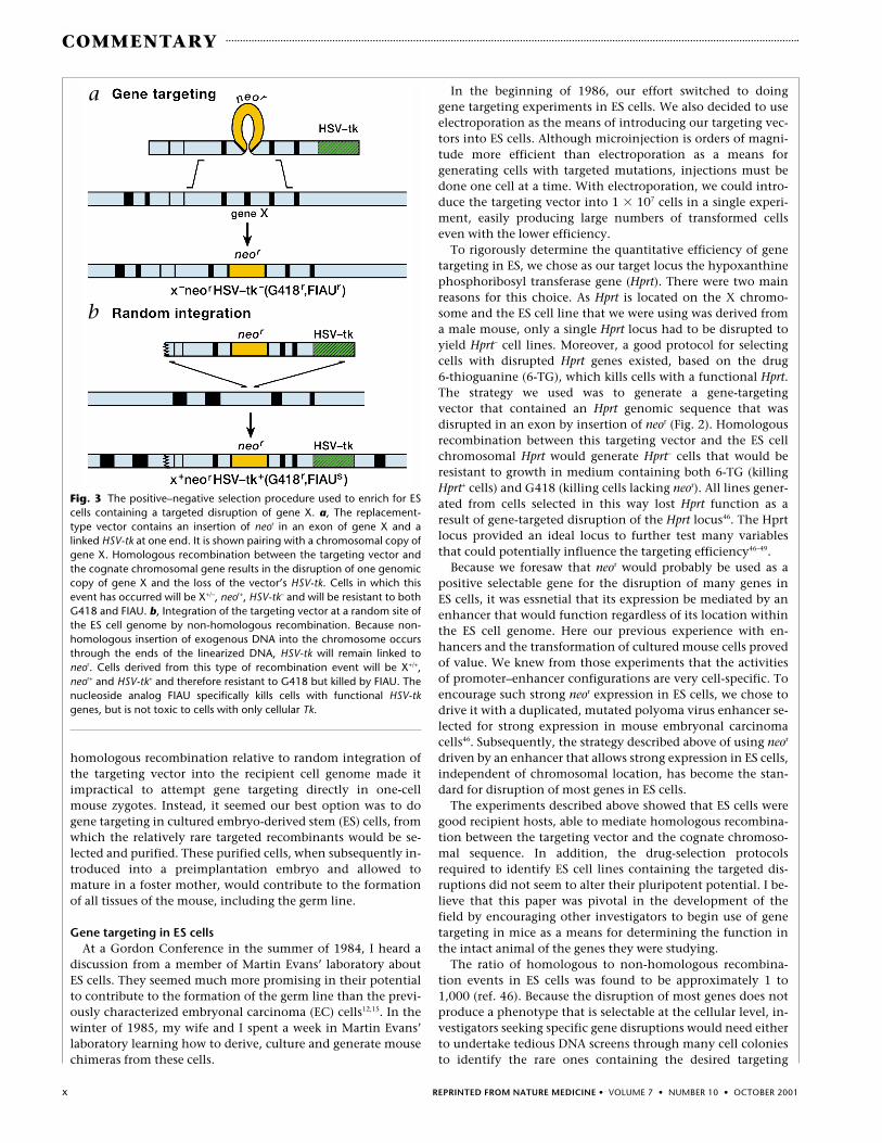
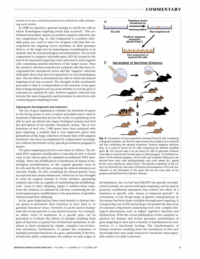
Fig. 3 The positive–negative selection procedure used to enrich for EScells containing a targeted disruption of gene X. a, The replacement-type vector contains an insertion of neor in an exon of gene X and alinked HSV-tk at one end. It is shown pairing with a chromosomal copy ofgene X. Homologous recombination between the targeting vector andthe cognate chromosomal gene results in the disruption of one genomiccopy of gene X and the loss of the vector’s HSV-tk. Cells in which thisevent has occurred will be X+/–, neor+, HSV-tk– and will be resistant to bothG418 and FIAU. b, Integration of the targeting vector at a random site ofthe ES cell genome by non-homologous recombination. Because non-homologous insertion of exogenous DNA into the chromosome occursthrough the ends of the linearized DNA, HSV-tk will remain linked toneor. Cells derived from this type of recombination event will be X+/+,neor+ and HSV-tk+ and therefore resistant to G418 but killed by FIAU. Thenucleoside analog FIAU specifically kills cells with functional HSV-tkgenes, but is not toxic to cells with only cellular Tk.
a
b
REPRINTED FROM NATURE MEDICINE • VOLUME 7 • NUMBER 10 • OCTOBER 2001 xi
COMMENTARY
events or to use a selection protocol to enrich for cells contain-ing such events.
In 1988 we reported a general strategy to enrich for cells inwhich homologous targeting events had occurred50. This en-richment procedure, known as positive–negative selection, hastwo components (Fig. 3). One component is a positive selec-table gene, neor, used to select for recipient cells that have in-corporated the targeting vector anywhere in their genomes(that is, at the target site by homologous recombination or atrandom sites by non-homologous recombination). The secondcomponent is a negative selectable gene, HSV-tk, located at theend of the linearized targeting vector and used to select againstcells containing random insertions of the target vector. Thusthe ‘positive’ selection enriches for recipient cells that have in-corporated the introduced vector and the ‘negative’ selectioneliminates those that have incorporated it at non-homologoussites. The net effect is enrichment for cells in which the desiredtargeting event has occurred. The strength of this enrichmentprocedure is that it is independent of the function of the genethat is being disrupted and succeeds whether or not the gene isexpressed in cultured ES cells. Positive–negative selection hasbecome the most frequently used procedure to enrich for cellscontaining gene-targeting events.
Subsequent development and extensionThe use of gene targeting to evaluate the functions of genes
in the living mouse is now a routine procedure and is used inhundreds of laboratories all over the world. It is gratifying to beable to pick up almost any major biological journal and findthe description of yet another ‘knockout’ mouse. The in vivofunctions of well over 7,000 genes have been analyzed withgene targeting, a number that is very impressive given thatgeneration of this large collection of mouse lines with targetedmutations has been accomplished by independent investiga-tors without the benefit of any special government program tofund it.
The gene-targeting protocol is now done as follows: The de-sired DNA sequence modification is introduced into a clonedcopy of the chosen gene by standard recombinant DNA tech-nology. Then, the modification is transferred, by means of ho-mologous recombination, to the cognate genomic locus in ES cells and the ES cell lines carrying the desired alteration areselected. Finally, ES cells containing the altered genetic locusare injected into mouse blastocysts, which are in turn broughtto term by surgical transfer to foster mothers, generatingchimeric mice that are capable of transmitting the modified ge-netic locus to their offspring. Figure 4 outlines these steps,from the isolation of cultured ES cell lines containing the de-sired targeted gene modification to the generation of germlinechimeras and their offspring.
So far, gene targeting has been used mainly to disrupt cho-sen genes to determine their function in mice (that is, to generate ‘knockout’ mice). However, it can be used to manip-ulate the mouse genome in any desired manner. For example,an allelic series of mutations in a specific gene can be generated to evaluate the effects of changes resulting fromgain-of-function or partial loss-of-function mutations, in ad-dition to those produced by simple, complete loss-of-func-tion mutations. Furthermore, to permit the evaluation ofmultiple potential functions of a gene, particularly if the loss-of-function allele compromises the embryo at early stages of
development, the Cre-loxP and Flp/FRT site-specific recombi-nation systems, in concert with gene targeting, can be used togenerate conditional mutations that restrict the effect of amutation to specific cells, tissues or temporal periods51. Inconclusion, a very broad range of genetic manipulations inthe mouse has been made available through gene targeting. Itis hoped that use of this technology will permit the discoveryof essential components underlying even very complex bio-logical phenomena such as higher cognitive function anddysfunction. With the recent publication of the complete se-quences for human and mouse genomes, practitioners ofgene targeting in mice have a bounty of information for con-version to a functional footing. The transformation ofhuman medicine resulting from the translation of this newknowledge base may make tomorrow’s medicine unrecogniz-able relative to today’s practices.
Fig. 4 Generation of mouse germline chimeras from ES cells containinga targeted mutation. a, The first step involves the isolation of a clonal EScell line containing the desired mutations. Positive–negative selection(Fig. 3) is used to enrich for ES cells containing the desired modifiedgene. b, The second step is to use those ES cells to generate chimericmice able to transmit the mutant gene to their progeny. To facilitate iso-lation of the desired progeny, the ES cells and recipient blastocysts arederived from mice with distinguishable coat color alleles (ES, agoutibrown mice; blastocyst, black mice). This permits evaluation of the ex-tent of chimerism by coat color chimerism and evaluation of ES cell con-tribution to the formation of the germ line by the coat color of theprogeny derived from the chimeric animals.
a
b

xii REPRINTED FROM NATURE MEDICINE • VOLUME 7 • NUMBER 10 • OCTOBER 2001
COMMENTARY
1. Stevens, L.C. The biology of teratomas. Adv. Morphog. 6, 1–31 (1967).2. Pierce, G.B. Teratocarcinoma: Model for a developmental concept of cancer.
Curr. Topics Dev. Biol. 2, 223–246 (1967).3. Kleinsmith, L.J. & Pierce, G.B. Multipotentiality of single embryonal carcinoma
cells. Cancer Res. 24, 1544–1551 (1964).4. Evans, M.J. The isolation and properties of a clonal tissue culture strain of pluripo-
tent mouse teratocarcinoma cells. J. Embryol. Exp. Morphol. 28, 163–196 (1972).5. Evans, M.J. & Martin, G.R. The differentiation of clonal teratocarcinoma cell cul-
ture in vitro. In Roche Symposium on Teratomas and Differentiation (eds. Solter, D.& Sherman, M.) (Academic Press, New York, 1975).
6. Martin, G.R & Evans, M.J. Differentiation of clonal lines of teratocarcinoma cells:formation of embryoid bodies in vitro. Proc. Natl. Acad. Sci. USA 72, 1441–1445,(1975).
7. Papaioannou, V.E., McBurney, M., Gardner, R.L. & Evans, M.J. The fate of terato-carcinoma cells injected into early mouse embryos. Nature 258, 70–73 (1975).
8. Gooi, H.C. et al. Stage-specific embryonic antigen involves 1-3 fucosylated type 2blood group chains. Nature 292, 156–158 (1981).
9. Stinnakre, M.G., Evans, M.J., Willison, K.R. & Stern, P.L. Expression of Forssmanantigen in the post-implantation mouse embryo. J. Embryol. Exp. Morphol. 61,117–131 (1981).
10. Lovell-Badge, R.H. & Evans, M.J. Changes in protein synthesis during differentia-tion of embryonal carcinoma cells and a comparison with embryo cells. J.Embryol. Exp. Morphol. 59, 187–206 (1980).
11. Evans, M.J. Origin of mouse embryonal carcinoma cells and the possibility of theirdirect isolation into tissue culture. J. Reprod. Fertil. 62, 625–631 (1981).
12. Evans, M.J. & Kaufman, M.H. Establishment in culture of pluripotential cells frommouse embryos. Nature 292, 154–156 (1981).
13. Rastan, S. & Robertson, E.J. X-chromosome deletions in embryo-derived (EK) cell-lines associated with lack of X-chromosome inactivation. J. Embryol. Exp. Morph.90, 379–388 (1985).
14. Evans, M.J., Bradley, A. & Robertson, E.J. EK cell contribution to chimeric mice:from tissue culture to sperm. In Genetic Manipulation of the Early MammalianEmbryo, Banbury Report. (Cold Spring Harbor Laboratory Press, Plainview, NY,1983).
15. Bradley, A., Evans, M.J., Kaufman, M.H. & Robertson, E.J. Formation of germ-linechimaeras from embryo-derived teratocarcinoma cell lines. Nature 309, 255–256(1984).
16. Mann, R., Mulligan, R.C. & Baltimore, D. Construction of a retrovirus packagingmutant and its use to produce helper-free defective retrovirus. Cell 33, 153–159(1983).
17. Kuehn, M.R., Bradley, A., Robertson, E.J. & Evans, M.J. A potential animal modelfor Lesch-Nyhan syndrome through introduction of HPRT mutations into mice.Nature 326, 295–298 (1987).
18. Smithies, O., Gregg, R.G., Boggs, S.S., Koralewski, M.A. & Kucherlapati, R.S.Insertion of DNA sequences into the human chromosomal β-globin locus by homologous recombination. Nature 317, 230–234 (1985).
19. Smithies, O. Zone electrophoresis in starch gels: Group variations in the serumproteins of normal human adults. Biochem. J. 61, 629–641 (1955).
20. Smithies, O. & Walker, N.F. Genetic control of some serum proteins in normalhumans. Nature 176, 1265–1266 (1955).
21. Sturtevant, A.H. The effects of unequal crossing over at the Bar locus inDrosophila. Genetics 10, 117–147 (1925).
22. Smithies, O., Connell, G.E. & Dixon, G.H. Chromosomal rearrangements and theevolution of haptoglobin genes. Nature 196, 232–236 (1962).
23. Smithies, O. Antibody variability. Science 157, 267–273 (1967).24. Slightom, J.L., Blechl, A.E. & Smithies, O. Human fetal Gγ and Aγ globin genes:
Complete nucleotide sequences suggest that DNA can be exchanged betweenthese duplicated genes. Cell 21, 627–638 (1980).
25. Goldfarb, M., Shimizu, K., Perucho, M. & Wigler, M. Isolation and preliminarycharacterization of a human transforming gene from T24 bladder carcinomacells. Nature 296, 404–409 (1982).
26. Smithies, O. A Dynamic osmometer for accurate measurements on small quanti-ties of material: Osmotic pressures of isoelectric β-lactoglobulin solutions.Biochem. J. 55, 57–67 (1953).
27. Kim, H.S. & Smithies, O. Recombinant fragment assay for gene targetting basedon the polymerase chain reaction. Nucleic Acids Res. 16, 8887–8903 (1988).
28. Doetschman, T. et al. Targetted correction of a mutant HPRT gene in mouse em-bryonic stem cells. Nature 330, 576–578 (1987).
29. Koller, B.H. et al. Germ-line transmission of a planned alteration made in a hy-poxanthine phosphoribosyltransferase gene by homologous recombination inembryonic stem cell. Proc. Natl. Acad. Sci. USA 86, 8927–8931 (1989).
30. Smithies, O. & Maeda, N. Gene targeting approaches to complex genetic dis-eases: Atherosclerosis and essential hypertension. Proc. Natl. Acad. Sci. USA 92,5266–5272 (1995).
31. Kim, H.-S. et al. Genetic control of blood pressure and the angiotensinogen locus.Proc. Natl. Acad. Sci. USA 92, 2735–2739 (1995).
32. Capecchi, M.R. Targeted gene replacement. Sci. Am. 270, 54–61 (1994).
33. Wigler, M. et al. Transfer of purified Herpes Virus thymidine kinase gene to cul-tured mouse cells. Cell 11, 223–232 (1977).
34. Capecchi, M.R. High efficiency transformation by direct microinjection of DNAinto cultured mammalian cells. Cell 22, 479–488 (1980).
35. Gordon, J.W., Scangos, G.A., Plotkin, D.J., Barbosa, J.A. & Ruddle, F.H. Genetictransformation of mouse embryos by microinjection of purified DNA. Proc. Natl.Acad. Sci. USA 77, 7380–7384 (1980).
36. Costantini, F. & Lacy, E. Introduction of a rabbit β-globin gene into the mousegerm line. Nature 294, 92–94 (1981).
37. Brinster, R.L. et al. Somatic expression of herpes thymidine kinase in mice follow-ing injection of a fusion gene into eggs. Cell 27, 223–231 (1981).
38. Wagner, E.F., Stewart, T.A. & Mintz, B. The human β globin gene and a func-tional thymidine kinase gene in developing mice. Proc. Natl. Acad. Sci. USA 78,5016–5020 (1981).
39. Wagner, T.E. et al. Microinjection of a rabbit β-globin gene in zygotes and itssubsequent expression in adult mice and their offspring. Proc. Natl. Acad. Sci. USA78, 6376–6380 (1981).
40. Levinson, B., Khoury, B.G., VandeWoude, G. & Gruss, P. Activation of SV40genome by 72-base pair tandem repeats of Moloney sarcoma virus. Nature 295,568–572 (1982).
41. Folger, K.R., Wong, E.A., Wahl, G. & Capecchi, M.R. Patterns of integration ofDNA microinjected into cultured mammalian cells: Evidence for homologous re-combination between injected plasmid DNA molecules. Mol. Cell. Biol. 2,1372–1387 (1982).
42. Folger, K.R., Thomas, K.R. & Capecchi, M.R. Nonreciprocal exchanges of infor-mation between DNA duplexes coinjected into mammalian cell nuclei. Mol. Cell.Biol. 5, 59–69 (1985).
43. Wong, E.A. & Capecchi, M.R. Homologous recombination between coinjectedDNA sequences peaks in early to mid-S phase. Mol. Cell. Biol. 7, 2294–2295(1987).
44. Thomas, K.R., Folger, K.R. & Capecchi, M.R. High frequency targeting of genesto specific sites in the mammalian genome. Cell 44, 419–428 (1986).
45. Folger, K.R., Thomas, K.R. & M. R. Capecchi. Analysis of homologous recombina-tion in cultured mammalian cells. Cold Spring Harbor Symp. Quant. Biol. 49,123–138 (1984).
46. Thomas, K.R. & Capecchi, M.R. Site-directed mutagenesis by gene targeting inmouse embryo-derived stem cells. Cell 51, 503–512 (1987).
47. Thomas, K.R., Deng, C. & Capecchi, M.R. High-fidelity gene targeting in embry-onic stem cells by using sequence replacement vectors. Mol. Cell. Biol. 12,2919–2923 (1992).
48. Deng, C. & Capecchi, M.R. Reexamination of gene targeting frequency as afunction of the extent of homology between the targeting vector and the targetlocus. Mol. Cell. Biol. 12, 3365–3371 (1992).
49. Deng, C., Thomas, K.R. & Capecchi, M.R. Location of crossovers during gene tar-geting with insertion and replacement vectors. Mol. Cell. Biol. 13, 2134–2140(1993).
50. Mansour, S.L., Thomas, K.R. & Capecchi, M.R. Disruption of the proto-oncogeneint-2 in mouse embryo-derived stem cells: a general strategy for targeting muta-tions to non-selectable genes. Nature 336, 348–352 (1988).
51. Gu, H., Marth, J.D., Orban, P.C., Mossmann, H. & Rajewsky, K. Deletion of aDNA polymerase beta gene segment in T cells using cell type-specific gene tar-geting. Science 265, 103–106 (1994).
Martin J. EvansCardiff UniversityBiomedical Sciences BuildingMuseum AvenueCardiff, UK
Oliver SmithiesDepartment of PathologyBrinkhous-Bullitt BuildingUniversity of North CarolinaChapel Hill, North Carolina, USA
Mario R. CapecchiHoward Hughes Medical InstituteDepartment of Human GeneticsUniversity of Utah School of MedicineSalt Lake City, Utah, USA


![Capablanca - Lasker Match 1921 [Capablanca, 1921]](https://static.fdocuments.us/doc/165x107/577cda471a28ab9e78a54085/capablanca-lasker-match-1921-capablanca-1921.jpg)