
LASER APPLICATIONS IN BIO-MEDICAL FIELD · LASER APPLICATIONS IN BIO-MEDICAL FIELD . 145 the energy...
11
144 Abstract In this topic are described the main parameters controlling the laser-biological matter interactions. They will be illus- trated the laser properties and the physical concepts at the base of the coherent light emission. In particular they will be discussed the role of the laser intensity, wavelength, dura- tion of the laser pulse, mechanisms of photon energy trans- fer to bio-medical materials, and many bio-medical and bio -engineering applications. A close examination will con- cern the classification of the physical laser-matter interac- tion mechanisms like photochemical, photothermal, photoablation and electromechanical. Certain uses in diag- nostics, therapeutic applications in Cardiology, Dentistry, Ophthalmology, deposition of biocompatible thin films and laser welding will be reported. Significant advantages of the laser over conventional techniques, will be discussed. INTRODUCTION The importance of knowledge of the mechanisms govern- ing the operation of laser requires the submission of as- pects of physics that are the basis for the study of the laser- matter interaction . An important parameter for the study of the effects of macroscopic and microscopic interaction with the matter and that characterizing the laser light is represented by the intensity, which is defined as the ratio of the emitted beam power and the unit of irradiated area. It is possible to distinguish the laser intensity in high ( >10 16 W/cm 2 ), medium ( ~10 10 W/cm 2 ) and low ( <10 6 W/cm 2 ). The latter in particular are used in medicine for diagnostic use, those intermediate for surgical use and those of high power mainly for research purposes. The capacity to concentrate in a tiny solid angle an enormous power makes the laser does lend itself to a large number of applications. The peculiarities of laser (high directionality and high spatial and temporal accuracy, an excellent haemostatic effect, reduction of pain and post-operative complications) make them a valuable and indispensable tool for the care and intervention on some of the patholo- gies otherwise not curable. The wavelength plays a funda- mental role in the laser-matter interaction as it is inherent in the value of the absorption coefficient and in its re- verse, or rather in the depths of absorption of the laser light. Also the duration of the laser pulse, associated with high or low energies, ensures a localized beam deposition with less or higher energy. STRUCTURAL MODEL To understand the idea of the laser operation one should consider the atomic theory which is based on phenomena such as absorption, spontaneous emission and stimulated emission [1]. Fig.1: A laser scheme at three (a) and four (b) levels. In a pattern of amplification at three levels, through a pumping operation is done by using another laser, a lamp or electrical discharges, a radiation forcing, induces an absorption process that brings the electrons from the ground state to the excited state. The electrons, will remain at this level for a time of about 10 -8 sec and then, will make a non radiative transition in a metastable level. Remain at this level for about 10 -3 sec, a sufficiently long time be- cause a photon emitted during a process of spontaneous emission, interacts with one of the electrons that are in the metastable level and, through a process of stimulated emis- sion, optical amplification is produced with a laser effect. The first laser, built in 1960 by T. Maiman, a Rubino la- ser, presented a diagram of this type [2]. The energy of photons emitted was 1.8 eV and fell in the visible spec- trum. A four layer structure, allows to reach threshold for the as a consequence of the faster population inversion. In this case, the photon is emitted during the relaxation be- tween E 3 and E 4 , where E 4 is a higher level than the funda- mental and therefore much less populated. The condition of population inversion is reached, with much lower en- ergy costs. The Nd: YAG laser has a pattern with four lev- els. It consists in a crystal of Y 3 Al 5 O 12 doped with Nd and M.Cutroneo 1,2,3 , L.Torrisi 1,2,* , C.Scolaro 1,2 1 Dipartimento di Fisica, Universita’ di Messina, V.le F. Stagno D’Alcontres 31, 98166 S. Agata, Messina, Italy 2 Dottoraro di Ricerca in Fisica, Universita’ di Messina, V.le F. S. D’Alcontres 31,98166 S. Agata, Messina, Italy 3 Centro Siciliano di Fisica Nucleare e Struttura della Materia, V.le A. Doria 6,95125 Catania, Italy *Corresponding author, e-mail: [email protected] LASER APPLICATIONS IN BIO-MEDICAL FIELD
Transcript of LASER APPLICATIONS IN BIO-MEDICAL FIELD · LASER APPLICATIONS IN BIO-MEDICAL FIELD . 145 the energy...

144
Abstract
In this topic are described the main parameters controlling
the laser-biological matter interactions. They will be illus-
trated the laser properties and the physical concepts at the
base of the coherent light emission. In particular they will be
discussed the role of the laser intensity, wavelength, dura-
tion of the laser pulse, mechanisms of photon energy trans-
fer to bio-medical materials, and many bio-medical and bio
-engineering applications. A close examination will con-
cern the classification of the physical laser-matter interac-
tion mechanisms like photochemical, photothermal,
photoablation and electromechanical. Certain uses in diag-
nostics, therapeutic applications in Cardiology, Dentistry,
Ophthalmology, deposition of biocompatible thin films and
laser welding will be reported. Significant advantages of the
laser over conventional techniques, will be discussed.
INTRODUCTION
The importance of knowledge of the mechanisms govern-
ing the operation of laser requires the submission of as-
pects of physics that are the basis for the study of the laser-
matter interaction . An important parameter for the study
of the effects of macroscopic and microscopic interaction
with the matter and that characterizing the laser light is
represented by the intensity, which is defined as the ratio
of the emitted beam power and the unit of irradiated area.
It is possible to distinguish the laser intensity in high
( >1016 W/cm2), medium ( ~1010 W/cm2) and low ( <106
W/cm2). The latter in particular are used in medicine for
diagnostic use, those intermediate for surgical use and
those of high power mainly for research purposes. The
capacity to concentrate in a tiny solid angle an enormous
power makes the laser does lend itself to a large number of
applications. The peculiarities of laser (high directionality
and high spatial and temporal accuracy, an excellent
haemostatic effect, reduction of pain and post-operative
complications) make them a valuable and indispensable
tool for the care and intervention on some of the patholo-
gies otherwise not curable. The wavelength plays a funda-
mental role in the laser-matter interaction as it is inherent
in the value of the absorption coefficient and in its re-
verse, or rather in the depths of absorption of the laser
light. Also the duration of the laser pulse, associated with
high or low energies, ensures a localized beam deposition
with less or higher energy.
STRUCTURAL MODEL
To understand the idea of the laser operation one should
consider the atomic theory which is based on phenomena
such as absorption, spontaneous emission and stimulated
emission [1].
Fig.1: A laser scheme at three (a) and four (b) levels.
In a pattern of amplification at three levels, through a
pumping operation is done by using another laser, a lamp
or electrical discharges, a radiation forcing, induces an
absorption process that brings the electrons from the
ground state to the excited state. The electrons, will remain
at this level for a time of about 10-8 sec and then, will make
a non radiative transition in a metastable level. Remain at
this level for about 10-3 sec, a sufficiently long time be-
cause a photon emitted during a process of spontaneous
emission, interacts with one of the electrons that are in the
metastable level and, through a process of stimulated emis-
sion, optical amplification is produced with a laser effect.
The first laser, built in 1960 by T. Maiman, a Rubino la-
ser, presented a diagram of this type [2]. The energy of
photons emitted was 1.8 eV and fell in the visible spec-
trum. A four layer structure, allows to reach threshold for
the as a consequence of the faster population inversion. In
this case, the photon is emitted during the relaxation be-
tween E3 and E4, where E4 is a higher level than the funda-
mental and therefore much less populated. The condition
of population inversion is reached, with much lower en-
ergy costs. The Nd: YAG laser has a pattern with four lev-
els. It consists in a crystal of Y3 Al5 O12 doped with Nd and
M.Cutroneo 1,2,3, L.Torrisi1,2,*, C.Scolaro1,2
1Dipartimento di Fisica, Universita’ di Messina, V.le F. Stagno D’Alcontres 31, 98166 S. Agata, Messina, Italy
2Dottoraro di Ricerca in Fisica, Universita’ di Messina, V.le F. S. D’Alcontres 31,98166 S. Agata, Messina, Italy 3Centro Siciliano di Fisica Nucleare e Struttura della Materia, V.le A. Doria 6,95125 Catania, Italy
*Corresponding author, e-mail: [email protected]
LASER APPLICATIONS IN BIO-MEDICAL FIELD

145
the energy of the photons emitted falls in the infrared re-
gion. The laser stores energy in a cavity between two mir-
rors. The geometry of the resonators can be confocal,
semifocal and, most widely used, with two flat parallel
mirrors, a resonator known as Fabry-Perot. Between the
two mirrors is an “active medium” within which, due to
the stimulated emission process, the optical amplification
effect occurs. Photons produced by stimulated emission
cross more times the active medium, increasing the inten-
sity of the stimulated emission. Photons off the optic axis
are lost and do not contribute to the amplification of the
stimulated emission. The resonant cavity selects the wave-
length of the laser emission. Within the cavity only the
wavelengths for which the distance between the two mir-
rors, L, is an integer multiple of half wavelengths (L =
N/2) can oscillate. The first mirror is fully reflective while
the second is partially reflective and 1-2% of it, is crossed
by laser light arisen in the cavity. Pumping through an
"active medium” provides electrons in order to be invested
by the source to generate laser radiation. The nature of the
active medium may be solid state (Nd, ruby, titanium-
sapphire, glass, semiconductor, ...), liquid (dye laser dyes,
organic liquids, ...) or gas (He-Ne, Ar, CO2, excimer, ...).
The pumping compensates exactly the spontaneous decay
from the upper laser level. It is possible to distinguish the
laser systems depending on four modes of operation be-
tween them:
- Continuous Emission: the emission takes place continu-
ously with a relatively low power which does not change
in time. The emission can be interrupted manually, or it is
possible to install electronic control mechanisms capable
to switch off the emission of the laser beam for a given
time (minimum of 0.01 sec) (i.e. carbon dioxide laser).
- Pulsed Emission: the emitted pulses are short and power-
ful. The duration and power of the peak pulses are fixed
parameters and specific for each device; the frequency may
be controlled by the operator (i.e., gas lasers).
- Giant-pulses (Q-Switching): through the use of electro-
optical systems placed in the cavities, which act as con-
trolled shutters, it is possible to reach high peaks of power
and short pulses with ns duration. In this case the laser
intensity can reach values higher than 1010 W/cm2 (i.e. Nd:
YAG laser).
- Tied-in modes (Mode Locking): through phase modula-
tors into cavities it will be possible to force the modes of
oscillation by amplifying only those who have a certain
phase relationship. It is possible to reach peak power
higher those obtained in Q-Switching mode and short
pulses of ps; the intensity can reach values even higher
than 1020 W/cm2 (i.e. titanium Sapphire laser).
APPLICATIONS To describe the use of lasers in the bio-medical field
should be considered the interaction between radiation and
biological tissues. Indeed, it is important to know the type
of interaction induced by the laser radiation and where this
process takes place within the tissue, in order to be able to
circumscribe the effects to the volume to be treated. A sys-
tem to classify the interaction between laser and biological
tissues is the use of so-called "medical laser interaction
map", so as that reported in Fig. 2, which classifies the
type of interactions depending on the intensity and dura-
tion of exposure to radiation. The diagonal lines indicate
irradiations with constant fluence (J/cm2) [3]. At equal
flow of energy supplied, by changing the exposure time
and wavelength () of the laser radiation there will be four
types of interactions of different nature:
• Photochemical interaction
• Photothermal interaction
• Photoablative interaction
• Electromechanical interaction.
These interactions will be examined later, discussing about
biomedical field, as a demonstration of several laser appli-
cations. In the medical field it exploits the properties of the
various biological tissues to absorb selectively the energy
carried by the beam emitted by the laser, leaving intact the
other surrounding tissues. The use of laser requires thus a
major change in operations compared to conventional in-
struments used in medicine. It works not in contact with
the tissue and in any case the action of the contact, does
not depend on pressure. From the optical point of view the
biological tissues, even if structurally complex, in a first
approach can be considered as isotropic and homogeneous
medium in which the propagation of the laser pulse
(pulsed or continuous) can be described by fundamental
properties such as reflection, absorption and scattering [4].
Fig. 2: Medical laser interaction map.
When a laser beam strikes perpendicularly on the surface
of the tissue, as a consequence of the jump of refractive
index between air and tissue, a small fraction of the radia-
tion is reflected back. The remaining fraction is propagated
into the tissue and undergoes absorption and scattering
processes. In fact, the light impinging on the material inter-
acts mainly with its electrons and produces ponderomotive

146
forces effects. If electrons are linked are not put in motion
and do not respond to the variation of the electromagnetic
field. The radiation is not absorbed and the material that
absorbs little is defined to be almost transparent. If the
electrons are spread, an oscillating current is induced and
electrons following the incident electromagnetic field,
electron-electron interactions occur and ionization effects
take place. The absorption of light energy, and the transfor-
mation of the light into heat, is indispensable for a tissue
reaction occurs. The optical response of the tissue is given
by the Beer-Lambert law, which describes in general the
exponential decrease of light intensity (power per unit sur-
face) E(z) as a function of the material depth z due to the
absorption and scattering processes:
where E (z) is the beam intensity attenuation in the tissue
(W/m2) at z depth; E0 is the incident irradiance (W/m2); r is
the Fresnel reflection coefficient for unpolarized light,
which is about 2 % for the normal light in the interspace
air-tissue; Fsc takes account of the scattering phenomena; z
is the penetration into the tissue; eff= a+ s is the effec-
tive absorption coefficient which takes account of absorp-
tion and scattering of the tissue at the used wavelength ,
as reported in Fig. 3.
Fig.3: Optical response in the tissue vs. depth
May be beneficial to consider the way that on average the
radiation travels before being absorbed, or rather the depth
of penetration of the radiation (or thickness or length of
extinction), defined as the depth at which the intensity of
collimated beam is attenuated by a factor 1 / e (37%). It is
expressed as a function of the absorption coefficient at a
(μeff = a+ s) of the tissue at wavelength:
The effects of diffusion tend to reduce the actual penetra-
tion of the light in the tissue. The Table I contains absorp-
tion coefficients and penetration depth of the laser light
for different tissues at different wavelengths [5, 6].
The absorption of optical radiation from the biological
material is the most important process for surgical-
therapeutic and diagnostic applications. Chemical species
that participate to the absorption of light in tissue are sev-
eral and their effect changes greatly with the wavelength of
the radiation, as reported in Fig. 4.
Fig.4: Absorption coefficient of water and of some chro-
mophores for bio-medical interest as a function of the
wavelength.
The absorption coefficient depends on the type and amount
of chromophores present in the tissue [7]. The chromopho-
res are molecules which confer a certain colour to a sub-
stance and which absorb a specific wavelength; their com-
position, which changes depending on the type of tissue,
determines the response of the tissue to the laser radiation
at a particular wavelength. Water is the chromophore pre-
sent in greater quantities in biological tissues, and it is
zEFrzE effsc exp)1()( 0
(1)
a1/ L )/1( effL (2)
Table I: Absorption coefficients and penetration depth for
different tissues measured at different wavelengths.
Laser Applications

147
practically transparent to visible light, while it is strongly
absorbing in the infrared. Between biological tissues, those
that can be considered highly transparent in the visible are
the cornea and the intraocular lens.
Organic compounds such as proteins, lipids and DNA ,in
part, absorb in the visible, but giving rise to important phe-
nomena of light scattering; for their stimulation is ex-
ploited then normally high ultraviolet absorption of or-
ganic molecular bonds. The absorption at wavelengths in
the visible is mainly due to specific pigments of the tissue,
such as hemoglobin in red blood cells and rhodopsin in the
retina. For this reason the main macroscopic effects for
radiation below 500 nm are due to the breaking of molecu-
lar bonds and responsible for the generation of ablations
cold; for radiation above 500 nm there is the production of
roto-vibrational motions of the molecules, and then genera-
tion of heat.
LASER—TISSUE INTERACTIONS
Now we describe the main four laser-tissue interactions,
which are used in medicine. In the following the lasers
used and the most important medical applications will be
presented. The interactions are based on the absorption of
radiation by the water contained in the tissues, by hemo-
globin in the blood and pigments or chromophores nor-
mally (or externally administered) present in some tissues.
- The photochemical interaction: occurs when the energy
of photons is greater than that of chemical energy bond,
typically greater than about 5 eV; occurs at very low levels
of intensity or irradiance (I ~ 1 W/cm2)) and for high expo-
sure times (duration time greater than a second). The field
of action is that of ultraviolet radiation that generates
chemical fragmentation effects. If the energy density de-
posited is very high it is possible to achieve an ablation
with high spatial control (KrF laser, ArF). In this case the
contours of the spots are well-defined for absence of ther-
mal spreads. The energy absorbed in the tissue is used for
structural modifications of the molecules existing, in fact
the reaction:
hν + A + B →(AB)* (3)
represents the photo-induced formation of the excited
molecule AB as a result of the absorption of the photon by
part of the molecule A or B. An application example of
this process is represented by PUVA therapy (Psoralen
UltraVioletA Therapy) regarding the treatment of psoriasis
or other skin diseases. In these cases, the psoralen mole-
cules bind with thymine (pyrimidine base of DNA) by
forming monofunctional adducts. Subsequently, the laser
irradiation also leads to the formation of bifunctional ad-
ducts or crosslinks in DNA that induces genotoxic effects.
When the deposited energy density is low, fluorescence
and phosphorescence effects can be obtained. This tech-
nique is useful for the diagnostic type of treatments as for
example in the use of optical marker to distinguish on a
sick part tissue from a healthy one (example hematopor-
phyrin). The energy absorbed in the tissue is used for the
production of new substances as a result of chemical reac-
tions triggered by laser radiation. In this case, the light
energy is used to excite a particular chromophore
(molecules which confer a particular color to a substance
and which absorb a specific wavelength) which gives rise
to a complex biological process whose final products may
have therapeutic relevance: the molecule acts with energy
transfer functions, after having undergone a photoexcita-
tion. A chromophore able to produce photoinduced reac-
tions in molecules which themselves do not absorb light in
the same spectral region is said photosensitizer. An exam-
ple is the photodynamic reaction (PDT) involves the
molecule which absorbs the photon (photosensitizer) and
an oxygen molecule. The advantage of this therapy lies in
the very low thermal damage to healthy tissue and in the
low risk of perforation of hollow organs. Biomedical tech-
niques which allow to diagnose cancer of the larynx,
esophagus and bladder, they can see the differentiations of
the tissues, and their metabolic activities, which allow a
precise diagnosis of the disease may consist in the analysis
of the fluorescence spectrum and acquiring of the fluores-
cence image of the organ.
In Fig.5 (a) it is possible to observe some examples of di-
agnostics based on the fluorescence of different tissues as a
function of the wavelength emitted by them when they are
hit by UV light.
Fig.5: Fluorescence spectrum emitted from different tis-
sues irradiated with UV light (a) and tumor tissues of the
bladder (b).
Fig.5 (b) shows the fluorescence spectrum in the case of a
papillary tumour of the bladder in which it is injected Pho-
tofrin 48 hours before the investigation, with a dose of
0.35 mg / kg of body weight. Irradiating healthy tissue and
the tumour with radiation of 405 nm wavelength, it is pos-
sible to observe a very strong reduction of fluorescence in
the blue-green with respect to the healthy tissue. However,
the limit of this technique lies in the fact that the data on
the fluorescence are "on time" and do not allow to see the
area tumour in its extension (especially in large tumours).
- The photothermal interactions: occurs when the energy of
a photon is lower than the binding energy and by using
M.Cutroneo, L.Torrisi, C.Scolaro.

148
infrared lasers, such as Nd: YAG or CO2 lasers, for power
densities above 100 W/cm2, or by irradiation with pulsed
lasers of durations up to microseconds (laser pulse duration
between 1 ms and 5 sec). In infrared and visible ranges
these interactions produce thermal effects on the irradiated
tissues. At a microscopic level the photothermal processes
are represented by the two stages of the reaction:
h+ A → A*; A* + M(E) → A + M(E+E) (4)
that is due to the absorption of a photon (IR) by a chromo-
phore present in the tissue, which brings it into an excited
state rotovibrational, and the subsequent de-excitation for
inelastic collision with the surrounding molecules M,
which increases the own kinetic energy or thermal energy.
Once the laser light is absorbed, the consequent rapid ther-
malization that takes place for non-radiative decay induces
a local temperature increase. The effects of temperature
can cause a different degree of damage as a function of
irradiation time, as reported in the scheme of Fig. 6. For
temperatures below 50 °C the thermal damage involves
heating and hyperthermia, and healthy tissue) up to 45 °C
with a consequent necrosis of the tissue itself.
At temperatures between 50° and 100° C occurs the dena-
turation of the biomolecules and their aggregates
(proteins, collagen, lipids, hemoglobin) or irreversible
coagulation of proteins. A high heating causes an irre-
versible deformation of these structures and losing of
protein function. The denatured cellular material is so
absorbed by the body and replaced with scar tissue. These
processes of photocoagulation are used for example in eye
surgery for the reduction of retinal detachments, and in
dermatology for the treatment of vascular lesions by us-
ing continuous Argon laser ( = 488 and 514,5 nm).
Fig.6: Scheme of the thermal interaction in biological
tissue for different temperatures.
When the temperature of the tissue reaches about 100 °C
the water constituting most of the soft tissues, vaporizes,
dehydrating the tissue. When the water present in the tis-
sue is completely evaporated, the tissue temperature rap-
idly increases up to about 300 °C and the tissue burns. In
this case the vaporization together with the carbonization
gives rise to the decomposition of the constituents tissue.
It is possible to control the temperature rise limiting ther-
mal damages to the predetermined target and to minimize
the spread of heat to adjacent tissue [8].
Fig. 6 shows a scheme of the thermal interaction in bio-
logical tissues for different temperatures.
- The fotoablative interactions: occurs when the intensity
and wavelength laser exceed certain threshold values that
allow the removal of layers of material from the irradiated
target; requires high power density (107-1010 W/cm2) and
pulse durations ranging typically from 10 to 100 ns. Many
biomolecules absorb strongly in the UV band (200-320
nm) and in the visible band (400-600 nm); such strong
absorptions involve a localized molecular dissociation that
can be accompanied by thermal effects or not, effects of
evaporation and ionization with the formation of plasma
and removal of material from the target. The photoablation
process is therefore in the photodissociation of macromole-
cules in photoproducts repulsive:
hν + AB → (AB)* → A + B* (5)
and the transfer of energy to atomic and molecular species
that are emitted at high speed from the irradiated target. In
the photoablation process the residual energy not used to
break molecular bonds in the photoproducts remains in the
form of translational kinetic energy. This behavior ex-
plains the instant high-speed ejection of photoproducts
from the area irradiated by the laser beam. Similar effects
may also be obtained by visible lasers with intensity of the
order of 1010 W/cm2.The process of photoablation is re-
lated to the product of laser intensity by the square of the
wavelength according to the equation [9]:
I 2 (6)
The pattern of interaction is the following: pulses laser are
focused on the tissue with an intensity greater than about
108 W/cm2; the radiation is strongly absorbed by the mole-
cules, for example by protein, starches and peptides, until
to depths of penetration of the order of 1 μm. It follows a
high excitation of macromolecules with the formation of
photodissocitation in repulsive photoproducts; finally an
expulsion of photo products at supersonic speed without
tissue necrosis or thermal effects are produced in the case
of UV band. The removal of atoms, molecules, clusters
from the tissue, through laser irradiation, is described by
the curves of the ablation rate, which represent the removal
rate as a function of the used laser fluence.
The ablation threshold represents the minimum level of
radiation above which is produced the removal of tissue
and below which substantially is induced only fluores-
cence effect and heating, without any ablative effect. This
parameter is important for the analysis of the ablation
process, and it is generally expressed by the laser fluence
threshold (J/cm2).
- The electro-mechanical interactions: takes place when the
Laser Applications

149
laser pulses of high intensity, greater than about 1010 W/
cm2 impinging the material that absorbs them. Ionization
effects are induced; they generate a plasma which causes
cavitation effects that can be summarized in photome-
chanical effects with creation of shock waves, with high
pressures, PM. Such schock wave pressures are higher than
the tensile strength of many materials, according to the
relation that expresses the plasma pulse pressure in units of
Mbar [10]:
(7)
where IL is the intensity of the pulse laser, the wave-
length of the laser photons, A the atomic weight and Z the
atomic number of the irradiated target. In the case of la-
sers working in the framework of "long pulse", with pulses
of tens or hundreds of nano-seconds, it is possible to ob-
tain effects of ablation with acceptable spatial resolution,
high ablation yield and high coefficients of crushing of the
target. However, in this case the shock waves propagates
in all directions starting from the laser spot until large dis-
tances with respect to the size of the spot. In the case of
the scheme "short pulse", with short pulses in the region of
the pico-seconds or even of femto-seconds, there is no
propagation of heat, the diffusion length is negligible and
the ablative effects are located in the spot area and in
the penetration depth of the light laser. In this case the
control of the grinding process becomes more controllable
and target-based on calcium compounds, as may be gall-
stones and kidney stones, can vaporize easily without a
detrimental impact on neighboring tissues (Endoscopic
Lithotripsy). In the field of urology Holmium laser ( =
2100 nm) free energy by thin rigid or flexible optical fi-
bres, which may act in liquid media, reaching endoscopi-
cally the kidney stone and proceed to lithotripsy to break
them without any surgical incision. A typical example of
Holmium lithotripsy is reported in Fig.7.
Fig 7: Endoscopic Lithotripsy in urological field (a); kid-
ney stone (b); crushing of the kidney stone (c).
FOTOABLATIVE APPLICATIONS Cardiology: In cardiovascular field the laser is used pri-
marily to treat problems caused by the formation of
plaques due to the accumulation of lipids and other sub-
stances within the arteries.
Transmyocardical Percutaneous Revascularization come
channel through which bood can flow towards areas of the
heart previously little drizzle, as schematized in Fig.8 (a).
Transmyocardical Laser Revascularization (TMLR) is a
more invasive technique Fig.8(b) because is necessary to
make a small incision in the left chest but also more effec-
tive in creating channels and provides for the creation of
channels in the left ventricle through the fiber that carries
light from a CO2 laser, in order to increase the flow of
blood within the heart [4].
Fig 9: Transmyocardical Percutaneous Revascularization
(PTMR) (a); Transmyocardical Laser Revascularization
(TMLR) (b).
Pulse Laser Deposition (PLD) of biocompatible
thin films and laser treatments: By means of PLD
(Pulse Laser Deposition) is possible to deposit bio-
compatible thin films in prosthesis to facilitate the
engagement and the use in the biological environ-
ment. Generally, the coatings are made with ceramic
materials, glass-ceramics or metals, graphite or poly-
mers, while the materials used to make protein de-
vices can be metals, polymers, ceramics, silicon, tita-
nium and composite materials. Even changing the
physical and chemical properties of materials, such as
hardness, wettability, chemical reactivity, wear resis-
tance, optical properties, morphology, mechanical
resistance, modellability can be controlled with high
power lasers suitable for the purposes. Usually the
treatment takes place under vacuum and are con-
trolled by the intensity and wavelength of the used
light. The experimental set-up of the PLD is schema-
tized in Fig.9(a). It consists of a vacuum chamber at
the center of which there is the irradiated target bio-
material. The pulse laser (e.i. excimer, XeCl, KrF,
Ruby, Nd: YAG laser operating at the second har-
monic (532 nm) with pulse duration from 9 to 20 ns,
a maximum pulse energy of 100 mJ-1 J and in repeti-
tion rate mode of 1-30 Hz is focused through a lens,
on a plate whirling often, chosen from biocompatible
3
1
3
23
2
14 2103,12
Z
AIP L
M
M.Cutroneo, L.Torrisi, C.Scolaro.

150
material. The vacuum in the chamber typically
reaches at least 10-6 mbar. The target is ablated at the
wavelength of the laser that causes a rapid release of
vapors, clusters and micro-grains, which form a
plasma emitting particles at high speed towards a
nearby substrate to be coated in the form of thin film.
The substrates are positioned in front of the plate at a
distance of about 30 cm and at different angles, from
0° up to 60°, with respect to the normal to the target
surface. The diameter of the laser spot measured gen-
erally ranges from about 1 to 20 mm2, depending on
the laser used.
Fig. 9: Experimental set-up of the PLD (a); orthopedic
prostheses(b); dental equipment (c).
In order to improve the adhesiveness between the
film and the substrate, the latter is kept at a tempera-
ture between 300° and 800° C. The photodeposited
films are often not uniform in thickness and can be
compact or highly porous, very adherent to the sub-
strate, with high osteoconduction and osseointegra-
tion. The conditions of photodeposition may change
greatly depending on the nature of the biocompatible
material that should be deposited and on the type of
substrate on which to deposit [6]. Examples of bio-
compatible materials used in orthopedic prostheses
and dental implants are Titanium, often present as
oxide TiO2, and Hydroxyapatite, HA - Ca10(PO4)3
(OH)2, because they have a high capacity to be rec-
ognized and accepted from bone tissue and from
other biological tissues and thus to be perfectly os-
teocompatible. The HA is present in high concentra-
tion (~ 70%) in cortical bone and tooth enamel (~
98%). Fig. 9 shows an example of titanium hip pro-
sthesis (b) and dental implant (c ), covered by a thin
PLD-HA film, to be anchored to
the human bone tissues. Seeking to improve the
quality of the deposits should be complied with cer-
tain conditions: high thick (100-300 mm); high mi-
crocrystallinity, high granulometry, and high poros-
ity, high adhesion to the substrate, high mechanical
strength, low content of other phases of Ca-P; low
content of contaminants, in order to avoid that trigger
processes of solubility of the material with the conse-
quent re-absorption in the biological environment,
inflammation or necrosis of tissues and the detach-
ment of the prosthesis.
Of course the type of the oxide surface (HA, TiO2,
bioglass ) on the implanted material, in particular, its
porosity and thickness affects the absorption of pro-
teins which follow the cell growth and then the os-
seointegration efficiency. By PLD it is possible to
induce nitridations, oxidations and carbonizations
processes that reduce the reactivity of the biomaterial
inserted as prosthesis inside the human body.
Particularly interesting appears the case of polymer
treatments by laser in order to improve their compati-
bility and functionality. For example, with laser irra-
diation it is possible to induce surface modifications
in Teflon (PTFE) until to make it hydrophilic accord-
ing to his characteristic hydrophobic. Laser irradia-
tion through a metal mesh mask can be employed
also to produce PTFE microfilters which are very
useful to filter biological liquids. PTFE micrometric
filters have been realized by lasers in micro-catheters
for the drainage of cerebral-spinal liquid in children
with peculiar pathologies. By way Fig.10(a) shows a
typical micro-filter from 20 microns in size of mesh
made on a Teflon catheter for drainage of the cere-
brospinal fluid in the case of Hydrocephalous pathol-
ogy, a condition in which there is an accumulation of
cerebrospinal fluid in the children brain ventricles
[11].
Fig. 10: Example of PTFE microfilters for drainage of
cerebral-spinal liquid.
Laser treatments may change the refractive index of
many polymers, such as PMMA, in order to adjust
the power of convergence of artificial crystalline lens
of the eye, can be realized polymer with special sur-
face roughness, hardness and geometries.
Dentistry: The Erbium Cromo laser (Erbium, Chro-
mium: Yttrium, Scandium Gallium Garnet) belongs
Laser Applications

151
to the middle infrared and interacts with water and
hydroxyapatite: is employed in dentistry, having
been specifically designed to handle both soft tissue
and hard tissues. This laser works in pulsed mode,
pulse duration of 140 s, 20 Hz repetition rate and 300
mJ of pulse energy, and transport fiber from 600 m
in diameter. Feature of this laser is to hold the fiber in
position on the target and to have a spray air-water
supplemented with varying proportions from 0 to
100, such that the water not only removes debris from
the ablation site and acts as a cooling agent, but also
determines the ablation of tissues for hydrokinetic
effect by means of microexplosions of the water it-
self. In Fig. 11(a) were compared the absorption coef-
ficients of water, hydroxyapatite (HA) and the dentin
as a function of the wavelength of the laser Er, Cr:
YSGG, Nd: YAG and CO2. At the wavelength of the
laser Er, Cr: YSGG (2,78 mm) we can see the peaks
due to the resonant absorption of water, dentine and
HA.
This result is due to high absorption coefficient and
the use
Fig.11: Absorption peak in water, dentin, and hydroxyapa-
tite (HA) vs. wavelength (a); laser ablation as a function of
fluence (b); SEM image of the channel in an enamel den-
tine product by laser (c).
of water-air-integrated spray to the laser provide high
values of ablation yields. In fact, the ablation yield
for lasers as Er, Cr: YSGG and for a laser fluence of
10 J/cm2 was evaluated approximately of 0,35 mg/
pulse [12]. By way of example Fig 11(b) shows a test
curve of laser ablation Er, Cr: YSGG. The ablation
threshold below which there is no removal of mole-
cules of hydroxyapatite, the main constituent of den-
tal hard tissue, is equal to 2.45 J/cm2, and the abla-
tion yield, experimentally measured from Torrisi
[12], is about 0.2 g/ pulse at the fluence of 6 J/
cm2.The insert of Fig 11(b) shows a SEM picture of
the channel created by laser irradiation in a tooth.
Ophthalmology: The excimer laser acts on the cor-
neal tissue without heating it by evaporating the dam-
age, thus allowing to shape the front surface of the
cornea (the main component of Dioptre eye) thereby
changing its refractive power. In practice "print" the
lens directly on the cornea, thus correcting all refrac-
tive defects such as myopia, hypermetropia and astig-
matism. The excimer laser (ArF: 193 nm) is charac-
terized by a beam of energy of 180 mJ with a Gaus-
sian energy profile, which operates in single mode or
repetition rate of 30 Hz, whose radiation is almost
totally absorbed by the corneal tissue [13]. Each
pulse, the duration of about 15 nsec, doesn‟t cut the
cells and doesn‟t burn it, but, by vaporization, re-
moves on average a quarter of a micrometer of the
corneal tissue exposed to radiation, leaving the
tissue surface smooth and free of roughness. The opera-
tion is designed to reshape the curvature of the cor-
nea, which is the natural lens of the eye and it is
more superficial. In
the case of a myopic eye one acts on the flattening of
the cornea, so as to make the eye more divergent and
also to ensure that the images of the most distant ob-
jects are formed on the retina. In the case of hyper-
metropia one acts on the cornea by increasing the
curvature. Finally for the astigmatic eye tends to
make the curvature of the cornea more spherical and
homogeneous [13]. The two most common tech-
niques in refractive surgery are PRK (PhotoRefrattive
Keratectomy) and LASIK (LAser in SItu Keratomil-
eusis). The intervention of PRK is carried out under
topical anesthesia, after which it removes the surface
layer of the cornea, the epithelium, then the front cor-
neal stoma, which will reform in the days following
the operation, to expose the underlying layer laser
action. Then with a trend to Flying spot of the beam
leaser it is possible to reshape the cornea correcting
defects, as reported in Fig.12(a). After the operation
is applied on the cornea a contact lens in order to
limit the postoperative pain and facilitate re-
epithelialization.
Fig. 12: PRK (a) e LASIK(b).
a) b)
M.Cutroneo, L.Torrisi, C.Scolaro.
152
LASIK is an operation where ablation occurs in the
deeper layers of the cornea after lifting a portion of
the cornea surface (it is therefore necessary that the
corneal thickness has a value sufficient to create such
a flap). It is placed on the eye a surgical high preci-
sion instrument (microkeratome), and it is made to
slide from right to left or top to bottom to obtain a
flap of the cornea (often 140-180 microns). This layer
is then raised and opened as if it were the page of a
book Fig.12(b). The corneal tissue exposed is treated
with the excimer laser. After treatment, the "page of
the book, i.e. the corneal flap is repositioned without
sutures.
A new frontier in refractive surgery is the femtosec-
ond (10-15 s) pulsed laser (solid-state laser), ultra-fast,
which emits a radiation of wavelength in the infrared
with spots of the size of a few microns and short du-
ration, less than the laser excimer. The femtosecond
laser is characterized
not only for extremely short light pulses, are also
very intense and powerful. Regardless of the type of
material employed, the incredible power of the fem-
tosecond laser, it evaporates almost instantly almost
without a trace [14, 16]. The action of the femtosec-
ond laser (also called femtolaser or intralaser) deter-
mines a very precise treatment with the maximum
safety related to the action non-mechanical engraving
and by the very high reproducibility of the results.
The mechanism governing the operation of the fem-
tosecond laser is to send pulses laser managed by a
computer in the context of the corneal stoma, with
adjustable accuracy and reproducibility ascertained in
accordance with the programming. The software is
very versatile and allows to choose the energy deliv-
ered and the distance between the spots in order to
optimize the penetration in the various layers of the
corneal stoma. The design of the cut can be obtained
by the succession of vertical cuts and resections lami-
nated with variable angle of incidence between them.
The laser pulse gives rise to the formation of cavita-
tion bubbles in the context of the corneal collagen
thus determining the separation of the blades them-
selves. The effect of photodestruction occurs only in
the focal point, i.e. in the area where is been cut the
corneal tissue. The tissue outside the defined area
remains unchanged. A full cut in the cornea, without
the use of blades with a pre-programmed diameter
and depth, is obtained by placing thousands of these
pulses laser next to each other creating a macro-
scopic effect. To create the flap, the spot laser moves
back and forth through the eye, and the bubbles are
connected to form a corneal flap. After, the beam of
femtolaser follows a program pattern along the cir-
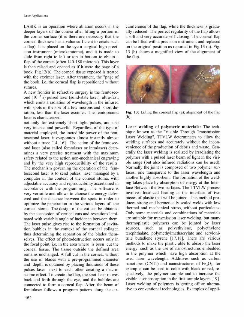
cumference of the flap, while the thickness is gradu-
ally reduced. The perfect regularity of the flap allows
a soft and very accurate self-closing. The corneal flap
can be lifted with a precision instrument and replaced
on the original position as reported in Fig.13 (a). Fig.
13 (b) shows a magnified view of the alignment of
the flap.
Fig. 13: Lifting the corneal flap (a); alignment of the flap
(b).
Laser welding of polymeric materials: The tech-
nique known as the "Visible Through Transmission
Laser Welding", TTVLW determinates to allow the
welding surfaces and accurately without the incon-
venience of the production of debris and waste. Gen-
erally the laser welding is realized by irradiating the
polymer with a pulsed laser beam of light in the visi-
ble range (but also infrared radiations can be used).
Normally the joint is composed of two polymer sur-
faces: one transparent to the laser wavelength and
another highly absorbent. The formation of the weld-
ing takes place by absorption of energy at the Inter-
face Between the two surfaces. The TTVLW process
involves localized heating at the interface of two
pieces of plastic that will be joined. This method pro-
duces strong and hermetically sealed welds with low
thermal and mechanical stress, without particulates.
Only some materials and combinations of materials
are suitable for transmission laser welding, but many
thermoplastic polymers can be jointed by laser
sources, such as polyethylene, polyethylene
terephthalate, polymethylmethacrylate and acryloni-
trile butadiene styrene [17,18]. There are various
methods to make the plastic able to absorb the laser
energy, such as the use of nanostructures embedded
in the polymer which have high absorption at the
used laser wavelength. Additives such as carbon
nanotubes (CNTs) and nanostructures of Fe2O3, for
example, can be used to color with black or red, re-
spectively, the polymer sample and to increase the
visible laser absorption in the first sample layers [19].
Laser welding of polymers is getting off an alterna-
tive to conventional technologies. Examples of appli-
a) b)
Laser Applications
153
cation can be found, amongst others, in the medical
devices, automotive, electronics, human care and
house hold devices industries [20]. In particular, in
medical devices the laser welding could be use to
produce filters, micro fluidic devices, medical pack-
aging (blood bags), catheters and prostheses. The in-
set of Fig.14(a) shows a scheme of polymer welding.
Different laser sources are commonly employed for
the welding of polymers, such as continuous CO2
high-power infrared lasers at 9.4 and 10.6 mm wave-
lengths, diode lasers and Nd:Yag lasers. Results
showed are achieved employed to irradiate polymers
in air by using 3 ns pulsed Nd:Yag lasers at the fun-
damental and second harmonic, in a single pulse or at
10 Hz repetition rate and at a laser intensity of about
8×10⁸W/cm2 with a maximum pulse energy of 150
mJ. Different physical and mechanical analyses were
employed in order to characterize the properties of
the single polymers before the welding and the joint
obtained just after the welding process. The poly-
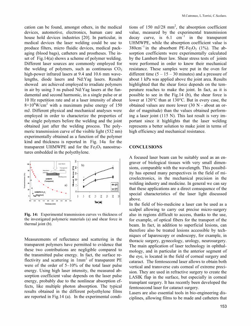
meric transmission curve of the visible light (532 nm)
experimentally obtained as a function of the polymer
kind and thickness is reported in Fig. 14a for the
transparent UHMWPE and for the Fe2O3 nanostruc-
tures embedded in the polyethylene.
Fig. 14: Experimental transmission curves vs thickness of
the investigated polymeric materials (a) and shear force in
thermal joint (b).
Measurements of reflectance and scattering in the
transparent polymers have permitted to evidence that
these two contributions are negligible compared to
the transmitted pulse energy. In fact, the surface re-
flectivity and scattering in 1mm2 of transparent PE
were of the order of 5–10% of the total laser pulse
energy. Using high laser intensity, the measured ab-
sorption coefficient value depends on the laser pulse
energy, probably due to the nonlinear absorption ef-
fects, like multiple photon absorption. The typical
results obtained in the different polyethylene films
are reported in Fig.14 (a). In the experimental condi-
tions of 150 mJ/28 mm2, the absorption coefficient
value, measured by the experimental transmission
decay curve, is 6.1 cm−1 in the transparent
UHMWPE, while the absorption coefficient value is
380cm−1 in the absorbent PE-Fe2O3 (1%). The ab-
sorption coefficients were experimentally calculated
by the Lambert-Beer law. Shear stress tests of joints
were performed in order to know their mechanical
resistance. These samples were put in the oven for
different time (5 – 15 – 30 minutes) and a pressure of
about 1 kPa was applied above the joint area. Results
highlighted that the shear force depends on the tem-
perature reaches to make the joint. In fact, as it is
possible to see in the Fig.14 (b), the shear force is
lower at 120°C than at 130°C. But in every case, the
obtained values are more lower (30 N - about an or-
der of magnitude) than the values obtained perform-
ing a laser joint (115 N). This last result is very im-
portant since it highlights that the laser welding
represents a better solution to make joint in terms of
high efficiency and mechanical resistance.
CONCLUSIONS
A focused laser beam can be suitably used as an en-
graver of biological tissues with very small dimen-
sions, comparable with the wavelength. This possibil-
ity has opened many perspectives in the field of mi-
croelectronics, in the mechanical precision in the
welding industry and medicine. In general we can say
that these applications are a direct consequence of the
special characteristics of the laser light discussed
above.
In the field of bio-medicine a laser can be used as a
scalpel allowing to carry out precise micro-surgery
also in regions difficult to access, thanks to the use,
for example, of optical fibers for the transport of the
beam. In fact, in addition to superficial lesions, can
therefore also be treated lesions accessible by tech-
niques of laparoscopy or endoscopy, for example, in
thoracic surgery, gynecology, urology, neurosurgery.
The main application of laser technology in ophthal-
mology, and in particular in the anterior segment of
the eye, is located in the field of corneal surgery and
cataract. The femtosecond laser allows to obtain both
vertical and transverse cuts corneal of extreme preci-
sion. They are used in refractive surgery to create the
LASIK flap in the surface, but especially in corneal
transplant surgery. It has recently been developed the
femtosecond laser for cataract surgery.
Lasers are also essential tools in bio-engineering dis-
ciplines, allowing films to be made and catheters that
0.0 0.5 1.0 1.5 2.0 2.5 3.0 3.5 4.0 4.5
0
20
40
60
80
100
UHMWPE+Fe2O
3
(Absorbent)
Tra
ns
mis
sio
n (
%)
Thickness (mm)
UHMWPE
(Transparent)
Laser Beam
Transparent
polymer Absorbent
polymer
Welding zone
a)
b)
M.Cutroneo, L.Torrisi, C.Scolaro.

154
are interesting applications in micro-perforated drain-
age of fluids: an example is the production of cathe-
ters in the pathology of hydrocephalus microperfo-
rated. Further developments have taken place also in
the field of fabrication of using the technique of
plasma spray with which it is possible to coat with
the film of biocompatible material orthopedic pros-
theses or dental implants.
Must also remember the importance of the laser
welding in medical devices to produce filters, medi-
cal packaging, catheters and prostheses. The basic
physics, combined with knowledge of chemistry, bi-
ology and medicine, now enables us to better control
the use of such radiation in order to be able to signifi-
cantly improve the quality of life of every living be-
ing.
BIBLIOGRAPHY
[1]W. T. Silfvast “Laser fundamentals”, Cambridge Uni-
versity Press, Cambridge 2004
[2]L. Garwin, T. Lincoln “A Century of Nature: Twenty-
One Discoveries that Changed Science and the World”,
Nature, 2003
[3] J.L.Boulnois, Photophysical processes in recent medi-
cal laser developments: A review, Lasers Med. Sci. Vol. 1,
pp. 47-66, 1986.
[4]L. Goldman “Lasers in Medicine”, R.W. Waynant Ed.,
CRC Press, 2002
[5] P.G. O‟Shea, H.P. Freund, Free-electron lasers: Status
and Application, Science Vol. 292, 8 June 2001
[6] L. Torrisi, S. Trusso, G. Di Marco and P. Parisi,
Physica Medica, XVII(4), 227, 2001
[7] M.K.Niemz, Laser-Tissue interaction, Springer,Berlin,
1996, p. 42.
[8] M.J.C. van Gemert and A.J. Welch, Clinical use of
laser-tissue interactions, IEEE Engineering in Medicine
and Biology Magazine, December 1989
[9] L.Torrisi et al. Rad. Eff & Defects in Solids: Inc.
Plasma Sc.& Pl. Tec. 165(6), 721, 2010
[10] A.P. Alloncle, D. Dufresne, M. Autric, J. de Physique
IV, Colloque C4, supp. 111, Volume 4, 1994, C4-131
[11] L.Torrisi Bio-Medical Mat. and Eng. 4(1), 17, 1994
[12] J.J. Beltrano, et al. Rad. Effects & Defects in Solids,
V. 163(4-6), 331-338, 2008
[13] AICCER, La Biometria, Fabiano Editore, Rivista Sci-
entifica D‟Informazione, 2009.
[14] Durrie DS, Kezirian GM., Femtosecond laser versus
mechanical keratome flaps in wavefront-guided laser in
situ keratomileusis: prospective contralateral eye study, J
Cataract Refract Surg. Gennaio 2005; 31(1).
[15] Martin Tp, Ree Jw, Legault C, Oberfeld Sm, Jacoby
Bg, Yu Dd, Dickens A, Johnson Hp. Cataract formation
and cataract extraction after penetrating keratoplasty. Oph-
thalmology 1994 Jan; 101(1).
[16] Buratto L., Böhm E. The use of the femtosecond laser
in penetrating keratoplasty. Am. J. Ophthalmol. 2007 143
(5).
[17] M.Chen,G.Zak,P.J.Bates, J. Mater. Process. Technol.
(2010). Doi10.1016/j.jmatprotec.2010.08.017
[18] N. Amanat, C. Chaminade, J. Grace, D.R. McKenzie,
N.L. James, Mater. Des. 31, 4823 (2010)
[19] V.V. Semak, R.J. Steele, P.W. Fuerschbach and B.K.
Damkroger, Journal of Physics D: Applied Physics, 33
(2000), 1179-1185.
[20] Pfleging,W.; Kohler, R.; Schierjott, P.; Hoffmann,W.
Sensor. Actuators B Chem. 2009, 138, 336–343.
Laser Applications


