Larger whole brain grey matter associated with long-term ...
18
RESEARCH ARTICLE Larger whole brain grey matter associated with long-term Sahaja Yoga Meditation: A detailed area by area comparison Sergio Elı ´as Herna ´ ndez ID 1 *, Roberto Dorta 2 , Jose ´ Suero 3 , Alfonso Barros-Loscertales 4 , Jose ´ Luis Gonza ´ lez-Mora 5 , Katya Rubia 6 1 Department of Ingenierı ´a Industrial, Universidad de La Laguna, Tenerife, Spain, 2 Department of Matema ´ ticas, Estadı ´stica e Investigacio ´ n Operativa, Universidad de La Laguna, Tenerife, Spain, 3 Centro de Salud Jazmı ´n, Sermas, Madrid, Spain, 4 Department of Psicologı ´a Ba ´ sica, Clı ´nica y Psicobiologı ´a, Universitat Jaume I, Castello ´ n, Spain, 5 Department of Fisiologı ´a, Universidad de La Laguna, Tenerife, Spain, 6 Institute of Psychiatry, Psychology and Neuroscience, King’s College London, London, United Kingdom * [email protected] Abstract Objectives Our previous study showed that long-term practitioners of Sahaja Yoga Meditation (SYM) had around 7% larger grey matter volume (GMV) in the whole brain compared with healthy controls; however, when testing individual regions, only 5 small brain areas were statistically different between groups. Under the hypothesis that those results were statistically conser- vative, with the same dataset, we investigated in more detail the regional differences in GMV associated with the practice of SYM, with a different statistical approach. Design Twenty-three experienced practitioners of SYM and 23 healthy non-meditators matched on age, sex and education level, were scanned using structural magnetic resonance imaging (MRI). Their GMV were extracted and compared using Voxel-Based Morphometry (VBM). Using a novel ad-hoc general linear model, statistical comparisons were made to observe if the GMV differences between meditators and controls were statistically significant. Results In the 16 lobe area subdivisions, GMV was statistically significantly different in 4 out of 16 areas: in right hemispheric temporal and frontal lobes, left frontal lobe and brainstem. In the 116 AAL area subdivisions, GMV difference was statistically significant in 11 areas. The GMV differences were statistically more significant in right hemispheric brain areas. Conclusions The study shows that long-term practice of SYM is associated with larger GMV overall, and with significant differences mainly in temporal and frontal areas of the right hemisphere and PLOS ONE PLOS ONE | https://doi.org/10.1371/journal.pone.0237552 December 28, 2020 1 / 18 a1111111111 a1111111111 a1111111111 a1111111111 a1111111111 OPEN ACCESS Citation: Herna ´ndez SE, Dorta R, Suero J, Barros- Loscertales A, Gonza ´lez-Mora JL, Rubia K (2020) Larger whole brain grey matter associated with long-term Sahaja Yoga Meditation: A detailed area by area comparison. PLoS ONE 15(12): e0237552. https://doi.org/10.1371/journal.pone.0237552 Editor: Niels Bergsland, University at Buffalo, UNITED STATES Received: July 24, 2020 Accepted: December 5, 2020 Published: December 28, 2020 Peer Review History: PLOS recognizes the benefits of transparency in the peer review process; therefore, we enable the publication of all of the content of peer review and author responses alongside final, published articles. The editorial history of this article is available here: https://doi.org/10.1371/journal.pone.0237552 Copyright: © 2020 Herna ´ndez et al. This is an open access article distributed under the terms of the Creative Commons Attribution License, which permits unrestricted use, distribution, and reproduction in any medium, provided the original author and source are credited. Data Availability Statement: All relevant data are within the paper and its Supporting information files.
Transcript of Larger whole brain grey matter associated with long-term ...

RESEARCH ARTICLE
Larger whole brain grey matter associated
with long-term Sahaja Yoga Meditation: A
detailed area by area comparison
Sergio Elıas HernandezID1*, Roberto Dorta2, Jose Suero3, Alfonso Barros-Loscertales4,
Jose Luis Gonzalez-Mora5, Katya Rubia6
1 Department of Ingenierıa Industrial, Universidad de La Laguna, Tenerife, Spain, 2 Department of
Matematicas, Estadıstica e Investigacion Operativa, Universidad de La Laguna, Tenerife, Spain, 3 Centro de
Salud Jazmın, Sermas, Madrid, Spain, 4 Department of Psicologıa Basica, Clınica y Psicobiologıa,
Universitat Jaume I, Castellon, Spain, 5 Department of Fisiologıa, Universidad de La Laguna, Tenerife,
Spain, 6 Institute of Psychiatry, Psychology and Neuroscience, King’s College London, London, United
Kingdom
Abstract
Objectives
Our previous study showed that long-term practitioners of Sahaja Yoga Meditation (SYM)
had around 7% larger grey matter volume (GMV) in the whole brain compared with healthy
controls; however, when testing individual regions, only 5 small brain areas were statistically
different between groups. Under the hypothesis that those results were statistically conser-
vative, with the same dataset, we investigated in more detail the regional differences in
GMV associated with the practice of SYM, with a different statistical approach.
Design
Twenty-three experienced practitioners of SYM and 23 healthy non-meditators matched on
age, sex and education level, were scanned using structural magnetic resonance imaging
(MRI). Their GMV were extracted and compared using Voxel-Based Morphometry (VBM).
Using a novel ad-hoc general linear model, statistical comparisons were made to observe if
the GMV differences between meditators and controls were statistically significant.
Results
In the 16 lobe area subdivisions, GMV was statistically significantly different in 4 out of 16
areas: in right hemispheric temporal and frontal lobes, left frontal lobe and brainstem. In the
116 AAL area subdivisions, GMV difference was statistically significant in 11 areas. The
GMV differences were statistically more significant in right hemispheric brain areas.
Conclusions
The study shows that long-term practice of SYM is associated with larger GMV overall, and
with significant differences mainly in temporal and frontal areas of the right hemisphere and
PLOS ONE
PLOS ONE | https://doi.org/10.1371/journal.pone.0237552 December 28, 2020 1 / 18
a1111111111
a1111111111
a1111111111
a1111111111
a1111111111
OPEN ACCESS
Citation: Hernandez SE, Dorta R, Suero J, Barros-
Loscertales A, Gonzalez-Mora JL, Rubia K (2020)
Larger whole brain grey matter associated with
long-term Sahaja Yoga Meditation: A detailed area
by area comparison. PLoS ONE 15(12): e0237552.
https://doi.org/10.1371/journal.pone.0237552
Editor: Niels Bergsland, University at Buffalo,
UNITED STATES
Received: July 24, 2020
Accepted: December 5, 2020
Published: December 28, 2020
Peer Review History: PLOS recognizes the
benefits of transparency in the peer review
process; therefore, we enable the publication of
all of the content of peer review and author
responses alongside final, published articles. The
editorial history of this article is available here:
https://doi.org/10.1371/journal.pone.0237552
Copyright: © 2020 Hernandez et al. This is an open
access article distributed under the terms of the
Creative Commons Attribution License, which
permits unrestricted use, distribution, and
reproduction in any medium, provided the original
author and source are credited.
Data Availability Statement: All relevant data are
within the paper and its Supporting information
files.

the brainstem. These neuroplastic changes may reflect emotional and attentional control
mechanisms developed with SYM. On the other hand, our statistical ad-hoc method shows
that there were more brain areas with statistical significance compared to the traditional
methodology which we think is susceptible to conservative Type II errors.
Introduction
Meditation is a general term that includes a large variety of practices that mainly focus on the
inner observation of the body and the mind. The western goal of most meditation techniques
is to achieve an improved control of attention and emotions in order to live a more balanced,
stress-free and healthier life. On the other hand, yoga includes many different techniques
among which meditation (dhayana in classical yoga) has a main role. If we travel back to the
origins of yoga, the first known treaty “The yoga sutras of Patanjali” mentions that “Yoga is the
suppression of the modifications of the mind” [1, 2]. In ancient yoga, a higher state of con-
sciousness called Nirvichara Samadhi was described, in today’s words Nirvichara could be
translated as “mental silence” or “thoughtless awareness”. In this state, the mind has none
thoughts and there is inner calm in a state of inner pure joy and the attention is focused on
each present moment. Sahaja Yoga Meditation (SYM) shares the goals of Patanjali’s Yoga
Sutras to achieve the state of Nirvichara or mental silence.
SYM, presumably through the regular achievement of the state of mental silence, has
shown health benefits in disorders that are often associated with recurrent or repetitive nega-
tive thoughts, such as: depression, stress, anxiety, and attention-deficit/hyperactivity disorder
[2–7]. Other studies on SYM have shown beneficial effects in treating physiological and neuro-
logical diseases such as asthma [8], high blood pressure [6], menopause [9] and epilepsy [10–
12], for a meta-analysis see [8]. Furthermore, the frequency with which the practitioners per-
ceive the state of mental silence has been shown to be associated with better physical and men-
tal health [13].
Neuroplasticity is one of the most commonly used terms in today’s neuroscience to express
the capacity of our human brain to change permanently. One of the key insights over the past
2 decades of neuroimaging research has been that the human brain, even in adulthood, is not
static, but on the contrary is a dynamic system that has the ability to shape itself. One of the
key fascinating questions that researchers try to answer is hence: how can we improve our
brain structure and function? One potential non-pharmacological way to shape our brain
could be through meditation [14].
Neuroplasticity can be measured by changes in grey matter volume (GMV). Many studies
have shown that brain areas that are more utilized through practice of a particular skill for
example, in music [15], or high performance sports [16], can become enlarged. It has even
been shown that relatively short periods of training of a particular skill, such as 3 months of
training to juggle or 3 months of studying for an exam in students can lead to transient
changes in the relevant brain areas such as visual-spatial perception regions for juggling [17,
18] or the hippocampus and parietal lobe for memory storage in medical students preparing
for an exam [19, 20].
Voxel Based morphometry (VBM) is the most used automated technique to measure GMV
by means of MRI scans. In most cases researchers follow the steps provided by the VBM
authors of the technique [21–24]. VBM has evolved [21] and the different steps like segmenta-
tion and normalization have been improved with each new software version [24, 25].
PLOS ONE Increases in whole brain grey matter associated with long-term Sahaja Yoga Meditation
PLOS ONE | https://doi.org/10.1371/journal.pone.0237552 December 28, 2020 2 / 18
Funding: The author(s) received no specific
funding for this work.
Competing interests: The authors have declared
that no competing interests exist.

In most cases, the statistical path followed to compare GMV mean differences between
groups has been with ANCOVAs, were typically total intracranial volumes (TIV), sex and age
are treated as nuisance covariates. This statistical method is based on random field theory [21,
26]. Another important point to consider is that structural images display local variation in
smoothness, which implies that cluster-level corrections should be applied using Random
Field Theory and non-stationary correction [27].
In our previous structural MRI study, we showed that 23 long-term practitioners of SYM
compared to healthy controls had 6.9% significantly larger GMV in the whole brain [28]
which represent, as far as we know, the highest GMV difference shown between groups of
healthy volunteers. However, this significant whole brain difference was related with only two
relatively small areas showing statistical significance located in right insula and right inferior
temporal gyrus with respective volumes of 564 and 739 mm3. Considering the concern of
incurring in Type II errors (false negatives or conservative assumptions), the aim of our study
was to analyse in more detail how the GMV differences are distributed across the whole brain.
This new study is based in two key issues: 1) The development of an statistical ad-hoc general
linear model (AH-GLM) that adapts itself on each brain area depending on the significance of
covariates of that particular area; and 2) The parcellation of the human brain using 2 different
methods i. Based on the human brain lobes: frontal, temporal, etc, that gives rise to 16 different
brain areas and ii. Using the more specific automated anatomical labelling (AAL) of 116 brain
areas [29, 30]. We used these two brain atlas subdivisions because they are widely used in the
neuroscience literature and because the proposed methodology allows us to study both of
them in detail. This way we could test the application of our ad-hoc method in 2 different sce-
narios and observe if the results obtained provided some overlap and coherence.
The key question for this analysis was whether there were any areas that differed between
long-term meditators and healthy controls which were overlooked in our previous paper [28]
due to a Type II error correction effect.
Materials and methods
Participants
Forty-six white Caucasian, right-handed, healthy volunteers, between 21 and 63 years partici-
pated in this study. Twenty-three of them were long-term, expert practitioners of SYM (17
females and 6 males) while the other 23 (also 17 females and 6 males) were non-meditators
matched on sex, education degree, body mass index and age (see Table 1). All volunteers
informed that they had no physical or mental illness, no history of neurological disorders, and
no addiction to alcohol, nicotine or drugs.
Table 1. Demographic characteristics of the groups.
Meditators Mean (SD) Controls Mean+ (SD) t(df = 44) p-value�
Volunteers N˚ 23 23
Age (years) 46.5 (11.4) 46.9 (10.9) -0.13 0.89
Age range (years) 20.3–63.1 21.3–63.3
Education degree, 0 to 6 3.78 (1.2) 4.04 (1.36) 0.69 0.50
Height (cm) 167.0 (8.8) 167.2 (7.6) 0.09 0.93
Weight (Kg) 69.5 (14.6) 71.7 (14.5) 0.53 0.60
Body mass index 24.9 (4.5) 25.5 (3.9) 0.54 0.60
�p-values represent group differences between meditators and controls using two-tailed independent samples t-tests.
https://doi.org/10.1371/journal.pone.0237552.t001
PLOS ONE Increases in whole brain grey matter associated with long-term Sahaja Yoga Meditation
PLOS ONE | https://doi.org/10.1371/journal.pone.0237552 December 28, 2020 3 / 18

Meditators had more than 5 years of daily meditation practice in SYM (mean 14.1 SD (6.1)
years); the daily average time dedicated to meditation was 84.7 (32.2) minutes.
Before their participation in this research, all volunteers filled in different questionnaires to
validate their individual health status, education and age. Additionally, meditators filled in
other questionnaire that asked about their experience in SYM, including: average time dedi-
cated to meditation per day, frequency of the perception of the state of mental silence, total
hours of meditation and years of practice of SYM.
All participants signed informed consent to participate freely. This study was approved by
the Ethics Committee of the University of La Laguna.
MRI acquisition
All images were obtained on a 3T MRI Scanner. High resolution sagittally oriented anatomical
images were collected. A 3D fast spoiled-gradient recalled pulse sequence was obtained
(TR = 8.8 ms, TE = 1.7 ms, flip angle = 10˚, matrix size = 256 × 256 pixels, 1 × 1 mm in plane
resolution, spacing between slices = 1 mm plus 0 mm interslice gap, slice thickness = 1 mm).
Total acquisition time was 13 minutes.
Voxel-based morphometry
Voxel-based morphometry (VBM) [21] with DARTEL was conducted using the SPM12 soft-
ware package (Statistical Parametric Mapping software: http://www.fil.ion.ucl.ac.uk/spm/).
Processing steps were performed as suggested by the method’s author [31]. VBM with DAR-
TEL has been shown to be more sensitive than standard VBM [24] and provides results com-
parable to those achieved with manual segmentation [32].
The procedure followed these steps: 1. All T1-weighted anatomical images were displayed
to screen to verify they were free from gross anatomical abnormalities. 2. For better registra-
tion, the T1 images were manually centred at the anterior commissure and reoriented accord-
ing to the anterior–posterior commissure line. 3. Using the New Segment procedure in
SPM12, images were segmented into: Grey matter (GM), White matter (WM) and Cerebrum
Spinal Fluid (CSF), a segmentation that provides acceptable substitute for labour intensive
manual estimates [25]. 4. The DARTEL routine inside SPM12 was used to spatially normalize
the segmented images [24]. The image intensity of each voxel was modulated by the Jacobian
determinants to ensure that regional differences in the total amount of GMV were conserved.
5. The final DARTEL template image modulated in the previous step was registered to MNI
space so that all the individual spatially normalised scans were brought into MNI space. 6.
Finally, the normalized modulated GMV images were smoothed with a 4-mm full-width at
half-maximum (FWHM) isotropic Gaussian kernel to increase the signal to noise ratio.
For each individual, total GM, WM and CSF were obtained with the Matlab script ‘get_to-
tals.m’ [33] and used to calculate the individual Total Intracranial Volume (TIV) by summing
the volumes of the three already mentioned components (GM, WM, CSF).
Regional GMV extractions
The WFU Pickatlas [29, 34] was used to generate ROI masks of the selected brain areas in
MNI space. Among the different brain areas subdivision generated by WFU Pickatlas, we
chose the lobar atlas, and the AAL subdivisions. The lobar ROI subdivisions were as follows:
right/left frontal lobe, right/left temporal lobe, right/left parietal lobe, right/left occipital lobe,
right/left limbic system, and right/left sublobar area (internal cerebrum: summation of basal
ganglia, thalamus, insula, and callosum), right/left brainstem and right/left cerebellum, the
AAL subdivision es the 116 area parcellation by Rolls et al. [30]. To automatically extract the
PLOS ONE Increases in whole brain grey matter associated with long-term Sahaja Yoga Meditation
PLOS ONE | https://doi.org/10.1371/journal.pone.0237552 December 28, 2020 4 / 18

GMV at each ROI for each subject, we programmed a Matlab script based on the MATLAB
code “get_totals” [33]. The output of the Matlab script was the regional GMV data for each
volunteer at each ROI. Similar or equivalent procedures to extract regional GMV have been
used in previous studies [35–37] To verify the truthfulness of the results obtained by the
MATLAB “get_totals.m” script, several comparisons were made with the equivalent Marsbar
toolbox (available at https://www.nitrc.org/projects/marsbar/). We verified that both tools pro-
vided the same results but because “get_totals” was easier to implement inside our script
Matlab program we used this method.
Statistical analysis
Differences in GMV between meditators and controls at each zone/area were analysed by con-
ducting an ad-hoc general linear model (AH-GLM)—ANCOVA that adapts it-self to every
area’s statistical specificities. The AH-GLM had the following terms Eq (1): the dependent vari-
able (DV) at each area Grey Matter Volume (GMV); the factor Meditator (Med) with two levels
(control Med = 0 and meditator Med = 1); two covariates, the volunteer’s age (Age) and the
volunteer’s Total Intracranial Volume (TIV); and two interactions, the factor with each covari-
ate: (Med × TIV) and (Med × Age) notice that the interactions could be significant only when
the associated covariate was significant. At Eq (1) each volunteer is represented by the sub-
script j and i represents each level of Meditator factor.
GMVij ¼ b0 þMedi þ b1 � Ageij þ b2 � TIVij þ b3 � ðMed� TIVÞij þ b4 � ðMed � AgeÞij þ εij ð1Þ
Each brain area classification into zones from Zone 1 till Zone 3D was dependent on the
statistical significance of each covariates (Age, TIV) and the corresponding interactions (Med× Age) and (Med × TIV). Covariates Age and TIV were considered significant at a threshold of
p<0.05, having a Pearson’s correlation coefficient with GMV of r>0.4. The interactions (Med× Age) and (Med × TIV) were considered significant when their associated covariate was sig-
nificant and the interaction had p<0.05. This way we differentiated zones starting from the
simplest Zone 1 where none of the covariates was significant, see Eq (2), to the zone 3D where
all covariates and interactions were significant represented by the full model Eq (1).
GMVij ¼ b0 þMedi þ εij ð2Þ
Sex was not included into the AH-GLM because one of the conditions to be able to carry
out an ANCOVA is that there is no effect of the factors on the covariates that are included in
the model. When studying whether there is an effect of sex on the covariate TIV it was verified
that this effect was highly significant p< 0.0001, because males had significant larger TIV than
females. Therefore, including TIV in the model intrinsically controls for the sex factor.
Standardized residuals for the GMV and for the overall model at each zone εij were nor-
mally distributed, as assessed by Shapiro-Wilk’s test (p> 0.05). There was homogeneity of var-
iances, as assessed by visual inspection of a scatterplot and Levene’s test of homogeneity of
variance (p<0.05). There were no outliers in the data, as assessed by no cases with standard-
ized residuals greater than ±3 standard deviations. These models require compliance with two
other assumptions: 1. To verify the existence of a non-zero linear relationship between the DV
and the covariates in all groups together. If there is no such relationship, conducting an
ANCOVA does not make sense, so a unifactorial ANOVA should be conducted alternatively;
2. To check the homogeneity of regression slopes; that is, to ensure that the linear relationship
of the DV and the covariate is the same in all groups.
The multiple comparison problem was solved by controlling the false discovery rate (FDR),
which manages the expected proportion of false positive findings among all the rejected null
PLOS ONE Increases in whole brain grey matter associated with long-term Sahaja Yoga Meditation
PLOS ONE | https://doi.org/10.1371/journal.pone.0237552 December 28, 2020 5 / 18
hypotheses [38], by means of the q-values estimated by Storey and Tibshirani’s method [39]
implemented in neuroscience research by Takeda et al [40]. We should consider that the q
value is similar to the p value, with the exception that it is a measure of significance in terms of
the false discovery rate rather than the false positive rate. From the distribution of p-values
obtained from the multiple comparison, the q-values were provided by means of the Biocon-
ductor´s q-value package from R software (3.6.1, R Foundation for Statistical Computing,
Vienna, Austria). Effect size was assessed by means of Z2partial following Cohen’s benchmarks to
define small (Z2partial ¼ 0:01), medium (Z2
partial ¼ 0:06), and large (Z2partial ¼ 0:14) effects [39].
Statistical significance was indicated by a false discovery rate (FDR) q-value <0.05 or p-
value<0.05 when corresponds. Further explanation of the AH-GLM and the Zone classifica-
tion is available at S1 Appendix.
Results
Our previous study [28] reported two main results: 1. The whole brain had statistically signifi-
cant larger GMV in meditators compared to controls. 2. There were 5 cluster areas with larger
GMV in meditators compared to controls: two from the direct VBM statistical results and
three from a priori hypothesised regions with more lenient threshold.
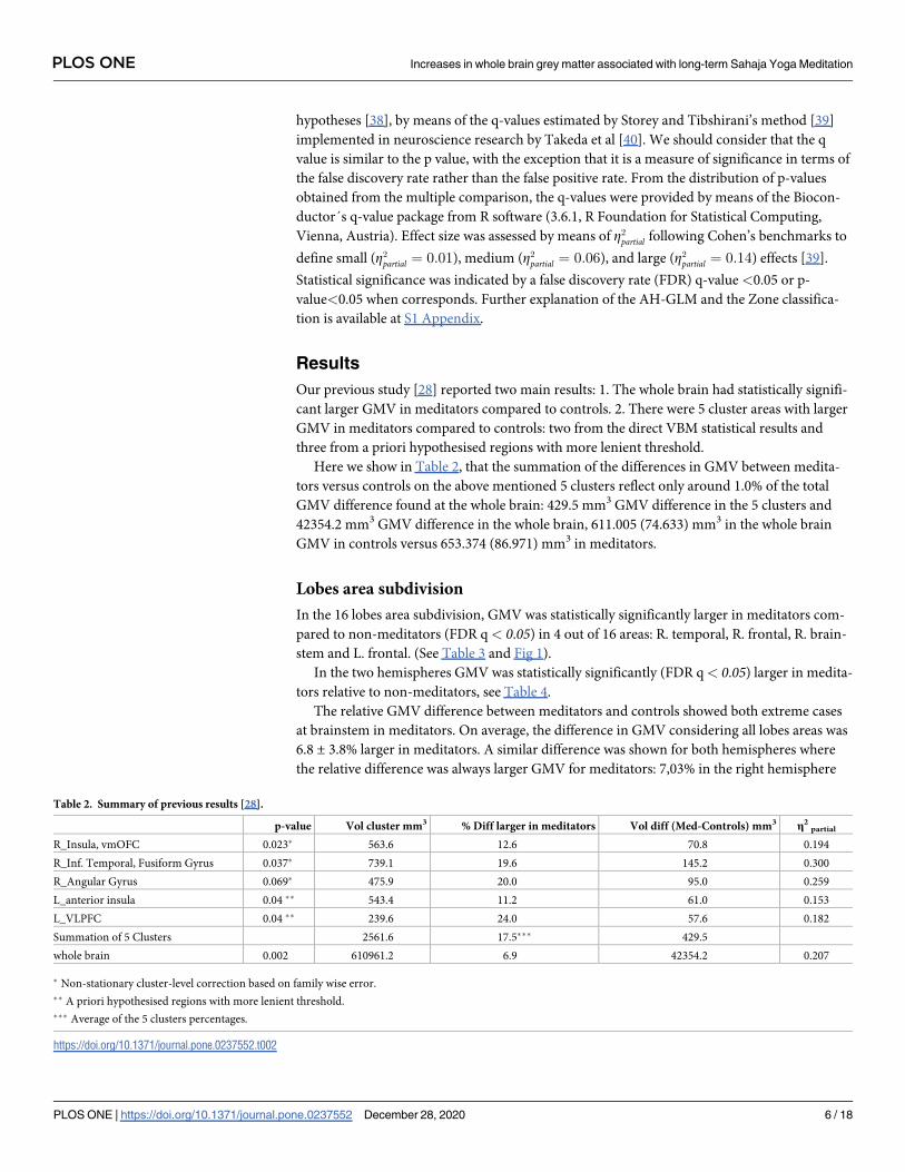
Here we show in Table 2, that the summation of the differences in GMV between medita-
tors versus controls on the above mentioned 5 clusters reflect only around 1.0% of the total
GMV difference found at the whole brain: 429.5 mm3 GMV difference in the 5 clusters and
42354.2 mm3 GMV difference in the whole brain, 611.005 (74.633) mm3 in the whole brain
GMV in controls versus 653.374 (86.971) mm3 in meditators.
Lobes area subdivision
In the 16 lobes area subdivision, GMV was statistically significantly larger in meditators com-
pared to non-meditators (FDR q < 0.05) in 4 out of 16 areas: R. temporal, R. frontal, R. brain-
stem and L. frontal. (See Table 3 and Fig 1).
In the two hemispheres GMV was statistically significantly (FDR q< 0.05) larger in medita-
tors relative to non-meditators, see Table 4.
The relative GMV difference between meditators and controls showed both extreme cases
at brainstem in meditators. On average, the difference in GMV considering all lobes areas was
6.8 ± 3.8% larger in meditators. A similar difference was shown for both hemispheres where
the relative difference was always larger GMV for meditators: 7,03% in the right hemisphere
Table 2. Summary of previous results [28].
p-value Vol cluster mm3 % Diff larger in meditators Vol diff (Med-Controls) mm3 η2partial
R_Insula, vmOFC 0.023� 563.6 12.6 70.8 0.194
R_Inf. Temporal, Fusiform Gyrus 0.037� 739.1 19.6 145.2 0.300
R_Angular Gyrus 0.069� 475.9 20.0 95.0 0.259
L_anterior insula 0.04 �� 543.4 11.2 61.0 0.153
L_VLPFC 0.04 �� 239.6 24.0 57.6 0.182
Summation of 5 Clusters 2561.6 17.5��� 429.5
whole brain 0.002 610961.2 6.9 42354.2 0.207
� Non-stationary cluster-level correction based on family wise error.
�� A priori hypothesised regions with more lenient threshold.
��� Average of the 5 clusters percentages.
https://doi.org/10.1371/journal.pone.0237552.t002
PLOS ONE Increases in whole brain grey matter associated with long-term Sahaja Yoga Meditation
PLOS ONE | https://doi.org/10.1371/journal.pone.0237552 December 28, 2020 6 / 18

and 6,72% in the left hemisphere (Table 4). In the whole brain the difference was 6.93%, which
was already shown on our previous study [28].
If we consider the reported GMV differences at lobes from Table 3 we see that the summa-
tion of the GMV differences in lobes between groups was 20,44 mL or 20440 mm3; this
Table 3. Statistics of GMV differences between groups in the significant lobes (16 areas).
Area Zone � F Nom. p-value FDR q-value GMV Controls (mean ± std) mL GMV Medit (mean ± std) mL ��Relat dif % η2partial
R. temporal 3A 10.52 0.002 0.016 46.65 ± 5.92 50.86 ± 7.28 9.02 0.200
R. frontal 3A 10.44 0.002 0.016 78.35 ± 11.61 85.68 ± 12.95 9.36 0.199
R. brainstem 1 9.82 0.003 0.016 1.67 ± 0.28 2.00 ± 0.42 19.68 0.182
L. frontal 3A 9.3 0.004 0.016 76.57 ± 11.35 83.48 ± 13.4 9.02 0.181
�Further explanation about Zone classification in S1 Appendix.
��Relat dif % = (GMV Medit—GMV Controls) x 100 / GMV Controls.
https://doi.org/10.1371/journal.pone.0237552.t003
Fig 1. Axial slices of the lobes area with different GMV between groups, in the order of 1 to 4, following statistical
significance. Z coordinates are shown in mm from the anterior-posterior commissure. The right side of the image
corresponds to the right side of the brain.
https://doi.org/10.1371/journal.pone.0237552.g001
Table 4. Statistics of GMV differences between groups in the hemispheres and whole brain.
Area Zone � F Nom. p-value FDR q-value GMV Controls (mean ± std) mL GMV Medit (mean ± std) mL ��Relat dif % η2partial
R.Hemisph. 3A 9.31 0.004 0.007 284.92 ± 35.02 304.95 ± 39.76 7.03 0.182
L.Hemisph. 3A 7.94 0.007 0.007 276.62 ± 33.46 295.22 ± 39.9 6.72 0.159
Whole brain GMV 3A 9.02 0.005 0.007 611 ± 74.63 653.37 ± 86.97 6.93 0.177
�Further explanation about Zone classification in S1 Appendix.
��Relat dif % = (GMV Medit—GMV Controls) x 100 / GMV Controls.
https://doi.org/10.1371/journal.pone.0237552.t004
PLOS ONE Increases in whole brain grey matter associated with long-term Sahaja Yoga Meditation
PLOS ONE | https://doi.org/10.1371/journal.pone.0237552 December 28, 2020 7 / 18

represent a 48,2% of the total GMV difference reported at the whole brain that was 42354.2
mm3. In the same way the reported GMV difference at the right hemisphere 20,03 mL repre-
sents a 47,3% of the whole brain difference while the left hemisphere difference 18.60 mL rep-
resents a 43,9%.
AAL area subdivision
In the 116 AAL area subdivision, GMV was statistically significant (FDR q< 0.05) larger in
meditators relative to non-meditators in 11 out of the 116 AAL areas: right middle temporal
gyrus (MTG.R), right paracentral lobule (PCL.R), right inferior frontal gyrus opercular part
(IFGoperc.R), right precentral gyrus (PreCG.R), right inferior temporal gyrus (ITG.R), right
inferior frontal gyrus orbital part (IFGorb.R), left postcentral gyrus (PoCG.L), left precentral
gyrus (PreCG.L), left middle frontal gyrus (MFG.L), left olfactory cortex (OLF.L), right middle
frontal gyrus orbital part (MFGorb.R), see Table 5 and Fig 2. In 59 AAL areas, the FDR q-value
was between 0.05 and 0.1.
The GMV difference between meditators and controls ranged from +15.3% larger GMV at
Right Parahippocampal gyrus to 0.0%, almost equal, at Right Lenticular nucleus—Pallidum.
On average the difference in GMV considering all AAL areas was a 6.7 ± 3.0% larger in
Meditators.
If we consider the 11 AAL areas with significant GMV differences, similar to the calculation
for the lobe areas, the summation of the difference in GMV between groups on those 11 areas
was 6,25 mL which represents a 14.8% of the total GMV difference at the whole brain.
Superposition of AAL and lobes results are shown in Fig 3, while superposition of AAL
results and 5 clusters from our first study [28] are shown in Fig 4.
Discussion
Discussion of the ad-hoc statistical method
In this study, the statistical analysis made use of an ad-hoc General Linear Model (AH-GLM)
which produces more explanatory results for the used subdivisions of the brain.
Table 5. Statistic of GMV differences between groups through significant AAL brain areas.
Area Zone � F Nom p- value FDR q-value GMV Controls
(mean) mm3GMV Controls
(std) mm3GMV Medit
(mean) mm3GMV Medit
(std) mm3
��Relat dif % η2partial
MTG.R 3A 11.84 0.001 0.0291 14.34 1.93 15.77 2.26 9.97 0.220
PCL.R 3A 11.00 0.002 0.0291 4.15 0.40 4.32 0.52 4.10 0.208
IFGoperc.R 3A 10.47 0.002 0.0291 3.68 0.60 4.12 0.67 11.96 0.200
PreCG.R 3A 9.75 0.003 0.0291 5.92 1.06 6.75 1.23 14.02 0.188
ITG.R 3A 9.30 0.004 0.0291 12.22 1.62 13.41 1.91 9.74 0.181
IFGorb.R 3A 9.08 0.004 0.0291 4.31 0.65 4.76 0.89 10.44 0.178
PoCG.L 3A 8.13 0.007 0.0382 7.70 1.25 8.42 1.22 9.35 0.162
PreCG.L 3A 7.90 0.007 0.0382 7.17 1.26 7.97 1.42 11.16 0.158
MFG.L 3A 7.52 0.009 0.0393 13.34 2.07 14.57 2.34 9.22 0.152
OLF.L 3A 7.45 0.009 0.0393 1.04 0.13 1.13 0.16 8.65 0.151
MFGorb.R 3A 6.88 0.012 0.0477 2.61 0.54 2.94 0.60 12.64 0.141
�Further explanation about Zone classification in S1 Appendix.
��Relat dif % = (GMV Medit—GMV Controls) x 100 / GMV Controls
https://doi.org/10.1371/journal.pone.0237552.t005
PLOS ONE Increases in whole brain grey matter associated with long-term Sahaja Yoga Meditation
PLOS ONE | https://doi.org/10.1371/journal.pone.0237552 December 28, 2020 8 / 18

As mentioned in the results section, the GMV differences between groups in the 5 clusters
reported in our previous study [28] represent only 1% of the total significant GMV difference
detected at the whole brain (see Table 2).
Following Cohen’s definitions [41], we observed that the effect size was large
(Z2partial > 0:14) in all resulting areas from our AH-GLM (see last columns in Tables 3, 4 and 5),
as was the case with the clusters shown in our previous manuscript, (see last column Table 2).
Fig 2. Horizontal slices of AAL areas with different GMV between groups, in the order of 1 to 11, following
statistical significance. Z coordinates are shown in mm distance from the anterior-posterior commissure. The right
side of the image corresponds to the right side of the brain.
https://doi.org/10.1371/journal.pone.0237552.g002
Fig 3. Lobes results in blue, AAL results in yellow, superposition of both results (AAL an lobes) in green.
https://doi.org/10.1371/journal.pone.0237552.g003
PLOS ONE Increases in whole brain grey matter associated with long-term Sahaja Yoga Meditation
PLOS ONE | https://doi.org/10.1371/journal.pone.0237552 December 28, 2020 9 / 18

The analysis showed that 11 out of the 116 AAL areas were significantly larger in meditators
which represents 14.8% of the total GMV difference at the whole brain (see Table 5). Four out
of 16 lobes areas were statistically different in GMV between meditators and non-meditators
and represent 20.4% of the GMV differences reported at the whole brain (see Table 3); the left
and right hemisphere GMV differences reported represent, respectively, 43.9% and 47,3% of
the GMV difference reported at the whole brain (see Table 4).
What these data seem to show is that the larger the number of area subdivisions tested, the
smaller the amount of GMV with statistical significance between groups. A possible explana-
tion is the dilution of significant differences at the whole brain with subsequent brain parti-
tions, presumably due to Type II error and to conservative assumptions. This conservative bias
may occur in other cross-sectional between-group studies where the whole brain GMV is sig-
nificantly different between groups, in which case we advocate the use of an AH-GLM like the
one presented here. In this situation, we recommend an AH-GLM exhaustive study of the dif-
ferences between the groups using atlas standard brain subdivisions. The AH-GLM ANCOVA
will allow to study in detail the different brain areas of the atlas subdivision used, and this
whole process may increase the statistical power of the analyses (see S1 Fig in the S1
Appendix).
Based on our AH-GLM method we present here a more sensitive and detailed examination
that reveals significantly different areas that were not detected with the statistical VBM stan-
dard procedure. The acknowledgment of these areas will allow to better understand the neuro-
plastic mechanisms associated with the practice of SYM and its inherent consciousness state of
mental silence, discussed in the next section. Our experience is that the standard VBM method
based on voxelwise statistics increases spatial specificity as mentioned by Woo et al [42]. On
the other hand, region of interest analyses like our AH-GLM may blur significant effects that
may extend across large brain regions as mentioned by Poldrack et al. [43]. Due to the impor-
tant difference in both methods, the results do not overlap (see Fig 4) so we think that the
results from both methods complement each other. Importantly, our results support the
Fig 4. Shows the results from the AAL areas in yellow and 5 clusters from our first study [28] in red.
https://doi.org/10.1371/journal.pone.0237552.g004
PLOS ONE Increases in whole brain grey matter associated with long-term Sahaja Yoga Meditation
PLOS ONE | https://doi.org/10.1371/journal.pone.0237552 December 28, 2020 10 / 18

common approach of regressing out TIV and age effects in VBM analysis. Nonetheless, our
results may suggest that the appropriateness of regressing out TIV and age effects are only nec-
essary at cortical ROI, but this was not the case at the brain stem (see S1 Appendix). A post-
hoc explanation is to consider that TIV is not related to regional volumes of brain stem regions
(e.g. larger brains do not have larger brain stems) and, therefore, there is no need to correct for
TIV (either age) effects on brain stem but on cortical brain regions. However, previous reports
have shown consistent TIV (but partial age) linear association with brain stem structures [44,
45] using different methodological approaches. Therefore, our results on ROI analyses support
the common ANCOVA design regressing out TIV (e.g. for estimation of regional brain vol-
umes) and age (e.g., usually indirectly related to regional brain volume across the brain) in cor-
tical brain regions. Moreover, further studies may explore whether AH-GLM ANCOVA better
adapts it-self to every area’s statistical specificities rather than other more common problem-
atic approaches based on voxelwise or clusterwise thresholds [46] at the cost of spatial resolu-
tion. In this sense, multi-modal parcellation of brain regions, merged within the context of the
Human Connectome, project may improve the definition of ROIs analyses with larger sample
sizes [47].
Discussion of the VBM results
The 3 lobe areas with the largest significant GMV differences were in the right hemisphere:
right temporal, right frontal and right brainstem. Furthermore, the 6 AAL areas with the larg-
est significant GMV differences were also in the right hemisphere: in middle and inferior tem-
poral lobe, in inferior and orbital frontal cortices, and in para- and precentral lobes (Tables 3
and 5, Figs 1 and 2).
Compared to our previous study [28] where we found GMV differences in insula/vmOFC,
inferior temporal and parietal lobes, with our new lobe and AAL area analyses, we detected
additional brain regions to be different between Meditators and Non-Meditators, including
inferior and orbitofrontal cortex, middle temporal lobe, precentral and paracentral gyrus and
brainstem while replicating findings in OFC and inferior temporal lobe. Below we discuss
mainly the findings in the novel areas detected.
This prevalence of larger differences in GMV in areas of the right hemisphere is in concor-
dance with our previous publications of functional and structural MRI associated with the
long-term practice of SYM [28, 48] where we found increased neuronal activation of right
hemispheric regions of right inferior frontal cortex and superior temporal lobe in long-term
SYM during their meditation relative to rest and significantly larger GMV in areas mainly of
the right hemisphere in anterior insula, inferior temporal gyrus and angular gyrus. It is also in
line with a study that tested only 4 weeks of SYM training and found an enlargement in right
inferior frontal cortex in the Meditation training group compared to controls [49].
The most significant AAL area shown in Table 5 was the right middle temporal gyrus
(MTG.R). Larger GMV in this region has been associated with feelings of “intimate relation-
ship with God and engaging in religious behaviour” in different religious practices [50]. Fur-
thermore, MTG.R has been shown to be activated in Carmelite nuns in relation to the
subjective impression of deepening into the spiritual dimension [51]. SYM practitioners also
mention that through their meditation they have subjective experiences of spirituality and
union with the divine (yoga = union).
The middle and inferior frontal lobes are crucial for higher order executive functions and
emotion control [52, 53]. The middle frontal lobes, also in the AAL area subdivision analysis,
are crucial for top-down emotion control, as well as for working memory, planning and other
executive functions [54].
PLOS ONE Increases in whole brain grey matter associated with long-term Sahaja Yoga Meditation
PLOS ONE | https://doi.org/10.1371/journal.pone.0237552 December 28, 2020 11 / 18

The inferior frontal lobes, which were observed also in the AAL area subdivision analysis,
are crucial for executive functions such as sustained attention, working memory, performance
monitoring, switching and inhibitory self-control [55]. The finding of larger GMV in these
regions is in line with previous VBM studies of other meditation techniques that also found
larger frontal lobe volumes in long-term Meditators, in particular in inferior frontal regions
[56]. The findings suggest that long-term meditation leads to enlargement of inferior frontal
lobe regions possibly due to the fact that meditation which teaches the practitioner to inhibit
unwanted thoughts and control their attention is a powerful attention and self-control training
which may lead to the enlargement of areas that mediate attention and inhibitory self-control
[57–60]. This would be in line with several studies that have shown that long-term Meditators
have better performance in tasks of executive functions, in particular in tasks of sustained
attention and inhibitory self-control [2, 61, 62]. Meditation, however, also has shown to lead to
better emotional detachment [63] and emotional self-control which is mediated by the orbito-
frontal and ventromedial frontal regions [53]. In fact, the orbitofrontal cortex was already
been shown to be enlarged in our previous more stringent VBM analysis of these data [64].
The enlargement in the temporal lobe is also interesting. The middle and inferior right tem-
poral lobes are closely connected to the limbic system and form crucial part of the emotion
control network [54–56]. The middle and inferior temporal gyri were also found to be
enhanced in activation in a meta-analysis of fMRI studies [65].
The enlargement in the brainstem is of particular interest, as previous studies have found
increased GMV in long-term meditators relative to controls in the brainstem [66, 67]; in a lon-
gitudinal study of mindfulness meditation, this increase of GMV in the brainstem in the medi-
tators was associated with better well-being [68]. The brainstem contains several production
areas of several modulatory neurotransmitter pathways, such as those arising from the raphe
nuclei (serotonergic; associated with modulation of mood and cognitive functions), ventral
tegmental area (dopaminergic; associated with motivation, working memory and attention)
and locus coeruleus (noradrenergic; associated with arousal and attention) [68, 69]. The state
of mental silence has been described subjectively in meditation scriptures as a state of
enhanced alertness, attention and arousal [1, 2].
The autonomic nervous system, brainstem and cortical systems are closely interconnected
in their mediation of the regulation of behaviour and cognition [70]. The enlargement of the
brainstem in long-term Meditators is therefore potentially a consequence of the long-term
practice of achieving the state of thoughtless awareness which leads to enhanced alertness and
arousal. It may also be related to the activation of the autonomic nervous system during medi-
tation [71] that is closely interconnected with brainstem regions. Given that the brainstem is
closely interconnected with frontal regions. It is also of note that brainstem and the two frontal
lobes were also increased in GMV in long-term Meditators.
The 6,9% larger GMV in meditators at the whole brain with a p-value of 0.002 constitutes
as far as we know the largest difference in GMV between healthy groups of similar age and
conditions. No other meditation technique or practice has shown such a large statistical differ-
ence in GMV at the whole brain. One of the assumptions of SYM is the spontaneous
(Sahaja = spontaneous) awakening of the Kundalini energy [72] during the meditation which
allows the practitioners to perceive the achievement of yoga (yoga = union) and the state of
mental silence, which meditators subjectively perceive as a cool breeze when they put their
hands some centimetres above of their head. It is possible that this experience, which is specific
to SYM, may be related to the enlargement of VBM and this needs to be further tested.
The pre, post and paracentral gyri were also different in Meditators. A meta-analysis of
structural and fMRI studies found precentral gyrus to be increased in volume and activation in
relation to meditation [65]. The precentral gyrus has furthermore been found to be larger in
PLOS ONE Increases in whole brain grey matter associated with long-term Sahaja Yoga Meditation
PLOS ONE | https://doi.org/10.1371/journal.pone.0237552 December 28, 2020 12 / 18

cortical gyrification in long-term Meditators [73] and increased in the integrity of a fiber tract
predominantly originating/terminating in motor areas (cortical-spinal tract) and been shown
to have larger GM tissue in paracentral regions [74, 75]. fMRI studies on interoceptive aware-
ness showed enhanced activity of the lateral somatomotor cortex among other regions such as
the insula [76]. It is thus possible that regions in the vicinity of the motor cortices, perhaps in
their associations with the insula might aid in mediating interoceptive attention and awareness
which is typically enhanced in Meditation [73]. There is furthermore evidence that meditation
leads to reduced pain sensitivity which is mediated by sensorimotor regions [77].
One important limitation of this research, which is inherent to all cross-sectional grey mat-
ter studies on group differences, is that we cannot assure that the only possible cause of the
group differences are differences in the behaviour tested, i.e., meditation in this case. It is pos-
sible that the GMV differences are attributable to other mediating factors such as personality,
lifestyle, etc. One possible way to address this confound would be to conduct longitudinal ran-
domised controlled trials where GMV is tested before and after several months of meditation
practice. One such longitudinal RCT, however, did find increased right frontal GMV after 4
weeks of SYM meditation practice [49], corroborating at least part of the here observed
findings.
Conclusions
In our previous study [28] where we used the standard statistical model for VBM, only 5 rela-
tively small brain areas were statistically different in GMV between groups. These 5 areas rep-
resented only around 1% of the total 6.9% larger GMV difference shown at the whole brain in
meditators compared to non-meditators. Hence the possibility of a Type II error or conserva-
tive results was considered. In this study, using an ad-hoc statistical method, we show in more
detail how this 6,9% larger GMV in meditators, the largest GMV difference in healthy groups
of similar age and conditions in the literature so far, is distributed in the brain subregions of
the meditators. The larger GMV in meditators is focused in particular in the right hemisphere
in frontal and temporal brain areas related to attention and emotional control.
Supporting information
S1 Appendix. Further explanation of the ad-hoc statistical model.
(DOCX)
S1 Table. Lobes GMV data of healthy controls and meditators.
(XLSX)
S2 Table. 116 AAL GMV data of healthy controls and meditators.
(XLSX)
S3 Table. Statistics of GMV differences between groups in the 16 lobes areas.
(XLSX)
S4 Table. Statistic of GMV differences between groups in the 116 AAL brain areas.
(XLSX)
Acknowledgments
We acknowledge the technical and logistical support of MRI services for Biomedical Studies
(Servicio de Resonancia Magnetica para Investigaciones Biomedicas) of the University of La
Laguna.
PLOS ONE Increases in whole brain grey matter associated with long-term Sahaja Yoga Meditation
PLOS ONE | https://doi.org/10.1371/journal.pone.0237552 December 28, 2020 13 / 18

Author Contributions
Conceptualization: Sergio Elıas Hernandez, Jose Suero, Alfonso Barros-Loscertales, Jose Luis
Gonzalez-Mora, Katya Rubia.
Data curation: Sergio Elıas Hernandez.
Formal analysis: Sergio Elıas Hernandez, Roberto Dorta, Alfonso Barros-Loscertales.
Funding acquisition: Sergio Elıas Hernandez.
Investigation: Sergio Elıas Hernandez, Roberto Dorta, Jose Suero, Alfonso Barros-Loscertales,
Jose Luis Gonzalez-Mora, Katya Rubia.
Methodology: Sergio Elıas Hernandez, Roberto Dorta, Jose Suero, Alfonso Barros-Loscertales,
Katya Rubia.
Project administration: Sergio Elıas Hernandez.
Resources: Sergio Elıas Hernandez, Alfonso Barros-Loscertales, Katya Rubia.
Software: Sergio Elıas Hernandez.
Supervision: Sergio Elıas Hernandez, Jose Suero, Alfonso Barros-Loscertales, Jose Luis Gonza-
lez-Mora, Katya Rubia.
Validation: Sergio Elıas Hernandez, Roberto Dorta, Alfonso Barros-Loscertales, Katya Rubia.
Visualization: Sergio Elıas Hernandez, Alfonso Barros-Loscertales, Katya Rubia.
Writing – original draft: Sergio Elıas Hernandez, Roberto Dorta, Alfonso Barros-Loscertales,
Katya Rubia.
Writing – review & editing: Sergio Elıas Hernandez, Roberto Dorta, Jose Suero, Alfonso Bar-
ros-Loscertales, Jose Luis Gonzalez-Mora, Katya Rubia.
References
1. Kokodoko A. The Yoga Sutra of Patanjali. Library Journal. 2014; 139(6):96-.
2. Rubia K. The neurobiology of Meditation and its clinical effectiveness in psychiatric disorders. Biological
Psychology. 2009; 82(1):1–11. https://doi.org/10.1016/j.biopsycho.2009.04.003 PMID: 19393712
3. Manocha R, Black D, Sarris J, Stough C. A Randomized, Controlled Trial of Meditation for Work Stress,
Anxiety and Depressed Mood in Full-Time Workers. Evidence-Based Complementary and Alternative
Medicine. 2011:1–8. https://doi.org/10.1155/2011/960583 PMID: 21716708
4. Harrison L, Manosh R, Rubia K. Sahaja Yoga Meditation as a family treatment program for attention
deficit hyperactivity disorder children. Journal of Clinical Psychology and Psychiatry. 2004; 9(4):479–
97.
5. Rubia K, Smith A, Taylor E. Performance of children with attention deficit hyperactivity disorder (ADHD)
on a test battery of impulsiveness. Child Neuropsychology. 2007; 13(3):276–304. https://doi.org/10.
1080/09297040600770761 PMID: 17453834
6. Chung S-C, Brooks MM, Rai M, Balk JL, Rai S. Effect of Sahaja Yoga Meditation on Quality of Life, Anx-
iety, and Blood Pressure Control. Journal of Alternative and Complementary Medicine. 2012; 18(6).
https://doi.org/10.1089/acm.2011.0038
7. Morgan A. Sahaja Yoga: an ancient path to modern mental health? Transpersonal Psychology Review
ed: Transpersonal Psychology; 2001.
8. Manocha R, Marks GB, Kenchington P, Peters D, Salome CM. Sahaia yoga in the management of mod-
erate to severe asthma: a randomised controlled trial. Thorax. 2002; 57(2):110–5. https://doi.org/10.
1136/thorax.57.2.110 PMID: 11828038
9. Manocha R, Semmar B, Black D. A pilot study of a mental silence form of meditation for women in peri-
menopause. Journal of Clinical Psychology in Medical Settings. 2007; 14(3):266–73. https://doi.org/10.
1007/s10880-007-9076-5
PLOS ONE Increases in whole brain grey matter associated with long-term Sahaja Yoga Meditation
PLOS ONE | https://doi.org/10.1371/journal.pone.0237552 December 28, 2020 14 / 18

10. Panjwani U, Gupta HL, Singh SH, Selvamurthy W, Rai UC. Effect of Sahaja yoga practice on stress
management in patients of epilepsy. Indian journal of physiology and pharmacology. 1995; 39(2):111–
6. MEDLINE:7649596. PMID: 7649596
11. Panjwani U, Selvamurthy W, Singh SH, Gupta HL, Thakur L, Rai UC. Effect of Sahaja yoga practice on
seizure control and EEG changes in patients of epilepsy. Indian Journal of Medical Research. 1996;
103:165–72. PMID: 9062044
12. Panjwani U, Selvamurthy W, Singh SH, Gupta HL, Mukhopadhyay S, Thakur L. Effect of Sahaja yoga
meditation on Auditory Evoked Potentials (AEP) and Visual Contrast Sensitivity (VCS) in epileptics.
Applied Psychophysiology and Biofeedback. 2000; 25(1):1–12. https://doi.org/10.1023/
a:1009523904786 PMID: 10832506
13. Manocha R, Black D, Wilson L. Quality of Life and Functional Health Status of Long-Term Meditators.
Evidence-Based Complementary and Alternative Medicine. 2012. https://doi.org/10.1155/2012/350674
PMID: 22611427
14. Yang C-C, Barros-Loscertales A, Pinazo D, Ventura-Campos N, Borchardt V, Bustamante J-C, et al.
State and Training Effects of Mindfulness Meditation on Brain Networks Reflect Neuronal Mechanisms
of Its Antidepressant Effect. Neural Plasticity. 2016. https://doi.org/10.1155/2016/9504642 PMID:
26998365
15. Jancke L. The plastic human brain. Restorative Neurology and Neuroscience. 2009; 27(5):521–38.
https://doi.org/10.3233/RNN-2009-0519 PMID: 19847074
16. Haenggi J, Langer N, Lutz K, Birrer K, Merillat S, Jaencke L. Structural Brain Correlates Associated with
Professional Handball Playing. Plos One. 2015; 10(4). https://doi.org/10.1371/journal.pone.0124222
17. Sampaio-Baptista C, Scholz J, Jenkinson M, Thomas AG, Filippini N, Smit G, et al. Gray matter volume
is associated with rate of subsequent skill learning after a long term training intervention. Neuroimage.
2014; 96:158–66. https://doi.org/10.1016/j.neuroimage.2014.03.056 PMID: 24680712
18. Draganski B, Gaser C, Busch V, Schuierer G, Bogdahn U, May A. Neuroplasticity: Changes in grey
matter induced by training—Newly honed juggling skills show up as a transient feature on a brain-imag-
ing scan. Nature. 2004; 427(6972):311–2. https://doi.org/10.1038/427311a PMID: 14737157
19. Brod G, Lindenberger U, Wagner AD, Shing YL. Knowledge Acquisition during Exam Preparation
Improves Memory and Modulates Memory Formation. Journal of Neuroscience. 2016; 36(31):8103–11.
https://doi.org/10.1523/JNEUROSCI.0045-16.2016 PMID: 27488631
20. Draganski B, Gaser C, Kempermann G, Kuhn HG, Winkler J, Buechel C, et al. Temporal and spatial
dynamics of brain structure changes during extensive learning. Journal of Neuroscience. 2006; 26
(23):6314–7. https://doi.org/10.1523/JNEUROSCI.4628-05.2006 PMID: 16763039
21. Ashburner J, Friston KJ. Voxel-based morphometry—The methods. Neuroimage. 2000; 11(6):805–21.
https://doi.org/10.1006/nimg.2000.0582
22. Ashburner J, Friston KJ. Why voxel-based morphometry should be used. Neuroimage. 2001; 14
(6):1238–43. https://doi.org/10.1006/nimg.2001.0961 PMID: 11707080
23. Ashburner J, Csernansky JG, Davatzikos C, Fox NC, Frisoni GB, Thompson PM. Computer-assisted
imaging to assess brain structure in healthy and diseased brains. Lancet Neurology. 2003; 2(2):79–88.
https://doi.org/10.1016/s1474-4422(03)00304-1 PMID: 12849264
24. Ashburner J. A fast diffeomorphic image registration algorithm. Neuroimage. 2007; 38(1):95–113.
https://doi.org/10.1016/j.neuroimage.2007.07.007 PMID: 17761438
25. Malone IB, Leung KK, Clegg S, Barnes J, Whitwell JL, Ashburner J, et al. Accurate automatic estimation
of total intracranial volume: A nuisance variable with less nuisance. Neuroimage. 2015; 104:366–72.
https://doi.org/10.1016/j.neuroimage.2014.09.034 PMID: 25255942
26. Mechelli A, Price CJ, Friston KJ, Ashburner J. Voxel-based morphometry of the human brain: Methods
and applications. Current Medical Imaging Reviews. 2005; 1(2):105–13. https://doi.org/10.2174/
1573405054038726
27. Hayasaka S, Phan KL, Liberzon I, Worsley KJ, Nichols TE. Nonstationary cluster-size inference with
random field and permutation methods. Neuroimage. 2004; 22(2):676–87. https://doi.org/10.1016/j.
neuroimage.2004.01.041 PMID: 15193596
28. Hernandez SE, Suero J, Barros A, Luis Gonzalez-Mora J, Rubia K. Increased Grey Matter Associated
with Long-Term Sahaja Yoga Meditation: A Voxel-Based Morphometry Study. Plos One. 2016; 11(3).
https://doi.org/10.1371/journal.pone.0150757 PMID: 26938433
29. Tzourio-Mazoyer N, Landeau B, Papathanassiou D, Crivello F, Etard O, Delcroix N, et al. Automated
anatomical labeling of activations in SPM using a macroscopic anatomical parcellation of the MNI MRI
single-subject brain. Neuroimage. 2002; 15(1):273–89. https://doi.org/10.1006/nimg.2001.0978 PMID:
11771995
PLOS ONE Increases in whole brain grey matter associated with long-term Sahaja Yoga Meditation
PLOS ONE | https://doi.org/10.1371/journal.pone.0237552 December 28, 2020 15 / 18

30. Rolls ET, Joliot M, Tzourio-Mazoyer N. Implementation of a new parcellation of the orbitofrontal cortex
in the automated anatomical labeling atlas. Neuroimage. 2015; 122:1–5. https://doi.org/10.1016/j.
neuroimage.2015.07.075 PMID: 26241684
31. VBM Tutorial. Available: [Internet]. 2010. http://www.fil.ion.ucl.ac.uk/~john/misc/VBMclass10.pdf.
32. Bergouignan L, Chupin M, Czechowska Y, Kinkingnehun S, Lemogne C, Le Bastard G, et al. Can voxel
based morphometry, manual segmentation and automated segmentation equally detect hippocampal
volume differences in acute depression? Neuroimage. 2009; 45(1):29–37. https://doi.org/10.1016/j.
neuroimage.2008.11.006 PMID: 19071222
33. Ridgway. Get_totals.m Matlab script
34. Maldjian JA, Laurienti PJ, Kraft RA, Burdette JH. An automated method for neuroanatomic and
cytoarchitectonic atlas-based interrogation of fMRI data sets. Neuroimage. 2003; 19(3):1233–9. https://
doi.org/10.1016/s1053-8119(03)00169-1 PMID: 12880848
35. Gonoi W, Abe O, Yamasue H, Yamada H, Masutani Y, Takao H, et al. Age-related changes in regional
brain volume evaluated by atlas-based method. Neuroradiology. 2010; 52(10):865–73. https://doi.org/
10.1007/s00234-009-0641-5 PMID: 20033142
36. Peelle JE, Cusack R, Henson RNA. Adjusting for global effects in voxel-based morphometry: Gray mat-
ter decline in normal aging. Neuroimage. 2012; 60(2):1503–16. https://doi.org/10.1016/j.neuroimage.
2011.12.086 PMID: 22261375
37. Taki Y, Thyreau B, Kinomura S, Sato K, Goto R, Kawashima R, et al. Correlations among Brain Gray
Matter Volumes, Age, Gender, and Hemisphere in Healthy Individuals. Plos One. 2011; 6(7). https://
doi.org/10.1371/journal.pone.0022734
38. Benjamini Y, Hochberg Y. CONTROLLING THE FALSE DISCOVERY RATE—A PRACTICAL AND
POWERFUL APPROACH TO MULTIPLE TESTING. Journal of the Royal Statistical Society Series B-
Statistical Methodology. 1995; 57(1):289–300. https://doi.org/10.1111/j.2517-6161.1995.tb02031.x
39. Storey JD, Tibshirani R. Statistical significance for genomewide studies. Proceedings of the National
Academy of Sciences of the United States of America. 2003; 100(16):9440–5. https://doi.org/10.1073/
pnas.1530509100 PMID: 12883005
40. Takeda Y, Suzuki K, Kawato M, Yamashita O. MEG Source Imaging and Group Analysis Using
VBMEG. Frontiers in Neuroscience. 2019; 13. https://doi.org/10.3389/fnins.2019.00241 PMID:
30967756
41. STATISTICAL POWER ANALYSIS FOR THE BEHAVIORAL-SCIENCES—COHEN, J. Perceptual and
Motor Skills. 1988; 67(3):1007-.
42. Woo C-W, Krishnan A, Wager TD. Cluster-extent based thresholding in fMRI analyses: Pitfalls and rec-
ommendations. Neuroimage. 2014; 91:412–9. https://doi.org/10.1016/j.neuroimage.2013.12.058
PMID: 24412399
43. Poldrack RA. Region of interest analysis for fMRI. Social Cognitive and Affective Neuroscience. 2007;
2(1):67–70. https://doi.org/10.1093/scan/nsm006 PMID: 18985121
44. Raininko R, Autti T, Vanhanen SL, Ylikoski A, Erkinjuntti T, Santavuori P. THE NORMAL BRAIN-STEM
FROM INFANCY TO OLD-AGE—A MORPHOMETRIC MRI STUDY. Neuroradiology. 1994; 36
(5):364–8. https://doi.org/10.1007/BF00612119 PMID: 7936176
45. Eugenio Iglesias J, Van Leemput K, Bhatt P, Casillas C, Dutt S, Schuff N, et al. Bayesian segmentation
of brainstem structures in MRI. Neuroimage. 2015; 113:184–95. https://doi.org/10.1016/j.neuroimage.
2015.02.065 PMID: 25776214
46. Eklund A, Nichols TE, Knutsson H. Cluster failure: Why fMRI inferences for spatial extent have inflated
false-positive rates. Proceedings of the National Academy of Sciences of the United States of America.
2016; 113(28):7900–5. https://doi.org/10.1073/pnas.1602413113 PMID: 27357684
47. Glasser MF, Coalson TS, Robinson EC, Hacker CD, Harwell J, Yacoub E, et al. A multi-modal parcella-
tion of human cerebral cortex. Nature. 2016; 536(7615):171-+. https://doi.org/10.1038/nature18933
PMID: 27437579
48. Hernandez SE, Suero J, Rubia K, Gonzalez-Mora JL. Monitoring the Neural Activity of the State of Men-
tal Silence While Practicing Sahaja Yoga Meditation. Journal of Alternative and Complementary Medi-
cine. 2015; 21(3):175–9. https://doi.org/10.1089/acm.2013.0450 PMID: 25671603
49. Dodich A, Zollo M, Crespi C, Cappa SF, Martinez DL, Falini A, et al. Short-term Sahaja Yoga meditation
training modulates brain structure and spontaneous activity in the executive control network. Brain and
Behavior. 2019; 9(1). https://doi.org/10.1002/brb3.1159 PMID: 30485713
50. Kapogiannis D, Barbey AK, Su M, Krueger F, Grafman J. Neuroanatomical Variability of Religiosity.
Plos One. 2009; 4(9). https://doi.org/10.1371/journal.pone.0007180 PMID: 19784372
51. Beauregard M, Paquette V. Neural correlates of a mystical experience in Carmelite nuns. Neuroscience
Letters. 2006; 405(3):186–90. https://doi.org/10.1016/j.neulet.2006.06.060 PMID: 16872743
PLOS ONE Increases in whole brain grey matter associated with long-term Sahaja Yoga Meditation
PLOS ONE | https://doi.org/10.1371/journal.pone.0237552 December 28, 2020 16 / 18

52. Badre D, Nee DE. Frontal Cortex and the Hierarchical Control of Behavior. Trends in Cognitive Sci-
ences. 2018; 22(2):170–88. https://doi.org/10.1016/j.tics.2017.11.005 PMID: 29229206
53. Dixon ML, Thiruchselvam R, Todd R, Christoff K. Emotion and the Prefrontal Cortex: An Integrative
Review. Psychological Bulletin. 2017; 143(10):1033–81. https://doi.org/10.1037/bul0000096 PMID:
28616997
54. Lindquist KA, Wager TD, Kober H, Bliss-Moreau E, Barrett LF. The brain basis of emotion: A meta-ana-
lytic review. Behavioral and Brain Sciences. 2012; 35(3):121–43. https://doi.org/10.1017/
S0140525X11000446 PMID: 22617651
55. Lorenz R, Violante IR, Monti RP, Montana G, Hampshire A, Leech R. Dissociating frontoparietal brain
networks with neuroadaptive Bayesian optimization. Nature Communications. 2018; 9. https://doi.org/
10.1038/s41467-018-03657-3 PMID: 29581425
56. Fox KCR, Nijeboer S, Dixon ML, Floman JL, Ellamil M, Rumak SP, et al. Is meditation associated with
altered brain structure? A systematic review and meta-analysis of morphometric neuroimaging in medi-
tation practitioners. Neuroscience and Biobehavioral Reviews. 2014; 43:48–73. https://doi.org/10.1016/
j.neubiorev.2014.03.016 PMID: 24705269
57. Rubia K, Smith AB, Brammer MJ, Taylor E. Right inferior prefrontal cortex mediates response inhibition
while mesial prefrontal cortex is responsible for error detection. Neuroimage. 2003; 20(1):351–8.
https://doi.org/10.1016/s1053-8119(03)00275-1 PMID: 14527595
58. Hung YW, Gaillard SL, Yarmak P, Arsalidou M. Dissociations of cognitive inhibition, response inhibition,
and emotional interference: Voxelwise ALE meta-analyses of fMRI studies. Human Brain Mapping.
2018; 39(10):4065–82. https://doi.org/10.1002/hbm.24232 PMID: 29923271
59. Ryman SG, El Shaikh AA, Shaff NA, Hanlon FM, Dodd AB, Wertz CJ, et al. Proactive and reactive
cognitive control rely on flexible use of the ventrolateral prefrontal cortex. Human Brain Mapping. 2019;
40(3):955–66. https://doi.org/10.1002/hbm.24424 PMID: 30407681
60. Vossel S, Geng JJ, Fink GR. Dorsal and Ventral Attention Systems: Distinct Neural Circuits but Collabo-
rative Roles. Neuroscientist. 2014; 20(2):150–9. https://doi.org/10.1177/1073858413494269 PMID:
23835449
61. Sedlmeier P, Eberth J, Schwarz M, Zimmermann D, Haarig F, Jaeger S, et al. The Psychological
Effects of Meditation: A Meta-Analysis. Psychological Bulletin. 2012; 138(6):1139–71. https://doi.org/
10.1037/a0028168 PMID: 22582738
62. Marciniak R, Sheardova K, Cermakova P, Hudecek D, Sumec R, Hort J. Effect of meditation on cogni-
tive functions in context of aging and neurodegenerative diseases. Frontiers in Behavioral Neurosci-
ence. 2014; 8. https://doi.org/10.3389/fnbeh.2014.00017 PMID: 24478663
63. Pavlov SV, Reva NV, Loktev KV, Korenyok VV, Aftanas LI. Impact of long-term meditation practice on
cardiovascular reactivity during perception and reappraisal of affective images. International Journal of
Psychophysiology. 2015; 95(3):363–71. https://doi.org/10.1016/j.ijpsycho.2015.01.002 PMID:
25583571
64. Hernandez SE, Barros-Loscertales A, Xiao YQ, Gonzalez-Mora JL, Rubia K. Gray Matter and Func-
tional Connectivity in Anterior Cingulate Cortex are Associated with the State of Mental Silence During
Sahaja Yoga Meditation. Neuroscience. 2018; 371:395–406. https://doi.org/10.1016/j.neuroscience.
2017.12.017 PMID: 29275207
65. Boccia M, Piccardi L, Guariglia P. The Meditative Mind: A Comprehensive Meta-Analysis of MRI Stud-
ies. Biomed Research International. 2015; 2015. https://doi.org/10.1155/2015/419808 PMID:
26146618
66. Hoelzel BK, Carmody J, Vangel M, Congleton C, Yerramsetti SM, Gard T, et al. Mindfulness practice
leads to increases in regional brain gray matter density. Psychiatry Research-Neuroimaging. 2011;
191(1):36–43. https://doi.org/10.1016/j.pscychresns.2010.08.006
67. Vestergaard-Poulsen P, van Beek M, Skewes J, Bjarkam CR, Stubberup M, Bertelsen J, et al. Long-
term meditation is associated with increased gray matter density in the brain stem. Neuroreport. 2009;
20(2):170–4. https://doi.org/10.1097/WNR.0b013e328320012a PMID: 19104459
68. Singleton O, Hoelzel BK, Vangel M, Brach N, Carmody J, Lazar SW. Change in brainstem gray matter
concentration following a mindfulness-based intervention is correlated with improvement in psychologi-
cal well-being. Frontiers in Human Neuroscience. 2014; 8. https://doi.org/10.3389/fnhum.2014.00033
PMID: 24600370
69. Venkatraman A, Edlow BL, Immordino-Yang MH. The Brainstem in Emotion: A Review. Frontiers in
Neuroanatomy. 2017; 11. https://doi.org/10.3389/fnana.2017.00015 PMID: 28337130
70. Bellato A, Arora I, Hollis C, Groom MJ. Is autonomic nervous system function atypical in Attention Deficit
Hyperactivity Disorder (ADHD)? A systematic review of the evidence. Neurosci Biobehav Rev. 2019.
Epub 2019/11/14. https://doi.org/10.1016/j.neubiorev.2019.11.001 PMID: 31722229.
PLOS ONE Increases in whole brain grey matter associated with long-term Sahaja Yoga Meditation
PLOS ONE | https://doi.org/10.1371/journal.pone.0237552 December 28, 2020 17 / 18

71. Tang YY, Ma YH, Fan YX, Feng HB, Wang JH, Feng SG, et al. Central and autonomic nervous system
interaction is altered by short-term meditation. Proceedings of the National Academy of Sciences of the
United States of America. 2009; 106(22):8865–70. https://doi.org/10.1073/pnas.0904031106 PMID:
19451642
72. Coward HG. JUNG AND KUNDALINI. Journal of Analytical Psychology. 1985; 30(4):379–92. https://
doi.org/10.1111/j.1465-5922.1985.t01-1-00379.x
73. Luders E, Kurth F, Mayer EA, Toga AW, Narr KL, Gaser C. The unique brainan atomy of meditation
practitioners: alterations in cortical gyrification. Frontiers in Human Neuroscience. 2012; 6. https://doi.
org/10.3389/fnhum.2012.00034
74. Luders E, Clark K, Narr KL, Toga AW. Enhanced brain connectivity in long-term meditation practition-
ers. Neuroimage. 2011; 57(4):1308–16. https://doi.org/10.1016/j.neuroimage.2011.05.075 PMID:
21664467
75. Luders E, Toga AW, Lepore N, Gaser C. The underlying anatomical correlates of long-term meditation:
Larger hippocampal and frontal volumes of gray matter. Neuroimage. 2009; 45(3):672–8. https://doi.
org/10.1016/j.neuroimage.2008.12.061 PMID: 19280691
76. Critchley HD, Wiens S, Rotshtein P, Ohman A, Dolan RJ. Neural systems supporting interoceptive
awareness. Nature Neuroscience. 2004; 7(2):189–95. https://doi.org/10.1038/nn1176 PMID: 14730305
77. Nakata H, Sakamoto K, Kakigi R. Meditation reduces pain-related neural activity in the anterior cingu-
late cortex, insula, secondary somatosensory cortex, and thalamus. Frontiers in Psychology. 2014; 5.
https://doi.org/10.3389/fpsyg.2014.01489
PLOS ONE Increases in whole brain grey matter associated with long-term Sahaja Yoga Meditation
PLOS ONE | https://doi.org/10.1371/journal.pone.0237552 December 28, 2020 18 / 18