Large Solid Pseudopapillary Neoplasm of the …...Large Solid Pseudopapillary Neoplasm of the...
12
633 JOP. Journal of the Pancreas - http://pancreas.imedpub.com/ - Vol. 16 No. 6 – Nov 2015. [ISSN 1590-8577] CASE REPORT JOP. J Pancreas (Online) 2015 Nov 09; 16(6):633-644. Large Solid Pseudopapillary Neoplasm of the Pancreas in the Oldest Known Male Patient Andrejs Vanags, Zane Simtniece, Ervins Vasko, Ilze Strumfa, Peteris Prieditis, Ivanda Franckevica Riga Stradins University, Latvia ABSTRACT Context Solid pseudopapillary neoplasm is an unusual low-grade pancreatic tumour of unknown origin affecting mainly young females. Its development in atypical age and gender groups requires increased awareness to ensure correct diagnosis. Case report An eighty-six-year- old male complained about dull epigastric pain lasting 6 weeks. The medical history was unremarkable. Objectively, a tender epigastric mass was palpable but otherwise the abdomen was soft and painless. Computed tomography revealed a large retroperitoneal mass. En bloc extirpation of it along with resection of the transverse colon, pancreatic corpus and portal vein was performed. End-to-end colonic anastomosis, end-to-side pancreatojejunal anastomosis and side-to-side enteroenteroanastomosis were created. The portal vein was reconstituted by end-to-end anastomosis. The resected tumour measured 20x17x10 cm. It showed solid pseudopapillary architecture with low-grade nuclei and invaded transverse colon and pancreas. Negative PAS stain and positive immunohistochemical expression of vimentin, CD56, progesterone receptors, CD99, cyclin D1 and CD10 confirmed the diagnosis of solid pseudopapillary neoplasm. The proliferation index was low (2.0%). Neoplastic cells lacked epithelial, mesothelial, neuroendocrine, vascular and myogenic markers. Resection lines and five lymph nodes were free of tumour. The patient developed postoperative pancreatitis and succumbed on the 27 th postoperative day due to repeated bleeding from necrotic pancreatic tissue. Conclusions To the best of our knowledge, the reported patient is the oldest person and the oldest male patient diagnosed with solid pseudopapillary neoplasm. Awareness of this entity and its occurrence in unusual sex and age groups would be helpful to plan age-adjusted treatment for this low-grade malignancy. Received June 22nd, 2015-Accepted July 25th, 2015 Keywords Aged, 80 and over; General Surgery; Immunohistochemistry; Pancreas; Pancreatic neoplasms Abbreviations IHC immunohistochemical visualisation; POD postoperative day; R0 surgical resection line free of tumour; R1 tumour present in the surgical resection line; SPN solid pseudopapillary neoplasm; TA arterial blood pressure Correspondence Andrejs Vanags Department of Surgery, Riga Stradins University 16 Dzirciema Street, Riga, LV-1007, Latvia Phone +371 6706 9223 Fax +371 6706 9395 E-mail [email protected] INTRODUCTION Solid pseudopapillary neoplasm (SPN) is an infrequent tumour constituting 0.2–3% of pancreatic neoplasms [1- 3]. In contrast with most pancreatic cancers carrying a dismal prognosis, it represents a low-grade malignancy [4, 5]. Radical resection is frequently possible despite large tumour size and can result in prolonged survival. In addition, surgical treatment of metastatic disease can also yield prolonged survival [6]. Therefore correct preoperative diagnosis would be helpful in planning the extent of surgery. Although an increasing proportion of cases are now diagnosed prior to operation and final histology [6, 7], the diagnosis mandates not only up-to-date imaging resources such as CT or MRI but also awareness of the possible diagnosis. SPN is characterised by unique biology. It occurs mainly in young females [5] and thus the diagnostics can be facilitated by gender and age data. However, SPN has also been reported in males [2, 5, 8], and it is important to be aware that SPN may arise in less typical age groups. Here we describe SPN in an aged man, who, to the best of our knowledge, is the oldest known male patient with SPN and thus atypical of the usual demographic characteristics. In addition, treatment of an elderly patient raises questions about whether certain type and extent of medical intervention is still appropriate. The difficulties experienced by our team are thoroughly discussed in the context of the medical literature and the significant newest achievements in pancreatic surgery. The aim of the present report is to expand knowledge by describing SPN in the oldest known male patient and by discussion of unusual treatment limitations. CASE REPORT An eighty-six-year-old male approached his doctor due to dull epigastric pain lasting for 6 weeks. He had had appendectomy in his youth and surgical treatment for benign prostatic hyperplasia 15 years earlier. Objectively, the man was lean. His skin was dry and well vascularised. His heart rate was 80 beats per minute and regular. Arterial blood pressure (TA) was 130/80 mm Hg. Vesicular breathing was audible in both lungs. A tender
Transcript of Large Solid Pseudopapillary Neoplasm of the …...Large Solid Pseudopapillary Neoplasm of the...

633JOP. Journal of the Pancreas - http://pancreas.imedpub.com/ - Vol. 16 No. 6 – Nov 2015. [ISSN 1590-8577]
CASE REPORT
JOP. J Pancreas (Online) 2015 Nov 09; 16(6):633-644.
Large Solid Pseudopapillary Neoplasm of the Pancreas in the Oldest Known Male Patient
Andrejs Vanags, Zane Simtniece, Ervins Vasko, Ilze Strumfa, Peteris Prieditis, Ivanda Franckevica
Riga Stradins University, Latvia
ABSTRACTContext Solid pseudopapillary neoplasm is an unusual low-grade pancreatic tumour of unknown origin affecting mainly young females. Its development in atypical age and gender groups requires increased awareness to ensure correct diagnosis. Case report An eighty-six-year-old male complained about dull epigastric pain lasting 6 weeks. The medical history was unremarkable. Objectively, a tender epigastric mass was palpable but otherwise the abdomen was soft and painless. Computed tomography revealed a large retroperitoneal mass. En bloc extirpation of it along with resection of the transverse colon, pancreatic corpus and portal vein was performed. End-to-end colonic anastomosis, end-to-side pancreatojejunal anastomosis and side-to-side enteroenteroanastomosis were created. The portal vein was reconstituted by end-to-end anastomosis. The resected tumour measured 20x17x10 cm. It showed solid pseudopapillary architecture with low-grade nuclei and invaded transverse colon and pancreas. Negative PAS stain and positive immunohistochemical expression of vimentin, CD56, progesterone receptors, CD99, cyclin D1 and CD10 confirmed the diagnosis of solid pseudopapillary neoplasm. The proliferation index was low (2.0%). Neoplastic cells lacked epithelial, mesothelial, neuroendocrine, vascular and myogenic markers. Resection lines and five lymph nodes were free of tumour. The patient developed postoperative pancreatitis and succumbed on the 27th postoperative day due to repeated bleeding from necrotic pancreatic tissue. Conclusions To the best of our knowledge, the reported patient is the oldest person and the oldest male patient diagnosed with solid pseudopapillary neoplasm. Awareness of this entity and its occurrence in unusual sex and age groups would be helpful to plan age-adjusted treatment for this low-grade malignancy.
Received June 22nd, 2015-Accepted July 25th, 2015 Keywords Aged, 80 and over; General Surgery; Immunohistochemistry; Pancreas; Pancreatic neoplasms Abbreviations IHC immunohistochemical visualisation; POD postoperative day; R0 surgical resection line free of tumour; R1 tumour present in the surgical resection line; SPN solid pseudopapillary neoplasm; TA arterial blood pressure Correspondence Andrejs Vanags Department of Surgery, Riga Stradins University 16 Dzirciema Street, Riga, LV-1007, Latvia Phone +371 6706 9223 Fax +371 6706 9395 E-mail [email protected]
INTRODUCTION
Solid pseudopapillary neoplasm (SPN) is an infrequent tumour constituting 0.2–3% of pancreatic neoplasms [1-3]. In contrast with most pancreatic cancers carrying a dismal prognosis, it represents a low-grade malignancy [4, 5]. Radical resection is frequently possible despite large tumour size and can result in prolonged survival. In addition, surgical treatment of metastatic disease can also yield prolonged survival [6]. Therefore correct preoperative diagnosis would be helpful in planning the extent of surgery. Although an increasing proportion of cases are now diagnosed prior to operation and final histology [6, 7], the diagnosis mandates not only up-to-date imaging resources such as CT or MRI but also awareness of the possible diagnosis.
SPN is characterised by unique biology. It occurs mainly in young females [5] and thus the diagnostics can be facilitated by gender and age data. However, SPN has also been reported in males [2, 5, 8], and it is important to be aware that SPN may arise in less typical age groups. Here we describe SPN in an aged man, who, to the best of our knowledge, is the oldest known male patient with SPN and thus atypical of the usual demographic characteristics.
In addition, treatment of an elderly patient raises questions about whether certain type and extent of medical intervention is still appropriate. The difficulties experienced by our team are thoroughly discussed in the context of the medical literature and the significant newest achievements in pancreatic surgery.
The aim of the present report is to expand knowledge by describing SPN in the oldest known male patient and by discussion of unusual treatment limitations.CASE REPORT
An eighty-six-year-old male approached his doctor due to dull epigastric pain lasting for 6 weeks. He had had appendectomy in his youth and surgical treatment for benign prostatic hyperplasia 15 years earlier. Objectively, the man was lean. His skin was dry and well vascularised. His heart rate was 80 beats per minute and regular. Arterial blood pressure (TA) was 130/80 mm Hg. Vesicular breathing was audible in both lungs. A tender

634JOP. Journal of the Pancreas - http://pancreas.imedpub.com/ - Vol. 16 No. 6 – Nov 2015. [ISSN 1590-8577]
JOP. J Pancreas (Online) 2015 Nov 09; 16(6):633-644.
mass measuring approximately 20 cm in diameter was palpable in the epigastrium, but otherwise the abdomen was soft and painless. Preoperative blood tests revealed neutrophilia and thrombocytosis; however, WBC and RBC counts and haemoglobin level were normal (Figure 1). The fibrinogen level was above normal at 4.0 g/L. Levels of urea, creatinine and alpha amylase were also higher than normal at 9.7 mmol/L, 124 mkmol/L and 116 U/L, respectively. The laboratory reference intervals are shown in Table 1.
CT revealed a large retroperitoneal mass (Figure 2). En bloc extirpation of the tumour along with middle segmental pancreatic resection as well as the resection of the transverse colon and portal vein was performed (Figure 3). Via the midline laparotomy access, the abdominal cavity was opened and inspected, revealing the giant tumour. The neoplasm was tightly associated with the posterior surface of the transverse colon. The Kocher manoeuvre was applied to the duodenum. After the duodenal mobilisation, the relationships between the
tumour and such large blood vessels as the portal vein, the superior mesenteric vein and artery were assessed for the resectability. An intimate association between the tumour and portal vein was found in a short segment (1 cm). The transverse colon was mobilised, providing 10 cm tumour-free distal and proximal access. The middle colic artery and its branches were dissected, ligated and cut. The large bowel was resected by scalpel then. The integrity of the colon was restored by end-to-end anastomosis with continuous monofilament absorbable sutures (Monocryl 3/0). The blood vessels, including portal vein, superior and inferior mesenteric veins were dissected as far as possible. The portal vein was mobilised, ligated proximally and distally, and cut. The vein was reconstituted by end-to-end anastomosis using continuous non-absorbable monofilament sutures (Prolene 5/0). Finally, middle segmental pancreatic resection was performed. Proximally from the tumour, resection was performed by stapler and the stump was covered by absorbable haemostatic sponge (Surgicel). Distally, pancreatic tissues were divided by scalpel and the distal remnant was mobilized
Figure 1. The dynamics of haematological and biochemical findings during the disease course. The following key events are highlighted by red arrows: day -1st, preoperative evaluation; day 0, operation; day 8th, necrotic pancreatitis revealed by computed tomography; day 17th, small bowel fistula identified on repeated laparotomy; day 24th, embolisation of the lower and upper pancreatoduodenal arteries carried out in an attempt to stop bleeding. Abbreviations in the Figure: WBC, white blood cells; Neu, neutrophilic leukocytes; HB, haemoglobin; RBC, red blood cells, Plt, platelets.

635JOP. Journal of the Pancreas - http://pancreas.imedpub.com/ - Vol. 16 No. 6 – Nov 2015. [ISSN 1590-8577]
JOP. J Pancreas (Online) 2015 Nov 09; 16(6):633-644.
distally from Treiz fascia. Pancreatojejunal end-to-side anastomosis was created using single layer of interrupted sutures (Monocryl 3/0) between the seromuscular layer of the jejunal loop and the capsule of the pancreas. To
from splenic vein and artery for 2 cm distance from the cut end. Bleeding from the cut pancreatic surface and small vascular tributaries was stopped by absorbable Vicryl 4/0 sutures. The small intestines were mobilised 40 cm
Table 1. Laboratory reference intervals.
Parameter Units Reference IntervalAlpha amylase (in blood) U/L 28 – 100Ca2+ mmol/L 2.20 – 2.60C-reactive protein mg/L 0 – 5.0 Creatine kinase, isoenzyme MB ng/mL less than 5.0Creatinine mkmol/L 53 – 113D-dimers mg/L less than or equal to 0.50Fibrinogen g/L 1.8 – 3.6Glucose mmol/L 3.5 – 6.2Haemoglobin, in male g/L 130 – 170 K+ mmol/L 3.6 – 5.1Platelet count 109/L 150 – 400 Red blood cell count, in male 1012/L 4.50 – 6.00Relative amount of neutrophilic leukocytes % 50.0 – 70.0Total protein g/L 60 – 82 Troponin ng/mL less than 0.78Urea mmol/L 2.0 – 7.1White blood cell count 109/L 4.0 – 10.0
Figure 2. Abdominal computed tomography (CT) findings. (a.). CT scan in horizontal plane. (b.). CT reconstruction in frontal plane. Note the large, rounded, heterogeneous mass (arrows).
Figure 3. The surgical intervention. (a.). Preoperative status, showing the relation between the solid pseudopapillary pancreatic neoplasm and adjacent organs. The sites of tumour invasion are highlighted by arrows: 1, in portal vein; 2, in pancreatic corpus; 3, in transverse colon. (b.). A view of the completed middle pancreatic resection along with resection of the portal vein and transverse colon. The anastomoses are highlighted by arrows: 4, end-to-end anastomosis of the portal vein; 5, end-to-side pancreatojejunal anastomosis between the distal pancreatic remnant and small intestinal loop; 6, side-to-side enteroenteroanastomosis; 7, end-to-end anastomosis of the large bowel. The proximal pancreatic remnant is stapled. No intervention on bile ducts has been performed.

636JOP. Journal of the Pancreas - http://pancreas.imedpub.com/ - Vol. 16 No. 6 – Nov 2015. [ISSN 1590-8577]
JOP. J Pancreas (Online) 2015 Nov 09; 16(6):633-644.
Intestinal contents were identified in the drain discharge on day 17th. Relaparotomy revealed small bowel fistula measuring 0.5 cm. On the 22nd POD, the level of haemoglobin decreased markedly (Figure 1) despite repeated blood transfusions. Internal bleeding was suspected. On the 24th POD, digital subtraction angiography revealed acute extravasation from the lower pancreatoduodenal artery. Embolisation of the lower and upper pancreatoduodenal arteries prevented leakage.
During the postoperative period, multiple biochemical abnormalities developed including elevated levels of alpha amylase, C-reactive protein and D-dimers (Figure 1).
The general status deteriorated because of repeated bleeding from necrotic pancreatic tissue and severe systemic inflammatory reaction, and the patient succumbed on the 27th postoperative day.
DISCUSSIONSPN Origin and Epidemiology
Solid pseudopapillary neoplasm of the pancreas is an infrequent tumour with unique biological properties.
ensure intestinal decompression and postoperative enteral feeding via nasojejunal tube, retrocolic side-to-side enteroenteroanastomosis (by Braun modification) was constructed using continuous suture (Monocryl 3/0) in a single layer. The abdominal cavity was inspected for haemostasis and drained leaving drainage adjacent to pancreatojejunal anastomosis, in the right flank and within the pelvic cavity. During the surgery, a significant intraoperative total blood loss was observed (2940 mL) and replaced with fresh frozen plasma and RBC concentrates. Crystalloids and colloids were infused as well (in total, 2000 mL). The operation lasted for 220 minutes.
The tumour measured 20x17x10 cm (Figure 4). It had a solid pseudopapillary architecture (Figure 5) with low-grade nuclei and necrosis. The tumour was partly delimited by a fibrous pseudocapsule, but focally invaded the transverse colon and pancreas. PAS stain was negative. The immunohistochemical visualisation (IHC) data confirmed the diagnosis of SPN via expression of vimentin, CD56, progesterone receptors, CD99, cyclin D1 and CD10. The proliferation index was low (2.0%). The neoplastic cells lacked pan-cytokeratin AE1/AE3, epithelial membrane antigen, calretinin, actin, desmin, CD34, CD31, S-100 protein, synaptophysin, prostate specific antigen, oestrogen receptor alpha, p53 protein and CD45. All resection lines were free of tumour. Five peritumoural lymph nodes showed reactive changes.
The postoperative period was characterised by hypotension (TA 80/50 mm Hg) starting from the 2nd postoperative day (POD) despite normal troponin and creatine kinase isoenzyme MB levels, electrocardiogram without signs of acute ischaemia, blood glucose 7.8 mmol/L and preserved diuresis. On the 8th POD, the drain discharge became purulent, containing detritic tissues. CT revealed necrotic pancreatitis and exudate in peripancreatic tissues as well as in the pleural, peritoneal and pelvic cavities, mesentery, major omentum, right kidney and right lung. Retrospectively, both the significant intraoperative blood loss and postoperative pancreatitis could be responsible for the arterial hypotension in the postoperative period.
Figure 4. Gross view of the bisected tumour. Note the solid (arrow), cystic (star) and necrotic (arrowhead) areas.
Figure 5. Morphological and immunohistochemical characteristics of the tumour. (a.). Solid and pseudopapillary architecture. H&E, original magnification (OM) 100x. (b.). Intense cytoplasmic expression of vimentin. Immunoperoxidase (IP), OM 100x. (c.). Intense membranous expression of CD56. IP, OM 400x. (d.). Complete lack of synaptophysin. IP, OM 400x. (e.). Intense perinuclear expression of CD99. IP, OM 400x. (f.). Intense cytoplasmic expression of CD10 with perinuclear enhancement. IP, OM 100x. (g.). Low nuclear grade. HE, OM 400x. (h.). Low proliferation fraction by Ki-67. IP, OM 400x.

637JOP. Journal of the Pancreas - http://pancreas.imedpub.com/ - Vol. 16 No. 6 – Nov 2015. [ISSN 1590-8577]
JOP. J Pancreas (Online) 2015 Nov 09; 16(6):633-644.
In large reviews, the mean age of tumour diagnosis was 28.5 years, 27.2 and 22 years [3, 5, 24], respectively, with the widest reported age range being from 2 to 85 years. Only a few cases have been observed above the age of 50 years [3, 12]. The mean age was 25 years in female SPN patients and 37 years in male patients, the difference being statistically significant [2]. Cases have been reported in males aged 72, 73 and 75 years [2, 12, 25]. In the largest reviews in which the age intervals of patients were specified [3, 24] the oldest patient was 85 years old with no gender specified. Thus, we are confident that we are reporting a unique case that includes two rare characteristics of SPN: male gender and extremely old age.
Clinical Picture
The clinical picture of SPN is non-specific. Abdominal pain is the most frequent symptom both in adults and paediatric patients, followed by a palpable mass [6, 26]. The frequency of pain is estimated as 41.7–80.0% [1, 10, 27]. A palpable mass was identified in 20.0–33.3% of patients [23, 27]. Nausea and vomiting were reported by 12.8–32.0% of patients [2, 26]. Gastrointestinal obstruction, anaemia, jaundice, pancreatitis, weight loss, bile duct obstruction and traumatic tumour rupture with haemoperitoneum have been described [2, 6, 20, 28, 29].
In addition, SPN can be discovered incidentally. The reported rate of asymptomatic cases differs between scientific articles, ranging from 15.6% (in an extensive review) to 19.0–21.0% (single institutional series) or even 50.0% in another large-scale review [2, 3, 10, 30]. In contrast, only 5.9% of patients were asymptomatic among 321 American cases [26]. Among patients suitable for laparoscopic treatment, the tumour was discovered incidentally in 40.0% of cases. The frequency of asymptomatic cases has increased during recent years; it is estimated that 90% of incidental cases have been detected since the year 2000 [5, 30].
Our patient had a typical clinical picture with predominant abdominal pain. The pain had been present for 6 weeks and was distressing to the patient, decreasing his quality of life and urging him to actively seek medical help. A palpable mass was found objectively.
Laboratory Diagnostics
In SPN patients, laboratory tests identify no specific deviations in biochemical or other parameters [29]. The levels of serum tumour markers are elevated in 2.0–12.7% of patients [24, 30]. Increased levels of alpha-fetoprotein, carcinoembryonic antigen, carbohydrate antigen CA19-9, CA125, CA242, CA72-4 and neuron-specific enolase are observed infrequently and are not specific either for the tumour histogenesis or malignant potential [24]. Elevated levels of aminotransferases or bilirubin, or pathological levels of blood glucose are reported in 3.0–4.9% of patients [30]. Thus, detection of routine laboratory parameters and most common tumour markers is not helpful in making a diagnosis [6].
Less than 2800 cases have been reported in the English literature over 51 years [5]. However, in the recent years SPN has been diagnosed more frequently due to the advances in the imaging modalities [9]. Although SPN is considered an exocrine pancreatic tumour, the exact cell of origin has not yet been identified. A ductal, acinar or even endocrine origin has been proposed. Development from centroacinar cells located between the pancreatic ducts and acini has been suggested. The frequent expression of progesterone receptors has led to the hypothesis that SPN originates from the genital ridge, which is located close to the pancreatic anlage during embryogenesis [10]. However, several extra-genital tumours, e.g., meningiomas, are reported to express progesterone receptors [11]. The complex immunophenotype of SPN suggests that it originates from undifferentiated pluripotential stem cells [6].
SPN is repeatedly reported to account for between 0.2 and 3% of pancreatic tumours [1, 2, 3, 12]. Differences in incidence can be attributed to intrinsic variations in the incidence of SPN or the selection of study groups (e.g. pancreatic masses vs. verified tumours vs. cancer. In addition, only surgically treated cases are included in most estimates as the case evaluation is limited by the availability of exact diagnosis). The rarity of the tumour may also cause observation bias. Thus, the rate was estimated as 0.17% among malignant neoplasms of the non-endocrine pancreas [13]. In contrast, SPN constituted 2.7% of all pancreatic tumours in another study [14]. Among all pancreatic resections, 0.8–1.4% cases were due to SPN [1, 15]. These tumours constituted 1.8% of pancreatic resections for neoplasms excluding enucleated neuroendocrine tumours [7]. The frequency of SPN among cystic tumours was higher, reaching 4.5–10.0% [16, 17]. Among solid pancreatic masses, only 0.27% tumours were SPN [18]. Due to the rare occurrence of pancreatic carcinoma in childhood, SPN constitutes up to 16.6% of childhood pancreatic tumours [19]. Some authors consider SPN to account for up to 6% of all exocrine pancreatic tumours [3, 20]. There has been a seven-fold increase in the number of cases detected since 2000 [5]. These changes may be due to a greater awareness of this entity among surgeons, radiologists and pathologists, as well as the higher quality and more frequent use of cross-sectional imaging. In addition, the opportunities for tissue investigations have broadened both qualitatively and quantitatively. Immunohistochemistry has brought an evidence-based approach into the diagnostic process, and the increased opportunity for surgical treatment [21, 22] and more widespread application of FNA have resulted in more frequent tissue availability for diagnostic evaluation.
Demography
SPN occurs mainly in young non-Caucasian women [23]. In the largest retrospective reviews, including 2744 [5], 718 [3] and 553 [24] cases, respectively, 87.8%, 90.7% and 89.3% of patients were female. In one study the percentage of male patients reached 21% [2]. In contrast, small case series occasionally report only female patients [1].

638JOP. Journal of the Pancreas - http://pancreas.imedpub.com/ - Vol. 16 No. 6 – Nov 2015. [ISSN 1590-8577]
JOP. J Pancreas (Online) 2015 Nov 09; 16(6):633-644.
In the reported case, the preoperative pathological laboratory findings included thrombocytosis and neutrophilia as well as increased levels of urea, creatinine and alpha amylase. None of these findings was specific for SPN.
Imaging
Abdominal imaging plays a crucial role in the preoperative diagnostics of SPN and evaluation of tumour spread. In general, SPN patients undergo CT (63.1–83.2% of patients or 48.9% of radiological procedures), transabdominal ultrasonography (44.6–60.9% of patients or 34.6% of radiological studies), MRI (7.8–16.6% of patients or 12.9% of radiological examinations) and endoscopic ultrasonography (4.7% of patients or 3.7% of radiological studies) as reported by Yu et al. and Law et al. [5, 24]. On CT, SPN is evident as a well-defined, low-attenuation mass with peripheral enhancement, complex cystic components, foci of necrosis and haemorrhage within the tumour [6]. The tumour capsule and intratumoural haemorrhage can be better visualised on MRI [7]. On transabdominal ultrasonography, SPN is seen as an encapsulated, heterogeneous mass with solid echogenic and cystic hypoechoic components, posterior enhancement, and displacement of surrounding structures. Peripheral calcifications can be present [6]. Small and completely solid tumours can be difficult to recognise correctly as SPN by radiological investigations. The proportion of solid tumours among SPN can be significant: in a series with a mean tumour diameter of 7.9 cm and range 1–25 cm, SPN were heterogeneous in 60.1%, cystic in 15.6% and solid in 24.3% of patients [24]. An entirely solid appearance is typical for SPN measuring 3 cm or less [9]. Despite the obstacles, SPN can be successfully identified by CT, especially in the context of increasing awareness and improved technology [7].
In our case, CT revealed the main features of SPN – a rounded outline and heterogeneous structure with a cystic component. In accordance with the algorithm of SPN diagnostics, the evaluation of a patient presenting with symptomatic abdominal mass was started by CT, not US [6].
Histology and Molecular Tests
The tumour has a solid architecture. The apical location of nuclei in perivascular tumour cells can cause a rosette-like arrangement. Degenerative changes and lack of cell cohesion lead to the development of pseudopapillae as perivascular cells survive and remain attached to a blood vessel, forming a finger-like complex with a central blood vessel, surrounded by tumour cells [28]. With tumour growth, the extent of secondary changes such as haemorrhage and necrosis increases, emphasizing the cystic architecture. Necrosis and prominent necrobiotic nests, vascular and perineural invasion, and a high proliferative index are described as criteria of malignant SPN [6]. However, clear-cut criteria of malignant SPN have not been established and it is difficult to predict the clinical course of SPN based on tissue structure [29]. In general, SPN can be considered as a low-grade malignancy [9].
In our case, the solid and pseudopapillary morphology was highly suggestive of SPN. Negative PAS stain excluded the diagnosis of acinar cell carcinoma. However, to ensure an evidence-based diagnosis and to rule out neuroendocrine tumour, IHC must be performed.
The immunophenotype of SPN shows several typical traits (Table 2). The frequent expression of vimentin [24, 27, 30] suggests mesenchymal differentiation that is predominant over epithelial features including cytokeratins such as AE1/AE3 and CAM 5.2 [29]. Indeed, marked expression of cytokeratin in a solid pancreatic neoplasm would favour the diagnosis of acinar cell carcinoma, and a negative keratin stain excludes it. However, alpha-1-antitrypsin and alpha-1-antichymotrypsin expression is frequent [6].
The neuroendocrine markers chromogranin A and synaptophysin are occasionally found in SPN. The reactivity is generally too focal to substantiate a hypothesis of endocrine origin of SPN, especially considering low atypia in SPN that would exclude loss of antigen as the consequence of de-differentiation. Negative chromogranin A and synaptophysin staining essentially rule out pancreatic or metastatic neuroendocrine neoplasm [6]. However, SPN shares membranous expression of CD56 with neuroendocrine tumours [30]. The expression of CD10 and galectin-3 can help to differentiate SPN from neuroendocrine tumours [29]. The presence of progesterone receptors is also typical of SPN [6, 29] and contrasts with the absence of oestrogen receptor alpha. Unusual perinuclear CD99 expression is characteristic [26]. SPN also features nuclear and cytoplasmic expression of beta-catenin and aberrant expression of E-cadherin in up to 100% of cases [6, 26]. SPN is characterised by the expression of cyclin D1, the downstream transcriptional target in the beta-catenin pathway [29]. The pathogenetic basis of this reactivity is a mutation in the CTNNB1 gene that is frequent in SPN in opposition to pancreatic ductal adenocarcinoma [6]. Therefore an important role for the Wnt signalling pathway is suspected in SPN pathogenesis [10].
The proliferation index is generally low, not exceeding 5% [29], and can be as low as 0.1%. This suggests slow growth of SPN in accordance with the doubling time studies in the few cases presenting with a long history and repeated CT scans. The reported values differ markedly (240 vs. 765 days), as might be expected given the low number of observations. However, tumours are classified as slow growing if the doubling time equals or exceeds 70 days and thus both values characterise slow growth [31].
In the present case, the co-expression of vimentin and CD10 and the perinuclear pattern of CD99 were diagnostic of SPN. Expression of CD56 was notable. Despite the male gender, nuclear expression of progesterone receptors was observed. Lack of synaptophysin allowed the exclusion of neuroendocrine tumour, and the complete lack of cytokeratin the exclusion of ductal adenocarcinoma or acinar carcinoma. The proliferation index was low despite invasive growth of the tumour.
639JOP. Journal of the Pancreas - http://pancreas.imedpub.com/ - Vol. 16 No. 6 – Nov 2015. [ISSN 1590-8577]
JOP. J Pancreas (Online) 2015 Nov 09; 16(6):633-644.
Preoperative Tissue Diagnosis and Cytological Features of SPN
FNA cytology is occasionally used for tumour diagnosis either preoperatively or in order to determine the need for chemoradiotherapy. In the huge review by Law et al. (2014), preoperative biopsies were performed in 9.2% patients and included percutaneous and FNA biopsies via endoscopic ultrasonography guidance [5]. Correct diagnoses were reached in 64.8% of these cases. However, the reported diagnostic rate in different series ranges from 0–100% [5]. Some groups have refrained from
preoperative core biopsy or FNA [23, 28] due to the risk of peritoneal dissemination [23], needle tract seeding and complications including bleeding and pancreatic fistula [18] as well as lack of utility. At present, FNA under EUS or CT guidance is an optional part of the diagnostic algorithm [3] if radiological diagnosis is equivocal or if neoadjuvant chemotherapy is planned [6]. It is suggested that with increased awareness, almost 50% of cases can be diagnosed preoperatively via the use of IHC [26]. However, the cytological diagnostics can be compromised by a lack of awareness, a thick tumour capsule and aspiration of necrotic material or cyst contents.
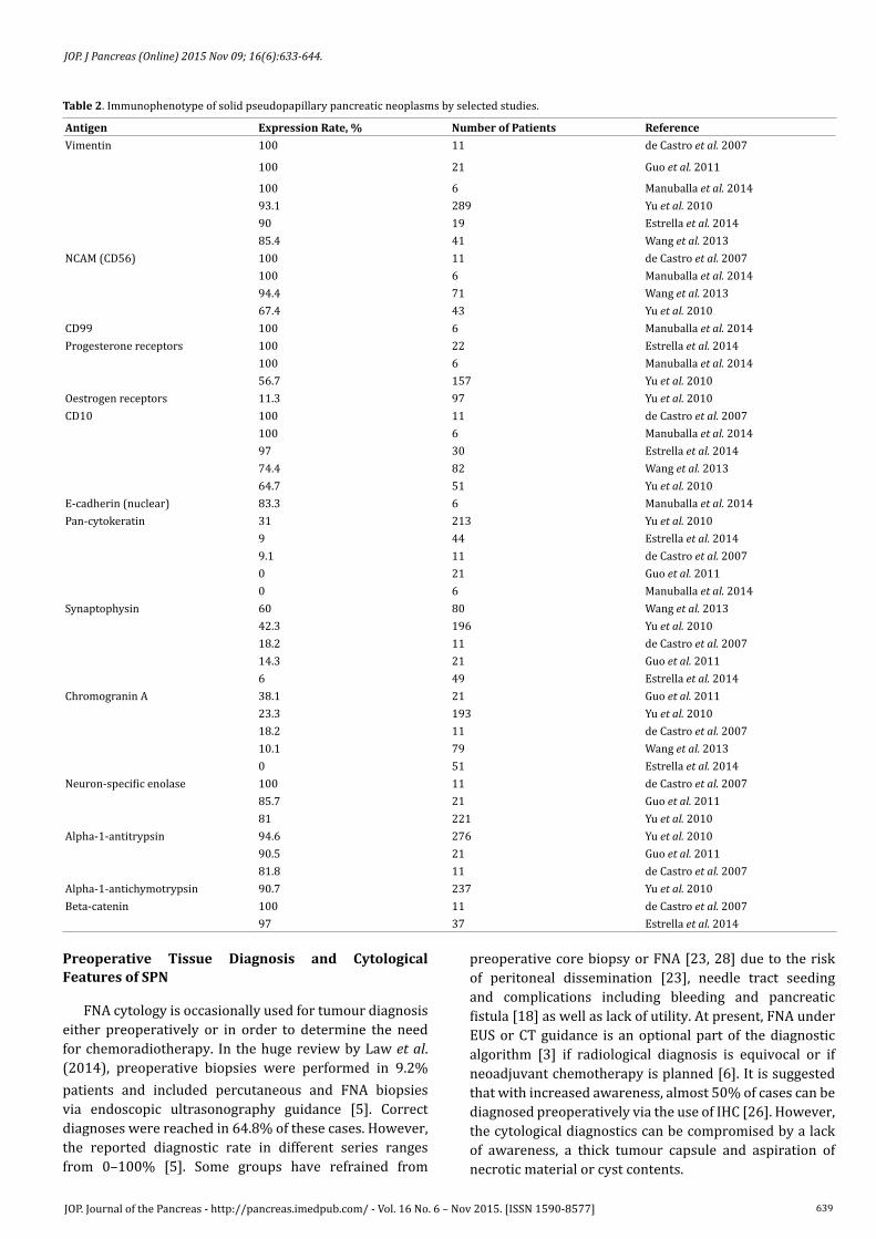
Table 2. Immunophenotype of solid pseudopapillary pancreatic neoplasms by selected studies.
Antigen Expression Rate, % Number of Patients ReferenceVimentin 100 11 de Castro et al. 2007
100 21 Guo et al. 2011
100 6 Manuballa et al. 201493.1 289 Yu et al. 201090 19 Estrella et al. 201485.4 41 Wang et al. 2013
NCAM (CD56) 100 11 de Castro et al. 2007100 6 Manuballa et al. 201494.4 71 Wang et al. 201367.4 43 Yu et al. 2010
CD99 100 6 Manuballa et al. 2014Progesterone receptors 100 22 Estrella et al. 2014
100 6 Manuballa et al. 201456.7 157 Yu et al. 2010
Oestrogen receptors 11.3 97 Yu et al. 2010CD10 100 11 de Castro et al. 2007
100 6 Manuballa et al. 201497 30 Estrella et al. 201474.4 82 Wang et al. 201364.7 51 Yu et al. 2010
E-cadherin (nuclear) 83.3 6 Manuballa et al. 2014Pan-cytokeratin 31 213 Yu et al. 2010
9 44 Estrella et al. 20149.1 11 de Castro et al. 20070 21 Guo et al. 20110 6 Manuballa et al. 2014
Synaptophysin 60 80 Wang et al. 201342.3 196 Yu et al. 201018.2 11 de Castro et al. 200714.3 21 Guo et al. 20116 49 Estrella et al. 2014
Chromogranin A 38.1 21 Guo et al. 201123.3 193 Yu et al. 201018.2 11 de Castro et al. 200710.1 79 Wang et al. 20130 51 Estrella et al. 2014
Neuron-specific enolase 100 11 de Castro et al. 200785.7 21 Guo et al. 201181 221 Yu et al. 2010
Alpha-1-antitrypsin 94.6 276 Yu et al. 201090.5 21 Guo et al. 201181.8 11 de Castro et al. 2007
Alpha-1-antichymotrypsin 90.7 237 Yu et al. 2010Beta-catenin 100 11 de Castro et al. 2007
97 37 Estrella et al. 2014

640JOP. Journal of the Pancreas - http://pancreas.imedpub.com/ - Vol. 16 No. 6 – Nov 2015. [ISSN 1590-8577]
JOP. J Pancreas (Online) 2015 Nov 09; 16(6):633-644.
In FNA samples, SPN is characterised by high cellularity in specimens obtained from a cystic mass, a finding that is not characteristic of true cystic neoplasms. The tumour cells form papillae lined with one or more layers of neoplastic cells. The shape of these straight and branching papillae has been compared to that of Chinese letters. The cells are monomorphic and bland, featuring round or oval nuclei, evenly dispersed chromatin, small nucleoli and nuclear grooves, long cytoplasmic processes, and extra- or intracellular hyaline globules. Ductal adenocarcinoma shows more marked polymorphism. Rosette formation and salt-and-pepper type chromatin favour an endocrine tumour but the differential diagnosis with pancreatic endocrine neoplasms can be more difficult, necessitating ancillary studies such as immunohistochemistry. Acinar cell carcinoma is characterised by loosely cohesive cell clusters, prominent nucleoli and granular, PAS-positive cytoplasm [6].
In our patient, the marked clinical symptoms necessitated prompt relief. As CT suggested a smooth and thus resectable lesion, FNA was not attempted. Retrospectively, the confirmation of SPN diagnosis by cytology would not have influenced the decision-making process.
Treatment
As summarised by Romics et al., surgical resection is the mainstay of SPN treatment [6]. Organ-sparing surgery is advocated, if possible. The typical operations include distal pancreatectomy for tumours located in the tail and corpus of the pancreas; pancreatoduodenectomy for pancreatic head tumours and middle segmental pancreatectomy with distal pancreatojejunostomy or pancreaticogastrostomy for SPN in the pancreatic neck. Regarding pancreatojejunostomy, the vast majority of studies report Roux-en-Y approach [32, 33, 34, 35]. Performing distal pancreatectomy, spleen preservation should be attempted in the absence of such contraindications as splenic artery or vein involvement or splenomegaly. The pylorus should be preserved in pancreatoduodenectomy if possible. Small SPN that are located apart from the pancreatic duct can be enucleated [6]. Although enucleation seems to be technically simpler and the pancreatoenteric anastomosis can be avoided, this type of operation has higher risk of complications, including long-standing pancreatic fistula or pancreatitis [36].
In the case of a large, invasive or metastatic tumour, the surgical attitude should change from less invasive to active since prolonged survival can be achieved [37]. En bloc resection must be performed if the tumour propagates to adjacent organs. Portal vein resection has been successfully performed for SPN invading the vein [9]. Metastases must be subjected to synchronous or metachronous resection. In contrast, routine lymphadenectomy is not advised due to the low rate of lymph node metastases [2, 6], estimated as 0.5–2.2% [5].
In surgical treatment, radical (R0) resection can be achieved in 92–100% of SPN patients [6, 10, 27].
The significance of palliative resection is somewhat controversial. In terms of resection margin status, the disease-specific survival in patients having residual tumour (R1) did not differ significantly in comparison with R0 cases [10]. In contrast, in a large Chinese study, only 1.3% patients who underwent radical (R0) resection died, in comparison with 40% in the palliatively resected group [24]. However, the residual tumour burden was not specified in this report. Guo et al. (2011) reported on 24 patients who were treated surgically and followed-up for 4–109 (median, 68) months [27]. Of these, all 22 patients having a R0 resection were alive with no evidence of disease while two patients in whom the resection was incomplete had a worse outcome. A patient with a microscopically positive margin developed liver metastases but received treatment and was alive 80 months after the first operation while a patient who had peritoneal metastases during the first operation died 42 months after surgery [27]. It can be concluded that a high postoperative tumour burden negatively influences survival and has more significant impact than microscopic residual tumour. Nevertheless, R0 resection is desirable.
The surgical approach can include either laparotomy or laparoscopy. At least 39 patients have had laparoscopic surgery for SPN, representing 1.7% of all SPN cases [5]. In a relatively large series including 10 patients from two large Italian centres, no cancer recurrence was observed within 47 months after radical laparoscopic treatment [23]. In contrast, poor results have been observed after laparoscopic biopsy in children: all patients developed either peritoneal dissemination or untreatable local recurrence [38].
In general, the central role of surgery in SPN treatment leaves few indications for adjuvant treatment [1, 12]. The role of neoadjuvant or adjuvant chemotherapy and radiotherapy is undefined because of limited experience with its use [6, 12, 18, 27]. However, radiotherapy can be used for unresectable SPN as the tumours are radiosensitive [3, 6, 27, 39].
In the largest English-language case review, adjuvant chemoradiotherapy was applied in 47 cases (6.3%), including 35 adjuvant chemotherapy and 12 radiotherapy patients. Among those patients who were followed-up, 75% (18/24) were alive at a mean of 51.1 months after surgery and combined treatment. Fluorouracil and gemcitabine were used most frequently. Three of five patients who were not suitable for surgery but received chemoradiotherapy were alive 18–60 months after diagnosis [5]. Tumour regression has been achieved via cisplatin, 5-FU and gemcitabine [6] or by combination therapy including cisplatin, cyclophosphamide, doxorubicin and vincristine [28]. However, ineffective treatment has also been observed [6]. In a Chinese study, chemotherapy with gemcitabin, cisplatin and anti-EGFR antibody was ineffective, and paclitaxel, carboplatin and anti-EGFR antibody induced intolerable abdominal and lumbosacral pain [30]. Neoadjuvant treatment using

641JOP. Journal of the Pancreas - http://pancreas.imedpub.com/ - Vol. 16 No. 6 – Nov 2015. [ISSN 1590-8577]
JOP. J Pancreas (Online) 2015 Nov 09; 16(6):633-644.
5-FU, doxorubicin, streptozocin, cisplatin, topotecan, iphosphamide and etoposide has been described as lacking efficacy [29].
In our case, the decision to perform radical surgery or not was the crucial point. When treating SPN, radical surgery can result in prolonged survival. Large size is not a contraindication for surgery in the case of SPN, given the low grade of malignancy. Lesions as large as 30 cm have been resected [29, 40]. En bloc resection of all involved organs with the aim of achieving R0 resection is consistent with the proposed algorithms [6]. Portal vein resection due to tumour invasion has been successfully performed in SPN patients [9]. Pancreatic surgery in patients older than 75 or 80 years is considered possible in selected patients [21, 22]. Middle segmental pancreatic resection is an oncologically appropriate option in patients with benign or low-grade malignant tumours located in the neck or body of the pancreas. This type of surgery allows to avoid extensive loss of healthy pancreatic parenchyma and thus preserves the exocrine and endocrine function [36]. However, old age, vascular operation and simultaneous resection of the large bowel are known risk factors in multivisceral pancreatic resections that are associated with significantly increased morbidity and a trend for increased mortality [4]. Middle segmental pancreatic resections also are reported to have higher incidence of complications than conventional pancreatoduodenectomy or distal pancreatectomy, possibly due to the development of two pancreatic remnants [32]. In our case, necrotic pancreatitis causing bleeding and a severe systemic inflammatory reaction were deadly complications. In a series of 40 middle segmental pancreatic resections, 10% of patients developed either pancreatic fistula, or pancreatic anastomotic insufficiency necessitating distal pancreatectomy. Bleeding complicated the postoperative course of 12.5% patients. Although radiologically guided arterial embolisation was helpful in some patients, delayed postoperative haemorrhage caused death in 2.5% of cases [36]. Acute pancreatitis after this type of pancreatic surgery is reported in 10% of patients [41].
Completion pancreatectomy or necrosectomy in a phased manner is used to treat postoperative pancreatic fistulas, bleeding and life-threatening postoperative pancreatitis [42, 43]. In a large series of 521 pancreatic surgery patients, completion pancreatectomy was necessary in 3.8% cases, because of the insufficiency of pancreaticojejunal anastomosis with resulting postoperative pancreatic fistula (70.0% of re-operated patients), severe bleeding (30.0%), portal vein thrombosis (5%) and remnant pancreatitis (35.0%). Although this approach represents the most active treatment of deleterious complications, the mortality of these complex patients remains significant (55%) even after completion pancreatectomy [42] and can reach almost 100% if the secondary surgical intervention is delayed. The general mortality of postoperative pancreatitis is reported to be 30–100%, exceeding the 5–15% death rate of primary pancreatitis [43].
Even in series, reporting comparable rates of complications after pancreatectomy in octogenarians compared with those younger than 80 years, the older patients had higher death risk once the complications occurred [44]. Thus, early diagnosis of complications would be necessary. Postoperative pancreatitis is clinically defined as abdominal pain in association with two- to three-fold increase of the blood levels of pancreatic enzymes. However, the clinical symptoms can be difficult to observe after major abdominal surgery. Circulatory instability or general abrupt change in clinical status can be the first manifestation of postoperative pancreatitis [43]. Retrospectively, the postoperative hypotension in our patient also most likely was an early manifestation of the pancreatitis although the significant intraoperative blood loss could contribute to it. The long-standing hypotension could become harmful by itself leading to low mesenteric blood flow [45] and thus facilitating development of postoperative pancreatic fistula. Ischemic acute pancreatitis also can be caused by hypotension [46]. In addition, mesenteric ischemia can also manifest by decreased arterial blood pressure. Therefore the differential diagnostics of hypotension in an aged patient after major abdominal surgery includes also compromised mesenteric circulation as the primary cause of arterial hypotension. The clinical distinction is complex; computed tomography angiography or magnetic resonance can be helpful. In our case, no signs of intestinal necrosis were found during the relaparotomy, retrospectively finally ruling out mesenteric thrombosis.
Among laboratory findings, CRP and calcium levels must be closely followed, if postoperative pancreatitis is suspected. An abrupt increase in the serum amylase and CRP levels in the time interval from 1st to 5th POD is suggestive of postoperative pancreatitis [43].
Although somewhat controversial, pancreatic resection with or without portal or supramesenteric vein resection has similar rate of complications and survival [4, 47]. However, the vascular invasion must be promptly detected preoperatively to ensure appropriate surgical planning. The preoperative diagnosis includes CT-based evaluation with multidetector helical CT with pre-contrast, late arterial and portal venous phases of enhancement [47-49]. The diagnostic accuracy of CT regarding vascular invasion reaches 62–92% [48] and has been improving in recent years due to the technological progress [50]. Axial images are combined with coronal oblique images to evaluate the relationship between the tumour and blood vessels; reconstructions are performed. The presence, site and extent of angioinvasion must be detected. In addition, individual anatomy (length and diameter of the left renal vein, anatomy of the kidneys and collateral veins, or anatomy of other possible autologous graft sites) must be exploited. During operation, the CT findings are re-evaluated to make the final decision on venous reconstruction. The vascular reconstruction can be performed as primary closure, end-to-end anastomosis or
642JOP. Journal of the Pancreas - http://pancreas.imedpub.com/ - Vol. 16 No. 6 – Nov 2015. [ISSN 1590-8577]
JOP. J Pancreas (Online) 2015 Nov 09; 16(6):633-644.
graft replacement. Autologous left renal vein is advocated for grafting due to appropriate diameter that matches the size of portal vein. In addition, the renal vein reconstruction is considered unnecessary due to gonadal vein ensuring collateral flow [47]. Alternatively, splenic, internal jugular, iliac, femoral or saphenous vein or vascular prosthesis can be used for reconstruction [49, 51-53]. To ensure patent blood flow, narrowing or bending of portal vein must be avoided. Intraoperative ultrasonography should be used to evaluate the reconstruction patency. After vascular operation, prostaglandin E1 treatment is recommended to increase the portal flow. The flow must be controlled daily by ultrasound investigation [47].
If a tumour is considered inoperable, alternative treatment would include chemoradiotherapy. However, there is no conclusive evidence of the efficacy of such treatment in SPN. In addition, the side effects could be harmful in such an old patient. Besides, the tumour was causing significant pain attributable to the large size of the mass as well as to large bowel invasion and compression. As a significant part of the tumour was cystic and the proliferation index was low, chemo- or radiotherapy would not have had a fast significant effect. A patient with unresectable SPN has been treated by gemcitabine-based chemotherapy resulting in prolonged survival of 26 months. However, the tumour size was not significantly reduced and the life quality was estimated as poor although the initial dominant symptom was loss of appetite [9]. In our patient, experiencing pain as the dominant symptom, even worse life quality could be expected.
Prognosis
In contrast to most other types of pancreatic cancer, SPN has a good prognosis. In a review by Law et al. (2014), only 4.4% of patients experienced tumour recurrence and 1.5% died due to the tumour during a mean follow-up of 36.1 months. The median time to recurrence was 50.5 months [5]. Thus, the follow-up of surgically treated SPN patients must be at least 5 years long and the survival data analysis should cover at least 4 or 5 years after operation [54]. However, some series have reported no recurrences during a 3.8–8-year follow-up. In contrast, late recurrences have been observed 7 and 14 years after primary resection [6]. The risk of recurrence is higher if the tumour invades the capsule, lymphatic or blood vessels, or synchronous metastases are present [54]. If recurrence develops, surgical treatment can be effective. The prognosis for a patient who is treated for SPN metastasis in the liver ranges from 6 months to 17 years [3]. Prolonged survival, reaching at least 34 months has been observed after resection of the primary SPN and 2 pulmonary metastases [9].
In two large reviews, the 1-, 2-, 3- and 5-year survival rates were 99.4%, 97%, 97.5% and 95.0–96.9%, respectively [3, 24]. The specific 10-year survival was 93–96% [3, 10].
In conclusion, to the best of our knowledge, the reported patient is the oldest person and the oldest male
patient diagnosed with this neoplasm. Awareness of this entity and its occurrence in unusual sex and age groups would be helpful when planning treatment for this low-grade malignancy.
AcknowledgementThe present work was carried out within the framework
of scientific project No. 2013/0004/1DP/1.1.1.2.0/13/APIA/VIAA/020, supported by ESF.
Conflict of InterestThe authors report no conflicts of interest to disclose.
References1. Adkisson CD, Harris AS, Bridges MD, Nguyen JH, Asbun HJ, Stauffer JA. Solid pseudopapillary tumor of the pancreas: report of five cases. International Journal of Hepatobiliary and Pancreatic Diseases 2012; 2:9-14.
2. Machado MC, Machado MA, Bacchella T, Jukemura J, Almeida JL, Cunha JE. Solid pseudopapillary neoplasm of the pancreas: distinct pattern of onset, diagnosis, and prognosis for male versus female patients. Surgery 2008; 143:29-34. [PMID: 18154930].
3. Papavramidis T, Papavramidis S. Solid pseudopapillary tumors of the pancreas: review of 718 patients reported in English literature. J Am Coll Surg 2005; 200:965-72. [PMID: 15922212].
4. Hartwig W, Hackert T, Hinz U, Hassenpflug M, Strobel O, Buchler MW, et al. Multivisceral resection for pancreatic malignancies: risk-analysis and long-term outcome. Ann Surg 2009; 250:81-7. [PMID: 19561478]
5. Law JK, Ahmed A, Singh VK, Akshintala VS, Olson MT, Raman SP, et al. A systematic review of solid-pseudopapillary neoplasms: are these rare lesions? Pancreas 2014; 43:331-7. [PMID: 24622060].
6. Romics L Jr, Olah A, Belagyi T, Hajdu N, Gyurus P, Ruszinko V. Solid pseudopapillary neoplasm of the pancreas – proposed algorithms for diagnosis and surgical treatment. Langenbecks Arch Surg 2010; 395:747-55. [PMID: 20155424]
7. de Castro SM, Singhal D, Aronson DC, Busch OR, van Gulik TM, Obertop H, et al. Management of solid-pseudopapillary neoplasms of the pancreas: a comparison with standard pancreatic neoplasms. World J Surg 2007; 31:1130-35. [PMID: 17429567].
8. Attaallah W, Javadov M, Ayranci FG, Filinte D, Dulundu E, Yegen C. Locally advanced pseudopapillary neoplasm of the pancreas in a male patient: a case report. JOP 2013; 14:438-41. [PMID: 23846943]
9. Hosokawa I, Shimizu H, Ohtsuka M, Kato A, Yoshitomi H, Furukawa K, et al. Preoperative diagnosis and surgical management for solid pseudopapillary neoplasm of the pancreas. J Hepatobiliary Pancreat Sci 2014; 21:573-8. [PMID: 24535774]
10. Estrella JS, Li L, Rashid A, Wang H, Katz MH, Fleming JB, et al. Solid pseudopapillary neoplasm of the pancreas: clinicopathologic and survival analyses of 64 cases from a single institution. Am J Surg Pathol 2014; 38:147-57. [PMID: 24418850]
11. Baxter DS, Orrego A, Rosenfeld JV, Mathiesen T. An audit of immunohistochemical marker patterns in meningioma. J Clin Neurosci 2014; 21:421-6. [PMID: 24231566]
12. Bouassida M, Mighri MM, Bacha D, Chtourou MF, Touinsi H, Azzouz MM, Sassi S. Solid pseudopapillary neoplasm of the pancreas in an old man: age does not matter. Pan Afr Med J 2012; 13:8. [PMID: 23308315]
13. Cubilla AL, Fitzgerald PJ. Classification of pancreatic cancer (nonendocrine). Mayo Clin Proc 1979; 54:449-58. [PMID: 221755].
14. Morohoshi T, Kanda M, Horie A, Chott A, Dreyer T, Kloppel G, et al. Immunocytochemical markers of uncommon pancreatic tumours. Acinar cell carcinoma, pancreatoblastoma, and solid cystic (papillary cystic) tumor. Cancer 1987; 59:739-47. [PMID: 3542187].
643JOP. Journal of the Pancreas - http://pancreas.imedpub.com/ - Vol. 16 No. 6 – Nov 2015. [ISSN 1590-8577]
JOP. J Pancreas (Online) 2015 Nov 09; 16(6):633-644.
15. Butte JM, Brennan MF, Gonen M, Tang LH, D’Angelica MI, Fong Y, et al. Solid pseudopapillary tumors of the pancreas. Clinical features, surgical outcomes, and long-term survival in 45 consecutive patients from a single center. J Gastrointest Surg 2011; 15:350-7. [PMID: 20824369]
16. Warshaw AL, Compton CC, Lewandrowski K, Cardenosa G, Mueller PR. Cystic tumours of the pancreas. New clinical, radiological, and pathological observations in 67 patients. Ann Surg 1990; 212:432-45. [PMID: 2171441].
17. Koito K, Namieno T, Nagakawa T, Shyonai T, Hirokawa N, Morita K. Solitary cystic tumor of the pancreas: EUS-pathologic correlation. Gastrointest Endosc 1997; 45:268-76. [PMID: 9087833].
18. Kersting S, Janot MS, Munding J, Suelberg D, Tannapfel A, Chromik AM, et al. Rare solid tumors of the pancreas as differential diagnosis of pancreatic adenocarcinoma. JOP 2012; 13:268-77. [PMID: 22572130].
19. Jaksic T, Yaman M, Thorner P, Wesson DK, Filler RM, Shandling B. A 20-year review of pediatric pancreatic tumours. J Pediatr Surg 1992; 27:1315-7. [PMID: 1328584].
20. Oliveira Lima S, Rocha Santana V, Correia Leao S, Faro Santos PS, de Albuquerque Jr RL. Solid-pseudopapillary tumor of pancreas in a young woman: a case report and literature review. Rev Med Chil 2012; 140:1179-84. [PMID: 23354641]
21. Sohn TA, Yeo CJ, Cameron JL, Lillemoe KD, Talamini MA, Hruban RH, et al. Should pancreaticoduodenectomy be performed in octogenarians? J Gastrointest Surg 1998; 2:207-16. [PMID: 9841976].
22. Ballarin R, Spaggiari M, di Benedetto F, Montalti R, Masetti M, de Ruvo N, et al. Do not deny pancreatic resection to elderly patients. J Gastrointest Surg 2009; 13:341-8. [PMID: 18784970]
23. Cavallini A, Butturini G, Daskalaki D, Salvia R, Melotti G, Piccoli M, et al. Laparoscopic pancreatectomy for solid pseudo-papillary tumors of the pancreas is a suitable technique; our experience with long-term follow-up and review of the literature. Ann Surg Oncol 2011; 18:352-7. [PMID: 20848223]
24. Yu PF, Hu ZH, Wang XB, Guo JM, Cheng XD, Zhang YL, Xu Q. Solid pseudopapillary tumour of the pancreas: A review of 553 cases in Chinese literature. World J Gastroenterol 2010; 16:1209-14. [PMID: 20222163]
25. Takahashi H, Hashimoto K, Hayakawa H, Kusakawa M, Okamura K, Kosaka A, et al. Solid cystic tumor of the pancreas in elderly man: report of a case. Surg Today 1999; 29: 1264-7. [PMID: 10639709].
26. Manuballa V, Amin M, Cappell MS. Clinical presentation and comparison of surgical outcome for segmental resection vs. Whipple’s procedure for solid pseudopapillary tumor: Report of six new cases and literature review of 321 cases. Pancreatology 2014; 14:71-80. [PMID: 24555981]
27. Guo N, Zhou QB, Chen RF, Zou SQ, Li ZH, Lin Q, et al. Diagnosis and surgical treatment of solid pseudopapillary neoplasm of the pancreas: analysis of 24 cases. Can J Surg 2011; 54:368-74. [PMID: 21939604]
28. Tipton SG, Smyrk TC, Sarr MG, Thompson GB. Malignant potential of solid pseudopapillary neoplasm of the pancreas. Br J Surg 2006; 93:733-7. [PMID: 16609955].
29. Vassos N, Agaimy A, Klein P, Hohenberger W, Croner RS. Solid-pseudopapillary neoplasm (SPN) of the pancreas: case series and literature review on an enigmatic entity. Int J Clin Exp Pathol 2013; 6:1051-9. [PMID: 23696922].
30. Wang LJ, Bai L, Su D, Zhang TT, Mao ZY, Guo XC, et al. Retrospective analysis of 102 cases of solid pseudopapillary neoplasm of the pancreas in China. J Int Med Res 2013; 41:1266-71. [PMID: 23812113]
31. Kato T, Egawa N, Kamisawa T, Tu Y, Sanaka M, Sakaki N, et al. A case of solid pseudopapillary neoplasm of the pancreas and tumor doubling time. Pancreatology 2002; 2:495-8. [PMID: 12378119].
32. Sperti C, Pasquali C, Ferronato A, Pedrazzoli S. Median pancreatectomy for tumors of the neck and body of the pancreas. J Am Coll Surg 2000; 190:711-6. [PMID: 10873007].
33. Sauvanet A, Partensky C, Sastre B, Gigot JF, Fagniez PL, Tuech JJ, et al. Medial pancreatectomy: a multi-institutional retrospective study of 53 patients by the French Pancreas Club. Surgery 2002; 132:836-43. [PMID: 12464868].
34. Efron DT, Lillemoe KD, Cameron JL, Yeo CJ. Central pancreatectomy with pancreaticogastrostomy for benign pancreatic pathology. J Gastrointest Surg 2004; 8:532-8. [PMID: 15239986].
35. Lavu H, Knuth JL, Baker MS, Shen C, Zyromski NJ, Schmidt M, et al. Middle segment pancreatectomy can be safely incorporated into a pancreatic surgeon’s clinical practice. HPB (Oxford) 2008; 10:491-7. [PMID: 19088938]
36. Muller MW, Friess H, Kleeff J, Hinz U, Wente MN, Paramythiotis D, et al. Middle segmental pancreatic resection: an option to treat benign pancreatic body lesions. Ann Surg 2006; 244:909-20. [PMID: 17122616].
37. Alves JR, Amico EC. Solid-pseudopapillary neoplasm of the pancreas: case series and literature review. JOP 2015; 16:218-26.
38. Fais PO, Carricaburu E, Sarnacki S, Berrebi D, Orbach D, Baudoin V, et al. Is laparoscopic management suitable for solid pseudo-papillary tumors of the pancreas? Pediatr Surg Int 2009; 25:617-21. [PMID: 19479267]
39. Fried P, Cooper J, Balthazar E, Fazzini E, Newall J. The role of radiotherapy in the treatment of solid and papillary neoplasms of the pancreas. Cancer 1985; 56:2783-5. [PMID: 4052952].
40. Wollmer CM Jr, Dixon E, Grant DR. Management of a solid pseudopapillary tumor of the pancreas with liver metastases. HPB (Oxford) 2003; 5:264-7. [PMID 18333000]
41. Adham M, Giunippero A, Hervieu V, Courbiere M, Partensky C. Central pancreatectomy: single-center experience of 50 cases. Arch Surg 2008; 143:175-81. [PMID: 18283143]
42. Nentwich MF, El Gammal AT, Lemcke T, Ghadban T, Bellon E, Melling N, et al. Salvage completion pancreatectomies as damage control for post-pancreatic surgery complications: a single-center retrospective analysis. World J Surg 2015; 39:1550-6. [PMID: 25651954]
43. Ryska M, Rudis J. Pancreatic fistula and postoperative pancreatitis after pancreatoduodenectomy for pancreatic cancer. Hepatobiliary Surg Nutr 2014; 3:268-75. [PMID: 25392838]
44. Tamirisa NP, Parmar AD, Vargas GM, Mehta HB, Kilbane EM, Hall BL, et al. Relative contributions of complications and failure to rescue on mortality in older patients undergoing pancreatectomy. Ann Surg 2015, Epub ahead of print, accessed 17 Jul 2015. [PMID: 25563871].
45. Sastry P, Hardman G, Page A, Parker R, Goddard M, Large S, Jenkins DP. Mesenteric ischaemia following cardiac surgery: the influence of intraoperative perfusion parameters. Interact Cardiovasc Thorac Surg 2014; 19:419-24. [PMID: 24939960]
46. Hackert T, Hartwig W, Fritz S, Schneider L, Strobel O, Werner J. Ischemic acute pancreatitis: clinical features of 11 patients and review of the literature. Am J Surg 2009; 197:450-4. [PMID: 18778810]
47. Yoshitomi H, Kato A, Shimizu H, Ohtsuka M, Furukawa K, Takayashiki T, et al. Tips and tricks of surgical technique for pancreatic cancer: portal vein resection and reconstruction (with videos). J Hepatobiliary Pancreat Sci 2014; 21:E69-74. [PMID: 24964060]
48. Marinelli T, Filippone A, Tavano F, Fontana A, Pellegrini F, Koninger J, et al. A tumour score with multidetector spiral CT for venous infiltration in pancreatic cancer: influence on borderline resectable. Radiol Med 2014; 119:334-42. [PMID: 24619824]
49. Tran Cao HS, Balachandran A, Wang H, Nogueras-Gonzalez GM, Bailey CE, Lee JE, et al. Radiographic tumor-vein interface as a predictor of intraoperative, pathologic, and oncologic outcomes in resectable and borderline resectable pancreatic cancer. J Gastrointest Surg 2014; 18:269-78. [PMID: 24129826]
50. Zhao WY, Luo M, Sun YW, Xu Q, Chen W, Zhao G, Wu ZY. Computed tomography in diagnosing vascular invasion in pancreatic and periampullary cancers: a systematic review and meta-analysis. Hepatobiliary Pancreat Dis Int 2009; 8:457-64. [PMID: 19822487].
51. Choi SH, Hwang HK, Kang CM, Lee WJ. Total pancreaticoduodenectomy and segmental resection of superior mesenteric vein-portal vein confluence with autologous splenic vein graft in mucinous cystadenocarcinoma of the pancreas. JOP 2010; 11:638-41. [PMID: 21068503].
52. Lee DY, Mitchell EL, Jones MA, Landry GJ, Liem TK, Sheppard BC, et al. Techniques and results of portal vein/superior mesenteric vein reconstruction using femoral and saphenous vein during pancreaticoduodenectomy. J Vasc Surg 2010; 51:662-6. [PMID: 20080375]
644JOP. Journal of the Pancreas - http://pancreas.imedpub.com/ - Vol. 16 No. 6 – Nov 2015. [ISSN 1590-8577]
JOP. J Pancreas (Online) 2015 Nov 09; 16(6):633-644.
53. Sgroi MD, Narayan RR, Lane JS, Demirjian A, Kabutey NK, Fujitani RM, Imagawa DK. Vascular reconstruction plays an important role in the treatment of pancreatic adenocarcinoma. J Vasc Surg 2015; 61:475-80. [PMID: 25441672]
54. Serrano PE, Serra S, Al-Ali H, Gallinger S, Greig PD, McGilvray ID, et al. Risk factors associated with recurrence in patients with solid pseudopapillary tumors of the pancreas. JOP 2014; 15:561-8. [PMID: 25435571]


![Men in the US with Solid Pseudopapillary Carcinomas …pancreas.imedpub.com/men-in-the-us-with-solid-pseudopapillary... · 10, 11]. Resection of SPNs using laparoscopic and robotic](https://static.fdocuments.us/doc/165x107/5b5ec8917f8b9a553d8d3a83/men-in-the-us-with-solid-pseudopapillary-carcinomas-10-11-resection-of-spns.jpg)


![Pediatric Solid Pseudopapillary Neoplasm[Spn] of The Pancreas … · Central Annals of Clinical Pathology Cite this article: Roganovic J, Matijasic N Jonjic N (2015) Pediatric Solid](https://static.fdocuments.us/doc/165x107/5ffdf42ed2be6c190c067e5b/pediatric-solid-pseudopapillary-neoplasmspn-of-the-pancreas-central-annals-of.jpg)