Large Conformational Changes in the Maturation of a Simple ...
12
COMMUNICATION Large Conformational Changes in the Maturation of a Simple RNA Virus, Nudaurelia capensis o Virus (NoV) Mary A. Canady 1 , Mariana Tihova 2 , Terry N. Hanzlik 3 , John E. Johnson 1 * and Mark Yeager 1,2,4 * 1 Department of Molecular Biology and 2 Department of Cell Biology The Scripps Research Institute 10550 N. Torrey Pines Rd La Jolla, CA 92037, USA 3 Division of Entomology Box 1700, Canberra, ACT Australia 4 Division of Cardiovascular Diseases, Scripps Clinic, 10666 N. Torrey Pines Rd, La Jolla CA 92037, USA An assembly intermediate of a small, non-enveloped RNA virus has been discovered that exhibits striking differences from the mature virion. Virus-like particles (VLPs) of Nudaurelia capensis o virus (NoV), a T 4 icosahedral virus infecting Lepidoptera insects, were produced in insect cells using a baculovirus vector expressing the coat protein. A procapsid form was discovered when NoV VLPs were purified at neutral pH con- ditions. These VLPs were fragile and did not undergo the autoproteolytic maturation that occurs in the infectious virus. Electron cryo-microscopy (cryoEM) and image analysis showed that, compared with the native vir- ion, the VLPs were 16 % larger in diameter, more rounded, porous, and contained an additional internal domain. Upon lowering the pH to 5.0, the VLP capsids became structurally indistinguishable from the authentic virion and the subunits autoproteolyzed. The NoV protein subunit coor- dinates, which were previously determined crystallographically, were modelled into the 28 A ˚ resolution cryoEM map of the procapsid. The resulting pseudo-atomic model of the NoV procapsid demonstrated the large rearrangements in quaternary and tertiary structure needed for the maturation of the VLPs and presumably of the virus. Based on this model, we propose that electrostatically driven rearrangements of interior helical regions are responsible for the large conformational change. These results are surprising because large structural rearrangements have not been found in the maturation of any other small RNA viruses. However, similarities of this conformational change to the maturational processes of more complex DNA viruses (e.g. bacteriophages and herpesvirus) and to the swelling of simple plant viruses suggest that structural changes in icosahedral viruses, which are integral to their function, have similar strategies and perhaps mechanisms. # 2000 Academic Press Keywords: conformational change; tetravirus; pseudo-atomic; virus assembly; modelling *Corresponding authors Introduction In the life-cycles of viruses, dramatic morpho- logical changes in their capsid structure are needed to allow them to carry out the diverse set of func- tions required for replication. All virus capsids must form readily, have structural integrity, and have the proper biological trigger in order to be infectious. It is clear that complex viruses undergo multi-stage assembly that is orchestrated by pro- tein-protein and/or protein-nucleic acid inter- actions. The most dramatic and best characterized of these changes occur in the double-stranded DNA (dsDNA) bacteriophages, where structural intermediates can often be characterized at low res- olution (Prasad et al., 1993). The dsDNA herpes simplex virus undergoes a maturational transition that is comparable to that of the dsDNA bacterio- M.A.C. and M.T., and J.E.J. and M.Y contributed equally to the work. Abbreviations used: NoV, Nudaurelia capensis o virus; dsDNA, double-stranded DNA; Ig, immunoglobulin; VLP, virus-like particle; cryoEM, electron cryo- microscopy; TBSV, tomato bushy stunt virus; CCMV, cowpea chlorotic mottle virus. E-mail addresses of the corresponding authors: [email protected]; [email protected] doi:10.1006/jmbi.2000.3723 available online at http://www.idealibrary.com on J. Mol. Biol. (2000) 299, 573–584 0022-2836/00/030573–12 $35.00/0 # 2000 Academic Press
Transcript of Large Conformational Changes in the Maturation of a Simple ...

doi:10.1006/jmbi.2000.3723 available online at http://www.idealibrary.com on J. Mol. Biol. (2000) 299, 573±584
COMMUNICATION
Large Conformational Changes in the Maturation of aSimple RNA Virus, Nudaurelia capensis ooo Virus (NoooV)
Mary A. Canady1, Mariana Tihova2, Terry N. Hanzlik3, John E. Johnson1*and Mark Yeager1,2,4*
1Department of MolecularBiology and2Department of Cell BiologyThe Scripps Research Institute10550 N. Torrey Pines RdLa Jolla, CA 92037, USA3Division of EntomologyBox 1700, Canberra, ACTAustralia4Division of CardiovascularDiseases, Scripps Clinic, 10666N. Torrey Pines Rd, La JollaCA 92037, USA
M.A.C. and M.T., and J.E.J. and Mequally to the work.
Abbreviations used: NoV, NudaudsDNA, double-stranded DNA; Ig,VLP, virus-like particle; cryoEM, elemicroscopy; TBSV, tomato bushy stcowpea chlorotic mottle virus.
E-mail addresses of the [email protected]; [email protected]
0022-2836/00/030573±12 $35.00/0
An assembly intermediate of a small, non-enveloped RNA virus has beendiscovered that exhibits striking differences from the mature virion.Virus-like particles (VLPs) of Nudaurelia capensis o virus (NoV), a T � 4icosahedral virus infecting Lepidoptera insects, were produced in insectcells using a baculovirus vector expressing the coat protein. A procapsidform was discovered when NoV VLPs were puri®ed at neutral pH con-ditions. These VLPs were fragile and did not undergo the autoproteolyticmaturation that occurs in the infectious virus. Electron cryo-microscopy(cryoEM) and image analysis showed that, compared with the native vir-ion, the VLPs were 16 % larger in diameter, more rounded, porous, andcontained an additional internal domain. Upon lowering the pH to 5.0,the VLP capsids became structurally indistinguishable from the authenticvirion and the subunits autoproteolyzed. The NoV protein subunit coor-dinates, which were previously determined crystallographically, weremodelled into the 28 AÊ resolution cryoEM map of the procapsid. Theresulting pseudo-atomic model of the NoV procapsid demonstrated thelarge rearrangements in quaternary and tertiary structure needed for thematuration of the VLPs and presumably of the virus. Based on thismodel, we propose that electrostatically driven rearrangements of interiorhelical regions are responsible for the large conformational change. Theseresults are surprising because large structural rearrangements have notbeen found in the maturation of any other small RNA viruses. However,similarities of this conformational change to the maturational processesof more complex DNA viruses (e.g. bacteriophages and herpesvirus) andto the swelling of simple plant viruses suggest that structural changes inicosahedral viruses, which are integral to their function, have similarstrategies and perhaps mechanisms.
# 2000 Academic Press
Keywords: conformational change; tetravirus; pseudo-atomic; virusassembly; modelling
*Corresponding authorsIntroduction
In the life-cycles of viruses, dramatic morpho-logical changes in their capsid structure are needed
.Y contributed
relia capensis o virus;immunoglobulin;ctron cryo-unt virus; CCMV,
ding authors:du
to allow them to carry out the diverse set of func-tions required for replication. All virus capsidsmust form readily, have structural integrity, andhave the proper biological trigger in order to beinfectious. It is clear that complex viruses undergomulti-stage assembly that is orchestrated by pro-tein-protein and/or protein-nucleic acid inter-actions. The most dramatic and best characterizedof these changes occur in the double-strandedDNA (dsDNA) bacteriophages, where structuralintermediates can often be characterized at low res-olution (Prasad et al., 1993). The dsDNA herpessimplex virus undergoes a maturational transitionthat is comparable to that of the dsDNA bacterio-
# 2000 Academic Press

Figure 1 (legend opposite)
574 N!V Procapsid to Capsid Conformational Change

N!V Procapsid to Capsid Conformational Change 575
phages (Trus et al., 1996). In both cases a roundedprecursor matures into a polyhedral capsid. Manyother large structural changes have been describedin the assembly and maturation of complex RNAand DNA viruses, both enveloped and non-envel-oped (Allison et al., 1995; Andres et al., 1998;Edvardsson et al., 1976; Salanueva et al., 1999;Turner & Summers, 1999). The molecular inter-actions that trigger these structural transitions arenot well understood, in part because no atomicresolution model is available for the assemblyintermediates. By combining the crystallographicmodel of Nudaurelia capensis o virus (NoV) withthe structure of the procapsid obtained using elec-tron cryomicroscopy (cryoEM), we have been able
Figure 1. Three-dimensional, surface-shaded (a) full particlsid (right) of NoV VLPs viewed down a 2-fold symmetry axangle) axes marked in (b). The procapsid is larger, roundeangular, solid shell. (c) The radial density plots reveal thattwo domains, whereas the procapsid (gray line) had a thicktion of each map is shown above the plot, the domains delincorrespond. (d) An SDS-PAGE gel showing that the procapwhile the capsid contains mostly 62 kDa and 8 kDa (not visiblysis upon lowering of the pH to 5.0. The recombinant bacuthe pFBWCap plasmid using the bacmid reagents supplied bblunt-ended RT-PCR product of the NoV coat protein into thduct was obtained by performing 12 PCR cycles (20-95 �C, 2cDNA made with SuperScript II (BRL) and RNA extractedsupplied by Don Hendry). The primers were designed usinand were the phosphorylated primers W21 (GCAGCAAAACWcapN (GGAGATGGACAGTAACTCAGCC), complementaATG of the coat protein in WcapN were inserted in order toing the endogenous context of the NoV coat protein ATG facells were grown in TC-100 medium supplemented with 10 %layers using a multiplicity of infection of 5. After incubationlysed with 0.5 % (v/v) NP40, and the virus-like particles (through a 30 % (w/v) sucrose cushion. The pellets were resTris (pH 7.5)). Debris was removed by low-speed centrifugatlayered onto a 10 %-40 % (w/v) sucrose gradient in buffer Aband containing the VLPs was collected and dialyzed againscapsids were obtained by diluting the procapsids 1:10 wit(�4 ml) of virus suspension (�1-2 mg/ml) were applied to hca four seconds with preheated ®lter paper, and then immtransmission electron microscope equipped with Gatan 62610 electrons/AÊ 2) images at a magni®cation of 45,000� thatwas used to select images that displayed minimal astigmatransfer function. Selected images were digitized at a 25 mmsponding to 5.6 AÊ on the specimen. The program X3D (Conwa circular mask and subtract background density. An initialsingle micrograph were determined using cross-correlatioCrowther, 1971; Fuller, 1987; Olson & Baker, 1989). To optiusing only a portion of the Fourier transform of the maskmap of NoV at 28 AÊ resolution (Johnson et al., 1994) was us(PFT) method (Baker & Cheng, 1996). The search proceduresid shell. Some ®ve to six cycles of re®nement were performusing 0.5 � increments. During the latter re®nement cycles,resolution was extended from 35 to 20 AÊ . The ®nal reconstand 259 particles, respectively. The ®nal reconstruction of thand had a resolution limit of 25 AÊ . To assess the resolution otwo independent reconstructions at higher resolution. The prture factors, and the program EMCTF03 used these data to cfrequency by comparing the structure factors of the two intations were visualized using AVS software (Upson et al., 1occupied by the capsid shell calculated from the number of c
to model a viral assembly intermediate for the ®rsttime.
NoV is a T � 4, icosahedral, single-strandedRNA virus that belongs to the tetravirus family,whose members infect insects of the lepidopteranorder (Murphy et al., 1995). The viral capsidsassemble from 240 copies of the 70 kDa coat pro-tein, which adopt four slightly different confor-mations in the capsid (designated A, B, C, and D)according to the theory of quasiequivalence(Caspar & Klug, 1962). After assembly, all of thesubunits are cleaved autocatalytically, resulting in62 kDa (b) and 8 kDa (g) products (Agrawal &Johnson, 1992). A similar phenomenon occurs inthe T � 3 insect nodaviruses, where autoproteo-
e and (b) sectioned views of the procapsid (left) and cap-is, with 3-fold (black triangle) and quasi 3-fold (white tri-d, and porous, while the mature capsid has a smaller,the capsid protein shell (black line) spanned 62 AÊ withness of 83 AÊ and comprised three domains. A cross-sec-eated and placed in register with the radii to which theysid sample contains the 70 kDa uncleaved coat protein,le) coat protein fragments which result from autoproteo-
lovirus expressing the NoV coat protein was made fromy BRL. The pFBWCap plasmid was made by inserting ae StuI site of the plasmid, pFastBac (BRL). The PCR pro-0-55 �C, 4-70 �C) with Pfu polymerase (Stratagene) uponfrom NoV puri®ed on a CsCl density gradient (kindlyg the NoV RNA2 sequence (Agrawal & Johnson, 1995)TGTCGTGTG), complementary to bases 2398-2416, and
ry to bases 267-285. Four bases prior to the initiatingimprove expression because baculovirus constructs hav-iled to give good expression. Spodoptera frugiperda (Sf21)
(v/v) fetal bovine serum. Cells were infected as mono-for four to ®ve days at 27 �C, the cells were harvested,
VLPs) were isolated by ultracentrifugation at 100,000 guspended overnight in buffer A (250 mM NaCl, 50 mMion, and supernatant containing the procapsid VLPs was. After ultracentrifugation at 113,000 g for 2.5 hours, thet buffer A, and then concentrated to 1-3 mg/ml. Matureh buffer B (70 mM sodium acetate (pH 5.0)). Aliquotsoley carbon ®lms on 400-mesh copper grids, blotted forediately plunged into liquid ethane. A Philips CM120
cryo-specimen holder was used to record low-dose (5 towere underfocussed by 0.8 to 1.2 mm. Optical diffractiontism and drift based on the Thon rings in the contrastinterval using a Perkin-Elmer microdensitometer, corre-ay et al., 1997) was used to extract particle images withset of origin and orientation values for particles from an and common line procedures (Baker et al., 1988;
mize the search procedure, calculations were performeded image (between 1/70 AÊ and 1/35 AÊ ). The publisheded as the starting model for the polar Fourier transformwas optimized by using particle radii spanning the cap-ed using 1.0 � increments, followed by six to eight cyclesadditional particles were progressively included and theructions of the procapsid and capsid were based on 57e authentic virion (not shown) was based on 46 particlesf the maps, each data set was divided in half to computeogram EMMAP3DT was used to generate a list of struc-ompute the correlation coef®cient as a function of spatialdependent reconstructions. All surface-shaded represen-989). Contour levels were chosen to include the volumeopies and the molecular mass of the coat protein.
576 N!V Procapsid to Capsid Conformational Change
Figure 2 (legend opposite)

N!V Procapsid to Capsid Conformational Change 577
lysis is required for infectivity (Schneemann et al.,1992). Although NoV shares little sequence hom-ology with the nodaviruses, the 2.8 AÊ crystal struc-ture of NoV showed that the subunit structureshad similar b-sandwich folds, but that the NoVsubunits have an immunoglobulin-like (Ig-like)domain protruding from the surface of the virus(Munshi et al., 1996). The autoproteolytic activesites are superimposable, both having similarlyplaced ``g peptide'' helices that are cleaved fromthe rest of the protein during the autoproteolysis.The placement of these a-helices in bundles interiorto the 5-fold axes led to the prediction that they areinvolved in RNA delivery (Cheng et al., 1994;Munshi et al., 1996). Analogously, VP4 of the T � 3picornaviruses is cleaved from the polyprotein andhas been implicated in the delivery of the viralgenome during infection (Rueckert, 1996).
Via a baculovirus vector, the 70 kDa NoV sub-unit was expressed in Sf21 insect cells, and thegene product spontaneously formed virus-like par-ticles (VLPs) containing heterologous cellular RNA.These VLPs were readily puri®ed at pH 7.5, butdid not undergo autoproteolytic maturation(Figure 1(d)). Negative stain electron microscopyshowed that the VLPs were fragile and structurallydistinct from authentic virions. Lowering the pH to5.0 resulted in more robust VLPs that resembledauthentic virions. Additionally, the coat protein awas cleaved into b (1-570) and g (571-644) pep-tides. Solution X-ray scattering revealed that thestructural transition occurred within seconds (datanot shown), while the autoproteolytic cleavagerequired hours for completion. We designate theimmature VLP as a procapsid because it is a struc-turally distinct precursor of the biologically rel-evant capsid form of the virus which does notallow maturation to take place. The biological rel-evance of procapsids isolated from VLP systemshas recently been substantiated by the ®nding thatthe herpes simplex virus procapsid discoveredin vitro was also identi®ed in infected cells(Newcomb et al., 2000).
Figure 2. The ®t of the color-coded (key shown on right),the cryoEM density of the (a), (b) procapsid and (c), (d) capfold axes of the procapsid reveal a good ®t with the Ig-likeunaltered crystallographically determined (Munshi et al., 19The X-ray coordinates of NoV (Munshi et al., 1996) were ®et al., 1991). Because the Ig-like domains were the most recogpoint. Additionally, the disposition of the domains was verdomains were ``excised'' from the subunits and ®tted to breasonable ®t had been obtained, each subunit was superimpMinor adjustments were then made, moving the subunits asthe best ®t was obtained when the shell and the Ig-like domafactors between 300 and 30 AÊ resolution were calculated fr(Collaborative Computational Project, 1994). Using the X-Pre®nement were performed for each trial. The ®nal positionsof the subunits, which were drawn using the O descriptorBacon, 1997). The Figure was generated using BobScript (Esn1997).
The structures of the procapsid, capsid, andauthentic virion (indistinguishable from capsid at25 AÊ resolution; data not shown here) were deter-mined using cryoEM and image reconstruction(Figure 1). While the procapsid has a round shapeand an external radius of 240 AÊ , the capsid has apolyhedral shape and a smaller radius of 210 AÊ
(Figure 1(c)). The appearance of the two particlesdiffers greatly because the Ig-like domains formconspicuous dumbbell-shaped dimers on theexterior of the procapsid while they are trimeric inthe capsid, resulting in a smoother surface(Figure 1(a)). The capsid shell is solid, but the pro-capsid is perforated by holes at all of the symmetryaxes (Figure 1(a) and (b)), the smallest present atthe 5-fold axes (Figure 2(b)). The quasi-hexamers,composed of two each of the B, C, and D subunits,are slightly distorted, causing the holes at the quasi6-fold axes to be ellipsoidal (Figure 2(a)). This dis-tortion is notable because it has been described atthe quasi 6-fold axes in bacteriophage (Conwayet al., 1995; Dokland & Murialdo, 1993; Prasad et al.,1993) and herpesvirus procapsids (Newcomb et al.,1996). In all cases, the symmetry is restored to near6-fold symmetry in the mature capsid.
The radial density plots derived from thecryoEM data (Figure 1(c)) revealed an additionalinternal domain in the procapsid. The mature cap-sid has density corresponding to two domains cen-tered at radii of 160 AÊ (shell) and 200 AÊ (Ig-like),whereas the procapsid has density correspondingto three domains, centered at radii of 160 (internal),190 AÊ (shell) and 230 AÊ (Ig-like). The internaldomain of the procapsid is triskelion-shaped andfound on the inner surface at both the 3-fold (DDDtrimer) and quasi 3-fold (ABC trimer) axes(Figures 1(b) and 4(a)).
To delineate the subunit rearrangements as theprocapsid form matures to the capsid, the NoVX-ray coordinates were ®tted into the procapsidcryoEM density (Figure 2(a) and (b)). An R-valueof 45 % was obtained after rigid body re®nementusing the entire subunit as a ®xed body. This
quasiequivalent NoV subunits (Munshi et al., 1996) intosid. Exterior views down the (a) quasi 6-fold and (b) 5-
and shell domains. (c) and (d) The same views of the96) positions are shown within the capsid cryoEM map.tted into the cryoEM maps using the program O (Jonesnizable feature in the map, they were used as a starting
y similar to their disposition in the CD dimer, so theseoth the AB and CD external domain densities. Once aosed, based on the position of the ®tted Ig-like domain.rigid bodies, with the exception of the C subunit, whereins were moved independently of one another. Structure
om the cryoEM map using the CCP4 program packageLOR package (BruÈ nger, 1996); 100 cycles of rigid body
of the subunits were analyzed using polyhedral modelslanguage and then rendered using Raster3D (Merritt &ouf, 1997; Kraulis, 1991) and Raster3D (Merritt & Bacon,
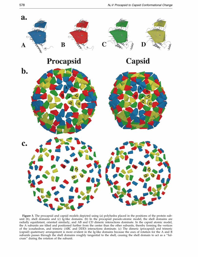
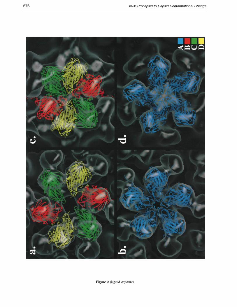
Figure 3. The procapsid and capsid models depicted using (a) polyhedra placed in the positions of the protein sub-unit (b), shell domains and (c) Ig-like domains. (b) In the procapsid pseudo-atomic model, the shell domains areradially equidistant, oriented similarly, and AB and CD dimeric interactions dominate. In the capsid atomic model,the A subunits are tilted and positioned further from the center than the other subunits, thereby forming the verticesof the icosahedron, and trimeric (ABC and DDD) interactions dominate. (c) The dimeric (procapsid) and trimeric(capsid) quaternary arrangement is more evident in the Ig-like domains because the axes of rotation for the A and Bsubunits passes through the shell domains roughly tangential to the shell, causing the shell domain to act as a ``ful-crum'' during the rotation of the subunit.
578 N!V Procapsid to Capsid Conformational Change

N!V Procapsid to Capsid Conformational Change 579
R-value was comparable to that determined in thecontrol experiment, using the unmodi®ed X-raycoordinates and the cryoEM map of the capsid.The procapsid model R-value improved onlyslightly when the shell and Ig-like domains werere®ned as independent units. Thus, the confor-mational change primarily involves rigid bodymovements of the subunits without signi®cant hin-ging between the Ig and shell domains. The result-ing pseudo-atomic model of the procapsid wasthen used to qualitatively examine the transitionwith the aid of simpli®ed models of the crystallo-graphic subunits (Figure 3(a)). These models sim-pli®ed the structures of the shell and Ig-likedomains, leaving out the internal domains becausethey are present only in the procapsid and are thusnot easily modelled using the capsid subunits.Analysis of these models revealed that the AB andCD dimers dominate the procapsid while the cap-sid is dominated by ABC and DDD trimers. How-ever, inspection of the interior of the procapsid(Figures 1(b) and 4(a)) shows that the internaldomains behave almost conversely: they areobviously trimeric in the procapsid, and in the cap-sid they lose these associations and become lessdistinct as domains (Figure 4(b)).
All four subunits translate towards the center ofthe particle during the transition, and the A and Bsubunits also undergo a signi®cant rotation. TheA/B and C/D dimers are roughly equivalent inthe procapsid, and both contribute equally to thecurvature of the rounded particle. The rotation ofthe A and B subunits during the transition causesthem to differentiate from the C/D dimer, leadingto a ``bent'' contact present between the facets ofthe polyhedral capsid. The description of the sub-unit interactions in the NoV procapsid is verysimilar to that of the T � 3 plant virus cowpeachlorotic mottle virus (CCMV) (Speir et al., 1995);namely, equivalent dimers both contributing to thecurvature of the capsid. This so-called truncatedicosahedral geometry is distinct from the canonicalicosahedral geometry, where a ``molecular switch''(Johnson, 1996) differentiates the two types ofdimers in the capsid.
While the movements of the subunits using thepseudoatomic model explain the procapsid to cap-sid transition, it is likely that the conformationalchange originates from a rearrangement of the pro-capsid internal domain. The crystallographicallyvisible N and C termini are interior to the capsidshell, so the internal domain is probably comprisedof N and C-terminal regions that are signi®cantlyrearranged from their dispositions in the capsid.The internal structure of the procapsid suggeststhat the internal domain stabilizes the procapsidand determines its curvature, and signi®cantalterations in this domain would be needed for theprocapsid to condense into the smaller, polyhedralcapsid (Figure 4). Similarly, the curvature of thehepatitis B core is determined by a positivelycharged C-terminal region (Zlotnick et al., 1996).That the N and C-terminal regions would be
involved in the conformational change followslogically from the discussion of the geometricalchange described above. In the truncated icosahe-dron, all subunits have nearly equivalent struc-tures, and this is seen especially in the internaldomain of the procapsid, where the density at the3-fold and quasi 3-fold axes is closely similar(Figure 1(b)). In the icosahedron, however, thequasi-symmetry is compromised by the molecularswitch which is formed in the capsid by the N andC termini (Munshi et al., 1996), thus requiring aconformational change in this region.
In the NoV capsid structure, the A and B sub-units have less ordered structure at their N and Ctermini. In the C and D subunits, these orderedfragments are a-helical and can be found along thequasi 2-fold axes between C and D in the capsid.We believe that in the procapsid, all four subunitshave comparable secondary structure with the cap-sid C and D subunits, and that these a-helices com-prise the procapsid internal domain (Figure 4(c)).During the procapsid to capsid conformationalchange, we propose the N and C-terminala- helices lose their trimeric associations at thequasi 3-fold and 3-fold axes and then reside at thequasi 2-fold axes. Additionally, for the A and Bsubunits, some a-helical regions (residues 44-57and 600-644) either become disordered or nolonger conform to icosahedral symmetry. In thepolyhedral capsid structure there is less room forthese a-helices at the ``bent'' quasi 2-fold axesbetween and internal to the A and B subunits. Asimilar situation has been described in the struc-ture of swollen tomato bushy stunt virus (TBSV),where the A and B subunits are thought to havemore ordered, interior structures than in theunswollen form (Robinson & Harrison, 1982). Bothtertiary (movement of a-helices) and perhaps sec-ondary (disordering of a-helices) structuralchanges are implicated in the NoV conformationalchange. Similarly, maturation of the bacteriophageP22 procapsid is thought to involve tertiary struc-tural changes in the coat protein which result instronger quaternary interactions (Tuma et al.,1998).
The atomic interactions responsible for the con-formational change are not yet clear because of thesigni®cant rearrangement that occurs during thetransition and the lack of a high-resolution struc-ture of the NoV procapsid. At this resolution, wecan expect that the pseudo-atomic model of theprocapsid could be used along with biochemicaldata to suggest groups of residues or regions ofthe coat protein that may be involved in the con-formational change, but not for a detailed descrip-tion of speci®c atomic interactions responsible. Thetransition described here occurs in an all-or-nonefashion when the pH is reduced from 7.6 to 5.0,but X-ray solution scattering demonstrated thatinitial changes in structure occur if the pH isreduced to �6.0 (data not shown). Therefore, weassume that the transition is caused byside-chain(s) which become protonated at pH 6.0,
Figure 4 (legend opposite)
580 N!V Procapsid to Capsid Conformational Change

N!V Procapsid to Capsid Conformational Change 581
causing the repulsion and/or attraction requiredfor rearrangement of the internal domain. A histi-dine residue is an obvious choice for protonationin this pH range, but is absent from the N and C-terminal regions. Analogies can be made to theswelling of plant viruses which occurs when thepH is raised. Mechanisms for expansion of TBSV(Robinson & Harrison, 1982) and CCMV (Speiret al., 1995) are thought to involve aspartic acidside-chains present at the quasi 3-fold axes whichnormally bind calcium ions. When the pH is raisedin the presence of EDTA, repulsion of negativelycharged carboxyl groups causes swelling of thecapsid. The transition takes place above neutralpH, and it is thought that the pKa values of theseside-chains must be anomalously high due to theirenvironment. Similar electrostatic repulsion maybe important for maintaining the NoV procapsid.It is also conceivable that arginine residues, whichare abundant in the N and C termini, might haveanomalously low pKa values (as has been observedfor lysine residues (Kokesh & Westheimer, 1971)),becoming charged at pH 6.0 and causing the dis-solution of the trimeric internal domain due toelectrostatic repulsion. Electrostatic interactionshave also been implicated in the scaffolding/coatprotein interactions in the maturation of bacterio-phage P22 (Parker & Prevelige, 1998), and theregion of the scaffolding protein thought to beinvolved is a helix-loop-helix domain with a regionthat is rich in positive charges.
It appears that the rearrangement of the internaldomain would be completed before the autopro-teolysis in the capsid would begin, althoughfurther rearrangement could occur as the autopro-teolysis proceeds. The autoproteolytic active site isfound in each subunit at the interface between the5-fold (A) and quasi 6-fold (B, C, D) related sub-units. The reaction is thought to be catalyzed bythe intrasubunit Glu103 (Munshi et al., 1996), bydirect analogy to the nodaviruses where site-directed mutagenesis showed that the correspond-ing residue (Asp75) is required for the reaction(Zlotnick et al., 1994). For the nodaviruses, a struc-turally distinct procapsid form has not beenobserved, and intersubunit interactions are thoughtto allow cleavage to occur only in the fullyassembled capsid. This intersubunit ``communi-cation'' is less clear in the structure of the NoVcapsid. The fact that signi®cant rearrangement of
Figure 4. The procapsid internal triskelion domain has a leof the procapsid (as shown in Figure 1(b), now semi-transpple. (b) The same view of the capsid with the innermost reg(c) View of these domains from the interior of the procapsidof the pseudo-atomic model (crystallographically determineterminal helices thought to be involved in the conformationfold symmetry in the crystallographic subunits is noticeableprocapsid), as well as the absence of trimeric interactions in tthe cryoEM density. (d) The same view as in (c) of the capsi(b), and BobScript (Esnouf, 1997) was used for (c) and (d).
the internal domain occurs during maturationsuggests that this rearrangement, rather than inter-subunit communication, may be suf®cient for acti-vating autoproteolysis in NoV.
Our results extend the familiar theme of theinitial formation of a round precursor particlewhich matures into a biologically functional poly-hedral form from the well-established complexDNA virus systems to a simple RNA virus. Thephenomenon can now be understood as a mechan-ism for viral subunits to assemble as nearly equiv-alent units, which then differentiate only afterassembly has completed (Figure 5). The roundshape, while it facilitates assembly, has compro-mised stability. The fact that many viral cores(hepatitis (BoÈ ttcher et al., 1997; Conway et al.,1997), Sindbis (Paredes et al., 1993), bluetonguevirus VP3 (Grimes et al., 1998)) are round sub-stantiates this idea, since these shells are protectedby an outer layer and appear to have been evolu-tionarily optimized for ease of assembly ratherthan for independent structural integrity. Ourstudy supports the idea that round viral cores andassembly intermediates in fact have truncated ico-sahedral geometry (Speir et al., 1995), whichexplains both their more rounded appearance andtheir nearly equivalent subunits (Wynne et al.,1999). The NoV procapsid internal domain, whichis ``lost'' as the capsid is formed, can be seen as afunctional equivalent of scaffolding proteins in bac-teriophage, which physically leave the capsid uponmaturation.
Parallels with complex and simpler viruses canalso be made concerning the quaternary rearrange-ments seen in the NoV procapsid to capsid tran-sition. The switching of the subunit interactionsfrom dimeric to trimeric has been seen in the lowpH-induced maturation of tick-borne encephalitisvirus (TBE) (Allison et al., 1995). It is also notablethat the swollen forms of plant viruses exhibitdimeric interactions which appear more equivalentand stronger than in the unswollen capsid (Speiret al., 1995; Robinson & Harrison, 1982). Manyplant viruses are thought to assemble from dimers(Choi & Loesch-Fries, 1999; Sorger et al., 1986;Rossmann et al., 1983), and recently the hepatitiscore has been shown to assemble from dimers ofthe coat protein (Zlotnick et al., 1999). By analogyto the NoV assembly intermediate observed here,it is tempting to speculate that when plant viruses
ss obvious organization in the capsid. (a) Cutaway viewarent) with the internal triskelion domain shown in pur-
ions (now no longer a distinct domain) colored purple.at the quasi 3-fold axis showing the A, B, and C subunitsd subunits ®tted to the cryoEM density). The N and C-al change are shown as rods. The breakdown of the 3-(compared with the symmetry they must possess in thehe pseudo-atomic model, which obviously are present ind. AVS (Upson et al., 1989) was used to generate (a) and

Figure 5. Schematic of the assembly of NoV based on the structure of the procapsid assembly intermediate. Dimers(gray) are assumed to be the building blocks of assembly, due to their tight associations in the procapsid. Becauseevery dimer exists in the non-differentiating milieu of the cytoplasm, we can assume that they are identical. Thedimers form associations with other dimers via their N and C-terminal regions, which form the trimeric internaldomain (purple lines which become triskelions; shown external to the procapsid for clarity). The dimers remain clo-sely similar in the procapsid, but may have become altered slightly from their structure in solution (denoted by achange in color from gray to purple). As the procapsid shrinks and the capsid begins to form, the subunits differen-tiate as they adjust to their slightly different environments, the endpoint being the four distinct subunits present inthe T � 4 polyhedral capsid (shown schematically, lower right). In the mature capsid, the internal domain has becomethe molecular switch which differentiates the AB and CD dimers (shown to the right of the T � 4 icosahedron).
582 N!V Procapsid to Capsid Conformational Change
swell, they are reverting to a previously formedassembly intermediate. The lack of observableswollen forms in T � 3 animal viruses such aspicornaviruses may be precluded by their proteo-lytic maturation, which may stabilize them and bea re¯ection on their different means of infection.
The pseudo-atomic model of the NoV procapsidpresented here demonstrates the extreme versati-lity of the viral coat protein. The structure of thecoat protein that initially assembles is surprisinglydifferent from that of the mature viral subunit,resulting in a dramatically different quaternaryarrangement in the NoV procapsid, causing it toexhibit very different structural properties from themature capsid. The mostly dimeric procapsidmatures into the trimeric capsid by putative elec-trostatic interactions in the trimeric internaldomain, which eventually lead to its disappearanceas a distinct domain. The similarities of this system
to both more complex and simpler viruses suggeststhat similar strategies are used by many virusesirrespective of their complexity to form a stablecapsid.
Acknowledgments
We thank Ian Wilson for critical reading of the manu-script, and Anette Schneemann and Dawn Marshall forassistance with cell culture and advice. This work wassupported by NIH grant GM54076 (J.E.J). M.Y. was sup-ported by grants from the NIAID (RO1 AI31535), theGustavus and Louise Pfeiffer Research Foundation, andthe Donald E. and Delia B. Baxter Foundation. Duringthis work M.Y. was an Established Investigator of theAmerican Heart Association and Bristol-Myers Squibband is now a recipient of a Clinical Scientist Award inTranslational Research from the Burroughs WellcomeFund.

N!V Procapsid to Capsid Conformational Change 583
References
Agrawal, D. K. & Johnson, J. E. (1992). Sequence andanalysis of the capsid protein of Nudaurelia capensiso Virus, an insect virus with T � 4 icosahedral sym-metry. Virology, 190, 806-814.
Agrawal, D. K. & Johnson, J. E. (1995). Assembly of theT � 4 Nudaurelia capensis o virus capsid protein,post-translational cleavage, and speci®c encapsida-tion of its mRNA in a baculovirus expression sys-tem. Virology, 207, 89-97.
Allison, S. L., Schalich, J., Stiasny, K., Mandl, C. W.,Kunz, C. & Heinz, F. X. (1995). Oligomericrearrangement of tick-borne encephalitis virusenvelope proteins induced by an acidic pH. J. Virol.69, 695-700.
AndreÂs, G., GarcõÂa-Escudero, R., SimoÂn-Mateo, C. &VinÄ uela, E. (1998). African swine fever virus isenveloped by a two-membraned collapsed cisternaderived from the endoplasmic reticulum. J. Virol.72, 8988-9001.
Baker, T. S. & Cheng, R. H. (1996). A model-basedapproach for determining orientations of biologicalmacromolecules imaged by cryoelectron micro-scopy. J. Struct. Biol. 116, 120-130.
Baker, T. S., Drak, J. & Bina, M. (1988). Reconstructionof the three-dimensional structure of simian virus40 and visualization of the chromatin core. Proc.Natl Acad. Sci. USA, 85, 422-426.
BoÈ ttcher, B., Wynne, S. A. & Crowther, R. A. (1997).Determination of the fold of the core protein ofhepatitis B virus by electron cryomicroscopy.Nature, 386, 88-91.
BruÈ nger, A. T. (1996). Recent developments for crystallo-graphic re®nement of macromolecules. Methods Mol.Biol. 56, 245-266.
Caspar, D. L. D. & Klug, A. (1962). Physical principlesin the construction of regular viruses. Cold SpringHarbor Symp. Quant. Biol. 27, 1-24.
Cheng, R. H., Reddy, V. S., Olson, N. H., Fisher, A. J.,Baker, T. S. & Johnson, J. E. (1994). Functionalimplications of quasi-equivalence in a T � 3 icosahe-dral animal virus established by cryo-electronmicroscopy and X-ray crystallography. Structure, 2,271-282.
Choi, J. & Loesch-Fries, L. S. (1999). Effect of C-terminalmutations of alfalfa mosaic virus coat protein ondimer formation and assembly in vitro. Virology,260, 182-189.
Collaborative Computational Project, No. 4 (1994). TheCCP4 suite: programs for protein crystallography.Acta Crystallog. sect. D, 50, 760-763.
Conway, J. F., Duda, R. L., Cheng, N., Hendrix, R. W. &Steven, A. C. (1995). Proteolytic and conformationalcontrol of virus capsid maturation: the bacterio-phage HK97 system. J. Mol. Biol. 253, 86-99.
Conway, J. F., Cheng, N., Zlotnick, A., Wing®eld, P. T.,Stahl, S. J. & Steven, A. C. (1997). Visualization of a4-helix bundle in the hepatitis B virus capsid bycryo-electron microscopy. Nature, 386, 91-94.
Crowther, R. A. (1971). Procedures for three-dimensionalreconstruction of spherical viruses by Fourier syn-thesis from electron micrographs. Phil. Trans. Roy.Soc. ser. B, 261, 221-230.
Dokland, T. & Murialdo, H. (1993). Structural transitionsduring maturation of bacteriophage lambda cap-sids. J. Mol. Biol. 233, 682-694.
Edvardsson, B., Everitt, E., JoÈ rnvall, H., Prage, L. &Philipson, L. (1976). Intermediates in adenovirusassembly. J. Virol. 19, 533-547.
Esnouf, R. M. (1997). An extensively modi®ed version ofMolScript that includes greatly enhanced coloringcapabilities. J. Mol. Graph. Model. 15, 132-134, 112-113 color plates.
Fuller, S. D. (1987). The T � 4 envelope of Sindbis virusis organized by interactions with a complementaryT � 3 capsid. Cell, 48, 923-934.
Grimes, J. M., Burroughs, J. N., Gouet, P., Diprose, J. M.,Malby, R., ZieÂntara, S., Mertens, P. P. C. & Stuart,D. I. (1998). The atomic structure of the bluetonguevirus core. Nature, 395, 470-478.
Johnson, J. E. (1996). Functional implications of protein-protein interactions in icosahedral viruses. Proc.Natl Acad. Sci. USA, 93, 27-33.
Johnson, J. E., Munshi, S., Liljas, L., Agrawal, D., Olson,N. H., Reddy, V., Fisher, A., McKinney, B.,Schmidt, T. & Baker, T. S. (1994). Comparativestudies of T � 3 and T � 4 icosahedral RNA insectviruses. Arch. Virol. Suppl. 9, 497-512.
Jones, T. A., Zou, J.-Y., Cowan, S. W. & Kjeldgaard, M.(1991). Improved methods for building proteinmodels in electron density maps and the location oferrors in these models. Acta Crystallog. sect. A, 47,110-119.
Kokesh, F. C. & Westheimer, F. H. (1971). A reportergroup at the active site of acetoacetate decarboxy-lase. II. Ionization constant of the amino group.J. Am. Chem. Soc. 93, 7270-7274.
Kraulis, P. J. (1991). MOLSCRIPT: a program to produceboth detailed and schematic plots of protein struc-tures. J. Appl. Crystallog. 24, 946-950.
Merritt, E. A. & Bacon, D. J. (1997). Raster3D: photorea-listic molecular graphics. Methods Enzymol. 277, 505-524.
Munshi, S., Liljas, L., Cavarelli, J., Bomu, W., McKinney,B., Reddy, V. & Johnson, J. E. (1996). The 2.8 AÊ
structure of a T � 4 animal virus and its impli-cations for membrane translocation of RNA. J. Mol.Biol. 261, 1-10.
Murphy, F. A., Fauquet, C. M., Bishop, D. H. L.,Ghabrial, S. A., Jarvis, A. W., Martelli, G. P., Mayo,M. A. & Summers, M. D. (1995). Editors of VirusTaxonomy: Classi®cation and Nomenclature of Viruses.The Sixth Report of the International Committee onTaxonomy of Viruses, Springer-Verlag, WõÂen.
Newcomb, W. W., Homa, F. L., Thomsen, D. R., Booy,F. P., Trus, B. L., Steven, A. C., Spencer, J. V. &Brown, J. C. (1996). Assembly of the herpes simplexvirus capsid: characterization of intermediatesobserved during cell-free capsid formation. J. Mol.Biol. 263, 432-446.
Newcomb, W. W., Trus, B. L., Cheng, N., Steven, A. C.,Sheaffer, A. K., Tenney, D. J., Weller, S. K. &Brown, J. C. (2000). Isolation of herpes simplexvirus procapsids from cells infected with a pro-tease-de®cient mutant virus. J. Virol. 74, 1663-1673.
Olson, N. H. & Baker, T. S. (1989). Magni®cation cali-bration and the determination of spherical virusdiameters using cryo-microscopy. Ultramicroscopy,30, 281-297.
Paredes, A. M., Brown, D. T., Rothnagel, R., Chiu, W.,Schoepp, R. J., Johnston, R. E. & Prasad, B. V.(1993). Three-dimensional structure of a membrane-containing virus. Proc. Natl Acad. Sci. USA, 90, 9095-9099.

584 N!V Procapsid to Capsid Conformational Change
Parker, M. H. & Prevelige, P. E., Jr (1998). Electrostaticinteractions drive scaffolding/coat protein bindingand procapsid maturation in bacteriophage P22.Virology, 250, 337-349.
Prasad, B. V. V., Prevelige, P. E., Marietta, E., Chen,R. O., Thomas, D., King, J. & Chiu, W. (1993).Three-dimensional transformation of capsids associ-ated with genome packaging in a bacterial virus.J. Mol. Biol. 231, 65-74.
Robinson, I. K. & Harrison, S. C. (1982). Structure of theexpanded state of tomato bushy stunt virus. Nature,297, 563-568.
Rossmann, M. G., Abad-Zapatero, C., Hermodson, M. A.& Erickson, J. W. (1983). Subunit interactions insouthern bean mosaic virus. J. Mol. Biol. 166, 37-73.
Rueckert, R. R. (1996). Picornaviridae: the viruses andtheir replication. In Fields Virology (Fields, B. N.,Knipe, D. N., Howley, P. M., Chanock, R. M.,Melnick, J. L., Monath, T. P., Roizman, B. & Straus,S. E., eds), 3rd edit., pp. 609-654, Lippincott-RavenPublishers, Philadelphia.
Salanueva, I. J., Carrascosa, J. L. & Risco, C. (1999).Structural maturation of the transmissible gastroen-teritis coronavirus. J. Virol. 73, 7952-7964.
Schneemann, A., Zhong, W., Gallagher, T. M. &Rueckert, R. R. (1992). Maturation cleavage requiredfor infectivity of a nodavirus. J. Virol. 66, 6728-6734.
Sorger, P. K., Stockley, P. G. & Harrison, S. C. (1986).Structure and assembly of turnip crinkle virus. II.Mechanism of reassembly in vitro. J. Mol. Biol. 191,639-658.
Speir, J. A., Munshi, S., Wang, G., Baker, T. S. &Johnson, J. E. (1995). Structures of the native andswollen forms of cowpea chlorotic mottle virusdetermined by X-ray crystallography and cryo-elec-tron microscopy. Structure, 3, 63-78.
Trus, B. L., Booy, F. P., Newcomb, W. W., Brown, J. C.,Homa, F. L., Thomsen, D. R. & Steven, A. C. (1996).The herpes simplex virus procapsid: structure, con-formational changes upon maturation, and roles ofthe triplex proteins VP19c and VP23 in assembly.J. Mol. Biol. 263, 447-462.
Tuma, R., Prevelige, P. E., Jr. & Thomas, G. J., Jr (1998).Mechanism of capsid maturation in a double-stranded DNA virus. Proc. Natl Acad. Sci. USA, 95,9885-9890.
Turner, B. G. & Summers, M. F. (1999). Structuralbiology of HIV. J. Mol. Biol. 285, 1-32.
Upson, C., Faulhaber, T., Jr, Kamins, D., Laidlaw, D.,Schlegel, D., Vroom, J., Gurwitz, R. & van Dam, A.(1989). The application visualization system: a com-putational environment for scienti®c visualization.IEEE Comput. Graph. Applic. 9, 30-42.
Wynne, S. A., Crowther, R. A. & Leslie, A. G. W. (1999).The crystal structure of the human hepatitis B viruscapsid. Mol. Cell, 3, 771-780.
Zlotnick, A., Reddy, V. S., Dasgupta, R., Schneemann,A., Ray, W. J., Jr, Rueckert, R. R. & Johnson, J. E.(1994). Capsid assembly in a family of animalviruses primes an autoproteolytic maturation thatdepends on a single aspartic acid residue. J. Biol.Chem. 269, 13680-13684.
Zlotnick, A., Cheng, N., Conway, J. F., Booy, F. P.,Steven, A. C., Stahl, S. J. & Wing®eld, P. T. (1996).Dimorphism of hepatitis B virus capsids is stronglyin¯uenced by the C terminus of the capsid protein.Biochemistry, 35, 7412-7421.
Zlotnick, A., Johnson, J. M., Wing®eld, P. W., Stahl, S. J.& Endres, D. (1999). A theoretical model success-fully identi®es features of hepatitis B virus capsidassembly. Biochemistry, 38, 14644-14652.
Edited by D. Rees
(Received 13 January 2000; received in revised form 17 March 2000; accepted 21 March 2000)