Laparoscopic Management of Stress Incontinence
6
Laparoscopic Management of Stress Incontinence INTRODUCTION Genuine stress incontinence is the involuntary loss of urine, which occurs when the intravesical pressure exceeds the maximum urethral pressure in the absence of a detrusor contraction. The preferred therapy for genuine stress incontinence is surgery. Urinary incontinence is becoming more prevalent as the population ages. A significant improvement in the psychological status of these patients after the successful surgical cure of stress incontinence has been demonstrated. PATHOPHYSIOLOGY In genuine stress incontinence, the proximal urethra is displaced outside the abdominal cavity. Stress incontinence then results from inadequate transmission of increase in intra-abdominal pressure to the proximal urethra. The urethra has, in fact, lost its retropubic position due to attenuated support. As a result, coughing will produce an immediate increase in intravesical pressure but not a concomitant increase in intraurethral pressure and a dribble of urine results (Fig. 1). More than 160 types of operations are available to correct stress urinary incontinence (SUI), an optimal approach has not yet been developed. On April 16, 2019, the Food and Drug Administration (FDA) ordered all manufacturers of surgical mesh intended for transvaginal repair of SUI. Surgical mesh has been used for urogynecologic procedures, including repair of pelvic organ prolapse (POP) and SUI. It is permanently implanted to reinforce the weakened vaginal wall for POP repair or support the urethra or bladder neck for the repair of SUI. There are three main surgical procedures performed with surgical mesh to treat pelvic floor disorders with surgical mesh: 1. Transvaginal mesh to treat POP 2. Transabdominal mesh to treat POP 3. Mesh sling to treat SUI Each of these procedures has unique risks and benefits and it is important not to confuse the procedures and their risks and benefits. Since the FDA has not received sufficient evidence to assure that the probable benefits of these devices outweigh their probable risks, the agency has concluded that these products do not have reasonable assurance of safety and effectiveness. The Burch procedure is considered by many to be the gold standard for surgical treatment of genuine stress incontinence (Fig. 2). The Burch procedure requires the elevation of the anterior wall of the vagina to the level of the origin of the paravaginal fascia by suspension from Cooper’s ligaments (iliopectineal ligaments). A properly performed Burch procedure cures in up to 93% of cases. Fig. 1: Pathophysiology of stress urinary incontinence. Prof. Dr. R. K. Mishra
Transcript of Laparoscopic Management of Stress Incontinence

Laparoscopic Management of Stress Incontinence
INTRODUCTIONGenuine stress incontinence is the involuntary loss of urine, which occurs when the intravesical pressure exceeds the maximum urethral pressure in the absence of a detrusor contraction. The preferred therapy for genuine stress incontinence is surgery. Urinary incontinence is becoming more prevalent as the population ages. A significant improvement in the psychological status of these patients after the successful surgical cure of stress incontinence has been demonstrated.
PATHOPHYSIOLOGYIn genuine stress incontinence, the proximal urethra is displaced outside the abdominal cavity. Stress incontinence then results from inadequate transmission of increase in intra-abdominal pressure to the proximal urethra. The urethra has, in fact, lost its retropubic position due to attenuated support. As a result, coughing will produce an immediate increase in intravesical pressure but not a concomitant increase in intraurethral pressure and a dribble of urine results (Fig. 1).
More than 160 types of operations are available to correct stress urinary incontinence (SUI), an optimal approach has not yet been developed. On April 16, 2019, the Food and Drug Administration (FDA) ordered all manufacturers
of surgical mesh intended for transvaginal repair of SUI. Surgical mesh has been used for urogynecologic procedures, including repair of pelvic organ prolapse (POP) and SUI. It is permanently implanted to reinforce the weakened vaginal wall for POP repair or support the urethra or bladder neck for the repair of SUI. There are three main surgical procedures performed with surgical mesh to treat pelvic floor disorders with surgical mesh:1. Transvaginal mesh to treat POP2. Transabdominal mesh to treat POP3. Mesh sling to treat SUI
Each of these procedures has unique risks and benefits and it is important not to confuse the procedures and their risks and benefits. Since the FDA has not received sufficient evidence to assure that the probable benefits of these devices outweigh their probable risks, the agency has concluded that these products do not have reasonable assurance of safety and effectiveness.
The Burch procedure is considered by many to be the gold standard for surgical treatment of genuine stress incontinence (Fig. 2). The Burch procedure requires the elevation of the anterior wall of the vagina to the level of the origin of the paravaginal fascia by suspension from Cooper’s ligaments (iliopectineal ligaments). A properly performed Burch procedure cures in up to 93% of cases.
Fig. 1: Pathophysiology of stress urinary incontinence.
Prof. Dr. R. K. Mishra

454 SECTION 3: Laparoscopic Gynecological Procedures
Fig. 2: Laparoscopic Burch suspension.
INDICATIONSEligible patients include women who decline or have persistent symptoms following a trial of conservative management. Women who have mixed urinary incontinence may be able to control their symptoms of urge incontinence and provide enough satisfaction that management of SUI is no longer necessary.
CONTRAINDICATIONS■ Patients with type III SUI (a fixed, nonfunctioning
proximal urethra) are not candidates for a Burch procedure, as no hypermobility exists to correct
■ Patients having cystocele, rectocele, or introital deficiency■ Patients not completed childbearing
LAPAROSCOPIC ANATOMYThe space of Retzius lies between the vesicoumbilical fascia posteriorly and the posterior rectus sheath and pubic bone anteriorly. This is the space first entered in Burch suspension.
Preoperative EvaluationPostmenopausal women should receive at least 3 months of estrogen replacement therapy. Preoperative evaluation includes history, physical, gynecologic, and neurologic examinations. The routine tests include stress test, Q-tip test, urinalysis, urine culture, and sensitivity. All women should undergo an urodynamic evaluation, with emphasis on voiding time, voiding volume, and postvoid residual urine volume. Stress incontinence is diagnosed by a positive stress test in the absence of simultaneous detrusor contractions or pressure equalization on the stress urethral closure pressure profile.
Operative TechniqueFollowing the induction of general endotracheal anesthesia, a Foley catheter should be applied in the bladder. A 10-mm
Fig. 3: Port position for Burch suspension surgery.
Fig. 4: Peritoneal incision in midline.
operative video laparoscope is inserted infraumbilical and two 5 mm accessory trocars are inserted according to baseball diamond concept (Fig. 3).
300 cc of methylene blue solution is filled into the bladder to keep the bladder weighted down and to allow the clear delineation of the dome of the bladder. Three-port techniques are used one in umbilicus and two 5 mm port 7.5 cm lateral and slightly below the umbilicus (Fig. 3).
The anterior abdominal wall peritoneum 2 cm above the delineated dome of distended bladder is cut and pulled down. To enter the space of Retzius intra-abdominally, the umbilical ligaments are identified laterally (Fig. 4).
The space of Retzius is developed using blunt and CO2 dissection of fibrofatty tissue. Care is taken to avoid obturator nerve and aberrant obturator vessel injury. The midline trocar entry and anatomic landmarks, including the round ligament from the internal ring, are used to avoid bladder injury. Blunt dissection is used to expose the retropubic space. Surgeon should stay close to the back of the pubic bone, dropping the anterior bladder wall, vaginal wall, and urethra downward. Dissection is limited over the urethra in the midline, to approximately 2 cm lateral to the

455CHAPTER 36: Laparoscopic Management of Stress Incontinence
Fig. 5: Retropubic dissection to detect Cooper’s ligament and tendinous arc of levator ani muscle.
Fig. 6: Retropubic dissection to detect Cooper’s ligament (arrows).
urethra, to protect its delicate musculature. An assistant should introduce one finger on each side of the catheterized urethra, elevating the lateral vaginal fornix. The overlying fibrofatty tissue is cleared laparoscopically from the anterior vaginal wall. The bladder is dissected medially from the paravaginal fascia from lateral to medial. Blunt dissection with atraumatic grasper is continued until the urethrovesical junction becomes apparent and the white glistening tissue of the paravaginal fascia appears.
Bladder muscle fibers of the urethrovesical junction should not be damaged at the time of dissection. Bleeding, if happens in this area, is controlled only with bipolar electrocoagulation because monopolar current can cause perforation of bladder or stricture of ureter. Retropubic dissection is continued until Cooper’s ligament is clearly exposed (Figs. 5 and 6).
Dissection in the space of Retzius is difficult in patients who have undergone previous laparotomy. Pneumoperitoneum pressure helps provide exposure of the space and its contents out to the obturator nerve.
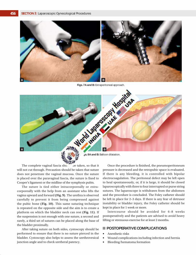
The space of Retzius also can be entered with a preperitoneal approach just like total extraperitoneal hernia surgery. The balloon dissector consists of a cannula and balloon system. The dissector is inserted through a 10-mm infraumbilical incision. It is advanced between the rectus muscle and the anterior surface of the posterior rectus sheath toward the symphysis pubis (Figs. 7A and B). The dissector’s external sheath is removed and the balloon is inflated with approximately 500 mL of saline solution. During inflation, the balloon unrolls sideways and exerts a perpendicular force that separates tissue layers. A blunt dissection of the connective tissues is propagated as the balloon expands. Full dissection takes about 1 minute but surgeon should keep the balloon inflated for 5 minutes to achieve proper hemostasis. Lateral pressure of balloon will stop capillary bleeding, if kept for 5 minutes. When maximal volume is reached, the balloon is deflated and removed through the incision (Figs. 8A and B).
The dissected space is insufflated with CO2 at a pressure of 8–10 mm Hg. The predefined shape of the balloon, its nonelastomeric material, and the incompressible character of the saline assure a large, relatively bloodless working space of predictable size and shape. The space is adequate for identification of pertinent landmarks and unencumbered manipulation of endoscopic surgical instruments.
After complete dissection, the paravaginal fascia is identified as glistening fascia over the finger of assistant. Using a needle holder, a suture is placed over the paravaginal fascia at the level of the urethrovesical junction, approximately 1–1.5 cm from the urethra. The urethrovesical junction can be identified easily because of the Foley bulb with the catheter under gentle traction. This suture is placed perpendicular to the vaginal axis to include approximately 0.5–1 cm of tissue.
456 SECTION 3: Laparoscopic Gynecological Procedures
Figs. 7A and B: Extraperitoneal approach.
Figs. 8A and B: Balloon dilatation.
The complete vaginal fascia should be taken, so that it will not cut through. Precaution should be taken that suture does not penetrate the vaginal mucosa. Once the suture is placed over the paravaginal fascia, the suture is fixed to Cooper’s ligament or the midline of the symphysis pubis.
The suture is tied either intracorporeally or extra-corporeally with the help from an assistant who lifts the vagina upward and forward (Fig. 9). The urethra is observed carefully to prevent it from being compressed against the pubic bone (Fig. 10). This same suturing technique is repeated on the opposite side and the aim is to create a platform on which the bladder neck can rest (Fig. 11). If the suspension is not enough with one suture, a second and rarely, a third set of sutures can be placed along the base of the bladder proximally.
After taking suture on both sides, cystoscopy should be performed to ensure that there is no suture pierced in the bladder. Cystoscopy also helps to assess the urethrovesical junction angle and to check uretheral patency.
Once the procedure is finished, the pneumoperitoneum pressure is decreased and the retropubic space is evaluated. If there is any bleeding, it is controlled with bipolar electrocoagulation. The peritoneal defect may be left open to heal spontaneously, or, if it is large, it should be closed laparoscopically with three to four interrupted or purse string sutures. The laparoscope is withdrawn from the abdomen and the procedure is concluded. The Foley catheter should be left in place for 2–3 days. If there is any fear of detrusor instability or bladder injury, the Foley catheter should be kept in place for 1 week or more.
Intercourse should be avoided for 6–8 weeks postoperatively and the patients are advised to avoid heavy lifting or strenuous exercise for at least 2 months.
POSTOPERATIVE COMPLICATIONS■ Anesthetic risks■ Wound complications including infection and hernia■ Bleeding/hematoma formation
A B
A B

457CHAPTER 36: Laparoscopic Management of Stress Incontinence
Fig. 9: Suturing of paravaginal fascia with Cooper’s ligament with the help of assistant finger.
Fig. 10: Suturing of paravaginal fascia with Cooper’s ligament (arrow).(1) Paravaginal fascia without suture applied; (2) Paravaginal fascia with suture applied; (3) Bladder; (4) Cooper's ligament of left side; (5) Pubic symphysis.
Fig. 11: Final view of suture placement in Burch suspension.
■ Deep venous thrombosis■ Voiding difficulty■ Urinary retention, which may require catheter drainage
or clean intermittent catheterization■ Development of urgency urinary incontinence■ Development of vaginal wall prolapse
Some of the less common problems are:■ New onset of intrinsic sphincteric deficiency■ Voiding pain■ Ureteral obstruction requiring reoperation■ Vesicovaginal fistula■ Vesicocutaneous fistula■ Foreign body erosion, if tacks were used for laparoscopic
repair
CONCLUSIONBurch colposuspension has been shown to be a highly effective surgical treatment for SUI with low recurrence rates, although there is some loss of efficacy with time similar to other procedures. Concerning the laparoscopic approach, the conclusion from the Cochrane review was that the available evidence suggests that it may be as effective as open colposuspension 2 years postoperatively. The National Institute for Health and Care Excellence (NICE) guidelines include among their recommendations that laparoscopic Burch colposuspension is not recommended as a routine procedure for the treatment of SUI in women. In current recommendations, Burch colposuspension remains an
option for secondary treatment. Because midurethral slings have recently become under scrutiny, it may return as a first-line treatment procedure. Laparoscopic Burch colposuspension should, therefore, nowadays be provided in fellowship programs worldwide.
BIBLIOGRAPHY 1. Alcalay M, Monga A, Stanton SL. Burch colposuspension:
a 10–20 year follow up. Br J Obst Gynaecol. 1995;102:740-5. 2. Ankardal M, Ekerydh A, Crafoord K, Milsom I, Stjerndahl JH,
Engh ME. A randomised trial comparing open Burch colposuspension using sutures with laparoscopic colpo-suspension using mesh and staples in women with stress urinary incontinence. BJOG. 2004;111:974-81.
3. Bo K, Talseth T, Holme I. Single blind, randomised, controlled trial of pelvic floor exercises, electrical stimulation, vaginal cones and no treatment in management of genuine stress incontinence in women. BMJ. 1999;318:487-93.
4. Boyles SH, Webber AM, Meyn L. Procedures for urinary incontinence in the United States 1979–1997. Am J Obstet Gynecol. 2003;189:70-5.

458 SECTION 3: Laparoscopic Gynecological Procedures
5. Burch JC. Urethro-vaginal fixation to Cooper’s ligament for correction of stress incontinence, cystocoele and prolapse. Am J Obstet Gynecol. 1961;81:281-90.
6. Burton G. A five year prospective randomised urodynamic study comparing open and laparoscopic colposuspension. Neurourol Urodyn. 1999;18:295-6.
7. Cardozo L, Drutz HP, Baygani SK, Bump RC. Pharmacological treatment of women awaiting surgery for stress urinary incontinence. Obstet Gynecol. 2004;104:511-9.
8. Cardozo LD, Cutner AA. Surgical management of stress incontinence. Contemp Rev Obstet Gynaecol. 1992;4:36-41.
9. Chaliha C, Stanton SL. Complications of surgery for genuine stress incontinence. Br J Obst Gynaecol. 1999;106:1238-45.
10. Christofi N, Hextall A. Which procedure for incontinence? J Br Menopause Soc. 2005;11:23-7.
11. Delorme E. Transobturator urethral suspension: a mini-invasive procedure on the treatment of stress urinary incontinence in women. Prog Urol. 2001;11:1306-13.
12. Duckett JRA. The use of periurethral injectables in the treatment of genuine stress incontinence. Br J Obst Gynaecol. 1998;105:390-6.
13. Eckford SD, Abrams P. Para-urethral collagen implantation for female stress incontinence. Br J Urol. 1991;68:586-9.
14. Foldspang A, Mommsen S. Overweight and urinary incontinence in women. Ugeskr Laeger. 1995;157:5848-51.
15. Gorton E, Stanton S, Monga A, Wiskind AK, Lentz GM, Bland DR. Periurethral collagen injection: a long-term follow-up study. BJU Int. 1999;84:966-71.
16. Groutz A, Gordon D, Woman I, Jaffa AJ, David MP, Lessing JB. Tension-free vaginal tape for stress urinary incontinence: is there a learning curve? Neurourol Urodyn. 2002;21:470-2.
17. Henalla SM, Hall V, Duckett JR, Link C, Usman F, Tromans PM, et al. A multicentre evaluation of a new surgical technique for urethral bulking in the treatment of genuine stress incontinence. BJOG. 2000;107:1035-9.
18. Hilton P, Mayne C. The Stamey endoscopic bladder neck suspension: a clinical and urodynamic investigation including actuarial follow-up over four years. Br J Obstet Gynaecol. 1999;98:1141-9.
19. Huang KH, Kung FT, Liang HM, Huang LY, Chang SY. Concomitant surgery with tension-free vaginal tape. Acta Obstet Gynecol Scand. 2003;82:948-53.
20. Iosif CF, Bekassy Z, Ryhdestrom H. Prevalence of urinary incontinence in middle age women. Int J Gynecol Obstet. 1980;26:255-9.
21. Iosif CF, Bekassy Z. Prevalence of genitourinary symptoms in the menopause. Acta Obstet Gynecol Scand. 1984;63:257-60.
22. Jarvis GJ, Hall S, Stamp S, Millar DR, Johnson A. An assessment of urodynamic examination in incontinent women. Br J Obst Gynaecol. 1980;87:893-6.
23. Jarvis GJ. Surgery for genuine stress incontinence. Br J Obst Gynaecol. 1994;101:371-4.
24. Jolleys JV. Reported prevalence of urinary incontinence in a general practice. BMJ. 1988;296:1300-2.
25. Khullar V, Cardozo LD, Abbott D, Anders K. GAX collagen in the treatment of urinary incontinence in elderly women: a two year follow-up. Br J Obstet Gynaecol. 1997;104:96-9.
26. Kligman AM, Armstrong RC. Histologic response to intradermal Zyderm and Zyplast (glutaraldehyde cross-linked) collagen in humans. J Dermatol Surg Oncol. 1986;12:351-7.
27. Kondo A, Saito M, Yamada Y, Kato T, Hasegawa S, Kato K. Prevalence of hand-washing incontinence in females in comparison with stress and urge incontinence. Neurourol Urodyn. 1990;19:330-1.
28. Mommsen S, Foldspang A. Body mass index and adult urinary incontinence. World J Urol. 1994;12:319-22.
29. Monga AK, Robinson D, Stanton SL. Peri-urethral collagen injections for genuine stress incontinence: a two year follow-up. Br J Urol. 1995;76:156-60.
30. Mukherjee K, Constantine G. Urinary stress incontinence in obese women: tension-free vaginal tape is the answer. BJU Int. 2001;88:881-3.
31. National Institute for Clinical Excellence (NICE). Guidance on the Use of TVT for Stress Incontinence. Technology Appraisal Guidance No. 56. London: NICE; 2003.
32. National Institute for Clinical Excellence (NICE). Interventional Procedures Review of Transobturator Tape Insertion for Stress Urinary Incontinence. London: NICE; 2004.
33. Pickard R, Reaper J, Wyness L, Cody DJ, McClinton S, N’Dow J. Periurethral injection therapy for urinary incontinence in women. Cochrane Database Syst Rev. 2003;2:CD003881.
34. Rekers H, Drogendijk AC, Valkenburg H, Riphagen F. Urinary incontinence in women from 35 to 79 years of age: prevalence and consequences. Eur J Obstet Gynecol Reprod Biol. 1992;43:229-34.
35. Schultheiss D, Höfner K, Oelke M, Grünewald V, Jonas U. Percutaneous bladder neck suspension with bone anchors: an improvement in the therapy of female stress urinary incontinence. Neurourol Urodyn. 1998;17:457-8.
36. Ulmsten U, Petros P. Intravaginal salingoplasty: an ambulatory surgical procedure for the treatment of female urinary incontinence. Scand J Urol Nephrol. 1995;29:75-82.
37. Ulmsten U, Henriksson L, Johnson P, Varhos G. An ambulatory surgical procedure under local anaesthesia for treatment for female urinary incontinence. Int Urogynecol J. 1996;7:81-6.
38. Van Kerrebroeck P, Abrams P, Lange R. Duloxetine urinary incontinence study group. Br J Obst Gynaecol. 2004;111:249-57.
39. Vancaillie TG, Schuessler W. Laparoscopic bladderneck suspension. J Laparoendosc Surg. 1991;1:169-73.
40. Vetter NJ, Jones DA, Victor CR. Urinary incontinence in the elderly at home. Lancet. 1981;2:1275-7.
41. Ward K, Hilton P. Prospective multicentre randomised trial of tension-free vaginal tape and colposuspension for primary urodynamic stress incontinence: two year follow-up. Am J Obstet Gynecol. 2004;190:324-31.
42. Ward K, Hilton P. Prospective multicentre randomised trial of tension-free vaginal tape and colposuspension as primary treatment for stress incontinence. BMJ. 2002;325:67-70.